1
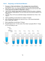
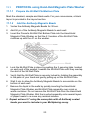
TM ProcartaPlex Multiplex Immunoassays For research use only. Not for diagnostic or therapeutic procedures. Using Magnetic Beads For serum, plasma and cell culture supernatants Instructions for Mouse Assays North America Europe/International* Technical Support: Research Products: 888.810.6168 858.642.2058 [email protected] Technical Support: +43 1 796 40 40-120 [email protected] Clinical Products: 877.726.8559 858.642.2058 [email protected] Customer Service: 888.999.1371 858.642.2058 [email protected] Fax:858.642.204 Customer Service: +43 1 796 40 40-304 [email protected] Fax:+43 1 796 40 40-400 Bender MedSystems GmbH Campus Vienna Biocenter 2 1030 Vienna, Austria www.eBioscience.com * Customers outside North America and Europe may contact their eBioscience distributor listed on our website at www.eBioscience.com/distributors. For Research Use Only. Not for use in diagnostic or therapeutic procedures. Limited License Subject to the eBioscience, an Affymetrix Company, terms and conditions that govern your use of eBioscience products, eBioscience grants you a non-exclusive, nontransferable, non-sub licensable license to use this eBioscience product only in accordance with the manual and written instructions provided by eBioscience. You understand and agree that, except as expressly set forth in the eBioscience terms and conditions, no right or license to any patent or other intellectual property owned or licensable by eBioscience is conveyed or implied by this eBioscience product. In particular, no right or license is conveyed or implied to use this eBioscience product in combination with a product not provided, licensed, or specifically recommended by eBioscience for such use. Citing ProcartaPlexTM Multiplex Immunoassays in Publications When describing a procedure for publication using this product, please refer to it as the ProcartaPlexTM Multiplex Immunoassay from eBioscience. eBioscience, Inc. reserves the right to change its products and services at any time to incorporate technological developments. This manual is subject to change without notice. Although this manual has been prepared with every precaution to ensure accuracy, eBioscience, Inc. assumes no liability for any errors or omissions, not for any damages resulting from the application or use of this information. © ® ® TM 2013 Affymetrix Inc. All rights reserved. Affymetrix , eBioscience and ProcartaPlex are trademarks or ® ® ® registered trademarks of Affymetrix, Inc. Luminex and xMAP are registered trademarks of the Luminex Corporation. All other trademarks are the property of their respective owners. 2 Table of Contents 1 Intended Use............................................................................... 4 2 About ProcartaPlexTM Mouse Immunoassay Kits ........................ 4 3 How it Works ............................................................................... 5 4 ProcartaPlexTM Immunoassay Workflow ..................................... 6 5 Analyte Overview ........................................................................ 7 6 ProcartaPlexTM Kit Contents ...................................................... 11 7 Required Equipment and Materials Not Supplied ..................... 13 8 Precautions and Technical Hints............................................... 14 9 Sample Preparation .................................................................. 16 10 Preparation of Reagents ........................................................... 17 11 Test Protocol ............................................................................. 22 12 Setup of the Luminex Instrument .............................................. 30 13 Analyzing Results ..................................................................... 31 14 Specificity .................................................................................. 31 15 Troubleshooting ........................................................................ 32 16 Blank Plate Layout .................................................................... 36 3 12.08.2013 (05) 1 INTENDED USE This user manual is for a ProcartaPlexTM Immunoassay Kit - Magnetic Beads from eBioscience to perform quantitative, multiplexed immunoassays based on the Luminex® technology. The procedure is for simultaneous measurements of multiple protein biomarkers in serum, plasma, and cell culture supernatant samples. Other biological samples might be suitable for use in the assay. The assay protocol and reagents supplied are not compatible with other manufacturer's reagents. Each 96-well plate kit is configured to allow for the following usage: 14 wells for a 7-point standard curve (in duplicate), 2 wells for blanks, and up to 80 wells for samples. NOTE: For the most current version of user documentation, go to our website at www.eBioscience.com 2 ABOUT PROCARTAPLEXTM MOUSE IMMUNOASSAY KITS ProcartaPlexTM Immunoassay Kits are available as: Pre-mixed panels contain all the reagents required to run the respective multiplex assay Simplex Kits designed for the measurement of one analyte in an immunoassay using xMAP technology. Together with a ProcartaPlexTM Basic Kit, Simplex kits can be used to detect one analyte alone or can be multiplexed with other Simplex Kits to measure a variety of analytes. Simplex Kits can also be combined with premixed panels as long as no Bead Region occurs twice in one experimental set-up. New custom assay development for analytes not listed on our website Please contact your local eBioscience sales representative for new custom assay development for analytes not listed on our website www.eBioscience.com. 4 3 HOW IT WORKS ProcartaPlexTM Mouse Immunoassays use the Luminex technology (multianalyte profiling beads) to enable the detection and quantitation of multiple protein targets simultaneously in diverse matrices. All ProcartaPlexTM assays use magnetic beads. The xMAP system combines a flow cytometer, fluorescent-dye microspheres (beads), dual laser design and digital signal processing to effectively allow multiplexing of up to 100 (50 for MAGPIX) unique assays within a single sample. The ProcartaPlexTM Immunoassay kits are compatible with all Luminex 100/200, MAGPIX and FLEXMAP 3D currently available. 5 4 PROCARTAPLEXTM IMMUNOASSAY WORKFLOW 6 5 ANALYTE OVERVIEW ProcartaPlexTM Immunoassays are available as predefined Multiplexing Panels or as Simplex assays.Together with a ProcartaPlexTM Basic Kit, Simplex kits can be used to detect one analyte alone or can be multiplexed with other Simplex Kits to measure a variety of analytes. Simplex Kits can also be combined with premixed panels as long as no Bead Region occurs twice in one experimental set-up. ProcartaPlex™ Mouse Panels are designed to detect up to 26 Mouse cytokines found in Mouse serum, plasma, and cell culture supernatants samples. See the following tables for a list of analytes detectable by these panels and a list of available Simplex assays and the respective bead regions. 7 ProcartaPlexTM Mouse Pre-mixed panel overview Cytokine & Chemokine Panel 1, 26Plex (EPX260-26088-901) Th9/Th17/Th22/Treg & Chemokine Panel, 15plex (EPX150-26089-901) Th1/Th2/Th9/Th17/Th22/Treg, 17plex (EPX170-26087-901) Th1/Th2 & Chemokine Panel 1, 20plex (EPX200-26090-901) IFN-gamma (38) Eotaxin (62) GM-CSF (42) Essential Th1/Th2, 6plex (EPX060-20831-901) Th1/Th2, 11plex (EPX110-20820-901) Th9/Th17/Th22/Treg, 6plex (EPX060-20822-901) IFN-gamma (38) IFN-gamma (38) IL-10 (13) Chemokine Panel 1, 9plex (EPX090-20821901) GRO-alpha (43) IL-12p70 (39) IL-12p70 (39) IL-17A (52) IP-10 (22) IL-4 (26) IL-13 (35) IL-22 (33) MCP-1 (51) IL-5 (27) IL-1ß (19) IL-23 (37) MCP-3 (48) IL-6 (28) IL-2 (20) IL-27 (36) MIP-1alpha (47) IL-12p70 (39) IL-4 (26) IL-4 (26) IL-9 (34) MIP-1beta (72) IL-13 (35) IL-5 (27) IL-5 (27) MIP-2 (55) IL-6 (28) IL-6 (28) RANTES (44) TNF-alpha (45) TNF-alpha (45) Eotaxin (62) GRO-alpha (43) IL-1ß (19) IL-2 (20) IL-18 (66) IP-10 (22) MCP-1 (51) MCP-3 (48) GM-CSF (42) MIP-1alpha (47) IL-18 (66) MIP-1beta (72) MIP-2 (55) RANTES (44) 8 TNF-alpha (45) ProcartaPlexTM Mouse Simplex kit overview Available Simplex Kits and respective Bead Regions Available Simplex Kits and respective Bead Regions EPX010-26038-901 Adiponectin 14 EPX010-26041-901 IL-9 34 EPX010-26008-901 Eotaxin/CCL11 62 EPX010-20614-901 IL-10 13 EPX010-26034-901 G-CSF/CSF-3 12 EPX010-26033-901 IL-12p40 39 EPX010-20612-901 GM-CSF 42 EPX010-26004-901 IL-12p70 39 EPX010-26031-901 Gro alpha 43 EPX010-26015-901 IL-13 35 EPX010-20606-901 IFN-gamma 38 EPX010-26001-901 IL-17A/CTLA-8 52 EPX010-26002-901 IL-1beta 19 EPX010-20618-901 IL-18 66 EPX010-20601-901 IL-2 20 EPX010-26022-901 IL-22 33 EPX010-26035-901 IL-3 14 EPX010-26017-901 IL-23 37 EPX010-20613-901 IL-4 26 EPX010-26024-901 IL-27 36 EPX010-20610-901 IL-5 27 EPX010-26040-901 LIF 18 EPX010-20603-901 IL-6 28 EPX010-26036-901 Leptin 65 9 Available Simplex Kits and respective Bead Regions EPX010-26018-901 IP-10 22 EPX010-26005-901 MCP-1 51 EPX010-26006-901 MCP-3 48 EPX010-26039-901 M-CSF 21 EPX010-26013-901 MIP-1alpha 47 EPX010-26014-901 MIP-1beta 72 EPX010-26032-901 MIP-2 55 EPX010-26037-901 RANKL 46 EPX010-26009-901 RANTES 44 EPX010-20607-901 TNF-alpha 45 EPX010-20619-901 VEGF-A 25 New Analytes are released regularly, all products listed on our website www.eBioscience.com 10 6 PROCARTAPLEXTM KIT CONTENTS ProcartaPlexTM Immunoassay Kits contain the components listed below. Refer to the Certificate of Analysis included in the kit for quantities and details of components supplied. Expiry of the kit is stated on the kit label. Expiry of the kit components can only be guaranteed if the components are stored properly, and if, in case of repeated use of one component, this reagent is not contaminated by the first handling. Included in: Component Description Multiplex Kits Simplex Kits Basic Kits Antigen Standards, premixed, lyophilized (2 vials each set) Please note that more than 1 set of vials may be shipped with each kit for certain products x x - Detection Antibody, premixed (50x) 1 Concentrated biotinylated detection antibodies x x - Magnetic Beads, premixed 1 Magnetic Beads coated with specific antibodies x x - Streptavidin-PE (SA-PE) 1 Streptavidin-conjugated Rphycoerythrin x - x Wash Buffer Concentrate (10x) 1 Concentrated aqueous buffered solution x - x Detection Antibody Diluent 1 For dilution of detection antibodies x - x Universal Assay Buffer 1 For dilution of standard and samples when analysing serum or plasma samples x - x Reading Buffer 1 Aqueous buffered solution x - x PCR 8-Tube Strip (2x) 0,2mL 8-tube strip for preparing standard curve x - x Flat bottom - and Filter Plate 96 well plates x - x Plate seals Adhesive-backed foil plate sealer x - x Black Microplate Lid Protecting the assay from light during incubation x 1 Contains Sodium Azide. See WARNING on next page. 11 x WARNING: All chemicals should be considered potentially hazardous. We recommend that this product and its components be handled by those trained in laboratory techniques and be used according to the principles of good laboratory practice. WARNING: This kit contains small quantities of sodium azide. Sodium azide is highly toxic and reactive in the pure form. At this product’s concentration, though not classified as hazardous, build up of sodium azide may react with lead and copper plumbing to form highly reactive explosive metal azide. Dispose of the product in accordance with all State and local regulations. 12 7 REQUIRED EQUIPMENT AND MATERIALS NOT SUPPLIED Luminex 100/200, MAGPIX, or other Luminex–based Instrument Glass-distilled or deionized water 5 ml and 10 ml graduated pipettes 10 - 1000 μl adjustable single channel micropipettes with disposable tips 20 μl to 300 μl adjustable multichannel micropipettes with disposable tips Multichannel micropipette reservoir Beakers, flasks, cylinders necessary for preparation of reagents Vortex mixer and Microplate shaker Note: Manual Wash Solutions for Magnetic Beads A) The Hand-Held Magnetic Plate Washer provides you a low cost opinion for magnetic separation and automated washing of magnetic bead-based assays without loss of assay performance. The Magnet employs a magnet layer located around the bottom of the well capable to pull the beads to the side, thus retaining them in the well while decanting the plate contents into a sink. The magnetic frame is white polycarbonate with a corrosion resistant plate seal below, both set up to polypropylene base. The clip system provided is capable of holding a variety of microplates.. It is recommended that the Hand-Held Magnet is used with the black 96 well flat-bottom plate from Greiner included with Multiplex and Basic Kits. The ring on the base plate facilitates gripping for all sizes of hands. (Refer to section 11.1 for detailed information) The Hand-Held Magnetic Plate Washer can be ordered via eBioscience in combination with the ProcartaPlexTM Kit; Cat. No. EPX-55555-000 B) In the case of the test procedure using the filter plate being chosen, a Filtration manifold and a vacuum pump is required. (Refer to section 11.2 for detailed information) We recommend using the “Multi Well Plate Vacuum Manifold” (PALL, cat # 5017 or directly from eBioscience, cat # BMS497FF) for bead washing. 13 8 PRECAUTIONS AND TECHNICAL HINTS Thoroughly read this user manual and certificate of analysis that is included with the assay kit. Before starting the assay, turn on the Luminex machine and initiate the startup protocol. It takes 30 min for the lasers to warm-up. Make sure the Luminex machine is calibrated according to the manufacturer’s instructions. Some samples may contain high analyte concentrations and require sample dilution for accurate quantitation. Please use Cell culture medium for cell culture supernatants or Universal Assay Buffer for serum and plasma samples. When working with samples and standards, change the pipette tips after every transfer and avoid creating bubbles when pipetting. Do not expose kit reagents to light during storage or incubation; the beads and Streptavidin-Phycoerythrin are photosensitive! During the incubation steps, cover the 96-well plate with the black lid. Be careful not to invert the 96-well Plate during the assay or allow contents from one well to mix with another well. Ensure that the Hand-Held Magnetic Plate Washer is securely locked into place prior to inverting when performing the wash steps. Use a multi-channel pipette and reagent reservoirs whenever possible to achieve optimal assay precision. For frozen samples, thaw completely and mix well prior to running the assay All chemicals should be considered as potentially hazardous. We therefore recommend that this product is handled only by those persons who have been trained in laboratory techniques and that it is used in accordance with the principles of good laboratory practice. Wear suitable protective clothing such as laboratory overalls, safety glasses and gloves. Care should be taken to avoid contact with skin or eyes. In the case of contact with skin or eyes wash immediately with water. See material safety data sheet(s) and/or safety statement(s) for specific advice (www.eBioscience.com). Reagents are intended for research use only and are not for use in diagnostic or therapeutic procedures. 14 Do not mix or substitute reagents with those from other lots or other sources. Do not use kit reagents beyond expiration date on label. Do not pipette by mouth. Do not eat or smoke in areas where kit reagents or samples are handled. Avoid contact of skin or mucous membranes with kit reagents or specimens. Rubber or disposable latex gloves should be worn while handling kit reagents or specimens. Avoid splashing or generation of aerosols. In order to avoid microbial contamination or cross-contamination of reagents or specimens which may invalidate the test use disposable pipette tips and/or pipettes. Use clean, dedicated reagent trays for dispensing the conjugates. Glass-distilled water or deionized water must be used for reagent preparation. Decontaminate and dispose of specimens and all potentially contaminated materials as if they could contain infectious agents. The preferred method of decontamination is autoclaving for a minimum of 1 hour at 121.5°C. Liquid wastes not containing acid and neutralized waste may be mixed with sodium hypochlorite in volumes such that the final mixture contains 1.0% sodium hypochlorite. Allow 30 minutes for effective decontamination. Liquid waste containing acid must be neutralized prior to the addition of sodium hypochlorite. 15 9 SAMPLE PREPARATION Cell culture supernatant, serum and plasma and were tested with this assay. Other biological samples might be suitable for use in the assay. Remove serum or plasma from the clot or cells as soon as possible after clotting and separation. A total volume of 25 µL per well of serum and plasma or 50 µL per well of cell culture supernatant samples is needed and a minimum of 2 replicates is recommended. Some samples may contain high concentrations of the analytes. Dilution of the samples may be needed if the analyte concentration is above the assay upper limit of quantitation. Serial dilution of the samples may need to be prepared to determine the appropriate dilution factor for accurately measuring the analytes of interest. Use Universal Assay Buffer to prepare dilutions of serum and plasma samples. Cell culture supernatants samples will be prepared by using appropriate Cell culture medium. Refer to the table of Recommended Sample Dilution for Analytes at the end of this manual. 9.1 Preparing Plasma Samples 1. 5. Collect samples in sodium citrate or EDTA tubes. When using heparin as an anticoagulant, no more than 10 IU of heparin per mL of blood collected should be used since an excess of heparin may give falsely high values of some of the analytes. Centrifuge samples at 1,000 x g at 4 °C for 10 min within 30 min of blood collection Collect the plasma fraction. (Optional) To minimize lipid and/or platelets in the sample, centrifuge the sample at 10,000 x g for 10 min at 2-8 °C and collect the plasma fraction. Use immediately or aliquot and store below -20 °C. 9.2 Preparing Serum Samples 2. 3. 4. 1. Allow blood to clot for 20-30 min at 20-25 °C. 2. Centrifuge at 1,000 x g for 10 min at 20-25 °C. 3. Collect the serum fraction. (Alternatively, use any standard serum separator tube following the manufacturer's instructions.) 4. (Optional) If there is a high lipid content in the sample, centrifuge at 10,000 x g for 10 min at 2-8 °C. Collect the serum fraction. 5. Use immediately or aliquot and store below -20 ° 16 10 PREPARATION OF REAGENTS 10.1 Wash Buffer Dilute the wash buffer concentrate (10x) 1:10 with deionized water. E.g. mix 20ml of the wash buffer concentrate (10x) with 180ml deionized water. Store the wash buffer (1x) at 2-8°C. Please note, that the wash buffer is stable for up to 6 months. 10.2 Standard This section provides instructions on how to make a 4-fold, 7-point standard curve for the assay panel. Each kit is shipped with two vials of identical premixed standards. Some higher plexed kits contain two or more different premixed standards. Please refer to the Certificate of Analysis provided in the kit for the content of analytes in each premixed Standard and when assigning Standard antigen concentrations for each analyte. It is recommended spinning down vials for a few seconds in a microcentrifuge before opening to collect lyophilized standard at the bottom. 10.2.1 Assay panels or combinations of Simplexes requiring only one premixed standard in the kit 1. 2. 3. 4. Centrifuge the antigen standard vial at 2000 x g for 10 sec. Add 250 μL of sample type-specific standard buffer (for serum or plasma use Universal Assay Buffer; for cell culture supernatants use cell culture medium) into the vial. Vortex gently for 30 sec. Incubate on ice for 5-10 min. 17 10.2.2 Panels or combinations of Simplexes with 2 or more different premixed standards in the kit 1. 2. 3. 4. 5. 6. 7. 8. Centrifuge all the different antigen standard vials at 2000 g for 10 sec. Add 250 μL of sample type-specific standard buffer (for serum or plasma use Universal Assay Buffer; for cell culture use cell culture medium) into one of the vials. Incubate the vial on ice for 5-10 min. Vortex gently for 30 sec. Transfer the entire content into the second vial with a different standard. Incubate on ice for 5-10 min. Vortex gently for 30 sec. If more than 2 of antigen standards are in the kit, repeat steps 5-7 until all the vials with different lot numbers are reconstituted. In cases where Simplex Kits are combined with other Simplex or Multiplex Kits check the contents of the Standard Mix in order to avoid that the same standard protein is added twice to the standard mix. If the combination of analytes you want to analyse in your multiplex assay includes two analytes that require the same premixed standard, use only one of these standards. If the kits you want to combine include two different batch numbers of the same premixed standard, you may choose any of the two for your multiplex assay. 18 10.2.3 1. 2. 3. 4. Preparing a 4-Fold Serial Dilution Prepare a 4-fold serial dilution of the standard mix (reconstituted according to 10.2.1 or 10.2.2.) using the PCR 8-tube strip provided. Add 200 μL of the reconstituted antigen standard mix into the first tube of the strip tube and label as Standard 1 (Std 1). Add 150 μL sample type-specific standard buffer (for serum or plasma use Universal Assay Buffer; for cell culture use cell culture medium) into Tubes 2-7. Transfer 50 μL of the reconstituted antigen standards from Tube 1 into Tube 2. 5. Mix by pipetting up and down for a total of 10 times. 6. After changing the pipette tip, transfer 50 μL of the mixed standards from Tube 2 into Tube 3 7. Mix by pipetting up and down 10 times. 8. Repeat Actions 4 to 7 for the rest of the tubes to prepare Std 4-7. Keep on ice until ready to use. 19 10.3 Detection Antibody Mixture (1x) All detection antibodies are provided as 50x concentrate in the kits. Some larger plexed panels include more than one premixed detection antibody concentrate in the kit. If you want to include additional simplex assays in your multiplex assay, the detection antibody concentrate provided in the simplex kits must also be included in the Detection Antibody Mixture (1x) Prepare the Detection Antibody Mixture (1x) according to the following calculation: a. Per test ( = well) 25 µl of the Detection Antibody Mixture (1x) is required. Consider tests for standard curves, blanks and samples. Calculate the final volume (V fin) of the Detection Antibody Mixture (1x) needed. V fin = number of wells x 25 µl Round up for pipetting reservoir. e.g.: 96 tests x 25 µl = 2400 µl, round up: V fin = 3000 µl b. Pipette 1/50 of final volume (V fin) of each (premixed or simplex) Detection Antibody Concentrate to a vial labelled “Detection Antibody Mixture (1x)”. e.g.: V fin = 3000 µl, 1/50 of final volume = 60 µl c. Fill up to the final volume (V fin) with Detection Antibody Diluent. 20 10.4 Bead Mixture Please note that the Certificate of Analysis provided with the assay lists the premixed antibody coated beads provided in the Kit. 10.4.1 Kits Including one Capture Bead Mix Follow the steps indicated under Assay Protocol 11.1.2 or 11.2.2. 10.4.2 Kits Including more than one Capture Bead Mix in one single Kit or combination of Kits For assays that are shipped with two or more bead mixtures included in the kit or if you want to add additional simplexes to your multiplex kit, refer to 11.1.2 or 11.2.2 for the first bead mix and repeat the same procedure (under 11.1.2 or 11.2.2) for each additional bead mix. In case of mixing two or more Simplex Kits or Simplex in combination with Multiplex Kits please check the bead regions assigned to the analytes you intend to combine. Some analytes use the same bead region and thus cannot be combined together in one multiplex assay 21 11 TEST PROTOCOL 1*For assays that require higher sensitivity, 120 min or overnight incubation is recommended 22 11.1 PROTOCOL using Hand-Held Magnetic Plate Washer 11.1.1 Prepare the 96-Well Flat Bottom Plate Mark the standard, sample and blank wells. For your convenience, a blank layout is provided in the Layout section. 11.1.2 Add the Antibody Magnetic Beads 1. Vortex the Antibody Magnetic Beads for 30 sec. 2. Add 50 μL of the Antibody Magnetic Beads to each well. 3. Insert the Procarta 96-Well Flat Bottom Plate into the Hand-Held Magnetic Plate Washer so that the A1 location of the 96-Well Plate matches up with the A1 on the washer. 4. Lock the 96-Well Plate in place by pushing the 2 securing tabs, located on each end of the washer, towards the 96-Well Plate until they overlap the skirt of the 96-Well Plate. 5. Verify that the 96-Well Plate is securely locked by holding the assembly in the palm of your hand and gently pulling up on the 96-Well Plate. 6. Wait 2 min to allow the Antibody Magnetic Beads to accumulate on the bottom of each well. 7. Remove the liquid in the wells by quickly inverting the Hand-Held Magnetic Plate Washer and 96-Well Plate assembly over a sink or waste container. Do not remove the 96-Well Plate from the Hand-Held Magnetic Plate Washer. Blot the inverted assembly onto several layers of paper towels to re move any residual solution. 8. Repeat actions 2-7 using the same plate with all Antibody coated beads you want to include in your Multiplexing assay. 23 11.1.3 Wash Antibody Magnetic Beads 1. Add 150 μL of 1X Wash Buffer into each well. 2. Wait 30 sec to allow the Antibody Magnetic Beads to accumulate on the bottom of each well. 3. Remove the wash buffer in the wells by quickly inverting the Hand-Held Magnetic Washer and 96-Well Plate assembly over a sink or waste container. 4. Blot the inverted assembly onto several layers of paper towels to remove any residual solution. 11.1.4 Add Antigen Standards and Samples 11.1.4.1 Serum and Plasma Samples 1. Add 25 μL of Universal Assay Buffer into each well. 2. Add 25 μL of standards or samples into dedicated wells 3. Add 25 μL of Universal Assay Buffer to the blank wells. 11.1.4.2 Cell Culture Supernatant Samples 1. Add 50 μL of samples or standards into dedicated wells. 2. Add 50 μL of cell culture medium to the blank wells. 11.1.5 Incubate the 96 Well Plate 1. Seal the 96-Well Plate using a Plate Seal provided. 2. Remove the 96-Well Plate from the Hand-Held Magnetic Plate Washer and cover the plate with the black microplate lid provided in the kit in order to protect it from light. 3. Incubation: A) Shake the 96-Well Plate at 500 rpm for 60 to 120 min at room temperature. 24 B) Alternatively, the 96-Well Plate can be incubated overnight: Shake the 96-Well Plate at room temperature for 30 min. Transfer the plate to 4 °C and store on a level surface. After incubation, remove the 96-Well Plate from 4 °C and shake for 30 min at room temperature and proceed to next step. NOTE: We recommend 120 min at room temperature or overnight incubation at 4°C for assays that require higher sensitivities. 11.1.6 Wash the 96-Well Plate 1. Insert the 96-Well Plate into the Hand-Held Magnetic Plate Washer and wait 2 min to allow the Antibody Magnetic Beads to accumulate on the bottom of each well. 2. Carefully remove the Plate Seal to avoid splashing the plate contents. 3. Remove the solution in the wells by quickly inverting the Hand-Held Magnetic Plate Washer and 96-Well Plate assembly over a sink or waste container. 4. Add 150 μL of 1X Wash Buffer into each well. 5. Wait 30 sec to allow the Antibody Magnetic Beads to accumulate on the bottom of each well. 6. Remove the supernatant in the wells by quickly inverting the 96-Well Plate over a sink or waste container. Repeat actions 4-7 two more times for a total of 3 washes. 7. After the last wash, blot the assembly onto several layers of paper towels to remove any residual solution. 11.1.7 Add Detection Antibodies Mix (1x) 1. Add 25 μL of Detection Antibodies Mix (1x) into each well. 2. Seal the 96-Well Plate with a new Plate Seal. 3. Remove the 96-Well Plate from the Hand-Held Magnetic Plate Washer and cover the plate with the black microplate lid provided in the kit. 4. Shake at 500 rpm for 30 min at room temperature. 25 11.1.8 Wash the 96-Well Plate Repeat step 11.1.6. 11.1.9 Add SA-PE 1. Add 50 μL of SA-PE solution into each well. 2. Seal the 96-Well Plate with a new Plate Seal. 3. Remove the 96-Well Plate from the Hand-Held Magnetic Plate Washer and cover the plate with the black microplate lid provided in the kit. 4. Shake at 500 rpm for 30 min at room temperature. 11.1.10 Wash the 96-Well Plate Repeat step 11.1.6. 11.1.11 Prepare the Plate for Analysis on a Luminex Instrument 1. Add 120 μL of Reading Buffer into each well. 2. Seal the 96-Well Plate with a new Plate Seal. 3. Remove the 96-Well Plate from the Hand-Held Magnetic Plate Washer and cover the plate with the black microplate lid provided in the kit. 4. Shake at 500 rpm for 5 min at room temperature. 5. Remove the Plate Seal prior to reading on the Luminex instrument. 26 11.2 PROTOCOL using a Filter Plate In this case a filtration manifold is required. 11.2.1 Prepare the 96-Well Filter Plate Mark the standard, sample and blank wells. For your convenience, a blank layout is provided in the Layout section. 11.2.2 Add the Antibody Magnetic Beads 1. Add 50 μl Reading Buffer (1x) to the filter plate to pre-wet the wells. Aspirate using the vacuum filtration manifold. Blot the bottom of the plate after filtration 2. Vortex the Antibody Magnetic Beads for 30 sec. 3. Add 50 μL of the Antibody Magnetic Beads to each well. 4. Remove the liquid in the wells by aspiration using the vacuum filtration manifold. Blot the bottom of the plate after filtration. 5. Repeat actions 3-4 using the same plate with all Antibody coated beads you want to include in your Multiplexing assay. 11.2.3 1. Wash Antibody Magnetic Beads Add 150 μL of 1X Wash Buffer into each well. 2. Remove the liquid in the wells by aspiration using the vacuum filtration manifold. Blot the bottom of the plate after filtration. 11.2.4 Add Antigen Standards and Samples 11.2.4.1 Serum and Plasma Samples 1. Add 25 μL of Universal Assay Buffer into each well. 2. Add 25 μL of standards or samples into dedicated wells 3. Add 25 μL of Universal Assay Buffer to the blank wells. 27 11.2.4.2 Cell Culture Supernatant Samples 1. Add 50 μL of samples or standards into dedicated wells. 2. Add 50 μL of cell culture medium to the blank wells. 11.2.5 Incubate the 96 Well Plate 1. Seal the 96-Well Plate using a Plate Seal provided. 2. Remove the 96-Well Plate from the filtration manifold and cover the plate with the black microplate lid provided in the kit. 3. Incubation: A) Shake the 96-Well Plate at 500 rpm for 60 to 120 min at room temperature. B) Alternatively, the 96-Well Plate can be incubated overnight: Shake the 96-Well Plate at room temperature for 30 min. Transfer the plate to 4 °C and store on a level surface. After incubation, remove the 96-Well Plate from 4 °C and shake for 30 min at room temperature and proceed to next step. NOTE: We recommend 120 min at room temperature or overnight incubation at 4°C for assays that require higher sensitivities. 11.2.6 1. Wash the 96-Well Plate Carefully remove the Plate Seal to avoid splashing the plate contents. 2. Remove the liquid in the wells by aspiration using the vacuum filtration manifold. Blot the bottom of the plate after filtration. 3. Add 150 μL of 1X Wash Buffer into each well. 4. Remove the liquid in the wells by aspiration using the vacuum filtration manifold. Blot the bottom of the plate after filtration. 5. Repeat actions 3-4 two more times for a total of 3 washes. NOTE: When washing the 96-Well Plate, we recommend using a multi-channel pipette or a multi-channel automatic liquid dispenser. Avoid touching the pipette tips to the sides of the wells when adding wash buffer using a multi-channel pipette. 28 11.2.7 Add Detection Antibodies Mix (1x) 1. Add 25 μL of Detection Antibodies Mix (1x) into each well. 2. Seal the 96-Well Plate with a new Plate Seal. 3. Cover the plate with the black microplate lid provided in the kit. 4. Shake at 500 rpm for 30 min at room temperature. 11.2.8 Wash the 96-Well Plate Repeat step 11.2.6. 11.2.9 Add SA-PE 1. Add 50 μL of SA-PE solution into each well. 2. Seal the 96-Well Plate with a new Plate Seal and cover the plate with the black microplate lid provided in the kit. 3. Shake at 500 rpm for 30 min at room temperature. 11.2.10 Wash the 96-Well Plate Repeat step 11.2.6. 11.2.11 Prepare the Plate for Analysis on a Luminex Instrument 1. Add 120 μL of Reading Buffer into each well. 2. Seal the 96-Well Plate with a new Plate Seal. 3. Cover the plate with the black microplate lid provided in the kit. 4. Shake at 500 rpm for 5 min at room temperature. Remove the Plate Seal prior to reading on the Luminex instrument. 29 12 SETUP OF THE LUMINEX INSTRUMENT Sample Size 100 DD Gate 5,000 - 25,000 Timeout 45 sec Bead Event/Region 50 - 100 If you are running assays on your Luminex instrument that uses both the 96Well Flat Bottom Plates for Magnetic Beads and Filter Plates for Polystyrene Beads verify the probe height for each plate type before reading. Failure to adjust the probe height can cause damage to the instrument. The Luminex system allows for calibration of Low and High RP1 target values. We recommend RP1 Low target value settings for ProcartaPlexTM Immunoassays. Please refer to the Certificate of Analysis provided with the kit for bead region and analyte associations when entering the information into the Luminex acquisition software. Please also refer to the Certificate of Analysis when assigning the Standard 1 (Std 1) concentration into the analysis software. Each analyte may have a different Std 1 concentration. A 4 fold dilution should be applied to each subsequent standard (Standard 2-7). For example if the starting concentration was 20,000, then a 4 fold dilution for Std 1-7 would be 20,000, 5000, 1250, 312, 78, 19.5 and 4.8 pg/ml. NOTE: If there is a malfunction of the Luminex instrument or software during the run, the 96-Well Plate can be re-read. Remove the 96-Well Plate from the instrument, insert the 96-Well Plate into the Hand-Held Magnetic Plate Washer, wait 2 min, then remove the buffer in the wells by quickly inverting the 96-Well Plate over a sink or waste container. Blot the assembly onto several layers of paper towels to remove any residual solution. Resuspend the beads in 120 μL of Reading Buffer, remove from the Hand-Held Magnetic Plate Washer, seal the 96-Well Plate with a new Plate Seal and shake at 500 rpm for 5 min at room temperature. The assayed samples may take longer to read since there will be less beads in the wells. 30 13 ANALYZING RESULTS The concentration of the samples can be calculated by plotting the expected concentration of the standards against the MFI generated by each standard. A 4PL or 5PL algorithm is recommended for the best curve fit. Analyze the assayed samples according to the operation manual for the Luminex, MAGPIX or Luminex-based instrument. 14 SPECIFICITY Cross reactivity was tested with combinable analytes of Simplex and Multiplex ProcartaPlexTM Assays. There was no relevant cross reactivity observed. (For detailed information refer to “Combination Table“ on www.eBioscience.com) 31 15 TROUBLESHOOTING Problem Probable Cause Solution Low Flow Rate Partial blockage of flow cell Remove the 96-well plate and perform a wash and rinse cycle Instrument needle is partially clogged Replace or clean needle according to the manufacturer's recommendations. Contamination from reusing the Plate Seal Use a new Plate Seal for each incubation step. Incomplete washing Blot the 96-Well Plate onto several layers of paper towels to remove any residual solution after each wash step. Contamination from contents from adjacent wells Avoid splashing the Wash Buffer during wash steps into adjacent wells Poor pipetting techniques Use appropriate pipetting techniques. Use new pipette tips for each well during sample and standard addition. Avoid touching pipette tips to sides of the wells when adding wash buffer. Probe height is incorrect Refer to the Luminex manual for proper adjustment of the needle height. Reading buffer volume added in the last step to resuspend the beads is too low Add 120 μL Reading Buffer into each well and shake at 500 rpm for 5 min at room temperature to resuspend the beads prior to reading on the Luminex instrument. Make sure sample size is set at 100 μL in the acquisition protocol. High bead aggregation Vortex the bead suspension well before using in the assay and ensure that the beads are properly mixed during the incubation steps. Low bead counts during data acquisition 32 Low bead counts during data acquisition Probable Cause Solution High bead aggregation Verify that beads were added at the correct concentration, and that correct bead regions and wells were selected during acquisition setup. Store bead solution in the dark and protect the 96-Well Plate from light by wrapping the 96-Well Plate with aluminum foil. Do not use expired beads. Do not expose the beads to ambient light for prolonged time. Avoid intense light. Dyes contained in the beads are photobleached from overexposure to light Beads are falling outside the bead region gates due to photo bleaching Beads settle on the bottom of the well Confirm that the plate shaker is set to 500 rpm and shaking for at least 5 min before reading. Remove the 96-Well Plate and perform a wash and rinse to the instrument Clear system of clogs or air using maintenance steps described in the instrument user manual (sanitize, alcohol flush, probe sonication, etc.). Make sure that the probe height is set correctly. Make sure that beads are in suspension by incubating plate for 3–5 minutes on plate shaker (750 rpm) immediately before analysis. Microbial growth in buffers can cause beads to stick to the filter plate membrane. Do not use contaminated reagents. Partial blockage of the flow cell Instrument needle is partially clogged Air bubble in the sample loop Did not use supplied 96well microtiter plate Beads were exposed to organic solvents Timeout limit is set too low 33 Replace or clean needle according to the manufacturer's recommendations Refer to the Luminex manual for proper removal of the air bubble. Only use the ProcartaPlexTM 96-Well Flat Bottom Plate supplied with the kit Do not use organic solvents in the immunoassay, as they will damage beads irreversibly. 50–100 events per bead region should be acquired within the 60 second timeout limit. If necessary, the timeout limit can be set higher, e.g. 75 seconds. Problem Probable Cause Solution Insufficient volume of an immunoassay reagent Solutions were not prepared or used as described in protocol Low signal or sensitivity Standards not reconstituted and diluted correctly Confirm correct buffer dilutions and use. If additional Universal Assay Buffer is needed, PBS can be used for the final bead resuspension step. Sample Dilution of sample is too low or If values are higher than the measurements not too high standard curve, dilute samples falling on the further in appropriate Sample standard curve Diluent. Target concentration is below Verify that curve fitting at the detection lower end of the standard curve is accurate. Not all serum/plasma samples contain detectable levels of all analytes. Expired reagents were used Suboptimal assay conditions Poor accuracy Did not use the appropriate assay diluents 34 Prepare fresh antigen standards following the instructions in the Preparing Antigen Standards Section. Expiry of the kit and reagents is stated on labels. Do not use expired reagents. Follow the recommended incubation times and temperature. Shake the 96- Well Plate during all incubations except during optional overnight incubation step. Use the same sample typespecific standard and assay buffers for standard and sample preparations (Universal Assay Buffer for serum and plasma Samples or Cell culture medium for cell culture supernatant samples). Example Plate Layout 1 2 3 4 5 6 7 Standards 8 9 10 11 12 Samples A Standard 1 Standard 1 1 1 9 9 17 17 25 25 33 33 B Standard 2 Standard 2 2 2 10 10 18 18 26 26 34 34 C Standard 3 Standard 3 3 3 11 11 19 19 27 27 35 35 D Standard 4 Standard 4 4 4 12 12 20 20 28 28 36 36 E Standard 5 Standard 5 5 5 13 13 21 21 29 29 37 37 F Standard 6 Standard 6 6 6 14 14 22 22 30 30 38 38 G Standard 7 Standard 7 7 7 15 15 23 23 31 31 39 39 H Blank Blank 8 8 16 16 24 24 32 32 40 40 35 16 BLANK PLATE LAYOUT 36