1
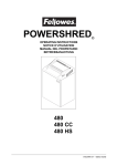
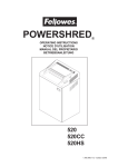
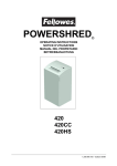
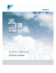
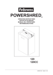
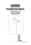
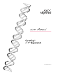
Chapter 6 Microfluidic-Based Manufacture of siRNA-Lipid Nanoparticles for Therapeutic Applications Colin Walsh, Kevin Ou, Nathan M. Belliveau, Tim J. Leaver, Andre W. Wild, Jens Huft, Paulo J. Lin, Sam Chen, Alex K. Leung, Justin B. Lee, Carl L. Hansen, Robert J. Taylor, Euan C. Ramsay, and Pieter R. Cullis Abstract A simple, efficient, and scalable manufacturing technique is required for developing siRNA-lipid nanoparticles (siRNA-LNP) for therapeutic applications. In this chapter we describe a novel microfluidicbased manufacturing process for the rapid manufacture of siRNA-LNP, together with protocols for characterizing the size, polydispersity, RNA encapsulation efficiency, RNA concentration, and total lipid concentration of the resultant nanoparticles. Key words Lipid nanoparticle, Microfluidics, siRNA, Nanoparticle manufacture, Solid-core, LNP, siRNA-LNP, NanoAssemblr, Nanoparticle formulation 1 Introduction There are numerous manufacturing methods for the preparation of lipid nanoparticles (LNP) for therapeutic applications [1–4]. The three main techniques are sonication, extrusion, and microfluidization [3, 4]. Sonication and extrusion differ only in the method used to alter LNP morphology and reduce particle size. A hydrated multilamellar vesicle (MLV) suspension is obtained after rehydrating dried lipid films in aqueous solution. Sonication using a bath sonicator or a probe sonicator reduces particle size and produces large unilamellar vesicles (LUV ~100 nm) or small unilamellar vesicles (SUV < 100 nm). In contrast, extrusion produces LUV or SUV by forcing the MLV suspensions through membranes of defined pore size for repeated cycles. In both cases, the size, morphology (LUV versus SUV), and polydispersity of the resultant LNP are dependent on lipid composition, lipid concentration, temperature, pH, volume and sonication time/power/tuning (sonication), or membrane pore size/number of extrusion cycles Kewal K. Jain (ed.), Drug Delivery System, Methods in Molecular Biology, vol. 1141, DOI 10.1007/978-1-4939-0363-4_6, © Springer Science+Business Media New York 2014 109 110 Colin Walsh et al. (extrusion) [3, 4]. Sonication and extrusion are sensitive techniques that suffer from batch-to-batch variation and are not easily scaled. Microfluidization provides an alternative LNP manufacturing technique. This method uses high pressure (up to 10,000 psi) to force a lipid suspension, which has been divided into two streams, through a small orifice into an interaction chamber where the two streams collide to produce LNP. The LNP are further reduced in size by cavitation, shear, and impact [3]. Similar to sonication and extrusion, particle size is dependent on LNP parameters such as lipid components, concentration, etc. In addition, pressure, size of the interaction chamber, and the number of microfluidization cycles influence LNP size. Microfluidization can reproducibly produce large volumes of LNP; however, despite reasonable encapsulation efficiencies (>75 %), the high shear force can damage encapsulated material. Moreover, problems with scale-up, material loss, and contamination limit the applicability of this LNP manufacturing method [3, 4]. Traditional manufacturing processes for the production of LNP are problematic [5]. LNP are formulated via bulk mixing and suffer from microscopic variations in the speed and local concentration of lipid components, resulting in nanoparticle heterogeneity, batchto-batch variability, and operator-dependent formulation characteristics [5]. Further, these “one-pot” manufacturing processes prevent precise control over temporal and spatial concentration of LNP components. Microfluidics provides a versatile and elegant approach to enable the simple and consistent production of LNP [6]. Microfluidic systems typically operate in the regime of laminar flow and afford precise, reproducible, and controlled mixing at the nanoliter scale, enabling conditions ideal for the formulation of nanoparticles and microparticles [7–10]. Further, microfluidic synthesis offers important practical advantages including low reagent consumption, parallelization, and automation, making it ideally suited to high-throughput optimization and scale-up. The NanoAssemblr™ is a recently developed microfluidic- based platform for the manufacture of nanoparticles such as liposomes, oil-in-water nanoemulsions, and short interfering RNA (siRNA) lipid nanoparticles (siRNA-LNP) (Precision NanoSystems Inc., Vancouver, Canada). The NanoAssemblr™ platform enables efficient encapsulation of siRNA in LNP, and data generated clearly demonstrates microfluidic production to be superior to current manufacturing methods. The NanoAssemblr™ platform comprises a benchtop instrument with computer software that controls the flow of fluids through a microfluidic mixing device. The microfluidic device consists of a Y-junction followed by a mixing region where staggered herringbone structures induce rapid mixing by chaotic advection (Fig. 1). The NanoAssemblr™ benchtop instrument uses syringe pumps to introduce a lipid/ethanol solution and an aqueous solution Microfluidic Manufacture of siRNA-Lipid Nanoparticles 111 Fig. 1 Schematic overview of the NanoAssemblr™ microfluidic mixer design. (A) and (B ) are inlets where the aqueous siRNA solution and the ethanolic lipid solution are introduced into the microfluidic channels. (C ) is an enlargement of the staggered herringbone mixer (SHM) used to promote rapid mixing of the aqueous and ethanol solutions. (D ) is the outlet where formulated siRNA-lipid nanoparticles (siRNA-LNP) are collected of siRNA into the device (inlet A and inlet B in Fig. 1) at flow rates of 4–12 mL/min. As the two streams pass through the herringbone mixer, the rapid mixing of the ethanol and aqueous streams results in a reduced solubility of lipids (Fig. 1 (C)), causing a state of supersaturation that drives the spontaneous aggregation of lipid into well-defined nanoparticles that simultaneously encapsulate the siRNA via electrostatic interaction with the cationic lipid component of the LNP. Following microfluidic manufacture, the residual ethanol is removed, the buffer exchanged, and the pH adjusted to pH 7.4 by dialysis. The final siRNA-LNP formulation is subsequently characterized according to particle size and polydispersity, RNA encapsulation efficiency, RNA concentration, and lipid concentration. Microfluidic-based manufacture results in siRNA-LNP formulations with superior characteristics to those produced using traditional methods, such as the preformed vesicle (PFV) or spontaneous vesicle formation (SVF) techniques. Microfluidic manufacture mediates (1) siRNA encapsulation efficiencies of 100 % versus 65–95 % for PFV and SFV methods; (2) defined particle size including the formulation of limit-size LNP (≥20 nm), which are unobtainable with either the PFV or SVF methods; and (3) LNP comprising ionizable cationic lipid in excess of 60 mol% compared to a maximum of 40 mol% achievable with the PFV approach. Collectively, this translates to reduced cost of goods, improved biodistribution, and increased potency in vitro and in vivo. 112 Colin Walsh et al. 2 Materials (See Note 1) 2.1 Aqueous siRNA Formulation Solution 1.Lyophilized siRNA. 2.RNA hydration buffer: 10 mM sodium acetate, pH 6.0. 3.Formulation buffer: 25 mM sodium acetate, pH 4.0. 2.2 Ethanolic Lipid Formulation Solution 1.Cationic or ionizable lipid. 2.1,2-Distearoyl-sn-glycero-3-phosphocholine (DSPC). 3.Cholesterol. 4.PEG-lipid. 5.Absolute ethanol. 2.3 Microfluidic Manufacturing Apparatus 1. NanoAssemblr™ benchtop instrument (Precision NanoSystems Inc., Vancouver, Canada). 2.NanoAssemblr™ microfluidic cartridge. 3.Disposable syringes: 1–10 mL. 2.4 Post- manufacture Processing 1.12–14,000 MW cutoff dialysis cassette. 2.PBS (calcium- and magnesium-free). 3.Disposable syringe. 4.0.2 μm filter. 5.Stir plate and magnetic stir bar. 2.5 siRNA Concentration and Encapsulation Efficiency 1.96-well plate. 2.siRNA-LNP characterization standard. 3.siRNA-LNP formulations. 4.RiboGreen Assay Kit (Life Technologies, R11490). 5.1× TE buffer pH 7.5. 6.1× TE buffer pH 7.5 with 2 % Triton X-100 (Triton buffer). 2.6 Lipid Quantification 1.siRNA-LNP formulations for testing: post-chip, post-dialysis, and post-filtration samples. 2.Wako Cholesterol E (CHOD-DAOS method): (a) Cholesterol E standard solution 200 mg/dL (2 mg/mL). (b) Cholesterol E color reagent. (c) Cholesterol E buffer. 2.7 LNP Characterization Equipment 1.Dynamic light-scattering particle sizer. 2.Fluorescence plate reader. 3.UV/Vis spectrophotometer. Microfluidic Manufacture of siRNA-Lipid Nanoparticles 113 3 Methods 3.1 Microfluidic Manufacture of siRNA-Lipid Nanoparticles (siRNA-LNP) 1. Preparation of siRNA formulation solution: Hydrate lyophilized siRNA in RNA hydration buffer to the desired concentration (generally 10–100 mg/mL). Measure final concentration using A260. Prepare the siRNA formulation solution by diluting the hydrated siRNA into formulation buffer to the desired final concentration (generally 0.1–1.5 mg/mL siRNA). Aliquot 60–80 μL and set aside for siRNA-LNP characterization (see Subheadings 3.3–3.5 below). 2.Preparation of lipid formulation solution stock: Solutions of individual lipids are prepared by dissolving a measured amount of dry lipid in absolute ethanol to the desired concentration (generally 10–50 mg/mL lipid, depending on solubility). The lipid formulation solution is prepared by mixing lipid stock solutions to provide a mixture at the appropriate molar ratios and total lipid content (see Note 2). This mixture is then diluted with absolute ethanol to provide a final solution at the desired volume and lipid concentration. Aliquot 20–30 μL and set aside for siRNA-LNP characterization (see Subheadings 3.3– 3.5 below). 3.Formulation of siRNA-LNP using the NanoAssemblr™ Benchtop Instrument: Load the NanoAssemblr™ microfluidic cartridge into the NanoAssemblr™ cartridge holder, and select the desired formulation conditions on the NanoAssemblr™ user interface (see Note 3). Attach a disposable syringe (BD) with formulation buffer into the first inlet of the NanoAssemblr™ microfluidic cartridge. Attach the second syringe containing absolute ethanol into the second inlet of the NanoAssemblr™ microfluidic cartridge. Prime the system by flowing ≥2 mL total of formulation buffer and absolute ethanol through the NanoAssemblr™ microfluidic cartridge using the desired formulation conditions (see Note 4). Ensure that the syringes are loaded with sufficient volume to run the desired total volume. Once the system has been primed, load the siRNA formulation solution into a disposable syringe of the appropriate size. Remove all air bubbles by gently agitating the syringe. Attach this syringe into the first inlet of the NanoAssemblr™ microfluidic cartridge. Load the lipid formulation solution into a disposable syringe of the appropriate size. Remove all air bubbles by gently agitating the syringe. Attach this syringe into the second inlet of the NanoAssemblr™ microfluidic cartridge. Insert sample collection tube into the left side and a waste collection tube into the right side of the NanoAssemblr™ sample collection block (see Note 5). Select the desired formulation parameters using the NanoAssemblr™ software interface and click “Go” (see Note 4). The NanoAssemblr™ Benchtop 114 Colin Walsh et al. Instrument will automate the manufacture of the siRNA-LNP and dispense the formulated nanoparticles into the sample collection tube. 3.2 Post- manufacture Processing of siRNA-LNP 1.Post-manufacture processing: Add PBS to a large beaker at room temperature and stir continuously. The total volume of PBS required is 200–400-fold the recovered sample volume (i.e., if 2 mL of manufactured siRNA-LNP was recovered, 400–800 mL of PBS should be used). Remove dialysis cassette from package and equilibrate in PBS for 10–15 min. Following cassette equilibration, load sample into dialysis cassette, place in beaker of PBS and stir at room temperature for ≥4 h to remove ethanol, and increase sample pH to 7.4. Remove cassette from PBS and recover sample. 2.Sample concentration: If necessary, the recovered sample can be concentrated using a centrifugal filter unit with 10,000 MW cutoff filter. Samples should be spun at 3,000 RPM until the desired concentration is achieved. 3. Sample sterilization: In a sterile hood, filter the recovered sample through a 0.2 μm filter to remove any aggregates and to sterile filter the siRNA-LNP formulation. Set aside approximately 75 μL of the final sample to use for characterization (see Subheadings 3.3–3.5 below). 3.3 siRNA-LNP Characterization: Particle Size and Polydispersity 1.Particle size and polydispersity are measured using dynamic light scattering: Dilute sample as needed to achieve an appropriate concentration for sizing (see Note 6). 3.4 siRNA-LNP Characterization: RNA Encapsulation Efficiency and Concentration (See Note 7) 1.Preparation of sample stock solutions: In the top row of the 96-well plate (Row A, Fig. 2), add 297 μL of TE buffer pH 7.5 to a single well for each sample plus a single well for a PBS blank using a multichannel pipette. Add 3 μL of sample to these wells for a final volume of 300 μL. Add 3 μL of PBS to the blank well. Pipette to mix. This is your stock solution for each sample. The final RNA concentration of these stock solutions should be approximately 4–7 μg/mL. 2.siRNA-LNP sample setup: Add 50 μL of TE buffer pH 7.5 to the two wells directly below each sample (Rows B and C, Fig. 2). Add 50 μL of sample stock solution from Row A into the wells in Rows B and C (this assay is run in duplicate. All liquid handling should be done using a multichannel pipette). Add 50 μL of Triton buffer to the wells in Rows D and E (Fig. 2) below each sample. Add 50 μL of sample stock solution from Row A into the wells in Rows D and E. Microfluidic Manufacture of siRNA-Lipid Nanoparticles 115 Fig. 2 Experimental design for RiboGreen assay in a 96-well plate. S1–S11 represent samples 1–11. B represents blank well. All numerical values represent μL volumes. Blue wells = sample; green wells = sample + TE buffer pH 7.5; red wells = sample + 2 % Triton buffer; brown wells = standard curve 3.RiboGreen RNA standard curve setup: Dilute the RNA standard to produce an RNA stock at a final concentration of 20 μg/mL in TE buffer pH 7.5. The final volume should be 150 μL (see Note 8). Set up a standard curve (in duplicate) as shown in Table 1 using the RNA stock (20 μg/mL siRNA), TE buffer pH 7.5, and Triton buffer. Once samples and standard curve are plated, incubate the plate at 37 °C for 10 min to lyse siRNA-LNP in the presence of Triton X-100 (see Note 9). 4.Preparation of RiboGreen Solution: Sum the total number of sample wells and standard curve wells. Add three to this number, and multiply the total by 100. This is the total volume, in μL, of RiboGreen Solution needed for this assay. In a 15 mL RNAse-free Falcon tube, dilute the RiboGreen Reagent 1:100 into TE buffer pH 7.5 to the total volume calculated. For example, if 3,000 μL of RiboGreen Solution is needed, add 30 μL of RiboGreen Reagent to 2,970 μL of TE buffer. Vortex the RiboGreen Solution for 10 s to mix. 5. Addition of RiboGreen Solution and sample readings: Remove a 96-well plate from 37 °C incubator. Add 100 μL of RiboGreen Solution to each well. Pop any air bubbles with a needle. Read using fluorescent plate reader (excitation = 480, emission = 525). 6.Sample analysis: Use the data generated by the RiboGreen standard curve to calculate the concentration of siRNA. 116 Colin Walsh et al. Table 1 Preparation of standard curve stock solutions for RiboGreen assay Final RNA concentration (μg/mL) RNA stock required (μL) TE buffer required (μL) Triton buffer required (μL) Total volume per well (μL) 2.5 25 25 50 100 1 10 40 50 100 0.5 5 45 50 100 0.25 2.5 47.5 50 100 0.1 1 49 50 100 3.5 siRNA-LNP Characterization: Cholesterol (Lipid) Concentration (See Note 10) 1.Mix the Cholesterol E color reagent and Cholesterol E buffer. This mixture is referred to as “Cholesterol Reagent” in this protocol. There is a separate dry color reagent for each of the two buffers included in the kit. Note the date that color reagent and buffer are mixed and use within 1 month. 2. 96-well plate setup: Sum the total number of Sample Standards and siRNA-LNP formulation samples that will be assayed. Multiply this number by two. This is the number of wells required to run this assay in duplicate (i.e., 12 samples require 24 wells). Add 185 μL of Cholesterol Reagent to the wells (Rows A and B, Fig. 3). 3.For the standard curve (Rows C and D, Fig. 3), add: (a)190 μL of Cholesterol Reagent into two wells. (b)195 μL of Cholesterol Reagent into two wells. (c)197.5 μL of Cholesterol Reagent into two wells. (d)199 μL of Cholesterol Reagent into two wells. (e)185 μL of Cholesterol Reagent into two wells (blank). 4.Sample addition, incubation, and plate reading: Add 15 μL of each sample to the wells from step 1 (Rows A and B, Fig. 3). This gives a final volume of 200 μL in each sample well. Add 10 μL of Cholesterol E standard solution to wells C1 and D1, 5 μL of Cholesterol E standard solution to wells C2 and D2, 2.5 μL of Cholesterol E standard solution to wells C3 and D3, and 1 μL of Cholesterol E standard solution to wells C4 and D4 (Fig. 4). This gives a final volume of 200 μL in each standard curve well. Add 15 μL of PBS into the two blank wells (wells C5 and D5, Fig. 4). This gives a final volume of 200 μL in each blank well. Place plate at 37 °C for 25 min. Remove plate and read samples at A595 using a 96-well plate reader. Microfluidic Manufacture of siRNA-Lipid Nanoparticles 117 Fig. 3 Experimental design for cholesterol assay. S1–S12 represent samples 1–12. All numerical values represent μL volumes of Cholesterol Reagent. Green wells = sample; brown wells = standard curve Fig. 4 Experimental layout for addition of Cholesterol E standard to a 96-well plate. S1–S12 represent samples 1–12. All numerical values represent μL volumes of Cholesterol E standard. Green wells = sample; brown wells = standard curve 5.Sample analysis: Use the data generated by the standard curve to calculate the cholesterol concentration, and use the relative proportion of cholesterol in the lipid composition to estimate the total lipid concentration. 118 Colin Walsh et al. 4 Notes 1.Prepare all buffers and solutions using RNAse-free water. All disposables should be sterile and RNAse-free. All surfaces, glassware, and labware should be thoroughly cleaned with a 70 % ethanol solution and RNAse Zap (Invitrogen) prior to formulation to prevent RNA degradation. 2.Typical lipid composition for the manufacture of siRNA-LNP is the following: cationic lipid/DSPC/cholesterol/PEG-lipid (50:10:38.5:1.5 mol:mol); siRNA/lipid ratio of 0.06 (wt/wt). 3.Operation of the NanoAssemblr™ Benchtop Instrument is controlled by a custom user interface that enables selection of manufacturing parameters: total formulation volume (range 1–20 mL); syringe size (1, 3, 5, or 10 mL); total flow rate of aqueous siRNA solution and ethanolic lipid solution through the microfluidic cartridge (range 2–12 mL/min); aqueous to ethanol flow rate ratio (1:1–5:1); and sample switching time (start of run, 0–24 s; end of run, 0–6 s). The sample switching function is designed to collect suboptimal particles that are manufactured at the beginning and at the end of the run when the mixing rate is not at steady state. This material is collected in the waste container. For more information, see the NanoAssemblr™ Benchtop Instrument user manual (www. nanoassemblr.com). 4.Typical manufacturing parameter settings on the Nano Assemblr instrument are: (a) Total formulation volume = 2 mL. (b) Syringe size = 3 and 1 mL. (c) Total flow rate = 12 mL/min. (d) Aqueous: ethanol flow rate ratio = 3:1. (e) Flow rate for aqueous siRNA solution = 9 mL/min. (f) Flow rate for ethanolic lipid solution = 4 mL/min. (g) Volume of aqueous siRNA solution dispensed = 1.5 mL. (h) Volume of ethanolic lipid solution dispensed = 0.5 mL. (i) Sample switching time at start of run = 0.25. (j) Sample switching time at the end of run = 0.05. 5. Refer to the NanoAssemblr™ Benchtop Instrument user manual for a detailed description of instrument operation (www. nanoassemblr.com). Microfluidic Manufacture of siRNA-Lipid Nanoparticles 119 6. Based on the lipid composition and the NanoAssemblr™ manufacturing settings described in Note 3 above, particle size is typically 60–65 nm with PDI < 0.05. 7.The RiboGreen assay (Life Technologies) is a fluorescence- based assay for the detection of RNA in solution. This 96-well plate assay is used to determine the encapsulation efficiency and total siRNA concentration of siRNA-LNPs formulated on the NanoAssemblr™. siRNA encapsulation efficiency is determined by measuring fluorescence in the absence or presence of detergent (Triton X-100). Sample Standards (aqueous siRNA and lipids in ethanol mixed by pipette at a ratio equivalent to the flow ratio on the NanoAssemblr™) are used as RNA concentration standards to account for the effect of lipids on RiboGreen fluorescence. Up to 11 samples can be run on a single plate. Please refer to the manufacturer’s protocol for more details. 8. VRNA Stock = ( 20 µg / mL ) ´ (150 µl ) C RNA Stock Volume TE buffer = 150 μL − VRNA Stock VRNA stock = volume of RNA stock required in μL CRNA stock = concentration of RNA stock in μg/mL 9.This assay uses the Sample Standard from siRNA-LNP formulation as the RNA standard. The Sample Standard is prepared by mixing aliquots of the siRNA and lipid stock solutions by pipetting. Aliquots should be mixed at the same ratio as the siRNA-LNP formulation (i.e., 3:1 aqueous/ethanol). 10.The cholesterol assay (Cholesterol E kit, Wako Diagnostics) is an enzymatic colorimetric assay to determine the cholesterol content of siRNA-LNP in the pre-manufacture (Sample Standard), post-manufacture, post-dialysis, and post-filtration steps of the formulation. This is used to calculate lipid losses and dilution during formulation. Up to 12 samples can be run on a single plate. Please refer to the manufacturer’s protocol for more details. Acknowledgements This work was supported by the National Science and Engineering Research Council of Canada (F09-04486), Canadian Institutes for Health Research (111627), and Genome British Columbia. 120 Colin Walsh et al. References 1.Mozafari MR (2005) Liposomes: an overview of manufacturing techniques. Cell Mol Biol Lett 10:711–719 2.Schwendener RA, Schott H (2010) Liposome formulations of hydrophobic drugs. In: Weissig V (ed) Liposomes methods and protocols, 2nd edn. Humana Press, Totowa, NJ, pp 129–138 3. Mozafari MR (2010) Nanoliposomes: preparation and analysis. In: Weissig V (ed) Liposomes methods and protocols, 2nd edn. Humana Press, Totowa, NJ, pp 29–50 4.Chatterjee S, Banerjee DL (2002) Preparation, isolation, and characterization of liposomes containing natural and synthetic lipids. In: Basu SC, Basu M (eds) Liposome methods and protocols, 1st edn. Humana Press Totowa, NJ, pp 3–16 5.Semple SC, Klimuk SK, Harasym TO et al (2001) Efficient encapsulation of antisense oligonucleotides in lipid vesicles using ionizable aminolipids: formation of novel small multilamellar vesicle structures. Biochim Biophys Acta 1510:152–166 6.Belliveau NM, Huft J, Lin PJ et al (2012) Generation and loading of lipid nanoparticles containing siRNA by microfluidic mixing. Mol Ther Nucleic Acids 1:e37 7.DeMello AJ (2006) Control and detection of chemical reactions in microfluidic systems. Nature 442:394–402 8. Jahn A, Vreeland WN, DeVoe DL et al (2007) Microfluidic directed formation of liposomes of controlled size. Langmuir 23:6289–6293 9.Song H, Chen DL, Ismagilov RF (2006) Reactions in droplets in microfluidic channels. Angew Chem Int Ed Engl 45:7336–7356 10. Song Y, Hormes J, Kumar CS (2008) Microfluidic synthesis of nanomaterials. Small 4:698–711