Transcript
www.biochain.com
Tel: 1-888-762-2568 Fax: 1-510-783-5386 Em ail: [email protected]
User’s Manual and Instructions
Uric Acid Assay Kit (Z5030021)
Quantitative Colorimetric Uric Acid Dete rmination at 590nm
DESCRIPTION
Uric acid is the waste product produced from the degradation of purines. In
healthy human, uric acid is filtered and removed from the blood by the
kidneys and excreted into urine. Because a number of kidney diseases are
know n to affect uric acid levels, uric acid determination is thus important
and useful in diagnosing and evaluating kidney diseases. For example,
when uric acid is present in the blood at abnormally high levels, it tends to
crystallize in body joints, resulting in gout, a very painful inflammatory
condition. Increased levels of uric acid are also known to be associated
with uremia, leukemia and pneumonia.
2. Add 1000 mL working reagent and tap lightly to mix.
3. Incubate 30 min at room temperature and read optical density at
590nm (510nm-630nm).
Simple, direct and automation-ready procedures for measuring uric acid
concentration in blood are becoming popular in Research and Drug
Discovery. Biochain’s uric acid assay kit is designed to measure uric acid
directly in serum w ithout any pretreatment. The improved method utilizes
2,4,6-tripyridyl-s-triazine that forms a blue colored complex specif ically with
iron in the presence of uric acid. The intensity of the color, measured at
590nm, is directly proportional to the uric acid concentration in the serum.
The optimized formulation substantially reduces interference by
substances in the raw samples.
ODBLANK, ODS TANDARD and ODSAMPLE are OD590nm values of Blank,
Standard and Sample, respectiv ely. It is not necessary to prepare a
calibration curve, because the concentration of the provided standard
lies w ithin the linear range.
Normal serum uric acid values: 1.0 to 7.0 mg/dL.
Conversions: 1 mg/dL uric acid equals 59.5 mM, 0.001% or 10 ppm.
KEY FEATURES
Sensitive and accurate. Use 5 mL samples. Linear detection range 0.22
mg/dL (13mM) to 30 mg/dL (2380mM) uric acid in 96-well plate assay.
Simple and high-throughput. The procedure involves addition of a single
working reagent and incubation for 30 min. Can be readily automated as a
high-throughput assay in 96-well plates for thousands of samples per day.
Improved reagent stability and versatility. The optimized formulation has
greatly enhanced reagent and signal stability. Cuvet or 96-well plate assay.
Low interference in biological samples. No pretreatments are needed.
Assays can be directly performed on serum samples.
APPLICATIONS :
CALCULATION
The uric acid concentration of Sample is calculated as
=
MATERI ALS REQUIRED, BUT NOT PROVIDED
Pipeting devices and accessories (e.g. 5 mL).
Procedure using 96-well plate:
Clear bottom 96-w ell plates (e.g. Corning Costar).
96-well plate absorbance (590nm) reader.
Procedure using cuvette:
Cuvets for measuring optical density at 510-630nm.
Spectrophotometer for measuring absorbance at 590nm.
EXAMPLES :
Samples were assayed using the 96-well protocol. The uric acid content
(mg/dL) was 1.3 ± 0.1 (n = 4) for mice serum, 2.6 ± 0.0 (n = 4) for fetal
bovine serum (Invitrogen), 1.4 ± 0.1 for goat serum, 1.3 ± 0.1 for rat
serum, 2.9 ± 0.1 for rat plasma, 3.4 ± 0.1 for human serum and 1.4 ± 0.1
for human plasma, respectively.
Direct Assays: uric acid in serum, plasma, urine and other biological samples.
Drug Discovery/Pharmacology: effects of drugs on uric acid metabolism.
1.2
KIT CONTENTS (250 tests in 96-well plates)
Uric Acid
1.0
OD590n m
Reagent A: 50 mL
Reagent B: 6 mL
Reagent C: 6 mL
Standard: 1 m L 10 mg/dL uric acid
Blank Control: 1 mL
Storage conditions. The kit is shipped at room temperature. Store
reagents at 4 °C, standard and blank control at -20 °C. Shelf lif e: 12
months after receipt.
Procedure using 96-well plate:
1. Set up standards and samples. Transfer 5 mL Blank, Standard and
samples in duplicate wells of a clear bottom 96-well plate.
2. Add 200 mL working reagent and tap lightly to mix.
3. Incubate 30 min at room temperature and read optical density at 510630nm (peak absorbance at 590nm).
Procedure using cuvette:
1. Set up test tubes labeled Blank, Standard, Samples. Transfer 20 mL
Blank, Standard and samples to appropriately labeled tubes.
0.6
0.4
R2 = 0.996
0.0
0
10
20
30
[Uric Acid], mg/dL
PROCEDURES
Reagent Preparation: shake Reagent C before use. Prepare
enough working reagent by mixing 10 volumes of Reagent A, 1 volume
Reagent B and 1 volume Reagent C. Fresh reconstitution is
recommended. Equilibrate to room temperature before assay. Metal
chelators (e.g. EDTA) interfere w ith this assay and should be avoided.
0.8
0.2
Precautions: reagents are for research use only. Normal precautions for
laboratory reagents should be exercised while using the reagents. Please
refer to Material Safety Data Sheet for detailed information.
F-753-3UMRevA
ODSAMPLE – ODBLANK
X 10 (mg/dL)
ODSTANDARD – ODBLANK
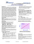
Standard Curve in 96-well plate assay
PUBLI CATI ONS
[1]. Viel, E.C. et al (2008). Xanthine oxidase and mitochondria contribute
to vascular superoxide anion generation in DOCA-salt hypertensive rats.
Am J Physiol Heart Circ Physiol. 295:H281-H288.
[2]. Kamel, A. H. (2007). Conventional and planar chip sensors for
potentiometric assay of uric acid in biological fluids using flow injection
analysis. J Pharm Biomed Anal. 45(2):341-348.
[3]. DiSilvestro R. A. et al (2009). Pomegranate extract mouth rinsing
effects on saliva measures relevant to gingivitis risk. Phytother Res.
23(8): 1123-1127.
Z5030021UA
Active Date: 09172012