1
FT-DR961d
IMAPlate
Products Description
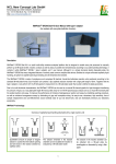
The IMAPlate is a disposable Lab device to perform manually high throughput liquid transfer, analysis and assay in a
miniature format.
Features:
• 96-channel pipette for liquid transfer
• 96 micro-cuvette array for UV, VIS or IR spectroscopy
• 96 microwell plate for parallel reactions and assays
IMAplates makes it possible pipette up to 96 individual samples simultaneously,
then to analyse them with your usual microplate reader, then if needed to recover
the samples!
Applications:
• UV, VIS or IR spectroscopy
• ELISA (chromogenic, fluorescent, luminometric)
• 96-channel pipetting / dispensing
This document presents some key applications and technical tips.
DNA/RNA quantification and quality analysis
Protein quantification: Bradford Protein Assay
Miniaturized Enzyme-Linked ImmunoSorbent Assay
A simple solution to improve the detection sensitivity of ELISA
A rapid miniature ELISA for the quantification of Troponin I using Perkin Elmer plate reader
Miniaturize Homogenous Assays
References
Contact your local distributor
[email protected]
Uptima, powered by
P.1/15
FT-DR961d
DNA/RNA quantification and quality analysis by IMAplate
Introduction
The quantification of DNA or RNA by the spectroscopy is a well-established routine method in many laboratories. The
absorbance of UV light at wavelength of 260 nm by a pure DNA or RNA provides a simple and accurate estimation of
the concentration of nucleic acids in a sample. For example, the absorption of 1 OD (A) is approximately equivalent to
50 μg/ml dsDNA, 33 μg/ml ssDNA, 40 μg/ml RNA or 30 μg/ml for oligonucleotides.
Besides, the quality of a DNA or RNA sample can also be checked by the spectroscopy. The ratio of A260/A280 or
A260/A230 is used to estimate the contamination of the sample by protein or by substances such as carbohydrates,
peptides, phenols or aromatic compounds, respectively. The pure DNA should have an A260/A280 ratio of
approximately 1.8, whereas pure RNA should give a value of approximately 2.0. The ratio A260/A230 should be
approximately 2.2 for pure samples.
IMAPlate is the world's first miniaturized analytical platform capable of manually performing high-throughput liquid
transfer, analysis, reaction and assay. It comprises 96 identical, funnel-like reaction units positioned according to
standard 96-well plate format and each reaction unit contains a 5 μl round reaction chamber with a light path of 5 mm
long. The IMAPlate uses capillary force to confine samples in the bottomless reaction chambers and therefore the
samples can spectroscopically be analyzed one-by-one in a microwell plate reader in any range of UV-VIS-IR spectra.
The use of IMAPlate combined with a microwell plate reader is an ideal routing method for quantification and quality
analysis of DNA or RNA samples, especially, with limited amount of sample volume.
Experimental to determine the concentration of samples
* Reagents and Materials
▪ DNA standards
▪ 96-well V-bottom plate
▪ Pipette
▪ IMAPlate and reader adaptor
▪ Microwell plate reader (with UV range)
* Procedure A: using capillary force based liquid transfer (high-throughput)
1. Transfer 15 to 20 μl of a standard series of DNA solution to the assigned wells of a 96-well V bottom plate.
2. Transfer 15 to 20 μl of sample solutions to the rest well of the 96-well V bottom plate.
3. Aspirate the standard and sample solutions to IMAPlate by capillary force.
4. Place the IMAPlate in the reader with the adaptor.
5. Measure absorbance at wavelength of 260 nm and base line absorbance at wavelength of 350 nm.
6. Use true absorbance values (A260 – A350) to calculate DNA concentration of samples according to the standards.
* Procedure B: using pipette loading
1. Pipette 1, 2, 3, 4 and 5 μl of a standard DNA solution to the assigned reaction chambers on IMAPlate.
2. Pipette appropriate amount of sample solutions (1 to 5 μl) to the rest reaction chambers on IMAPlate according to the
expected concentration.
3. Place the IMAPlate in the reader with the adaptor.
4. Measure absorbance at wavelength of 260 nm and base line absorbance at wavelength of 350 nm.
5. Use true absorbance values (A260 – A350) to calculate DNA concentration of samples according to the standards.
Contact your local distributor
[email protected]
Uptima, powered by
P.2/15
FT-DR961d
Results and Discussion
Figure 1 shows spectra of calf thymus DNA solution and water
in the reaction chambers of an IMAPlate measured by a
microwell plate reader. The obtained spectrum is a typical
spectrum of a pure DNA sample measured by a
spectrophotometer and can be used for both quantitative and
quality analysis.
The data of calf thymus DNA standards, generated with the
Procedure A, can very well be fitted with linear regression
equation (Figure 2). The coefficient of variation from 96
reaction chambers of an IMAPlate is 5.8 % for measuring a
known concentration of calf thymus DNA solution in the low
detection range (Figure 3). According to the results, the
concentration of a DNA sample within 5 μg/ml to 250 μg/ml
could accurately be determined by this procedure. The
Procedure A is an easy, fast, robust and effective means for the
analysis of a large number of samples.
Figure 4 represents a diagram of the true absorbance against the volume of a DNA sample solution pipetted in the
reaction chambers. The true absorbance shows a linearity relation with the volume at the range from 1 μl to 5 μl.
Therefore, the use of a pipette can provide an alternative to prepare a standard curve, in which only one standard with
an appropriate concentration can be added into the assigned reaction chambers on IMAPlate with a serial volume. In
such a way, the preparation of a standard series of DNA solution is no longer necessary. It may also eliminate the need
for dilution of samples with high concentration just by adding 1 μl of samples to the reaction chambers for
measurement.
Conclusion
The IMAPlate is an easy-to-use, robust, miniaturized analytical platform suitable for quantification and quality analysis
of DNA/RNA samples with 96-well plate readers. It offers users a wide range of UV-VIS-IR spectrometric analysis of 1
μl to 5 μl of samples and a variable light path from 1 mm to 5 mm. The measured samples can totally be recovered
without cross contamination risk. The flexible, simple handling of the IMAPlate not only saves hands on time and
material cost for the analysis, but also enhances the lab productivity
Contact your local distributor
[email protected]
Uptima, powered by
P.3/15
FT-DR961d
Protein quantification: Bradford Protein Assay by IMAplate
Introduction
The Bradford protein assay is a simple and rapid method to determine the total protein concentration in a sample.
The assay uses Coomassie G-250 Dye as a colorimetric reagent for the quantification of protein. In the acidic
environment of the reagent mixture, the absorbance peak of the dye will change from 465 nm to 595 nm when the dye
binds to protein. Within the linear range, the absorbance value at 595 nm is proportion to the total amount of protein
existing in the reagent mixture.
IMAPlate is the world's first miniaturized analytical platform capable of manually performing high-throughput liquid
transfer, analysis, reaction and assay. It comprises 96 identical, funnel-like reaction units positioned according to
standard 96-well plate format and each reaction unit contains a 5 μl round reaction chamber with a light path of 5 mm.
The bottomless reaction chamber uses capillary force to confine sample solution inside it; therefore up to 96 samples
can be analyzed one-by-one in a microwell plate reader.
The use of IMAPlate for the Bradford protein assay would provide scientists an easy-to-use, miniaturized analytical tool
for protein quantification. It offers benefits such as:
·■ minimize the consumption of delicate protein samples - only requiring 0.1 μl to 4 μl sample
·■ no need for time-consuming sample dilutions - flexible sample volume
·■ large linear measurement range - up to 4000 μg/ml
·■ high throughput - obtaining up to 96 individual data in one measurement
·■ save reagent and produce less chemical waster
Experimental
* Reagents and Materials
▪ Bradford Reagent – Uptima CooAssay #UPF86400
▪ Protein standards
▪ Pipettes (can accurately transfer 1 and 4 μl)
▪ IMAPlate™ start kit
▪ Microwell plate reader (e.g. BioTek PowerWave™ Microplate Spectrophotometer)
* Procedure A (high concentration of protein sample)
1. Pipette 4μl of 1:4 diluted Bradford reagent (fresh prepared mixture of one part of the reagent plus 3 part of distilled
water) to the reaction chambers.
2. Pipette 1μl of protein standards and sample (mixing very well before use) to the assigned reaction chambers.
3. Invert the IMAPlate several times to mix the solution.
4. Place the IMAPlate in the reader with the adaptor.
5. Measure the peak absorbance at wavelength of 595 nm and base line absorbance at wavelength of 800 nm.
If desired, the peak absorbance and base line absorbance can also be measured at other wavelengths between 575 nm
to 615 nm and 750 nm to 850 nm respectively.
6. Use true absorbance values (A595 – A800) to plot the standard curve and calculate the concentration of samples
according to the standards.
* Procedure B (low concentration of protein sample)
1. Pipette 1μl of un-diluted Bradford reagent to the reaction chambers.
2. Pipette 4μl of protein standards and sample (mixing very well before use) to the assigned reaction chambers.
3. Invert the IMAPlate several times to mix the solution.
4. Place the IMAPlate in the reader with the adaptor.
5. Measure the peak absorbance at wavelength of 595 nm and base line absorbance at wavelength of 800 nm.
6. Use true absorbance values (A595 – A800) to plot the standard curve and calculate the concentration of samples
according to the standards.
Contact your local distributor
[email protected]
Uptima, powered by
P.4/15
FT-DR961d
Results and Discussion
The plot of the true absorbance values against BSA
concentration gives a typical binding curve as expected (see
left figure). If the data are treated by a graph fitting
software with binding mode, the detection range of the
BSA can be from 0 to 10000 μg/ml. But a straight line can
be obtained in a narrow range with low concentration of
protein. For example, in Procedure A the concentration of
BSA is between 0 to 400 μg/ml or in Procedure B the
concentration of BSA is between 0 to 100 μg/ml. the linear
range also can be extended up to 4000 μg/ml if 0.1 μl of
sample is mixed with 5 μl of 1:5 diluted Bradford reagent.
To the current setup (the final reagent concentration is 1:5
dilution) the amount BSA in the reaction chamber from
0.04 to 0.4 μg shows a near-linear relationship with the true
absorbance values.
Conclusion
The IMAPlate is an easy-to-use, robust, miniaturized analytical platform suitable for quantification and quality analysis
of protein samples with 96-well plate readers. It offers users a wide range of UV-VIS-IR spectrometric analysis of 5 μl
of samples. The measured samples can totally be recovered without cross contamination risk. The flexible, simple
handling of the IMAPlate not only saves hands on time and material cost for the analysis, but also enhances the lab
productivity.
The method can be adapted with other protein dyes –colorimetric, fluorimetric, luminometric.
Contact your local distributor
[email protected]
Uptima, powered by
P.5/15
FT-DR961d
Miniaturized Enzyme-Linked ImmunoSorbent Assay by IMAplate
Introduction
Enzyme linked immunosorbent assay (ELISA), as a simple, sensitive, specific and economical method for qualification
and quantification of a particular molecule (e.g. protein, peptide or small molecule) in a sample, is routinely used in life
sciences, health care, and many other different industries.
Although it is developed in late 70’s, the ELISA has almost kept its original assay format; the assay is performed in a
96-well plate with a working volume of 100μl or 200μl. Attempt to reduce the reaction volume of the ELISA by using
384-well plate with a working volume of 20μl or 50μl has been reported successful. But technical difficulties in liquid
handling hampered such kind of approaches being used.
IMAPlate is the world's first miniaturized analytical platform, capable of manually performing high-throughput liquid
transfer, analysis, reaction and assay. Its unique liquid handling concept has resolved the technical difficulties for
transferring tiny amount of solution. Up to 96 individual samples or solution can simultaneously be transferred in and
out of the 5 μl reaction chambers of the IMAPlate by touch-loading or touch-unloading procedure. Therefore, the
processing of each step for ELISA is in parallel and high-throughput. This unique liquid handling concept especially
eases the tedious washing steps of the ELISA.
The use of IMAPlate for ELISA would bring scientists many benefits such as:
·■ minimizing the consumption of delicate samples and reagents - 5 μl
·■ reducing time to result by at least half
·■ high productivity
·■ cost effectiveness
·■ user friendly
·■ benefit environment - producing less biological and chemical wasters
ELISA benefits
IMAplate microplates have unique bottom free chambers instead of wells, that are designed to:
-have a slightly larger surface for immunological reactions
-a smaller volume of reagent
-a slightly longer path lenght
-be able to load samples and reagent quickly capillary force.
·■ A a result, ELISA assays performed in IMAplate show quiker kinetic of immunological reaction, and higher signal
(increase absorbance values up to 7-folds) while using only 5µL of samples and reagents and gaining considerable
time in handling.
·■ The miniaturazation of the method is critical when analysing rare or precious samples, when using expensive
antibodies (primary antibodies will be used, and eventuelly more diluted) or other costly reagents such a ECL
substrates or saturating agents. The method also reduce consumption of the washing buffer, furthermore that ther is no
need of washer that need large volume for setting up the process.
·■ The benefits are combined in in High Throught Put analysis (HTS), and when combining several analysis on each
samples.
Experimental
* Reagents and Materials
▪ IMAPlate start kit
▪ Human IL-6 DuoSet ELISA Development kit
▪ U-bottomed 96-well plates
▪ Pastuer pipette and/or pipettes
▪ Microwell plate reader
* Miniaturized ELISA - High-throughput Protocol
Plate Preparation - coating
1. Dilute the Capture Antibody to the working concentration (2 μg/ml) in PBS according to the protocol provided by the
manufacturer. Add one drop (between 20μl to 50μl) of the diluted Capture Antibody to each well of the U-bottomed 96Contact your local distributor
[email protected]
Uptima, powered by
P.6/15
FT-DR961d
well plate by a pasture pipette or a pipette. Immediately coat the IMAPlate plate(s) by touch-loading.
Incubate 60 minutes in a humidity environment at room temperature.
2. Touch-unload the reaction chambers and wash with Wash Buffer by touch-loading and touch-unloading Wash Buffer
three times.
3. Block IMAPlate by touch-loading of Reagent Diluent (1mg/ml BSA in PBS) to each reaction chamber.
Incubate 10 minutes in a humidity environment at room temperature.
4. Wash three times as the step 2. The IMAPlate plates are now ready for sample addition.
Sample & Reagent Saving Protocol
The plate preparation and the assay procedure in this Sample & Reagent Saving Protocol basically follow the same
protocol as the belox High-throughput Protocol except of the way to load reaction chamber. When it needs to save the
samples or reagents, the reaction chambers are loaded by a pipette instead of by the touch-loading. It is recommended to
turn the IMAPlate upside down and to use the reverse pipetting technique to add 5μl of samples or reagents into the
reaction chambers through the bottom openings. Be careful for the sample and standard orientation. In order to avoid
the orientation mistakes, mark the backside of the IMAPlate.
Assay Procedure - High-throughput
1. Touch-load sample or standard in Reagent Diluent from the U-bottomed 96-well plate, which is prepared in advance
by adding a drop of the samples or the standards to the appropriate wells.
Incubate 60 minutes in the humidity chamber at room temperature.
2. Wash three times as the step 2 of Plate Preparation.
3. Touch-load the Detection Antibody diluted in Reagent Diluent from a U-bottomed 96-well plate.
Incubate 60 minutes in the humidity chamber at room temperature.
4. Wash three times as the step 2 of Plate Preparation.
5. Touch-load 1:200 diluted Streptavidin-HRP solution from a U-bottomed 96-well plate.
Incubate 15 minutes in the humidity chamber at room temperature.
6. Wash five times as the step 2 of Plate Preparation.
7. Touch-load Substrate Solution. Incubate 5 - 10 minutes at room temperature.
8. Place the IMAPlate on a U-bottomed 96-well plate containing 15μl of stop solution (2 - 5M H2SO4) in each well.
Make sure the bottom openings are contacted with the stop solution. After touch-exchange 30 seconds, slowly remove the IMAPlate from the Ubottomed 96-well plate and gently invert several times until the blue colored TMB substrate solution turning to yellow.
9. Immediately determine the true absorbance of each reaction chamber by a two-wavelength measurement: measure
both peak absorbance at 450nm and the baseline absorbance at 650nm (any wavelength between 550nm to 700nm can be used) and
subtract the peak absorbance from the correspondent baseline absorbance to get true absorbance (Abstrue = Abs450nm – Abs650nm). The true
absorbance should be used for the calculation and plotting. If the reader has spectral scan mode, it is recommended to use the spectral measurement
setting for the two-wavelength measurement e.g. starting at 450nm and ending at 650nm with a step of 200nm.
Results and Discussion
Figure 1 shows a typical plot of the data set of human IL-6
standards from an ELISA that was performed in IMAPlate
with a high-throughput protocol, and a fitting curve of the
standards with one-site binding mode. The standards were
very well distributed around the fitting curve and the CV%
of the IL-6 standards was usually less than 10% in
triplicates (typically around 5%).
When the same concentration of all the reagents was used
to perform ELISA on conventional 96 well plate and
IMAPlate, the/ slope of the standard curve markedly
increased with IMAPlate. Therefore, the sensitivity was
correspondently increased even that the time for reactions
in IMAPlate was reduced to half.
Contact your local distributor
[email protected]
Uptima, powered by
P.7/15
FT-DR961d
It has to be pointed out that, when the sensitivity increases, the
detection range may be narrowed due to the overflow of the
absorbance for higher concentration standards (each plate
reader has a certain absorbance detection range). Using a plate
reader with a wide detection range may avoid the overflow for
higher concentration standards. However, the missing peak
absorbance data at 450nm can always be derived from the data
set measured at a less sensitive, off-peak wavelength (e.g. at
485nm) by the linearity relationship between the data set at
peak wavelength and the one at off-peak wavelength (Fig.2).
Of course, further shortening reaction time for the color
development or reducing Streptavidin-HRP concentration are
possible alternative ways to avoid the overflow if the
sensitivity does not need to be increased.
Conclusion
Using the IMAPlate for the ELISA can dramatically reduce the consumption of the sample and reagents. The time to
results for a typical ELISA can be cut at least to half. The sensitivity is also increased. The unique touch-loading and
touch-unloading procedure provides a parallel, high-throughput liquid transfer and especially eases the wash steps.
Besides, the self-dosed solution up-taking may not need to use low volume precision pipette and can save the pipette
tips. The IMAPlate is a user friendly, robust, miniaturized, high-throughput lab device for performing miniaturized
ELISA.
Contact your local distributor
[email protected]
Uptima, powered by
P.8/15
FT-DR961d
IMAplate simple solution to improve the detection sensitivity of ELISA
Introduction
ELISA usually uses the same 96-well microplate for the reaction and the readout, typically 100µL. The following
method takes advantage of unique IMAplate feature to increas the detection sensitivity:
A standard ELISA procedure is performed in conventionnal microplates, taking advantage of the large surface of the
96-wells, but the final staining of immobilized enzyme is done with only 25µL (instead of 100µL). Hence the
concentration of the colored product formed is about 4 times higher if all the immobilized enzyme is able to react. Then
the colored solution is pipetted by IMAplate 5µL funnel-like chambers (5µL) and made available for reading in a
standard microplate reader with a higher lenght pass so higher signal can be recorded. All together, up 7-fold higher
signal can be achieved.
Alternatively, all the ELISA assay procedure can be performed directly in IMAplate with additionnal benefits. See
above applications 'Miniaturized ELISA assay'.
Experimental
* Reagents and Materials
▪ Ready to use ELISA kit or ELISA reagent kit
▪ 96-well ELISA plate
▪ Pipette
▪ IMAPlate start kit
▪ Microwell plate reader
* Procedure for ELISA using the IMAPlate for result readout*
1. Coat 96-well ELISA plate according to the protocol provided in the reagent kit with capture antibody.
Note: A reduced volume for coating (e.g. 50 μl for flat bottom or 40 μl for V and U bottom) may reduce the
background.
2. Block remaining protein-binding sites of the well with blocking reagent.
Note: above preparation steps are only for ELISA reagent kit
3. Perform the ELISA according to the protocol provided by manufacturer. In order to increase the detectable rate, an
increased volume of sample may be preferred for low abundant molecule measurement.
4. Add reduced volume of substrate solution e.g. TMB solution and vortex for 15 to 30 minutes.
The volume of substrate solution used depends on the demands of the sensitivity and the bottom format of the 96-well plate. 25 μl
would be the low limit for a flat bottom plate, otherwise not all immobilized enzymes are able to make contact with the substrate
solution. If the volume of the substrate solution is the same as that for coating, the vortex is not necessary.
5. After incubation, transfer 5 μl of the substrate solution (with or without the addition of acidic stop solution) from 96well ELISA plate to IMAPlate by pipette or aspirate by capillary force, if the volume is large enough (e.g. 75 μl for flat
bottom or 20 μl for V and U bottom).
It is suggested to measure the substrate solution without addition of acidic stop solution because it may cause the precipitation for
standards and samples with high concentration and make the absorbance value of these standards and samples invalid.
6. Place the IMAPlate in the reader with the adaptor and measure absorbance at wavelength of 665 nm and base line
absorbance at wavelength of 500 nm without acidic stop solution or at wavelength of 450 nm and base line absorbance
at wavelength of 650nm with acidic stop solution.
For standards and samples having absorbance value over the upper detection limit of the reader, pipetting 1 μl or 2 μl to IMAPlate
for the measurement can make these standards and samples valid and may extend the upper detection limit of the ELISA.
7. Use true absorbance values (A665 – A500) or (A455 – A650) to calculate the concentration of samples according to
the standards.
Contact your local distributor
[email protected]
Uptima, powered by
P.9/15
FT-DR961d
Results and Discussion
As predicted, the detection sensitivity and the low detection limit can be significantly improved with the new ELISA
setup.
Figure 1 shows the plot of two ELISA data sets for human TNF-a
standard curves with a series of 3-fold dilution. The red curve is the plot of
the data from the conventional ELISA setup and the green one is the plot
of the data from the new ELISA setup. Both ELISAs were performed in
96-well plate with flat bottom according to the protocol provided in the kit
except that, for the green curve, 25 μl of TMB substrate solution was used
instead of 100 μl for the incubation with vortexing and the result was read
out using the IMAPlate with the volume of 5 μl.
The detection sensitivity (the slope of the initial increase) was increased
about 7-fold and the low detection limit also went down correspondently.
If a 96-well plate with V or U bottom is used and the/ substrate solution wareduced to 15 μl,
the detection sensitivity is expected to increase more than 10-fold and the low detection limit
can go down 0-fold as well. The detectable rate for those low abundant analytes can be
increased further when an increased sample volume is applied. It has to be pointed out that to
use higher concentration substrate solution would be preferred in order to avoid quickly
running out of the substrate for standards of high concentration defined in the provided
protocol.
Since the absorbance value of two standards with the highest
concentration was over the/ upper detection limit of the plate
reader, a second measurement using 1.5 μl of the reaction
solution was performed. Because the obtained absorbance value
with 1.5 μl of the reaction solution decreased about 3.3-fold and
matched the calculation based on the decrease of the light path,
the linear fitting curve of standards in Figure 2 is used to derive
the absorbance data for the two tandards with 5 μl. Alternatively,
it is also ractical to measure the absorbance value irectly from
the 96-well ELISA plate after adition of 100 μl of suitable buffer
and derive the data from the linear fitting curve of the standards
in both measurements.
Conclusion
Using the IMAPlate6 for the ELISA readout can dramatically increase the detection sensitivity of commercial ELISA
kit with the described ELISA setup, in which the ELISA is basically carried out according to user’s protocol, except the
readout step. So scientists can directly use commercial ELISA kits and perform the ELISA with their familiar routine
procedure but the addition of several lower standards and the alternation of the readout. The described approach
provides scientists a very simple but very effective method to improve the detection sensitivity of ELISA and will
accelerate the discovery and applications of low abundant proteins in research and diagnostics.
The IMAPlate6 is an easy-to-use, robust, miniaturized analytical platform and the multi-utility of IMAPlate6 and the
approach described will have a substantial impact not only on ELISA, but also on other type of assays.
Contact your local distributor
[email protected]
Uptima, powered by
P.10/15
FT-DR961d
IMAPlate-based rapid miniature ELISA for the quantification of Troponin
I using Perkin Elmer Enspire 2300 plate reader
Introduction
The cardiac Troponin I (cTnI) plays an important role in the regulation of cardiac muscle
contraction. It does not ordinarily exist in peripheral circulation but will be released into
circulation in myocardial necrosis. Therefore, the cTnI is a reliable cardiac biomarker for the
cardiac muscle injury. The amount of cTnI in peripheral circulation is usually quantified by
Enzyme-linked immunosorbent assay (ELISA), which is performed in a 96-microwell plate.
The conventional cTnI ELISA suffers from a number of limitations: e.g. long assay
incubation times, large sample volumes and laborious liquid handling.
The IMAPlateTM 5RC96 is a miniaturized analytical platform, which can replace the 96-well
plate for carrying out reactions and assays. It comprises 96 identical, funnel-like reaction units and each reaction unit
contains a 5 μL capillarity reaction chamber with a light path length of 5 mm.
Besides being economic, sensitive and flexible for liquid handling by pipetting, the unique self-dosed liquid taking-up
feature of IMAPlate also supports manual high-throughput, in which up to 96 individual samples can simultaneously be
loaded in a second by simply dipping the open bottom of the capillarity reaction chambers and emptied by applying
them to a filter paper.
Loaded solution can then be measured fluorimetrically or colorimetrically by plate readers. In this application, we use
Perkin Elmer Enspire 2300 plate reader to measure the cTnI ELISA performed in IMAPlate.
This IMAPlate-based miniature ELISA demonstrates better features over the conventional cTnI ELISA such as
dramatically reducing the sample and reagents consumption and the time to results.
Materials and Methods
Reagents and Materials:
▪ Capture antibody (clone 560) and detection antibody coupled with HRP (clone MF4) for cardiac Troponin I, Hytest, Ref. 4T21
▪ Human cardiac Troponin I-T-C complex, Hytest, Ref. 8T62
▪ TMB (Tetramethylbenzidine) substrate, Substrate Reagent Pack, R&D Systems, Ref.DY999
▪ IMAPlateTM 5RC96 start kit
▪ Nunc 96-well Polysorp plates
▪ Perkin Elmer plate reader: ENSPIRE 2300 MULTILABEL READER
▪ Reagents: PBS pH 7.4; fat free powder milk; Tween-20, H2SO4
▪ Cardiac troponin I (cTnI) free serum, Hytest, Ref. 8TFS
▪ Washing buffer: PBS - 0.05 % Tween-20
▪ Detection buffer: PBS - 0.05 % Tween-20 - 0.2 % fat free powder milk
The capture antibody and detection antibodies have been diluted to 7.5 μg/mL in PBS and to 1μg/mL in detection
buffer, respectively.
IMAPlate protocol:
1. Coat the IMAPlate 5RC96 plate by directly pipetting 5 μL of capture antibody solution into the reaction chamber
from the bottom opening.
Incubate 30 minutes at room temperature in a high humidity environment to prevent from evaporation.
[When working with very low sample volumes, small interferences (e.g. bubbles, particles, homogeneity of solution) can have a large impact on the
results. Make sure to mix the sample solution thoroughly before pipetting. Reverse pipetting is recommended in order to avoid bubble formation or
incomplete dispensing from the tip.]
2. Wash the plate 4 times.
Empty the IMAPlate by placing it on a filter paper and pushing slightly against filter paper for 10 seconds to let the solution
completely absorbed by the filter paper.
Touch-load the IMAPlate with wash buffer and empty the IMAPlate four times.
[Touch-load procedure: place IMAPlate on a plate cover containing sufficient wash buffer and move up and down several times to ensure the
capillarity reaction chambers fully loaded. Instead of the plate cover, a flat bottomed 96-well plate containing 100 μL of wash buffer in each well can
also be used.]
3. Block IMAPlate by touch-loading. Incubate 10 minutes in a high humidity environment at room temperature.
4. Wash the plate 4 times (as in step 2).
Contact your local distributor
[email protected]
Uptima, powered by
P.11/15
FT-DR961d
5. Prepare cTnI standard concentrations at 12.5; 6.25; 3.125; 1.56; 0.78; 0.39; 0.2 and 0 ng/mL in PBS. Add standard or
sample by directly pipetting 5 μL standard concentrations into the reaction chamber from the bottom opening. 2 or 3
replicates are prepared. Incubate 30 minutes at room temperature in a high humidity environment.
6. Wash the plate 4 times (as in step 2).
7. Add detection antibody in IMAPlate by directly pipetting 5 μL of antibody solution into the reaction chamber
through the bottom opening. Incubate 30 minutes at room temperature in a high humidity environment.
8. Wash the plate 4 times (as in step 2).
9. Add TMB substrate by touch-loading of TMB solution from a plate cover or a 96-well plate.
Incubate 5 - 20 minutes at room temperature.
[For a U- or V-bottomed 96-well plate, 25 μL TMB solution in the wells is enough and for a flat bottomed 96-well plate 80 – 100 is needed.]
10. Pipette 0.5 μL of 3M H2SO4 stop solution directly into the reaction chamber from the bottom opening.
Slowly inverse the IMAPlate several times to make sure the solutions are mixed well.
Alternatively, place the IMAPlate on a 96-well plate that contains the stop solution [15 μL for a U- or V-bottomed 96-wll plate and 80 μL for a flat
bottomed 96-well plate] and wait for several seconds to allow the solution in the reaction chamber to partially exchange with the stop solution through
the bottom openings. Slowly lift the IMAPlate and inverse several times to make sure the solutions are mixed well.
11. Measure absorbance at both peak wavelength (450 nm for TMB) and base-line wavelength (e.g. 650 nm for TMB)
by using the IMAPlate adaptor according to the IMAPlate user manual.
Calculate the true absorbance (Abstrue = Abspeak - Absbaseline).
96-well plate protocol:
1. Coat the plate with 50 μL/well of capture antibody solution. Incubate overnight at 4°C.
2. Wash the plate 4 times (200 μL/well wash buffer).
3. Block the plate with 100 μL of Blocking solution to each well. Incubate 2 hours at room temperature with agitation.
4. Wash the plate 4 times.
5. Prepare cTnI standards concentrations at 12.5; 6.25; 3.125; 1.56; 0.78; 0.39; 0.2 and 0 ng/mL in PBS.
Add 50 μL/well standard or sample. Incubate 2 hours at room temperature with agitation.
6. Wash the plate 4 times.
7. Add 50 μL/well of detection antibody solution. Incubate 2 hours at room temperature with agitation.
8. Wash the plate 4 times.
9. Add 100 μL of TMB substrate to each well. Incubate 5 - 20 minutes at room temperature with agitation.
10. Add 100 μL of stop solution to each well.
11. Measure absorbance at 450 nm according to TMB solutions instructions.
Results ()
Standard curves:
Figure 1 shows the cTnI standard curves obtained from both IMAPlate (red) and NUNC 96-well plate (blue).
Two independent experiments have been performed for the two different assays (IMAPlate and NUNC 96-well plate). Samples were run in triplicate
for IMAPlate assay and in duplicate for NUNC 96-well plate assay. The standard curves from the IMAPlate and the 96-well plate are comparable.
The maximum absorbance/background ratio is very close (41.31 fold background versus 42.31 for the IMAPlate and the 96-well Nunc plate,
respectively) while the concentration of cTnI to reach half maximum absorbance/background is around 30% lower for IMAPlate than 96-well Nunc
plate (4.2 vs 5.9 ng/ml for the IMAPlate and the 96-well Nunc plate respectively). Therefore, the IMAPlate should have a better resolution towards
the low concentration.
Figure 1: 4 PL fit of the absorbance/background ratio versus
log of cTnI concentration using the Graphpad Prism Software.
Sensitivity, Limit of Detection (LOD):
Mean +/- SD. N=1 (the results are representative of the two
Limit of detection (LOD) was calculated as: Average of
independent experiments.)
background + 2.5 SD of background and are shown in
Table 1 below. The IMAPlate offers a slightly better
sensitivity compared to conventional 96-well plate assay.
Table 1: LOD values (mean of 2 independent experiments
Contact your local distributor
[email protected]
Uptima, powered by
P.12/15
FT-DR961d
for each assay, N=2)
Intra-assay precision:
The CV% of the cTnI standards was usually less than 20% in triplicates for IMAPlate assay and in
duplicates for 96-well plate assay, from cTnI concentrations from 1.56 to 12.5 ng/mL.
Recovery in serum:
Additional samples have been prepared by spiking pure Troponin–free serum with known
concentrations of cTnI. No difference could be observed between IMAPlate and 96-wells plate
assays for concentrations around 5 ng/mL.
Discussion
The IMAPlate technology is the world’s first miniaturized analytical platform that also supports manual highthroughput liquid handling. Due to the very small dimension of the capillarity reaction chamber, using high quality plate
readers such as from Perkin Elmer are recommended for the measurement of IMAPlate. The rapid auto new plate
definition function of the Perkin Elmer Enspire 2300 multilabel reader makes it much more attractive for the
measurement of IMAPlate to obtain high quality data.
The IMAPlate based miniature cTnI ELISA assay showed comparable results to conventional 96-wells plate for cTnI
ELISA assay. The shape of two standard curves was quite similar except that the one from IMAPlate was shifted in
parallels towards low concentration. Combined the observation of lower limit of detection in IMAPlate, the IMAPlatebased cTnI ELISA should have a better sensitivity. Besides, the requirement for samples and reagents was greatly
decreased. The time-to-results is reduced from overnight coating plus 7 hours to less than 3 hours including coating
procedure. The unique self-dosed liquid taking-up feature also leads to very simple washing steps. However the touchloading procedure may require some technical practices for a new user.
In conclusion, the Perkin Elmer Enspire multilabel reader is compatible with the IMAPlate technology. The
combination of these two technologies provides a very useful miniature lab tool for routinely performing assays as well
as analyzing samples.
Contact your local distributor
[email protected]
Uptima, powered by
P.13/15
FT-DR961d
Homogenous assays
Miniature, high sensitive homogeneous assays
Introduction:
Due to relatively low cost of absorbance plate readers, cheap and uncomplicated reagents preparation, low sample consumption and simple “add-mixmeasure” procedure, 96-well plate based homogeneous colorimetric assays are still popularly used in the labs for routine analysis, research
application and compound screening. Attempting to miniature the assays in 384- or 1536-well plate seems not practicable for manual pipetting, and
remains a challenge even for automations. Although the “add-mix-measure” assay procedure is straightforward, false results can easily be generated
due to the air gap between the solutions caused by multiple pipetting into a small well.
IMAPlate comprises 96 identical, funnel-like bottomless reaction units, in which the diameter of the upper compartment
is larger than the well of 384-well plate while the diameter of the lower compartment is smaller than the well of 1536well plate. The unique design of the IMAPlate enables each reaction unit to hold more than 50 μl of solution.
When the lower bottom opening is sealed, the solution pipetted into the upper compartment does not flow into the lower
compartment. For example, by using a parafilm sheet to temperately seal the bottom, reagent and sample can stay in the
upper compartment for thoroughly mixing and incubation. Once the parafilm sheet removed, assay solutions will flow
and fill up the lower compartment, and immediately increase total liquid thickness about 5 mm. Therefore, for the
absorbance measurement, the light path length is increased ultimately. Using IMAPlate can overcome the
incomplete mix and reaction observed in 384- and 1536-well plate based assays. It not only reduces the assay volume,
but also keeps high or increases the sensitivity of the assay. The IMAPlate based miniature homogeneous assay can fit
for both manual operation and automated liquid handling workstation.
IMAPlate Macro Volume Miniature Assay Procedure:
Contact your local distributor
[email protected]
1. Prepare a piece of
parafilm (e.g. 100 x 150
mm) and place it on the top
of an empty 96-well plate
without the wax paper.
2. Push a new IMAPlate
down to the 96-well plate
and the parafilm will
automatically seal the
bottom openings.
3. Add assay components
one by one into the upper
compartment. (Total assay
volume* is recommended
between 15 μl to 25 μl.)
4. Mix thoroughly with a
plate shaker and incubate.
5. Before measurement, peel
off the parafilm and the
assay solution will flow into
the lower compartment.
6. Place the IMAPlate in an
IMAPlate adaptor and gently
tap it in horizontal direction
several times to ensure the
assay solution filling up the
lower compartment.
7. Load the IMAPlate
adaptor with the assay
IMAPlate into the reader and
measure both the peak
absorbance and base line
absorbance.
8. Use true absorbance
values (Abspeak – Absbaseline ) to plot the standard
curve and calculate the
concentration of samples.
Uptima, powered by
P.14/15
FT-DR961d
* The assay volume should not be over the maximum volume, which the IMAPlate can hold to withstand each step of the assay
procedure from dropping off.
Examples:
Figure 1. Concentration curves of Alkaline Phosphatase: the
line with solid circle is performed in IMAPlate with total 20 μl
of assay solution and the line with open circle is performed in
Nunc 96-well plate with 150 μl of total assay solution.
Figure 2. Concentration curves of Candida Anterctica Lipase B:
the line with solid circle is performed in IMAPlate with total 20 μl
of assay solution and the line with open circle is performed in
Nunc 96-well plate with 200 μl of total assay solution.
References
1. Spies P. et al. Establishment of a miniaturized enzyme-linked immunosorbent assay for human transferrin quantification using an
intelligent multifunctional analytical plate, Analytical Biochemistry, 382 (2008): 35-39. Article
2. Spies P. et al. A simple approach to improve the sensitivity of ELISA: using IMAPlate 5RC96 for result readout, Analytical
Biochemistry, 397 (2010): 48-55. Articlepdf
3. Sciotti M. A. et al. IMAPlate Based Miniature, High Sensitive, Rapid Screening Method for Detecting Bioengineered, Secreted
Lipase Activities in Yeast Expression Systems, CHIMIA, 64, (2010): 789-792. Articlepdf
Ordering information
Designation
IMAplate™ Start Kit*
IMAplate™ (96 µcuves) – White§
IMAplate™ (96 µcuves) – Black§
IMAplate™ (96 µcuves) – Yellow§
Product number
DR9601 1 Kit*
DR9611 5 ea
DT5431 , 5 ea
DT5441 , 5 ea
Quantity
Technical sheet and price
5 ea
5 ea
Product Highlight
*the starter kit contains 5 white IMAplates and Reader adapter
§ White plate are recommended for luminescence measurments, Black for fluorescence measurements, and yellow plates for UV-vis spectrometry and sample handling
Catalog size quantities and prices may be found at http://www.interchim.com.
Please inquire for higher quantities (availability, shipment conditions).
For any information, please ask : Uptima / Interchim; Hotline : +33(0)4 70 03 73 06
Contact your local distributor
[email protected]
Rev.L7E-J11E
Uptima, powered by
P.15/15