1
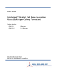
ly
!
AMPK Kinase Assay Kit
User’s Manual
For Research Use Only, Not for use in diagnostic procedures
On
Non-Radioisotopic Kit for Measuring AMPK Kinase Activity
CycLex AMPK Kinase Assay Kit
en
ce
Pu
rp
Intended Use................................................ 1
Storage......................................................... 1
Introduction ................................................. 2
Principle of the Assay.................................. 3
Materials Provided ...................................... 4
Materials Required but not Provided .......... 4
Precautions and Recommendations............. 5
Detailed Protocol.......................................... 6-12
Evaluation of Results ................................... 13
Assay Characteristics ...................................13
Troubleshooting ........................................... 13
Reagent Stability .......................................... 13
Sample Preparation.......................................14-16
Example of Test Results................................ 17-21
References......................................................22
Related Product..............................................23
os
e
Cat# CY-1182
Intended Use
er
The CycLex Research CycLex AMPK Kinase Assay Kit is primarily designed to measure the
activities of purified AMP-activated protein kinase (AMPK) or recombinant AMPK for the rapid and
sensitive evaluation of activators or inhibitors. The phospho-serine specific monoclonal antibody used in
this assay kit has been demonstrated to recognize the phospho-serine 789 in mouse Insulin Receptor
Substrate-1 (IRS-1), which is efficiently phosphorylated by AMPK. Additionally, column fractions of
cultured primary cell, cell line, or tissue can be assayed for AMPK activity with the CycLex Research
Product CycLex AMPK Kinase Assay Kit if the appropriate dose of AMPK specific inhibitor e.g.
Compound C is used.
ef
Applications of this kit include:
1) Screening activators or inhibitors of AMPK.
2) Evaluating the effects of pharmacological agents on AMPK activity in vitro.
3) Monitoring the purification of AMPK activity.
Storage
rR
• Upon receipt store all components at 4°C.
• Don’t expose reagents to excessive light.
Fo
Cat#: CY-1182
1
Version#: 120420
On
Introduction
ly
!
AMPK Kinase Assay Kit
User’s Manual
For Research Use Only, Not for use in diagnostic procedures
Measurement of AMPK activity
Pu
rp
os
e
AMP-activated protein kinase (AMPK) regulates cellular energy metabolism by phosphorylation of
key enzymes in metabolic pathways, such as acetyl–coenzyme A carboxylase (ACC) (1) or mTOR
(mammalian target of rapamycin) (2), by modulating their activities, and by regulating the activity of
transcription factors (3) and transcriptional cofactors (4). Its functions have been extensively studied in
muscles and liver. AMPK stimulates pathways, which increase energy production (glucose transport,
fatty acid oxidation) and switches off pathways, which consume energy (lipogenesis, protein synthesis,
gluconeogenesis). Activation of AMPK in adipose tissue can be achieved through situations such as
fasting and exercise. Adiponectin and leptin as well as hypoglycaemic drugs are activators of adipose
tissue AMPK. This activation probably involves changes in the AMP/ATP ratio, and the upstream kinase
LKB1 (5). These results have led to the concept that AMPK has an interesting pharmaceutical potential
in situations of insulin resistance (type 2 diabetes) and it is indeed the target of existing drugs, e.g.
Metformin and hormones which improve insulin sensitivity.
Mammalian AMPK is a trimeric enzyme comprising a catalytic α subunit (63 kDa) and non-catalytic
β and γ subunits (6, 7). Multiple isoforms of each mammalian enzyme exist (α1, α2, β1, β2, γ1, γ2, γ3),
each encoded by a different gene. The β subunits have a calculated molecular mass of 30 kDa, but
migrate on SDS-PAGE with apparent masses of 38 kDa (β1) and 34 kDa (β2), whereas the three γ
isoforms have molecular masses of 37 kDa (γ1), 63 kDa (γ2) and 55 kDa (γ3) (8). While AMPK
complexes containing α2 (the first catalytic isoform to be cloned) (9) predominate in skeletal and cardiac
muscle (10), approximately equal levels of α1 and α2 complexes are present in the liver (11). In contrast,
pancreatic islet β-cells largely express α1 complex activity (12, 13).
rR
ef
er
en
ce
The protocol generally regarded as most sensitive for the quantitative measurement of AMPK activity
involves incubation of the AMPK sample with substrate, either a natural or synthetic polypeptide (such
as SAMS peptide), in the presence of Mg2+and 32P-labeled ATP. The reaction is terminated by "spotting"
a sample onto a phosphocellulose P81 filter paper disc, followed by washing extensively to remove
unincorporated radiolabel and the incorporated radioactivity on P81 filter is counted. While sensitive,
this method is labor-intensive, generates hazardous radioactive waste and depends on a radioisotope of
short half-life. It is particularly unsuitable when kinase assays are only performed on an infrequent basis.
The CycLex AMPK Kinase Assay Kit uses anti-phospho-mouse IRS-1 S789 monoclonal antibody
(AS-4C4) and peroxidase coupled anti-mouse IgG antibody as a reporter molecule in a 96-well ELISA
format. This assay provides a non-isotopic, sensitive and specific method to measure the activities of
AMPK
Fo
Cat#: CY-1182
2
Version#: 120420
On
Principle of the Assay
ly
!
AMPK Kinase Assay Kit
User’s Manual
For Research Use Only, Not for use in diagnostic procedures
rp
os
e
The CycLex Research Products CycLex AMPK Kinase Assay Kit is a single-site, semi-quantitative
immunoassay for AMPK activity. Plates are pre-coated with a substrate-peptide corresponding to
surrounding mouse IRS-1 S789, which contains serine residue that can be efficiently phosphorylated by
AMPK. The detector antibody specifically detects only the phosphorylated form of serine residue on
substrate-peptide. The CycLex AMPK Kinase Assay Kit may be used to study the kinetics of a purified
AMPK as well as to screening AMPK activator or inhibitor. To perform the test, the sample is diluted in
Kinase Buffer, pipetted into the wells and allowed to phosphorylate the bound substrate following the
addition of Mg2+ and ATP. The amount of phosphorylated substrate is measured by binding it with a
AS-4C4, an anti-phospho-mouse IRS-1 S789 monoclonal antibody, followed by binding with
horseradish peroxidase conjugated anti-mouse IgG, which then catalyzes the conversion of the
chromogenic substrate tetra-methylbenzidine (TMB) from a colorless solution to a blue solution (or
yellow after the addition of stopping reagent). The color is quantitated by spectrophotometry and reflects
the relative amount of AMPK activity in the sample. For kinetic analysis, the AMPK-containing sample
is added to the wells in a similar fashion and at varying times the reaction is stopped by the addition of
the chelator, sodium ethylenediaminetetraacetate (EDTA) and the amount of phosphorylated substrate
determined as before.
Pu
Summary of Procedure
Add 100 µL of sample to the wells
Incubate for 30 min at 30°C.
en
ce
Wash the wells
Add 100 µL of Anti-Phospho-mouse IRS-1 S789 monoclonal Antibody.
Incubate for 30 min at room temp.
Wash the wells
Add 100 µL of HRP conjugated anti-mouse IgG
Incubate for 30 min at room temp.
er
Wash the wells
ef
Add 100 µL of Substrate Reagent
rR
Add 100 µL of Stop Solution
Fo
Cat#: CY-1182
Measure absorbance at 450 nm
3
Version#: 120420
On
Materials Provided
ly
!
AMPK Kinase Assay Kit
User’s Manual
For Research Use Only, Not for use in diagnostic procedures
All samples and standards should be assayed in duplicate. The following components are supplied and
are sufficient for the one 96-well microtiter plate kit.
os
e
Microplate: One microplate supplied ready to use, with 96 wells (12 strips of 8-wells) in a foil, zip-lock
bag with a desiccant pack. Wells are coated with mouse IRS-1 S789 -peptide as AMPK substrate.
10X Wash Buffer: One bottle containing 100 mL of 10X buffer containing 2 %Tween®-20
Kinase Buffer: One bottle containing 20 mL of 1X buffer; used for Kinase Reaction Buffer and sample
dilution.
rp
20X ATP: Lyophilized ATP Na2 salt. Reconstitute contents of vial with 1.6 mL of ddH2O. (See section
“Preparation of Working Solution” page 6.)
Anti-Phospho-mouse IRS-1 S789 Monoclonal Antibody (AS-4C4): One vial containing 12 mL of
anti-phospho-mouse IRS-1 S789 monoclonal antibody AS-4C4). Ready to use.
Pu
HRP conjugated Anti-mouse IgG: One vial containing 12 mL of HRP (horseradish peroxidase)
conjugated anti-mouse IgG. Ready to use.
Substrate Reagent: One bottle containing 20 mL of the chromogenic substrate, tetra-methylbenzidine
(TMB). Ready to use.
en
ce
Stop Solution: One bottle containing 20 mL of 1 N H2SO4. Ready to use.
Materials Required but not Provided
rR
ef
er
• AMPK Active enzyme(s): Available from CycLex Co., Ltd. (See page 6, section “ Preparation of
Working Solution” and page 24, section “Related Product”). e.g. The AMPK (α1/β1/γ1), Active
(Cat# CY-SEA11)
• 10X AMP (100 µM soln): AMP (Adenosine 5-monophosphate sodium salt) is available from Sigma,
Cat#. A1752.
• 10X Compound C (250 µM): Compound C is available from Sigma, Cat#. P5499. 2.5 mM stock
solution (DMSO) diluted 1:10 in Kinase Buffer.
• Pipettors: 2-20 µL, 20-200 µL and 200-1000 µL precision pipettors with disposable tips.
• Precision repeating pipettor
• Wash bottle or multichannel dispenser for plate washing.
• Microcentrifuge and tubes for sample preparation.
• Vortex mixer
• Plate reader capable of measuring absorbance in 96-well plates at dual wavelengths of 450 nm/540
nm. Dual wavelengths of 450/550 or 450/595 nm can also be used. The plate can also be read at a
single wavelength of 450 nm, which will give a somewhat higher reading.
• 500 or 1000 mL graduated cylinder
• Reagent reservoirs
• Deionized water of the highest quality
• Disposable paper towels
Fo
Cat#: CY-1182
4
Version#: 120420
ly
!
Precautions and Recommendations
On
AMPK Kinase Assay Kit
User’s Manual
For Research Use Only, Not for use in diagnostic procedures
• Store the ATP at -20°C when not in use. Store all other components at 4°C. Do not expose reagents to
excessive light. Avoid freeze/thaw cycles.
• Allow all the components to come to room temperature before use.
os
e
• All microplate strips that are not immediately required should be returned to the zip-lock pouch, which
must be carefully resealed to avoid moisture absorption.
• Do not use kit components beyond the indicated kit expiration date.
• Use only the microtiter wells provided with the kit.
rp
• Rinse all detergent residue from glassware.
• Use deionized water of the highest quality.
Pu
• Do not mix reagents from different kits.
• The buffers and reagents used in this kit contain either sodium Kathon-CG as preservatives. Care
should be taken to avoid direct contact with these reagents.
• Do not mouth pipette or ingest any of the reagents.
en
ce
• Do not smoke, eat, or drink when performing the assay or in areas where samples or reagents are
handled.
• Dispose of tetra-methylbenzidine (TMB) containing solutions in compliance with local regulations.
• Avoid contact with Substrate Solution which contains hydrogen peroxide.
• Avoid contact with Stop Solution which contains Sulfuric Acid.
er
• In case of contact with the Stop Solution and the Substrate Solution, wash skin thoroughly with water
and seek medical attention, when necessary.
• Biological samples may be contaminated with infectious agents. Do not ingest, expose to open
wounds or breathe aerosols. Wear protective gloves and dispose of biological samples properly.
rR
ef
• CAUTION: Sulfuric Acid is a strong acid. Wear disposable gloves and eye protection when
handling Stop Solution.
Fo
Cat#: CY-1182
5
Version#: 120420
On
Detailed Protocol
ly
!
AMPK Kinase Assay Kit
User’s Manual
For Research Use Only, Not for use in diagnostic procedures
os
e
The CycLex AMPK Kinase Assay Kit is provided with removable strips of wells so the assay can be
carried out on separate occasions using only the number of strips required for the particular
determination. Since conditions may vary, running an aliquot of the appropriate AMPK Active enzyme
(See page 24, section “ Related Product”), separately available from CycLex, should be included in
each assay. Disposable pipette tips and reagent troughs should be used for all transfers to avoid
cross-contamination of reagents or samples.
Preparation of Working Solution
1. Prepare a working solution of Wash Buffer by adding 100 mL of the 10X Wash Buffer (provided) to
900 mL of ddH2O. Mix well. Store at 4°C for two weeks or -20°C for long-term storage.
rp
2. Prepare 20X ATP Solution by adding 1.6 mL of ddH2O to the vial of 20X ATP (provided,
lyophilized). Mix gently until dissolved. The final concentration of the 20X ATP Solution should be
1.25 mM. Store the solution in small aliquots (e.g. 100 µL) at -20°C.
Kinase Buffer (provided)
20X ATP (provided)
96 assays
10 assays
9.95 mL
0.05 mL
995 µL
5 µL
199 µL
1 µL
10 mL
1,000 µL
200 µL
en
ce
Total
Pu
3. Prepare Kinase Reaction Buffer (5 µM ATP plus) by mixing following reagents for activator
screening.
2 assay
You will need 80-90 µL of Kinase Reaction Buffer (5 µM ATP plus) per assay well. Mix well.
Discard any unused Kinase Reaction Buffer (ATP plus) after use.
4. Prepare Kinase Reaction Buffer (50 µM ATP plus) by mixing following reagents for inhibitor
screening and Standard Assay.
10 assays
1 assay
Kinase Buffer (provided)
20X ATP (provided)
9.5 mL
0.5 mL
950 µL
50 µL
95 µL
5 µL
Total
10 mL
1000 µL
100 µL
er
96 assays
rR
ef
You will need 80-90 µL of Kinase Reaction Buffer (50 µM ATP plus) per assay well. Mix
well. Discard any unused Kinase Reaction Buffer (ATP plus) after use.
Fo
Cat#: CY-1182
6
Version#: 120420
ly
!
AMPK Kinase Assay Kit
User’s Manual
For Research Use Only, Not for use in diagnostic procedures
os
e
On
5. Prepare AMPK Active enzyme as a Positive Control for phosphorylation.
Add a Positive Control to the first well in each assay.
e.g. 0.05-0.20 ng / 10 µL of the AMPK (α1/β1/γ1), Active (CycLex Co., Ltd., See page 24, section
“Related Product”). Use BSA-containing buffer to dilute the enzyme for the stability of enzyme
activity.
Dilution Buffer (as an example, not provided):
20mM Hepes-KOH (pH 7.5), 1% BSA, 1mM EDTA, 2mM DTT, 50mM NaCl and 50% glycerol
Dilution:
1st Step: Dilute the enzyme 1:100 with the Dilution Buffer.
2nd Step: Dilute 1st Step enzyme 1: 50-200 with Kinase Buffer (provided).
Standard Assay
rp
1. Remove the appropriate number of microtiter wells from the foil pouch and place them into the well
holder. Return any unused wells to the foil pouch, refold, seal with tape and store at 4°C.
2. Prepare all samples (diluted with Kinase Buffer as needed). All samples should be assayed in
duplicate.
Pu
3. To assay partially purified recombinant AMPK, add 10 µL of each fraction to the wells of the
assay plate on ice. AMPK Active enzyme should be included in duplicate wells in each assay as a
positive control for phosphorylation, e.g. 0.1 ng/10 µL of AMPK (α1/β1/γ1), Active (Cat#
CY-SEA11) (See page 6, section “ Preparation of Working Solution” and page 24, section
“Related Product”).
en
ce
4. Begin the kinase reaction by addition of 90 µL Kinase Reaction buffer (50 µM ATP plus) per
well, cover with plate sealer, and incubate at 30°C for 30 minutes.
5. Wash wells five times with Wash Buffer making sure each well is filled completely. Remove
residual Wash Buffer by gentle tapping or aspiration.
6. Pipette 100 µL of Anti-Phospho-mouse IRS-1 S789 Monoclonal Antibody (AS-4C4) into each
well, cover with plate sealer or lid, and incubate at room temperature (ca.25°C) for 30 minutes.
7. Wash wells five times as same as in step 5.
er
8. Pipette 100 µL of HRP-conjugated Anti-mouse IgG into each well, cover with plate sealer or lid,
and incubate at room temperature (ca.25°C) for 30 minutes. Discard any unused conjugate after
use.
ef
9. Wash wells five times as same as in step 5.
10. Add 100 µL of Substrate Reagent to each well and incubate at room temperature (ca.25°C) for
5–15 minutes.
rR
11. Add 100 µL of Stop Solution to each well in the same order as the previously added Substrate
Reagent.
12. Measure absorbance in each well using a spectrophotometric plate reader at dual wavelengths of
450/540 nm. Dual wavelengths of 450/550 or 450/595 nm can also be used. Read the plate at 450
Fo
Cat#: CY-1182
7
Version#: 120420
ly
!
AMPK Kinase Assay Kit
User’s Manual
For Research Use Only, Not for use in diagnostic procedures
On
nm if only a single wavelength can be used. Wells must be read within 30 minutes of adding the
Stop Solution.
Note-1: Complete removal of liquid at each step is essential to good performance. After the last wash,
remove any remaining Wash Buffer by aspirating or decanting. Invert the plate and blot it
against clean paper towels.
os
e
Note-2: Reliable signals are obtained when either O.D. values do not exceed 0.25 units for the blank
(no enzyme control), or 2.5 units for a positive control when using the 0.1 ng of AMPK
(α1/B1/G1), Active (Cat# CY-SEA11) .
rp
Note-3: If the microplate reader is not capable of reading absorbance greater than the absorbance of the
AMPK Active enzyme, perform a second reading at 405 nm. A new O.D. values, measured at
405 nm, is used to determine AMPK activity of off-scale samples. The readings at 405 nm
should not replace the on-scale readings at 450 nm.
Kinetic Assay
Pu
1. Remove the appropriate number of microtiter wells from the foil pouch and place them into the well
holder. Return any unused wells to the foil pouch, refold, seal with tape and store at 4°C.
2. Prepare all samples (diluted with Kinase Buffer as needed). All samples should be assayed in
duplicate.
en
ce
3. To assay partially purified recombinant AMPK, add 10 µL of each fraction to the wells of the
assay plate on ice. AMPK Active enzyme should be included in duplicate wells in each assay as a
positive control for phosphorylation, e.g. 0.1 ng/10 µL of AMPK (α1/β1/γ1), Active (Cat#
CY-SEA11) (See page 6, section “Preparation of Working Solution” and page 24, section “Related
Product”).
4. Begin kinase reaction by addition of 90 µL Kinase Reaction Buffer (50 µM ATP plus) in
duplicate per well in timed intervals (suggested interval is 4 minutes but should be individually
determined for each system). After the final addition, incubate at 30°C for 20 minutes.
er
5. Stop the reaction by flicking out the contents. (Alternatively, the reaction may be terminated by the
addition of 150 µL 0.1 M Na EDTA, pH 8.0 to each well).
6. Wash wells five times with Wash Buffer making sure each well is filled completely. Remove
residual Wash Buffer by gentle tapping or aspiration.
ef
7. Pipette 100 µL of Anti-Phospho-mouse IRS-1 S789 Monoclonal Antibody (AS-4C4) into each
well, cover with plate sealer or lid, and incubate at room temperature (ca.25°C) for 30 minutes.
8. Wash wells five times as same as in step 6.
rR
9. Pipette 100 µL of HRP-conjugated Anti-mouse IgG into each well, cover with plate sealer or lid,
and incubate at room temperature (ca.25°C) for 30 minutes. Discard any unused conjugate after
use.
10. Wash wells five times as same as in step 6.
Fo
Cat#: CY-1182
8
Version#: 120420
ly
!
AMPK Kinase Assay Kit
User’s Manual
For Research Use Only, Not for use in diagnostic procedures
On
11. Add 100 µL of Substrate Reagent to each well (Avoid exposing the microtiter plate to direct
sunlight Covering the plate with e.g. aluminum foil is recommended). Return Substrate A to 2-8°C
immediately after the necessary volume is removed
os
e
12. Incubate the plate at room temperature (ca.25°C) for 5–15 minutes. (Appropriate incubation
time may vary, and incubation time can be extended up to 20 minutes if the reaction temperature
is below than 20°C).
13. Measure absorbance in each well using a spectrophotometric plate reader at dual wavelengths of
450/540 nm. Dual wavelengths of 450/550 or 450/595 nm can also be used. Read the plate at 450
nm if only a single wavelength can be used. Wells must be read within 30 minutes of adding the
Stop Solution.
Recommendations
For activator screening
Assay reagents
Pu
rp
Special considerations when screening activators or inhibitors
In order to estimate the activating or inhibitory effect on individual AMPK activity in the test
chemicals correctly, it is necessary to conduct the control experiment of “Solvent control” at least once
for every experiment and “Activator control” or Inhibitor control” at least once for the first experiment,
in addition to “Test sample”, as indicated in the following table. When test chemicals cause an activating
effect on AMPK activity, the level of A450 is strengthen as compared with “Solvent control”.
Test sample For
Activator
80 µL
Solvent Control
For Activator
80 µL
Activator
control
80 µL
10 µL
-
-
Solvent for Activator
-
10 µL
-
10X AMP (100 µM) *
-
-
10 µL
10 µL
10 µL
10 µL
en
ce
Kinase Reaction buffer (5 µM ATP)
10X Activator or equivalent
AMPK Active enzyme **
or Purified enzyme sample
er
* Sigma (Cat# A1752): See page 4, section “Materials Required but not Provided”
** e.g. 0.005 ng/µL of AMPK (α1/β1/γ1), Active (CycLex Co., Ltd., Cat# CY-SEA11) : See page 6, section “Preparation
of Working Solution” and page 24, section “Related Product”
ef
1. Following the above table, add the Reagents to each well of the microplate. Finally, initiate reaction
by adding 10 µL of “AMPK Active enzyme” to each well and mixing thoroughly at room temperature.
Cover with plate sealer or lid, and incubate at 30°C for 30 minutes.
rR
2. Follow the Standard Assay steps 5-12, page 7.
Note: Although we suggest to conduct experiments as outlined in the table above, the optimal
experimental conditions will vary depending on the parameters being investigated, and must be
determined by the individual user. Especially, an appropriate amount of AMPK Active enzyme
must be optimized by titration of the AMPK Active enzyme and setting the amount, which shows
OD value does not exceed plateau range in dose response curve.
Fo
Cat#: CY-1182
9
Version#: 120420
On
For inhibitor screening
ly
!
AMPK Kinase Assay Kit
User’s Manual
For Research Use Only, Not for use in diagnostic procedures
Kinase Reaction buffer (50 µM ATP)
80 µL
Solvent
Control
For
Inhibitor
80 µL
10X Inhibitor or equivalent
10 µL
-
-
10 µL
-
-
10 µL
10 µL
10 µL
Solvent for Inhibitor
-
10X Compound C (250 µM) *
-
AMPK Active enzyme **
or Purified enzyme sample
80 µL
rp
10 µL
Inhibitor
control
os
e
Assay reagents
Test sample
For Inhibitor
* Sigma (Cat# P5499): See page 4, section “Materials Required but not Provided”
** e.g. 0.01 ng/µL of AMPK (α1/β1/γ1), Active (CycLex Co., Ltd., Cat# CY-SEA11) : See page 6, section “Preparation
of Working Solution” and page 24, section “Related Product”
Pu
1. Following the above table, add the Reagents to each well of the microplate. Finally, initiate reaction
by adding 10 µL of “AMPK Active enzyme” to each well and mixing thoroughly at room temperature.
Cover with plate sealer or lid, and incubate at 30°C for 30 minutes.
2. Follow the Standard Assay steps 5-12, page 7.
rR
ef
er
en
ce
Note: Although we suggest to conduct experiments as outlined in the table above, the optimal
experimental conditions will vary depending on the parameters being investigated, and must be
determined by the individual user. Especially, an appropriate amount of AMPK Active enzyme
must be optimized by titration of the AMPK Active enzyme and setting the amount, which shows
OD value does not exceed plateau range in dose response curve.
Fo
Cat#: CY-1182
10
Version#: 120420
On
Special considerations when measuring precise AMPK activity
ly
!
AMPK Kinase Assay Kit
User’s Manual
For Research Use Only, Not for use in diagnostic procedures
In order to measure the activity of AMPK correctly, it is necessary to conduct the control experiment
of “Inhibitor control” at least once for every experiment and “ATP minus control” at least once for the
first experiment, in addition to “No enzyme control” as indicated in the following table. Although the
level of A450 increases in “Test sample” when AMPK enzyme activity is in the sample, the high level of
A450 is not observed in “Inhibitor control”, “ATP minus control” and “No enzyme control”.
Inhibitor
control
80 µL
80 µL
Kinase Buffer (ATP minus)
-
-
10X Compound C (250 µM) *
-
10 µL
Vehicle for 10X Compound C
10 µL
-
Your enzyme sample
AMPK Active enzyme **
Buffer for your enzyme sample
10 µL
-
80 µL
No
enzyme
control
80 µL
80 µL
-
-
-
-
-
10 µL
10 µL
10 µL
10 µL
-
10 µL
-
10 µL
rp
10 µL
-
Positive
control
Pu
Kinase Reaction buffer (50 µM ATP)
ATP
minus
control
-
os
e
Test
Sample
Assay reagents
* Sigma (Cat# P5499): See page 4, section “Materials Required but not Provided”
** e.g. 0.01 ng/µL of AMPK (α1/β1/γ1), Active (CycLex Co., Ltd., Cat# CY-SEA11) : See page 6, section “Preparation
of Working Solution” and page 24, section “Related Product”
en
ce
1. Following the above table, add the Reagents to each well of the microplate. Finally, initiate the
reaction by adding 10 µL of “Your enzyme sample” or “Buffer” to each well and mixing thoroughly at
room temperature. Cover with plate sealer or lid, and incubate at 30°C for 30 minutes.
2. Follow the Standard Assay steps 5-12, page 7.
rR
ef
er
Note: Although we suggest to conduct experiments as outlined in the table above, the optimal
experimental conditions will vary depending on the parameters being investigated, and must be
determined by the individual user. Especially, an appropriate amount of AMPK Active enzyme
must be optimized by titration of the AMPK Active enzyme and setting the amount, which shows
OD value does not exceed plateau range in dose response curve.
Fo
Cat#: CY-1182
11
Version#: 120420
ly
!
AMPK Kinase Assay Kit
User’s Manual
For Research Use Only, Not for use in diagnostic procedures
Assay reagents
Kinase Reaction buffer (50 µM ATP)
Test Sample
80 µL
os
e
On
Special considerations when measuring relative AMPK activity in cell lysate
In order to measure the relative activity of AMPK in cell lysate, it is necessary to conduct the control
experiment of “Inhibitor control” for every sample and “Positive control” at least once for the first
experiment as indicated in the following table.
Here “relative AMPK activity” is defined as Compound C sensitive protein kinase activity in cell
lysate using this kit. Thus “relative AMPK activity” can be calculated by subtracting A450 of “Inhibitor
control” from A450 of Test Sample”.
Although the level of A450 increases in “Test sample” when AMPK enzyme activity is in the sample,
the high level of A450 is not observed in “Inhibitor control”, usually less than 0.4.
Inhibitor control
80 µL
Positive control
80 µL
-
-
10 µL
-
-
10X Compound C (250 µM) *
-
Vehicle for 10X Compound C
10 µL
-
10 µL
Your enzyme sample
AMPK Active enzyme***
Buffer
10 µL
-
10 µL
-
10 µL
-
Pu
rp
Kinase Buffer (ATP minus)
* Sigma (Cat# P5499): See page 4, section “Materials Required but not Provided”
** e.g. 0.01 ng/µL of AMPK (α1/β1/γ1), Active (CycLex Co., Ltd., Cat# CY-SEA11) : See page 6, section “Preparation
of Working Solution” and page 24, section “Related Product”
en
ce
1. Following the above table, add the Reagents to each well of the microplate. Finally, initiate the
reaction by adding 10 µL of “Your cell lysate sample” or “AMPK Active enzyme” to each well and
mixing thoroughly at room temperature. Cover with plate sealer or lid, and incubate at 30°C for 30
minutes.
2. Follow the Standard Assay steps 5-12, page 7.
CAUTION: It should be noted that this assay kit detects not only AMPK activity but also other
protein kinases, e.g. Salt-inducible kinase-2, in crude extract and column sample. You should trace
AMPK protein level by western blotting in column fractions.
rR
ef
er
Note: Although we suggest to conduct experiments as outlined in the table above, the optimal experimental
conditions will vary depending on the parameters being investigated, and must be determined by the
individual user. Especially, an appropriate amount of AMPK Active enzyme must be optimized by
titration of the AMPK Active enzyme and setting the amount, which shows OD value does not exceed
plateau range in dose response curve.
Fo
Cat#: CY-1182
12
Version#: 120420
On
Evaluation of Results
ly
!
AMPK Kinase Assay Kit
User’s Manual
For Research Use Only, Not for use in diagnostic procedures
os
e
1. Average the absorbance values for the AMPK sample duplicates (positive control) and all
experimental sample duplicate values (when applicable). When AMPK positive control (0.1 ng/assay)
is included as an internal control for the phosphorylation reaction, the absorbance value should be
greater than 1.0 with a background less than 0.25 when using Kinase Reaction buffer (50 µM ATP
plus).
2. For kinetic analysis, on graph paper, plot the mean absorbance values for each of the time points on
the Y-axis versus the time of each reaction (minutes) on the X-axis.
Assay Characteristics
rp
The CycLex Research Product CycLex AMPK Kinase Assay Kit has been shown to detect the
activity of purified recombinant AMPK and column fractions containing AMPK. The assay may be used
to follow the purification of AMPK or may be used to detect the presence of AMPK in cell lysates.
Troubleshooting
Pu
Note: It should be noted that this assay kit detects not only AMPK activity but also other protein
kinase activities, e.g. Salt-inducible kinase-2, in column samples in the absence of AMPK inhibitor.
en
ce
1. The AMPK Active enzyme (See page 24, section “Related Product”) should be run in duplicate, when
a standard assay is being performed, using the protocol described in the “Detailed Protocol”.
Incubation times or temperatures significantly different from those specified may give erroneous
results.
2. The reaction curve is nearly a straight line if the kinetics of the assay is of the first order. Variations in
the protocol can lead to non-linearity of the curve, as can assay kinetics of other than first order. For a
non-linear curve, point to point or quadratic curve fit methods should be used.
3. Poor duplicates, accompanied by elevated values for wells containing no sample, indicate insufficient
washing. If all instructions in the “Detailed Protocol” were followed accurately, such results indicate
a need for washer maintenance.
er
4. Overall low signal may indicate that desiccation of the plate has occurred between the final wash and
addition of Substrate Reagent. Do not allow the plate to dry out. Add Substrate Reagent immediately
after wash.
ef
Reagent Stability
rR
All of the reagents included in the CycLex Research Product CycLex AMPK Kinase Assay Kit have
been tested for stability. Reagents should not be used beyond the stated expiration date. Upon receipt kit
reagents should be stored at 4°C, except the ATP must be stored at -20°C. Coated assay plates should be
stored in the original foil bag sealed by the zip lock and containing a desiccant pack.
For research use only, not for use in diagnostic or therapeutic procedures
Fo
Cat#: CY-1182
13
Version#: 120420
On
Sample Preparation
ly
!
AMPK Kinase Assay Kit
User’s Manual
For Research Use Only, Not for use in diagnostic procedures
os
e
Numerous extraction and purification methods can be used to isolate AMPK. The following protocols
have been shown to work with a number of different tissues and enzyme sources and are provided as
examples of suitable methods. Crude samples can frequently be used without dilution while more
concentrated or highly purified AMPK should be diluted. It is strongly advised that the user always
perform an initial experiment to determine the proper dilution to be used in subsequent experiments.
This need not be any more than a single time point assay using serial dilutions of the crude extract, cell
lysate or sample fraction taken prior to a purification step. One eight well strip of the substrate plate
should be sufficient for this initial experiment. All sample preparation should be performed at 4°C and
recovered fractions should be kept at 4°C to prevent loss of enzymatic activity.
rp
CAUTION: It should be noted that this assay kit detects not only AMPK activity but also other
protein kinases, e.g. Salt-inducible kinase-2, in crude extract and column sample. You should trace
AMPK protein level by western blotting in column fractions.
Purification AMPK-Rich Fractions
Pu
1. Homogenize 10 g of fresh tissues (liver, skeletal muscle, kidney etc.) in five volumes of an
appropriate homogenize buffer (for example; 50 mM Tris-HCl, pH 8.5, 250 mM sucrose, 5 mM
sodium pyrophosphate, 50 mM NaF, 1 mM EGTA, 1 mM EDTA, 10 mM beta-mercaptoethanol, 10
mM benzamidine 4HCl, 1 mg/ml soybean trypsin inhibitor, and 0.5 mM PMSF, 1 µg/mL pepstatin,
0.5 µg/mL leupeptin, 2 mM Na3VO4) using a Potter-Elvehjem tissue grinder or a Polytron
homogenizer.
en
ce
2. Centrifuge the homogenate for 30 min at 30,000 x g at 4°C to pellet the insoluble membrane/organelle
fraction. Take a supernatant.
3. Add PEG6,000 to the to the supernatant to final concentration of 2.5 % (w/v). Stir for 30 min at 4°C.
4. Centrifuge for 15 min at 30,000 x g at 4°C. Take the supernatant.
5. Add PEG6,000 to the supernatant to final concentration of 7 % (w/v). Stir at for 30-60 min at 4°C.
er
6. Centrifuge for 15 min at 30,000 x g at 4°C. Discard the supernatant and dissolve the pellet with 5 ml
of homogenize buffer without sucrose <2.5-7.0 % PEG fraction>.
7. Add saturated (NH4)2SO4 solution to the 2.5-7.0 % PEG fraction to final 38 % saturated (NH4)2SO4.
Stir for 30-60 min at 4°C.
rR
ef
8. Centrifuge for 15 min at 30,000 x g at 4°C. Discard the supernatant and dissolve the pellet with 5 ml
of B-buffer (20 mM Tris-HCl, pH 7.5, 0.1 % Triton X-100, 0.5 mM EDTA, 1 mM EGTA, 1 µg/mL
pepstatin, 0.5 µg/mL leupeptin, 5 mM beta-glycerophosphate, 5 mM NaF, 2 mM Na3VO4, 10 mM
beta-mercaptoethanol) containing 50 mM NaCl <38 % SAS fraction>.
9. Centrifuge the 38 % SAS fraction for 15 min at 30,000 x g at 4°C to pellet the insoluble fraction
10. Load the cleared 38 % SAS fraction to a 1 x 4 cm column of Blue-Sepharose (GE-Biosciences) that
has been equilibrated with B-buffer containing 300 mM NaCl.
.
Fo
Cat#: CY-1182
14
Version#: 120420
On
11. Wash the column with five column volumes of B-buffer containing 300 mM NaCl.
ly
!
AMPK Kinase Assay Kit
User’s Manual
For Research Use Only, Not for use in diagnostic procedures
12. Sequentially elute the protein with three column volumes of 1 M NaCl in Buffer B. Pool the 1 M
NaCl eluate and concentrate by 10% (w/v) PEG 6,000 precipitation. After Centrifugation, dissolve
the resultant precipitate with 2 ml of B-buffer. < Blue-Sepharose fraction>.
os
e
13. Centrifuge the Blue-Sepharose fraction for 15 min at 30,000 x g at 4°C to pellet the insoluble
fraction
14. Load the cleared Blue-Sepharose fraction onto 1 ml MonoQ column or 1 ml Resource Q column
(GE-Biosciences) previously equilibrated with B-buffer containing 50 mM NaCl.
15. Wash the column with ten column volumes of B-buffer containing 50 mM NaCl
rp
16. Elute the protein with a linear gradient of NaCl (0.05-0.6 M) in B-buffer collecting 1 mL fractions.
These samples are now ready for analysis according to the instructions provided in the Detailed
Protocol. Collect the AMPK containing fraction. Usually, AMPK-activity is enriched in the 0.3 M
NaCl eluent. (See Fig. 7, page 21)
Pu
Note: Although we suggest to conduct experiments as outlined above, the optimal experimental
conditions will vary depending on the parameters being investigated, and must be determined by
the individual user. Especially, an appropriate cell lysate must be optimized by titration of the cell
lysate and setting the amount, which shows OD value does not exceed plateau range in a
dose-response curve. NO WARRANTY OR GUARANTEE OF PERFORMANCE USING
THESE PROCEDURES IS MADE OR IMPLIED.
en
ce
Preparation of Cell Lysate
A. Preparation of Cell Lysis Buffer
20 mM Tris HCl, pH 7.5, 250 mM NaCl, 10 % glycerol, 0.5 % Nonidet® P-40, 1 mM EDTA, 1 mM
EGTA, 0.2 mM PMSF, 1 µg/mL pepstatin, 0.5 µg/mL leupeptin, 5 mM NaF, 2 mM Na3VO4, 2 mM
β-glycerophosphate, 1mM DTT
er
B. Preparation of poly-l-lysine coated plate
Coat the plate with 25 µg/mL poly-l-lysine (PLL) in PBS for 4-12 h at 37°C. Subsequent to a
washing step with PBS.
C. Treatment of Cells
1. Plate adherent cells in PLL-coated 24 well-plate at 60-70 % confluency (around 2.5 x 10⁵
cells/well).
ef
2. Incubate the culture dish at 37°C for 7-12 hours in CO2 incubator.
rR
3. Change the medium to FCS-free, glucose-free medum (Invitrogen), or low glucose medium (5 mM
D-glucose). Incubate at 37°C overnight in CO2 incubator.
4. Add appropriate amount of test compound or equivalent and vehicle for test compound to each
well.
5. Incubate the culture dish at 37°C for appropriate time.
Fo
Cat#: CY-1182
15
Version#: 120420
On
ly
!
AMPK Kinase Assay Kit
User’s Manual
For Research Use Only, Not for use in diagnostic procedures
D. Cell Extraction
Note: This protocol has been successfully applied to HepG2 cell line. Users should optimize the cell
extraction procedure for their own applications.
os
e
1. Wash cells three times with ice-cold PBS. Remove any remaining PBS by decanting. Invert the
plate and blot it against clean paper towels.
At this point the cells in the plate can be frozen at below -70°C and lysed at a later date.
2. Lyse the cells by adding 0.1 mL* of Cell Lysis Buffer for 60-90 minutes at 4°C, with rotating at ca.
300 rpm by an orbital microplate shaker.
rp
*To get a rough idea you could adjust the cell concentration to around 5 x 10⁶ cells/mL Cell
Lysis Buffer. Resulting protein concentration of the HepG2 cell lysate should be 0.8-1.0 mg/mL
using this procedure.
Pu
**The volume of Cell Lysis Buffer depends on the cell line, the cell number and the amount of
active AMPK. For example, 5 x 10⁵ HepG2 cells can be lysed in 0.1 mL of Cell Lysis Buffer.
3. Centrifuge at 3,500 rpm for 15 minutes at 4°C using a microplate bucket. (or transfer the cell
lysates to microcentrifuge tubes and centrifuge at 15,000 rpm for 10 minutes at 4°C.)
en
ce
4. Transfer the clear lysates to new 96-well microplate or clean microcentrifuge tubes. 10 µL of these
cell lysates are ready for assay. Go to the section “Special considerations when measuring relative
AMPK activity in cell lysate” at page 13.
The cell lysate can be stored at below -70°C. Avoid multiple freeze/thaw cycles. After thaw the
cell lysate, Centrifuge at 15,000 rpm for 15 minutes at 4°C again since the cell lysate should be
clear of any sediments or particulate matter.
rR
ef
er
Note: Although we suggest to conduct experiments as outlined above, the optimal experimental
conditions will vary depending on the parameters being investigated, and must be determined by
the individual user. Especially, an appropriate cell lysate must be optimized by titration of the cell
lysate and setting the amount, which shows OD value does not exceed plateau range in a
dose-response curve. NO WARRANTY OR GUARANTEE OF PERFORMANCE USING
THESE PROCEDURES IS MADE OR IMPLIED.
Fo
Cat#: CY-1182
16
Version#: 120420
On
Example of Test Results
ly
!
AMPK Kinase Assay Kit
User’s Manual
For Research Use Only, Not for use in diagnostic procedures
Fig.1 Dose dependency of recombinant AMPK (α1/β1/γ1) (Cat# CY-SEA11) enzyme reaction
3.5
os
e
3.0
A 450
2.5
2.0
1.5
rp
1.0
0.0
0
50
100
150
Pu
0.5
200
250
300
350
AMPK ( pg / 100 uL reaction )
ATP ; 50 μM
en
ce
Fig.2 Time course of recombinant AMPK (α1/β1/γ1) (Cat# CY-SEA11) enzyme reaction
3.5
3.0
2.0
1.5
er
A450
2.5
ef
1.0
0.5
rR
0.0
0
Fo
Cat#: CY-1182
10
20
30
40
Reaction Time (min.)
50
17
60
ATP ; 50 μM
AMPK ;
100 pg / 100 μL reaction
Version#: 120420
On
Fig.3 Km for ATP (AMPK (α1/β1/γ1), Active: Cat# CY-SEA11)
2500
y = 10.769x + 103.57
2
R = 0.9995
Km for ATP
9.6 (uM)
1500
os
e
OD450/min. < s/v >
2000
1000
0
AMPK ;
100 pg / 100 μL reaction
200
Pu
50
100
150
ATP conc. (uM) < s >
rp
500
0
ly
!
AMPK Kinase Assay Kit
User’s Manual
For Research Use Only, Not for use in diagnostic procedures
100
80
70
60
50
40
Staurosporine
K252a
IC50
Staurosporine ; 250 pM
K252a ; 15 nM
er
Relative Activity (% control)
90
en
ce
Fig.4 Effect of broad-spectrum kinase inhibitor staurosporine on AMPK (α1/β1/γ1) (Cat# CY-SEA11)
activity
30
20
ATP ; 50 μM
AMPK ;
100 pg / 100 μL reaction
ef
10
0
rR
0.0001
Fo
Cat#: CY-1182
0.001
0.01
0.1
1
10
Drug conc. (uM)
18
Version#: 120420
250
On
Fig.5 Effect of AMP on AMPK (α1/β1/γ1) (Cat# CY-SEA11) activity
5μM ATP
50μM ATP
os
e
150
5 μM ATP
AMPK ;
100 pg / 100 μL reaction
-------------------------50 μM ATP
AMPK ;
50 pg / 100 μL reaction
rp
100
50
0
0.01
0.1
1
AMP (uM)
Pu
Relative intensity (% of control)
200
0.001
ly
!
AMPK Kinase Assay Kit
User’s Manual
For Research Use Only, Not for use in diagnostic procedures
10 100 1000
110
100
80
70
er
60
5 μM ATP, 0 μM AMP
5 μM ATP, 10μM AMP
50μM ATP, 0 μM AMP
50μM ATP, 10μM AMP
50
40
IC50
---------------------------------------------300 nM
in the presence of
5 μM ATP, 100 pg AMPK / 100 μL reaction
--------------------------------------------------------2 μM
in the presence of
50 μM ATP, 50 pg AMPK / 100 μL reaction
---------------------------------------------------------
ef
Relative Activity (% control)
90
en
ce
Fig.6 Effect of AMPK specific inhibitor compound C on AMPK (α1/β1/γ1), Active (Cat# CY-SEA11)
activity
30
20
rR
10
0
0.001
Fo
Cat#: CY-1182
0.01
0.1
1
compound C (uM)
19
10
100
Version#: 120420
ATP minus
50uM ATP 10 uM Compound C
NaCl conc.
(mM)
os
e
50uM ATP
On
Fig.7 RESOURCE Q column elution profile of AMPK (Blue-Sepharose fraction from rabbit liver
extract), according to “Purification AMPK-Rich Fractions” (page 15-16)
ly
!
AMPK Kinase Assay Kit
User’s Manual
For Research Use Only, Not for use in diagnostic procedures
700
600
500
300
200
100
0
rR
ef
er
en
ce
Pu
rp
400
Fo
Cat#: CY-1182
20
Version#: 120420
ly
!
AMPK Kinase Assay Kit
User’s Manual
For Research Use Only, Not for use in diagnostic procedures
3.5
3.0
Effect of AICAR on AMPK activity of HepG2 in vivo
AMPK Assay
os
e
Plus Compound C
2.5
2.0
1.5
rp
A450
On
Fig.8 Effect of AICAR on AMPK activity of HepG2 (Cell lysates of AICAR-treated HepG2 were used
for measurement of relative AMPK activity)
1.0
Pu
0.5
0.0
0.000 0.063 0.125 0.250 0.500 1.000 2.000 4.000
AICAR conc. (mM)
en
ce
Fig.9 Effect of DNP on AMPK activity of HepG2 (Cell lysates of DNP-treated HepG2 were used for
measurement of relative AMPK activity)
3.5
3.0
Effect of DNP on AMPK activity of HepG2
AMPK Assay
Plus Compound C
2.5
1.5
ef
A450
er
2.0
1.0
rR
0.5
Fo
Cat#: CY-1182
0.0
0.000 0.063 0.125 0.250 0.500 1.000 2.000 4.000
DNP conc. (mM)
21
Version#: 120420
On
References
ly
!
AMPK Kinase Assay Kit
User’s Manual
For Research Use Only, Not for use in diagnostic procedures
rR
ef
er
en
ce
Pu
rp
os
e
1. Sim ATR, Hardie DG. The low activity of acetyl-CoA carboxylase in basal and glucagon-stimulated
hepatocytes is due to phosphorylation by the AMP-activated protein kinase and not cyclic
AMP-dependent protein kinase. FEBS Lett. 233: 294 –298, 1988
2. Bolster DR, Crozier SJ, Kimball SR, Jefferson LS. AMP-activated protein kinase suppresses protein
synthesis in rat skeletal muscle through downregulated mTOR signaling. J Biol Chem. 277:
23977–23980, 2002
3. Kawaguchi T, Osatomi K, Yamashita H, Kabashima T, Uyeda K. Mechanism for fatty acids sparing
effect on glucose-induced transcription: regulation of carbohydrate response element binding protein
by AMP-activated protein kinase. J Biol Chem. 277: 3829 –3835, 2001
4. Yang W, Hong YH, Shen XQ, Frankowski C, Camp HS, Leff T. Regulation of transcription by
AMP-activated Protein Kinase. Phosphorylation of p300 blocks its interaction with nuclear receptors.
J Biol Chem. 276: 38341–38344, 2001
5. Hardie, D.G. The AMP-activated protein kinase pathway: new players upstream and downstream. J.
Cell Sci. 117:5479–5487, 2004
6. Stapleton, D., Woollatt, E., Mitchelhill, K. I., Nicholl, J. K., Fernandez, C. S., Michell, B. J., Witters,
L. A., Power, D. A., Sutherland, G. R. and Kemp, B. E.; AMP activated protein kinase isoenzyme
family: subunit structure and chromosomal location. FEBS Lett. 409: 452–456, 1997
7. Kemp, B. E., Stapleton, D., Campbell, D. J., Chen, Z. P., Murthy, S., Walter, M., Gupta, A., Adams, J.
J., Katsis, F., van Denderen, B. et al.; AMP-activated protein kinase, super metabolic regulator.
Biochem. Soc. Trans. 31: 162–168, 2003
8. Cheung, P. C. F., Salt, I. P., Davies, S. P., Hardie, D. G. and Carling, D.; Characterization of
AMP-activated protein kinase gamma-subunit isoforms and their role in AMP binding. Biochem. J.
346: 659–669, 2000
9. Hardie, D. G. and Carling, D.; The AMP-activated protein kinase – fuel gauge of the mammalian cell?
Eur. J. Biochem. 246: 259–273, 1997
10. Stapleton, D., Mitchelhill, K. I., Gao, G., Widmer, J., Michell, B. J., Teh, T., House, C. M., Fernandez,
C. S., Cox, T., Witters, L. A. and Kemp, B. E.; Mammalian AMP-activated protein kinase subfamily. J.
Biol. Chem. 271: 611–614, 1996
11. Woods, A., Azzout-Marniche, D., Foretz, M., Stein, S. C., Lemarchand, P., Ferre, P., Foufelle, F. and
Carling, D.; Characterization of the role of AMP-activated protein kinase in the regulation of
glucose-activated gene expression using constitutively active and dominant negative forms of the
kinase. Mol. Cell. Biol. 20: 6704–6711, 2000
12. da Silva Xavier, G., Leclerc, I., Salt, I. P., Doiron, B., Hardie, D. G., Kahn, A. and Rutter, G. A. ; Role
of AMP-activated protein kinase in the regulation by glucose of islet beta-cell gene expression. Proc.
Natl. Acad. Sci. U.S.A. 97: 4023–4028, 2000
13. Salt, I. P., Johnson, G., Ashcroft, S. J. and Hardie, D. G.; AMP-activated protein kinase is activated
by low glucose in cell lines derived from pancreatic beta cells, and may regulate insulin release.
Biochem. J. 335: 533–539, 1998
Fo
Cat#: CY-1182
22
Version#: 120420
On
Related Products
ly
!
AMPK Kinase Assay Kit
User’s Manual
For Research Use Only, Not for use in diagnostic procedures
en
ce
Pu
rp
os
e
AMPK Active enzymes
* AMPK(α1/β1/γ1), Active : Cat# CY-SEA11
* AMPK(α1/β1/γ2), Active : Cat# CY-SEA12
* AMPK(α1/β1/γ3), Active : Cat# CY-SEA13
* AMPK(α1/β2/γ1), Active : Cat# CY-SEA14
* AMPK(α2/β1/γ1), Active : Cat# CY-SEA21
er
PRODUCED BY
ef
CycLex Co., Ltd.
1063-103 Terasawaoka
Ina, Nagano 396-0002
Japan
Fax: +81-265-76-7618
e-mail: [email protected]
URL: http://www.cyclex.co.jp
rR
CycLex/CircuLex products are supplied for research use only. CycLex/CircuLex products and
components thereof may not be resold, modified for resale, or used to manufacture commercial
products without prior written approval from CycLex Co., Ltd.. To inquire about licensing for
such commercial use, please contact us via email.
Fo
Cat#: CY-1182
23
Version#: 120420