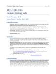
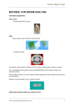
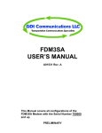
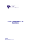
Download VIDEOTEST-SPERM 2.1 - Digital Imaging Systems
Transcript
VIDEOTEST-SPERM 2.1 System for sperm concentration, motility, and morphology analysis according to WHO requirements (with the software for Windows 98, 2000, XP) developed by VideoTesT SPECIFICATION Saint-Petersburg 2004 VideoTesT-Sperm 2.1: specification VideoTesT, ltd., [email protected] VideoTesT, ltd. 190000 Russia, St.-Petersburg, P.O. Box 356 Phone: +7(812)314-8100, 314-8445 Fax: +7(812)325-6494 e-mail: [email protected] http://www.videotest.ru VideoTesT-Metal © 1999-2004 VideoTesT, ltd. All rights reserved. Information in this document is subject to change without notice. This document is a property of VideoTesT, ltd. It may be copied or transmitted to a third party only in its complete form with no omissions; all trade marks and copyrights be preserved. Any changes or amendments shall be agreed upon in writing with VideoTesT, ltd. 2 VideoTesT-Sperm 2.1: specification VideoTesT, ltd., [email protected] 1. INTRODUCTION VideoTesT-Sperm 2.1 system is designed for automatic analysis of the sperm concentration, motility, and morphology. The analysis is performed in accordance with WHO (World Health Organization) requirements. The sperm concentration, motility, and morphology are important parameters for sperm fertility assessment. Their quantitative analysis is performed for the diagnosis of the men infertility. GENERAL INFORMATION The sperm ejaculate is a viscous heterogeneous opaque liquid. Its volume varies from 1-2 to 10 ml and more, 3-3,5 ml at average. The sperm ejaculate contains spermatozoa (sperms), the haploid male sex cells. The normal mature spermatozoon consist of head, middle piece, and tail (see Figure 1). Figure 1. Spermatozoon The total sperm length is around 50-60 microns. The upper part of the sperm head is called acrosome. It contains ferments necessary for sperm penetration into the egg cell. The data on normal sperm ejaculate values is in Table 1. Criterion Volume Color Smell Relative viscosity рН Erythrocytes Leucocytes Agglutinates (*) Sperm concentration Motile sperms Fast and progressively motile Alive sperms Table 1 Normal values 2 - 6 ml grayish white fresh chestnut 0-5 mm 7,2 - 7,6 n/a or traces Traces ≥20mln/ml > 50% ≥25% ≥75% (*) Agglutination is sticking or clumping together of live sperm cells in the ejaculate. The agglutination influences on the sperm motility (reduces it). 3 VideoTesT-Sperm 2.1: specification VideoTesT, ltd., [email protected] The sperm motility, concentration, and morphology are important characteristics which determines sperm fertility. The sperm motility is an important characteristic of the sperm quality. Motility involves the ability of a sperm to swim strongly and vigorously in one direction. Motility is critical for sperm to travel up the cervical canal, into the uterus and the fallopian tubes and, finally, penetrate the egg. The sperm motility is estimated by its velocity and track linearity. The sperm velocity varies, and can be as high as 10 - 50 microns per second. WHO recommends to classify the human sperms into 4 groups by their velocity (A, B, C, D): A) fast and progressively motile sperms (i.e. >25microns/sec at 37ºС and >20microns/sec at 20ºС B) slow and low progressively motile C) non-progressively motile (<5microns/sec) D) non motile sperms. The normal sperm shall contain >50% sperms with progressive motility (A and B groups) or >25% fast and progressively motile sperms (A group). The sperm concentration is a measure of the number of sperm cells in a milliliter of semen. The sperm ejaculate may contain more than 150 mln spermatozoa. Normal concentration is at least 20 million sperm cells per milliliter of semen or >40mln spermatozoa in the ejaculate. The sperm morphology analysis is performed for estimation of the number of spermatozoa with the normal morphology. The sperm morphology estimation is very important for sperm fertility analysis. A healthy sperm cell has an oval head containing DNA or genetic material, and a center which provides energy to propel the sperm tail giving forward movement. Abnormally shaped sperm can cause infertility since they often cannot fertilize the egg. In the normal sperm sample at least 20% of all sperms shall have normal morphology. The normal spermatozoon has the following parameters: Head length is 4—5 microns, width is 2,5—3,5 microns, elongation (length to width) is 1,50—1,75. Acrosome percent is 40—70% of total head area. Middle piece length is 6—7 microns, width is less than 1 micron. Tail length is 40-50 microns. The sperm morphology analysis done according to the strict criteria suggested by Krueger and Menkveld in 1986 provides an ability to forecast the in vitro fertilization results with high accuracy. The WHO recommends to use the strict criteria of sperm morphology as standard for human sperm analysis. COMPUTER ASSISTED SPERM ANALYSIS The computer-assisted semen analysis (CASA) uses high technologies of video image processing. The CASA provides the objective and reliable estimation of sperm parameters. The computer assisted system performs automatic analysis of sperm motility, concentration and morphology. VideoTesT-Sperm system provides an ability to perform the computer-assisted sperm analysis. VIDEOTEST-SPERM SYSTEM COMPONENTS VideoTesT-Sperm system (see Figure 2) includes the following items: 1. Upright transmitted light microscope with a phase contrast and stage micrometer, 2. Micro-cell sperm chamber or the set of standard disposable sperm chambers, 3. Color analogue image acquisition system to capture images of the native sperm ejaculates and sperm smears, 4. Computer and printer, 5. VideoTesT-Sperm 2.1 specialized software to process and analyze the acquired images. 4 VideoTesT-Sperm 2.1: specification VideoTesT, ltd., [email protected] Figure 2. VideoTesT-Sperm system The microscope is an important part of the complete system. It shall provide the sharp and contrast images of the sperm samples. The microscope used for VideoTesT-Sperm system shall comply with the following requirements: flat and evenly illuminated image field, Koeler illumination properly adjusted, high resolution, high image contrast, stabilized power supply unit, conversion filter included. The stage micrometer shall be included for system calibration. The microscope shall be equipped with 1x video adapter, the phase contrast (recommended), 20x long distance lens (if the micro-cell sperm chamber is used) to capture the native sperm ejaculate images (video sequencies), and 100x lens to capture the images of the sperm stained smears. The micro-cell sperm chamber or standard disposable sperm chamber (see Figure 3) is used to place the sperm ejaculate. The special sperm chambers provide that all spermatozoa move in the same plane. The monolayer sperm movement is required to ensure the high accuracy of the sperm concentration and motility analysis. 3a 3b Figure 3. Sperm chambers. 3a-micro-cell Makler sperm chamber, 3b – standard disposable sperm chamber The advantages of the standard disposable sperm chamber are as follows: 1. No need to equip the microscope with 20x long distance lens, 2. Minimization of the direct contact with sperm ejaculate, and so minimization of the risk of infection, 3. No need to clean the chamber and reduction of the total analysis time. The advantage of the micro-cell sperm chamber is that the investments are made once and the chamber can be used for many years (if it is properly maintained and cleaned). The image acquisition system shall capture the high quality images from the microscope to the computer and record the sperm movies to .avi files for further analysis of the sperm concentration and motility. The 5 VideoTesT-Sperm 2.1: specification VideoTesT, ltd., [email protected] image acquisition system recommended for capturing the images and their further analysis in the software: color analogue camera with the resolution of at least 470 TVL and image acquisition board (framgrabber) recording at 50 fps. The camera control (the settings adjustment, movie recording, and static image capture) shall be performed from VideoTesT-Sperm software for comfortable and fast operation. This is provided if MV-500 framegrabber is used. The computer and its operation system shall provide fast and reliable system operation and processing the big volumes of information. The sperm movie processing time depends on the PC configuration. The recommended PC configuration for VideoTesT-Sperm system is as follows: • • • • • • • • • Pentium 4 1,6 GHz or higher processor, 32 Mb videocard or higher, 512 Mb RAM, Windows XP Professional operation system, 17” monitor with 1280x1024 or higher resolution, 150Mb free hard disk space (hard disk Fast HDD, ATA100 or SCSI), Windows compatible mouse or other pointing device, Keyboard and CD-ROM, Free USB port. GENERAL REQUIREMENT TO SPERM SAMPLES Requirements to the native sperm ejaculates. The native sperm ejaculate shall be fresh and does not contain the artifacts. For concentration and motility analysis the native ejaculate is diluted and placed into the clean and dry sperm camber. Requirements to the stained sperm smears. For automatic sperm morphology analysis the sperms in the stained smears shall have good contrast, sharp contours, and the number of the contacting and overlaid sperms shall be minimized. The example images of the quality sperm smears are given on Figure 4. (б) (а) Figure 4. Example images of the sperm smears VideoTesT-Sperm system includes two routines of the automatic sperm analysis: • «Motility» • «Morphology». It also includes the additional routine «Test» designed for testing the software operability. 2. ROUTINES: GENERAL INFORMATION AND DESCRIPTION OF WORK This chapter contains the general information on the «Motility» and «Morphology» routines and describes their operation. «MOTILITY» ROUTINE Motility routine is designed for sperm concentration analysis in the native ejaculate and estimation of the sperm motility. This routine provides processing the video images (files in .avi format) of the native sperm ejaculates. 6 VideoTesT-Sperm 2.1: specification VideoTesT, ltd., [email protected] For sperm motility and concentration analysis the sperm chamber with the native ejaculate is placed on the microscope stage. The sperm movie recording is performed with the image acquisition system installed on the microscope. Prior to start video image processing in the Motility routine it is necessary to calibrate the system. The calibration is performed once for the working microscope lens used for image observation and shall be performed again only if any system modifications are done (change of the microscope lens, video adapter, or image acquisition system). Starting images processing in this routine the user shall first fill database blank in (patient details, sperm ejaculate volume, its properties, etc). The possible database blank view is represented on Figure 5. The user can easily modify the blank view in accordance with the internal requirements. After the database blank if filled in the one second sperm movie is recorded for further processing in the routine. It is also possible to open the previously recorded sperm movies (.avi files) from disk and process them. Figure 5. Possible view of the database blank After the movie is loaded in the program window its automatic processing starts. The moving sperms are thresheld, and the sperm motility and concentration are estimated. Figure 6 shows the program window view in the movie processing mode. 7 VideoTesT-Sperm 2.1: specification VideoTesT, ltd., [email protected] Figure 6. Program window view in the movie processing mode During the automatic analysis of the moving sperms the sperm motility parameters are measured and all sperms are classified by their motility. The scheme of the sperm movement is shown on Figure 7. Figure 7. Sperm movement scheme The sperm motility parameters calculated in the software: VAP – average path velocity (micron/sec), VSL – straight-line velocity (micron/sec), VCL – curvilinear velocity (micron/sec), 8 VideoTesT-Sperm 2.1: specification VideoTesT, ltd., [email protected] ALH – lateral head displacement, BCF – beat cross frequency (Hz), STR – sperm track straightness (%). STR = VSL / VAP x 100. LIN – linearity (%). LIN = VSL / VCL x 100. In accordance with WHO requirements all sperms are classified into 4 classes by their motility: А, В, С, D (see Figure 8). Figure 8. Example tracks of the A, B, and C sperms. Motility class A includes the progressively moving sperms, i.e. the sperms with high straight-line velocity (VSL) and linear track (high LIN). Motility class B includes the following sperms: - with the linear track and average straight-line velocity (B1 type), - with the non-linear track and high or average straight-line velocity (B type). Motility class C includes the sperms with the non-linear track and high or average straight-line velocity. Motility class D includes non-motile or very low motile sperms. In the Motility routine the «round» cells (all cells of the rounded form) are also thresheld and counted. The round cells include the leucocytes, erythrocytes, epithelial cells, and spermatogenesis cells. The round cells differ from the sperms by their size and form. The data on percent count of A, B, C, and D spermatozoa is automatically sent to the image database blank (see Figure 9). The sperm concentration is automatically calculated and the information (in mln per ml) is sent in the corresponding field of the database blank. The round cells concentration is also measured. 9 
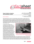
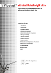
VideoTesT-Sperm 2.1: specification VideoTesT, ltd., [email protected] Figure 9. The database blank (fragment) with the data on sperm concentration and motility. The total number of sperms analyzed in three movies is 40. The data on average sperm motility parameters for each motility class is displayed in the corresponding fields (see Figure 10). Figure 10. The database blank (fragment) with the data on average sperm motility parameters for each class. The user can review the processed movie (by frames if required). The sperm tracks are displayed on the processed movie (see Figure 11). The track colors correspond to the sperm motility class. Figure 11. Processed movie with the sperm tracks 10 VideoTesT-Sperm 2.1: specification VideoTesT, ltd., [email protected] When the first sperm movie processing is finished the user can process the next ones recorded from the same ejaculate. The analysis results are accumulated in the database. After all required movies recorded from the same ejaculate are processed, the next ejaculate is placed into the sperm chamber, the next database blank is filled in, and the motility analysis starts again in the routine as it is described above. Attention! If you install VideoTesT-Sperm 2.1 demo version please use .avi files from the Images/Sperm folder for their processing in the «Motility» routine. «MORPHOLOGY» ROUTINE The stained sperm smear is placed on the microscope stage. The smear images are captured with the image acquisition system installed on the transmitted light microscope. The spermatozoon in the stained smear is an elongated cell consisting of the head, tail, and midpiece. The upper part of the sperm head is called acrosome. Morphology routine is designed for sperm morphology analysis in the stained smears. The morphological parameters of the sperm head, midpiece, and tail are measured (including the acrosome area percentage). Prior to start image processing in the Morphology routine it is necessary to calibrate the system. The calibration is performed once for the working microscope lens used for image observation and shall be performed again only if any system modifications are done (change of the microscope lens, video adapter, or image acquisition system). Starting images processing in this routine the user shall first fill database blank in (patient details, sperm ejaculate volume, its properties, etc). The possible database blank view is represented on Figure 4. The user can easily modify the blank view in accordance with the internal requirements. If the user wants to add the sperm morphology data to the database blank containing the sperm concentration and motility analysis results, than he does not need fill in the database blank. The image of the stained sperm smear can be acquired to the software with the image acquisition system installed on the microscope or opened from the disk. The spermatozoa are thresheld automatically. The automatic thresholding settings can be adjusted by the user if necessary. Figure 12 shows the source sperm smear image (left), and the software window with the sperms automatically thresheld (right). (a) (b) Figure 12. Automatic sperm thresholding. 12а – source image, 12b – automatic sperm thresholding 11 VideoTesT-Sperm 2.1: specification VideoTesT, ltd., [email protected] The user can correct the automatically thresheld sperms by manual editing the sperm midpieces and tails. For all threshed sperms the following parameters are measured: • head length and width, • head form, elongation, and asymmetry, • head front and back tapering • head average brightness and color, • acrosome area percent, • midpiece length, width and area, • tail length and asymmetry. By some of the measured parameters the sperms are classified into normal and abnormal (pathological). The following parameters are used for this classification: • head length, width, form, and elongation, • head asymmetry and its front and back tapering, • acrosome percent area. The sperm brightness and color parameters are used for sperm separation from the artifacts. For sperm morphology analysis the required number of image fields/smears is processed (the total number of spermatozoa to be estimated is at least 200). When image processing is finished all analyzed sperms (normal and pathological) are represented in a gallery. The Gallery view is shown on Figure 13. Figure 13. Sperms gallery The user can delete any spermatozoon(oa) or change its class by moving the sperm thumbnail from one cell to another. The morphology analysis results are accumulated in the database. For each sperm class (Norm and Pathology) the data on average head length and width, elongation and asymmetry, acrosome part, midpiece and tail length is calculated and sent to the corresponding fields (see Figure 14). Figure 14. The database blank (fragment) with the data on average sperm morphology parameters (for Norm and Pathology classes) Attention! If you install VideoTesT-Sperm 2.1 demo version please use .bmp files from the 12 VideoTesT-Sperm 2.1: specification VideoTesT, ltd., [email protected] Images/Sperm folder for their processing in the «Morphology» routine. FINALIZATION OF THE ANALYSIS After the sperm concentration, motility, and morphology analysis is finished the user can print out the analysis results as a the standard report. The report templates are easily created by the user according to the company requirements. The report example is shown on Figure 15. Figure 15. Possible report view The sperm motility, concentration, and morphology analysis results are automatically sent to the built-in database. The database blank view, the number and types of the database fields can be modified by the user of the system. The database is designed for the analysis data storage and quick search for the required information. The database blank view is shown on Figure 5. 13 VideoTesT-Sperm 2.1: specification VideoTesT, ltd., [email protected] The database can be viewed in two modes: Blank and Table. In the Blank mode (see Figure 15) the full database record is displayed on the screen. The record contains a number of fields (set by the user) arranged as the user defined. In the Blank mode it is possible to enter the necessary information into the database (manually and automatically) and view full data for each patient. The Blank can contain the text, number, and date fields. The fields are labeled and filled in by the user. Some fields are filled in automatically (the sperm motility, concentration and morphology analysis results received during the image processing in the Motility and Morphology routines are sent there). By default in all date fields the current date is displayed. In the Table mode the database records are displayed in the table form (see Figure 16). The table mode is used for entries sort and filtration. Figure 16. Image database in the table mode The image database provides an ability to filter entries by any condition(s), e.g. by the analysis date, patient name, etc. The database entries can be sorted by any field (text, number, or date fields). The image database provides an ability to search for the text fragments in the text, number, or date fields. 3. ABOUT DEVELOPER Since 1990 VideoTesT, ltd. has been focusing its activity on development of image analysis software, integration of PC-based VideoTesT Image Analysis Systems, automation and implementation of the image analysis methods in Life Sciences, Medicine, Materials Sciences, and Manufacturing fields. VideoTesT software includes several universal and specialized programs. Universal programs: • VideoTesT-Size 5.0 – new software for image acquisition, annotation, enhancement and manual measurements on the images. • VideoTesT-Album 4.0 - image database. Special programs: • VideoTesT-Master (Morphology) 4.0 – image analysis in life sciences with pre-installed bio-medical routines • VideoTesT-Master (Structure) 4.0 - image analysis in material sciences with pre-installed material analysis routines • VideoTesT-Metal 1.0 – metal structure analysis • VideoTesT-Karyo 3.0 - chromosome analysis: karyotyping and ideograms • VideoTesT-Karyo 3.1 – high accuracy of automatic karyotyping the human and pig chromosomes, ability to train the software to automatically recognize the animal chromosomes • VideoTesT-FISH - FISH analysis • VideoTesT-CGH - CGH analysis • VideoTesT-Sperm 2.1 - sperm analysis (motility, concentration, and morphology). Ability to adjust the software for animal sperm analysis • VideoTesT-Colony Count - count and sizing the bacteria colonies. For additional information on VideoTesT products, please contact: VideoTesT, ltd., 190000 Russia, St.-Petersburg, P.O. Box 356 Phone: +7(812)314-8100, +7(812)314-8445 Fax: +7(812)325-6494 e-mail: [email protected] http://www.videotest.ru 14