1
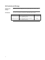
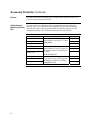
pBudCE4.1 Catalog no. V532-20 Rev. date: 26 August 2009 Manual part no. 25-0389 MAN000206 Corporate Headquarters Invitrogen Corporation 1600 Faraday Avenue Carlsbad, CA 92008 T: 1 760 603 7200 F: 1 760 602 6500 E: [email protected] For country-specific contact information visit our web site at www.invitrogen.com User Manual ii Table of Contents Kit Contents and Storage........................................................................................................................... iv Introduction ................................................................................................................... 1 Product Overview ........................................................................................................................................1 Experimental Outline...................................................................................................................................2 Methods ......................................................................................................................... 3 Cloning into pBudCE4.1..............................................................................................................................3 Transfection and Analysis...........................................................................................................................7 Creating Stable Cell Lines .........................................................................................................................10 Appendix...................................................................................................................... 13 Human EF-1 Promoter ............................................................................................................................13 pBudCE4.1 Vector ......................................................................................................................................14 pBudCE4.1/lacZ/CAT...............................................................................................................................17 Zeocin™ ........................................................................................................................................................18 Recipes .........................................................................................................................................................20 Accessory Products ....................................................................................................................................21 Technical Support.......................................................................................................................................23 Purchaser Notification ...............................................................................................................................24 References....................................................................................................................................................25 iii Kit Contents and Storage Shipping and Storage pBudCE4.1 vectors are shipped on wet ice. Upon receipt, store vectors at –20°C. Kit Contents All vectors are supplied as detailed below. Store the vectors at –20°C. Vector iv Composition Amount pBudCE4.1 40 L of 0.5 g/μL vector in 10 mM TrisHCl, 1 mM EDTA, pH 8.0 20 g pBudCE4.1/lacZ/CAT 40 L of 0.5 g/μL vector in 10 mM TrisHCl, 1 mM EDTA, pH 8.0 20 g Introduction Product Overview pBudCE4.1 pBudCE4.1 is a 4.6 kb vector designed for simultaneous expression of two genes in mammalian cell lines. The vector contains the human cytomegalovirus (CMV) immediate-early promoter and the human elongation factor 1α-subunit (EF-1α) promoter for high-level, constitutive, independent expression of two recombinant proteins (see page 13 for more information on the EF-1α promoter). Features of the vector allow detection and purification of expressed proteins (see pages 15–16) for more information). High-level stable and transient expression studies can be carried out in most mammalian cell types. In addition to the two promoters, the vector contains the following elements: C-terminal peptides encoding the myc (c-myc) epitope or the V5 epitope and a polyhistidine (6xHis) metal-binding tag for detection and purification of recombinant proteins Zeocin™ resistance gene for selection in E. coli and creation of stable, mammalian cell lines (Mulsant et al., 1988) (see pages 18–19 for more information) SV40 origin for episomal replication in cell lines that are latently infected with SV40 or that express the SV40 large T antigen (e.g., COS7) pBudCE4.1/lacZ/CAT is included for use as a positive control for transfection, expression, and detection in the cell line of choice. pBudCE4.1 is an improved version of pBudCE4. During construction of the original vector, an ATG was inadvertently created in the multiple cloning site (672–674 bp) 3 to the CMV promoter. Since it may interfere with proper translation of the cloned gene, this ATG was changed to ATT to create pBudCE4.1. Continued on next page 1 Experimental Outline Experimental Outline Use the following outline to clone and express your genes of interest in pBudCE4.1. Step 2 Action Page 3–5 1 Determine a cloning strategy. 2 Ligate your inserts into the vector and transform into E. coli. Select transformants on Low Salt LB containing 25–50 μg/mL Zeocin™. 6, 20 3 Analyze your transformants for the presence of both inserts by restriction digestion. 6 4 Select a transformant with the correct restriction pattern and sequence to confirm that both genes are cloned in frame with the C-terminal peptide (if desired). 6 5 Transfect your construct into the cell line of choice. 7 6 Test for expression of your recombinant proteins by western blot analysis or functional assay. For antibodies to the myc epitope, the V5 epitope, or the C-terminal polyhistidine tag, see page 22. 7 Purify your recombinant proteins using a metal-chelating resin such as ProBond™ (see page 21 for ordering information). 8 Generate a stable cell line, if desired. 8–9 9 10–11 Methods Cloning into pBudCE4.1 General Molecular Biology Techniques For help with DNA ligations, E. coli transformations, restriction enzyme analysis, purification of single-stranded DNA, DNA sequencing, and DNA biochemistry, refer to Molecular Cloning: A Laboratory Manual (Sambrook et al., 1989) or Current Protocols in Molecular Biology (Ausubel et al., 1994). E. coli Strain Most E. coli strains are suitable for the growth of this vector including TOP10 and DH5™ T1R. We recommend that you propagate vectors containing inserts in E. coli strains that are recombination deficient (recA) and endonuclease A deficient (endA). See below for an important note about E. coli strains. For your convenience, TOP10 and DH5™ T1R are available from Invitrogen as chemically competent or electrocompetent cells (TOP10 only) in One Shot® format (see page 21). Important Any E. coli strain that contains the complete Tn5 transposable element (i.e. DH5 F´IQ, SURE, SURE2) encodes the ble (bleomycin resistance gene). These strains will confer resistance to Zeocin™. We recommend that you choose an E. coli strain that does not contain the Tn5 gene (i.e. TOP10). Transformation Method You may use your method of choice for transformation. Chemical transformation is the most convenient for most researchers. Electroporation is the most efficient and the method of choice for large plasmids. Maintaining pBudCE4.1 To propagate and maintain the pBudCE4.1 vector, use a small amount of the supplied 0.5 g/μL stock solution in TE, pH 8.0 to transform a recA, endA E. coli strain like TOP10 or equivalent. Select transformants on Low Salt LB plates containing 25–50 μg/mL Zeocin™ (see page 20). Be sure to prepare a glycerol stock of each plasmid for long-term storage (see page 6). Cloning Considerations Your insert should contain a Kozak consensus sequence with an ATG initiation codon for proper initiation of translation (Kozak, 1987; Kozak 1990). An example of a Kozak consensus sequence is provided below. Other sequences are possible, but the G or A at position –3 and the G at position +4 (shown in bold) illustrates the most commonly occurring sequence with strong consensus. Replacing one of the two bases at these positions provides moderate consensus, while having neither results in weak consensus. The ATG initiation codon is shown underlined. (G/A)NNATGG If you wish to express your protein WITHOUT the C-terminal peptide, be sure to include a stop codon. Continued on next page 3 Cloning into pBudCE4.1, Continued CMV Multiple Cloning Site Below is the multiple cloning site of pBUDCE4.1 located downstream of the CMV promoter. Restriction sites are labeled to indicate the cleavage site. Potential stop codons are underlined. The arrow indicates the predicted start of transcription using T7 RNA polymerase. Sequencing primers are available separately (see page 22). CMV Forward priming site 501 CAACGGGACT TTCCAAAATG TCGTAACAAC TCCGCCCCAT TGACGCAAAT GGGCGGTAGG CAAT TATA 561 3 end of CMV Putative start of transcription CGTGTACGGT GGGAGGTCTA TATAAGCAGA GCTCTCTGGC TAACTAGAGA ACCCACTGCT T7 promoter/priming site 621 Pst I/Sse8387 I 677 Hind III TACTGGCTTA TCGAAATTAA TACGACTCAC TATAGGGAGA C CCA AGC TTG CAT TCC Pro Ser Leu His Ser Sal I Acc I Sca I Xba I BamH TGC AGG TCG ACA TCG ATC TTA AGC AGT ACT TCT AGA GGA TCC GAA CAA AAA Cys Arg Ser Thr Ser Ile Leu Ser Ser Thr Ser Arg Gly Ser Glu Gln Lys Polyhistidine (6xHis) tag myc epitope 728 CTC ATC TCA GAA GAG GAT CTG AAT ATG CAT ACC GGT CAT CAT CAC CAT CAC Leu Ile Ser Glu Glu Asp Leu Asn Met His Thr Gly His His His His His 779 CAT TGA GTTTGA TCCCCGGGAA TTCAGACATG ATAAGATACA TTGATGAGTT TGGACAAACC His *** 841 ACAACTAGAA TGCAGTGAAA AAAATGCTTT ATTTGTGAAA TTTGTGATGC TATTGCTTTA SV40 polyadenylation signal 901 TTTGTAACCA TTATAAGCTG CAATAAACAA GTTGGGGTGG GCGAAGAACT Continued on next page 4 Cloning into pBudCE4.1, Continued EF-1α Multiple Cloning Site Below is the multiple cloning site of pBUDCE4.1 located downstream of the EF-1α promoter. Restriction sites are labeled to indicate the cleavage site. The promoter is marked using the convention of Uetsuki et al., 1989. For more information see page 13. Sequencing primers are available separately (see page 22). 2940 GCACTTGATG TAATTCTCGT TGGAATTTGC CCTTTTTGAG TTTGGATCTT GGTTCATTCT 3000 EF-1a Forward priming site 3´end of hEF-1a Intron 1 CAAGCCTCAG ACAGTGGTTC AAAGTTTTTT TCTTCCATTT CAGGTGTCGT GAACACGTGG 5´ end of hEF-1a Exon 2 3060 Not I BstX I Xho I Bgl II T CGC GGC CGC TTC GAA GGT ACC AGC ACA GTG GAC TCG AGA GAT CTG GCC Arg Gly Arg Phe Glu Gly Thr Ser Thr Val Asp Ser Arg Asp Leu Ala Sfi I 3109 Kpn I BstB I* V5 epitope BstB I* GGC TGG GCC CGT TTC GAA GGT AAG CCT ATC CCT AAC CCT CTC CTC GGT Gly Trp Ala Arg Phe Glu Gly Lys Pro Ile Pro Asn Pro Leu Leu Gly Polyhistidine (6xHis) tag 3157 CTC GAT TCT ACG CGT ACC GGT CAT CAT CAC CAT CAC CAT TGA G Leu Asp Ser Thr Arg Thr Gly His His His His His His *** BGH Reverse priming site 3200 TTTAAACCCG CTGATCAGCC TCGACTGTGC CTTCTAGTTG CCAGCCATCT GTTGTTTGCC BGH polyadenylation signal 3260 CCTCCCCCGT GCCTTCCTTG ACCCTGGAAG GTGCCACTCC CACTGTCCTT TCCTAATAAA 3320 ATGAGGAAAT TGCATCGCAT TGTCTGAGTA GGTGTCATTC TATTCTGGGG GGTGGGGTGG 3380 GGCAGGACAG CAAGGGGGAG GATTGGGAAG ACAATAGCAG GCATGCTGGG GATGCGGTGG *Note that there are two BstB I sites in the polylinker. Continued on next page 5 Cloning into pBudCE4.1, Continued MEND ION AT RECOM E. coli Transformation Preparing a Glycerol Stock 6 Transform your ligation mixtures into a competent recA, endA E. coli strain (e.g., TOP10) and select on Low Salt LB plates containing 25–50 μg/mL Zeocin™ (see page 20). Select 10–20 clones and analyze for the presence and orientation of your insert. We recommend that you sequence your construct to confirm that each of your genes is fused in frame with the C-terminal peptide. Several primers are available separately that you may use to sequence your construct. These are marked in the multiple cloning site diagrams on pages 4–5. For ordering information, see page 22. Alternatively, you may design your own primers for sequencing. Once you have identified the correct clone, be sure to purify the colony and make a glycerol stock for long-term storage. It is also a good idea to keep a DNA stock of your plasmid at –20°C. 1. Streak the original colony out on a Low Salt LB plate containing 25 μg/mL Zeocin™. 2. Incubate the plate at 37°C overnight. 3. Isolate a single colony and inoculate into 1–2 mL of Low Salt LB containing 25 μg/mL Zeocin™. 4. Grow the culture to stationary phase (OD600 = 1–2). 5. Mix 0.85 mL of culture with 0.15 mL of sterile glycerol and transfer to a cryovial. 6. Store at –80°C. Transfection and Analysis Introduction Once you have confirmed that your inserts are in the correct orientation and fused in frame with the C-terminal peptide (if desired), you are ready to transfect your cell line of choice. We recommend that you include the positive control vector and a mock transfection to evaluate your results. Plasmid Preparation Plasmid DNA for transfection into eukaryotic cells must be very clean and free from phenol and sodium chloride. Contaminants will kill the cells, and salt will interfere with lipid complexing decreasing transfection efficiency. We recommend isolating plasmid DNA using the PureLink™ HiPure Miniprep Kit or the PureLink™ HiPure Midiprep Kit (see page 21 for ordering information). Methods of Transfection For established cell lines (e.g. HeLa), consult original references or the supplier of your cell line for the optimal method of transfection. We recommend that you follow exactly the protocol for your cell line. Pay particular attention to medium requirements, when to pass the cells, and at what dilution to split the cells. Further information is provided in Current Protocols in Molecular Biology (Ausubel et al., 1994). Methods for transfection include calcium phosphate (Chen and Okayama, 1987; Wigler et al., 1977), lipid-mediated (Felgner et al., 1989; Felgner and Ringold, 1989), and electroporation (Chu et al., 1987; Shigekawa and Dower, 1988). Invitrogen offers the Lipofectamine™ 2000 Reagent for lipid-mediated transfection. Positive Control pBudCE4.1/lacZ/CAT is provided as a positive control vector for mammalian cell transfection and expression and may be used to optimize transfection conditions for your cell line (see page 17). The gene encoding -galactosidase is expressed from the CMV promoter as a fusion to the myc epitope in mammalian cells. The gene encoding chloramphenicol acetyltransferase (CAT) is expressed as a fusion to the V5 epitope from the EF-1α promoter. A successful transfection results in -galactosidase and CAT expression that can be easily assayed (see page 9. Continued on next page 7 Transfection and Analysis, Continued Detecting Fusion Proteins Antibodies are available from Invitrogen to detect expression of fusion proteins from pBudCE4.1 (see page 22). In pBudCE4.1/lacZ/CAT, -galactosidase and CAT are expressed as fusion proteins to the myc epitope or the V5 epitope, respectively. In addition you may assay for activity of either control protein using one of the assays described on the next page. To detect fusion protein by western blot, you will need to prepare a cell lysate from transfected cells. We recommend that you perform a time course to optimize expression of the fusion protein (e.g. 24, 48, 72 hours, etc. after transfection). To lyse cells: Polyacrylamide Gel Electrophoresis 1. Wash cell monolayers (~106 cells) once with phosphate-buffered saline (PBS). 2. Scrape cells into 1 mL PBS and pellet the cells at 1,500 × g for 5 minutes. 3. Resuspend in 50 μL Cell Lysis Buffer (see recipe on page 20). Other lysis buffers may be suitable. 4. Incubate cell suspension at 37°C for 10 minutes to completely lyse the cells. Note: You may prefer to lyse the cells at room temperature or on ice if degradation of your proteins are a potential problem. 5. Centrifuge the cell lysate at 10,000 × g for 10 minutes at room temperature to pellet nuclei and transfer the supernatant to a fresh tube. Assay the lysate for protein concentration. Note: Do not use protein assays utilizing Coomassie Blue or other dyes. NP-40 interferes with the binding of the dye with the protein. 6. Add SDS-PAGE sample buffer to a final concentration of 1X and boil the sample for 5 minutes. 7. Load 20 μg of lysate onto an SDS-PAGE gel (see below) and electrophorese. Use the appropriate percentage of acrylamide to resolve your fusion protein. To facilitate separation of your recombinant protein by polyacrylamide gel electrophoresis, a wide range of pre-cast Novex® NuPAGE® and Tris-Glycine polyacrylamide gels and electrophoresis apparatus are available from Invitrogen. The patented Novex® NuPAGE® Gel System prevents the protein modifications associated with Laemmli-type SDS-PAGE, ensuring optimal separation for protein analysis. In addition, Invitrogen also carries a large selection of molecular weight protein standards and staining kits for visualization of recombinant proteins. For more information about the appropriate gels, standards, and stains to use to visualize your recombinant protein, refer to our website (www.invitrogen.com) or call Technical Support (see page 23). Continued on next page 8 Transfection and Analysis, Continued Western Analysis To detect expression of your recombinant fusion protein by western blot analysis, you may use the Anti-myc, Anti-V5, or the Anti-His(C-term) antibodies available from Invitrogen (see page 22 for ordering information) or an antibody to your protein of interest. The ready-to-use WesternBreeze® Chromogenic Kits and WesternBreeze® Chemiluminescent Kits are available from Invitrogen to facilitate detection of antibodies by colorimetric or chemiluminescent methods (see page 21 for ordering). For more information, refer to our website (www.invitrogen.com) or call Technical Support (see page 23). The C-terminal peptide containing the myc epitope and the polyhistidine tag or the V5 epitope and polyhistidine tag will add approximately 3 kDa to the size of your protein. Assay for galactosidase Activity You may assay for -galactosidase expression by activity assay using cell-free lysates (Miller, 1972) or by staining the cells for activity. Invitrogen offers the –Gal Assay Kit and the -Gal Staining Kit for fast and easy detection of –galactosidase expression (see page 21). Assay for CAT Activity You may assay for CAT expression by ELISA assay, western blot analysis, fluorometric assay, or radioactive assay (Ausubel et al., 1994; Neumann et al., 1987). The CAT assay kit is available from Invitrogen for detection of CAT protein (see page 21). Purifying Cells You will need 5 × 10⁶ to 1 × 10⁷ transfected cells for purification of your protein on a 2 mL ProBond™ column (or other metal-chelating column). Refer to the manufacturer's instructions before attempting to purify your fusion protein. To prepare cells for lysis, refer to the protocol on page 20. 9 Creating Stable Cell Lines Introduction pBudCE4.1 contains the Zeocin™ resistance gene for selection of stable cell lines using Zeocin™. We recommend that you test the sensitivity of your mammalian host cell to Zeocin™ as natural resistance varies among cell lines. General information and guidelines are provided below for your convenience. For more information about Zeocin™, refer to page 18. Effect of Zeocin™ on Sensitive and Resistant Cells The method of killing with Zeocin™ is quite different from neomycin (G418) and hygromycin. Cells do not round up and detach from the plate. Sensitive cells will exhibit the following morphological changes upon exposure to Zeocin™: Selection in Mammalian Cell Lines Vast increase in size (similar to the effects of cytomegalovirus infecting permissive cells) Abnormal cell shape Presence of large empty vesicles in the cytoplasm (breakdown of the endoplasmic reticulum and Golgi apparatus or scaffolding proteins) Breakdown of plasma and nuclear membrane (appearance of many holes in these membranes) Eventually, these "cells" will completely break down and only "strings" of protein will remain. Zeocin™-resistant cells should continue to divide at regular intervals to form distinct colonies. There should not be any distinct morphological changes in Zeocin™-resistant cells when compared to cells not under selection with Zeocin™. To generate a stable cell line expressing your protein, you need to determine the minimum concentration of Zeocin™ required for killing your untransfected host cell line. Typically, concentrations between 50 and 1,000 μg/mL Zeocin™ are sufficient to kill the untransfected host cell line. Test a range of concentrations (see below) to ensure that you determine the minimum concentration necessary for your cell line. 1. Seed cells (2 × 10⁵ cells/60 mm plate) for each time point and allow cells to adhere overnight. 2. The next day, substitute culture medium with medium containing varying concentrations of Zeocin™ (e.g., 0, 50, 125, 250, 500, 750, and 1000 g/mL). 3. Replenish the selective medium every 3–4 days, and observe the percentage of surviving cells. 4. Observe the cells at regular intervals to determine the appropriate concentration of Zeocin™ that prevents growth. 5. Select the concentration that kills cells in 7–10 days. Continued on next page 10 Creating Stable Cell Lines, Continued Possible Sites for Linearization To obtain stable transfectants, you may choose to linearize your vector before transfection. While linearizing your vector may not improve the efficiency of transfection, it increases the chances that the vector does not integrate in a way that disrupts the gene of interest or elements necessary for expression of the gene. The table below lists unique sites that may be used to linearize your construct prior to transformation. Other restriction sites are possible. Be sure that your insert does not contain the restriction enzyme site you wish to use to linearize your vector. Enzyme Restriction Site (bp) Location Nhe I 1877 Upstream of EF-1α promoter Many BspH I 4240 Backbone New England Biolabs Fsp I 4547 Backbone Many Pvu I 4568 Backbone Many Selection of Stable Integrants Supplier Once the appropriate Zeocin™ concentration is determined, you can generate a stable cell line with your construct. 1. Transfect your cells using the appropriate protocol for your cell line. Include a sample of untransfected cells as a negative control. 2. After transfection, wash the cells once with 1X PBS and add fresh medium to the cells. 3. 48 hours after transfection, split the cells into fresh medium (no Zeocin™) and allow cells to attach. 4. Remove medium and add medium containing Zeocin™ at the appropriate concentration for your cell line. Split the cells such that they are no more than 25% confluent. 5. Replenish selective medium every 3–4 days until Zeocin™-resistant colonies are detected. 6. Pick and expand colonies. Continued on next page 11 Creating Stable Cell Lines, Continued Preparing Cells for Lysis Lysis of Cells Use the procedure below to prepare cells for lysis prior to purification of your protein on ProBond™. You will need 5 × 10⁶ to 1 × 10⁷ cells for purification of your protein on a 2 mL ProBond™ column (see ProBond™ Purification System manual). 1. Seed cells in either five T-75 flasks or 2 to 3 T-175 flasks. 2. Grow the cells in selective medium until they are 80–90% confluent. 3. Harvest the cells by treating with trypsin-EDTA for 2 to 5 minutes or by scraping the cells in PBS. 4. Inactivate the trypsin by diluting with fresh medium (if necessary) and transfer the cells to a sterile microcentrifuge tube. 5. Centrifuge the cells at 1,500 rpm for 5 minutes. Resuspend the cell pellet in PBS. 6. Centrifuge the cells at 1,500 rpm for 5 minutes. You may lyse the cells immediately or freeze in liquid nitrogen and store at –80°C until needed. If you are using ProBond™ resin, refer to the ProBond™ Purification System manual for details about sample preparation for chromatography. The ProBond™ Purification System manual is available for downloading at our website (www.invitrogen.com) or by contacting Technical Support (see page 23). If you are using other metal-chelating resin, refer to the manufacturer's instruction for recommendations on sample preparation. 12 Appendix Human EF-1 Promoter Description The diagram below shows the features of the EF-1α promoter used in pBudCE4.1 (Mizushima and Nagata, 1990). Features are marked as per Uetsuki et al., 1989(Uetsuki et al., 1989). 5´ end of human EF-1a promoter AGCTAGCTTC GTGAGGCTCC GGTGCCCGTC AGTGGGCAGA GCGCACATCG CCCACAGTCC CCGAGAAGTT GGGGGGAGGG GTCGGCAATT GAACCGGTGC CTAGAGAAGG TGGCGCGGGG TAAACTGGGA AAGTGATGTC GTGTACTGGC TCCGCCTTTT TCCCGAGGGT GGGGGAGAAC Start of Transcription TATA box CGTATATAAG TGCAGTAGTC GCCGTGAACG TTCTTTTTCG CAACGGGTTT GCCGCCAGAA Exon I 5´ end of Intron 1 CACAGGTAAG TGCCGTGTGT GGTTCCCGCG GGCCTGGCCT CTTTACGGGT TATGGCCCTT GCGTGCCTTG AATTACTTCC ACCTGGCTGC AGTACGTGAT TCTTGATCCC GAGCTTCGGG TTGGAAGTGG GTGGGAGAGT TCGAGGCCTT GCGCTTAAGG AGCCCCTTCG CCTCGTGCTT GAGTTGAGGC CTGGCCTGGG CGCTGGGGCC GCCGCGTGCG AATCTGGTGG CACCTTCGCG CCTGTCTCGC TGCTTTCGAT AAGTCTCTAG CCATTTAAAA TTTTTGATGA CCTGCTGCGA CGCTTTTTTT CTGGCAAGAT AGTCTTGTAA ATGCGGGCCA AGATCTGCAC ACTGGTATTT Sp 1 CGGTTTTTGG GGCCGCGGGC GGCGACGGGG CCCGTGCGTC CCAGCGCACA TGTTCGGCGA Sp 1 GGCGGGGCCT GCGAGCGCGG CCACCGAGAA TCGGACGGGG GTAGTCTCAA GCTGGCCGGC Sp 1 Sp 1 CTGCTCTGGT GCCTGGCCTC GCGCCGCCGT GTATCGCCCC GCCCTGGGCG GCAAGGCTGG CCCGGTCGGC ACCAGTTGCG TGAGCGGAAA GATGGCCGCT TCCCGGCCCT GCTGCAGGGA Sp 1 GCTCAAAATG GAGGACGCGG CGCTCGGGAG AGCGGGCGGG TGAGTCACCC ACACAAAGGA Ap 1 AAAGGGCCTT TCCGTCCTCA GCCGTCGCTT CATGTGACTC CACGGAGTAC CGGGCGCCGT CCAGGCACCT CGATTAGTTC TCGAGCTTTT GGAGTACGTC GTCTTTAGGT TGGGGGGAGG GGTTTTATGC GATGGAGTTT CCCCACACTG AGTGGGTGGA GACTGAAGTT AGGCCAGCTT GGCACTTGAT GTAATTCTCC TTGGAATTTG CCCTTTTTGA GTTTGGATCT TGGTTCATTC 3´ end of Intron 1 TCAAGCCTCA GACAGTGGTT CAAAGTTTTT TTCTTCCATT TCAGGTGTCG TGA... 5´ end of Exon 2 13 pBudCE4.1 Vector Map of pBudCE4.1 The figure below summarizes the features of the pBudCE4.1 vector. The vector sequence is available for downloading from our website (www.invitrogen.com) or by contacting Technical Support (see page 23). +1 ori C pU PE oc V5 Hi s Hind III Pst I/Sse8387 I Sal I Acc I Sca I Xba I BamH I in 4595 bp myc His SV4 pA 0 pBudCE4.1 F- Comments for pBudCE4.1 4595 nucleotides MV BG Hp A PC BstB I Sfi I Bgl II Xho I BstX I Kpn I BstB I Not I 1a P SV40 EM 7 Ze CMV promoter: bases 7-594 Nhe I CMV Forward priming site: bases 544-564 T7 promoter/priming site: bases 638-657 CMV multiple cloning site: bases 664-713 myc epitope: bases 719-748 6xHis tag: bases 764-782 SV40 polyadenylation sequence: bases 803-933 Zeocin resistance gene: bases 1063-1437 (complementary strand) EM7 promoter: bases 1456-1510 (complementary strand) SV40 early promoter: bases 1547-1869 (complementary strand) EF-1a promoter: bases 1885-3051 EF-1a Forward priming site: bases 2999-3019 EF-1a multiple cloning site: bases 3062-3126 V5 epitope: bases 3127-3168 6xHis tag: bases 3178-3195 BGH Reverse priming site: bases 3218-3235 (complementary strand) BGH polyadenylation sequence: bases 3224-3447 pUC origin: bases 3521-4194 Continued on next page 14 pBudCE4.1 Vector, Continued Features of pBudCE4.1 pBudCE4.1 (4595 bp) contains the following elements. All features have been functionally tested. Feature Benefit Human cytomegalovirus (CMV) immediate-early promoter/enhancer Permits efficient, high-level expression of recombinant protein (Andersson et al., 1989; Boshart et al., 1985; Nelson et al., 1987). CMV Forward priming site Permits sequencing through the insert from the 5 end. T7 promoter/priming site Permits sequencing through the insert from the 5 end. Allows for in vitro transcription in the sense orientation. CMV Multiple cloning site Seven unique sites allow insertion of your gene. myc epitope Allows detection of your recombinant protein (Glu-Gln-Lys-Leu-Ile-Ser-Glu- with the Anti-myc Antibody, Anti-myc-HRP Antibody, or Anti-myc-AP Antibody (Evan et Glu-Asp-Leu) al., 1985) (see page 22 for ordering). C-terminal polyhistidine (6xHis) tag Permits purification of your recombinant protein on metal-chelating resin such as ProBond™. In addition, the C-terminal polyhistidine tag is the epitope for the Anti-His(C-term) Antibody, the Anti-His (C-term)-HRP Antibody, or the Anti-His(C-term)-AP Antibody (Lindner et al., 1997) (see page 22 for ordering). SV40 polyadenylation signal Efficient transcription termination and polyadenylation of mRNA. Note: The SV40 late polyadenylation signal terminates transcription for the gene cloned into the CMV MCS while the SV40 early polyadenylation signal terminates transcription for the Zeocin™ resistance gene. The signals are encoded on opposite strands in the same fragment of DNA. Zeocin™ resistance gene Selection of transformants in E. coli and stable transfectants in mammalian cells (Drocourt et al., 1990; Mulsant et al., 1988). EM7 promoter Synthetic promoter based on the bacteriophage T7 promoter for expression of the Zeocin™ resistance gene in E. coli. Continued on next page 15 pBudCE4.1 Vector, Continued Features of pBudCE4.1, Continued Feature 16 Benefit SV40 early promoter and origin Allows efficient, high-level expression of the Zeocin™ resistance gene and episomal replication in cells expressing the SV40 large T antigen. Human elongation factor 1α (EF-1α) promoter Permits efficient, high-level expression of recombinant protein (Goldman et al., 1996; Mizushima and Nagata, 1990). EF-1α Forward priming site Permits sequencing through the insert from the 5 end. EF-1α Multiple cloning site Seven unique sites allow insertion of your gene. V5 epitope (Gly-Lys-Pro-Ile-Pro-Asn-ProLeu-Leu-Gly-Leu-Asp-SerThr) Allows detection of your recombinant protein with the Anti-V5 Antibody, Anti-V5-HRP Antibody or the Anti-V5-AP Antibody (Southern et al., 1991) (see page 22 for ordering). 6xHis tag See previous page. Bovine growth hormone (BGH) reverse priming site Permits sequencing through the insert from the 3 end. BGH polyadenylation signal Efficient transcription termination and polyadenylation of mRNA (Goodwin and Rottman, 1992). pUC origin High-copy number replication and growth in E. coli. pBudCE4.1/lacZ/CAT Description pBudCE4.1/lacZ/CAT is an 8432 bp control vector containing the gene for β-galactosidase and the gene for chloramphenicol acetyltransferase (CAT). The lacZ gene was excised from pIND/lacZ using Hind III and Xba I and cloned into Hind III/Xba I digested pBudCE4.1. The CAT gene was cloned by digesting pBudCE4.1/lacZ and pBudCE4/lacZ/CAT with Bgl II and Mun I. A fragment containing the CAT gene and part of the EF-1 promoter from pBudCE4/lacZ/CAT was cloned into Bgl II/Mun I digested pBudCE4.1/lacZ to generate pBudCE4.1/lacZ/CAT. Map of Control Vector The figure below summarizes the features of the pBudCE4.1/lacZ/CAT vector. The nucleotide sequence for pBudCE4.1/lacZ/CAT is available for downloading from our website (www.invitrogen.com) or by contacting Technical Support (see page 23). +1 ori C pU myc His SV4 pA 0 pA BG H pBudCE4.1/ lacZ/CAT PE in 8432 bp Comments for pBudCE4.1/lacZ/CAT 8432 nucleotides CMV promoter: bases 7-594 1a CMV Forward priming site: bases 544-564 T7 promoter/priming site: bases 638-657 LacZ fusion: bases 673-3966 LacZ ORF: bases 673-3840 myc epitope: bases 3901-3963 6xHis tag: bases 3946-3963 SV40 polyadenylation sequence: bases 3986-4116 Zeocin resistance gene: bases 4245-4619 (complementary strand) EM7 promoter: bases 4638-4693 (complementary strand) SV40 early promoter: bases 4728-5051 (complementary strand) EF-1a promoter: bases 5067-6236 EF-1a Forward priming site: bases 6181-6201 CAT fusion: bases 6268-7032 CAT ORF: bases 6268-6924 V5 epitope: bases 6964-7005 6xHis tag: bases 7015-7032 BGH Reverse priming site: bases 7055-7072 (complementary strand) BGH polyadenylation sequence: bases 7061-7285 pUC origin: bases 7358-8031 oc F- P SV40 E M7 Ze lacZ V5 Hi s MV CAT PC Sfi I Bgl II Hind III Pst I Xba I BamH I 17 Zeocin™ Zeocin™ Zeocin™ is a member of the bleomycin/phleomycin family of antibiotics isolated from Streptomyces. Antibiotics in this family are broad spectrum antibiotics that act as strong anti-bacterial and anti-tumor drugs. They show strong toxicity against bacteria, fungi (including yeast), plants, and mammalian cells. The Zeocin™ resistance protein has been isolated and characterized (Calmels et al., 1991; Drocourt et al., 1990). This protein, the product of the Sh ble gene (Streptoalloteichus hindustanus bleomycin gene), is a 13.7 kDa protein that binds Zeocin™ in a stoichiometric manner to inhibit its DNA strand cleavage activity. Expression of this protein in eukaryotic and prokaryotic hosts confers resistance to Zeocin™. Molecular Weight, Formula and Structure The formula for Zeocin™ is C55H86O21N20S2Cu.HCl and the molecular weight is 1,527.5 daltons. Zeocin is an HCl salt. The diagram below shows the structure of Zeocin™. CONH2 H H2 N N H O H N CH3 HO N Cu NH O N H N N H N O O N O O ++ H2N H N CH3 HO R S N S CH3 H OH O O CH3 R = HN NH2 N NH NH2 OH H2N O O HO O HO OH OH O Applications of Zeocin™ Zeocin™ is used for selection in mammalian cells (Mulsant et al., 1988); plants (Perez et al., 1989); yeast (Baron et al., 1992); and prokaryotes (Drocourt et al., 1990). Suggested concentrations of Zeocin™ for selection in mammalian cell lines and E. coli are listed below: Organism Zeocin™ Concentration and Selective Medium E. coli 25–50 g/mL in Low Salt LB medium* (see page 20 for recipe) Mammalian Cells 50-1,000 g/mL (varies with cell line) *Efficient selection requires that the concentration of NaCl be no more than 5 g/liter (< 90 mM). Continued on next page 18 Zeocin™, Continued Handling Zeocin™ High salt and acidity or basicity inactivates Zeocin™. Therefore, we recommend that you reduce the salt in bacterial medium and adjust the pH to 7.5 to keep the drug active (see next page). Note: The salt concentration should not be adjusted for mammalian cells. Changes to the salt concentration are detrimental to cells. Store Zeocin™ at –20°C and thaw on ice before use. Zeocin™ is light sensitive. Store drug, plates, and medium containing drug in the dark. Wear gloves, a laboratory coat, and safety glasses or goggles when handling solutions containing Zeocin™. Zeocin™ is toxic. Do not ingest or inhale solutions containing the drug. 19 Recipes Low Salt LB Medium with Zeocin™ For Zeocin™ to be active, the salt concentration of the medium must remain low (<90 mM) and the pH must be 7.5. For selection in E. coli, it is imperative that you prepare LB broth and plates using the following recipe. Note the lower salt content of this medium. Failure to use low salt LB medium will result in non-selection due to inactivation of the drug. For more information about Zeocin™, refer to page 18. Low Salt LB Medium: 10 g Tryptone 5 g NaCl 5 g Yeast Extract 1. Combine the dry reagents above and add deionized, distilled water to 950 mL. Adjust pH to 7.5 with 5 M NaOH. Bring the volume up to 1 liter. For plates, add 15 g/L agar before autoclaving. 2. Autoclave on liquid cycle at 15 lbs./sq. in. and 121°C for 20 minutes. 3. Thaw Zeocin™ on ice and vortex before removing an aliquot. 4. Allow the medium to cool to at least 55°C before adding the Zeocin™ to 25–50 μg/mL final concentration. 5. Store plates at 4°C in the dark. Plates containing Zeocin™ are stable for 1–2 weeks. For your convenience Low Salt LB medium containing 25 g/ml Zeocin™ is available as premixed, pre-sterilized E. coli growth medium (imMedia™) that contains everything you need in a convenient pouch. Liquid and agar media are available, depending upon your application (see page 21). Cell Lysis Buffer 50 mM Tris, pH 7.8 150 mM NaCl 1% Nonidet P-40 1. This solution can be prepared from the following common stock solutions. For 100 ml, combine: 1 M Tris base 5 mL 5 M NaCl 3 mL Nonidet P-40 1 mL 2. Bring the volume up to 90 mL with deionized water and adjust the pH to 7.8 with HCl. 3. Bring the volume up to 100 mL. Store at room temperature. Note: Protease inhibitors may be added fresh at the following concentrations: 1 mM PMSF; 1 μg/mL pepstatin; 1 μg/mL leupeptin. 20 Accessory Products Introduction The products listed below are designed for use with pBudCE4.1. For details, visit www.invitrogen.com or contact Technical Support (page 23). Quantity Catalog no. 21 × 50 μL C4040-03 One Shot TOP10 Electrocomp Cells 21 × 50 μL C4040-52 One Shot® Max Efficiency® DH5α™ T1R 20 × 50 μL 12297-016 PureLink™ HiPure Plasmid Miniprep Kit 100 preps K2100-03 PureLink™ HiPure Plasmid Midiprep Kit Item ® One Shot TOP10 Chemically Competent E. coli ® ™ 25 preps K2100-04 ™ Lipofectamine 2000 Reagent 1.5 mL 11668-019 -Gal Assay Kit 80 mL K1455-01 1 kit K1465-01 6 purifications K850-01 50 mL R801-01 150 mL R801-15 1 gram R250-01 5 grams R250-05 200 mL Q620-20 -Gal Staining Kit ™ ProBond Purification System ProBond™ Resin Zeocin™ ™ imMedia Zeo Liquid ™ imMedia Zeo Agar 8–10 agar plates Q621-20 ® 1 kit WB7103 ® 1 kit WB7105 ® 1 kit WB7107 ® WesternBreeze Chemiluminescent Kit, AntiMouse 1 kit WB7104 WesternBreeze® Chromogenic Kit, Anti-Rabbit 1 kit WB7106 WesternBreeze Chromogenic Kit, Anti-Goat 1 kit WB7108 Fast Cat® Chloramphenicol Acetyltransferase Assay Kit 1 kit F2900 WesternBreeze Chromogenic Kit, Anti-Mouse WesternBreeze Chromogenic Kit, Anti-Rabbit WesternBreeze Chromogenic Kit, Anti-Goat ® Continued on next page 21 Accessory Products, Continued Primers For your convenience, Invitrogen offers a custom primer synthesis service. Visit www.invitrogen.com for more details. Antibodies and Western Detection Kits If you do not have an antibody specific to your protein, Invitrogen offers the Anti-myc, Anti-V5, or Anti-His(C-term) antibodies to detect your recombinant fusion protein. Horseradish peroxidase (HRP)- and alkaline phosphatase (AP)conjugated antibodies are available for convenient one-step detection. Antibody Anti-myc Anti-myc-HRP Anti-myc-AP Anti-V5 Anti-V5-HRP Anti-V5-AP Anti-His(C-term) Anti-His(C-term)-HRP Anti-His(C-term)-AP 22 Epitope Catalog no. Detects a 10 amino acid epitope derived from c-myc (Evan et al., 1985): EQKLISEEDL R950-25 R951-25 R952-25 Detects a 14 amino acid epitope derived from the P and V proteins of the paramyxovirus, SV5 (Southern et al., 1991): GKPIPNPLLGLDST R960-25 Detects the C-terminal polyhistidine tag (requires the free carboxyl group for detection) (Lindner et al., 1997): HHHHHH-COOH R930-25 R961-25 R962-25 R931-25 R932-25 Technical Support Web Resources Contact Us Visit the Invitrogen website at www.invitrogen.com for: Technical resources, including manuals, vector maps and sequences, application notes, MSDSs, FAQs, formulations, citations, handbooks, etc. Complete technical support contact information Access to the Invitrogen Online Catalog Additional product information and special offers For more information or technical assistance, call, write, fax, or email. Additional international offices are listed on our website (www.invitrogen.com). Corporate Headquarters: 5791 Van Allen Way Carlsbad, CA 92008 USA Tel: 1 760 603 7200 Tel (Toll Free): 1 800 955 6288 Fax: 1 760 602 6500 E-mail: [email protected] Japanese Headquarters: LOOP-X Bldg. 6F 3-9-15, Kaigan Minato-ku, Tokyo 108-0022 Tel: 81 3 5730 6509 Fax: 81 3 5730 6519 E-mail: [email protected] European Headquarters: Inchinnan Business Park 3 Fountain Drive Paisley PA4 9RF, UK Tel: +44 (0) 141 814 6100 Tech Fax: +44 (0) 141 814 6117 E-mail: [email protected] MSDS Material Safety Data Sheets (MSDSs) are available on our website at www.invitrogen.com/msds. Certificate of Analysis The Certificate of Analysis provides detailed quality control and product qualification information for each product. Certificates of Analysis are available on our website. Go to www.invitrogen.com/support and search for the Certificate of Analysis by product lot number, which is printed on the box. Limited Warranty Invitrogen (a part of Life Technologies Corporation) is committed to providing our customers with high-quality goods and services. Our goal is to ensure that every customer is 100% satisfied with our products and our service. If you should have any questions or concerns about an Invitrogen product or service, contact our Technical Support Representatives. All Invitrogen products are warranted to perform according to specifications stated on the certificate of analysis. The Company will replace, free of charge, any product that does not meet those specifications. This warranty limits the Company’s liability to only the price of the product. No warranty is granted for products beyond their listed expiration date. No warranty is applicable unless all product components are stored in accordance with instructions. The Company reserves the right to select the method(s) used to analyze a product unless the Company agrees to a specified method in writing prior to acceptance of the order. Invitrogen makes every effort to ensure the accuracy of its publications, but realizes that the occasional typographical or other error is inevitable. Therefore the Company makes no warranty of any kind regarding the contents of any publications or documentation. If you discover an error in any of our publications, please report it to our Technical Support Representatives. Life Technologies Corporation shall have no responsibility or liability for any special, incidental, indirect or consequential loss or damage whatsoever. The above limited warranty is sole and exclusive. No other warranty is made, whether expressed or implied, including any warranty of merchantability or fitness for a particular purpose. 23 Purchaser Notification Limited Use Label License No. 22: Vectors and Clones Encoding Histidine Hexamer This product is licensed under U.S. Patent Nos. 5,284,933 and 5,310,663 and foreign equivalents from Hoffmann-LaRoche, Inc., Nutley, NJ and/or Hoffmann-LaRoche Ltd., Basel, Switzerland and is provided only for use in research. Information about licenses for commercial use is available from QIAGEN GmbH, Max-Volmer-Str. 4, D-40724 Hilden, Germany. Limited Use Label License No. 28: CMV Promoter The use of the CMV promoter is covered under U.S. Patent Nos. 5,168,062 and 5,385,839 owned and licensed by the University of Iowa Research Foundation and is sold for research use only. Commercial users must obtain a license to these patents directly from the University of Iowa Research Foundation (UIRF), 214 Technology Innovation Center, Iowa City, Iowa 52242. For further information, please contact the Associate Director of UIRF, at 319-335-4546. Limited Use Label License No. 60: EF-1 Promoter EF-1alpha promoter products are the subject of one or more of 5,225,348 and 5,266,491, and sold under license for research purposes only. The use of this product for any commercial purpose, including but not limited to, use in any study for the purpose of a filing of a new drug application, requires a license from: Mochida Pharmaceutical Co., Ltd., 7, Yotsuya 1Chome, Shinjuku-Ku, Tokyo 160, Japan. Tel: 81-3-3225-5451; Fax: 81-3-3225-6091. 24 References Andersson, S., Davis, D. L., Dahlbäck, H., Jörnvall, H., and Russell, D. W. (1989). Cloning, Structure, and Expression of the Mitochondrial Cytochrome P-450 Sterol 26-Hydroxylase, a Bile Acid Biosynthetic Enzyme. J. Biol. Chem. 264, 8222-8229. Ausubel, F. M., Brent, R., Kingston, R. E., Moore, D. D., Seidman, J. G., Smith, J. A., and Struhl, K. (1994). Current Protocols in Molecular Biology (New York: Greene Publishing Associates and WileyInterscience). Baron, M., Reynes, J. P., Stassi, D., and Tiraby, G. (1992). A Selectable Bifunctional -Galactosidase: Phleomycin-resistance Fusion Protein as a Potential Marker for Eukaryotic Cells. Gene 114, 239-243. Boshart, M., Weber, F., Jahn, G., Dorsch-Häsler, K., Fleckenstein, B., and Schaffner, W. (1985). A Very Strong Enhancer is Located Upstream of an Immediate Early Gene of Human Cytomegalovirus. Cell 41, 521-530. Calmels, T., Parriche, M., Burand, H., and Tiraby, G. (1991). High Efficiency Transformation of Tolypocladium geodes Conidiospores to Phleomycin Resistance. Curr. Genet. 20, 309-314. Chen, C., and Okayama, H. (1987). High-Efficiency Transformation of Mammalian Cells by Plasmid DNA. Molec. Cell. Biol. 7, 2745-2752. Chu, G., Hayakawa, H., and Berg, P. (1987). Electroporation for the Efficient Transfection of Mammalian Cells with DNA. Nucleic Acids Res. 15, 1311-1326. Drocourt, D., Calmels, T. P. G., Reynes, J. P., Baron, M., and Tiraby, G. (1990). Cassettes of the Streptoalloteichus hindustanus ble Gene for Transformation of Lower and Higher Eukaryotes to Phleomycin Resistance. Nucleic Acids Res. 18, 4009. Evan, G. I., Lewis, G. K., Ramsay, G., and Bishop, V. M. (1985). Isolation of Monoclonal Antibodies Specific for c-myc Proto-oncogene Product. Mol. Cell. Biol. 5, 3610-3616. Felgner, P. L., Holm, M., and Chan, H. (1989). Cationic Liposome Mediated Transfection. Proc. West. Pharmacol. Soc. 32, 115-121. Felgner, P. L. a., and Ringold, G. M. (1989). Cationic Liposome-Mediated Transfection. Nature 337, 387-388. Goldman, L. A., Cutrone, E. C., Kotenko, S. V., Krause, C. D., and Langer, J. A. (1996). Modifications of Vectors pEF-BOS, pcDNA1, and pcDNA3 Result in Improved Convenience and Expression. BioTechniques 21, 1013-1015. Goodwin, E. C., and Rottman, F. M. (1992). The 3´-Flanking Sequence of the Bovine Growth Hormone Gene Contains Novel Elements Required for Efficient and Accurate Polyadenylation. J. Biol. Chem. 267, 16330-16334. Kozak, M. (1987). An Analysis of 5´-Noncoding Sequences from 699 Vertebrate Messenger RNAs. Nucleic Acids Res. 15, 8125-8148. Kozak, M. (1991). An Analysis of Vertebrate mRNA Sequences: Intimations of Translational Control. J. Cell Biology 115, 887-903. Kozak, M. (1990). Downstream Secondary Structure Facilitates Recognition of Initiator Codons by Eukaryotic Ribosomes. Proc. Natl. Acad. Sci. USA 87, 8301-8305. Lindner, P., Bauer, K., Krebber, A., Nieba, L., Kremmer, E., Krebber, C., Honegger, A., Klinger, B., Mocikat, R., and Pluckthun, A. (1997). Specific Detection of His-tagged Proteins With Recombinant Anti-His Tag scFv-Phosphatase or scFv-Phage Fusions. BioTechniques 22, 140-149. Miller, J. H. (1972). Experiments in Molecular Genetics (Cold Spring Harbor, New York: Cold Spring Harbor Laboratory). Continued on next page 25 References, Continued Mizushima, S., and Nagata, S. (1990). pEF-BOS, a Powerful Mammalian Expression Vector. Nucleic Acids Res. 18, 5322. Mulsant, P., Tiraby, G., Kallerhoff, J., and Perret, J. (1988). Phleomycin Resistance as a Dominant Selectable Marker in CHO Cells. Somat. Cell Mol. Genet. 14, 243-252. Nelson, J. A., Reynolds-Kohler, C., and Smith, B. A. (1987). Negative and Positive Regulation by a Short Segment in the 5´-Flanking Region of the Human Cytomegalovirus Major Immediate-Early Gene. Molec. Cell. Biol. 7, 4125-4129. Neumann, J. R., Morency, C. A., and Russian, K. O. (1987). A Novel Rapid Assay for Chloramphenicol Acetyltransferase Gene Expression. BioTechniques 5, 444-447. Perez, P., Tiraby, G., Kallerhoff, J., and Perret, J. (1989). Phleomycin Resistance as a Dominant Selectable Marker for Plant Cell Transformation. Plant Mol. Biol. 13, 365-373. Sambrook, J., Fritsch, E. F., and Maniatis, T. (1989). Molecular Cloning: A Laboratory Manual, Second Edition (Plainview, New York: Cold Spring Harbor Laboratory Press). Shigekawa, K., and Dower, W. J. (1988). Electroporation of Eukaryotes and Prokaryotes: A General Approach to the Introduction of Macromolecules into Cells. BioTechniques 6, 742-751. Southern, J. A., Young, D. F., Heaney, F., Baumgartner, W., and Randall, R. E. (1991). Identification of an Epitope on the P and V Proteins of Simian Virus 5 That Distinguishes Between Two Isolates with Different Biological Characteristics. J. Gen. Virol. 72, 1551-1557. Uetsuki, T., Naito, A., Nagata, S., and Kaziro, Y. (1989). Isolation and Characterization of the Human Chromosomal Gene for Polypeptide Chain Elongation Factor-1. J. Biol. Chem. 264, 5791-5798. Wigler, M., Silverstein, S., Lee, L.-S., Pellicer, A., Cheng, Y.-C., and Axel, R. (1977). Transfer of Purified Herpes Virus Thymidine Kinase Gene to Cultured Mouse Cells. Cell 11, 223-232. ©2009 Life Technologies Corporation. All rights reserved. For research use only. Not intended for any animal or human therapeutic or diagnostic use. 26 Corporate Headquarters 5791 Van Allen Way Carlsbad, CA 92008 T: 1 760 603 7200 F: 1 760 602 6500 E: [email protected] For country-specific contact information, visit our web site at www.invitrogen.com User Manual