1
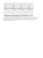
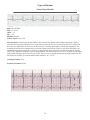
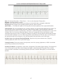
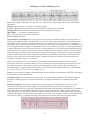
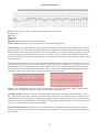
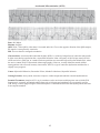
Ventricular Fibrillation
Rate: Unable to measure due to rapid and uncoordinated electrical activity.
P waves: None
PRI: None
QRS: None
QT: none
Rhythm: Rapid and chaotic. No discernable rhythm.
Source of pacer: Multiple sources of ectopy in the Ventricular/Purkinje Fibers
Characteristics: Ventricular fibrillation (VF) is the result of highly irritable ventricle(s), which begin to send out
rapid electrical stimuli. The stimuli are chaotic resulting in no organized ventricular depolarization. The ventricles
do not contract because they never depolarize. For you visual learners... just imagine shaking a bowl full of Jell-O.
Gently shake the bowl and watch the Jell-O quiver or “fibrillate.” This is similar to what is happening within the
heart. Because the ventricles are fibrillating and never contracting, the patient does not have a pulse, cardiac output,
or blood pressure.
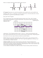
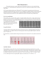
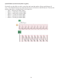
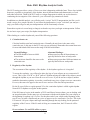
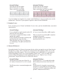
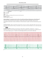
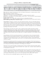
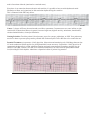
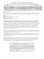
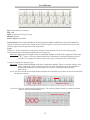
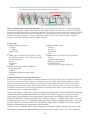
The terms coarse and fine have been used to describe the amplitude of the waveforms in VF. With Coarse VF, the
fibrillatory waves are more easily seen and are usually greater than 3mm in height (3 small boxes tall). Coarse VF
usually indicates a more recent onset of VF, which could be more easily converted by prompt defibrillation. The
presence of fine VF (which looks a bit like asystole and is less than 3mm in height) often means there has been a
considerable delay since collapse, and successful resuscitation is more difficult.
Causes: CAD, AMI, trauma, hypoxia, acidosis, antiarrhythmics, electrolyte imbalances, cardiac catheterization,
cardioversion, accidental electrocution, cardiac pacing and extreme hypothermia.
Nursing Priorities: Check for an airway, breathing, and pulse per Basic Life Support (BLS) standards. Call for
help. The patient will be apneic, pulseless, and unresponsive. Begin CPR. VF treatment requires electrical therapy
(defibrillation). Resuscitation requires defibrillation and often requires emergency drugs per ACLS VF guidelines.
The sooner the patient is defibrillated, the more likely of achieving spontaneous circulation. The longer the patient
is in VF, the more difficult it is to convert the rhythm.
Potential Treatments: Initial treatment is always defibrillation. Only defibrillation provides definitive therapy.
Other priorities include securing an airway, making sure the patient has IV access, and administering medications
per guidelines.
38