1
8
TranSignalTM TF Protein Array
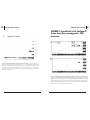
5. INCUBATION OF PROTEIN WITH THE ARRAY
MEMBRANE
Use this protocol with catalogue items MA3501, MA3502, MA3503 and
MA3504.
TranSignalTM TF Protein Array
6. DETECTION
Important: Do not let the membrane dry out during detection.
6 .1 Prepare 500µl of detection solution by combining 250µl Detection
Buffer A and 250 µl of Detection Buffer B per membrane.
6.2
Using forceps to hold the notched corner, carefully remove each
membrane from its tray. Drain the excess Wash Buffer from the
membrane by touching the edge against tissue. Place membrane
protein-side-up on a clean plastic sheet protector or overhead
transparency, by orienting the notch to the top, right-hand
corner.
6.3
Pipet the mixed Detection Buffers onto the membrane. Overlay
the membrane with a second plastic sheet and ensure that the
buffer mixture is evenly distributed over the membrane without
air bubbles.
5.3 Dilute the bacterial extract, containing overexpressed protein to a
final concentration of 60 µg/ml in 4 ml of 1X Blocking Buffer I. If you
have purified protein, dilute 3 -5 µg purified protein in 4 ml of 1X
Blocking Buffer I.
6.4
Incubate for 5 min at room temperature.
6.5
Remove excess substrate by gently applying pressure to the top
sheet. Using a paper towel, remove excess detection solution
remaining on the surface of the sheets.
5.5 Incubate the membrane with the diluted bacterial extract or purified
protein at room temperature for 2 hr with gentle shaking.
6.6
Expose the membranes using either Hyperfilm TM ECL or a
chemiluminescence imaging system, such as the FluorChemTM
imager from Alpha Innotech Corp. In either case, we recommend
that you try several different exposures of varying lengths of time
(e.g., 30 sec–5 min).
This section describes incubating bacterial extract containing your protein
of interest or purified protein with the array membrane. Note that the
array membranes are notched at the top right-hand corner for orientation
purposes.
Note: Be sure that the membrane is fully submerged in assay buffer at all times. Never
let the membrane dry out.
5.1 Place each membrane into a container containing 4 ml of 1X Blocking
Buffer I.
5.2 Place the tray on a shaker and incubate for 2 hr at room temperature.
5.6 Wash the membrane twice with 4 ml of 1X Wash Buffer for 5 min
(each wash) at room temperature.
5.7 Incubate the membrane with 4 ml of 1X Blocking Buffer II containing
primary antibody:
Note: Use an amount of the antibody suitable for western blot
detection of the protein or protein tag, specified by the manufacturer.
5.8 Incubate the membrane with the primary antibody solution for 2
hours at room temperature,
5.9
Wash the membrane twice with 4ml 1X Wash Buffer for 5-min (each
wash).
5.10 Incubate the membrane with the appropriate secondary antibody
diluted 1:5,000–1:15,000 in 4ml 1X Blocking Buffer II for 1 hr at room
temperature.
5.11 Wash the membrane four times with 4 ml of 1X Wash Buffer for 5 min
(each wash) at room temperature.
For Technical Support, call 1.877.726.6642 (PANOMIC) or visit our Web site at www.panomics.com.
For Technical Support, call 1.877.726.6642 (PANOMIC) or visit our Web site at www.panomics.com.
9