1
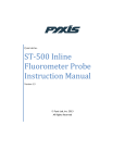
molecular biology reagents
protocol
DNaseAlert™ Substrate
Nuclease Detection System
User Manual
See what we can do for you at www.idtdna.com.
For Research Use Only.
IDT_Protocol Template_DNaseAlert_v5.indd 1
(14-01-01-13)
3/31/15 5:36 PM
protocol
molecular biology reagents
Introduction
Nucleases are widely present in the laboratory environment and can interfere
with many experiments. IDT developed DNaseAlert™ and RNaseAlert®
Substrate Nuclease Detection Systems for rapid, sensitive detection of
DNases and RNases, respectively. These reagents are fluorescence-quenched
oligonucleotide probes that fluoresce after nuclease degradation. The assays
can be read visually or measured and quantified using a fluorometer. Assays
can be used qualitatively or quantitatively to test lab reagents, work surfaces,
equipment, and supplies for nuclease contamination. The reagents can also
be used as highly sensitive substrates for enzyme kinetic studies.
The DNaseAlert Substrate is a synthetic DNA oligonucleotide that has a HEX™
reporter dye (hexachlorofluorescein, R) on one end and a dark quencher (Q)
on the other end (Figure 1). Its sequence has been carefully optimized to
react with a wide variety of nucleases; it contains domains that will react
with single-stranded endonucleases, single-stranded exonucleases, and
double-stranded nucleases. Intact, the substrate has little or no fluorescence.
When cleaved by a DNase, the substrate fluoresces pink (536 nm or UV
excitation, 556 nm emission).
The DNaseAlert test is fast and simple. Lab surfaces or liquid reagents can
be tested and verified as being “nuclease-free” or “contaminated” in less
than one hour. For speed and ease of use, a simple tube-based visual assay
can be performed directly at the site of testing. Alternatively, quantitative
fluorescent results (cuvette or microtiter plate formats) can be obtained
and used to document nuclease testing for GLP or manufacturing needs.
Figure 1. DNaseAlert™ Assay.
DNaseAlert™ Substrate Nuclease Detection System
IDT_Protocol Template_DNaseAlert_v5.indd 2
2
3/31/15 5:36 PM
protocol
molecular biology reagents
Kit Components and Storage Conditions
DNaseAlert™
Substrate Nuclease
Detection System
Component
Quantity
Storage Condition*
25 single-use tubes (50 pmol
–20°C (protect from
per tube) or 2 tubes bulk
light to prevent
substrate (2 nmol per tube)
photobleaching)
DNaseAlert Buffer
250 µL
–20°C
Nuclease-Free Water
2 mL
Room temperature
DNase I Enzyme (positive control)
25 µL
–20°C
Nuclease Decontamination Solution
50 mL
Room temperature
DNaseAlert Substrate
™
* Reagent quality is guaranteed for 6 months from date received when stored as directed.
Before you start:
• Use gloves when handling kit components and performing the assay.
• Perform all steps under nuclease-free conditions:
–
Use nuclease-free pipette tips and tubes.
–
If necessary, clean pipettes and other lab surfaces with
Nuclease Decontamination Solution before use.
Single-use tubes—testing a liquid solution
A
Set up one DNaseAlert assay for each sample to be tested.
Include 2 additional tubes for the positive and negative controls.
1. Add 5 µL of Nuclease-Free Water (provided) to a DNaseAlert Substrate
single-use tube for each assay.
2. Add 5 μL of 10X DNaseAlert Buffer to each tube.
3. Add 40 µL of test sample to each sample tube. Add 40 µL of Nuclease-Free
Water to each control tube. Add 1 µL of DNase I to the positive control
tube. Mix thoroughly. The final concentration of substrate is 1 μM for the
visual assay; lower substrate concentrations can be used for fluorometric
detection (see Quantitative measurement, below).
4. Incubate at 37°C for 30–60 min. Greater sensitivity is achieved with
longer incubations. If a temperature-regulated incubator or water bath is
not available, incubation can be done at room temperature (2–3X longer
incubation time is recommended to achieve similar sensitivity).
3
IDT_Protocol Template_DNaseAlert_v5.indd 3
See what we can do for you at www.idtdna.com.
3/31/15 5:36 PM
protocol
molecular biology reagents
5. Perform detection.
Quick visual confirmation: Place tube on a shortwave (300 nm) UV
transilluminator. For best results, use a darkened room.
If the tube remains clear, the assay is negative and the sample is free of detectable
DNase contamination. If the tube glows pink, DNase contamination is present.
A longwave (365 run) UV source can be used, but sensitivity will be lower.
Important safety note: Never look directly into a UV light source.
Always use protective face shielding.
Quantitative measurement: Place contents in a nuclease-free cuvette or
microtiter plate. Measure fluorescence using a fluorometer set to the HEX
channel (536 nm excitation, 556 nm emission). After the incubation at Step 4
(above) is complete, the sample can be diluted with up to 2 mL Nuclease-Free
Water, as needed, to accommodate the size of the detection chamber.
6. Validate all negative assays. Add 1 µL of DNase I (or other DNase) to each
tube. Mix and incubate at 37°C for 10 min. Repeat Step 5 (Perform detection).
All negative tubes should now be positive. Any assays that fail to fluoresce
during this validation step must be considered “failed”, and the nuclease
detection assay should be repeated. See troubleshooting guide at the end
of this manual for help with assay failures.
B
Single-use tubes—testing a solid object
or dry surface
Set up one DNaseAlert assay for each sample to be tested.
Include 2 additional tubes for the positive and negative controls.
1. Add 5 µL of Nuclease-Free Water (provided) to a DNaseAlert Substrate
single-use tube for each assay.
2. Add 5 μl of 10X DNaseAlert Buffer to each tube.
3. Add 40 µL of Nuclease-Free Water to each tube. Mix thoroughly.
4. Place the object to be tested into a sample tube. Add 1 µL of DNase I
to the positive control tube.
• Pipette tips, electrodes, or other small solids can be dipped directly
into the assay solution.
• To test surfaces that cannot be dipped into an assay tube:
a. Wipe the surface of interest with a piece of nuclease-free filter
paper pre-wetted with Nuclease-Free Water.
b. Soak the filter paper in a small amount of Nuclease-Free Water.
c. Transfer the liquid to an assay tube and proceed as instructed in
A. Single-use tubes—testing liquid samples.
DNaseAlert™ Substrate Nuclease Detection System
IDT_Protocol Template_DNaseAlert_v5.indd 4
4
3/31/15 5:36 PM
molecular biology reagents
protocol
5. Incubate at 37°C for 30–60 min.
6. Read assay in a fluorometer using the HEX channel (536 nm excitation, 556 nm emission). After Step 5 incubation is complete, the sample can
be diluted using up to 2 mL Nuclease-Free Water, as needed, to
accommodate the size of the detection chamber.
7. Validate all negative assays. Add 1 μL of DNase I (or other DNase) to
each negative (non-fluorescent) tube. Mix and incubate at 37°C for 10 min.
Repeat detection procedure as before. All negative tubes should now be
positive. Any assays that fail to fluoresce during this validation step must be
considered “failed”, and the nuclease detection assay should be repeated.
See troubleshooting guide at the end of this manual for help with assay failures.
C
DNaseAlert™
Substrate Nuclease
Detection System
Bulk substrate—testing a liquid solution
Bulk substrate assays can be performed in individual tubes; however, it is
more convenient to perform the assays in microtiter plates. We recommend
using black opaque plates that minimize scatter and cross-talk between wells.
Always include negative and positive control wells. Use duplicate or triplicate
sample wells for quantitative measurements.
1. Add 1 mL of Nuclease-Free Water (provided) to the bulk DNaseAlert
Substrate tube. Mix thoroughly. Final substrate concentration is 2 µM
(20 pmol substrate in 10 µL).
2. Add 10 µL of DNaseAlert Substrate to each sample and control well.
3. Add 10 µL of 10X DNaseAlert Buffer to each well.
4. Add 80 µL of sample to the sample wells. Add 80 µL of Nuclease-Free
Water to each control well. Add 1 µL of DNase I to the positive control well.
Mix thoroughly.
5. Incubate at 37°C for 30–60 min. The recommended final concentration
of substrate is 200 nM for a standard 96-well fluorometer; more dilute
solutions can be used for sensitive cuvette fluorometers.
6. Read assay in fluorometer using the HEX channel (536 nm excitation,
556 nm emission). Assays can be read as simple endpoint measurements
or examined in real time to obtain quantitative kinetic curves.
7. Validate all negative assays. Add 1 μL of DNase I (or other DNase) to
each well. Mix and incubate at 37°C for 10 min. Repeat detection procedure
as before. All negative tubes should now be positive. Any assays that fail
to fluoresce during this validation step must be considered “failed”, and
the nuclease detection assay should be repeated. See troubleshooting
guide at the end of this manual for help with assay failures.
5
IDT_Protocol Template_DNaseAlert_v5.indd 5
See what we can do for you at www.idtdna.com.
3/31/15 5:36 PM
protocol
molecular biology reagents
Troubleshooting
Observation
False
Negatives
Possible Problem
Solution
Presence of DNase inhibitors
Solutions with extreme pH, strong ionic strength, or
detergents can block action of DNases, preventing
detection of contaminants that are present.
Test your solution for inhibitors of the DNaseAlert assay.
Set up a standard assay with your test solution and add
1 μL DNase I (or other DNase).
Low pH solutions
Solutions with pH <7.0 will decrease the efficiency
of HEX fluorescence, lowering assay sensitivity.
If possible, adjust the pH of the test solution to obtain
pH >7.0 and <9.0.
Substrate loss
1. The DNaseAlert Substrate is provided dried
down. The dry pellet can become dislodged
from the bottom of the tube and may be lost
from the tube when opened; dry oligos can be
electrostatically attracted to laboratory gloves.
2. Prolonged exposure of the DNaseAlert Substrate
to light can lead to photobleaching of the
fluorescent dye and decrease assay sensitivity.
If the assay does not convert to “positive” after a
1-hour incubation, your test solution is incompatible
with the DNaseAlert system.
1. a. Spin down tubes before opening.
b. Perform a positive control assay.
Always perform a positive control test on single-use
tube assays with negative results. A positive result in
another tube, even from the same kit, is not sufficient.
2. Store bulk substrate and assay tubes in the dark.
Nuclease–substrate incompatibility
The DNaseAlert Substrate contains a fluorescent dye
at the 5' end and a fluorescence quencher at the 3'
end. Activity of some exonucleases is blocked by
terminal end-groups and, therefore, such nucleases
(e.g., E. coli Exo I) cannot be detected using this assay.
Contamination
Nuclease contamination of tubes, pipette tips, and
other lab equipment can lead to false positives.
Perform a negative control assay.
A negative control must be included with each set of
assays performed. Fluorescence in the negative control
tube (Nuclease-Free Water test) indicates contamination
in the experimental setup
Obtain new supplies and clean work area.
If contamination is suspected, obtain fresh tubes and
pipette tips and clean all lab surfaces with Nuclease
Decontamination Solution before repeating the assay.
False
Positives
Quencher exhaustion
Prolonged exposure of the substrate to UV light
can damage the quencher. A visual assay left on an
intense shortwave (254 nm) UV source can turn
positive even when no nuclease is present.
Read your assay immediately to limit
exposure to UV light.
Substrate degradation
The DNaseAlert Substrate contains DNA bases that
will be depurinated and degraded when exposed to
acidic conditions.
Avoid use of solutions with pH <4.5.
Substrate stimulation
Certain organic solvents disrupt quenching.
In particular, DNaseAlert Substrate always glows
in acetonitrile.
DNaseAlert™ Substrate Nuclease Detection System
IDT_Protocol Template_DNaseAlert_v5.indd 6
6
3/31/15 5:36 PM