1
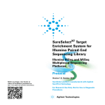
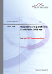
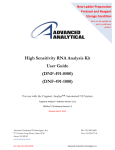
PacBio SampleNet – Shared Protocol
Please note: the shared protocols described herein may not have been validated by Pacific Biosciences and are provided as-is and
without any warranty. Use of these protocols is offered to those customers who understand and accept the associated terms and
®
conditions and wish to take advantage of their potential to help prepare samples for analysis using the PacBio system. If any of these
protocols are to be used in a production environment, it is the responsibility of the end user to perform the required validation.
10 kb to 20 kb Template Preparation and
Sequencing with Low-Input DNA
Before You Begin
To perform this procedure, you must have the PacBio®:
•
•
•
•
•
•
DNA Template Prep Kit
DNA/Polymerase Binding Kit
MagBead Kit
DNA Sequencing Kit
DNA Control Complex
SMRT® Cells for standard sequencing
This procedure can be used to prepare 10-20 kb libraries from 50 ng up to 200 ng of sheared and concentrated
DNA, or at least 100 ng into shearing. Note: for input amounts between 200 ng and 1 µg, the standard 10 kb library
prep protocol may be used.
Insert Size Target
10 to 20 kb
Insert Size Range
8 kb to 22 kb
Sheared and
Concentrated DNA
Amount
50 to 200 ng
Ligation
DNA Damage
Repair
Blunt
Required
Fragment and Concentrate DNA
Use a Covaris® g-TUBE® device to shear your DNA sample, following the g-TUBE user manual available for
download from the Covaris website, with one change -- reduce the sample volume from 150 µL to 50 µL.
Note: After the first spin, make sure that all of the sample has passed into the lower chamber. If any sample
remains, re-spin. If necessary, add 10 µL EB or TE to the upper chamber, flick the g-TUBE or pipette up and
down several times, and spin again. Depending upon the quality of your sample, approximately 20% to 50%
sample loss is to be expected as a result of the shearing and concentration process.
Page 1
PacBio SampleNet – Shared Protocol
STEP
1
Concentrate DNA
Add 0.5X volume of AMPure® PB magnetic beads.
μL of sample X 0.5X =
μL of beads
Note that the beads must be brought to room temperature and all AMPure PB
bead purification steps should be performed at room temperature.
Before using, mix the bead reagent well until the solution appears homogenous.
Pipette the reagent slowly since the bead mixture is viscous and precise
volumes are critical to the purification process.
Consistent and efficient recovery of your sample is critical to successful
SMRTbell™ template preparation. If using this protocol for the first time, we
strongly recommend that you process a control sample first. Using the DNA
shearing methods and subsequent AMPure PB bead purification steps
described below, you should recover approximately 50%-80% of your input DNA
(by mass). Typical yields, from pre-purified DNA (where smaller fragments are
already eliminated as a result of the shearing process) are between 80-100%.
2
Mix the bead/DNA solution thoroughly.
3
Quickly spin down the tube (for 1 second) to collect the beads.
4
Allow the DNA to bind to beads by mixing in a VWR® vortex mixer at 2000 rpm for
10 minutes at room temperature. Note that the bead/DNA mixing is critical to yield.
After mixing, the bead/DNA mixture should appear homogenous.
We recommend using a VWR vortex mixer with a foam microtube attachment
(see the Guide’s Overview section for part numbers). If using other
instrumentation, ensure that the mixing is equally vigorous. Failure to thoroughly
mix the DNA with the bead reagent will result in inefficient DNA binding and
reduced sample recoveries.
5
Spin down the tube (for 1 second) to collect beads.
6
Place the tube in a magnetic bead rack until the beads collect to the side of the
tube and the solution appears clear. The actual time required to collect the beads
to the side depends on the volume of beads added.
7
With the tube still on the magnetic bead rack, slowly pipette off cleared
supernatant and save in another tube. Avoid disturbing the bead pellet.
If the DNA is not recovered at the end of this Procedure, you can add equal
volumes of AMPure PB beads to the saved supernatant and repeat the AMPure
PB bead purification steps to recover the DNA.
8
Wash beads with freshly prepared 70% ethanol.
Note that 70% ethanol is hygroscopic and should be prepared FRESH to
achieve optimal results. Also, 70% ethanol should be stored in a tightly capped
polypropylene tube for no more than 3 days.
– Do not remove the tube from the magnetic rack.
– Use a sufficient volume of 70% ethanol to fill the tube (1.5 mL for 1.5 mL
tube or 2 mL for 2 mL tube). Slowly dispense the 70% ethanol against the
side of the tube opposite the beads. Let the tube sit for 30 seconds.
– Do not disturb the bead pellet.
– After 30 seconds, pipette and discard the 70% ethanol.
9
Repeat step 8 above.
Page 2
Notes
PacBio SampleNet – Shared Protocol
STEP
10
Concentrate DNA
Remove residual 70% ethanol and dry the bead pellet.
– Remove tube from magnetic bead rack and spin to pellet beads. Both the
beads and any residual 70% ethanol will be at the bottom of the tube.
– Place the tube back on magnetic bead rack.
– Pipette off any remaining 70% ethanol.
11
Check for any remaining droplets in the tube. If droplets are present, repeat step
10.
12
Remove the tube from the magnetic bead rack and allow beads to air-dry (with the
tube caps open) for 30 to 60 seconds.
13
Add 37 μL of Pacific Biosciences’ Elution Buffer to the beads to elute the DNA.
– Mix until homogeneous.
– Vortex for 1-2 minutes at 2000 rpm.
– Spin the tube down to pellet beads, then place the tube back on the magnetic
bead rack.
– Carefully collect the eluted sample.
– Discard the beads.
14
Proceed to the next step, if possible. If necessary, store at -20°C to continue later.
Page 3
Notes
PacBio SampleNet – Shared Protocol
Repair DNA Damage
Use the following table to repair any DNA damage.
1. In a LoBind microcentrifuge tube, add the reagents below.
Note: premix damage repair buffer, NAD+, ATP high, and dNTPs if you are preparing more than 1 sample.
Reagent
Sheared DNA
Tube Cap Color
Stock Conc.
−
Volume
Final Conc.
37 μL
−
DNA Damage Repair
Buffer
10 X
5.0 μL
1X
NAD+
100 X
0.5 μL
1X
ATP high
10 mM
5.0 μL
1 mM
dNTP
10 mM
0.5 μL
0.1 mM
DNA Damage Repair
Mix
2.0 μL
Total Volume
50.0 μL
Notes
−
*To determine the correct amount of H2O to add, use your actual DNA amount noted in the Notes column.
2. Mix the reaction well by pipetting or flicking the tube.
3. Spin down contents of tube with a quick spin in a microfuge.
4. Incubate at 37ºC for 20 minutes, then return the reaction to 4ºC for 1 minute.
Repair Ends
Use the following table to prepare your reaction then purify the DNA.
Reagent
DNA (Damage
Repaired)
End Repair Mix
Tube Cap Color
Stock Conc.
−
20 X
Total Volume
Volume
Final Conc.
50 μL
−
2.0 μL
1X
52.0 μL
−
1. Mix the reaction well by pipetting or flicking the tube.
2. Spin down contents of tube with a quick spin in a microfuge.
3. Incubate at 25ºC for 5 minutes (no longer), return the reaction to 4ºC.
Page 4
Notes
PacBio SampleNet – Shared Protocol
STEP
Purify DNA
1
Add 0.5X volume of AMPure PB beads to the End-Repair reaction. (For detailed
instructions on AMPure PB bead purification, see the Concentrate DNA section).
2
Mix the bead/DNA solution thoroughly.
3
Quickly spin down the tube (for 1 second) to collect the beads. Do not pellet beads.
4
Allow the DNA to bind to beads by shaking in a VWR vortex mixer at 2000 rpm for
10 minutes at room temperature.
5
Spin down the tube (for 1 second) to collect beads.
6
Place the tube in a magnetic bead rack to collect the beads to the side of the tube.
7
Slowly pipette off cleared supernatant and save (in another tube). Avoid disturbing
the bead pellet.
8
Wash beads with freshly prepared 70% ethanol.
9
Repeat step 8 above.
10
Remove residual 70% ethanol and dry the bead pellet.
– Remove tube from magnetic bead rack and spin to pellet beads. Both the
beads and any residual 70% ethanol will be at the bottom of the tube.
– Place the tube back on magnetic bead rack.
– Pipette off any remaining 70% ethanol.
11
Check for any remaining droplets in the tube. If droplets are present, repeat step
10.
12
Remove the tube from the magnetic bead rack and allow beads to air-dry (with
tube caps open) for 30 to 60 seconds.
13
Elute the DNA off the beads in 32-33 μL Elution Buffer.:
– Mix until homogeneous.
– Vortex for 1-2 minutes at 2000 rpm.
– Spin the tube down to pellet beads, then place the tube back on the magnetic
bead rack.
– Carefully collect the eluted sample.
– Discard the beads.
14
Proceed to the next step if possible. If necessary, store at -20°C to continue later.
Page 5
Notes
PacBio SampleNet – Shared Protocol
Prepare Blunt-Ligation Reaction
Use the following table to prepare your blunt-ligation reaction:
1. In a LoBind microcentrifuge tube (on ice), add the following reagents in the order shown. If preparing a
Master Mix, ensure that the adapter is NOT mixed with the ligase prior to introduction of the inserts. Add the
adapter to the well with the DNA. All other components, including the ligase, should be added to the Master
Mix.
Tube Cap Color
Reagent
Stock
Volume
Conc.
DNA (End Repaired)
Final Conc.
Notes
32 µl
Annealed Blunt
Adapter (20 μM)
20 μM
1.0 μL
0.5 μM
Mix before proceeding
Template Prep Buffer
10 X
4.0 μL
1X
ATP low
1 mM
2.0 μL
0.05 mM
Mix before proceeding
Ligase
Total Volume
−
30 U/μL
1.0 μL
0.75 U/μL
−
40.0 μL
−
2.
3.
4.
5.
Mix the reaction well by pipetting or flicking the tube.
Spin down contents of tube with a quick spin in a microfuge.
Incubate at 25ºC for 45 minutes.
Incubate at 65ºC for 10 minutes to inactivate the ligase, then return the reaction to 4ºC. You must proceed
with adding exonucleases after this step.
Add exonucleases to remove failed ligation products.
Reagent
Tube Cap Color
Stock Conc.
Ligated DNA
Volume
40 μL
Mix reaction well by pipetting
ExoIII
100.0 U/μL
0.5 μL
ExoVII
10.0 U/μL
0.5 μL
Total Volume
41 μL
1. Mix the reaction well by pipetting or flicking the tube.
2. Spin down contents of tube with a quick spin in a microfuge.
3. Incubate at 37ºC for 45 minutes, then return the reaction to 4ºC. Do not exceed 1 hour incubation time. You
must proceed with purification after this step.
Page 6
PacBio SampleNet – Shared Protocol
Purify SMRTbell™ Templates
There are 2 final purification steps. The first uses 0.5X volumes of AMPure PB beads, followed by purification
with 0.45X volumes of AMPure PB beads.
STEP
Purify SMRTbell™ Templates - First Purification
1
Add 0.5X volume of AMPure PB beads to the exonuclease-treated reaction. (For
detailed instructions on AMPure PB bead purification, see the Concentrate DNA
section).
2
Mix the bead/DNA solution thoroughly.
3
Quickly spin down the tube (for 1 second) to collect the beads. Do not pellet
beads.
4
Allow the DNA to bind to beads by shaking in a VWR vortex mixer at 2000 rpm for
10 minutes at room temperature.
5
Spin down the tube (for 1 second) to collect beads.
6
Place the tube in a magnetic bead rack to collect the beads to the side of the
tube.
7
Slowly pipette off cleared supernatant and save (in another tube). Avoid
disturbing the bead pellet.
8
Wash beads with freshly prepared 70% ethanol.
9
Repeat step 8 above.
10
Remove residual 70% ethanol and dry the bead pellet.
– Remove tube from magnetic bead rack and spin to pellet beads. Both the
beads and any residual 70% ethanol will be at the bottom of the tube.
– Place the tube back on magnetic bead rack.
– Pipette off any remaining 70% ethanol.
11
Check for any remaining droplets in the tube. If droplets are present, repeat step
10.
12
Remove the tube from the magnetic bead rack and allow beads to air-dry (with
tube caps open) for 30 to 60 seconds.
13
Elute the DNA off the beads in 50 μL of Elution Buffer. Mix for 10 minute at 2000
rpm:
– Mix until homogeneous.
– Vortex for 1-2 minutes at 2000 rpm.
– Spin the tube down to pellet beads, then place the tube back on the
magnetic bead rack.
– Carefully collect the eluted sample.
– Discard the beads.
Page 7
Notes
PacBio SampleNet – Shared Protocol
STEP
Purify SMRTbell™ Templates - Second Purification
1
Add 0.45x volume of AMPure PB beads to the 50 μL of eluted DNA.
2
Mix the bead/DNA solution thoroughly.
3
Quickly spin down the tube (for 1 second) to collect the beads.
Do not pellet beads.
4
Allow the DNA to bind to beads by shaking in a VWR vortex mixer at 2000 rpm for
10 minutes at room temperature.
5
Spin down the tube (for 1 second) to collect beads.
6
Place the tube in a magnetic bead rack to collect the beads to the
side of the tube.
7
Slowly pipette off cleared supernatant and save
(in another tube). Avoid disturbing the bead pellet.
8
Wash beads with freshly prepared 70% ethanol.
9
Repeat step 8 above.
10
Remove residual 70% ethanol and dry the bead pellet.
– Remove tube from magnetic bead rack and spin to pellet beads. Both the
beads and any residual 70% ethanol will be at the bottom of the tube.
– Place the tube back on magnetic bead rack.
– Pipette off any remaining 70% ethanol.
11
Check for any remaining droplets in the tube. If droplets are present, repeat step
10.
12
Remove the tube from the magnetic bead rack and allow beads to
air-dry (with tube caps open) for 30 to 60 seconds.
13
Elute the DNA off the beads in 8-10 μL of Elution Buffer:
– Mix until homogeneous.
– Vortex for 1-2 minutes at 2000 rpm.
– Spin the tube down to pellet beads, then place the tube back on the
magnetic bead rack.
– Carefully collect the eluted sample.
– Discard the beads.
14
Check quantitation with the Qubit® dsDNA HS Assay Kit. If there is too little
sample, estimate concentration based on 10% yield of input amount into
damage repair.
Page 8
Notes
PacBio SampleNet – Shared Protocol
Anneal and Bind SMRTbell™ Templates
You must have a PacBio DNA Polymerase Binding Kit for this step. To anneal sequencing
primer and bind polymerase to SMRTbell templates, follow the Calculator recommendations
with the following set-up:
1. Under Edit Sample, enter the Volume to Use and DNA Concentration (measured or estimated assuming 10% library yield). Then select the following:
- Magnetic Beads: Yes
- Preparation Protocol: Small scale
- DNA Control Complex: No
- Non-standard: Yes
Note the
suggested number
of SMRT Cells
PacBio SampleNet – Shared Protocol
2. Under Optional, enter a Custom Concentration on Plate:
-For P4 polymerase, enter 0.025 nM
-For P5 polymerase, enter 0.040 nM
3. Under Annealing, pre-mix 10x Primer Buffer and Diluted Sequencing Primer at higher volumes
to eliminate small volume pipetting
a. Prepare a 10x pre-mix by
combining 9.0 µL 10x Primer
Buffer and 1.3 µL Diluted
Sequencing Primer
b. Add 1.03 µL pre-mix to the
sample
4. Dilutions. If polymerase is diluted to <8.0 nM, increase the polymerase:template ratio until 8.0
nM final concentration is obtained (or simply dilute polymerase 1/200 in Binding Buffer v3):
If this number is <8.0 nM, see
recommendation under “Optional”
below.
PacBio SampleNet – Shared Protocol
Increase this number as required until
the polymerase dilution is at least 8.0 nM
(see below).
5. Use the entire complex to sequence the number of SMRT Cells recommended (by the
calculator) in one run.
6. If desired, the bead-bound complex from sample plate can be used the next day by doing the
following:
-Pool the remaining beads from multiple SMRT Cells.
-Add the Bead Binding Buffer to bring the volume to 19 µL (for 1 SMRT Cell), or the required
volume for the closest number of SMRT Cells. For example, if 3 cells were run, and the
remaining pooled volume is 26 µL, add 2 µL Bead Binding Buffer to bring the volume to 28
µL for 2 SMRT Cells. The required volume for any number of SMRT Cells may be
determined with the binding calculator.
The expected yield from reused beads depends on the amount of Bead Binding buffer added. If
none is needed, it may be close to the original yield (if used the following day). The yield will
decrease with longer times between runs.
For Research Use Only. Not for use in diagnostic procedures. © Copyright 2014, Pacific Biosciences of California, Inc. All rights reserved. Information in this
document is subject to change without notice. Pacific Biosciences assumes no responsibility for any errors or omissions in this document. Certain notices, terms,
conditions and/or use restrictions may pertain to your use of Pacific Biosciences products and/or third party products. Please refer to the applicable Pacific
Biosciences Terms and Conditions of Sale and to the applicable license terms at http://www.pacificbiosciences.com/licenses.html. Pacific Biosciences, the Pacific
Biosciences logo, PacBio, SMR, SMRTbell, and Iso-Seq are trademarks of Pacific Biosciences in the United States and/or certain other countries. All other
trademarks are the sole property of their respective owners.