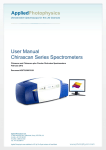
Download SX20 Hardware User Guide
Transcript
NEW
SX20 Hardware
User Guide
DIMENSIONS TO KINETICS
This section provides a complete guide to the hardware of the
SX20 stopped flow system. It includes a technical description
of the hardware, instrument set-up, hardware-related
aspects of operation and a basic maintenance guide.
ADDING
Applied Photophysics Limited
203/205 Kingston Road
Leatherhead KT22 7PB
United Kingdom
NEW
Tel: +44 (0)1372 386537
(USA) 1- 800 543 4130
Fax: +44 (0)1372 386477
Email: [email protected]
URL: www.photophysics.com
DIMENSIONS TO KINETICS
1
Updated January 2006
Contents
2.0 Introduction................................................................................................................ 5
2.1.1 Description..............................................................................................................5
2.1.2 Components ............................................................................................................5
2.2 SX20 Optional Accessories ....................................................................................... 7
2.2.1 SQ.1 Sequential (Double) Mixing Option...............................................................7
2.2.2 SEM.1 Emission Scanning Option..........................................................................7
2.2.3 AM.1 Extended Absorbance Option .......................................................................7
2.2.4 PDA.1 Photodiode Array Option ............................................................................8
2.2.5 UV.1 Deuterium Light Source Option ....................................................................8
2.2.6 DD.1 Dual Channel Detection Option ....................................................................8
2.2.7 DF.1 Dual Channel Fluorescence Detection Option ...............................................9
2.2.8 FP.1 Fluorescence Polarisation Option ...................................................................9
2.2.9 RC.1 5μL Rapid Kinetics Cell Option ....................................................................9
2.2.10 QFA.1 Quench Flow Adaptor Option ...................................................................9
2.2.11 AN.1 Anaerobic Accessory...................................................................................9
2.3 SX20 Instrument Set-up........................................................................................... 10
2.3.1 Site Requirements .................................................................................................10
2.3.2 SX20 Hardware Set-up .........................................................................................11
2.3.3 SX20 Electrical Set-up..........................................................................................15
2.3.4 SX20 Optional Accessories Basic Set-up .............................................................20
2.3.4.1 SEM.1 Scanning Emission Monochromator Option..........................................20
2.3.4.2 AM.1 Extended Absorbance Option ..................................................................21
2.4 SX20 Hardware Operation....................................................................................... 22
2.4.1 Sample Handling Unit...........................................................................................22
2.4.1.1 Introduction........................................................................................................22
2.4.1.2 Description of a Stopped-Flow Drive ................................................................24
2.4.1.3 Stopped-Flow Circuit.........................................................................................24
2.4.1.4 Drive Syringes ...................................................................................................26
2.4.1.5 Drive Valves ......................................................................................................26
2.4.1.6 Stop Syringe.......................................................................................................27
2.4.1.7 Stop Valve..........................................................................................................28
2.4.1.8 Flow Tubing.......................................................................................................29
2.4.1.9 Interchangeable Cell Cartridge System..............................................................32
5.1.10 Optical Cells .......................................................................................................34
2.4.1.11 Absorbance Pathlength Selection.....................................................................35
2.4.1.12 The Auto-Stop Mechanism ..............................................................................36
2.4.1.13 Pneumatic Drive and Drive Rams....................................................................39
2.4.1.14 Temperature Control ........................................................................................40
2.4.2 Absorbance Detector.............................................................................................42
2
Updated January 2006
2.4.2.1 Description.........................................................................................................42
2.4.2.2 Basic Set-Up and Electrical Connections...........................................................42
2.4.2.3 PMT Characteristics...........................................................................................43
2.4.3 Fluorescence Detector...........................................................................................44
2.4.3.1 Description.........................................................................................................44
2.4.3.2 Basic Set-Up and Electrical Connections...........................................................44
2.4.3.3 PMT Characteristics...........................................................................................45
2.4.3.4 Dual Channel Fluorescence Set-Up ...................................................................46
2.4.4 Monochromator.....................................................................................................47
2.4.4.1 Description.........................................................................................................47
2.4.4.2 Basic Set-Up and Electrical Connections...........................................................48
2.4.4.3 Selecting a Suitable Monochromator Slit Width (Band Pass)............................48
2.4.4.4 Band Pass Calculation........................................................................................48
2.4.4.5 Dual Monochromator Operation ........................................................................49
2.4.4.6 Photodiode Array Operation ..............................................................................49
2.4.4.7 Absorbance Measurements in the near IR (>650nm).........................................49
2.4.4.8 Maintenance.......................................................................................................49
2.4.5 150W Lamp Housing ............................................................................................50
2.4.5.1 Description.........................................................................................................50
2.4.5.2 Basic Set-Up and Electrical Connections...........................................................51
2.4.5.3 Lamp Stability Magnet.......................................................................................51
2.4.5.4 Lamp Types .......................................................................................................52
2.4.5.5 Purging the Lamp Housing (Ozone-Producing Lamp Only) .............................54
2.4.5.6 Lamp Alignment Procedure ...............................................................................56
2.4.5.7 Lamp Lifetime ...................................................................................................57
2.4.5.8 Lamp Stability Test............................................................................................57
2.4.6 Lamp Power Supply Unit......................................................................................58
2.4.6.1 Description.........................................................................................................58
2.4.6.2 Basic Set-Up and Electrical Connections...........................................................58
2.4.6.3 Operation ...........................................................................................................59
2.4.6.4 Use of Xenon and Mercury-Xenon Lamps ........................................................59
2.4.6.5 Maintenance.......................................................................................................60
2.4.6.6 Electrical Characteristics....................................................................................60
2.4.7 Electronics Unit ....................................................................................................61
2.4.7.1 Description.........................................................................................................61
2.4.7.2 Operation ...........................................................................................................64
2.4.7.3 Maintenance.......................................................................................................65
2.4.7.4 Electrical Characteristics....................................................................................65
2.4.8 Workstation...........................................................................................................66
2.4.8.1 Description.........................................................................................................66
2.4.8.2 Basic Set-Up and Electrical Connections...........................................................66
2.4.8.3 Operation ...........................................................................................................67
2.4.9 Photo Diode Array Detector (Option PDA.1).......................................................68
3
Updated January 2006
2.4.9.1 Description.........................................................................................................68
2.4.9.2 Basic Set-Up and Electrical Connections...........................................................68
2.5 SX20 Experimental Operation ................................................................................. 69
2.5.1 Single Mixing Stopped-Flow Operation ...............................................................69
2.5.2 Sequential Mixing Stopped-Flow Operation.........................................................73
2.5.3 Optical Filters........................................................................................................78
2.5.3.1 Absorbance Measurements in the near IR (>650nm).........................................78
2.5.3.2 Fluorescence Measurements ..............................................................................78
2.5.4 Asymmetric (Variable Ratio) Mixing ...................................................................81
2.5.4.1 Setting the Drive Pressure for Asymmetric Mixing Experiments......................81
2.5.4.2 Setting the Drive Volume for Asymmetric Mixing Experiments ......................82
2.5.5 Anaerobic Operation (using option AN.1)............................................................83
2.5.5.1 Purging the Drive Syringes ................................................................................83
2.5.5.2 Flow Circuit Preparation....................................................................................84
2.5.5.3 Sample Introduction Care ..................................................................................84
2.5.5.4 Procedure for Bench Top Anaerobic Stopped-Flow Studies .............................85
4
Updated January 2006
2.0 Introduction
This is a reference guide to the hardware of the SX20 stopped-flow reaction analyser. It
describes the set-up, operation and routine maintenance of the hardware of the
instrument. A trouble-shooting guide is also included to help the user fault find
commonly encountered problems.
2.1 SX20 Stopped-Flow Reaction Analyser
This section describes the experimental capabilities of the standard configuration SX20
stopped-flow reaction analyser and the components that comprise the system.
2.1.1 Description
The SX20 stopped-flow system in its standard configuration, without any of the
optional accessories described later in this section allows the user to carry out single
mixing stopped-flow measurements. The user can select between detection of
absorbance kinetics at a single wavelength or fluorescence kinetics using an optical
filter to isolate the fluorescence signal.
2.1.2 Components
The standard configuration SX20 stopped-flow system consists of the following major
hardware components: 1)
2)
3)
4)
5)
6)
7)
8)
9)
10)
11)
12)
150W Xenon lamp power supply and igniter unit
150W Xenon lamp housing (lamp fitted)
Monochromator (Mono 1)
Optical rail for mounting of the lamp housing and monochromator
Fibre optic light guide
Sample handling unit fitted with 20µL cell cartridge
Absorbance detector
Fluorescence detector
Electronics Unit fitted with single detection channel
Computer fitted with fibre optic interface card
Monitor
Printer
5
Updated January 2006
These components will be described in further detail later in this section. In addition to
the major components, the SX20 system is supplied with the following ancillary
components:1) Mains electricity distribution board and mains cables
2) Set of cables for electrical connection of the SX20 major components
3) Pneumatic tubing and connectors for connecting the system to an 8bar gas
supply
4) In-line pressure regulator for controlling drive pressure
5) Storage case including toolkit and consumable spares kit
6) User manual and back-up software
6
Updated January 2006
2.2 SX20 Optional Accessories
This section describes the optional accessories that are available for the SX20 system.
These options are available for new systems or as upgrades to existing systems as the
research interests of the user evolve.
2.2.1 SQ.1 Sequential (Double) Mixing Option
The sequential (double) mixing option equips the sample handling unit with four drive
syringes and permits the mixing of two reagents (A+B) with a first mixing drive and
after a user defined delay period, mixes the aged solution with a third reagent (C). This
option consists of the following components:1) A sequential mixing sample handling unit
2) 2 sequential mixing drive rams
3) Stop syringe brake mechanism
2.2.2 SEM.1 Emission Scanning Option
This emission scanning option extends the fluorescence detection capability of the
SX20. An emission monochromator located between the optical cell and the
fluorescence detector allows collection of fluorescence kinetic traces at a single
wavelength and collection of steady state and time resolved emission spectra. It consists
of the following components: 1) Emission monochromator (Mono 2)
2) Second optical rail and four supports
3) Emission fibre optic light guide
2.2.3 AM.1 Extended Absorbance Option
The extended absorbance option provides improved stray light performance in the far
UV enabling accurate absorbance measurements up to 2AU below 250nm. It consists of
the following components: -
7
Updated January 2006
1) Second monochromator (Mono 2)
2) Extended optical rail
3) Mono 1 to Mono 2 optical coupler
2.2.4 PDA.1 Photodiode Array Option
The Photodiode Array (PDA) option allows collection of time resolved absorbance
spectra from a single stopped-flow drive. There are two different versions available: A
UV region PDA operates in the wavelength region 180-750nm. A visible region PDA
operates in the wavelength range 350-1100nm. The option consists of the following
components: 1) Photodiode array unit (UV or visible version)
2) PDA fibre optic light guide (xenon light source version)
3) Electronics Unit PDA module
2.2.5 UV.1 Deuterium Light Source Option
The deuterium light source option allows the collection of PDA data using the UV
version PDA accessory down to 200nm. The option consists of the following
components:1) Deuterium lamp power supply unit
2) Deuterium lamp housing (lamp fitted)
3) PDA fibre optic light guide (deuterium light source version)
2.2.6 DD.1 Dual Channel Detection Option
The dual channel detection accessory allows fluorescence and absorbance data to be
collected simultaneously using the absorbance and fluorescence PMT detectors. It
consists of the following components:1) Dual channel PMT module fitted in the Electronics Unit.
2) Detector cable for connecting the detector to the PMT module.
8
Updated January 2006
2.2.7 DF.1 Dual Channel Fluorescence Detection Option
The dual channel fluorescence detection accessory allows fluorescence data to be
collected simultaneously using two fluorescence PMT detectors. It requires the DD.1
dual channel option and consists of the following additional components:1) Additional fluorescence detector
2) Fluorescence detector mounting fittings and filter holder
2.2.8 FP.1 Fluorescence Polarisation Option
The fluorescence polarisation accessory allows collection of polarisation and anisotropy
signals. It consists of the following components:1) Additional fluorescence detector
2) T-format excitation and emission polarizer accessory
2.2.9 RC.1 5μL Rapid Kinetics Cell Option
The 5μL rapid kinetics cell allows collection of stopped-flow data with a dead time of
0.45ms. The cell is mounted in an interchangeable cell cartridge supplied in a protective
storage case and is supplied with a 1mL stop syringe.
2.2.10 QFA.1 Quench Flow Adaptor Option
The quench flow accessory utilises the sequential mixing capability of the SHU. The
adaptor is mounted in the standard interchangeable cell cartridge and is fitted with a
basic sample collection loop.
2.2.11 AN.1 Anaerobic Accessory
The anaerobic accessory improves the ease with which anaerobic experiments can be
performed without the need for a glove-box environment. It consists of the following
components: 1) Pack of ten 3-way valves for loading anaerobic samples into the SHU
2) Nitrogen purging manifold that connects to the sample handling unit
9
Updated January 2006
2.3 SX20 Instrument Set-up
This section describes the basic set-up procedure and configuration of the SX20
stopped-flow reaction analyser. More detailed information relating to the set-up and
connections of each component of the system is provided later in this chapter.
2.3.1 Site Requirements
The SX20 should be set-up in a clean laboratory environment on a sturdy work
surface. The laboratory temperature should be maintained within the range 15-35oC.
The minimum space requirement for the SX20 (length x width x height) is as follows:
SX20 system: 2.0m x 0.7m x 0.5m
SX20 system fitted with option AM.1: 2.5m x 0.7m x 0.5m
The SX20 requires a stable mains electricity supply of 85-264V.
The sample handling unit is pneumatically driven and requires a compressed gas
supply (preferably nitrogen) capable of operating at 8bar (120psi). The supply should
be fitted with a push on tube adaptor to accept the flexible hose (6mm internal
diameter) that connects to the SHU inlets.
For connection to a water circulator unit, the SHU is provided with an inlet and outlet
fitting compatible with a flexible hose of 8mm internal diameter.
10
Updated January 2006
2.3.2 SX20 Hardware Set-up
The picture below shows the standard arrangement of the major hardware components
of the system.
Sample Handling Unit
PC Workstation
Electronics Unit
Monochromator
Light Guide
Optical Rail
Lamp Housing
Power Supply Unit
The major components of the SX20 spectrophotometer should be assembled according
to the picture above, with the lamp housing and monochromator positioned on the
optical rail to the right of the sample handling unit. (Note: The sample-handling unit is
shown with the sequential mixing SQ.1 option fitted).
SX20 systems equipped with the SEM.1 and AM.1 options are supplied with two
monochromators and an extended optical rail. The set-up of these systems should be
assembled according to the instructions provided later in this Chapter.
The following descriptions are provided to assist the user with the set-up of the major
components of the SX20:
Lamp Power Supply Unit
The 150W xenon lamp power supply unit is generally located to the right of the optical
rail. Suitable clearance to the rear and left hand side should be allowed for ventilation
of this unit.
11
Updated January 2006
Xenon Arc Lamp Housing
The 150W xenon lamp housing is mounted on the right hand side of the optical rail. A
magnet assembly is sometimes attached to the rear of the housing. This provides
magnetic stabilisation of the lamp arc.
Monochromator
The monochromator is located on the left hand side of the optical rail to the right of the
150W xenon arc lamp housing. It is essential that the lamp housing and monochromator
are correctly coupled together. The two units must be pushed together so that the exit
port of the lamp locates firmly with the entrance port of the monochromator. Both units
should be locked in position on the optical rail using the four plastic screws on each
unit
Fibre Optic Light Guide
The fibre optic light guide transmits light exiting the monochromator to the optical cell
of the stopped-flow sample handling unit. The large coupler of the light guide is pushed
into the exit slit assembly of the monochromator and secured using 2x M3 finger
screws. The other end is connected to one of the two lower ports of the optical cell
block. The short pathlength is accessed from the lower right hand port and the long
pathlength is accessed from the lower left hand port. A plastic blanking plug should be
placed over the unused port.
Sample Handling Unit
Pneumatic Connections - The stopped flow operation of the sample handling unit is
driven by a compressed gas supply. A nitrogen cylinder is recommended for this
purpose and must be regulated to 8bar (125psi). A length of flexible tube (6mm
internal diameter) is supplied for connecting the cylinder to the sample handling unit.
The cylinder pressure regulator should be fitted with a push-on fitting to accommodate
the flexible tube which is clamped onto the fitting with the finger-tightened hose clamp
supplied with the system.
12
Updated January 2006
The main pneumatic inlets (labelled 1 & 2) are located on the rear of the sample
handling unit. Port 1 is internally connected to the auto-stop and, on sample handling
units equipped for sequential mixing, the right hand drive ram. Port 2 is connected to
the left hand drive ram.
An h-shaped pneumatic tubing arrangement allows both of these ports to be connected
to the flexible tube of the compressed gas supply (see picture). The arrangement also
incorporates an in-line pressure regulator that is used to adjust the drive pressure of the
left hand drive ram.
This connector may additionally be used for supplying nitrogen to the lamp housing
when using ozone-producing lamps or when using the anaerobic purging manifold.
Detailed instructions for operating with ozone-producing lamps are provided in the
xenon lamp operation section of the manual.
13
Updated January 2006
Stopped-Flow Circuit - The flow circuit of the sample handling unit requires
connection of the waste flow tube to the outlet of the stop valve. Place the free end of
the waste tube into a suitable receptacle.
Note – To prevent damage to the optical cell during shipping, the flow circuit is filled
with an antifreeze solution. The flow circuit should be flushed thoroughly with distilled
water prior to use.
Water Circulator Connections - Two tubing connections on the lower right of the
sample handling unit water bath permit connection of a circulating thermostatic water
bath to the stopped-flow sample handling unit. The front port is the inlet and the rear
port is the outlet. It is important that the inlet and outlet are connected correctly
otherwise the sample handling unit water bath will not fill correctly. It is recommended
that the circulating thermostatic water bath is positioned below the sample-handling
unit to aid the drainage procedure for the water bath.
Absorbance Detector
The absorbance photomultiplier tube detector is connected to one of the two upper ports
of the cell housing and opposite the port used for the light input. A plastic blanking
plug should be placed over each of the unused ports.
Fluorescence Detector
The fluorescence photomultiplier tube detector is connected to the rear port on the cell
housing. The light guide is normally connected to the short pathlength port (lower right
hand side) of the cell housing.
Electronics Unit
The electronics unit should be positioned to the left of the sample handling unit.
Workstation, Monitor and Printer
The workstation is normally positioned to the left of the electronics unit. The monitor is
positioned above the electronics unit.
14
Updated January 2006
2.3.3 SX20 Electrical Set-up
This section describes the basic electrical set-up of the SX20 stopped-flow reaction
analyser. It systematically lists the connections that must be made between the various
components of the system.
Mains Electricity Supply
The lamp power supply unit and electronics unit feature a universal mains power
supply and do not need to be set for the mains electricity voltage of your particular
country. These units are provided with mains cables terminated in IEC plugs. These
plugs should be connected to the distribution board provided. The distribution board
should be fitted with a plug suitable for the mains sockets fitted in your laboratory. This
means that only one socket is required to power the entire system.
The workstation PC, monitor and printer are each supplied with a mains cable for
connection to the distribution board.
The diagram overleaf shows the mains supply connections.
Earth (Ground) Connections
The electronic circuitry used in the stopped-flow spectrometer is very sensitive and
must be correctly grounded. The lamp power supply unit, the lamp housing and the
mains distribution board all have earth posts that must be connected to the earth post on
the optical rail via the braided earth straps provided with the system.
The system is earthed through the mains supply earth. It is vital that the laboratory
mains electrical supply where the system will be used is correctly earthed.
The diagram overleaf shows the basic configuration of the earth connections.
Note that the other components of the system are separately earthed.
15
Updated January 2006
Monitor
Sample Handling Unit
Monochromator
150W Lamp Housing
Lamp Supply
Electronics Unit
Earth Strap
to Monitor
to Computer
to Printer
Earth Strap
Earth Strap
Mains Distribution Board
Laboratory Mains Supply
SX20 mains supply and earth connections
Fluorescence PMT
MAINS
COMMS
CONTROL
SHU
PMT
PDA (Optional)
Sample Handling Unit
Monochromator
150W Lamp Housing
Lamp Supply
Computer
Absorbance PMT
PDA
SX20 electrical connections
16
Updated January 2006
Lamp Power Supply Unit
One loose, braided cable provides the earth link between the lamp power supply and the
optical rail. In addition to the mains inlet socket, there are sockets for two output cables
from the safe-start igniter of the lamp power supply. These outputs are connected to the
inlet sockets on the lamp housing unit using the loose black and red cables. The black
cable is the negative and also carries the 13000V ignition pulse to start the lamp, hence
the heavy insulation. The red cable is the positive.
150W Xenon Arc Lamp Housing
One loose, braided cable provides the earth link between the lamp housing and the
optical rail. The lamp housing is fitted with two input terminals (black negative and red
positive) that are connected to the lamp power supply unit.
Monochromators
For systems with one monochromator, the grey monochromator cable fitted with a 9pin D-connector is connected to the Drive 1 port on the control module of the
electronics unit. For SX20 systems fitted with the SEM.1 or AM.1 option (i.e. two
monochromators) Mono 1 must be connected to the Drive 1 port and Mono 2 must be
connected to the Drive 2 port on the control module of the Electronics Unit.
Sample Handling Unit
The sample handling unit is connected to the KSHU module in the Electronics Unit via
a black cable fitted with a 44-pin D-connector.
Absorbance Detector
The absorbance detector is connected to channel 1 of the PMT module on the
electronics rack via a black cable fitted with a 10-pin D-connector.
Fluorescence Detector
The end window fluorescence detector is connected to channel 2 of the PMT module on
the electronics rack via a black cable fitted with a 10-pin D-connector.
17
Updated January 2006
Electronics Unit
The electronics unit contains a number of plug-in modules for controlling the various
operations of the stopped-flow system. It is fitted with one mains supply input. The
modules are fitted with a range of sockets and must be connected as follows:
12V and 5V Supply Modules - These modules are not connected to other components
on the SX20.
Communications Module - This is connected to the PCI card housed in the
workstation PC via a fibre optic communication cable. The Temp Inputs port is
currently redundant on the SX20.
Control Module 1 - This provides control of the monochromators. For systems with
one monochromator, the monochromator must be connected to the Drive 1 port. For
SX20 systems fitted with the SEM.1 or AM.1 options, (i.e. two monochromators),
Mono 1 must be connected to the Drive 1 port and Mono 2 must be connected to the
Drive 2 port.
KSHU Module - This provides control of the stopped-flow sample handling unit. The
sample handling unit is connected via a black cable fitted with a 44-pin D-connector.
The Lamp Cont port is currently redundant on the SX20 system.
PMT Module - This provides control of the photomultiplier tubes. There are two
versions of the PMT module. The single channel PMT module provides a single
channel for fluorescence or absorbance measurement. The detector is connected via a
black cable fitted with a 10-pin D-connector. In the case of the dual channel PMT
18
Updated January 2006
module, the absorbance detector is connected to Channel 1 via a black cable fitted with
a 10-pin D-connector. The fluorescence detector is connected to Channel 2 via a black
cable fitted with a 10-pin D-connector.
PDA Module - For systems equipped with the photodiode array option, this module
controls the PDA unit which is mounted on the back of the sample handling unit. The
PDA unit is connected to the module via a black cable fitted with a 26-pin D-connector.
Workstation, Monitor and Printer
Refer to the user manuals supplied with these units for detailed instructions relating to
electrical connections.
The workstation houses a PCI card that is connected to the communications module of
the Electronics Unit via a fibre optic communication cable.
19
Updated January 2006
2.3.4 SX20 Optional Accessories Basic Set-up
This section describes the set-up of the optional accessories for the SX20 system. The
basic instrument configuration does not alter significantly for any of the optional
accessories with the exception of options SEM.1 and AM.1 which feature a second
monochromator. Instrument set-up for other optional accessories will be described fully
in the relevant operating instructions section.
2.3.4.1 SEM.1 Scanning Emission Monochromator Option
This option features a two-tiered optical rail and second monochromator. The
instrument layout for this option is shown in the picture below:
Sample Handling Unit
Emission Light Guide
Excitation Light Guide
Emission Mono 2
Excitation Mono 1
Extended Optical Rail
Power Supply Unit
Lamp Housing
The general assembly of the components is similar to the basic system. However, the
following points should be noted:The lamp is always connected to monochromator 1 and never to monochromator 2.
Monochromator 2 is connected to the Drive 2 port of Control Module 1 in the
Electronics Unit.
The excitation light guide has a rectangular bundle of fibres of dimensions 5mm x 1mm
at the SHU end. The emission light guide has a circular bundle of fibres of 3.5mm
diameter at the SHU end.
20
Updated January 2006
2.3.4.2 AM.1 Extended Absorbance Option
This option features an extended optical rail and second monochromator. The
instrument layout for this option is shown in the picture below:
Sample Handling Unit
Light Guide
Monochromator 2
Monochromator 1
Opto-Coupler
Lamp Housing
Power Supply Unit
Extended Optical Rail
The general assembly of the components is similar to the basic system. However, the
following points should be noted:The lamp is always connected to monochromator 1 and never to monochromator 2.
Monochromator 2 is connected to the Drive 2 port of Control Module 1 in the
Electronics Unit.
The monochromators are connected using the mono-to-mono opto-coupler.
21
Updated January 2006
2.4 SX20 Hardware Operation
This section provides detailed information relating to the hardware of the SX20
stopped-flow reaction analyser.
2.4.1 Sample Handling Unit
2.4.1.1 Introduction
The Sample Handling Unit (SHU) is the heart of the SX20 stopped-flow reaction
analyser where reagents are loaded, mixed and monitored for a signal change. The
picture below shows an SHU equipped with the sequential (double) mixing (SQ.1)
option. Principal components of the system are labelled for reference purposes and are
described in detail in this section.
Reservoir Syringes
Optical Cell Housing
Drive Valve Controls
Water Bath Housing
Auto-Stop Assembly (Top)
Stop Valve
Stop Syringe
Drive Syringes
Sequential Mix Drive Ram
Auto-Stop Assembly (Bottom)
Single Mix Drive Ram
22
Updated January 2006
The picture overleaf shows the SHU fitted with the sequential (double) mixing option.
This option equips the SHU with 4 drive syringes and 2 pneumatic drive rams. The
standard single-mix system is equipped with 2 drive syringes and 1 pneumatic drive
ram. The operation of each SHU is identical for single mixing stopped-flow
experiments.
The flow circuit comprises 2 drive syringes, 2 drive valves, 3 flow tubes, optical cell,
stop valve and stop syringe. With the exception of the last two components, the flow
circuit is enclosed by a water bath to provide temperature control of the reagents and
their reaction.
Reagents are loaded into separate drive syringes from reservoir syringes. The direction
of flow into and out of the drive syringe is controlled by a drive valve. The direction of
flow is manually selected by the user via the drive valve control knob.
The SHU features a pneumatic drive system that drives the reagents from the drive
syringes through two micro volume flow tubes into an optical flow through cell. The
optical cell contains an integral T-mixer which under pressure causes highly efficient
mixing of the two reagents. This reaction mixture is stopped in the observation chamber
of the optical cell.
Each pneumatic drive displaces the current reaction mixture from the observation
chamber to the stop syringe via a waste flow tube and subsequently to a waste vessel.
The volume of each drive is accurately controlled via the Auto-Stop mechanism. This
mechanism is also responsible for triggering the data acquisition procedure.
The cell housing employs a removable cartridge design in which the optical cell is
mounted. This provides the capability for rapidly changing the optical cell according to
experimental requirements.
The cell housing features five observation ports that provide user friendly flexibility in
measurement of absorbance and fluorescence based signals.
23
Updated January 2006
2.4.1.2 Description of a Stopped-Flow Drive
The following sequence of events occurs for each stopped-flow drive:
1) The stop syringe is emptied of a set volume of solution by the auto-stop
mechanism. This volume equates to the drive volume that will be mixed later
in the sequence by the drive ram. There are three steps to this process. Firstly,
the stop valve is turned to the Empty position. Secondly, the return cylinder is
raised to displace a fixed volume of solution from the stop syringe. Finally, the
return cylinder returns to its initial position and the stop valve is switched to
the Drive position.
2) The drive ram fires, pushing the reagents from the two drive syringes through
the flow tubes into the optical cell. The optical cell contains a T-mixer that
accounts for thorough mixing of the two reagents prior to the observation
window.
3) The flow of the mixed solution is stopped at the moment the stop syringe
plunger contacts the copper trigger on the Auto-Stop mechanism. I.e. once the
selected drive volume has been mixed.
4) A signal is monitored as set-up in the software.
2.4.1.3 Stopped-Flow Circuit
A simplified view of the single mix flow circuit is portrayed in the diagram overleaf.
The flow circuit consists of the following key components:2 drive valves for loading the reagents into the drive syringes
2 drive syringes for storing the reagents prior to mixing
2 flow tubes for transferring the reagents from the drive syringes to the optical cell
1 optical cell with an integral T-mixer where the reagents are mixed and an observation
chamber with two different optical pathlengths.
1 waste flow tube for transferring the mixed reagents from the optical cell to the AutoStop unit.
1 stop valve that controls the flow from the optical cell to the stop syringe and from the
stop syringe to the waste collection vessel.
1stop syringe that is emptied and filled during the stopped-flow process.
The sequential (double) mixing flow circuit features two additional drive valves and
drive syringes plus extra flow tubing. This configuration will be described fully later in
this Chapter.
24
Updated January 2006
Optical Cell
Stop Valve Actuator
Water Bath Adapter
Left Valve to Cell Tube
Waste Tube
Right Valve to Cell Tube
Stop Syringe
F
C
Drive Ram
25
Updated January 2006
2.4.1.4 Drive Syringes
Two 2.5mL drive syringes are fitted to the standard single mixing SHU. The drive
syringe is screwed into the base plate of the drive valve and is filled with the reagent
solution that is mixed during the stopped-flow drive. The plastic piston is constructed of
PEEK to protect against corrosion. The tip of the piston is Teflon. The SHU water bath
is fitted with rubber grommets to provide a leak-free seal between the drive syringe and
the water bath housing.
In addition to the standard Kloehn manufactured 2.5mL syringe, use of Hamilton
syringes of different volumes may be used to achieve asymmetric mixing ratios. This is
described in detail later in this Chapter. However, it is essential that if asymmetric
mixing ratios are attempted the drive pressure must be reduced using the external
pressure regulator fitted to the pneumatic inlet on the rear of the SHU.
The normal temperature operating range of the drive syringes determines the operating
range of the SX20 to 5-50oC. Above 50oC, the adhesive used to bond the syringe will
deteriorate. Below 5oC the syringe piston tip does not seal with the glass barrel. A
special low temperature syringe is available for work below 10oC.
2.4.1.5 Drive Valves
Two PEEK drive valves are fitted to the
standard single mixing sample handling
unit. There are left-hand and right-hand
versions of the drive valve. The drive valve
is fixed in position using a stainless steel
base plate and two securing screws. SHUs
fitted with the sequential (double) mixing
option will be fitted with four drive valves.
The picture to the right shows a right-hand
drive valve with its respective base plate
and screws. Note that the two O-rings fitted
to seal the valve are not shown th
26
Updated January 2006
The drive valves control the flow of solution between the drive syringe and either the
reservoir syringe or the flow-circuit. The Load position is used to control the flow of
the drive syringe to and from the reservoir syringes via a female leur fitting. The Drive
position is used to control the flow from the drive syringe towards the optical cell via a
PEEK micro volume flow tube. Each drive valve is controlled manually by turning the
control knob 90o between the Load and Drive positions. The picture below shows the
orientation of the valves in both positions.
Valves in Drive Position
Valves in Load Position
2.4.1.6 Stop Syringe
A 2.5mL stop syringe is fitted to the SHU. Its purpose is to act as a small reservoir of
waste solution from which a volume equal to the drive volume may be displaced prior
to the stopped-flow drive. At the time of the drive, the stop syringe is refilled and the
action of the plunger striking the trigger indicates that the drive is completed and
activates the acquisition of data. The stop syringe is screwed into the lower threaded
port of the stop valve and supported in this position by the stop syringe clamp. The
plastic piston is constructed of PEEK to protect against corrosion. The tip of the piston
is Teflon.
27
Updated January 2006
In addition to the standard Kloehn manufactured 2.5mL stop syringe, use of other stop
syringes may be used for specific experiments. For rapid kinetic measurements (rate
constants in excess of 500s-1) a smaller volume 1mL Hamilton manufactured stop
syringe is recommended for improved data fitting. This is described in detail later in
this Chapter.
2.4.1.7 Stop Valve
The stop valve controls the flow of solution from the flow circuit into the stop syringe
and from the stop syringe to the waste vessel. The stop valve may be turned manually
using a control knob or automatically via the software controlled Auto-Stop
mechanism. The Drive position is used to control the flow from the flow circuit to the
stop syringe. The Empty position is used to control the flow from the stop syringe to the
waste vessel. The Drive and Empty positions are shown in the diagrams below.
Stop Valve in Drive Position
Stop Valve in Empty Position
The body of the valve is constructed of PEEK with a plastic spindle. The 180o rotation
of the spindle provides control of the flow direction. The spindle is retained in the valve
body by a stainless steel plate.
A stainless steel adaptor is screwed into the entrance port of the stop valve to
accommodate the PEEK flow tube connected to the optical cell. A polypropylene tube
is connected to the exit port to flow to the waste vessel.
28
Updated January 2006
2.4.1.8 Flow Tubing
The flow circuit utilises custom-made PEEK (polyetheretherketone) tubing terminated
with compression fittings to link the drive valves with the optical cell and the cell with
the stop valve. The rigidity and chemical inertness of PEEK make it ideally suited for
this purpose. The internal diameter of all the flow tubing before the cell entrance is
1.56mm. The waste tube connected between the cell and stop valve is 3.12mm.
All the flow tubes are connected to the various valves and connectors in the flow circuit
using threaded fittings with the exception of those that connect to the optical cell face.
These tubes are attached to the cell using a pressure plate, which enables both inlet
tubes and the outlet tube to be tightened or loosened simultaneously.
The arrangement of the pressure plate and flow lines is displayed in the picture below.
For the purpose of clarity, the drive valves have been removed.
Pressure Plate
Securing Screw
Waste Flow Tube
Reagents Out
Valve C Flow Tube
(Reagent In)
Valve F Flow Tube
(Reagent In)
29
Updated January 2006
The single mixing SHU flow circuit comprises the following tubes:
Description
Left Hand Valve to Cell
Right Hand Valve to Cell
Cell to Stop Valve
Part Number
PK25R
PK15R
PK20R
The sequential (double) mixing SHU flow circuit comprises the following tubes:
Description
Valve A to Pre-Mixer
Valve B to Pre-Mixer
Pre-Mixer
Pre-Mixer to Connector
Connector/Pre-Mixer to Cell
Valve F to Connector
Valve C to Cell
Cell to Stop Valve
30
Part Number
PK18
PK17
PK22
PK16R
PK19
PK15R
PK20R
Updated January 2006
PEEK is an inert thermoplastic material that is compatible with a wide range of organic
solvents, acids and bases making it an ideal choice for the stopped-flow circuit. PEEK
also offers high anaerobic performance for oxygen sensitive stopped-flow experiments
provided that the flow-circuit has been prepared carefully.
The compatibility of PEEK with various reagents encountered in stopped-flow
measurements is outlined in the table below:
Reagent
1,1,1 Trichloroethane
1,2 Dichloroethane
Carbon Tetrachloride
Chloroform
Dichloromethane
Dichlorobenzene
Ethylene Dichloride
Trichloroethylene
Aliphatic Esters
Butyl Acetate
Ethyl Acetate
Diethylether
Dioxane
Petroleum Ether
Tetrahydrofuran
Benzene
Toluene
Xylene
Hexane
Phenol (dilute)
Phenol (conc)
Dimethylsulphoxide
Diphenylsulphone
Acetonitrile
Compatibility
A
B
A
A
B
A
A
A
A
A
A
A
A
A
A
A
A
A
A
A
C
B
B
A
Reagent
Urea
Ammonium Chloride
Calcium Salts
Copper Salts
Iron(II) Chloride
Iron(III) Salts
Manganese Salts
Magnesium Salts
Nickel Salts
Potassium Salts
Silver Nitrate
Sodium Salts
Tin(II) Chloride
Sulphites
Hydrogen Peroxide
Bleach
Soap Solution
Sodium Hydroxide
Nitric Acid (Conc)
Sulphuric Acid (Con)
Hydrochloric Acid
Compatibility
A
A
A
A
B
A
A
A
A
A
A
A
A
A
A
A
A
A
B
B
A
A – No attack – no or little adsorption
B – Slight attack
C – Severe attack
31
Updated January 2006
2.4.1.9 Interchangeable Cell Cartridge System
The SX20 sample handling unit features a novel cell cartridge system that facilitates
cartridge mounted optical cells to be interchanged with a minimum of work. The
diagram below shows a cell cartridge removed from the cell block.
Sealing O-Ring
Observation Windows
Sealing O-Ring
Captive Fixing Screws
Procedure for changing the optical cell
The cell currently fitted in the cell block of the sample handling unit should be removed
according to the following procedure:
1) Remove the light guide, fluorescence detector and fluorescence cut-off filter
holder from the cell block.
2) Drain the contents of the thermostat bath housing.
3) Loosen the pressure plate that seals the three flow tubes against the face of the
cell cartridge. (No more than one turn is sufficient).
4) Undo the four fixing screws that secure the cell cartridge to the cell block.
5) Carefully withdraw the cell cartridge to the rear.
The refitting procedure is the reverse of the removal:
1) Ensure that the O-ring seals are present.
2) Insert the cell cartridge into the cell block so that the bevelled corner of the
cartridge is aligned with that on top of the cell block.
3) Secure the cartridge in position with the four captive fixing screws.
4) Tighten the tubing pressure plate screw to ensure a good seal between the three
flow tubes and the cell.
5) Replace the light guide and detectors.
32
Updated January 2006
6) Flush the flow circuit with distilled water until all bubbles have been expelled.
7) Reattach the thermostat bath cover.
Whenever any component of the flow circuit has been altered, upon reassembly the user
is recommended to check the flow circuit for leak-free operation.
Checking the flow circuit for leaks
Leaks in the flow circuit will result in collection of unreliable data and loss of sample.
The flow circuit may be checked for leaks using the following procedure:1) Fill the flow circuit with water or solvent.
2) In the software, set-up a stopped-flow drive for 10s with the drive pressure
held.
3) During the 10s drive, closely observe the tips of the drive syringe pistons.
4) If the pistons do not creep upwards the flow circuit is leak-free.
The flow circuit is full when the stop syringe contacts the auto-stop trigger initiating
data acquisition. At this moment the drive syringes should not be able to push more
solution from the drive syringes and the pistons should not creep upwards.
If the flow circuit leaks, the pressure held on the drive syringes over 10s will push the
solution from the drive syringes out of the flow circuit at the location of the leak. In this
case the pistons will appear to creep upwards during the 10s period.
A more detailed description of identifying leaks and their repair is given in the
maintenance section of this chapter.
33
Updated January 2006
5.1.10 Optical Cells
All optical cells available for use on the SX20 are construction from quartz and feature
an integral flow circuit and T-mixer located in the quartz. The dimensions of the optical
windows of the cell provide the user with different optical pathlengths and dead times.
The diagram below shows the internal design of the 20µL optical cell:
2mm Pathlength
Observation Window
Reagent Inlet Port
Waste Outlet Port
Fluorescence
Observation Window
Reagent Inlet Port
10mm Pathlength
Observation Window
T-Mixer
Cell Pathlengths and Dead Times
The table below lists the pathlengths and approximate stopped-flow dead times of the
optical cells available from Applied Photophysics.
Cell Volume
20µL
5µL
Pathlengths
10mm +2mm
5mm + 1mm
Uses
Standard SF
Rapid SF
Dead Time
1ms
500µs
All SX20 systems are supplied with the standard 20µL optical cell. This cell is the most
sensitive for emission based measurements and has a dead time of approximately 1ms.
The 5µL cell provides extra pathlength options and a shorter dead time (500µs)
however the fluorescence sensitivity is lower than the standard cell owing to the smaller
fluorescence window in the 5µL cell.
Note that the 5µL cell must be operated with the 1mL stop syringe provided with the
cell. Rapid kinetic measurement will be described in detail later in this chapter of the
User Manual.
34
Updated January 2006
2.4.1.11 Absorbance Pathlength Selection
The optical cells feature a long and short pathlength (10mm and 2mm for the standard
20µL cell). The pathlength is selected according to the positioning of the light guide
and absorbance detector on the cell block. The diagram shows the rear of the cell block
and the configuration of the light guide and detector to select each pathlength.
Cell Cartridge Screws
Cell Cartridge Ports
A
Air Bleed
B
E
D
C
The long pathlength is between ports C and A. The short pathlength is between ports D
and B. The optical pathlength is selected by moving the absorbance PMT detector (A or
B ports) and light guide (C or D ports) to the respective ports on the cell block.
The absorbance pathlength also applies to using the optional photodiode array detector.
Further information on use of the PDA is provided later in this chapter.
Fluorescence Excitation Pathlength Selection
The fluorescence detector mounting (port E) is at the rear of the cell block and the
emission PMT detector should be mounted using the rubber light seal, finger screws
and nylon washers provided. When measuring fluorescence the excitation light guide
should be connected to the D port to give the best illumination and lowest inner filter
effect.
35
Updated January 2006
2.4.1.12 The Auto-Stop Mechanism
The Auto-Stop mechanism is mounted on the front left hand side of the Sample
Handling Unit. It has two main functions:1) To control the volume of each stopped-flow drive.
2) To trigger data acquisition at the moment the flow is stopped.
The Auto-Stop mechanism, shown below comprises a number of important
components:Auto-Stop Actuator - This provides automated
stop valve control via the software. It switches
between the Drive and Empty positions driven by
compressed gas (8bar). The plastic control knob
provides an option to control the stop valve
manually.
Control Knob
Auto-Stop Actuator
Stop Valve - This controls the flow of reagents
into the stop syringe (Drive position) and out of
the stop syringe to the waste vessel (Empty
position).
Stop Syringe - The emptying of this syringe
controls the drive volume. A number of syringe
options are available.
Stop Syringe Clamp - This provides support for
the stop syringe.
Stop Valve
Stop Syringe
Stop Syringe
Clamp
Brake Assembly
Brake Assembly - This is required only for
sequential (double) mixing experiments.
Stopped-Flow Trigger - This completes a circuit
once contacted by the piston of the stop syringe
to start the data acquisition.
Return Cylinder - This upward movement of the
cylinder empties the stop syringe. It also contains
the adjuster for setting the drive volume. The
Return Cylinder is pneumatically driven by
compressed gas.
36
Stopped-Flow
Trigger
Return Cylinder
Updated January 2006
Setting the Drive Volume
The volume of a stopped-flow drive is
controlled using the adjuster screw on the
return cylinder. This adjuster sets the vertical
limit of travel for the return cylinder. The
zero drive volume is set by turning the screw
clockwise to the limit of its travel. The
volume is increased by releasing the screw a
set number of turns.
Using the standard 2.5mL stop syringe, one
full turn of the adjuster screw equates to a
total drive volume of approximately 40µL.
The exact drive volume required for a
stopped-flow experiment will depend on a
number of factors including the type of
measurement, the acquisition time and
physical properties of the reagents.
Recommended drive volumes are provided in
the relevant sections of this chapter that
discuss
Software Control of the Auto-Stop Mechanism
The stop syringe is emptied by clicking on the Empty button in the Pro-Data software
Control Panel. This action completes the empty cycle of the Auto-Stop mechanism.
This function is useful for priming the flow-circuit during the experimental set-up
routine to maximise sample economy.
The software provides control of three solenoid valves that open and close to apply high
pressure to the actuator and return cylinder via three plastic tubes labelled A, B and C.
A rotates the stop valve to the Empty position, B raises the return cylinder to empty the
stop syringe by a set volume and C rotates the stop valve back to the Drive position.
The timing of these operations is critical and is controlled via the Waste Timings
function in the Pro-Data software Control Panel. Under normal circumstances the user
will not need to alter these timings
37
Updated January 2006
Manual Control of the Auto-Stop Mechanism
The Auto-Stop mechanism may be manually operated to empty the stop syringe. This is
particularly useful for cleaning the flow circuit before and after measurements. To
empty the stop syringe, the following procedure should be used:
1) Turn the control knob clockwise 180o to the rear. This is the Empty position.
2) Grip the lower part of the Stop Syringe plunger and push upward expelling the
contents of the syringe to the waste vessel.
3) Return the control knob anti-clockwise 180o to the front. This is the Drive
position.
Stop Syringe Brake Mechanism
The brake mechanism, (see Auto-Stop picture), is provided to improve the results
obtained when performing sequential (double) mixing experiments. It is used to provide
friction on the stop syringe plunger and hence prevent over-run after the first drive. This
would result in cavitation in the flow line and inconsistent results. The friction is
increased by tightening the mechanism, decreased by loosening it. It must be loose for
single mixing stopped-flow so that it does not impair the dead time of the instrument,
but tightened up, as necessary, when performing sequential mix experiments.
Note that the sequential mixing brake assembly is only fitted to SHUs equipped with
the sequential (double) mixing option (SQ.1)
38
Updated January 2006
2.4.1.13 Pneumatic Drive and Drive Rams
The SX20 Sample Handling Unit features a pneumatic drive system. The drive ram acts
as the interface between the pneumatic drive system and the flow circuit. To satisfy the
differing needs of the single mixing stopped-flow and sequential (double) mixing
stopped-flow techniques, three drive rams are supplied on the full sequential-flow
machine. On the single-mixing instrument, only one drive ram is supplied. The picture
below shows the three drive rams compatible with the SX20.
Single Mix Drive Ram
Flush and Pre-Mix Drive Rams
Sequential mixing experiments require great care in setting up the volumes for the first
and second mixing drives. To assist the user in setting up the correct drive volumes, an
electronic transducer is fitted in each drive ram mounting platform. Movement of the
transducer is reported in the software and may be used to calculate the drive volume.
The limited travel of the transducer necessitates the use of special sequential mix drive
rams with a similar travel limit. In addition, the pre-mix drive ram contains a self-stop
mechanism that allows the volume of the pre-mix drive to be set via this mechanism.
The flush ram is not fitted with this mechanism and the corresponding drive volume is
set via the auto-stop mechanism.
Further information on the use of the transducer profile information to set up sequential
mixing experiments is provided later in this chapter.
39
Updated January 2006
2.4.1.14 Temperature Control
The sample handling unit is fitted with a water bath to provide temperature control of
stopped-flow experiments. A circulator unit may be connected to pump thermostatic
fluid into the water bath housing surrounding the drive syringes, drive valves and flow
tubing. The thermostatic fluid also fills the cell block ensuring temperature control of
the entire flow circuit.
A thermocouple located in the PEEK housing reports an accurate temperature
measurement inside the water bath to the software. This temperature is recorded with
each stopped-flow drive.
Circulator Unit Connections
A circulator unit is connected to the SHU water bath via 8mm tubing. The SHU is fitted
with quick fit Legris-type fittings to allow the circulator to be connected and
disconnected rapidly without the need to drain the water bath. The rear port is the outlet
and the front port is the inlet.
For circulator units equipped with an external temperature probe to compensate for
temperature loss between the circulator and the external water bath, a probe port is
provided on the right side of the
Software Controlled Circulator Units
Certain circulator units may be connected to the serial port of the workstation PC via a
RS232 cable to provide temperature control in the SX20 control software. The
following circulator units may be linked to the workstation PC:
Neslab RTE 200
Fisher Scientific 3016
For further information on circulator unit compatibility please contact the Technical
Support Team at Applied Photophysics.
40
Updated January 2006
Recommended Thermostatic Fluids
Applied Photophysics recommend the addition of an anti bacterial agent and antifreeze
to distilled water as a suitable thermostatic fluid.
Draining the Water Bath
The water bath can be drained according to the following procedure:
1) Switch off the circulator unit.
2) Loosen the bleed screw on top of the cell block using a 2.5mm hexagonal
wrench.
3) Allow the water bath to drain.
4) Tighten the bleed screw on top of the cell block.
41
Updated January 2006
2.4.2 Absorbance Detector
2.4.2.1 Description
The SX20 is fitted with a side-window 9-stage photomultiplier tube for absorbance
detection measurements. The standard detector features a Hamamatsu R928 PMT
capable of operation in the wavelength range 185-900nm.
2.4.2.2 Basic Set-Up and Electrical Connections
The detector is mounted to one of the two upper ports on the cell block using two
mounting screws. The detector position must always be opposite that of the light-guide
(fitted to one of the lower ports) and this configuration determines which optical
pathlength of the cell is selected for the measurement. See the section describing the
cell block and optical cells for further information on available pathlengths.
Absorbance measurements normally employ the 10mm pathlength of the standard 20µL
cell (or 5mm pathlength of the 5µL cell). These require the detector to be mounted to
the upper right port and the light-guide to the lower left port of the cell block.
Remember to blank off any unused ports.
42
Updated January 2006
Caution! Before switching the detector between ports, it is essential that any high
voltage applied to the detector is switched off via the software to prevent damage
to the PMT.
For SX20 systems fitted with a single detection channel, there will be one cable
connected to the PMT Module of the Electronics Unit. When switching between
absorbance and fluorescence detection, the cable must be connected to the appropriate
detector.
For SX20 systems fitted with dual detection channels, there will permanently be two
detector cables connected to the PMT Module of the Electronics Unit. The absorbance
detector is connected to Channel 1 and the fluorescence detector is connected to
Channel 2.
2.4.2.3 PMT Characteristics
The response characteristics of the Hamamatsu R928 PMT are displayed in the diagram
below:
43
Updated January 2006
2.4.3 Fluorescence Detector
2.4.3.1 Description
The SX20 is fitted with an end-window 11-stage photomultiplier tube for emission
detection measurements. The standard detector features a Hamamatsu R6095 PMT
capable of operation in the wavelength range 300-650nm. In addition to the standard
PMT, Applied Photophysics offer alternative PMTs for applications requiring different
detection characteristics.
2.4.3.2 Basic Set-Up and Electrical Connections
The detector is mounted to the fluorescence port at the rear of the cell block using 3
finger screws. When fitting the detector, ensure that the detector light seal and the seals
in the cell block are present to prevent detection of stray light.
Fluorescence measurements are normally carried out using the 2mm pathlength of the
standard 20µL cell (or 5mm pathlength of the 5µL cell). These are accessed by fitting
the excitation light-guide to the lower right port on the cell block. Remember to blank
off any unused ports.
44
Updated January 2006
Caution! Before removing the detector from the cell block, it is essential that any
high voltage applied to the detector is switched off via the software to prevent
damage to the PMT.
For SX20 systems fitted with a single detection channel, there will be one cable
connected to the PMT Module of the Electronics Unit. When switching between
absorbance and fluorescence detection, the cable must be connected to the appropriate
detector.
For SX20 systems fitted with dual detection channels, there will permanently be two
detector cables connected to the PMT Module of the Electronics Unit. The absorbance
detector is connected to Channel 1 and the fluorescence detector is connected to
Channel 2.
2.4.3.3 PMT Characteristics
The response characteristics of the Hamamatsu R6095 PMT are displayed in the chart
below:
45
Updated January 2006
2.4.3.4 Dual Channel Fluorescence Set-Up
For SX20 systems fitted with the dual channel fluorescence detection or fluorescence
polarisation options, the second fluorescence detector must be connected to Channel 1
of the Electronics Unit PMT Module. This channel is normally used for absorbance
measurements and the fluorescence detector is best connected by disconnecting the
detector cable from the absorbance detector and connecting to the second fluorescence
detector.
For dual channel fluorescence detection, the second fluorescence detector should be
mounted on the upper right port of the cell block according to the picture below using
the twist-lock fittings supplied. The male fitting is connected to the cell block using 4
countersunk screws ensuring that the light sealing O-ring is fitted. The female fitting is
connected to the end of the fluorescence detector using 3 screws.
46
Updated January 2006
2.4.4 Monochromator
2.4.4.1 Description
The high precision automated monochromator contains a 250nm holographic
diffraction grating that provides a useful operating wavelength range of 190 to beyond
850nm (limited by PMT detector range). The monochromator is accurately pre-aligned
and calibrated using a He-Ne laser and mercury emission lines.
The wavelength of monochromatic light is selected by rotation of the grating using a
stepper motor housed within the monochromator unit. This motor is controlled via
Control Module 1 in the Electronics Unit. This allows the monochromator wavelength
to be set via the SX20 control software.
Entrance Slit
Control
Exit Port
Exit Slit
Control
Locking Screws
The monochromator entrance and exit slits are manually adjustable using the dials on
the front of the monochromator. The dials are calibrated in millimetres and are
mechanically linked to a pair of slit blades on the entrance and exit. A 1mm
monochromator slit opening is equivalent to a wavelength bandwidth of 4.65nm. The
slits provide a bandwidth range from below 0.5nm up to 37 nm.
47
Updated January 2006
2.4.4.2 Basic Set-Up and Electrical Connections
The monochromator is located on the optical rail to the left of the lamp housing. It is
essential that the lamp housing and monochromator are correctly coupled together. The
two units must be pushed together so that the exit port of the lamp locates firmly with
the entrance port of the monochromator. To prevent vibration, both units should be
locked in position on the optical rail using the four plastic locking screws on each unit.
The monochromator control cable is connected to Port 1 of Control Module 1 in the
Electronics Unit. For systems with two monochromators, Mono 1 must be connected to
the Drive 1 port and Mono 2 must be connected to the Drive 2 port.
2.4.4.3 Selecting a Suitable Monochromator Slit Width (Band Pass)
The monochromator slit width setting is an important variable in the set-up of a
stopped-flow experiment. Typically these values lie in the range 0.25 to 2mm. A
suitable value will depend on numerous factors, the most important factors being the
type of signal being measured and the stability of the reagents to photochemical
degradation (photo-bleaching). The entrance and exit slit widths are normally set to
identical values.
A suitable setting for absorbance experiments is 0.5mm.
Fluorescence experiments tend to be more sensitive and require higher light throughput
using wider slit width settings. 1mm and 2mm settings are commonly used.
It should be noted that samples which are susceptible to photochemical reaction should
be investigated with a narrower slit setting to reduce the possibility of photo-bleaching.
2.4.4.4 Band Pass Calculation
The wavelength bandpass is calculated by multiplying the slit width readings (in mm)
by a factor of 4.65. Bandpass below 2 nm are most accurately set using feeler gauges.
The final bandpass adjustment should always be set by moving the dial in a clockwise
direction. This procedure will give maximum accuracy and reproducibility.
48
Updated January 2006
2.4.4.5 Dual Monochromator Operation
Systems equipped with two monochromators are labelled Mono 1 and Mono 2. Mono 1
is always connected to the lamp housing on the lower rail. Mono 2 may either be
positioned on the upper rail in the emission configuration or on the lower rail to the left
of Mono 1 in the UV absorbance configuration according to the experimental
requirement.
2.4.4.6 Photodiode Array Operation
When the SX20 is used with the optional photodiode array accessory (PDA.1), the
monochromator is automatically set to the zero order position by the software. This
allows white light from the lamp source to pass directly through the monochromator to
the optical cell. The user must adjust the entrance and exit slit widths to maximise the
signal reaching the photodiode array unit. A detailed description of photodiode array
operation is provided later in this chapter of the user manual.
2.4.4.7 Absorbance Measurements in the near IR (>650nm)
It is essential that absorbance measurements collected at wavelengths greater than
650nm use an appropriate filter to eliminate second order stray light from detection.
Second order stray light is a phenomenon common to all monochromators fitted with
diffraction gratings. In addition to monochromatic light of the required wavelength, a
small proportion of light with half this wavelength is also generated.
All SX20 systems are supplied with a 645nm cut-off filter that should be positioned
between the optical cell and the absorbance detector when measuring absorbance in the
near IR region (>650nm).
2.4.4.8 Maintenance
The monochromator should not require any maintenance from the user. Under no
circumstances should the user attempt to recalibrate the monochromator.
49
Updated January 2006
2.4.5 150W Lamp Housing
2.4.5.1 Description
The arc lamp source uses a 150W gap shortened xenon bulb and is ignited and powered
by the lamp power supply unit. Three different lamps may be fitted to the housing to
accommodate experimental requirements.
A manually operated shutter is located on the exit port of the lamp housing to control
the passage of light to the monochromator. The shutter is open when the shutter is
pushed to the rear. The shutter is closed when pulled to the front.
Lamp Shutter
50
Updated January 2006
2.4.5.2 Basic Set-Up and Electrical Connections
The 150W lamp housing is mounted on the right hand side of the optical rail. A magnet
assembly is sometimes attached to the rear of the housing to provide greater stability of
the xenon arc lamp.
The lamp housing is fitted with two input terminals (black negative and red positive)
that are connected to the lamp power supply unit. The earth post should be connected to
the earth post on the optical rail using the braided cable supplied.
Lamp Stability Magnet
-ve Terminal
+ve Terminal
Earth Point
2.4.5.3 Lamp Stability Magnet
The SX20 lamp housing is sometimes fitted with an adjustable magnet assembly that
stabilises the xenon arc. The magnet is attached to the rear of the lamp housing in the
orientation that provides the greatest stability.
51
Updated January 2006
2.4.5.4 Lamp Types
There are three types of 150W arc lamp approved for use in the SX20 lamp housing.
The choice of lamp will depend on individual experimental requirements. Systems are
normally supplied fitted with a 150W ozone-free xenon lamp unless requested
otherwise. This lamp has excellent long term stability and a spectral range of 2501000nm.
For studies in the far UV region, a 150W ozone-producing xenon lamp is
recommended, with a spectral range of 200-1000nm. Operating with an ozone
producing lamp requires the lamp housing to be purged with an inert gas such as
nitrogen. The purging procedure is described later in this section.
For fluorescence-based measurements, a 150W mercury-xenon lamp is available that
offers significantly higher output at particular wavelengths. The lamp emission profile
relative to the standard 150W source is shown overleaf and is accompanied by a table
listing the most significant mercury emission lines. Fluorescence excitation at these
wavelengths provides an improved signal to noise level in fluorescence data compared
to a standard 150W xenon lamp.
Additional lamps can be supplied mounted on an extra lamp housing unit back-plate
and storage case. This allows for more rapid lamp changeovers and eliminates the need
for lamp alignment following the swap.
The table below lists the three lamps approved for use with the SX20 system:
Lamp
150W Xe (O3 free)
150W Xe (O3 producing)
150W Hg-Xe
Manufacturer
Osram
XBO 150W/CR OFR
Osram
XBO 150W/4
Hamamatsu
L2482
52
Uses
general SF
Far UV region SF
Sensitive
fluorescence SF
Updated January 2006
The output profiles for the xenon lamp (red) and mercury xenon lamp (blue) are
displayed in the diagram below:
The principle emission lines of the mercury-xenon lamp are listed in the table below:
Wavelength
222
230
240
250
265
277
283
290
297
Intensity
6.0%
9.8%
14.2%
19.2%
45.6%
23.9%
28.8%
30.7%
63.1%
Wavelength
302
313
334
365
405
436
546
577
53
Intensity
52.0%
100%
37.1%
89.5%
60.8%
72.8%
47.9%
25.4%
Updated January 2006
2.4.5.5 Purging the Lamp Housing (Ozone-Producing Lamp Only)
Caution! Ozone inhalation is harmful to health.
This procedure applies only to lamp housings fitted with an ozone-producing xenon
lamp. Ozone is produced by the reaction of oxygen in the atmosphere with UV light.
Inhalation of ozone is harmful to health and will rapidly degrade seals and optical
surfaces in the lamp housing and monochromator. Purging the lamp housing with an
inert gas such as nitrogen will ensure there is no build-up of ozone in the lamp housing.
A fitting is provided on the right hand side of the lamp housing to connect a purging
line from the main stopped-flow drive supply.
Connection of the Lamp Housing Purging Accessory
If nitrogen is being used as the main drive gas supply then the elbow on the h-shaped
tubing arrangement used to connect the gas supply to the back of the sample handling
unit can be replaced with an additional flow regulator assembly. The tubing on the
outlet of the flow regulator should be connected to the purge inlet on the right hand side
of the lamp housing as shown in the picture below.
54
Updated January 2006
Purging Procedure
Before igniting the lamp, the flow regulator should be opened to allow a minimal
steady flow of nitrogen through the lamp housing. This flow should be continued for at
least 15 minutes prior to igniting the lamp. This will ensure that all the air in the lamp
housing is replaced with nitrogen. The nitrogen flow must be maintained when the
lamp is running.
55
Updated January 2006
2.4.5.6 Lamp Alignment Procedure
The lamp alignment should be checked frequently to ensure optimum performance of
the SX20 system. Any misalignment will result in excess noise, due to both a lower
light level and, more importantly, increased vibrational noise. The alignment procedure
involves adjusting the horizontal and vertical position of the lamp in the lamp housing
to optimize the signal monitored in the live display window of the software.
Caution! To prevent risk of electrical shock and damage to the instrument, the
alignment procedure must only be performed using the insulated 4mm hexagonal
wrench supplied with the SX20 system.
1) Remove the magnet assembly from the rear of the lamp housing. Note the
orientation of the magnet if fitted.
2) Loosen the lamp alignment mechanism locking screw on the rear of the lamp
housing. Generally no more than one quarter turn should be necessary.
3) Remove the two plastic plugs covering the alignment screws.
4) The sample handling unit should be configured in the absorbance mode. Set
the monochromator to a suitable wavelength for aligning the lamp. Applied
Photophysics recommend a value of 350nm with monochromator slits set to
0.5mm.
5) Click the Reference button in the baseline panel of the control software to set
the absorbance baseline.
6) Adjust the horizontal alignment setting by turning the hexagonal wrench until
the signal reaches an optimum level (i.e. minimum absorbance).
7) Adjust the vertical alignment setting by turning the hexagonal wrench until the
signal reaches an optimum level (i.e. minimum absorbance).
8) Repeat the horizontal and vertical alignment steps until the optimum signal is
reached.
9) Retighten the lamp alignment mechanism locking screw and refit the magnet
assembly to the rear of the lamp housing.
10) Replace the two plastic plugs covering the alignment screws.
56
Updated January 2006
2.4.5.7 Lamp Lifetime
The 150W arc lamps are generally rated for 1000 operational hours. The life time of a
lamp often exceeds 1000 hours depending on the type of work it is used for. Symptoms
of an aging lamp include difficulty to ignite, poor lamp stability and reduced output
(particularly in the UV region). When the lamp starts to exhibit these symptoms it
should be replaced.
Note that lamps operated in excess of 1000 hours are more susceptible to break or
explode causing damage to the interior of the lamp housing.
2.4.5.8 Lamp Stability Test
The stability of a lamp is a performance measure that will determine the quality of the
data that can collected on the SX20. Lamp stability can be measured according to the
following procedure:
1) The sample handling unit should be configured in the absorbance mode with a
2mm optical pathlength.
2) Set the monochromator to 350nm with entrance and exit slits set to 0.5mm.
3) Click the Reference button to apply a high voltage to the detector.
4) Acquire data over a 10s period.
The maximum peak-to-peak noise on the signal during the acquisition period should not
exceed 0.001AU for a new system. As systems and lamps age, the stability level
decreases. It is recommended that the lamp alignment should be checked if the lamp
stability has deteriorated significantly. If the stability exceeds 0.003AU the lamp should
be considered for replacement.
57
Updated January 2006
2.4.6 Lamp Power Supply Unit
2.4.6.1 Description
The lamp power supply unit (PSU) is a purpose-designed unit for igniting and running
the 150W xenon arc lamp. The unit contains a safe-start igniter that is harmless to
surrounding electronic equipment. The igniter delivers a high voltage pulse to strike the
arc lamp and once ignited supplies a stable running voltage to the lamp.
2.4.6.2 Basic Set-Up and Electrical Connections
The 150W xenon lamp power supply unit is generally located to the right of the optical
rail. Suitable clearance to the rear and left hand side should be allowed for the
ventilation fan to operate efficiently.
The PSU has one mains inlet socket and two sockets (red and black) for connecting the
cables to the lamp housing. The red cable is the positive. The black cable is the negative
and also carries the 13000V ignition pulse to start the lamp, hence the heavy insulation.
The earth post should be connected to the earth post on the optical rail using the braided
cable supplied.
The Lamp Cont socket on the rear of the PSU is currently redundant.
58
Updated January 2006
2.4.6.3 Operation
The mains electricity supply to the power supply unit is controlled by the black rocker
switch on the front panel of the PSU. The power is switched on by moving the switch to
the on (I) position. The red LED indicates that power is present.
To ignite the lamp, press the red Start button on the front panel of the PSU. If ignition is
successful the digital timer display will be activated. Xenon arc lamps typically require
30 minutes to warm up and stabilise.
The digital timer displays the number of hours that the lamp has been running. Adjacent
to the timer is a recessed button to reset the timer when the lamp is replaced.
The lamp is switched off by moving the rocker switch to the off (O) position.
2.4.6.4 Use of Xenon and Mercury-Xenon Lamps
The lamp PSU is designed to supply a running current appropriate for a 150W Xenon
lamp and a 150W Mercury-Xenon Lamp. A switch on the rear panel labelled 8.5A and
7.5A provides a simple way to control the running current for these lamps. When using
a xenon lamp, ensure the switch is set to the 8.5A position. When using a mercuryxenon lamp set the switch to the 7.5A position. An incorrect setting will result in
increased noise visible on data traces.
59
Updated January 2006
2.4.6.5 Maintenance
The lamp PSU contains no user serviceable components and should not require any
maintenance. In case of a damaged fuse, the 3.15A mains fuse holder is located above
the mains inlet socket.
2.4.6.6 Electrical Characteristics
The characteristics of the lamp power supply and igniter unit are as follows: Supply voltage
Supply frequency
Power rating
Fuse
85-264V
47-63Hz
250VA
3.15A
60
Updated January 2006
2.4.7 Electronics Unit
2.4.7.1 Description
The Electronics Unit provides automated control of the sample handling unit and
monochromator wavelength setting plus signal acquisition from the photomultiplier
tube detectors and data processing.
The modular design of the SX20 electronics means only required features need be
installed and any faults that develop are localised and can be easily repaired by
substitution of the appropriate module. All modules have a built in self-test capability
which communicates any operational problem to the user.
61
Updated January 2006
The rear view of the SX20 Electronics Unit is shown in the picture below:
The full functionality and connections of the Electronics Unit modules are described in
order from left to right:
Mains Inlet – The Electronics Unit mains electricity supply inlet is connected to the
mains distribution board. The universal mains
12V Power Supply Module – This provides 12V power to all electronics modules of
the SX20 except the Communications Module. A red LED indicates that the module is
operational. Two sockets are provided for monitoring the output level of the module.
5V Power Supply Module - This provides 5V power to all electronics modules of the
SX20. A red LED indicates that the module is operational. Two sockets are provided
for monitoring the output level of the module.
Communications Module – This is the interface of the electronics and the fibre-optic
link to the computer, it also provides several extra inputs for analogue temperature
probes and a general-purpose digital I/O.
The module is connected to the PCI card housed in the workstation PC via a fibre-optic
communication cable. The Temp Inputs port is currently redundant on the SX20.
62
Updated January 2006
The function of the red Tx and Rx LEDs are identical to those on the front panel of the
Electronics Unit. These LEDs flash to indicate that information is being transmitted and
received. The green Status LED reports a fault on the module electronics. This is
constantly illuminated if the module passes the self test routine and flashes if the test is
failed.
Control Module 1 – This provides stepper motor control used to drive the
monochromators.
For systems with one monochromator, the monochromator must be connected to the
Drive 1 port. For SX20 systems fitted with the SEM.1 or AM.1 options, (i.e. two
monochromators), Mono 1 must be connected to the Drive 1 port and Mono 2 must be
connected to the Drive 2 port.
The green Status LED below each port indicates the operational status of the respective
Drive. The LED is constantly illuminated if the Drive passes the self test routine and
flashes if the test is failed.
KSHU Module – This provides control of the sample handling unit pneumatics. It also
manages inputs from the drive ram transducers and temperature probe.
The sample handling unit is connected via a black cable fitted with a 44-pin Dconnector. The Lamp Cont port is currently redundant on the SX20 system.
The green Status LED indicates the operational status of the module electronics. This is
constantly illuminated if the module passes the self test routine and flashes if the test is
failed. The red TRG LED indicates triggering of the Auto-Stop of the sample handling
unit.
PMT Module – This controls the photomultiplier tube detector high voltage and signal
measurement. There are single and double channel versions that allow for connection
and measurement on one and two channels respectively.
For systems with one data acquisition channel, the detector cable is connected to the
Channel 1 port. For systems fitted with two data acquisition channels, the absorbance
detector is connected to Channel 1 of the module. The fluorescence detector is
connected to Channel 2.
63
Updated January 2006
The green Status LED below each port indicates the operational status of the Channel.
The LED is constantly illuminated if the Channel passes the self test routine and flashes
if the test is failed.
PDA Module (only fitted on systems equipped with the optional photodiode array
detector) – This controls all communication with the externally mounted PDA
accessory.
The PDA unit is connected to the module via a black cable fitted with a 26-pin Dconnector.
The three yellow LEDs labelled 1024, 512 and 256 indicate the type of diode array that
is connected to the module. The diode labelled 256 should be illuminated when a PDA
is attached as the currently supplied units are 256 element diode arrays. The green
Status LED reports a fault on the module electronics. This is constantly illuminated if
the module passes the self test routine and flashes if the test is failed.
Two vacant slots in the electronics rack are available for future instrument expansion.
2.4.7.2 Operation
Refer to the picture below for aspects relevant to operation of the Electronics Unit:
The mains electricity supply to the Electronics Unit is controlled by the black rocker
switch on the front panel of the unit. The power is switched on by moving the switch to
the on (I) position. The green LED labelled Status indicates that power is present. This
LED should be constantly illuminated.
64
Updated January 2006
When the Electronics Unit is first switched on, the instrument will automatically check
its zero positions (the monochromator motor will be audible) and perform an
electronics self-test procedure. If the self test procedure fails, the green Status LED will
flash indicating an electronics error. In the event of a self-test failure, restart the
Electronics Unit to remedy the fault. If the fault persists, please contact the Technical
Support Team at Applied Photophysics.
The two red LEDs labelled Tx and Rx indicate that the Electronics Unit is transmitting
and receiving information respectively.
The Electronics Unit is switched off by moving the rocker switch to the off (O)
position.
2.4.7.3 Maintenance
The Electronics Unit contains no user serviceable components and should not require
any maintenance. In case of a damaged fuse, the 1A mains fuse holder is located above
the mains inlet socket.
2.4.7.4 Electrical Characteristics
The electrical characteristics of the system are as follows:Supply voltage
Supply frequency
Power rating
Fuse
85-264V
47-63Hz
80VA
1A
65
Updated January 2006
2.4.8 Workstation
2.4.8.1 Description
The SX20 features a Hewlett Packard PC fitted with a fibre-optic interface card that is
linked to the Electronics Unit of the SX20 and provides computer control of the
stopped-flow data acquisition via the SX Control Software.
The PC is connected to a flat-screen monitor, ink-jet printer and network socket. A
programmable water circulator may also be connected to provide temperature control of
the stopped-flow unit.
2.4.8.2 Basic Set-Up and Electrical Connections
The set-up of the computer is fully described in the Hewlett Packard PC user manual
supplied with the computer. Please refer to this manual for detailed instructions for
setting up the computer. The interface card is connected to the Electronics Unit via a
fibre-optic cable.
Fibre Optic PC Interface Card
Fibre-Optic Cable To
Electronics Unit
66
Updated January 2006
2.4.8.3 Operation
Please refer to the Hewlett Packard user manual supplied with the computer for full
details on operation of the computer. The operation of the SX20 via the workstation is
described fully in the software chapters of the User Manual.
67
Updated January 2006
2.4.9 Photo Diode Array Detector (Option PDA.1)
2.4.9.1 Description
The SX20 may be equipped with an optional photodiode array detector for collection of
time-resolved absorbance spectra from a single stopped-flow drive.
There are two versions of the 256 element photodiode array accessory: The visible
region PDA has an operational spectral range of 300-1100nm with a wavelength
resolution of 3.3nm. The UV region PDA has an operational spectral range of 190735nm with a wavelength resolution of 2.2nm. Each PDA is capable of an integration
speed of 1.4ms per scan.
2.4.9.2 Basic Set-Up and Electrical Connections
The cream coloured photodiode array unit is normally positioned on the rear of the
sample handling unit. The PDA is connected to the cell housing via a fibre-optic light
guide. The light guide is attached to the cell housing and the PDA via two finger
screws. The light guide has a rectangular bundle of fibres of dimensions 7mm x 1mm at
the SHU end and a circular bundle of fibres of 3.5mm diameter at the PDA end.
The PDA unit is connected to the PDA module in the Electronics Unit via a black cable
fitted with 26-pin D-connectors. The yellow diode labelled 256 will be illuminated
when a PDA is attached.
68
Updated January 2006
2.5 SX20 Experimental Operation
This section provides detailed information relating to experimental operation of the
SX20 stopped-flow reaction analyser hardware.
2.5.1 Single Mixing Stopped-Flow Operation
The following procedure describes the use of the syringes and the various valves during
sample loading, sample flow circuit flushing and stopped-flow mixing. This is the basic
experimental template for stopped-flow operation. Variations of this template will be
described in a later section of this chapter.
STEP 1 Sample Handling Unit Preparation (Sequential Mixing Systems Only)
For systems equipped with the sequential mixing option (SQ.1), the sample handling
unit should be set-up as shown in the diagram below with the single mix drive ram
connected to the left hand ram platform. The single mixing experiment always utilises
this drive ram and the reagents are always loaded in the F (left) and C (right) drive
syringes. The black plastic dust cover should be fitted to the right hand (Pre-Mix) ram
platform.
69
Updated January 2006
STEP 2 Flow Circuit Configuration (Sequential Mixing Systems Only)
For systems equipped with the sequential mixing option (SQ.1), the flow circuit should
be set-up as shown in the diagram below with the flow tubing connecting directly
between drive valve F and the optical cell.
Systems not equipped with the sequential mixing option are only configured for single
mixing experiments. No additional preparation is required.
Optical Cell
Stop Valve Actuator
Waterbath Adapter
Valve F to Cell
Plug
Waste Tube
4-Way
Valve C to Cell Tube
PK22
Straight Connector
PK19
PK17
PK18
B
A
Stop Syringe
F
C
Drive Ram
70
Updated January 2006
STEP 3 Adjusting the Drive Volume
Screw the knurled volume adjuster on the Auto-Stop fully clockwise (i.e. front edge
moves from left to right), then screw it three and a half turns anti-clockwise (counterclockwise). This equates to a total flow volume of approximately 140 µL.
STEP 4 Flushing the Flow Circuit
Fit two reservoir syringes filled with distilled water to the C and F luer fittings. It is
recommended that 5 or 10mL plastic disposable syringes be used.
Turn the C and F drive valves to the Load (side) position. Ensure that the drive ram is
pushed fully down. Fill the C and F drive syringes by pushing down the plungers of the
reservoir syringes. Expel any air bubbles by flushing backwards and forwards between
the drive syringes and the reservoir syringes several times.
Turn the F drive valve to the Drive (forward) position and manually push the water
from the drive syringe, through the flow circuit and into the stop syringe.
Empty the stop syringe by clicking on Empty in the Pro-Data control software.
Repeat the above two steps until the drive syringe is empty.
Turn the F drive valve to the Load (side) position and the C drive valve to the Drive
(forward) position and manually push the water from the drive syringe, through the
flow circuit and into the stop syringe.
Empty the stop syringe by clicking on Empty in the Pro-Data control software.
Repeat the above two steps until the drive syringe is empty.
If there are bubbles in the stop syringe that cannot be expelled by manual pushing, the
syringe must be removed and the bubbles removed. To collect quality data it is essential
that air bubbles are not present in the flow circuit.
71
Updated January 2006
STEP 5 Loading the Reagents
Ensure that all the control valves are set to the Load (side) position. Replace the C and
F reservoir syringes with ones containing the samples. Where applicable, the solution of
greatest density must be loaded in the F drive syringe. If both samples are of the same
density then the most precious sample, should be loaded in the C syringe as it has the
smaller priming volume.
Fill the drive syringes by pushing down the pistons of the reservoir syringes (this
ensures that there is no cavitation inside the drive syringes and, therefore, no bubbles).
Prime the sample handling unit by manually pushing 200µL from each drive syringe.
Completely fill both of the drive syringes and ensure that there is no gap between the
drive ram and the drive syringe plungers.
Set both drive valves to the Drive (forward) position.
STEP 6 Data Acquisition
The experimental procedures for the various modes of data acquisition are described in
detail in the SX20 Operation chapter of this User Manual.
Twenty-five drives can be made in quick succession before the drive syringes will need
to be refilled.
72
Updated January 2006
2.5.2 Sequential Mixing Stopped-Flow Operation
The sequential mixing stopped-flow experiment allows investigation of the reactivity of
reaction intermediate species.
The following procedure describes the use of the syringes and the various valves during
sample loading, sample flow circuit flushing and sequential stopped-flow mixing.
The sequential mixing experiment employs a double pneumatic drive. The first (PreMix) drive mixes the contents of the two right hand drive syringes (A and B) in an
aging loop. After a user defined aging period (from 0.010s to 1000s) the second (Flush)
drive mixes the contents of the aging loop with a third reagent C.
STEP 1 Sample Handling Unit Preparation
The sample handling unit should be set-up as shown in the diagram below with the
flush drive ram connected to the left hand ram platform and the pre-mix drive ram
connected to the right hand ram platform.
73
Updated January 2006
STEP 2 Flow Circuit Configuration
The flow circuit should be set-up according to the following instructions and referring
to the diagram below:-
Optical Cell
Stop Valve Actuator
Waterbath Adapter
115μl Ageing Loop
4-Way Coupler
Waste Tube
Valve C to Cell Tube
PK22
Straight Connector
PK19
PK17
PK18
B
A
Stop Syringe
F
C
Flush Ram
Pre-mix Ram
74
Updated January 2006
Drain the thermostat solution from the water bath by removing the bleed screw from the
top of the cell block.
Remove the front cover of the water bath.
Remove the aging loop from the straight through connector that connects to the flush
line. Remove the plugs from the pre-mixer and the end of the extension tube.
Connect the extension tube to the straight through connector on the end of the flush line
and the aging loop to the pre-mixer. Pressure test the flow line to make sure that it is
not leaking. Replace the front of the water bath and the bleed screw.
STEP 3 Adjusting the Pre-Mix Drive Volume
Screw the fine adjuster on the pre-mix drive ram fully upwards so that the ram will
have no upward travel (this is the zero drive volume position). Screw the fine adjuster
five turns to increase the drive volume. This equates to a pre-mix drive volume of
approximately 220µL. It is essential that the pre-mix drive volume should be
220µL±10µL.
Use of the Profiles function in the Pro-Data control software allows accurate
measurement of the drive volume and will be described in STEP 6.
STEP 4 Adjusting the Total Drive Volume
Screw the volume adjuster on the Auto-Stop fully clockwise (i.e. front edge moves
from left to right, until the pneumatic connection starts to rotate), then screw it eight
and a half turns anti-clockwise (counter-clockwise). This equates to a total drive
volume of approximately 400µL and will result in a drive 2 volume of 180µL (±10µL)
assuming a pre-mix (drive 1) volume of 220µL (±10µL).
Use of the Profiles function in the Pro-Data control software allows accurate
measurement of the drive volume and will be described in STEP 6.
75
Updated January 2006
STEP 5 Flushing the Flow Circuit
Fit four reservoir syringes filled with distilled water or another suitable solvent to the
A, B, C and F luer fittings. It is recommended that 5mL or 10mL plastic disposable
syringes are used.
Turn the A, B, C and F drive valves to the Load position. Ensure that the drive rams are
pushed fully down. Fill the drive syringes by pushing down the plungers of the
reservoir syringes. Expel any air bubbles by flushing backwards and forwards between
the drive syringes and the reservoir syringes several times.
Turn the F drive valve to the Drive position and manually push the water from the drive
syringe, through the flow circuit and into the stop syringe.
Empty the stop syringe by clicking on Empty in the Pro-Data control software.
Repeat the above two steps until the drive syringe is empty.
Flush from the other three drive syringes in the same manner as described above.
Fill all four drive syringes with water and turn all of the drive valves to the Drive
position.
Enable the sequential mixing mode in the Pro-Data control software. With a delay time
of 1s, carry out a stopped-flow drive by clicking the Drive button in the window shown
below. The drive profiles will be displayed automatically after the drive and should be
used to check the accuracy of the drive 1 and 2 volumes.
76
Updated January 2006
STEP 6 Checking the Drive Volume Using the Profiles Function
Carry out several drives and check that the drive 1 volume is 220µL (±10µL) and the
drive 2 volume is 180µL (±10µL) from the Drive Profiles data shown in the window
below. The drive volumes and measured delay time should be consistent from run to
run.
STEP 7 Loading the Reagents
Ensure the all four drive valves are set to the Load position. Replace the reservoir
syringes with ones containing the samples. The F (flush) reservoir syringe should be
filled with a neutral buffer solution. Fill the drive syringes by pushing down the pistons
of the reservoir syringes (this ensures that there is no cavitation inside the drive
syringes and, therefore, no bubbles). Ensure that there are no air bubbles in the drive
syringes.
Set the four drive valves to the Drive position.
STEP 8 Data Acquisition
The experimental procedures for the various modes of data acquisition are described in
detail in the Operation Manual chapter of this user manual.
The first trace acquired with new reagents will prime the flow lines and should be
discarded. The drive syringes must be refilled after each acquisition.
77
Updated January 2006
2.5.3 Optical Filters
The SX20 is designed to operate with a variety of optical filters according to
experimental requirements.
2.5.3.1 Absorbance Measurements in the near IR (>650nm)
It is essential that absorbance measurements collected at wavelengths greater than
650nm use an appropriate filter to eliminate second order stray light from detection.
Second order stray light is a phenomenon common to all monochromators fitted with
diffraction gratings. In addition to monochromatic light of the required wavelength, a
small proportion of light with half this wavelength is also generated.
All SX20 systems are supplied with a 645nm cut-off filter that should be positioned
between the optical cell and the absorbance detector when measuring absorbance in the
near IR region.
2.5.3.2 Fluorescence Measurements
Optical filters are used in fluorescence measurements to eliminate the scattered
excitation light from detection by the fluorescence PMT. The fluorescence signal is
measured at a 90o angle to the excitation light and the filter is mounted between the
optical cell and detector.
The filter (25mm diameter) is mounted in the circular filter holder and retained in
position using the split ring. The filter holder shown below is designed for use with cutoff and band-pass filters.
Filter Holder
78
Updated January 2006
Cut-Off Filters
Cut-off filters eliminate light of shorter wavelength than its cut-off value. Applied
Photophysics stock the following range of Schott manufactured filters suitable for use
with the SX20.
295
305
320
335
360
375
395
400
420
455
475
495
515
530
550
570
590
610
630
645
The transmittance profiles for these filters are displayed below:
79
Updated January 2006
Band-Pass Filters
Band-pass filters transmit a band of light of a certain wavelength. Band-pass filters are
most suited to applications where more than one fluorophor is present in the reaction
mixture. Applied Photophysics offer a 435, 465 and 505nm filters for use with the
SX20. The transmittance profiles for these filters are displayed in the diagram below:
100
90
80
Internal transmittance
70
60
IF435
IF465
50
IF505
40
30
20
10
0
300
320
340
360
380
400
420
440
460
480
500
520
540
560
580
600
Wavelength [nm]
80
Updated January 2006
2.5.4 Asymmetric (Variable Ratio) Mixing
The SX20 is fitted with 2.5mL Kloehn manufactured drive syringes as standard. This
provides a 1:1 stopped-flow mixing ratio. Other mixing ratios may be achieved by
fitting drive syringes of different volumes to the sample handling unit.
For single mixing stopped-flow measurements the following ratios may be achieved
using one standard 2.5mL Kloehn syringe and one of the Hamilton manufactured Salt
Line syringes.
Mixing Ratio
1:1
2.5:1
5:1
10:1
25:1
Syringe Configuration
2.5mL + 2.5mL
2.5mL + 1mL
2.5mL + 500µL
2.5mL + 250µL
2.5mL + 100µL
2.5.4.1 Setting the Drive Pressure for Asymmetric Mixing Experiments
Caution! To prevent damage to the flow circuit from excessive pressures produced
when using drive syringes smaller than the standard 2.5mL Kloehn syringe, the
pneumatic drive pressure must be reduced via the pressure regulator fitted to the
rear of the sample handling unit.
The drive pressure is set at 4bar for standard 1:1 mixing experiments. For 1:5, 1:10 and
1:25 mixing ratios the drive pressure must be reduced to 2 bar using the regulator
connected to the gas inlet on the rear of the sample handling unit.
Left Hand Drive
Ram Pressure
Regulator
81
Updated January 2006
2.5.4.2 Setting the Drive Volume for Asymmetric Mixing Experiments
The total drive volume should be increased as the ratio increases in order to obtain good
reproducible results. The table below lists some recommended drive volumes that may
be used as a guide during experimental set-up.
Mixing Ratio
1:1
2.5:1
5:1
10:1
25:1
Drive Volume
140µL
160µL
200µL
250µL
300µL
Preparing the SHU for Asymmetric Mixing
1) Check that the drive pressure has been set to 2bar.
2) Drain the water bath housing and remove the front plate.
3) Unscrew the F drive syringe from the drive valve retaining plate by gripping
the metal tip. Withdraw the syringe down through the rubber water bath seal.
4) Repeat the reverse of the removal procedure to fit the Hamilton syringe. It will
be necessary to fit a rubber sleeve to the glass barrel of smaller diameter
syringes to prevent water bath leaks. Tighten the syringe into the drive valve
retaining plate carefully to provide a leak-free fit.
5) Test the flow circuit for leaks using the procedure described previously and
tighten the drive syringe further if necessary.
6) Replace the front plate of the water bath housing.
7) Adjust the drive volume on the Auto-Stop according to the values listed in the
table above.
82
Updated January 2006
2.5.5 Anaerobic Operation (using option AN.1)
The AN.1 anaerobic accessory equips the SX20 with a high performance bench top
anaerobic capability. In such an environment, operating under anaerobic conditions
requires stringent care with respect to eliminating oxygen dissolved in the thermostat
medium and adsorbed into the wall material of the sample flow circuit. The
accessory comprises a purging manifold and a set of 3-way valves.
2.5.5.1 Purging the Drive Syringes
The most significant source of oxygen ingress is between the plunger and barrel of the
drive syringe. Although the syringes are gas tight, the tips of the plungers are made of
Teflon which does not act as a barrier to oxygen. In order to stop oxygen from passing
between the barrel and plunger, the end of the syringe barrel needs to be maintained in
an inert environment. This is achieved using the anaerobic purging manifold shown
attached to the sample handling unit in the picture below.
83
Updated January 2006
To fit the accessory, push it over the drive syringe plungers and screw the accessory to
the underside of the sample handling unit water bath so that the ends of the drive
syringe plungers are enclosed. Connect an inert gas supply (nitrogen or argon) to the
male fitting on the side of the manifold and gently purge so that the ends of the drive
syringes are in an inert environment. The previous diagram shows a cut away view of
the assembled accessory.
2.5.5.2 Flow Circuit Preparation
Another significant source of oxygen contamination is through the flow circuit. The
SX20 sample handling unit is fitted with PEEK tubing and PEEK drive valves as
standard. This minimises the danger of oxygen contamination from the tubing and the
valves.
The thermostatic liquid in the circulator should be purged of oxygen by gently bubbling
nitrogen through it at 25°C ± 1°C. 1g of sodium dithionite should be added to the
thermostat liquid. The circulator must be left running for the duration of anaerobic
operation. A solution of 600ng/ml Glucose Oxidase and 10mM glucose in 100ml of
sodium acetate, pH5, should be flushed through the internal flow lines of the samplehandling unit. After 1 hour deoxygenated buffer should be used to flush out the Glucose
Oxidase solution. The sample handling unit is now ready for anaerobic use.
2.5.5.3 Sample Introduction Care
Another source of oxygen contamination is at the point where the samples are
introduced into the drive syringes. For basic anaerobic work the use of gas tight luer tip
reservoir syringes is recommended. Prepare the anaerobic samples as usual, draw them
into gas tight syringes, bring them quickly to the sample handling unit, as you are about
to fit them to the luer connections expel a small amount of the sample to prevent an air
bubble from being trapped between the reservoir syringe and the fitting.
Advanced anaerobic work requires the use of three-way valves to introduce the samples
into the system. These are supplied as part of the AN.1 accessory and may be used
according to the following procedure:
84
Updated January 2006
2.5.5.4 Procedure for Bench Top Anaerobic Stopped-Flow Studies
1) Flush the drive syringes and flow circuit of the SHU with anaerobic buffer.
After flushing, a little anaerobic buffer should be left in the drive syringes.
2) Prepare all reagents in a glove box and load these into gas-tight (luer lock)
syringes with a 3-way stop valve on the end of the syringe, ensure this valve is
closed.
3) The syringe and 3-way valve should be transferred to the stopped flow
instrument.
4) Connect an empty disposable syringe on to the unused port on the 3-way
valve. With the 3-way valve in the same position (i.e. closed), turn the drive
valve to the Load position and push up on the drive syringe to expel the
anaerobic buffer from the drive syringe into the empty disposable syringe on
the side of the 3-way valve. This effectively scrubs the drive valve, luer fitting
and part of the 3-way valve making these components anaerobic.
5) Open the 3-way valve by turning it 90 degrees and introduce a small amount
of the anaerobic sample into the drive syringe.
6) Turn the 3-way valve back to the closed position and then push this small
volume of anaerobic sample out through the 3-way valve to the disposable
syringe (containing anaerobic buffer). This fills the valve, syringe and luer
fitting with sample.
7) Open the 3-way valve and load the sample into the drive syringes. The system
is now ready for the stopped-flow experiment.
85
Updated January 2006
86
Updated January 2006


![IFW451_p01 [Konvertiert]](http://vs1.manualzilla.com/store/data/006663837_2-b9fc3b4fb1770fb647e0a7a561979307-150x150.png)


