1
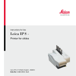
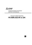
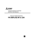
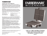
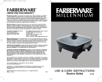
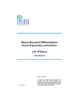
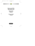
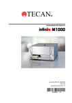
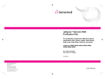
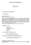
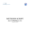
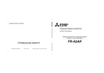
2
Immunohistochemical Detection With Quantum Dots
Rizwan S. Akhtar, Cecelia B. Latham, Dario Siniscalco, Carlo Fuccio,
and Kevin A. Roth
Summary
Quantum dot (QD) conjugates have many immunohistochemical applications. The
optical, excitation/emission, and photostable properties of QDs offer several advantages
over the use of chromogens or organic fluorophores in these applications. Here, we
describe the use of QD conjugates to detect primary antibody binding in fixed tissue
sections. We also describe the use of QDs in simultaneous and sequential multilabeling
procedures and in combination with enzyme-based signal amplification techniques. QD
conjugates expand the arsenal of the immunohistochemist and increase experimental
flexibility in many applications.
Key Words: Immunostaining; detection methods; multi-labeling; tyramide signal
amplification; fluorophore; antibody binding.
1. Introduction
Detection of biologically relevant molecules in human and animal fixedtissue sections is often accomplished by applying primary antibodies that bind
specifically to the antigen of interest followed by application of labeled secondary antibodies. Traditionally, the secondary antibodies used in immunohistochemical detection have been directly conjugated to organic fluorophores.
These labels are easily visualized using a suitably equipped fluorescence microscope. Alternatively, the secondary antibodies may not be labeled per se, but
rather conjugated to enzymes that catalyze the deposition of chromogenic or
fluorescent substrates. For example, horseradish peroxidase (HRP) or alkaline
phosphatase (AP)-conjugated secondary antibodies can be used to deposit
fluorescent or chromogenic substrates at and/or near the location of primary
antibody binding. In this manner, enzyme-based amplification can greatly
increase the sensitivity of immunohistochemical detection (1).
From: Methods in Molecular Biology, vol. 374: Quantum Dots: Applications in Biology
Edited by: M. P. Bruchez and C. Z. Hotz © Humana Press Inc., Totowa, NJ
11
12
Akhtar et al.
Quantum dot (QD) conjugates have recently become commercially available
(as Qdot® Conjugates from Quantum Dot Corporation, Hayward, CA) and can
be used in several immunohistochemical applications. These reagents offer
several advantages over typical fluorophores. First, many organic fluorophores
undergo a rapid and irreversible photobleaching. QDs photobleach minimally,
if at all, even with extended periods of viewing (2–5). Second, all QDs have a
similar excitation spectrum and can be excited in the near 360-nm range. This
feature, coupled with each QD narrow emission spectra, facilitates their use in
simultaneous multilabeling techniques (2,6). QD-based immunohistochemistry
can allow multilabeling visualization without the need for computer image
overlay or multiple custom filtersets, one or both of which is typically required
with organic fluorophores.
In this chapter, we describe how to use QD conjugates in several immunohistochemical applications. We demonstrate multilabeling QD protocols and how to
combine QD conjugates with tyramide signal amplification (TSA) for exquisitely
sensitive immunohistochemical detection. Finally, we describe how to combine
these techniques to accomplish simultaneous triple labeling. For completeness,
we begin with an overview of our tissue section preparation protocol.
2. Materials
2.1. Preparation of Tissue for Immunohistochemical Study
1. 10X Phosphate buffered saline (PBS): dissolve 80 g NaCl, 2.0 g KCl, 14.2 g
Na2HPO4, 2.0 g KH2PO4, and 0.1 g NaN3 in 1 L distilled H2O. Adjust pH to 7.2
with NaOH. Store 10X solution at room temperature. Dilute 10X using distilled
H2O for 1X PBS and store at room temperature.
2. 4% (w/v) Paraformaldehyde: dissolve 4 g paraformaldehyde (Electron
Microscopy Sciences, Fort Washington, PA; cat. no. 19200) in PBS, heat to
58–60°C, and add 1 N NaOH dropwise until solution is clear. Fill to 100 mL volume
with PBS and pH to 7.2. Use immediately. If solution is heated above 60°C, do not
use.
3. Bouin’s fixative: 750 mL saturated aqueous picric acid (Sigma, St. Louis, MO,
cat. no. P-6744), 250 mL formaldehyde solution (Fisher Scientific, Pittsburgh, PA,
cat. no. F75-1GAL), and 50 mL glacial acetic acid (Fisher Scientific, cat. no. A58500). Store at 4°C.
4. Tissue-freezing medium (VWR International, West Chester, PA, cat. no. 15148-031).
5. Slides: Snowcoat X-tra (Surgipath Medical Industries, Inc., Richmond, IL, cat. no.
15148-031).
6. Slide rack/staining dish: Wheaton 900200 (VWR International, cat. no. 25461003).
7. Antigen retrieval container: TPX staining jar (VWR International, cat. no. 25460907).
Immunohistochemistry and Quantum Dots
13
8. CitriSolv (Fisher Scientific, cat. no. 22-143975).
9. Isopropanol (Fisher Scientific, cat. no. HC-500-1GAL).
10. Citrate antigen retrieval buffer: working solution (10 mM citrate buffer, pH 6.0)
consists of 9 mL 0.1 M citric acid monohydrate solution (Sigma, cat. no. C-7129),
41 mL 0.1 M sodium citrate solution (Sigma, cat. no. S-4641), and 450 mL distilled H2O for a total of 500 mL. Store all solutions at 4°C.
11. Steamer: Farberware FRA500 (domestic rice cooker) or equivalent.
2.2. Detection of Single Antibody Immunoreactivity
With QD-Conjugated Secondary Antibody
1. Plastic slide folder (VWR International, cat. no. 48443-850).
2. Pap pen (Research Products International, Mount Prospect, IL; cat. no. 195504) was
used in this study. Quantum Dot Corporation recommends the use of the ImmEdge
Hydrophobic Barrier Pen (Vector Labs, Burlingame, CA; cat. no. H-4000).
3. PBS-blocking buffer (BB): dissolve 1.0 g bovine serum albumin (Fisher Scientific,
cat. no. BP1600-100), 0.2 g nonfat powdered skim milk (Carnation, Nestle,
Glendale, CA), and 0.3 mL Triton X-100 (Sigma, cat. no. T-8787) in 100 mL 1X
PBS. Store at 4°C.
4. Bisbenzimide (Hoescht 33,258) stock solution: prepare Hoescht 33,258 (Sigma,
cat. no. B-2883) at 2 mg/mL stored at 4°C. Dilute 1 μL in 10 mL PBS when applying to slides.
5. 90% (v/v) Glycerol in PBS (Fisher Scientific, cat. no. G33-1).
6. Microscope cover glasses: various sizes (VWR International, cat. no. 48393).
7. Optical fluorescence filters: usable excitation filters include 325 ± 25 nm (UV),
360 ± 40 nm (DAPI), 470 ± 40 nm (FITC), or 545 ± 30 nm (rhodamine). Usable
emission filters include 400 nm long pass, 450 ± 50 nm (DAPI), 535 ± 40 nm
(FITC), or 610 ± 75 nm (Cy3). Custom filtersets for QDs are available from
Chroma Technology (Rockingham, VT) or Omega Optical (Brattleboro, VT).
2.3. Simultaneous Detection of Two Antibodies Raised in Different
Species Using QD-Conjugated Secondary Antibodies
1. Mouse anti-MAP2 primary monoclonal antibody (Sigma, cat. no. M4403).
2. Rabbit anti-GFAP primary antiserum (Dako, Glostrup, Denmark, cat. no. Z334).
3. Quantum dot 565 anti-rabbit secondary antibody: QD 565 goat F(ab’)2 antirabbit IgG conjugate (Quantum Dot Corp., cat. no. 1143-1).
4. QD 605 anti-mouse secondary antibody: QD 605 goat F(ab’)2 anti-mouse IgG
conjugate (Quantum Dot Corp., cat. no. 1100-1).
2.4. Detection of Single Antibody Immunoreactivity by TSA
and a QD-Conjugated Streptavidin
1. 3% Hydrogen peroxide: dilute stock 30% hydrogen peroxide (Fisher Scientific,
cat. no. BP2633) in PBS at time of use. Store stock solution at 4°C.
14
Akhtar et al.
2. HRP-conjugated secondary antibody: peroxidase AffiniPure™ donkey anti-rabbit
IgG (cat. no. 711-035-152) and peroxidase AffiniPure donkey anti-mouse IgG
(cat. no. 715-035-151) both from Jackson ImmunoResearch Laboratories, West
Grove, PA.
3. TSA™ Biotin System (Perkin-Elmer Life Sciences Products, Boston, MA; cat. no.
NEL700).
4. QD 525 streptavidin conjugate (Quantum Dot Corp., cat. no. 1014-1).
5. QD 605 streptavidin conjugate (Quantum Dot Corp., cat. no. 1010-1).
3. Methods
3.1. Preparation of Tissue for Immunohistochemical Study
1. Sample tissue should be harvested and immediately fixed in a sufficient volume of
fixative, such as 4% paraformaldehyde, Bouin’s fixative, or methanol (see Note 1).
In general, overnight immersion fixation at 4°C is satisfactory; however, for some
antigens, perfusion fixation and/or special fixation procedures may be required for
optimal immunohistochemical detection.
2. Sample tissue can be prepared as either paraffin embedded or frozen sections.
Paraffin-embedded tissue sections require deparaffinization prior to use. Frozen
sections should be allowed to warm to room temperature (approx 10 min at room
temperature) prior to further processing. Go to step 6 for frozen slides.
3. For paraffin sections, deparaffinize sections by washing slides twice in CitriSolv,
first for 10 min and second for 5 min (see Notes 2–4). Then, rehydrate sections by
three washes with isopropanol for 5 min each. Then, wash slides in running tap
water for 5 min. Finally, wash slides in distilled water for 5 min.
4. To increase the immunohistochemical detection of some antigenic epitopes in
fixed tissue, antigen retrieval (AR) may be required (see Note 1). We typically use
a citric acid AR method. Perform AR by placing slides in an AR container containing citrate buffer. Place the container inside a steamer with an adequate amount
of distilled water and turn it on. Once steam can be seen exiting the steamer, continue AR for 20 min. Then, remove the slide container, place it on the lab bench,
and allow it to cool at room temperature for 20 min.
5. Remove slides from AR container and wash in running tap water for 5 min.
6. Incubate sections in PBS for 5 min at room temperature. Slides are now ready for
immunostaining.
3.2. Detection of Single Antibody Immunoreactivity
With QD-Conjugated Secondary Antibody
Direct immunofluorescence can be used to detect primary antibody binding in
fixed-tissue sections. Many protocols use organic fluorophore-conjugated secondary antibodies that recognize species-specific epitopes on primary antibodies.
QD-conjugated secondary antibodies can replace organic fluorophore-conjugated
secondary antibodies in a number of immunohistochemical applications. An
Immunohistochemistry and Quantum Dots
15
Fig. 1. Detection of single antibody immunoreactivity with quantum dot (QD)conjugated secondary antibody. (A–C) Immunostained section of postnatal day 7
mouse cerebellum. (A) Calbindin, a marker of Purkinje cells in the cerebellum, is
immunostained with a rabbit polyclonal antiserum, subsequently detected with QD
565-conjugated goat anti-rabbit secondary antibody and imaged with a 470 ± 40-nm
excitation filter and a 535 ± 40-nm emission filter. (B) Hoescht 33,258 counterstaining of nuclei is imaged with a 360 ± 40-nm excitation filter and a 450 ± 50-nm emission filter. (C) Calbindin and Hoescht 33,258 counterstaining is simultaneously
visualized with a 325 ± 25-nm excitation filter and a 400LP emission filter. Scale
bar = 40 μm.
example of single antibody immunoreactivity detection with QD-conjugated
secondary antibody is shown in Fig. 1.
1. Follow Subheading 3.1. to prepare slides (see Note 5).
2. Prepare a humidified chamber by cutting a paper towel in half lengthwise and
placing it in a plastic slide folder. Wet the paper towel with distilled water. The
paper towel should be damp but not completely saturated with water and should
be flat. When slides are placed in the folder, the underside of the slides should not
come into contact with the paper towel.
3. Quickly blot excess moisture from around the tissue on each slide with a paper
towel and encircle the tissue with a Pap pen or equivalent (see Note 6). Place the
slides in the humidified chamber and immediately add PBS-BB on top of the
tissue within the Pap pen boundary. One slide should be processed at a time and
care should be taken to avoid drying of the tissue.
4. To inhibit nonspecific antibody binding, incubate the slides in PBS-BB (blocking
buffer) for 30 min at room temperature. As mentioned in step 3, excess PBS-BB
can be added to individual slides one at a time. Alternatively, slides can be
processed in bulk by placing the slides in a slide rack and immersing the rack in a
staining dish containing PBS-BB.
5. Prepare primary antibody diluted in PBS-BB. Remove the PBS-BB from the slide
(see Note 7) and apply primary antibody. When applying any antibody or reagent
to slides, use a sufficient volume to cover the tissue (typically 50–200 μL).
16
Akhtar et al.
6. Incubate the slides in primary antibody for 24 h at 4°C (see Note 8). Incubation
can be shortened to 12 h at 4°C or to 60–90 min at room temperature. In general,
longer antibody incubations at lower temperatures result in better signal-to-noise
ratios in immunohistochemical detection.
7. Wash the slides three times in PBS for 5 min each (see Note 9).
8. Dilute QD-conjugated secondary antibody in PBS-BB at a 1:100 dilution. Because
the Pap pen may interfere with QD signals (7), remove the Pap pen from slides
before applying QD conjugates (see Note 6). Apply secondary antibody and incubate slides in a humidified chamber for 1 h at room temperature.
9. Wash the slides three times in PBS for 5 min each. If nuclear counterstaining is
desired, dilute stock bisbenzimide (Hoescht 33,258) solution in the PBS during
the second wash.
10. Cover slip slides in 90% glycerol in PBS (see Note 10).
11. View slides with a fluorescence microscope equipped with a 360 ± 40 nm (DAPI)
excitation filter and a 400-nm long pass emission filter (see Note 11) (see
Subheading 3.7. for information on optimal filterset selection).
3.3. Simultaneous Detection of Two Antibodies Raised in Different
Species Using Two QD-Conjugated Secondary Antibodies
There are several variations and extensions of the protocol in Subheading
3.2. that will allow the detection of multiple antigens in fixed tissue. Multiple
primary antibodies can be applied simultaneously to a single tissue section.
After an overnight incubation period, multiple secondary antibodies can then be
applied to detect primary antibody binding. As long as each primary antibody
is raised in a different species, this technique retains specificity and is very
straightforward. An example of double antibody immunoreactivity detection
with two QD-conjugated secondary antibodies is shown in Fig. 2.
1. Begin immunostaining as described in Subheading 3.2., steps 1–4.
2. Dilute multiple primary antibodies directed toward antigens of interest in the same
antibody cocktail. For example, one can make a cocktail containing:
a. 1 μL Mouse-derived anti-MAP2 antibody.
b. 1 μL Rabbit-derived anti-GFAP antiserum.
c. 198 μL PBS-BB.
For a 1:200 dilution of both primary antibodies.
3. Apply primary antibody cocktail to each slide and incubate overnight at 4°C.
4. Wash the slides three times in PBS for 5 min each. Remove Pap pen from slides
(see Note 6).
5. Prepare a single solution of QD-conjugated secondary antibodies diluted in QD
buffer at their respective optimal concentrations. For example, one can make the
following secondary antibody cocktail to detect the primary antibodies used in
step 2:
Immunohistochemistry and Quantum Dots
17
Fig. 2. Simultaneous detection of two antibodies, raised in different species, with
two quantum dot (QD)-conjugated secondary antibodies. (A–D) Immunostained
section of postnatal day 7 mouse brainstem. (A) Microtubule-associated protein-2 is
immunostained with a mouse monoclonal antibody, subsequently detected with QD
605-conjugated goat anti-mouse secondary antibody, and imaged with a 545 ± 30-nm
excitation filter and a 610 ± 75-nm emission filter. (B) Glial fibrillary-associated protein (GFAP) is immunostained with a rabbit polyclonal antiserum, subsequently
detected with QD 565-conjugated goat anti-rabbit secondary antibody, and imaged with
a 470 ± 40-nm excitation filter and a 535 ± 40-nm emission filter. (C) Hoescht 33,258
counterstaining of nuclei is imaged with a 360 ± 40-nm excitation filter and a 450 ± 50-nm
emission filter. (D) Merged image of MAP2, GFAP, and Hoescht 33,258 counterstaining. Scale bar = 40 μm.
18
Akhtar et al.
a. 1 μL of QD 605 conjugated goat-derived anti-mouse antibody.
b. 1 μL of QD 565 conjugated goat-derived anti-rabbit antibody.
c. 98 μL of QD buffer.
For a 1:100 dilution of both secondary antibodies.
6. Apply secondary antibody cocktail to each slide and incubate for 1 h at room
temperature.
7. Wash the slides three times in PBS for 5 min each. If nuclear counterstaining is
desired, dilute stock bisbenzimide (Hoescht 33,258) solution in the PBS during
the second wash.
8. Cover slip slides in 90% glycerol in PBS (see Note 10).
9. View slides with a fluorescence microscope equipped with a 360 ± 40-nm excitation filter and a 400-nm long pass emission filter, which will display both labeled
colors. Alternatively, use a narrow-band filter set for each of the QDs used and
view the signals individually (see Subheading 3.7. for information on optimal
filter set selection).
3.4. Detection of Single Antibody Immunoreactivity by TSA
and QD-Conjugated Streptavidin
QD streptavidin conjugates can be successfully incorporated in enzymebased signal amplification techniques, including TSA (4). Enzyme-based signal
amplification in immunohistochemical detection methods effectively increases
the number of fluorescent molecules at antigenic sites of interest (1). Although
several enzyme-based signal amplification methods are available, TSA is the
most versatile. TSA can greatly increase the sensitivity of immunohistochemical detection, thereby aiding the detection of antigens that are expressed at low
levels (8,9). Furthermore, signal amplification can increase the ability to detect
antigens within an autofluorescent or otherwise noisy sample. TSA is based on
HRP-catalyzed deposition of tyramide-conjugated molecules at sites of antibody binding (8,10,11). One can either deposit fluorophore-conjugated tyramide (direct TSA) or biotinylated tyramide with subsequent detection with a
fluorophore-labeled streptavidin (indirect TSA). Next, we outline a protocol in
which HRP-conjugated secondary antibody catalyzes the deposition of biotinylated tyramide at sites of primary antibody binding. Deposited biotin is then
detected with QD-conjugated streptavidin. Figure 3 depicts binding of two primary antibodies raised in different species, where one primary antibody is
detected by TSA and QD-conjugated streptavidin, and the other primary antibody is detected by QD-conjugated secondary antibody.
1. Follow Subheading 3.1. to prepare slides (see Note 5).
2. Incubate sections in 3% hydrogen peroxide in PBS for 5 min at room temperature.
Hydrogen peroxide will help destroy endogenous peroxidase activity in the tissue
that could otherwise lead to false-positive signals during the enzyme amplification.
Immunohistochemistry and Quantum Dots
19
Fig. 3. Sequential detection of two antibodies, raised in different species, with tyramide signal amplification (TSA) and quantum dot (QD)-conjugated streptavidin and
with QD-conjugated secondary antibody. (A–D) Immunostained section of embryonic
day 13 mouse telencephalon. (A) Musashi, a marker of neural precursor cells in the
ventricular zone, is immunostained with a rabbit polyclonal antiserum, subsequently
detected with TSA and QD 525-conjugated streptavidin, and imaged with a 470 ± 40-nm
excitation filter and a 535 ± 40-nm emission filter. (B) TuJ1, a marker of immature
neurons in the intermediate zone, is immunostained with a mouse monoclonal
antibody, subsequently detected with QD 605-conjugated goat anti-mouse secondary
antibody, and imaged with a 545 ± 30-nm excitation filter and a 610 ± 75-nm emission filter. (C) Hoescht 33,258 counterstaining of nuclei is imaged with a 360 ± 40-nm
excitation filter and a 450 ± 50-nm emission filter. (D) Musashi, TuJ1, and Hoescht
33,258 counterstaining is simultaneously visualized with a 325 ± 25-nm excitation
filter and a 400LP emission filter. Scale bar = 40 μm.
20
Akhtar et al.
3. Wash slides in running tap water for 5 min.
4. Wash sections in PBS for 5 min at room temperature.
5. For primary antibody application, follow Subheading 3.2., steps 2–7. Note that
when using TSA, the optimal primary antibody concentration is often dramatically
reduced compared with non-TSA immunohistochemistry detection techniques.
6. Dilute HRP-conjugated secondary antibody in PBS-BB. Apply 50–200 μL of
diluted HRP-conjugated secondary antibody to each slide and incubate for 1 h at
room temperature.
7. Wash the slides three times in PBS for 5 min each.
8. Dilute biotinylated tyramide in amplification buffer as recommended by the manufacturer. Apply biotinylated tyramide to each slide.
9. Gently rotate slides in the humidified chamber on an elliptical rotator (25 rpm) for
3–10 min at room temperature.
10. Wash the slides three times in PBS for 5 min each. Remove Pap pen from slides
(see Note 6).
11. Dilute QD streptavidin conjugate in QD buffer at a 1:400 dilution and incubate for
30 min at room temperature prior to use.
12. Apply 50–200 μL of diluted QD streptavidin conjugate to each slide and incubate
for 30 min at room temperature.
13. Wash the slides three times in PBS for 5 min each. If nuclear counterstaining is
desired, dilute stock bisbenzimide (Hoescht 33,258) solution in the PBS during
the second wash.
14. Cover slip slides in 90% glycerol in PBS.
15. View slides with a fluorescence microscope equipped with a 360 ± 40-nm (DAPI)
excitation filter and a 400-nm long pass emission filter (see Note 11) (see
Subheading 3.7. for information on optimal filter set selection.
3.5. Sequential Detection of Multiple Antibody Immunoreactivity
by TSA and QD-Conjugated Streptavidin and QD-Conjugated
Secondary Antibodies
The protocol described in Subheading 3.3. can only be used if the primary
antibodies employed are raised in different species. If the antibodies are raised
in the same species (e.g., the detection of two mouse-derived primary antibodies), special techniques can be used to retain specificity while using organic
fluorophores or QDs (11). Here we will describe a technique called “dilutional
neglect” using TSA and multiple QDs. The first primary antibody is detected
using TSA and QD-conjugated streptavidin, and the second primary antibody is
detected by QD-conjugated secondary antibody. Because TSA significantly
increases sensitivity, lower concentrations of primary antibody are required for
successful immunostaining. As long as the first antibody that is to be detected
with TSA and QD-conjugated streptavidin is used at a lower concentration than
that required for detection by QD-conjugated secondary antibodies, the detection method for the second primary antibody will “neglect” the first primary
Immunohistochemistry and Quantum Dots
21
Fig. 4. Sequential detection of two antibodies, raised in the same species, with tyramide signal amplification (TSA) and quantum dot (QD)-conjugated streptavidin and
with QD-conjugated secondary antibody. (A–D) Immunostained section of postnatal
day 7 mouse cerebellum. (A) Calbindin is immunostained with a rabbit polyclonal antiserum, subsequently detected with TSA and QD 605-conjugated streptavidin, and
imaged with a 545 ± 30-nm excitation filter and a 610 ± 75-nm emission filter. (B) Glial
fibrillary-associated protein (GFAP) is immunostained with a rabbit polyclonal
antiserum, subsequently detected with QD 565-conjugated goat anti-rabbit secondary
antibody, and imaged with a 470 ± 40-nm excitation filter and a 535 ± 40-nm emission
filter. (C) Hoescht 33,258 counterstaining of nuclei is imaged with a 360 ± 40-nm excitation filter and a 450 ± 50-nm emission filter. (D) Calbindin, GFAP, and Hoescht
33,258 counterstaining is simultaneously visualized with a 325 ± 25-nm excitation
filter and a 400LP emission filter. Scale bar = 40 μm.
22
Akhtar et al.
antibody and only indicate binding of the second primary antibody. QDs can
easily be integrated into this type of protocol, as outlined next. In Fig. 4, TSA
using QD-conjugated streptavidin is combined with QD secondary antibody to
perform double labeling where the two primary antibodies used are raised in the
same species.
1. Prepare slides as described in Subheading 3.1. (see Note 5).
2. Begin immunostaining as described in Subheading 3.4., steps 2–4. Next, complete Subheading 3.2., steps 2–4.
3. Prepare the first primary antibody diluted in PBS-BB. For example, we will use
rabbit derived anti-calbindin antiserum at a 1:10,000 dilution. At this dilution,
calbindin antiserum binding will not be detected by QD-conjugated secondary
antibody, which requires a 1:100 dilution. Apply primary antibody to each slide and
incubate overnight at 4°C. Do not apply the second primary antibody at this time.
4. Wash the slides three times in PBS for 5 min each.
5. Dilute HRP-conjugated secondary antibody in PBS-BB. Apply 50–200 μL of
diluted HRP-conjugated secondary antibody to each slide and incubate for 1 h at
room temperature.
6. Wash the slides three times in PBS for 5 min each.
7. Dilute biotinylated tyramide in amplification buffer as recommended by the manufacturer. Apply biotinylated tyramide to each slide.
8. Gently rotate slides in the humidified chamber on an elliptical rotator (25 rpm) for
10 min at room temperature.
9. Wash the slides three times in PBS for 5 min each.
10. Repeat Subheading 3.2., steps 2–4.
11. Prepare the second primary antibody diluted in PBS-BB. In our example, we will
use rabbit derived anti-GFAP antiserum at a 1:100 dilution. Apply primary antibody to each slide and incubate overnight at 4°C.
12. Wash the slides three times in PBS for 5 min each.
13. Prepare QD-conjugated secondary antibody and QD streptavidin conjugate in a
reagent cocktail at their respective concentrations. Be sure the QDs used emit fluorescence at different wavelengths. For example, the following will detect the
primary antibodies used in steps 3 and 11:
a. 1 μL of QD 605 streptavidin conjugate.
b. 4 μL of QD 565-conjugated goat-derived anti-rabbit antibody.
c. 395 μL of QD buffer.
For a 1:400 dilution of QD 605-conjugated streptavidin and a 1:100 dilution of
QD 565-conjugated goat derived anti-rabbit antibody.
14. Apply reagent cocktail to each slide and incubate for 1 h at room temperature.
15. Wash the slides three times in PBS for 5 min each. If nuclear counterstaining is
desired, dilute stock bisbenzimide (Hoescht 33,258) solution in the PBS during
the second wash.
16. Cover slip slides in 90% glycerol in PBS (see Note 10).
Immunohistochemistry and Quantum Dots
23
17. View slides with a fluorescence microscope equipped with a 360 ± 40-nm excitation filter and a 400-nm long pass emission filter, which will display both labeled
colors. Alternatively, use a narrow-band filter set for each of the QDs used and
view the signals individually (see Subheading 3.7. for information on optimal
filter set selection).
3.6. Triple Labeling Using QD-Conjugated Streptavidin and Multiple
QD-Conjugated Secondary Antibodies
In practice, several QD-conjugated secondary antibodies may be used to
simultaneously and specifically detect multiple primary antibody binding in a
given tissue sample, provided that all primary antibodies are raised in different
species. Therefore, the protocols in Subheadings 3.3. and 3.5. can be combined
to achieve triple labeling using QDs. Several primary antibodies, which will be
detected using different QD-conjugated secondary antibodies, should be prepared as an antibody cocktail and applied to slides. If enzyme-based signal
amplification is desired, it may be performed for one antibody before the use
of an antibody cocktail. An example of triple primary antibody immunoreactivity detection with a combination of the techniques described here is
depicted in Fig. 5.
3.7. Selecting Filter Sets for Use With QDs and Immunohistochemical
Detection
To successfully view QDs in immunohistochemical applications, the appropriate filter sets must be used. Investigators may misinterpret their results if
an incorrect or suboptimal filter set is used to view the staining. General
red/green/blue emission filters may be adequate to passage the narrow emission
wavelengths of QDs. It is recommended to use custom filter sets for QD viewing, but purchasing and maintaining these filters in addition to standard filter sets
may not be cost effective. Based on our observations, we discuss next several
aspects of filter set selection for immunohistochemical applications.
1. Quantum Dot Corporation recommends a 460-nm short-pass excitation filter, a
475-nm dichroic beamsplitter, and a narrow-band pass filter centered at the wavelength corresponding to the QD to be viewed. For example, a QD 605-conjugated
secondary antibody should be viewed with a 605 ± 20-nm narrow-band pass emission filter. Custom filtersets that include these filters are currently available from
Chroma Technology or Omega Optical.
2. QDs are optimally excited at wavelengths below 460 nm, and the use of a short
pass 460-nm filter will provide maximal excitation. No currently available QDs
emit light at or below 460 nm. However, several other dyes, such as DAPI and
Hoescht 33,258, emit fluorescence at or around 460 nm. If it is desired to use these
dyes concurrently with QDs, an ultraviolet excitation filter that does not exceed
24
Akhtar et al.
Immunohistochemistry and Quantum Dots
25
400 nm can be used. We have had good results using a 325 ± 25-nm or 360 ± 40-nm
excitation filter with QDs. Using a 460-nm short pass excitation filter would preclude the simultaneous visualization of QDs and fluorescent dyes such as DAPI or
Hoescht 33,258.
3. Conventional narrow-band filters for fluorophores, such as Cy3 or FITC, can
be used to visualize certain QDs. For example, a Cy3 filter set containing a
545 ± 30-nm excitation filter, a 570-nm long pass dichroic beamsplitter, and a
610 ± 75-nm emission filter would adequately allow the visualization of QD 605.
The 610 ± 75-nm emission filter, while not centered on the 605-nm wavelength,
would allow the passage of emitted light at 605 nm because of its relatively wide
bandwidth (75 nm). Furthermore, a FITC filter set containing a 470 ± 40-nm excitation filter, a 500-nm long pass dichroic beamsplitter, and a 525 ± 40-nm emission filter would transmit fluorescence from both QD 525 and 565. Therefore, an
investment in custom filter sets for QD immunohistochemistry is not absolutely
required. However, specific visualization of QD 565 would require a custom filterset because that wavelength does not correspond to any other commonly used
fluorophore. The custom filtersets are optimized for detection of QDs and can be
expected to produce the best signal-to-noise ratio.
4. If the simultaneous visualization of several QDs is desired, a long-pass emission
filter can be used. For example, QD 525, 565, and 605 can be simultaneously
visualized with a 470 ± 40-nm excitation filter, a 510-nm long-pass dichroic
beamsplitter, and a 520-nm long-pass emission filter. In this way, all three QDs
can be seen at the same time, and currently our laboratory uses a similar filter for
QD visualization. If a fourth fluorescent dye is to be visualized (such as DAPI or
Hoescht 33,258), one can use a 325 ± 35-nm excitation filter, a 370-nm long-pass
dichroic beamsplitter, and a 400-nm long-pass emission filter. When using longpass emission filters, there may be a decrease in the signal-to-noise ratio because
Fig. 5. Sequential detection of three antibodies with tyramide signal amplification
(TSA) and quantum dot (QD)-conjugated streptavidin and with two QD-conjugated
secondary antibodies. (A–E) Immunostained section of adult mouse brain. (A) NeuN,
a marker of mature neurons, is immunostained with a mouse monoclonal antibody, subsequently detected with TSA and QD 525-conjugated streptavidin, and imaged with a
470 ± 40-nm excitation filter and a 535 ± 40-nm emission filter. (B) Glial fibrillaryassociated protein (GFAP) is immunostained with a rabbit polyclonal antiserum, subsequently detected with QD 565-conjugated donkey anti-rabbit secondary antibody, and
imaged with a 470 ± 40-nm excitation filter and a 535 ± 40-nm emission filter. (C)
Microtubule-associated protein-2 (MAP2) is immunostained with a mouse monoclonal
antibody, subsequently detected with QD 605-conjugated goat anti-mouse secondary
antibody, and imaged with a 545 ± 30-nm excitation filter and a 610 ± 75-nm emission
filter. (D) Hoescht 33,258 counterstaining of nuclei is imaged with a 360 ± 40-nm excitation filter and a 450 ± 50-nm emission filter. (E) Merged image of NeuN, GFAP,
MAP2, and Hoescht 33,258 counterstaining. Scale bar = 40 μm.
26
Akhtar et al.
of the additional wavelengths of light transmitted, thereby leading to increased
visible autofluorescence.
4. Notes
1. The protocols outlined here are robust in our laboratory and should be used as
starting points for the development of protocols in other laboratories. There are a
large number of potential fixatives, and we recommend that several fixatives be
tried during protocol optimization. The particular fixative used is selected based
on the tissue to be immunostained, the requirement for perfusion, the stability or
availability of the antigen to be detected, and the characteristics of the antibody to
be used. Furthermore, there are also many methods of performing antigen
retrieval, and we recommend that several be tried for each antigen and tissue.
2. Washing slides can best be accomplished by placing slides in a suitable glass slide
rack that will fit inside a rectangular staining dish and transferring the entire rack
from one wash solution to another. Transferring individual slides between various
solutions for washing is typically not necessary. Also, slides can be washed by
removing the entire slide rack from the staining dish, quickly emptying the dish
into the sink, replacing the rack in the dish, and rapidly pouring a new solution in
the dish. Care should be taken to minimize the time slides are exposed to air.
3. We recommend establishing a “deparrafinization station” to be used by several
investigators in a laboratory. Use two staining dishes that contain CitriSolv and
three that contain isopropanol. Use each dish sequentially to perform deparrafinization and rehydration. After several rounds of use, replace dish 1 with dish
2, and dish 2 with dish 3. Then, clean dish 1, fill with solution, and use as dish 2
(CitriSolv) or 3 (isopropanol).
4. At no time should the tissue on a slide be allowed to dry. Dried tissue is perhaps
the most frequent cause of suboptimal or uninterpretable staining. The tissue must
remain moist at all times. To ensure that slides remain wet, they must be quickly
transferred between containers during washes. Also, the slides should be inspected
periodically during incubations with antibody or detection reagents to ensure that
solution adequately covers the tissue. Even partial or temporary dryness may lead
to nonspecific absorption of reagents to the tissue resulting in high background
and/or nonspecific signals.
5. When performing any type of immunohistochemistry, control slides should be run
in parallel that do not receive primary antibody but do receive all subsequent
detection reagents. These negative control slides will illustrate any background or
false-positive signals generated by the detection method that is independent of primary antibody binding. Some tissue has significant autofluorescent material that
will be evident in these negative control slides. In addition, we have found that if
used at inappropriately high concentrations, QD reagents may nonspecifically
label neurons (4). This nonspecific labeling will also be evident in negative control slides, and will allow for better interpretation of truly labeled slides.
Nonimmune serum controls or preabsorbed primary antibody controls are also
useful for interpreting immunohistochemical results.
Immunohistochemistry and Quantum Dots
27
6. Pap pens are very useful to create a boundary on a slide to prevent the lateral
movement of solution from capillary action. This way, a smaller volume of antibody/
reagent can be used to cover tissue completely. However, Pap pen may interfere with
QD signals and photostability (7), and we recommend removing Pap pen prior to the
addition of QD reagents. Removal is accomplished by using a damp paper towel or
a fingernail. Pap pen is not absolutely required and a grease pencil, scoring with a
diamond tip, or an aerosolized hydrocarbon coating around the tissue can alternatively be used with similar results. Quantum Dot Corporation suggests the use of the
ImmEdge Pap pen from Vector Labs to minimize interference with signal intensity.
7. To remove antibody or reagent solutions from a slide, firmly tap the slide on its
side onto the lab bench. Then, use a folded dry paper towel to dab over the Pap
pen boundary. There should be no wetness “over” the boundary. Otherwise,
solution that is added within the boundary will easily pass to the other side via
capillary action. This process must be done quickly and carefully to ensure the tissue section does not dry or become scratched or damaged.
8. While slides are in the humidified chamber (slide folder), ensure that the folder
remains as horizontal as possible. Be sure that any refrigerator shelves that are
used to house slides overnight are level, so that slides remain adequately wet.
Also, when removing slides from the chamber for washing, the underside of the
slide folder “leaves” should be inspected and washed if any residual antibody or
reagent is present. The slide folder can then be rinsed and reused for the next step
of immunostaining.
9. When washing slides, it is important to use large volumes of fresh PBS. One can
also agitate slides at 100 rpm on a rotator for better washing.
10. Slides that are cover slipped with 90% glycerol in PBS may have undesirable air
bubbles beneath the cover slip. To overcome this problem, the cover slip should be
gently removed and reapplied until the air bubbles are either eliminated or are not
over any pieces of tissue.
11. QD fluorescence may require a brief period of illumination prior to viewing to
maximize their signal-to-noise ratio. The immunostained slide should be placed in
the light path of the microscope and for 2–4 min for the specific fluorescence to
become optimally apparent.
Acknowledgments
We would like to thank all the members of the Roth laboratory for their
insights and assistance. These protocols were developed in part by support from
NIH grants NS35107 and NS41962. RSA received support from the UAB
Medical Scientist Training Program (NIH grant 08361).
References
1. Roth, K. A. and Baskin, D. G. (2005) Enzyme-based fluorescence amplification for
immunohistochemistry and in situ hybridization. In: Molecular Morphology in
Human Tissues: Techniques and Applications, (Tubbs, R. R. and Hacker, G. W.,
eds.), CRC Press, Washington, D.C., pp. 65–80.
28
Akhtar et al.
2. Bruchez, M., Jr., Moronne, M., Gin, P., Weiss, S., and Alivisatos, A. P. (1998)
Semiconductor nanocrystals as fluorescent biological labels. Science 281,
2013–2016.
3. Wu, X., Liu, H., Liu, J., et al. (2003) Immunofluorescent labeling of cancer marker
Her2 and other cellular targets with semiconductor quantum dots. Nat. Biotechnol.
21, 41–46.
4. Ness, J. M., Akhtar, R. S., Latham, C. B., and Roth, K. A. (2003) Combined tyramide
signal amplification and quantum dots for sensitive and photostable immunofluorescence detection. J. Histochem. Cytochem. 51, 981–987.
5. Watson, A., Wu, X., and Bruchez, M. (2003) Lighting up cells with quantum dots.
BioTechniques 34, 296–303.
6. Jaiswal, J. K., Mattoussi, H., Mauro, J. M., and Simon, S. M. (2003) Long-term
multiple color imaging of live cells using quantum dot bioconjugates. Nat.
Biotechnol. 21, 47–51.
7. Qdot Streptavidin Conjugates User Manual. (2005) Molecular Probes, Inc., Eugene,
OR, pp. 1–14. http://probes.invitrogen.com/media/pis/mp19000.pdf.
8. Bobrow, M. N., Litt, G. J., Shaughnessy, K. J., Mayer, P. C., and Conlon, J. (1992)
The use of catalyzed reporter deposition as a means of signal amplification in a
variety of formats. J. Immunol. Methods 150, 145–149.
9. van Gijlswijk, R. P., Zijlmans, H. J., Wiegant, J., et al. (1997) Fluorochromelabeled tyramides: use in immunocytochemistry and fluorescence in situ hybridization. J. Histochem. Cytochem. 45, 375–382.
10. Bobrow, M. N., Harris, T. D., Shaughnessy, K. J., and Litt, G. J. (1989) Catalyzed
reporter deposition, a novel method of signal amplification. Application to
immunoassays. J. Immunol. Methods 125, 279–285.
11. Shindler, K. S. and Roth, K. A. (1996) Double immunofluorescent staining using
two unconjugated primary antisera raised in the same species. J. Histochem.
Cytochem. 44, 1331–1335.