1
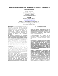
myDAQ Biomedical Instrumentation Board SD May1212 Design Document Team: Jeremy Bruecken Kossi Sessou Alex Weiss Advisor: Mani Mina Client: Andrew Watchorn Contents 1. 2. 3. 4. Introduction .......................................................................................................................................... 3 1.1 Executive Summary ....................................................................................................................... 3 1.2 Project Goal................................................................................................................................... 3 1.3 Operating Environment ................................................................................................................ 3 1.4 Limitations..................................................................................................................................... 3 1.5 Deliverables................................................................................................................................... 3 Concept ................................................................................................................................................. 4 2.1 Concept Sketch ............................................................................................................................. 4 2.2 Functional Specifications .............................................................................................................. 4 2.3 Non Functional Requirements ...................................................................................................... 4 System Description ............................................................................................................................... 4 3.2 Filter and Amplification Board ...................................................................................................... 6 3.3 LabView Computer Interface ........................................................................................................ 6 Functional Decomposition .................................................................................................................... 6 4.1 4.1.1 Amplification ......................................................................................................................... 6 4.1.2 Filtering ................................................................................................................................. 7 4.2 Amplification ......................................................................................................................... 7 4.2.2 Filtering ................................................................................................................................. 7 LabView Interface Design.............................................................................................................. 8 Testing and Evaluation .......................................................................................................................... 9 5.1 Safety Testing ................................................................................................................................ 9 5.2 Signal Quality ................................................................................................................................ 9 5.2.1 Requirements ........................................................................................................................ 9 5.2.2 Lab Testing ............................................................................................................................ 9 5.2.3 LabVIEW Testing ................................................................................................................. 10 5.3 6. EMG Signal Conditioning............................................................................................................... 7 4.2.1 4.3 5. ECG Signal Conditioning ................................................................................................................ 6 System Functionality ................................................................................................................... 11 Standards ............................................................................................................................................ 11 6.1 Explicit Standards ........................................................................................................................ 11 6.1.1 Safety .................................................................................................................................. 11 SDMay1212 Design Document Page 1 6.1.2 6.2 EKG Filtering ........................................................................................................................ 11 Implicit Standards ....................................................................................................................... 12 6.2.1 Portability ............................................................................................................................ 12 6.2.2 User Interface...................................................................................................................... 12 6.3 Standards Interactions ................................................................................................................ 12 6.4 Meeting Standards ...................................................................................................................... 12 Appendix I: User Manual............................................................................................................................. 13 Introduction ............................................................................................................................................ 13 Installing LabVIEW .................................................................................................................................. 13 Making Connections ............................................................................................................................... 13 Performing Measurements ..................................................................................................................... 15 SDMay1212 Design Document Page 2 1. Introduction 1.1 Executive Summary National Instruments requires a board that plugs into their product, the myDaq, with the necessary components to perform electromyography and read a pulse. The functionality should be comparable to the functions covered by the BioPac in an introductory biomedical engineering course at a 40x lower price with standard safety requirements. 1.2 Project Goal The goal of this project is to create a system that will connect the human body to the myDAQ for biomedical instrumentation. The system will be able to create accurate, useable signals to the myDAQ from electrocardiogram and electromyography. Additionally, LabView software to measure and analyze the signals will be included. To make this product competitive on the market, it should be at a 40x lower production price than the current selling price of the BioPac. This product will expand National Instrument’s educational market offering. 1.3 Operating Environment The system should operate in a wide range of environments. This includes the standard commercial operating range of 00 C to 700 C. Additionally, the system must be robust in ability to plug and unplug sensors, travel in a backpack, and protect the end user from unsafe electrical exposure. 1.4 Limitations The limitations of this project are time and experience. There is a limited amount of time to develop a system that will compete with a mature product, the BioPac. Additionally, the team consists of undergraduate students with very little industry experience. These limitations indicate this product will most likely be a test of concept and provide the base for a fully developed system that will compete in the market. Finally, there are strong limitations set by cost and safety. The system must protect the end user and meet an extremely low price point. 1.5 Deliverables At the end of our project term the client, National Instruments, would like a working prototype board that plugs directly into the myDAQ. LabView software to acquire and analyze signals will be included with the board. Finally, the submission will include a design document, specifications, and test. SDMay1212 Design Document Page 3 2. Concept 2.1 Concept Sketch In Figure 1 it can be seen that the ECG/EMG sensors will be run from the myDaq biomedical instrumentation board. This board will amplify and filter the signals coming in and protect the end user. Figure 1 – A systems level view of the biomedical engineering package for the myDaq 2.2 Functional Specifications 2.3 Gain EMG and EKG signals by ~2000 Eliminate DC component of EKG, EMG signals Protect end user from up to 5000V signal Provide display of signals through LabView Maximum power consumption of 500mW Non Functional Requirements User friendly LabView interface Robust modular connection design 40x lower price than the BioPac 3. System Description The description can be broken down into three main parts: the sensors, filter and amplification board, and LabView computer interface. Each of these components play a vital role in the functional system. SDMay1212 Design Document Page 4 Figure 3.1 – System description flow diagram Figure 3.2 – Physical view of the system The hardware set up will be simillar to Figure 3.2. In this test set up, we use a lab kit board to place an operational amplifier (741) and resistors to provide a gain of about -2. The input signal will be provided by the sensors as opposed to this case where the myDaq is used to generate an arbitrary sinusoidal signal through the analog output port (AO1). The measurement is then done by using the analog input port (AI1). SDMay1212 Design Document Page 5 3.1 Sensors The sensors provide the physical connection from the human body to the rest of the system. There are two ECG/EMG sensors which detect potential differences in the skin for amplfication. A final pad will provide a ground connection for clearer signal readings. These are simple connection pads whose conductivity may be improved by the use of a conductive great at the point of contact. All electrical connections to a human test subject will be run through a Human Protection Circuit (HPS) to ensure safety in the event of an overvoltage fault. 3.2 Filter and Amplification Board This board consists of three main areas: safety, amplification, and filtering. The safety blocks are circuit elements designed to protect against potentially harmful signals being applied to users. These are designed to meet regulation. Amplification takes place through two amplifier blocks: a differential amplifier and an op amp stage to gain up the potential between the two ECG/EMG sensor pads. Finally, filters located after the amplifiers eliminate interference in the signal as preparation for processing. 3.3 LabView Computer Interface The LabView interface provides an interface for each of the three signal capture modes: ECG, EMG, and pulse oximeter. By selecting one of these modes and plugging in the corresponding sensor a user will be able to perform stimulus, sample, and analysis operations depending on the operation mode. ECG will have a sample and analysis mode in which the input signal will be captured an then analyzed to determine the test subject’s heartbeat in real time. Similarly, an EMG requires sampling of the EMG sensors. Once again, analysis may be performed on the signal to determine the nature of the tested muscle. Finally, the pulse oximeter will concurrently stimulate the LED inputs and read the photodector output to collect data for analysis. The nature of the pulse oximeter makes analysis a much more involved task. 4. Functional Decomposition 4.1 ECG Signal Conditioning The human body controls muscles through electrical signals. One of the most important signals is that of the heart. ECG signals are commonly measured and well understood. The American Association of Medical Instrumentation (AAMI) and American National Standards Institute (ANSI) have developed standards on what filters should be applied as well as how the signal should be captured. 4.1.1 Amplification The human body generates ECG signals at with amplitude of about 1mV. Figure 4.1.1 demonstrates the basic circuit design used to capture this signal. This circuit drives the right leg to ground via and OPA2131. Provisions for protection against signals picked up in SDMay1212 Design Document Page 6 the wires connecting to the human are included with the shield driven by another OPA2131. Finally, the captured signal is gained up by the INA121 instrumentation amplifier. To achieve a large enough output signal there will be a second gain stage of 200 times shown as part of the filter circuit in Figure 4.1.2. Figure 4.1.1 – Burr-Brown INA121 Application Note 4.1.2 Filtering Once the signal is acquired from the body it must be properly filtered to eliminate noise that may have been gained up in the first stage. A human heart rate is typically within the range from 40 – 200 beats per minute. This translates to a range of .67 – 3.33 Hz. The high pass filter will eliminate the direct current (DC) component of the signal while preserving the desired heart rate signal. Additionally, a low pass 40Hz RC filter will be implemented following the gain stage to reduce high frequency noise and eliminate a common source of interference: 60Hz power. Rather than increase costs and complexity with a physical filter, the signal will be processes through a digital filter in LabVIEW® to remove excess noise. 4.2 EMG Signal Conditioning The human body controls muscle movement with electrical signals. Sensors place on the skin with proper conductivity can pick up these signals for analysis. To properly view the signals generated, analog circuits must gain up the signal and remove excess noise. 4.2.1 Amplification EMG signals are generally in the range of 1mV to 5mV. The same type of signal acquisition circuit described in Figure 3.1.1 can be used to capture the EMG signals. The second gain stage will provide a total gain of ~2000, which will put the output signal well within the myDAQ acquisition range of 1-15V. 4.2.2 Filtering The signals generated by skeletal muscles are often in the 100 – 500Hz range. These would be filtered out by the circuit prescribed for the EKG measurement. For this reason, a separate filtering system must be implemented. This system will provide a band pass filter with a range of .67 – 1000Hz. SDMay1212 Design Document Page 7 4.3 LabView Interface Design The user interface will consist of three main menus: ECG, EMG and Pulse Oximeter. In each menu a start, stop and pause buttons will be implemented for the control along with graph or numerical display depending on the test being performed. The signal processing will be done by using the Daq Assistant that allows an access to the myDaq I/O as shown in Figure 4.3.1 below that is associated with the setup of Figure 3.2. Figure 4.3.1 – Block diagram of a sample signal acquisition and processing in LabView The user interface of the above block diagram will allow the user to monitor the signal and display the result of the different computations. In Figure 4.4.2, the input and output are displayed with their respective amplitudes and peak-to-peak values. The gain is calculated by taking the ratio of output over the input and the result is displayed. SDMay1212 Design Document Page 8 Figure 4.4.1 –User interface associated with the block diagram of Figure 4.3.1 5. Testing and Evaluation Testing will be performed in three separate areas: safety, signal quality, and system functionality. First and foremost, the safety circuitry must meet IEC specifications to protect the end user. Tests to ensure proper safety will be determined when the specifications are acquired. Next, we will test for signal quality through our gain and filtering stages. Finally, the system will be tested as a whole to ensure entire system functionality. 5.1 Safety Testing Safety testing will include applying large transients to the circuit’s computer connection and viewing the signal that is transmitted to the human connections. Based on IEC specifications, the safety circuitry will be tested to protect the end user from large transients. However, applying very large transients will most likely destroy the circuitry. Therefore our tests swept the voltage at the connection to the human body. It was observed that any increase in voltage above around .7V required a current not supported by the connections upstream. 5.2 Signal Quality 5.2.1 Requirements Signal quality will be tested by performing signal analysis with a human connected. The system must be able to filter and gain a recognizable ECG signal. It must also provide a responsive output to muscle movements in the EMG mode. 5.2.2 Lab Testing Tests were carried out on multiple individuals with the lab equipment in 2046 Coover. It was Figure 5.2.2 contains a sample of the acquired signal. The single capacitor on the output was able to eliminate the DC signal with some slight attenuation of the AC component. However, it is clear that the pulse signals can easily be extracted for analysis. SDMay1212 Design Document Page 9 Since the signal filtering is performed in the digital domain, EMG signals are easily captured with this circuitry. Figure 5.2.2 – Signal capture during testing of EKG measurement 5.2.3 LabVIEW Testing Next the board was connected to the myDAQ and tested for the ability to capture and analyze signals provided by the human body. Figure 5.2.3 contains the capture signal in the LabVIEW interface. SDMay1212 Design Document Page 10 Figure 5.2.3 – LabVIEW signal capture 5.3 System Functionality The complete system should meet all the functional specification described in Section 4. It should be able to perform measurements at the request of the user and display the result in form of graph or numbers depending on the task. After ensuring the quality of the signal this was a simple test of interface functionality. The user interface provided the information regarding pulse and EMG signal representation. 6. Standards 6.1 Explicit Standards 6.1.1 Safety Several standards were applicable to this myDAQ biomedical acquisition add-on board project. First and foremost are the safety standards set by International Elecrotechnical Commission. Since this is an educational device not used in a medical environment it is subject to the less stringent standards that were met with our relatively simple Human Protection Circuitry (HPS). 6.1.2 EKG Filtering The American Association of Medical Instrumentation (AAMI) and American National Standards Institute (ANSI) have established the filtering requirements for measuring an electrical pulse signal. These proved helpful to our design as they have already been optimized for eliminating noise and achieving a high quality signal. SDMay1212 Design Document Page 11 6.2 Implicit Standards 6.2.1 Portability The add-on board is a compliment to the myDAQ which is inherently a student friendly portable device. To be an appropriate complement our device was required to have similarly robust mechanical characteristics and portable size. 6.2.2 User Interface The user interface is something difficult to standardize. New designs are always coming out that provide a more intuitive and simple interface. However, the standard lies in the constant search for improvement. It is standard that the interface requires minimum explanation to use and does not include unnecessary complications. 6.3 Standards Interactions Unfortunately, sometimes standards interact and result in conflicts. Such standards as safety and portability may not always be synonymous. In this situation it is important to have an understanding of each standards implication. This is as much a practical issue as an ethical one. Obviously, user safety must always come first in the implementation of standards. Fortunately, we were not faced complications in our standards interactions. 6.4 Meeting Standards The standards defined in the previous sections were met through careful research and thoughtful design. Standards are set to help simplify and improve the design process. They prevent the repetition of mistakes and set the bar for future designs. The process of researching standards and implementing them in design work is fundamental to the engineering discipline. Through this work we can better understand and perform this process. SDMay1212 Design Document Page 12 Appendix I: User Manual myDAQ Biomedical Instrumentation Manual National Instruments Introduction This manual will walk you through the steps to perform biomedical measurements with the myDAQ via an add-on board. In the essence of safety, only healthy individuals should be subjected to these measurements. They should by no means be used as diagnostic information. The designers of the addon board have taken precautionary steps to prevent any possible injury to the subject. However, by performing these measurements the end-user takes full responsibility for any injuries that may arrive from the usage of this device. Installing LabVIEW Follow the instructions provided with the myDAQ kit to install the accompanying version of LabVIEW on the computer that will be used to perform measurements. After this is complete an additional package for the add-on board will need to be installed. It should come packaged with the device; however, it may also be available on the LabVIEW website supported by National Instruments. This software will allow measurements to be taken and analyzed with just a few clicks. Making Connections First the myDAQ will need to be connected to the computer with LabVIEW installed via the USB cable provided. Instructions for this procedure are included in the myDAQ documentation. Next the biomedical instrumentation add-on board will plug in to the data bus on the side of the myDAQ. Press firmly until it is fully inserted. Figures 1 and 2 provide visual detail for these steps. Now the user will be connected. Place the electrodes provided in the kit on the subject to be tested. The back film peels off and has an adhesive with conducting gel to provide a strong connection. There are three locations: the right arm, left arm, and right leg as indicated by Figure 3. The red wires clamp on to the metal tie of the electrode at either arm and the black wire connects to the right leg. Remember, the quality of the measurements relies significantly on these connects. Make sure the pads are pressed firmly down on the skin and that the subject remains still throughout testing. Now the other end of the cable may be plugged into the add-on board. The fully connected device is seen in Figure 4. SDMay1212 Design Document Page 13 Figure 2: The board aligned with the myDAQ Figure 3: The board plugged into the myDAQ SDMay1212 Design Document Page 14 Figure 4: Connection locations Figure 5: Fully connected device Performing Measurements Simply run the LabVIEW file provided with the add-on board to begin. This will setup the myDAQ to perform measurements and open the interface in Figure 5. Click Acquire ECG Signal to begin the signal acquisition. Note this is the same button used for EMG signal capture. After acquiring the signal simply click the ECG Feature Extraction to obtain a real time heart rate measurement from the signal. SDMay1212 Design Document Page 15 Figure 6: LabVIEW user interface SDMay1212 Design Document Page 16