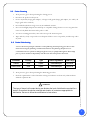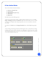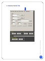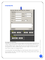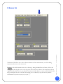
1
SeeMoreTM USB Probes
We revolutionize imaging devices.
© Copyright Interson Corporation all rights reserved 2009 - 2010. No part of this
manual or software may be reproduced in any form or by any means, electronic or
mechanical, including but not limited to, photocopying, recording, or by any
information storage and retrieval systems, without the express written consent of
Interson.
Rev: 6-15-10
Windows is a trademark of Microsoft Corporation.
Equipment Manufacturer
Interson Corporation
7026 Koll Center Parkway, Suite 201
Pleasanton, CA 94566 USA.
Ph o n e: 92 5. 4 62 . 49 4 8
800.766.4664
Fax:
925.462.4833
Email: [email protected]
www.interson.com
EC Representative
MediTech
Maastrichterlaan 127
NL-6291 EN Vaals
Net h e rl an d s
Phone: +31.43.306.3320
Fax: +31.43.306.3338
0197
This user guide is applicable to but not limited to the following probes:
3.5 MHz General Purpose (GP/AB)
5.0 MHz General Purpose (GP/AB)
7.5 MHz Small Parts (SP/PI)
7.5 MHz Endocavity (EC/EB)
7.5 MHz Vascular (SR/VA)
12.0 MHz Endo-rectal (ER/ES)
12.0 MHz Micro Vascular (MV)
12.0 MHz Ophthalmic (OP)
SEEMORE™ USER GUIDE
Table of Contents
I. INTRODUCTION ............................................................................................................ 2
A. MINIMUM SYSTEM REQUIREMENTS ..................................................................................... 2
B. WARNINGS, SAFETY INFORMATION .................................................................................... 2
1.1 MEANING OF SIGNAL WORDS .......................................................................................... 2
1.2 MEANING OF SAFETY SYMBOL.......................................................................................... 3
1.3 GENERAL CAUTIONS AND WARNINGS ................................................................................ 3
1.4 OPERATOR QUALIFICATIONS .......................................................................................... 4
D. CARE AND HANDLING OF PROBES ...................................................................................... 4
2.1 CLEANING AND DISINFECTION .................................................................................4
2.2 PROBE CLEANING ...............................................................................................5
2.3 PROBE DISINFECTING ...........................................................................................5
2.4 SURFACE CLEANING ..................................................................................................... 6
2.5 SURFACE DISINFECTION ................................................................................................. 6
E. ACOUSTIC ENERGY........................................................................................................ 7
F. ELECTROMAGNETIC COMPATIBILITY (EMC) .......................................................................... 7
G. PRESCRIPTION DEVICE STATEMENT .................................................................................... 7
H. TRAINING .................................................................................................................. 7
I. GENERAL .................................................................................................................... 7
II. SEEMORETM DISPLAY .............................................................................................8
III.IMAGING WINDOW ...............................................................................................9
IV. USER INTERFACE WINDOW .................................................................................. 10
A.PREFERENCES TAB (PREF. TAB) ......................................................................................... 11
B. NEW EXAM TAB .......................................................................................................... 13
C. AUTOSCAN TAB .......................................................................................................... 14
D. ADVANCED SCAN TAB (ADV. SCAN) .................................................................................. 16
E. MEASURE TAB ............................................................................................................ 18
A. SAVING AND VIEWING IMAGES......................................................................................... 20
B. ELECTROMAGNETIC COMPATIBILITY .................................................................................. 22
C.STORAGE AND TRANSPORTATION ............................................................................ 24
D.TRANSPORTATION .............................................................................................. 24
E.CARE OF THE USB PROBE ...................................................................................... 24
F.DISPOSAL.......................................................................................................... 25
APPENDIX A - SEEMORETM SOFTWARE INSTALLATION GUIDE ................................................ 26
APPENDIX B - INTERSON PROBE SYSTEM SPECIFICATIONS ……………………………….......... 28
APPENDIX C- INTERSON PROBES AND THEIR APPLICATIONS …………………………….......... 29
APPENDIX D - SUMMARY OF THE ACOUSTIC QUANTITIES ……………………………….......... 31
APPENDIX E -INTERSON PROBES INDICATIONS FOR USE.. ………………………………...........37
APPENDIX F -PATIENT PRIVACY AND CONFIDENTIALITY ………………………………............40
SeeMore™ User Guide | 01-9911-00 REV 1
V. SCANNING ....................................................................................................... 20
1
I.
Introduction
Congratulations on your purchase of the Interson USB Ultrasound Imaging Probe, the ultrasound imaging probe that
plugs into the USB port of your computer. Please review this user guide before you begin scanning. Contact
Interson or your sales representative if you have any questions. Note: There is also an on-line Help guide that
explains basic functions that is built into the SeeMoreTM user software.
Note: The sale of this item is subject to regulation by the U.S. Food and Drug Administration and state and local
regulatory agencies.
A . Minimum System Requirements
•
•
•
•
•
Computer Operating System – Windows XP, Windows 7, Vista, or Mac (running Windows)
Minimum processor – 1 GHz
Minimum RAM – 512 MB RAM (recommend 1 GB or more)
USB 2.0 port
Minimum display – 1024 X 600 resolution with 32bit color
B . Warnings, Safety Information
1.1 Meaning of Signal Words
In this users manual, the signal words “Warning” and “Caution” are used regarding safety and important instructions.
These signal words and their meanings are as follows. All users of the Interson Ultrasound Probe system must
understand the meanings of these signal words.
Meaning
!
WARNING
Indicates an potentially hazardous situation, which if not avoided could cause injury or
harm to the equipment.
!
CAUTION
Indicates a potentially hazardous situation which of not avoided, may result in minor
injury or harm to the equipment.
CAUTION
Indicates a potentially hazardous situation which if not avoided, may result in property
damage.
Type BF Equipment
SeeMore™ User Guide | 01-9911-00 REV 1
Signal Word
2
1.2
Meaning of Safety Symbol
!
1.3
“Attention” (refer to Users Manual.)
General Cautions and Warnings
!
CAUTION
Probes must be cleaned after each use. Cleaning the probe is an essential step prior to
effective disinfection. Follow the manufacturer’s instructions when using disinfectants.
!
WARNING
Do not allow sharp objects, such as scalpels or cauterizing knives, to touch probes
or cable.
!
WARNING
Equipment not suitable for use in the presence of flammable mixtures
WARNING
If the probe is used with other devices, current leakage may increase and electric shock
may be caused. It is the user’s responsibility to ensure safety when the probe is to be
used with other devices. If safety cannot be ensured, use of the probe with other devices
is not allowed.
!
WARNING
The use of a “Non-Medical” grade AC Adapter could potentially cause harm to the system,
the probe, the operator and/or the patient.
SeeMore™ User Guide | 01-9911-00 REV 1
!
3
1.4
Operator Q ualifications
The medical professional operating the USB ultrasound probe system must have a general knowledge of the use of
ultrasound imaging devices and imaging protocols.
Do not plug into the Ultrasound Probes until the Software has been installed. See software installation
guide (Appendix I, at the end of this document.)
Do not use in the presence of flammable anesthetics or other flammable materials.
Interson probes use very low acoustic power output, and ultrasound imaging has been found, in many
studies, to be safe when used correctly. However, as with all medical procedures, risks and benefits must
be weighed. It is important to use the lowest power settings and the shortest scan times possible while
attaining the needed clinical information.
D. Care and Handling of Probes
Although Interson probes are very durable, reasonable care must be taken to avoid damaging them. Handle
the membrane on the tip of the probe and the cable attachment at the other end of the probe with care.
Keep the probe membrane away from sharp objects to avoid damage. Clean and store the probe in its
padded case. This will protect the probe and the delicate scanning membrane. Do not put stress on or use
the cable to carry the probe, as this may damage the probe and cable. Your probe should give you many
years of reliable service if these simple precautions are followed.
Cleaning and disinfection
!
WARNING
Always disconnect the ultrasound probe system from the host computer before
performing maintenance or cleaning.
Always follow the manufacturer’s instructions when cleaning and disinfecting
probes and biopsy guide adapters.
Do not use a surgeon’s brush when cleaning probes. The use of even soft brushes
can damage the probe.
SeeMore™ User Guide | 01-9911-00 REV 1
2. 1
4
2.2
Probe Cleanin g
1. Wear protective gloves when performing the cleaning process.
2. Disconnect the probe from the system.
3. Remove any sheaths, biopsy guide adapters, or biopsy needle guides (biopsy guide adapter, if re-usable, the
biopsy guide can be sterilized.)
4. Discard sheaths (sheaths are single-use item) in a Biohazard container.
5. Use a soft cloth lightly dampened in a mild soap or compatible cleaning solution to remove any particulate
matter or body fluids that remain on the probe or cable.
6. To remove remaining particulates, rinse with water up to the immersion point.
7. Wipe with a dry cloth; or wipe with a water-dampened cloth to remove soap residue, and then wipe with a
dry cloth.
2.3
Pro be Disinfe ctin g
A 10-6 reduction in pathogens should be reached following the disinfecting procedures in this
manual and using the following recommended solutions. The following disinfectants are
recommended because of both its biological effectiveness (as qualified through the FDA 510(k)
process) and its chemical compatibility with Interson ultrasound product materials.
Solutions
Country
Type
Active ingredient
FDA 510(k)
Cidex®
USA
Liquid
Gluteraldehyde
K934434
Cidex Plus®
USA
Liquid
Gluteraldehyde
K923744
1. Wear protective gloves when performing the disinfecting procedure.
!
WARNING
The type of tissue it will contact during use dictates the level of disinfection required for a
device. Ensure that the solution strength and duration of contact are appropriate for
disinfection. Be sure to follow the manufacturer’s instructions.
SeeMore™ User Guide | 01-9911-00 REV 1
2. Check the expiration date on the solution that is being used solution is used. Use only solutions that are
within the expiration date.
5
!
WARNING
Using a non-recommended disinfection solution, incorrect solution strength, or immersing
a probe deeper or for a period longer than recommended can damage or discolor the
probe and will void the probe warranty.
Do not immerse probes longer than one hour. Probes may be damaged by longer
immersion times.
Disinfect probes using only liquid solutions. Using autoclave, gas (EtO), or other nonInterson-approved methods will damage the probe and void the warranty.
3. Mix the disinfection solution compatible with the probe according to label instructions for solution
strength. A disinfectant qualified by the FDA 510(k) process is recommended.
4. Immerse the probe into the disinfection solution per the manufactures’ recommendations of duration.
5. Follow the instructions on the disinfection label for the duration of probe immersion.
6. Using the instructions on the disinfectant or sterilization label, rinse the probe up to the point of
immersion, and then air dry or towel dry with a clean cloth.
7. Examine the probe for damage such as cracks, splitting, fluid leaks, or sharp edges or projections. If damage
is evident, discontinue use of the probe and contact a customer service representative.
2.4
Surface Cleaning
Refer to “Probe Cleaning”
2.5
Surface Disinfection
Do not immerse the probe or use heat or radiation to sterilize it. This will
permanently damage the probe and void the warranty.
Draping: For endocavity scanning, a clean/nonsterile drape or condom covering may be used.
Ultrasound does not penetrate through air, so you must put scanning gel on the inside of the drape, as well
as some couplant (gel or water) between the outer surface of the drape and the region being scanned.
Visit the Interson YouTube channel for helpful, instructional videos:
http://www.youtube.com/user/IntersonCorp
SeeMore™ User Guide | 01-9911-00 REV 1
Refer to “Probe Disinfecting”
6
E . Acoustic Energy
The effects of acoustic energy on human tissue are currently under investigation.
Therefore, it is recommended that diagnostic ultrasound output power be set to the lowest possible levels in
accordance with the principle of ALARA (As Low As Reasonably Achievable).
See section 6 of this manual for Acoustic measurements.
F . Electromagnetic Compatibility (EMC)
The Interson GP, EC & SP family of USB powered Ultrasound Probe have completed and passed EN 60601-1-2:
2007 standard.
G . Prescription Device Statement
!
CAUTION
US Federal law restricts this device to sale by or on the order of a physician.
!
WARNING
The sheath may contain natural rubber and talc, which may cause allergic reactions.
For more information, see the FDA’s March 29, 1991, Medical Alert on
latex products.
This USB probe system is intended to be used by trained medical professionals only. The specific probe functions are
described in this manual and are also available through the system software help menu.
I. General
This user guide is for the USB probe system. Prior to using the probe, become familiar with the operating
instruction in this guide.
The USB probe system is a unique concept where the Ultrasound system is built entirely into the probe.
This USB probe system allows the user to image in real-time and review cine or freeze-frame images on the screen
in B-Mode scan format.
SeeMore™ User Guide | 01-9911-00 REV 1
H. Training
7
II. SeeMore T M Display
The computer monitor screen is divided into two sections:
On the left side is the Imaging Window. This displays the ultrasound scan, and includes other
information about the scan.
On the right side of the screen is the User Interface Window. It contains a number of tabs that allow
the user to set preferences and conduct the ultrasound exam (see description and diagrams below).
User Interface Window
SeeMore™ User Guide | 01-9911-00 REV 1
Imaging Window
8
Imaging Window
Data
Top: Measurements (Distance, Area)
Bottom: Patient name, Facility, Clinician, Date, Frequency, Depth, Probe type
Annotation/Pointer on image
Image Orientation Marker (Asterisk)
Note the asterisk in the upper left side of the sector/image. This corresponds to the patient’s right side, or
superior aspect, if the orientation button on the probe is pointed toward the patient’s right side or
superior/head. This may be flipped left to right using the imaging orientation button.
Grid lines may be displayed on the left side of the image to aid in assessing size and scale of the image
viewed. Default is 10 mm increments. May be adjusted within the preferences tab.
SeeMore™ User Guide | 01-9911-00 REV 1
III.
9
IV. User Interface
Interf ace Window
There are five tabs in the User Interface Window:
•
•
•
•
•
Preferences Tab (Pref. Tab)
New Exam Tab
Advanced Scan Tab (Adv. Scan)
AutoScan Tab
Measure Tab
Each will be discussed in the following sections.
At the bottom of the User Interface Window, available regardless of which tab you select, are the Save
Image, Load Image, Save Cine, and Load Cine controls. These functions allow users to save current
screen image(s) or recall prior exams. Cine play/stop allows user to review saved frames, and save the
frozen image.
You will also see a Zoom button and an Image Orientation button, which will flip the image right to
left.
SeeMore™ User Guide | 01-9911-00 REV 1
To freeze an image, click on the green Scan arrow in the image box. (Note: you may also freeze an image
by clicking on the scan/freeze button on the probe, or by depressing your computer’s space bar.) To play
cine loop, click on the green arrow. You may review, frame by frame, by using arrow keys. You may then
save any of the still images within the loop.
10
SeeMore™ User Guide | 01-9911-00 REV 1
A. Preferences T ab (Pref. Tab)
11
Select the Preferences tab in the user interface window, and fill out the following:
1) Facility (Click within window and type in name. Click on the “Add” button. You may enter as
many names as you like). To delete a name, click “Remove.”
2) Clinician (Same as above. You may enter as many names as you like)
3) Cine Frames
a. Choose frames saved (32, 64, 128, or 256). Default is 32 frames. (More frames = longer
length, more memory is used)
b. Grid lines (options)
Off; 10 mm, 5mm, 2mm scale. (Default is 10 mm scale)
4) Image Store
a. Location of folder to store images on. Browse to select location and folder. Default is hard
drive folder c://Seemoredata/patientdata
5) Auto Save
a. Save image on button press (freeze)
b. Save on space bar press
c. Save on either
d. Off (default)
6) Invert image – flips image vertically.
Center Line/Biopsy Guideline
SeeMore™ User Guide | 01-9911-00 REV 1
The Center Line radio button toggles on and off a center line. The Biopsy Guideline only works with the
endocavity probe, and is used in conjunction with a biopsy guide.
12
Entering Patient Information
Click on the “New Exam” tab. Fill in any of the information on the form, and click on “Start Exam” button
to begin scanning. Saved images (cines and reports) will be stored in a folder labeled with the last name of
the patient (within the SeeMoreTM images folder). User may select the facility and clinician to be displayed
from a pull-down list. Click the “End Exam button” when exam is complete. You may use the “Load
Patient” button to navigate to an existing patient folder.
You may also perform a study without filling in “New Exam” information. Click on the “New Exam” tab
but do not fill in the data. Click on “Start Exam” to begin scanning. Saved images will be stored in a folder
labeled with the date of the study.
SeeMore™ User Guide | 01-9911-00 REV 1
B. New Exam Tab
13
AutoScan is the default mode for scanning. The goal is to have the system as easy-to-use as possible,
while producing an optimal image without the user having to adjust a lot of controls. The limited control
options include: intensity, contrast, depth, and a choice of preset imaging settings. The software autodetects the type of probe plugged in (i.e. AB, PI, EC, etc), and will default to a preset exam type. This
will load gain, DGC, depth, and frequency settings from a look up table. If there are multiple probes
plugged in, the user may select a probe from this menu. Most probes have multiple frequencies, and these
may be selected from this menu as well. Finally, user defined presets may be selected, rather than default.
SeeMore™ User Guide | 01-9911-00 REV 1
C. AutoScan Tab
14
SeeMore™ User Guide | 01-9911-00 REV 1
The preset includes DGC settings, gain, depth, and contrast. The user may save preferred settings, and
give them a unique name associated with a specific probe. (See Advanced tab for saving custom settings)
15
The user may adjust TGC (time gain compensation – Near/Mid/Far) and other controls that can optimize
the image (but can also degrade it if the user doesn’t adjust them correctly). Intensity and Contrast
controls operate in the same manner as within the AutoScan tab. You can adjust the Power from low to
high. The default is high power and is applicable for most applications.
SeeMore™ User Guide | 01-9911-00 REV 1
D. Advanced Scan Tab (Adv. Scan)
16
Creating and Saving Custom Probe Settings
While in advanced mode, the user may adjust the DGC, depth, gain and contrast to their liking. In order
to save as a preset, place the cursor in the preset window, and type in a new name (i.e. clinician name or a
description; Example: DrSmith, Kidney, or AB5.0MHz) and click “Save.”
SeeMore™ User Guide | 01-9911-00 REV 1
The DEFAULT setting cannot be modified or deleted. This is the factory default and is defined for
every supported probe to give reasonable results for a wide range of applications. Use it as a starting point
to find the settings that you prefer and then create new settings with your own, useful names. Then it will
be easy for you to recall your preferences each time you start a session.
17
The Measure Tab allows user to choose between distance and area measurements, as well as adding
annotation and arrows to the exam screen.
Distance is the default measurement. Freeze the image. Using the RIGHT mouse button, click on the
starting point then drag the cursor to the end point. Release the mouse button. The measurement data is
displayed on the main screen as you make the measurement. This distance (in mm’s) will be labeled on the
image and displayed as D1 on the left of the imaging screen. This may be repeated as many distance
measurements as you choose (D1, D2, D3, D4…).
SeeMore™ User Guide | 01-9911-00 REV 1
E. Measure Tab
18
Area – Click on the “Area” button. Freeze the image. Right click the mouse to trace the circumference of
the object of interest. The area and circumference will be displayed on the left side of the screen.
Zoom/Magnify Image --Any area of the image window can be zoomed to fill the image window by
clicking the left mouse button on the center of the area to be zoomed and then dragging the mouse to
enlarge the area to be zoomed. The resulting rectangle will show the extent of the image that will be
displayed in the image window.
You can zoom while imaging live or on a static image. You can zoom in farther on an already zoomed
image in the same manner of left-clicking on the center of the area to be zoomed and dragging the mouse
to the extent of the area to zoom. To return to normal size, click on “Full View” or freeze/unfreeze the
image.
Annotation – Right click mouse anywhere on image, and type in annotation. After typing text, hit
return/enter key.
Pointer - Right click mouse anywhere on image, and release to place arrow.
Clear/Clear All -- Click “clear” button to erase last measurements, annotations, or zoom action. To
clear all measurements and annotations, click on the “Clear All” button.
SeeMore™ User Guide | 01-9911-00 REV 1
Note: Optional Bladder Volume, OB calculation package and Urology calculation packages are available.
19
V. Scanning
Plug in probe. Launch software. Click on the scan/freeze button on the probe (this is also the
orientation button, which you should point toward the patient’s right side or superior/head and
corresponds to the orientation mark on the screen). Alternately, you may click on the space bar or Scan
arrow on the screen to freeze or unfreeze the image.
Image Orientation: The sector is oriented with the probe membrane displayed at the top of the screen.
An asterisk/ orientation mark on the left side of the screen corresponds to the button on the probe, which
is traditionally pointed to the patient’s right side or superior. User may change image orientation (flip left
to right) by clicking on the “Image Orientation” button at the bottom of the screen.
Image Controls:
Intensity
The intensity control adjusts the overall intensity, or brightness, of the image.
Contrast
The contrast control adjusts the overall contrast of the image.
The probe controls settings also allow you to select the probe to use, select from the available pulse
frequencies and/or depth modes, and to adjust the pulse power. Depending on what probe(s) you have
attached to the system, the available selections will vary.
Depth
The displayed depth can be changed, and choices vary with the pulse frequency you choose.
A. Saving and Viewing Images
There are two types of exams:
1. STAT EXAM: Scanning without first creating a new patient on the “New Exam” tab. Images and cine
loops may be saved, and they are all stored in the STAT Images folder. User must name files before
saving.
SeeMore™ User Guide | 01-9911-00 REV 1
Frequency
Each probe has a center frequency, but since our probes have wide-band transducers, they can be pulsed at
higher frequencies to give better resolution (with the trade-off of less penetration).
20
2. NEW EXAM: The user first creates a study within the New Exam tab by filling in the form and
clicking on “Start Exam.” If a last name is entered, a folder with that name is created within the Patient
Data folder, and all images saved during the study are located there. If an exam is created, but no last
name entered, a folder with the current date is created, and all images will be located there. User
must “Stop Exam” at the conclusion of the study.
There are several ways to save images:
1) First, freeze the image, and then click on the “Save Image” button. It will be stored in a folder as
described above. You may later rename the image, or change location of storage when saved
manually. Images are stored in both raw data format (back scatter images, file type *.bs as well as
jpg (*.jpg). JPG images are compressed and take less storage. They can easily be printed or
emailed.
2) Viewing and Managing Raw Data/Backscatter Images: Click on the “Review Image”
button. A high-resolution image will be displayed which may be manipulated (post-processed) to
some extent. The image controls for contrast and intensity, as well as measurements and zoom
may be used to adjust the image. When the image is saved, a JPG image is automatically created.
3) Viewing and Managing JPG Images: Minimize the SeeMore application and click on the
SeeMore Data desktop shortcut on the desktop to display the STAT Exam and Patient Data folders
as described above. These images contain annotations, measurements, and patient information not
found on the raw data images. They can be viewed with any standard image viewing software, and
are substantially smaller in size than the raw data/backscatter images.
4) If preference is set to “Save on Freeze,” every time user freezes an image, it will automatically
be stored in the default folder. User can chose whether this is triggered by pressing the
scan/freeze button on the probe, space bar, or either.
5) Cine Loops: Each time you start scanning, the cine buffer is cleared and will be filled with
captured frames from the current scanning session. To save a Cine Loop, click on the “Save Cine”
button. The last number of frames of the exam will be stored in the patient’s folder (length can be
set in preferences). You can play back the entire buffer, at the rate you captured it, or to manually
select frames to display. You may replay the image and stop at any frame, and may save a still
image from there. Individual frames may be advanced using the left/right keyboard arrow keys.
SeeMore™ User Guide | 01-9911-00 REV 1
A desktop shortcut link to a folder called “SeeMore Data” displays both the STAT Exam Folder and named
Patient Data Folders. Within these folders, the user may select JPG files for printing, or may send them to
an external drive or attach to emails using the Windows “send to” command (right click on image and
select “Send To” option).
21
B.
Electromagnetic Compatibility
Like other medical equipment, Interson USB Ultrasound Probes require special precautions to ensure
electromagnetic compatibility with other electrical medical devices. To ensure electromagnetic
compatibility (EMC), Interson USB Ultrasound Probes must be installed and operated according to the
EMC information provided in this manual.
The Interson USB Ultrasound Probes have been designed and tested to comply with IEC 60601-1-2: 2002
requirements for EMC with other devices.
!
CAUTION
Portable and mobile RF communications equipment may affect the normal function of
the Interson USB Ultrasound Probes.
!
CAUTION
SeeMore™ User Guide | 01-9911-00 REV 1
Do not use cables or accessories other than those provided with the Interson USB
Ultrasound Probe, as they may result in increased electromagnetic emissions or
decrease immunity to such emissions.
22
Guidance and Manufacturer’s Declaration: Electromagnetic Emissions & Immunity
Interson USB Ultrasound Probes are intended for use in the electromagnetic environment specified below. The customer or the user of the
Interson USB Ultrasound Probe should ensure that it is used in such an environment.
Environmental
Phenomena
Test In
Accordance to
Radiated
Emissions
EN60601-1-2
Electrostatic
Discharge
EN60601-1-2
Level
Group 1
Criteria
Basic
Standard
Notes
Under Limit
CISPR 11
Measure at 5 meters
36.202.1 (j)
EN61000-4-2
Apply to all accessible components
Class a
±2kV ±4kV ±8kV
contact discharge
±2kV ±4kV ±8kV
air discharge
Radiated
Immunity
EN60601-1-2
80MHz-2.5GHz
3V/m 80%@1kHz
36.202.1 (j)
EN61000-4-3
Expose all parts of EUT to field
EFT
EN60601-1-2
±2kV
36.202.1 (j)
EN61000-4-4
None
36.202.1 (j)
EN61000-4-6
None
I/O Only
Conducted
Immunity
5/50 5kHz
EN60601-1-2
0.15 – 80MHz
3Vrms 80%@1kHz
I/O Only
Interson’s USB Ultrasound Probes are intended for use in the electromagnetic environment
specified below. The customer or the user of the Interson USB Ultrasound Probe should ensure that
it is used in such an environment.
Field strength form fixed transmitters, such as base stations for radio (cellular/cordless) telephones
and land mobile radios, armature radio, AM and FM radio broadcast and TV broadcast, cannot be
predicted theoretically with accuracy. To assess the electromagnetic environment due to fixed RF
transmitters, an electromagnetic site survey should be considered. If the measured field strength in
the location in which the Interson USB Ultrasound Probe is used exceeds the applicable RF
compliance level, the Interson USB Ultrasound Probe should be observed to verify normal
operation. If abnormal performance is observed, additional measures may be necessary, such as
reorienting or relocating the Interson USB Ultrasound Probe system.
SeeMore™ User Guide | 01-9911-00 REV 1
Guidance and Manufacturer’s Declaration: Electromagnetic Immunity
23
C . Storage and Transportation
When the Probe is not being used, it should be stored clean, dry area.
!
CAUTION
Do not store the probe in the shipping case. It may become a source of infection.
To prevent damage to the probe, do not store in areas where it might be exposed to:
• Excessive vibration
• Excessive dust & dirt
Store the probe under the following ambient conditions:
•
•
•
Temperature:
Relative Humidity:
Atmospheric pressure:
-10°C to 50C° (14°F to 122°F)
20% to 80% (no condensation)
700 hPa to 1060 hPa
D . Transportation
When transporting the probe to a different field location or being returned for repair and/or maintenance, use the
disinfected carrying case or enclosure that the probe was originally packaged in. Call Interson for an RMA number
before returning probe.
If the original package is not available, pack in such a way that the probe is protected.
E . Care of the USB Probe
USB probe(s) and their cables are completely sealed units. The probe may be submersed in water up to the cable
during normal use.
DO NOT OPEN ANY PROBE
Be careful when handling the USB probe. If the USB probe dropped on a hard surface it can be damaged.
DO NOT DISCONNECT or REMOVE USB CABLE
Be sure to keep the USB probe plug dry at all times.
SeeMore™ User Guide | 01-9911-00 REV 1
Never carry the probe by the cable. The cable could disconnect from the probe allowing it to drop and possibly
damaging the probe.
Never bend the USB cable in a tight radius. This could result in damage to the cable.
Transport the probe under the following ambient conditions:
• Temperature:
-10°C to 50C° (14°F to 122°F)
• Relative Humidity:
20% to 80% (no condensation)
• Atmospheric pressure:
700 hPa to 1060 hPa
24
The probe should be cleaned after every use. Regularly check the transducer housing, front face for cracks, as this
may cause a loss of fluid, which would impair the performance of the probe. Regularly check the cable for cuts
cracks and kinks. This could also impair the performance of the probe.
Cleaning
Ensure the USB probe is at room temperature, rinse off any visible contamination (such as scanning gel or biological
substances) with a detergent and tap water at a maximum of 40°C (104°F). Do not use water at temperatures below
10°C (50°F). Dry with a sterile cloth.
Maintenance
Periodic testing and maintenance of the Interson USB Ultrasound probe is NOT required.
!
Warning
Users of this USB probe(s) have an obligation and responsibility to provide the highest degree of infection control
possible to patients, co-workers and themselves. To avoid cross contamination, follow all infection control policies
established for the office, department or hospital as they apply to personnel and equipment.
F . Disposal
2. Concerning the WEEE label.
The following information is for EU member states:
The use of this symbol indicates that this product should not be treated as household waste.
By ensuring that this product is disposed of correctly, you will help prevent potential negative
consequences for the environment and human health, which could otherwise be caused by
inappropriate waste-handling of this product For more information concerning the return and
recycling of this product, please consult Interson Corporation
SeeMore™ User Guide | 01-9911-00 REV 1
1. Contact Interson Corporation before disposing of the probe.
25
Appendix A - SeeMore T M Software Installation Guide
Warning: Do not plug in your USB Ultrasound Probe until the Software has been
installed.
Note: Visit our website (www.interson.com) and go to the Download page to assure you have the
latest version of SeeMoreTM software.
Note: For VISTA systems, must install software as administrator. Right click on installation
program and choose: “Run as Administrator.”
Minimum system Requirements
•
•
•
•
•
Computer Operating System – Windows XP, Windows 7, Vista, or Mac (running Windows)
Minimum processor – 1 GHz
Minimum RAM – 512 MB RAM (recommend 1 GB or more)
USB 2.0 port
Minimum display – 1024 x 600 resolution with 32bit color **
** Note: Display resolution and color depth are set through the Display control within the Windows
Control Panel. The software will not load unless these settings are correct. At least one USB 2.0 port is
required. If in doubt, check these settings before loading software.
Installing the software is a two-step process. Step 1 is software installation. Step 2 is USB
driver installation.
1. Plug in the Memory Stick to one of the computer’s USB 2.0 ports.
2. The following file will be displayed once you have opened the Removable Disk:
“SeeMoreSetup.exe.”
3. Copy this file to your “My Documents” folder.
4. Click on the icon “SeeMoreSetup.exe.”
5. Follow the on-screen instructions.
6. When installation is complete, click “CLOSE.”
7. The software is now installed.
8. Remove the Memory Stick and store it in a safe place.
(Note: warranty information, software license, and other useful information are also included on this
Memory Stick.)
SeeMore™ User Guide | 01-9911-00 REV 1
Step 1: Software Installation
26
Step 2: USB Drivers Installation
(Note: this process must be done for each USB port you will be using. Two sets of drivers will be
installed.)
After the software has been installed, launch the SeeMore application by double-clicking on the new icon
on your desktop. Windows will display a “FOUND NEW HARDWARE” message when the probe is
inserted. The drivers will begin to be installed.
Note: With the Windows Operating System, you will get a saying: “Windows can’t verify the publisher of
this driver software.” Click on “Install this driver software anyway.”
When the installation is complete, click on “FINISHED” (Please be patient; it may take several minutes).
Quit application. Plug in probe, and launch application again. Windows will once again display “FOUND
NEW HARDWARE” and the second set of drivers will be installed, the same as the first set of drivers.
When the installation is complete, click on “FINISHED.”
SeeMore™ User Guide | 01-9911-00 REV 1
You are now ready to scan. Plug in your probe. Double-click the new SeeMore icon on your desktop. The
software will load and start running on your system.
27
APPENDIX B
Interson Probe System Specifications
Imaging Mode
B Scan
•
•
•
•
Image Resolutions
0.1 to 2.0 mm resolution *
Gray Shades
True 256 (8 bits) shades of gray
Sector Size
60, 90 or 180 degree sector *
Transducers
High Bandwidth, single element: 3.5, 5, 7.5, and 12
MHz *
Depth Selections
5, 10, 15 and 20 cm depth *
Measurements
Distance, area, volume measurements
Signal Processing
•
•
•
•
TGC controls, near, mid and far
Contrast and Image Intensity controls
Frame averaging
Interpolation
Archive Functions
•
•
•
Exam data
Cine buffer range 32-356 frames
Open system architecture
Power Supply Requirements
Environmental
Storage Temperature
*Probe Dependent
5.0 V at 50 mA (max) obtained from the USB2 port
•
Max operating temperature 40°C (104°F)
•
Min operating temperature 10°C (50°F)
•
Operating humidity range 20-80% noncondensing
-10°C to 50°C (14°F to 122°F)
SeeMore™ User Guide | 01-9911-00 REV 1
Functions
Standard USB Port (2.0) connectivity
Multiple freeze method: button on probe,
keyboard, or soft key on screen
Zoom with enhanced resolution (4 times over
sampling)
Auto Image saves on Freeze
28
APPENDIX C
Interson probes and their applications
USB PROBE
MV 12.0 MHz
USB PROBE
SP 7.5 MHz
PI 7.5 MHz
USB PROBE
EC 7.5 MHz
EB 7.5 MHz
USB PROBE
SR 7.5 MHz
VA 7.5 MHz
USB PROBE
ER 12.0 MHz
ES 12.0 MHz
Product Design
Product Targeted Use
Human
Vascular - Phlebotony
Focal Point - 0.5cm
Max depth - 2.0cm
Patient contact area - 25mm
Displayed depth - 3cm
Human
Superfical Anatomy
Focal Point - 2.0cm
Max depth - 10.0cm
Patient contact area - 20mm
Displayed depth - 5cm; 10cm
Human
endo-cavity/trans
vagnial - OB/GYN
Focal Point - 2.5cm
Max depth - 10 cm
Patient contact area - 21mm
Displayed depth - 5cm; 10cm
Human
Superfical Anatomy
Focal Point - 2.0cm
Max depth - 10.0cm
Patient contact area - 20mm
Displayed depth - 3cm; 5cm;
6cm; 10cm
Human endo-cavity
trans rectal
Focal Point - 2.5cm
Max depth - 10 cm
Patient contact area - 64mm
Displayed depth - 5cm; 10cm
SeeMore™ User Guide | 01-9911-00 REV 1
Description
29
APPENDIX C (Cont…)
USB PROBE,
GP 3.5 MHz
AB 3.5 MHz
Human - Abdominal
Focal Point - 7.5mm
Max depth - 20cm
Patient contact area - 35mm
DIsplayed depth - 10cm; 15cm;
20cm
SeeMore™ User Guide | 01-9911-00 REV 1
USB PROBE
GP 5.0 MHz
AB 5.0 MHz
Human - Abdominal
Focal Point - 6mm
Max depth - 20cm
Patient contact area - 32mm
DIsplayed depth - 10cm; 15cm;
20cm
30
APPENDIX D
Summary of the acoustic quantities
GP 3.5 MHz/ AB 3.5 MHz Probe
Summary of the acoustic quantities (GP 3.5 MHz/ AB 3.5 MHz Probe)
MI
-
TIS
Scanning
TIS
Non-scanning
Aaprt =1 cm²
NA
TIS
TIB
Non-scanning Scanning
Aaprt >1 cm²
NA
3.30
Acoustic working
3.30
3.30
frequency (MHz)
Output power (mW) 3.30
38.8
NA
NA
Bounded output
38.8
38.8
NA
NA
power
(mW)
Attenuated output
11.3
11.3
NA
NA
power (mW)
Spatial-peak temporal- 13.5
13.5
NA
NA
average intensity
(mW/cm2)
Attenuated spatial3.98
3.98
NA
NA
peak temporal-average
intensity (mW/cm2)
Peak-rarefactional
1.66
1.66
NA
NA
acoustic pressure
(MPa)
Attenuated peak0.896 0.896
NA
NA
rarefactional acoustic
pressure (MPa)
-1 2 dB output beam 2.27
2.27
NA
NA
area (cm2)
Equivalent aperture
1.7
1.7
NA
NA
diameter
Depth for TIS
0
0
NA
NA
Depth for TIB
0
0
NA
NA
Depth at max.
5.43
5.43
NA
NA
attenuated pulseintensity integral
Supplementary information:
B-Mode only with 90 degree scan angle, 15 Hz scan rate and 256 lines per scan
TIB
TIC
Non-scanning NA
NA
38.8
38.8
NA
NA
NA
NA
11.3
NA
NA
13.5
NA
NA
3.98
NA
NA
1.66
NA
NA
0.896
NA
NA
2.27
NA
NA
1.7
NA
NA
0
0
5.43
NA
NA
NA
NA
NA
NA
SeeMore™ User Guide | 01-9911-00 REV 1
Index
Mode
31
GP 5.0 MHz/ AB 5.0 MHz Probe
Index
Mode
MI
-
TIS
Scanning
TIS
TIS
TIB
Non-scanning Non-scanning Scanning
Aaprt =1 cm²
NA
Aaprt >1 cm²
NA
Acoustic working
3.66
3.66
frequency (MHz)
Output power (mW) 38.2
38.2
NA
NA
Bounded output power 38.2
38.2
NA
NA
(mW)
Attenuated output
14.1
14.1
NA
NA
power (mW)
Spatial-peak temporal- 18.7
18.7
NA
NA
average intensity
(mW/cm2)
Attenuated spatial-peak 6.92
6.92
NA
NA
temporal-average
intensity (mW/cm2)
Peak-rarefactional
2.22
2.22
NA
NA
acoustic pressure
(MPa)
Attenuated peak1.35
1.35
NA
NA
rarefactional acoustic
pressure (MPa)
-1 2 dB output beam
1.13
1.13
NA
NA
area (cm2)
Equivalent aperture
1.2
1.2
NA
NA
diameter (cm2)
Depth for TIS
0
0
NA
NA
Depth for TIB
0
0
NA
NA
Depth at max.
3.93
3.93
NA
NA
attenuated pulseintensity integral
Supplementary information:
B-Mode only with 90 degree scan angle, 15 Hz scan rate and 256 lines per scan
TIB
Nonscanning
TIC
-
3.66
NA
NA
38.2
38.2
NA
NA
NA
NA
14.1
NA
NA
18.7
NA
NA
6.92
NA
NA
2.22
NA
NA
1.35
NA
NA
1.13
NA
NA
1.2
NA
NA
0
0
3.93
NA
NA
NA
NA
NA
NA
SeeMore™ User Guide | 01-9911-00 REV 1
TABLE: Summary of the acoustic quantities (GP 5.0 MHz/AB 5.0 MHz Probe)
32
SP 7.5 MHz/ VA 7.5 MHz Probe
Index
Mode
MI
-
TIS
Scanning
TIS
TIS
TIB
Non-scanning Non-scanning Scanning
Aaprt =1 cm²
NA
Aaprt >1 cm²
NA
Acoustic working
4.72
4.72
frequency (MHz)
Output power (mW)
16.5
16.5
NA
NA
Bounded output power 11.4
11.4
NA
NA
(mW)
Attenuated output
10.6
10.6
NA
NA
power (mW)
Spatial-peak temporal- 62.2
62.2
NA
NA
average intensity
(mW/cm2)
Attenuated spatial-peak 40.3
40.3
NA
NA
temporal-average
intensity (mW/cm2)
Peak-rarefactional
3.10
3.10
NA
NA
acoustic pressure (MPa_
Attenuated peak2.49
2.49
NA
NA
rarefactional acoustic
pressure (MPa)
-1 2 dB output beam
0.64
0.64
NA
NA
area (cm2)
Equivalent aperture
0.90
0.90
NA
NA
diameter (cm)
Depth for TIS (cm)
0
0
NA
NA
Depth for TIB (cm)
0
0
NA
NA
Depth at max.
1.33
1.33
NA
NA
attenuated pulseintensity integral (cm)
Supplementary information:
B-Mode only with 90 degree scan angle, 15 Hz scan rate and 256 lines per scan
TIB
Nonscanning
TIC
-
4.72
NA
NA
16.5
11.4
NA
NA
NA
NA
10.6
NA
NA
62.2
NA
NA
40.3
NA
NA
3.10
NA
NA
2.49
NA
NA
0.64
NA
NA
0.90
NA
NA
0
0
1.33
NA
NA
NA
NA
NA
NA
SeeMore™ User Guide | 01-9911-00 REV 1
TABLE: Summary of the acoustic quantities (SP 7.5 MHz Probe/ VA 7.5 MHz
Probe)
33
SR 7.5 MHz/ VA 7.5 MHz Probe
TABLE: Summary of the acoustic quantities (SR 7.5 MHz/VA 7.5 MHz Probe)
MI
-
TIS
Scanning
TIS
Non-scanning
Aaprt =1 cm²
NA
TIS
TIB
Non-scanning Scanning
Aaprt >1 cm²
NA
4.75
Acoustic working
4.75
4.75
frequency (MHz)
Output power (mW) 17.7
17.7
NA
NA
Bounded output
13.4
13.4
NA
NA
power (mW)
Attenuated output
11.5
11.5
NA
NA
power (mW)
Spatial-peak temporal- 55.4
55.4
NA
NA
average intensity
(mW/cm2)
Attenuated spatial36.2
36.2
NA
NA
peak temporal-average
intensity (mW/cm2)
Peak-rarefactional
2.80
2.80
NA
NA
acoustic pressure
(MPa)
Attenuated peak2.27
2.27
NA
NA
rarefactional acoustic
pressure (MPa)
-1 2 dB output beam 0.64
0.64
NA
NA
area (cm2)
Equivalent aperture
0.90
0.90
NA
NA
diameter (cm)
Depth for TIS (cm)
0
0
NA
NA
Depth for TIB (cm)
0
0
NA
NA
Depth at max.
1.30
1.30
NA
NA
attenuated pulseintensity integral (cm)
Supplementary information:
B-Mode only with 60 degree scan angle, 18 Hz scan rate and 256 lines per scan
TIB
TIC
Non-scanning NA
NA
17.7
13.4
NA
NA
NA
NA
11.5
NA
NA
55.4
NA
NA
36.2
NA
NA
2.80
NA
NA
2.27
NA
NA
0.64
NA
NA
0.90
NA
NA
0
0
1.30
NA
NA
NA
NA
NA
NA
SeeMore™ User Guide | 01-9911-00 REV 1
Index
Mode
34
EC 7.5 MHz/ EB 7.5 MHz Probe
Index
Mode
MI
-
TIS
Scanning
TIS
TIS
TIB
Non-scanning Non-scanning Scanning
Aaprt =1 cm²
NA
Aaprt >1 cm²
NA
Acoustic working
4.60
4.60
frequency (MHz)
Output power (mW)
23.5
23.5
NA
NA
Bounded output power 19.8
19.8
NA
NA
(mW)
Attenuated output
12.4
12.4
NA
NA
power (mW)
Spatial-peak temporal- 35.7
35.7
NA
NA
average intensity
(mW/cm2)
Attenuated spatial-peak 18.9
18.9
NA
NA
temporal-average
intensity (mW/cm2)
Peak-rarefactional
3.16
3.16
NA
NA
acoustic pressure (MPa)
Attenuated peak2.30
2.30
NA
NA
rarefactional acoustic
pressure (MPa)
-1 2 dB output beam
0.64
0.64
NA
NA
area (cm2)
Equivalent aperture
0.9
0.9
NA
NA
diameter (cm)
Depth for TIS (cm)
0
0
NA
NA
Depth for TIB (cm)
0
0
NA
NA
Depth at max.
2.0
2.0
NA
NA
attenuated pulseintensity integral (cm)
Supplementary information:
B-Mode only with 90 degree scan angle, 15 Hz scan rate and 256 lines per scan
TIB
Nonscanning
TIC
-
4.60
NA
NA
23.5
19.8
NA
NA
NA
NA
12.4
NA
NA
35.7
NA
NA
18.9
NA
NA
3.16
NA
NA
2.30
NA
NA
0.64
NA
NA
0.9
NA
NA
0
0
2.0
NA
NA
NA
NA
NA
NA
SeeMore™ User Guide | 01-9911-00 REV 1
TABLE: Summary of the acoustic quantities (EC 7.5 MHz/ EB 7.5 MHz Probe)
35
MV 12.0 MHz Probe
Index
Mode
MI
-
TIS
Scanning
TIS
TIS
TIB
Non-scanning Non-scanning Scanning
Aaprt =1 cm²
NA
Aaprt >1 cm²
NA
Acoustic working
6.39
6.39
frequency (MHz)
Output power (mW) 0.72
0.72
NA
NA
Bounded output
0.27
0.27
NA
NA
power (mW)
Attenuated output
0.69
0.69
NA
NA
power (mW)
Spatial-peak temporal- 3.64
3.64
NA
NA
average intensity
(mW/cm2)
Attenuated spatial3.49
3.49
NA
NA
peak temporal-average
intensity (mW/cm2)
Peak-rarefactional
1.45
1.45
NA
NA
acoustic pressure
(MPa)
Attenuated peak1.42
1.42
NA
NA
rarefactional acoustic
pressure (MPa)
-1 2 dB output beam 0.38
0.38
NA
NA
area (cm2)
Equivalent aperture
0.70
0.70
NA
NA
diameter (cm)
Depth for TIS (cm)
0
0
NA
NA
Depth for TIB (cm)
0
0
NA
NA
Depth at max.
0.10
0.10
NA
NA
attenuated pulseintensity integral (cm)
Supplementary information:
B-Mode only with 60 degree scan angle, 15 Hz scan rate and 256 lines per scan
TIB
Nonscanning
TIC
-
6.39
NA
NA
0.72
0.27
NA
NA
NA
NA
0.69
NA
NA
3.64
NA
NA
3.49
NA
NA
1.45
NA
NA
1.42
NA
NA
0.38
NA
NA
0.70
NA
NA
0
0
0.10
NA
NA
NA
NA
NA
NA
SeeMore™ User Guide | 01-9911-00 REV 1
TABLE: Summary of the acoustic quantities (MV 12.0 MHz Probe)
36
MV 12 MHz
A
A
A
A
B
X
X
X
X
X
X
X
X
B
B
EC 7.5 MHz
EB 7.5 MHz
VA 7.5 MHz
SR 7.5 MHz
B
B
A
B
X
X
X
X
X
X
X
X
X
X
SeeMore™ User Guide | 01-9911-00 REV 1
A = A Scan
B = B SCAN
A
SP 7.5 MHz
PI 7.5 MHz
Clinical Application
Opthalmic
Fetal
Abdominal
Intra-Operative (Specify)
Intra-Operative Neurological
Pediatric
Small Organ
Neonatal Cephalic
Adult Cephalic
Cardiac
Transesophageal
Trans-Rectal
Trans-Vaginal
Trans-Urethral
Intra-Vascular
Peripheral -Vascular
Laparascopic
Muscular-Skeletal Conventional
Muscular-Skeletal Superficial
Others (Specify)
GP 5.0 MHz
AB 5.0 MHz
GP 3.5 MHz
AB 3.5 MHz
APPENDIX E
Interson probes indications for use
37
APPENDIX E (Continued)
Interson probes indications for use
1. USB Transducer GP 3.5 MHz/ AB 3.5 MHz:
This device is a hand-held, single element, mechanical sector probe intended for transcutaneous use with
INTERSON USB ULTRASOUND PROBE SYSTEM. The nominal operating frequency is 3.5 MHz. In B-mode the
transducer operates over a 35 mm area as an end-firing probe. This device is intended for use with INTERSON USB
ULTRASOUND PROBE SYSTEM for the transcutaneous imaging of neonatal, abdominal organs and structures
including the gastrointestinal tract, kidney, bladder, etc., to aid in the detection and assessment of physical and
functional abnormalities using established diagnostic criteria.
2. USB Transducer GP 5.0 MHz/ AB 5.0 MHz:
This device is a hand-held, single element, mechanical sector probe intended for transcutaneous use with
INTERSON USB ULTRASOUND PROBE SYSTEM. The nominal operating frequency is 5.0 MHz. In B-mode the
transducer operates over a 32 mm area as an end-firing probe. This device is intended for use with INTERSON USB
ULTRASOUND PROBE SYSTEM for the transcutaneous imaging of neonatal, abdominal organs and structures
including the gastrointestinal tract, kidney, bladder, etc., to aid in the detection and assessment of physical and
functional abnormalities using established diagnostic criteria.
5. USB Transducer MV 12.0 MHz:
This device is a hand-held, single element, mechanical sector probe intended for transcutaneous use with
INTERSON USB ULTRASOUND PROBE SYSTEM. The nominal operating frequency is 12 MHz. In B-mode the
transducer operates over a 29 mm area as an end-firing probe. This device is intended for use with INTERSON USB
ULTRASOUND PROBE SYSTEM for the transcutaneous imaging of peripheral vessels and as a small organs aid in
the detection and assessment of physical and functional abnormalities using established diagnostic criteria.
6. USB Transducer SR 7.5 MHz/VA 7.5 MHz:
This device is a hand-held, single element, mechanical sector probe intended for transcutaneous use with
INTERSON USB ULTRASOUND PROBE SYSTEM. The nominal operating frequency is 7.5 MHz. In B-mode the
transducer operates over a 29 mm area as an end-firing probe. This device is intended for use with INTERSON USB
ULTRASOUND PROBE SYSTEM for the transcutaneous imaging of peripheral vessels and as a small organs aid in
the detection and assessment of physical and functional abnormalities using established diagnostic criteria.
SeeMore™ User Guide | 01-9911-00 REV 1
3. USB Transducer SP 7.5 MHz/ PI 7.5 MHz:
This device is a hand-held, single element, mechanical sector probe intended for transcutaneous use with
INTERSON USB ULTRASOUND PROBE SYSTEM. The nominal operating frequency is 7.5 MHz. In B-mode the
transducer operates over a 20 mm area as an end-firing probe. This device is intended for use with INTERSON USB
ULTRASOUND PROBE SYSTEM for the transcutaneous imaging of neonatal, abdominal organs and structures
including the gastrointestinal tract, kidney, bladder, etc., peripheral vessels and as an small organs aid in the
detection and assessment of physical and functional abnormalities using established diagnostic criteria.
38
SeeMore™ User Guide | 01-9911-00 REV 1
7. USB Transducer EC 7.5 MHz/EB 7.5 MHz:
This device is a hand-held, single element, mechanical sector probe intended for transcutaneous use with
INTERSON USB ULTRASOUND PROBE SYSTEM. The nominal operating frequency is 7.5 MHz. In B-mode the
transducer operates over a 21 mm area as a side-firing probe. This device is intended for use with INTERSON USB
ULTRASOUND PROBE SYSTEM for the transcutaneous imaging of endocavity etc. and as a small organs aid in the
detection and assessment of physical and functional abnormalities using established diagnostic criteria.
39
Appendix F
Patient Privacy and Confidentiality:
Steps You Can Take to Safeguard Your Data
Ultrasound images, patient data files, and reports may include identifying patient information. HIPAA regulations
and other patient privacy and regulatory standards require that users take reasonable care to protect this
information.
It is important that you comply with your own hospital or clinic HIPAA guidelines regarding privacy and
safeguarding patient information. As yourselves these questions: What would my liability be if someone stole my
laptop, computer, or storage device? What would I do if my computer hard drive crashed? Ultimately, it is the
user’s responsibility to assure the security of their data.
Several strategies may be used to protect data:
1) Control the computer and storage devices at all times. They should be locked up when not under direct
control of user.
2) Limit access to computer to authorized users
3) Password protect computer – require logon.
4) Password protect any folders or files that include patient information. This should have adequate
encryption to prevent unauthorized viewing.
5) Regularly back up your data, and store in a safe place.
SeeMore™ User Guide | 01-9911-00 REV 1
Note: Interson/SeeMore software does not include electronic signature control, and is not meant to substitute for
an electronic medical record.
40