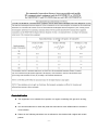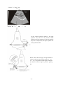
1
WED-9618
Full Digital Ultrasonic Diagnostic System
Operation/Technical Manual
(Version: V1.2, PN NO.: WED-20-11060911E)
SHENZHEN WELL.D MEDICAL ELECTRONICS CO. LTD.
Preface........................................................................................................................................................... 1
Statement................................................................................................................................................. 1
Manufacturer's warranty.......................................................................................................................... 1
Matters need Attention.............................................................................................................................2
Safety labels.............................................................................................................................................2
Safety classification................................................................................................................................. 3
General tips for equipment operation...................................................................................................... 3
General Safety Message...........................................................................................................................4
Contraindication...................................................................................................................................... 9
1 Summary................................................................................................................................................... 11
1.1 Brief Introduction.............................................................................................................................11
1.2 Range Of Application...................................................................................................................... 11
1.3 Appearance...................................................................................................................................... 12
1.4 Technical Specificatio......................................................................................................................13
1.5 Electric principle block diagram......................................................................................................14
1.6 Standard configurations................................................................................................................... 14
1.7 Optional pieces................................................................................................................................ 14
2 Installation.................................................................................................................................................15
2.1 Operation environmental requirements............................................................................................15
2.2 Connecting probe and main unit...................................................................................................... 15
2.3 Image management software installation.........................................................................................16
2.4 Equipotential connection................................................................................................................. 16
3 Operating procedures............................................................................................................................. 18
3.1 Introduction of keyboard................................................................................................................. 18
3.2 Display interface.............................................................................................................................. 18
3.3 Operating procedures.......................................................................................................................19
4 Image parameter adjustment................................................................................................................. 20
4.1 Display mode switching...................................................................................................................20
4.2 Image freezing................................................................................................................................. 21
4.3 Gain adjustment............................................................................................................................... 21
4.4 Depth adjustment............................................................................................................................. 21
4.5 Scanning angle adjustment.............................................................................................................. 21
4.6 Dynamic range adjustment.............................................................................................................. 22
4.7 Focus adjustment............................................................................................................................. 22
4.8 probe work frequency...................................................................................................................... 22
4.9 Change probe................................................................................................................................... 23
4.10 Frame-related adjustment.............................................................................................................. 23
4.11 M rate adjustment.......................................................................................................................... 23
4.12 Power Adjustment..........................................................................................................................23
5 Image processing.................................................................................................................................... 24
5.1 Overview..........................................................................................................................................24
5.2 Image smoothing............................................................................................................................. 24
5.3 Tissue harmonic imaging................................................................................................................. 24
5.4 Image Flip........................................................................................................................................25
5.5 Gray correction................................................................................................................................ 25
5.6 Bar chart...........................................................................................................................................25
5.7 Zoom................................................................................................................................................26
6 Explanation............................................................................................................................................... 27
6.1 Overview..........................................................................................................................................27
-I-
6.2 Type the basic information of the patient and hospital.................................................................... 27
6.3 Image explanation............................................................................................................................28
6.4 Body mark....................................................................................................................................... 28
6.5 date and clock adjustment................................................................................................................29
6.6 Screen cleaning................................................................................................................................ 30
7 Image storage and Cine loop.................................................................................................................31
7.1 Static image storage......................................................................................................................... 31
7.2 Image export.................................................................................................................................... 31
7.3 Image cleaning.................................................................................................................................32
7.4 Cine loop..........................................................................................................................................32
8 Measurement........................................................................................................................................... 33
8.1 Ordinary measurement.....................................................................................................................33
8.1.1 Distance measurement.................................................................................................... 33
8.1.2 perimeter/area measurement.......................................................................................... 33
8.1.3 Volume measurement.......................................................................................................34
8.2 Cardiac Measurement...................................................................................................................... 35
8.2.1 Left Ventricular Measurement......................................................................................... 35
8.2.2 Aortic Valve Measurement............................................................................................... 37
8.2.3 Mitral Valve Measurement............................................................................................... 37
8.2.4 Pulmonary Valve Measurement...................................................................................... 38
8.2.5 Tricuspid Valve Measurement......................................................................................... 39
8.2.6 Heart Rate Measurement.................................................................................................40
8.3 Obstetrics calculation.......................................................................................................................41
9 Report........................................................................................................................................................43
10 Operating Instruction for Function of Puncture Guide..................................................................... 45
10.1 Fixation of Puncture Frame and Probe.......................................................................................... 45
10.2 Selection of Puncture Needles....................................................................................................... 46
10.3 Call out the Puncture Guding Line................................................................................................ 46
10.4 Calibration of Puncture Line..........................................................................................................47
10.5 Insertion of Puncture Needle......................................................................................................... 48
10.6 After Puncture................................................................................................................................ 49
11 Check and Maintenance....................................................................................................................... 50
11.1 Service life..................................................................................................................................... 50
11.2 Main unit maintenance...................................................................................................................50
11.3 Probe maintenance......................................................................................................................... 51
11.4 Correct usage of probe................................................................................................................... 52
12 Simple Trouble Shooting...................................................................................................................... 53
12.1 Check............................................................................................................................................. 53
12.2 Simple trouble shooting................................................................................................................. 53
13 Transportation and Storage................................................................................................................. 54
13.1 Environment requirements on transportation and storage............................................................. 54
13.2 Transportation................................................................................................................................ 54
13.3 Storage........................................................................................................................................... 54
Appendix A Acoustic output reporting table.............................................................................................55
Appendix B Obstetrics................................................................................................................................63
- II -
Preface
Statement
�
This publication, including pictures and illustrations, is property of Shenzhen Well.D Medical
Electronics Co., Ltd. and under protection of international copyright law.
�
Information in this document is not annotated to change. The manufacture shall not state nor
observe any warranty basing on this point, and definitely give up any implied warranty basing on
any special purpose of selling or making benefit.
�
Without previous written permission from the producer, this document must not be photocopied,
reproduced or translated into other languages.
�
We preserve the right of revision on this document without still further notice.
�
Some pictures in this manual, which are schematic diagrams for indication only, may disaccord
with the real object, and then the real object should be regarded as the final.
�
is trade marks of Shenzhen Well.D Medical Electronics Co., Ltd. Any abuse of these trade
marks without permission will be sued to assume legal responsibility according to laws.
Manufacturer's warranty
�
Shenzhen Well.D Medical Electronics Co., Ltd. assumes the responsibility for equipment security,
reliability and performance only under the preconditions that the disassembly, assembly and
maintenance of the equipment are all performed by its assigned professional and the equipment is
used strictly in compliance with the operation manual.
�
Shenzhen Well.D Medical Electronics Co., Ltd. ensures a guarantee period within a year and a
half since the delivery day and promises there is no problem with the new equipment in material
and technology. Within the guarantee period, Shenzhen Well.D Medical Electronics Co., Ltd. will
maintain the equipment and replace the parts of non-man-made damages free of charge. But will
not repair or replace the equipment surface if it is damaged.
�
This guarantee is only available for failures occurred when the equipment is operated in
compliance with the operation manual. And the guaranteed equipment can only be used in the
prescribed range given in manual.
�
This guarantee excludes losses or damages caused by external reasons such as thunder struck,
earthquake, theft, unsuitable use or abuse and refitting the equipment.
�
Shenzhen Well.D Medical Electronics Co., Ltd. shall not be responsible for damages caused by
other devices or arbitrary connection to other devices.
-1-
�
Shenzhen Well.D Medical Electronics Co., Ltd. shall not be responsible for losses, damages or
injuries caused by delayed service request.
�
When there is problem with the products, please contact Shenzhen Well.D Medical Electronics
Co., Ltd. and explain the equipment model, serial number, date of purchase and the problem.
Matters need Attention
�
To ensure operational safety and long-term stable equipment performance, please read this
operation manual closely and understand the equipment functions, operation and maintenance at
all points before operating the equipment, especially contents of "Warning", "Caution" and
"Note".
�
Maloperation or inobservance of the instructions given by manufacturer or its agents may result
in equipment damage or personal injury.
�
The following convention works through this manual to lay special emphasis on some
information.
�
"Warning":
Stands for neglect of it will cause severe personal injury, death or realized
property loss.
�
"Caution": Stands for neglect of it will cause slight personal injury or property damage.
�
"Note": to remind user of installation, operation or maintenance information. These
information is very significant but with no risk. Any warning against dangers shall not be
contained in NOTE.
Safety labels
Equipment labels explanation:
Type B equipment
Note!
documents
Refer
to
accompanying
Turn-on (general supply)
Disconnect (general supply)
VIDEO
Equipotential
Risky voltage
IPX7
Protection against dripping water
-2-
USB
RS-232
Packing and transportation labels explanation:
Handle carefully
Temperature limit
Upward
Piling layer limit
Keep dry
Protect against heat
Safety classification
According to the degree of safety of application in the presence of a flammable anaesthetic
mixture with air or with oxygen or nitrous oxide
oxide::
WED-9618 not suitable for use in the presence of a flammable anaesthetic mixture with air or with
oxygen or nitrous oxide;
According to the mode of operation:
Continuous operation;
According to the degree of protection against harmful ingress of water as detailed in the
current edition of IEC 529
529:
Main unit is general equipment,the probe is enclosed equipment protected against the effects of
immersion,IPX7.
According to the type of protection against electric shock:
ClassⅠequipment energized from an external electrical power source equipment;
According to the degree of protection against electric shock:
Type B
General tips for equipment operation
◆
In operation
1.
Heat radiation holes are strictly prohibited to be covered.
-3-
2.
After closedown, do not switch on the equipment within 2 - 3 minutes.
3.
On scanning, if any abnormal case is found, stop scanning immediately and shut down
the equipment.
4.
The patient is prohibited to touch any non-applied part of the equipment.
5.
When operating, do not press the keyboard panel too much, otherwise the equipment
might be damaged.
◆
After operation
1.
Power off the equipment.
2.
Pull out the plug from power supply socket instead of pulling the cable.
3.
Clean off the couplant on the probe with soft medical sterilized cotton ball.
4.
Put the probe into the special case.
General Safety Message
Safety of the operator and patients and reliability of the equipment are taken into consideration
during designing and producing, the following safety precaution must be implemented:
1.
The equipment shall be operated by qualified operating staff or under their instructions.
2.
Do not open the equipment and change the parameters without permission. If necessary,
please turn to for Shenzhen Well.D Medical Electronics Co., Ltd. or its authorized agent
for service.
3.
The equipment has already been regulated into its optimal performance. Do not adjust
any preset control or switch unless operate as per instructions in the manual.
4.
If there is equipment failure, please shut down the equipment at once and contact for
Shenzhen Well.D Medical Electronics Co., Ltd. or their authorized agent.
5.
If it is needed to connect the equipment with other company’s' electronic or mechanical
devices , please contact Shenzhen Well. D Medical Electronics Co,. Ltd. before
connection.
6.
Equipment operation, storage and transportation environment
Environmental requirements on normal operation:
a) Environment temperature range: 0℃~+40℃
b) Relative humidity range: ≤80%
c)
Atmosphere pressure range: 70KPa~106KPa
Environment requirements on equipment storage and transportation:
d) Environment temperature range: -40℃~+55℃
-4-
e) Relative humidity range: <80%(20℃)
7.
Do not hit the fragile TFT-LCD display. If it cracks, deal carefully with it in case the liquid
crystal gets into eyes or mouths.
8.
Must not hit the inner chargeable Li-ion battery nor throw it into fire in case it trigger an
explosion ; Do not short circuit the battery output electrodes in case the battery be
damaged; and please use the original binding charger to charge the battery. More over,
because used battery will cause environment pollution, please handle the battery
correctly for recovery processing.
9.
Must not disassemble the power supply adapter. If failures happen, it should be handled
by the professional; the charging output can only be used for charging the battery of the
equipment, any improper use on other battery may cause explosion, fire and other
unexpected hazards.
10. Must not short circuit the output of the adapter, a long term short circuit shall result in
adapter damage.
11. Please use standard power cord as the input line of the network power supply for the
adapter to reduce risk.
12. Shenzhen Well.D Medical Electronics Co., Ltd. shall not take any responsibility for any
risk resulted from unauthorized re-fitment by the users.
13. To disconnect the equipment from the power supply network by unplug the adapter from
the power supply network.
14. Refer to appendix A for sound output parameters.
-5-
EMC statement
statement:
WED
WED- 9618 shall not affect the basic performance of radio service and other equipments and can
work well in its stated electromagnetic environment.
-6-
-7-
-8-
Contraindication
★ The equipment is not suitable for inspection on organs containing with gas such as lung,
etc.
★ It is recommended not to check the parts with wounded or acute inflammation to avoid the
cross infection.
★ Patient in the following situations are not allowed to be checked with vagina and rectum
probes:
-9-
inflammation of the vagina , such as trichomonas virginities, colpomycosis, venereal
disease etc ; The unmarried, deformity of vagina, menstrual period, postmenopausal
vagina atrophy, person with difficulty carry on the vagina ultrasonic examination,
colporrhagia, placenta previa patient, etc.
★ The following patient is prohibited to use the puncture
High blood pressure, coronary heart disease, disturbance of blood coagulation and the
bleeding tendency patient
Warning
When WED-9618 works in strong electromagnetic environment exceeding its statement,
its image might be interfered and the diagnoses could be influenced. At this time,
please stop operating until the EMC interference is removed.
Warning
When WED-9618 works at the state of overlapping or paralleling with other equipments,
If it must work close to other equipments,
there might be unexpected EMC problems
problems;If
please observe carefully and check if some equipment is influenced by unexpected EM
coupling.
Warning
Replacement of non-standard probe parts may cause unexpected EMC problem.
NOTE
Accessory equipment connected to the analog and digital interfaces must be certified
according to the respective IEC standards (e.g. IEC 60950 for data processing
equipment and IEC 60601-1-1 for medical equipment). Furthermore, all configurations
shall comply with the valid version of the system standard IEC 60601-1-1. Everybody
who connects additional equipment to the signal input or signal output part configures
a medical system, and is therefore responsible that the system complies with the
requirements of the valid version of the system standard IEC60601-1. IF in doubt,
consult the technical service department or your local representative.
- 10 -
1 Summary
1.1 Brief Introduction
�
This equipment is high resolution linear/convex ultrasound scanning diagnostic equipment. It
adopts micro-computer control and digital scan converter (DSC), digital beam-forming (DBF),
real time dynamic aperture (RDA), real time dynamic receiving apodization, real time Dynamic
receiving focusing (DRF), Digital frequency Scan (DFS), 8 segments TGC, frame correlation
technologies to endue its image with clarity, stability and high resolution.
�
There are six display modes: B, B+B, B+M, B+M/M, M and 4B; And 256 gray scale.
�
The system can process real time image display, freeze, save, load, zoom, up and down
flip, left and right flip, black and white flip, and capacity cine loop; Multi-level scanning
depth, angle, dynamic range, acoustic power, frame correlation factor regulation and
focus number, focal space, focus position, etc. It offers more than 40 body marks.
�
Date, clock display; Name, sex, age, doctor, hospital annotation; Distance, circumference,
area, volume, heart rate measurement; preset two obstetric tables to measure GA, FW
and EDD. Many probes are optional for clinic diagnosis demands.
�
PAL-D video output offers connection to external video image printer and big display and
other equipments. High speed USB port provides real time image transfer to the PC.
�
Adoption of folded soft push keyboard and trackball provides immediate, convenient and
flexible operation. The equipment is jet molding enclosure and potable structure, the
usage of non-industrial frequency transformer switching power supply, programmable
parts (FPGA) and surface mounting technology (SMT) make the whole unit highly
compact.
1.2 Range Of Application
Application in abdominal, Obstetric, Cardiac, small parts sonography.
- 11 -
1.3 Appearance
Contrast
Brightness
Overall gain
Front Panel
Video out
USB 2.0 port
Fuse
RS-232 port
GND
(Equipotent connector)
Power switch
Heat radiator
Rear Panel
- 12 -
Power socket
1.4 Technical Specificatio
Probe
C1-7/60R/3.5MHz
L1-3/7.5MHz
C1-8/20R/5.0MHz
EC1-2/13R/6.5MHz
convex array
HF linear
micro convex
endo-vaginal
Display depth (mm)
250 (max)
≥190
≥80
≥90
≥90
Lateral
≤2
(depth≤80)
≤3
(80<depth≤130)
≤1
(depth≤60)
≤1
(depth≤40)
≤1
(depth≤40)
Axial
≤1
(depth≤80)
≤0.5
(depth≤80)
≤0.5
(depth≤40)
≤0.5
(depth≤40)
≤3
≤3
≤2
≤2
Maximal detect depth (mm)
Resolution
(mm)
Blind zone(mm)
Geometric
position
precision
Horizontal
≤0
≤3
≤4
≤2
Vertical
≤3
≤5
≤2
≤2
Monitor size
10 Inch
Display mode
B, B+B, B+M, B+M/M, M, 4B
Image gray scale
Cine loop
256 Scale
≥500 Frame
Image storage
64 Frame
Scan angle
Adjustable
Scan depth
40mm-250mm
Image flip
Up/down, left/right, black/ white
Image Process
Focus
Measure
Character display
Notation
GAMA, Image Smoothen, THI, Histogram, Zoom
Focus Number, Focus position, Focal space
Distance, circumference, area, volume, heart, GA, FW, EDD
Date, clock, name, PID, age, Sex, hospital name, doctor,
full-screen character editor, posture mark, Position indication
Output report
4 types
USB port
USB2.0
power consumption (MAX)
100VA
Net weight
6.4kg
Size
253mm×315mm×353mm
- 13 -
1.5 Electric principle block diagram
1.6 Standard configurations
�
Mainframe
�
C1-7/60R/3.5MHz convex array
�
PS cable
�
2 pieces of fuse tube F2AL250V
�
Coupling gel 250ml
�
User Manual
�
Final examination report
�
Packing List
1.7 Optional pieces
�
C1-8/20R/5.0MHz Micro-convex probe
�
L1-3/7.5MHz HF linear probe
�
EC1-2/13R/6.5MHz Endo-vaginal probe
�
High speed USB cable
�
Monitor
�
Video printer
Warning:
Please use the spare parts specified above. The manufacturer shall not bear the
responsibility of safety problem, unexpected EMC performance drop caused by
arbitrary adoption of non-specified spare parts.
- 14 -
2 Installation
2.1 Operation environmental requirements
�
Environment temperature range: 0℃ ~+40℃
�
Relative humidity range: ≤80%
�
Atmosphere pressure range: 70kPa~106kPa
�
Power supply: a.c.110V~130V/220V~240V,50Hz/60Hz
�
Avoid strenuous vibration during operation; Keep away from equipments with high
electric field, high magnetic field and high voltage; avoid strong sunlight on the display;
Keep the equipment well-ventilated, damp proof and dustproof.
Note:
Check according to the "packing list" after uncasing and make sure that there is no
shipping damage, and then install the equipment according to requirements and
methods described in “Installation
”.
Installation”
Warning:
If there is breakage at unpacking check, it is banned to use the device to ensure
security.
The probe should be protected from felling off or crashing and the manufacturer
manufacturer..
ng probe and main unit
2.2 Connecti
Connecting
Check the power supply too see if it is in the expected range (a.c.110V~130V/220V~240V,50Hz/60Hz),
then connect the equipment and the power socket with the cable.
Connect the probe:
①Insert the probe
- 15 -
② Locking the
probe
along
this direction
Warning
Warning::
Do not by all means unplug or plug the probe connector at state of log on in case the
probe and main unit be damaged.
Once the probe is connected with the main unit, do not unplug nor plug it at discretion
in case poor contact happen.
The probe should be protected from felling off or crashing and the manufacturer
assumes no responsibility for the kind of hazard.
Must not touch the contact pin in the probe connector.
Please handle the equipment carefully
carefully..
2.3 Image management software installation
Installation steps, see the documentation in CD, you can see the help document to use.
USB Cable
Insert USB port
2.4 Equipotential connection
See the figure:
Equipment back panel
Equipotential terminal
Equipotential line
Equipotential terminal
- 16 -
Other device
Warning
Equipotential: When this equipment is used together with other device, equipotential
should be in consideration.
When the equipment is in use, the doctor and patient are under the risk of
uncontrollable compensating current influence, which is caused by different
electric
potential between facilities and tangible current carrying parts. The most safe solution
and connect the medical equipments with the
is set up a united equipotential network
network,and
equipotential network in the treatment room.
- 17 -
3 Operating procedures
3.1 Introduction of keyboard
Keyboard
(alphanumeric
keys)
TGC
Function keys
Trackball
Number character includes below double function keys:
“1” —————— Press this key to move cursor or sampling line left
“2” —————— Press this key to move cursor up
“3” —————— Press this key to move cursor down
“4” —————— Press this key to move cursor or sampling line right
“0” —————— Control the cursor moving speed by direction keys
3.2 Display interface
Hospital
Name
P sex
P age
Date
Clock
Frequency
Doctor
PID
Scan direction
power
Focus no. & space
Scan angle
Frame rate
Frame-correlation
Total gain
Dynamic range
Gamaconr
Image smoothing
Gray level bar
Scan area
Cine loop Status
Depth
- 18 -
Freeze mark
3.3 Operating procedures
1.
Switch on the equipment.
2.
Press any button to enter the B-type ultrasonic scan interface.
3.
Type the basic information of the patient.
Press “Annotation” to type the information including name, PID, age, gender, hospital, doctor.
4.
Spread ultrasonic coupling agent on the probe acoustic shadow surface, and close to the scanning area,
then the real-time image can be observed on the screen.
5.
Freeze the image, and conduct measurement on the image.
6.
Create a report, print the report and image by the connected video printer.
- 19 -
4 Image parameter adjustment
4.1 Display mode switching
B mode switching
B”button to switch current image to real-time single B mode. (The default mode when the
Press “B
equipment is power on)
BB mode switching
BB
Press “BB
BB” button to switch the current image to double B mode. Real-time image and freeze image are
displayed on the screen. Press “BB” button continuously to switch the real-time and freeze states of two
images.
B+M mode switching
BM/M
Press “BM/M
BM/M” button to switch the current image to B+M mode. B type and M type real-time images can
be displayed on the screen at the same time. The sample line of B type image can be moved around by
mouse and arrow button.
B+M/M mode switching
Press “4B” button to switch the current image to B+M/M mode in the B+M type or M type real-time
scanning mode. B type and 2 M type real-time images can be displayed on the screen at the same time. The
L/R
sample line of B type image can be moved around by mouse and arrow button. Press “L/R
L/R” button to
Change the current sampling line.
M mode switching
Press “BM/M” button to switch the current image to M mode in the B+M type real-time scanning
mode. And then an M type image is displayed on the screen.
4B mode switching
4B
Press “4B
4B” button to switch the current image to 4B mode in the B type, BB type real-time scanning mode.
4B
Four B type images are displayed, one of which is in a real-time state. Press “4B
4B” button continuously to
switch the real-time and freeze states of four images.
B
BB
- 20 -
B+M
B+M/M
M
4B
Attention:
When you call up the menu, please exit the menu before switching the display mode.
4.2 Image freezing
Freeze
Press “Freeze
Freeze” button to switch real-time state and freeze state. “FREEZE” on the bottom of screen is the
freeze mark.
4.3 Gain adjustment
G” key to set them circularly.
Total gain control key. There are four steps: 25, 30, 35, 40. Press “G
Regulate the gain knob to control gain, The gain setting range is 0-62
4.4 Depth adjustment
Press Dept
Depth↑ or Dept
Depth↓ key in the B BB BM type real-time mode to switch the current scanning depth.
The current depth is displayed on the bottom right of screen. The depth adjustment range changes with
different probe type. The following table is presented as a reference.
Probe type
C1-7/60R/3.5MHz
L1-3/7.5MHz
C1-8/20R/5.0MHz
depth(mm)
90~250
40~105
80~150
Depth 90
Depth 180
EC1-2/13R/6.5MHz
50~120
Depth 240
4.5 Scanning angle adjustment
Press “ Angle ” button in real-time state to active this function. The “Angle” on the right of screen is
lightening. Then switch the scanning angle with↑↓key . When mentioning probe angle, please refer to the
following table.
Cancel
Press “Cancel
Cancel” in turn to exit.
- 21 -
Probe type
C1-7/60R/3.5MHz
EC1-2/13R/6.5MHz
C1-8/20R/5.0MHz
angle
60º visual and adjustable
120º visual and adjustable
90º visual and adjustable
4.6 Dynamic range adjustment
Press“DR”button in real-time state to active this function. The “DR” on the right of screen is lightening.
Then adjust the dynamic range with ↑↓key. The dynamic range is 0-192dB, and the adjustment range is
64-192dB.
Cancel
Press “Cancel
Cancel” in turn to exit.
4.7 Focus adjustment
Focus number
Press“FPin”key to lighten up the “FPIN” on the screen right top, press again to change the focus numbers.
There are two focus at most. Press this key to switch between one focus to two.
Cancel
Press “Cancel
Cancel” in turn to exit.
Attention:
When the focus is set to 2, the current image frame frequency would be reduced.
Focus space selection
When set the focus number as two, their distance can be adjusted. There are 5 levels focus space 2, 3, 4, 5,
6 and press“Span”key repeatedly to switch among them circularly.
Focal position
Press“FPin”key to lighten up the “FPIN” on the screen right top, and then move the focus up and down
with ↑↓key, and change the focal position.
Cancel
Press “Cancel
Cancel” in turn to exit.
Tip
Tip:
The focusing of image near the focus indicator
is better, and you can change the focus
number and focal position, so that the focus will point to the interest areas.
4.8 probe work frequency
Frequency
At real time estate, press“Frequency
Frequency”key, change the working frequency of the probe. The frequency of
each probe is given bellow:
C1-7/60R/3.5MHz —— 2.5MHz、3.5MHz、5.0MHz
C1-8/20R/5.0MHz —— 4.5MHz、5.0MHz、5.5MHz
EC1-2/13R/6.5MHz —— 5.5MHz、6.5MHz、7.5MHz
- 22 -
L1-3/7.5MHz
—— 6.5MHz、7.5MHz、8.5MHz
The current probe frequency displays on the up right of the screen.
4.9 Change probe
probe
-S
This equipment is fitted with two probe connectors (A, B). Press “probe
probe-S
-S” key to switch the probes. The
current probe model appears on the right top of the screen.
Tips
Tips:
The device can automatically identify probes.
When it is connected with two probes. The system default working probe is the one connecting to
socket A.
Please shut down the system first before replacing probes. Restart the system, it can realize
automatic identification.
4.10 Frame-related adjustment
Press“M Volocity Correlation”buttons in real-time B mode, BB, 4B mode states to active frame-related
adjustment. “FMAVG” on the right of screen is lighting. Change the frame size with ↑ ↓ key, and the
result will be displayed on the right of screen, such as “FMAVG: 0.55”. The adjustment range is 0.20-0.95.
Cancel
Press “Cancel
Cancel” in turn to exit.
Tip:
” on the right of
If the frame-related adjustment is conducted in the BM mode, “Frame Related
Related”
the screen should be lightened in B or BB mode before entering the adjustment mode.
4.11 M rate adjustment
Press “M Volocity Correlation” button in real-time B+M, B+M/M, M mode to adjust the refresh rate of
M-type image on the screen. The current scan rate is displayed on the bottom of screen, such as
“ST=2.50S”. There are four levels listed as 3.00S, 2.50S, 2.00S, 1.25S to adjust in B+M mode, and four
levels listed as 6.00S, 5.00S, 4.00S, 2.50S to adjust in M mode.
4.12 Power Adjustment
At real time or freeze, press “ D ” key adjust acoustic , Right of the screen shows “POWER: 80%” or
“POWER: 100%”.
Attention:
Be sure to avoid prolonged exposure to high power
Use
Ultrasound may cause harm to human
human,Be
power,Use
with caution
caution..
- 23 -
5 Image processing
5.1 Overview
Image Processing
Press “Image
Processing” button to display the menu which including some image processing functions,
then press the corresponding number to enter. The “V1.12” on the top of menu presents current software
Cancel
version number. Press“Cancel
Cancel”button to exit the menu.
-----V1.12----1. IMG-PROC
2. NOM-IMG
3. NOM-THI
4. POSI-NEGA
5. GAMA-GREY
6. HISTOGRAM
5.2 Image smoothing
Image Processing
Press “Image
Processing” → “1” button in real-time state to active this function. The “IM” on the right of
screen is lightening. Then adjust sharpen level with ↑↓key, there are 4 levels can be chosen, which listed
as IM0, IM1, IM2, IM3.
Common image
Image after smoothing
5.3 Tissue harmonic imaging
Press “Image Processing” → “3” button in real-time state to open the tissue harmonic imaging, “THI” on
the bottom of screen is the tissue harmonic imaging mark.
Close tissue harmonic imaging, Press “Image Processing” → “2” button.
Close THI
Open THI
- 24 -
5.4 Image Flip
Image polarity
Press “Image Processing” → “4” button continuously to change the image polarity.
Upside down
U/D
Press “U/D
U/D” continuously in non-4B mode to realize the upside down of image.
Turning around
L/R
Press “L/R
L/R” continuously in real-time scanning state of B, BB, 4B mode to realize the turning around of
image. The
which on top left of screen is the indicator of image scanning direction.
5.5 Gray correction
Press “Image Processing” → “5” button to active this function. The “GY” on the right of screen is
lightening. Then adjust the gray scale of image with ↑↓key. The adjustment range is GY0-GY7.
Tip:
When an external monitor is applied, the image might be properly displayed only by gray scale
correction.
5.6 Bar chart
Press “Image Processing” → “6” in turn to call out sample frame after the image is frozen. Move the frame
to the interest image area, Adjust the size of the sample frame with ↑↓key, press “Measure” to conduct
measurement. Meanwhile the statistics coordinate and statistics result are displayed on the bottom right of
screen.
- 25 -
In the coordinates, X axis presents gray value and Y axis presents pixels number.
In the statistics result, PT presents pixels number of the sample frame, GM and PM present the
corresponding gray value and pixels number of the highest point in the statistics chart.
Clear
Press “Clear
Clear” to exit the bar chart statistics.
5.7 Zoom
At real time or freeze state, press “Zoom” key to display or close sampling windows. Move “sampling
window” by trackball. In order to change the sizes of windows, press ↑↓keys.
- 26 -
6 Explanation
6.1 Overview
Press “Annotation” to display explanation menu which including image explanation functions, then press
the corresponding number to enter. The “V1.12” at the top of menu presents current software version
number. Press“Cancel”button to exit the menu.
-----V1.12----1. NAME
2. PID
3. AGE
4. SEX
5. COMMENT
6. TIME
7. HOSP
8. DOCT
9. ERASE
A. OBTABLE
6.2 Type the basic information of the patient and hospital
First press the "NEW-P" key to clear patient information , Type the basic information according to the
following method.
NAME
Press “Annotation”→“1”in turn to display the following dialog box:
PLEASE ENTER NAME:
■
15 characters maximum can be put in (letter, number or space), press“ ←”to delete the input mistakes.
After input, press“Confirm”key to exit.
PATIENT ID
Press “Annotation”→“2” in turn to display the following dialog box:
PLEASE ENTER PID
■
You can enter numbers and letters, input method with the "Name", enter up to 8 characters.
AGE
Press “Annotation”→“3” in turn to display the following dialog box:
- 27 -
PLEASE ENTER AGE:
■
Age is composed by three numbers, and the input method is the same with the method which “NAME”
adapts.
GENDER
Press “Annotation”→“4” in turn to display the following dialog box:
PLEASE ENTER SEX:
1.MALE
2.FEMALE
Press the corresponding number to select.
HOSPITAL
Press “Annotation”→“7” in turn to display the following dialog box:
PLEASE ENTER HOSP:
■
The input method is the same with the method which “NAME” adapts, and the maximum character number
is 18.
DOCTOR
Press “Annotation”→“8” in turn to display the following dialog box:
PLEASE ENTER DOCT:
■
The input method is the same with the method which “NAME” adapts, and the maximum character number
is 14.
6.3 Image explanation
Press “Annotation”→“5” in turn into the image explanation function. The white cursor will display on the
screen. Move the trackball cursor, you can annotate anywhere within the image area. At the same time the
bottom of the screen will display the Chinese character’s shortcut key, press the corresponding number keys
(0-9), the cursor will input the same characters.
When finishing the comment, press “Annotation” or“Cancel” key on the board to end this operation.
6.4 Body mark
Body mark
Body mark
Press “Body
mark” key to active body mark. There are 40 body marks, press this key repeatedly to display
- 28 -
these marks circularly
1. Abdomen
Body front
(M)
Body left side
Body front
(F)
Body back
Body right side
2. Obstetric
Mother
3. Head
(F)
Fetus
4. Heart
5. Others
Position indicator
Confirm
Reference
When the Body mark shows, Press “Confirm
Confirm” key to tune out Position indicator↖, Press “Reference
Reference” key
Cancel
to adjust directions, Then move it with trackball to make marks on the Body mark, Finally Press “Cancel
Cancel”
key to complete and exit.
Redo the above operation Can display more Position indicator.
6.5 date and clock adjustment
Press “Annotation”→“6” in turn to enter the date and clock adjustment dialog box:
YY-MM-DD
■
HH-MM-SS
Type method: for example now is nine thirty-five thirty second on January 1st, 2010, then type
100101093530. Press“Confirm”to complete the typing and exit, the following box is listed as a reference.
YY-MM-DD
100101
HH-MM-SS
093530
- 29 -
6.6 Screen cleaning
press "Clear" key to clear the screen measurement mark and measurement results.
- 30 -
7 Image storage and Cine loop
7.1 Static image storage
The equipment offers storage space for 64 static images, and the date wouldn’t lost when the power-down.
Store image by the following steps:
1.
First scan and freeze an image.
2.
Save
Press “Save
Save” button in turn to save the image. The prompt “SAVING……05”displayed on top
left of screen indicates that the image saving is conducting, any other operation is forbidden. And
the prompt will disappear when the saving is completed.
3.
Press “freeze” button to back to the real-time scanning state.
The image memory can number the image (01-64) automatically. If 01-20 is used, it would save from 21
the next time. When the memory is full (save over 64 images), the following dialog box would display
during the image saving.
STORAGE IS FULL. ERASE NO.01?
1. YES
2. NO
Prompt whether the image NO.01 should be overwritten, you may choose yes or not.
Tip:
When the memory is full, you may export one image, and then conduct image saving. The system
would prompt whether clear or save from the removed image number.
7.2 Image export
Recall
1. Press “Recall
Recall” button in turn to display the following dialog box:
PLEASE ENTER STORAGE NO.:
■
2. Type the memory number(such as “01”, press “←” to delete if there is wrong type.), then press
Confirm
“Confirm
Confirm” to export the first saved image. On the bottom left of screen it displays 01/64 to
present current image number and memory capacity. Now press“↑↓”to export other images in
turn.
Freeze
3. Press “Freeze
Freeze” to back to real-time scanning mode, and repeat the above steps to export other
images.
- 31 -
7.3 Image cleaning
Annotation
Press “Annotation
Annotation”→“9” in turn to display the following dialog box:
ERASE ALL STORAGE?
1.YES 2.NO
Press “11” to confirm the image cleaning. The prompt “ERASING... ...”displayed on top left of screen
indicates that the image cleaning is conducting, any other operation is forbidden. And the prompt will
disappear when the cleaning is completed.
Press “22”to stop the image cleaning.
7.4 Cine loop
First of all, save all pieces of static image in temporary memory (the data in temporary memory will be lost
when the power is off), then play these static image continuously to realize the function of cine loop.
Operate as the following steps:
1. Start the equipment into real-time scanning mode, and allow the equipment to conduct the cine
loop image collection, the collect time is about 30 seconds.
Cine loop
2. Freeze the image, press “Cine
loop” button in turn to playback, and the image is played in a
continuous loop. The playback state “120/508
→
”is displayed on the
bottom left of the screen. “120/508” present current frame and total frame separately.
Cine loop
3. During the playback to realize frame step play with ↑ ↓ key or Trackball. Press “Cine
loop”
button in turn again to back to auto repeat mode.
Freeze
4. Press “Freeze
Freeze” button to exit the cine loop.
Tip:
Press “BB
BB”” button to switch different windows during the playback in BB mode.
If the image scanning way, probe or display mode is changed, the cine loop operation wouldn
wouldn’’t
work until ordinary scanning is conducted for 30 seconds.
The cine loop time will be changed by the change of angle. The current playback information is
displayed on the bottom left of the screen.
- 32 -
8 Measurement
8.1 Ordinary measurement
8.1.1 Distance measurement
1.
First freeze the image.
2.
Press “DIST” to display the measurement cursor.
3.
Move the cursor to measurement start point.
4.
Press “Reference”, and move the cursor to measurement end point. (press “Reference” to switch
the start and end cursor)
5.
Press “Cancel” to end the measurement.
Repeat steps 2-5 to measure continuously. Up to 4 groups of measurement is allowed. If you want to
continue the measurement after 4 groups, the original data would be overwritten. The current measure
result would be displayed on the right of screen. The distance rate is listed as “D1/D2, D3/D4”. Press
“Clear” to clear the measurement mark and measurement result.
Distance measurement chart
8.1.2 perimeter/area measurement
Free measurement
1.
First clean the origin date on the screen.
2.
A/C
Press “A/C
A/C” in turn to display following dialog box:
PLEASE ENTER:
1.FREEHAND 2.ELLIPSE
3.
Press “1” to choose free method, and the measure cursor is displayed on the screen.
4.
Move the cursor to measurement start point.
5.
Press “Reference”, move the cursor to end point through the edge of measure areas to form a
closed path.
6.
Press “Reference” to complete the measurement. If the curve is unclosed, the system would form
a closed path automatically according to the shortest distance.
- 33 -
A/C
Press “A/C
A/C” key, Repeat steps 4-6 to measure continuously. Up to 2 groups of measurement is allowed. The
result would be displayed on the right of screen. “C1/C2, A1/A2”are perimeter rate, area rate. Press “Clear”
to re-measure.
Free measurement chart
Elliptical method measurement
1.
First freeze the image.
2.
Clean the origin date on the screen.
3.
A/C
Press “A/C
A/C” in turn to display following dialog box:
PLEASE ENTER:
1.FREEHAND 2.ELLIPSE
4.
Press “2” to choose elliptical method, and the elliptical sample frame is displayed on the screen.
5.
Move sample frame to measure area.
6.
Press "Confirm", adjust the size by direction button.
7.
Press "Reference" key, adjust the sample frame direction by trackball.
8.
Press "Confirm", repeat steps 5-7 to re-adjust sample frame.
9.
Press “Cancel” to end the measurement.
A/C
Press “A/C
A/C” key, Repeat steps 5-9 to measure continuously. Up to 2 groups of measurement is allowed.
Press the middle button to end the measurement. The result would be displayed on the right of screen. Clear
screen first if you want to re-measure.
Elliptical measurement chart
8.1.3 Volume measurement
Triaxial method
- 34 -
1.
Measure the distance of 3 groups or more according to distance measurement.
2.
Press “Measure”or “Cancel” to display volume value.
“Vm1”on the right of screen indicates the current volume value.
Elliptical method
Measure the perimeter/area value of two groups by elliptical method, the current volume value“Vm1”
would be displayed on the right of screen.
Volume measurement chart
8.2 Cardiac Measurement
Measure
Obtain the satisfied image under the B/M or M mode and freeze. Press the“Measure
Measure”key and the cardiac
measurement menu will appear, The “V1.12” at the top of menu presents current software version number.
Press“Cancel”button to exit the menu.
-----V1.12----1. LV<VF>
2. AV
3. MV
4. PV
5. TV
6. HR
1. Left Ventricular
2. Aortic Valve
3. Mitral Valve
4. Pulmonary Valve
5. Tricuspid Valve
6. Heart Rate
8.2.1 Left Ventricular Measurement
By measuring the diastolic period and systolic period: posterior wall of the right ventricular, anterior wall
of the interventricular septum, posterior wall of the interventricular septum, endocardium of the posterior
wall of the left ventricular, epicardium; to calculate the diastolic period and systolic period respectively:
thickness of the left cardiac wall, diameter of the right ventricular, diameter of the left ventricular, thickness
of the posterior wall of the left ventricular, thickness of the interventricular septum, volume of the left
ventricular, heartbeat flow, ECG output, systolic fraction and ejection fraction.
Operation Procedure:
1.
2.
3.
4.
Enter the B/M mode, and move the mouse to change the position of the sampling lime. Press the
“Freeze” key when the proper M mode image appears;
Measure
Press the“Measure
Measure”key and the cardiac measurement menu will appear;
Press the“1”key to select “LV<VF>”, a vertical time bar will appear in the M image;
Move the time bar to the end of the diastolic period and press the “Confirm”key, mark the anterior
wall of the right ventricular (D1) with Confirm key. Some mark points of the diastolic period are
- 35 -
5.
6.
described as follows and mark the following several points with the same method:
D2——Posterior Wall of the Right Ventricular in the Diastolic Period
D3——Anterior Wall of the Interventricular Septum in the Diastolic Period
D4——Posterior Wall of the Interventricular Septum in the Diastolic Period
D5——Endocardium of the Posterior Wall of the Left Ventricular in the Diastolic Period
D6——Epicardium in the Diastolic Period
(D6) After marking, the second time bar will appear, move the time bar to the end of the diastolic
period and press“Confirm”key and mark the anterior wall of the right ventricular (S1) of the systolic
period with“Confirm”key. Some mark points of the diastolic period are described as follows and mark
the following several points with the same method:
S2——Posterior Wall of the Right Ventricular in the Systolic Period
S3——Anterior Wall of the Interventricular Septum in the Systolic Period
S4——Posterior Wall of the Interventricular Septum in the Systolic Period
S5——Endocardium of the Posterior Wall of the Left Ventricular in the Systolic Period
S6——Epicardium in the Systolic Period
(S6)After the marking finishes, the measurement results will display on the right side of the screen
automatically.
Left
Ventricular
Measurement
RHWD—Thickness of the Right Cardiac Wall in the Diastolic Period (mm)
RHWS—Thickness of the Right Cardiac Wall in the Systolic Period (mm)
RVD—Right Ventricular Diameter in the Diastolic Period (mm)
RVS—Right Ventricular Diameter in the Systolic Period (mm)
LVDD—Left Ventricular Diameter in the Diastolic Period (mm)
LVDS—Left Ventricular Diameter in the Systolic Period (mm)
PLVWD—Thickness of the Posterior Wall of the Left Ventricular in the Diastolic Period (mm)
PLVWS—Thickness of the Posterior Wall of the Left Ventricular in the Systolic Period (mm)
IVSD—Thickness of the Interventricular Septum in the Diastolic Period (mm)
IVSS—Thickness of the Interventricular Septum in the Systolic Period (mm)
LVEDV—Left Ventricular Volume at the end of the Diastolic Period (ml)
LVESV—Left Ventricular Volume at the end of the Systolic Period (ml)
SF—Systolic Fraction (%)
EF—Ejection Fraction (%)
SV—Heartbeat Volume (ml)
CO—ECG Output (ml)
- 36 -
8.2.2 Aortic Valve Measurement
Calculate the aortic valve diameter, open size of the aortic valve and the ratio between the aortic valve and
left atrium to study the aortic valve.
Operation Procedure:
1.
2.
3.
4.
5.
6.
7.
Enter the B/M mode and move the mouse to change the position of the sampling line. Press the
“Freeze”key when proper M mode image appears;
Measure
Press the“Measure
Measure”key and the cardiac measurement menu will appear;
Press the“2”key to select “AV”, and a vertical time bar will appear in the M image;
Move the time bar to the end of the diastolic period and press“Confirm”key, mark the anterior wall of
the aortic valve (D1) and the posterior wall of the aortic valve (D2) with“Confirm”key, the second
time bar will appear after the marking;
Move the time bar to the open point of the aortic valve and press“Confirm”key, mark the anterior lobe
of the aortic valve (O1) and posterior lobe of the aortic valve (O2) with the same method, the third
time bar will appear after the marking;
Move the time bar to the close point of the aortic valve and press“Confirm”key, mark the posterior
wall of the aortic valve (C1) and posterior lobe arterial wall (C2);
When all the measurements finish, the calculation results of the aortic valve will display on the right
side of the screen.
Aortic Valve Measurement
AO—The Aortic Root Diameter at the end of the Diastolic Period (mm)
LA—Diameter of the Left Ventricular at the end of the Systolic Period (mm)
AVO—Distance between the Aortic Anterior Lobe and Posterior Lobe and the Open Point of the
Aortic Valve (mm)
LAR—Left Atrium/Aorta Ratio
LVET—Left Ventricular Ejection Time (second)
8.2.3 Mitral Valve Measurement
The research of the mitral valve allows the user to estimate the D-E and E-F displacement, D-E and E-F
slope, D-E and E-F time on the D wave, E wave and F wave under the M mode and BM mode.
Operation Procedure:
1. Enter the B/M mode and move the mouse to change the position of the sampling line, press the
“Freeze”key when the proper M mode image appears;
- 37 -
2.
3.
4.
5.
Measure
Press the“Measure
Measure”key and the cardiac measurement menu will appear;
Press the“3”key to select “MV”, move the cursor to the Q wave position of the M mode image area
and press“Confirm”key, mark “D”;
Mark the following mitral valve waves with the same method;
E: E Wave
F: F Wave
After the marking finishes, the measurement results will display on the right side of the screen
automatically.
Mitral
Valve
Measurement
DE<EX>—Distance from Point D to E on the Y Axis (mm)
EF<EX>—Distance from Point E to F on the Y Axis (mm)
DE<TM>—DE Time (second)
EF<TM>—EFTime (second)
DE<SL>—slope= (Point E on the Y Axis –Point D on the Y Axis)/ (Point F on the X Axis-Point D on
the X Axis) (mm/second)
EF<SL>—slope= (Point E on the Y Axis –Point F on the Y Axis)/(Point F on the X Axis- Point E on
the X Axis) (mm/second)
8.2.4 Pulmonary Valve Measurement
In this measurement, it is a characteristic calculation parameter. Calculate the pulmonary valve according to
the following point marking position.
A: Maximum Downward Position of the Valve of the Atrial Systolic Period
B: Start Point of the Ventricular Systolic Period
C: Maximum Open Point of the Lobule
E1: Start Point of the Ventricular Diastolic Period
E2: Complete Close Point of the Valve
F: Start Point of the Atrial Systolic Period
Mark the above points with the method described above, when the marking finishes, the measurement
results will display on the right side of the screen automatically.
- 38 -
Pulmonary Valve
Measurement
A<DEP>——Depth:
The distance from point F to point A is the distance from the start point of the atrial systolic
period to the maximum downward position of the pulmonary valve. The distance from point F to
A on the Y axis is the maximum movement range of the pulmonary valve in the atrial systolic
period.
A<DEP>Depth=AY-FY (mm)
AY is point A on the Y axis, and FY is point F on the Y axis.
EF<SL>——Slope:
When the initial blood flow from the atrium, E1 is the start point of the ventricular diastolic
period and F is the inflow start point of the atrial systolic period.
EF<SL>Slope=(FY-E1Y)/(FX-E1X)(mm/second)
FY is point F on the Y axis, E1Y is point E1 on the Y axis, FX is point F on the X axis and E1X is
point E1 on the X axis.
BC<DS>——Diameter:
BC is the open part of the pulmonary valve in the ventricular systolic period and the part on the Y
axis the movement distance of lobule when the pulmonary valve open to the maximum.
BC<DS> diameter =CY-BY (mm)
CY is point C on the Y axis, and BY is point B on the Y axis.
BC<SL>——Slope:
BC<SL>Slope= (CY-BY)/ (CX-BX) (mm/second)
CY is point C on the Y axis, BY is point B on the Y axis, CX is point C on the X axis and BX is
point B on the X axis.
RVET——Ejection Time of the Right Ventricular:
Ejection time of the right ventricular is the time from point B to E2 of the opening of the
pulmonary valve.
RVET=E2X-BX (second)
E2X is point E2 on the X axis, and BX is point B on the X axis.
8.2.5 Tricuspid Valve Measurement
Tricuspid valve measurement is similar to mitral valve measurement, and the following three points are
used for:
D: Open point of the tricuspid valve and the end of the systolic period of the right ventricular
E: Maximum Point of the Tricuspid Valve Orifice
F: The tricuspid valve closes completely
Mark the above points with the method described above, when the marking finishes, the measurement
- 39 -
results will display on the right side of the screen automatically.
Tricuspid
Valve
Measurement
DE<EX>— Distance from point D (right ventricular in the front of the tricuspid valve orifice) to point
E (maximum tricuspid valve orifice) (mm)
EF<EX>—Distance from point E to point F on the Y axis (mm)
DE<TM>—DE time (second)
EF<TM>—EF time (second)
DE<SL>—Slope= (Point E on the Y axis–Point D on the Y axis)/ (Point F on the X axis – Point D on
the X axis) (mm/second)
EF<SL>—Slope= (Point E on the Y axis–Point F on the Y axis) / (Point F on the X axis – Point E on
the X axis) (mm/second)
8.2.6 Heart Rate Measurement
Operation Procedure:
1. Enter the B/M mode, move the mouse to change the position of the sampling line, press the “Freeze”
key when proper M mode image appears;
Measure
2. Press the “Measure
Measure” key and the cardiac measurement menu will appear;
3. Press the“6”key to select “HR”, a vertical time bar appears in the M image;
4. Move the time bar to the first wave peak and press“Confirm”key, the second time bar appears;
5. Move the time bar to the third wave peak and press“Confirm”key;
6. The measurement finishes and the measurement results will display on the right side of the screen.
Heart Rate
Measurement
- 40 -
TM—Time (second)
HR— Heart Rate (times/minute)
8.3 Obstetrics calculation
The equipment can calculate gestation age, expected date of delivery according to 7 parameters such as
BPD, FL, AC , HC, CRL,GS, first day of the last menses. Therein, BPD can be used to calculate fetus
weight.
OB table
Cancel
At freeze state, Press“OB
table”key to display obstetrics table menu and press“Cancel
Cancel”button to exit the
menu.
1.
2.
3.
4.
5.
6.
7.
8.
TOKYO
BPD-FW
FL
AC
HC
CRL
AD
GS
LMP
For BPD, FL, CRL, AD and GS, Measure the distance according to distance measurement method, the
corresponding obstetrical table data display on screen right.
For AC, HC, Measure as per Ellipse measurement method for circumference and area, the device will
automatically calculate the related data and display them on screen right.
Select "7. LMP", a reminder displays
PLEASE ENTER LMP: MM—DD
■
This reminder ask to put in the first date of the pregnant woman's last menses in the date format MM-DD.
For example: May 11, put in 05-11. When the correct date is put, measuring result will displays at "EDD"
on screen right in the format of YY-MM-DD, For example 07-02-21 means that the expected date of
delivery is on February 21,2007.
Note
In expected date of delivery calculation, the system date must be correct.
The default standard pregnancy period in the system is 40 weeks. In last menses method
measuring, if the time interval between the input date and the system date exceeds 40 weeks, the
inward date will not be accepted and need to be put in again.
Under the condition of item 1, if the inward date is bigger than the current system date, it will be
regarded as the date of the last year.
- 41 -
Set Obstetrics formula
Press “Annotation” to select "A. OBTABLE ", The remind box is given bellow:
PLEASE ENTER OBTABLE: < TOKYO >
1.TOKYO 2.HADLOCK
Table TOKYO is suitable for Asian, and Table HADLOCK is suitable for European.
Tips:
、 abdomen circle (AC) only have the Hadlock form
, this equipment does Not
Head circle (HC)
(HC)、
form,
provide the HC, AC form under the Tokyo formula
- 42 -
9 Report
The equipment will store the patient information, diagnosis annotation, measurement result and hospital,
data, time, doctor and some other information in the report page with the diagnosis. The abdominal, cardiac,
obstetrics and urological reports will generate automatically according to different measurement items, and
store the last measurement results.
R”key, The report menu will display:
When the corresponding measurement finishes, press“Freeze”→“R
1.ABDOMEN
2.CARDIAC 3. OB
4. UROLOGY
Enter the corresponding number key to enter the related report and press any key to exit this menu.
Abdominal Report, Urological Report:
Cardiac Report:
Obstetrics Report:
- 43 -
R” to exit the report.
Press “R
Tips:
You can press the “Annotation
Annotation
Annotation” key on the report interface and then annotate information in
” to exit
the annotation box, Press “Confirm
Confirm”
exit..
In the obstetrics report 1, 2, 3 means that biparietal diameter (BPD), femur length (FL),
Crown-rump length (CRL) and gestational sac (GS) can be measured at most for three times,
and AVG is the average value of the three measurement results. TOTAL-AVG is the average
value of the gestational age (GA) and expected date of delivery (EDD).
In the obstetrics report, fetal weight (BPD, AC) is calculated through the biparietal diameter
(BPD) and abdominal circumference (AC), and when the measured BPD and AC are unequal to
zero, fetal weight will display here automatically.
Clear the contents of the report
At real time or freeze, press“NEW-P”key to clear notation and report information on the image or the
characters area.
- 44 -
10 Operating Instruction for Function of Puncture Guide
Warning
Warning::
The puncture guiding line must be calibrated before performing a new puncture .If the puncture
needle is not consistent with the puncture guiding line , please do not perform the puncture
operation.
It is with risk when performing the puncture guided under ultrasound, thus it should be operated by
operators with the proper qualification and capability ,and the preparative work before operation
such as examination of clotting time ,BPC,electrocardiogram, blood pressure, sterilization of
puncture set and puncture probe and signing of operation agreement should be done strictly.
10.1 Fixation of Puncture Frame and Probe
Put the probe aside, then loosen
the fixing screw nut on the
puncture frame.
Mount the puncture frame on the
probe
- 45 -
Buckle on the puncture frame and
turn over the probe , then fasten
the fixing screw nut of the
puncture frame
10.2 Selection of Puncture Needles
The puncture needles can be divided into 3 specification numbers such as No.7, No.9 and No.12. You can
choose the proper puncture needle and needle guard based on the practical need. Please ensure that the
specification number of selected puncture needle should be consistent with the selected needle guard.
10.3 Call out the Puncture Guding Line
Under the real time estate (single B mode), press“P”key,the screen will display the PT Sampling line, it
calls puncture lead line, Press “ 1, 4 ” keys move to confirm the position, if want to exit, please press
- 46 -
“Cancel”or“Clear”key.
10.4 Calibration of Puncture Line
Fix the selected puncture needle on the angle
regulator unit, then turn the puncture needle
guard in ward for 180 degree. Thus the puncture
angle can be adjusted by the angle regulator unit
on the puncture frame
Please ensure that each type of angle should be
identical with the detent (Detent Angle: 25°、40°、
55°、70°). The puncture needle angle should also
be identical with the angle of puncture guiding
line
- 47 -
10.5 Insertion of Puncture Needle
Please ensure that the tissue for puncture is on the
puncture guiding line first. Then adjust the angle
and insert the puncture needle slowly in the
direction of needle guard
Please do not move the probe when
inserting the puncture needle for
ensuring the accuracy of the puncture
operation
- 48 -
10.6 After Puncture
Turn the puncture needle guard outward for 180
degree to make the rabbet of the puncture needle
guard outward ,then remove the probe from it
Press“Cancel”or“Clear”key to exit the puncture
guiding line
- 49 -
11 Check and Maintenance
11.1 Service life
Bases on the manufacturer’s design, production related files, this model’s use life is six years. The
Product’s material will gradually aging, if the product continually use over the designed use life, it
may bring the problem of the performance reduced and fault rate raise.
Note
Note:
The Discard the device according to local law.
Do not discard it mixing with other household garbage.
Warning
Warning:
The manufacturer shall not assume the responsibility of risks caused by using the
device beyond its service life.
11.2 Main unit maintenance
�
Instrumentation environment should accord with "Operation environmental requirement".
�
If device enclosure needs cleaning,shutdown the device first and then wipe with alcohol sponges.
�
Device should not turn on and off frequently.
�
When the device does not work for a long time, pack the device according to the instructions on
the packing. Store it properly in the warehouse. The storage environment should accord with
“Transportation and storage environmental requirements".
Caution
Caution:
Please refer to instructions prescribed by the manufacturer closely when using
detergents.
Be careful with cleaning of the display, because it is very easy to scratch and spoil.
Please wipe it with dry soft cloth.
Please do not clean the inner base of the device.
Please do not place the device in liquid.
Do not leave any detergent on the device surface.
Though there will be no chemical reaction between the device enclosure and most of
those detergents, We still suggest no detergent in cleaning lest the device surface is
spoiled.
- 50 -
11.3 Probe maintenance
�
Probe is an expensive and frangible part. Never hit it or drop it on floor. When diagnoses pauses,
Freeze
put the probe in its case and press “Freeze
Freeze” key to keep it in a state of "Frozen".
�
See to use medical ultrasound coupling gel during diagnosis. Degree of protection against
harmful ingress of water is IPX7. For the probes, water should not immerge over the probe
acoustic window (refer to figure). Regularly check the probe enclosure to ensure it is good incase
liquid ingression spoil the inner components.
C1-7
�
L1-3
EC1-2
C1-8
Probe and host machine once tie, be request not to take down at will , for fear probe pin and
socket's contract badness.
Note:
Probe might be damaged due to long time covered coupling gel.
Clean the probe head after every use.
Do not clean the probe with a surgical brash neither soft brash. Only soft cloth can be
used to clean it.
Do not press the probe on the patient too long to avoid discomfort.
Warning:
Must not use extender, ethylene oxide or any other organic solvent which tend to
deface the probe's protective foil.
Must not place the probe in liquid or detergent.
Must keep the equipment or probe from any type of liquid's infiltration.
Must not clean device or probe by airing or heating.
- 51 -
11.4 Correct usage of probe
In order to prolong probe's service life and obtain optimum performance,follow these instructions:
1.
Periodic inspection on probe cable, socket and acoustic window.
2.
Shutdown the device first and then connect or disconnect the probe.
3.
Do not drop probe or flint body,and never hit the probe acoustic window, otherwise probe should
be damaged.
4.
Never heat the probe.
5.
Never bend or pull probe cable, otherwise the internal connection should be broken.
6.
Use couplant only on probe header and then clean probe.
7.
Inspect probe acoustical window, enclosure and cable seriously after probe cleaning. Never use
the probe again if any crack or breakage is found.
- 52 -
12 Simple Trouble Shooting
12.1 Check
�
Check if the power supply is ok or not, main unit power cable is connected well or not. When the
supply voltage surpasses the equipment specified voltage range (a.c.110V~130V/220V~240V,
50Hz/60Hz), must not switch on the equipment.
�
Check if the probe and main unit are connected well or not.
�
Check power cable and probe lines regularly and replace the damaged or broken one if found.
12.2 Simple trouble shooting
No.
Malfunction phenomena
Troubleshooting
1
Open power switch of the device, with 1. Examine power supply;
no signal appeared on the screen and the 2. Examine supply line and plug;
indicator light is not on.
3. Examine whether the fuse is burned-out;
4. Examine the lightness adjusting knob of the
monitor.
2
Discontinuous striae and snow appear 1. Examine power supply: strike fire interfere of
on the screen.
the other devices;
2. Environmental examination: Electric and
magnetic field interfere;
3. Examine power and probe plugs: whether
they are well connected.
3
Unclear image display
1.Adjust the brightness and contrast knobs on
the front panel;
2.Adjust the 8 level TGC and the total gain knob
on the front panel;
3.Clean the screen optical filter.
4
Unclear near field
1. Adjust the total gain knob on the front panel
and the 8 level TGC;
2. Adjust the focus position to the near field;
5
Unclear far field
1. Adjust the total gain knob on the front panel
and the 8 level TGC;
2.Adjust the focus number, space and position to
set the focus in the far field.
- 53 -
13 Transportation and Storage
13.1 Environment requirements on transportation and storage
Environment temperature range:-40℃~+55℃
Relative humidity range:<80%(20℃)
.2 Transportation
13
13.2
Signs on the packing box conform to 《Iconograph and sign of packing, storage and transportation》
(GB/T191-2008). Simple shockproof establishment is fitted within the box,which apply to aviation,
railway, highway or steamship transportation. Keep dry, avoid inversion and collision.
.3 Storage
13
13.3
Equipment should be taken out from the packing when storage time exceeds six months,power on it for
four hours, and then pack it correctly and keep it in a warehouse. The device must not be piled, and do not
place it closely against the floor, walls or roof.
Keep it well ventilated, do not expose it to strong sunlight or caustic gases.
- 54 -
Appendix A Acoustic output reporting table
B mode
Nominal frequency: 3.5MHz
Trans ducer Model: C1-7/60R/3.5MHz
Manufactured By: SHENZHEN WELL.D MEDICAL ELECTRONIC CO.,LTD.
TIS
Index label
Pra
0.85
(MPa)
P
Scan
Aaprt≤1cm2
0.98
-
115.2
-
Aaprt>1
1cm
1cm2
-
Min.of [Pα(zs), Izpta,αzs)] (mW)
-
Zs
-
(cm)
Zbp
acoustic
Zb
(cm)
Z at max. Ipi,α
(cm)
deq (Zb)
(cm)
(cm)
fawf
(MHz)
Nonscan
TIC
-
(a)
-
#
1.46
(mW)
Associated
parameters
Non-scan
MI
Maximum index value
TIB
5.30
2.91
2.91
-
-
-
#
X
(cm)
1.64
-
-
-
#
Y
(cm)
1.30
-
-
-
#
Dim of Aaprt
td
(μsec)
0.52
prr
(Hz)
3787
pr at max. Ipi
(MPa)
2.48
deq at max. Ipi
(cm)
Other
information
Ipa,α at max. MI
Operating
(W/cm2)
207.9
F-PIN
1
1
-
-
-
#
Power,%
80
80
-
-
-
#
Angle,°
30
30
Focus position
7
7
control
conditions
Notes:
(a)This probe is not intended for transcranial or neonatal cephalic use;
- 55 -
Acoustic output reporting table
M mode(Inc B mode)
Nominal frequency: 3.5MHz
Transducer Model: C1-7/60R/3.5MHz
Manufactured By: SHENZHEN WELL.D MEDICAL ELECTRONIC CO.,LTD.
TIS
Index label
Pra
0.89
(MPa)
P
Scan
Aaprt≤1cm2
1cm2
Aaprt>1
Nonscan
0.49
-
0.036-
0.11
(a)
82.0
-
4.7
#
Min.of [Pα(zs), Izpta,αzs)] (mW)
2.61
Zs
2.94
(cm)
Zbp
acoustic
Zb
(cm)
Z at max. Ipi,α
(cm)
deq (Zb)
(cm)
(cm)
fawf
(MHz)
TIC
1.51
(mW)
Associated
parameters
Non-scan
MI
Maximum index value
TIB
2.94
5.07
5.30
0.35
2.91
2.91
-
2.90
2.90
#
X
(cm)
2.35
-
2.34
2.34
#
Y
(cm)
1.30
-
1.30
1.30
#
Dim of Aaprt
Other
information
td
(μsec)
0.53
prr
(Hz)
4166
pr at max. Ipi
(MPa)
2.58
deq at max. Ipi
(cm)
Ipa,α at max. MI
(W/cm2)
0.34
203.9
F-PIN
1
1
-
1
1
#
Operating
Power,%
80%
80%
-
80%
80%
#
control
Angle,°
30
30
30
30
#
conditions
Focus position
7
7
-
7
7
#
M Speed,S
1.25
1.25
-
1.25
1.25
#
Notes:
(a)This probe is not intended for transcranial or neonatal cephalic use;
- 56 -
Acoustic output reporting table
B mode
Nominal frequency: 5.0MHz
Transducer Model: C1-8/20R/5.0MHz
Manufactured By: SHENZHEN WELL.D MEDICAL ELECTRONIC CO.,LTD.
TIS
Index label
Pra
0.64
(MPa)
P
Scan
Aaprt≤1cm2
1cm2
Aaprt>1
Nonscan
0.19
-
-
-
(a)
10.0
-
-
#
Min.of [Pα(zs), Izpta,αzs)] (mW)
-
Zs
-
(cm)
Zbp
acoustic
Zb
(cm)
Z at max. Ipi,α
(cm)
deq (Zb)
(cm)
(cm)
fawf
(MHz)
TIC
1.26
(mW)
Associated
parameters
Non-scan
MI
Maximum index value
TIB
2.98
3.87
3.87
-
-
-
#
X
(cm)
0.52
-
-
-
#
Y
(cm)
0.79
-
-
-
#
Dim of Aaprt
Other
information
td
(μsec)
0.43
prr
(Hz)
4784
pr at max. Ipi
(MPa)
1.87
deq at max. Ipi
(cm)
Ipa,α at max. MI
Operating
(W/cm2)
117.2
F-PIN
1
1
-
-
-
#
Power,%
80
80
-
-
-
#
Angle,°
90
90
Focus position
6
6
-
-
-
#
control
conditions
Notes:
(a)This probe is not intended for transcranial or neonatal cephalic use;
- 57 -
Acoustic output reporting table
M mode(Inc B mode)
Nominal frequency: 5.0MHz
Transducer Model: C1-8/20R/5.0MHz
Manufactured By: SHENZHEN WELL.D MEDICAL ELECTRONIC CO.,LTD.
TIS
Index label
Pra
0.66
(MPa)
P
Scan
Aaprt≤1cm2
1cm2
Aaprt>1
Nonscan
0.22
0.025
-
0.05
(a)
11.6
1.35
1.35
#
Min.of [Pα(zs), Izpta,αzs)] (mW)
-
Zs
-
(cm)
Zbp
acoustic
Zb
(cm)
Z at max. Ipi,α
(cm)
deq (Zb)
(cm)
(cm)
fawf
(MHz)
TIC
1.29
(mW)
Associated
parameters
Non-scan
MI
Maximum index value
TIB
2.68
3.00
0.31
3.86
3.86
3.88
-
3.88
#
X
(cm)
0.63
0.63
-
0.63
#
Y
(cm)
0.79
0.80
-
0.80
#
Dim of Aaprt
Other
information
td
(μsec)
0.43
prr
(Hz)
4807
pr at max. Ipi
(MPa)
1.93
deq at max. Ipi
(cm)
Ipa,α at max. MI
(W/cm2)
0.30
119.1
F-PIN
1
1
1
-
1
#
Operating
Power,%
80%
80%
80%
-
80%
#
control
Angle,°
90
90
90
-
90
#
conditions
Focus position
6
6
6
-
6
#
M Speed,S
1.25
1.25
1.25
-
1.25
#
Notes:
(a)This probe is not intended for transcranial or neonatal cephalic use;
- 58 -
Acoustic output reporting table
B mode
Nominal frequency: 7.5MHz
Transducer Model: L1-3/7.5MHz
Manufactured By: SHENZHEN WELL.D MEDICAL ELECTRONIC CO.,LTD.
TIS
Index label
Pra
0.54
(MPa)
P
Scan
Aaprt≤1cm2
1cm2
Aaprt>1
Nonscan
0.64
-
-
-
(a)
24.0
-
-
#
Min.of [Pα(zs), Izpta,αzs)] (mW)
-
Zs
-
(cm)
Zbp
acoustic
Zb
(cm)
Z at max. Ipi,α
(cm)
deq (Zb)
(cm)
(cm)
fawf
(MHz)
TIC
1.30
(mW)
Associated
parameters
Non-scan
MI
Maximum index value
TIB
4.9
5.70
-
-
5.70
-
-
-
#
X
(cm)
1.02
-
-
-
#
Y
(cm)
0.71
-
-
-
#
Dim of Aaprt
Other
information
td
(μsec)
0.28
prr
(Hz)
5649
pr at max. Ipi
(MPa)
3.41
deq at max. Ipi
(cm)
Ipa,α at max. MI
(W/cm2)
109.6
Operating
F-PIN
1
1
-
-
-
#
control
Power,%
80%
80%
-
-
-
#
conditions
Focus position
3
3
-
-
-
#
Notes:
(a)This probe is not intended for transcranial or neonatal cephalic use;
- 59 -
Acoustic output reporting table
M-mode(Inc B mode)
Nominal frequency: 7.5MHz
Transducer Model: L1-3/7.5MHz
Manufactured By: SHENZHEN WELL.D MEDICAL ELECTRONIC CO.,LTD.
TIS
Index label
Pra
0.56
(MPa)
P
Scan
Aaprt≤1cm2
0.60
0.029
22.1
1.06
TIC
1cm2
Aaprt>1
Nonscan
-
0.045
(a)
1.06
#
1.34
(mW)
Min.of [Pα(zs), Izpta,αzs)] (mW)
-
Zs
-
(cm)
Associated
Zbp
acoustic
Zb
(cm)
Z at max. Ipi,α
(cm)
deq (Zb)
(cm)
parameters
Non-scan
MI
Maximum index value
TIB
(cm)
fawf
(MHz)
2.47
4.90
0.20
5.70
5.70
5.70
-
5..70
#
X
(cm)
1.01
1.00
-
1.00
#
Y
(cm)
0.70
0.70
-
0.70
#
Dim of Aaprt
Other
information
td
(μsec)
0.28
prr
(Hz)
5681
pr at max. Ipi
(MPa)
3.52
deq at max. Ipi
(cm)
Ipa,α at max. MI
Operating
(W/cm2)
0.19
109.7
F-PIN
1
1
1
-
1
#
Power,%
80%
80%
80%
-
80%
#
Focus position
3
3
3
-
3
#
M Speed,S
1.25
1.25
1.25
-
1.25
#
control
conditions
Notes:
(a)This probe is not intended for transcranial or neonatal cephalic use;
- 60 -
Acoustic output reporting table
B mode
Nominal frequency: 6.5MHz
Transducer Model: EC1-2/13R/6.5MHz
Manufactured By: SHENZHEN WELL.D MEDICAL ELECTRONIC CO.,LTD.
TIS
Index label
Pra
0.64
(MPa)
P
Scan
Aaprt≤1cm2
1cm2
Aaprt>1
Nonscan
0.23
-
-
-
(a)
10.0
-
-
#
Min.of [Pα(zs), Izpta,αzs)] (mW)
-
Zs
-
(cm)
Zbp
acoustic
Zb
(cm)
Z at max. Ipi,α
(cm)
deq (Zb)
(cm)
(cm)
fawf
(MHz)
TIC
1.38
(mW)
Associated
parameters
Non-scan
MI
Maximum index value
TIB
2.48
4.66
4.66
-
-
-
#
X
(cm)
0.63
-
-
-
#
Y
(cm)
0.79
-
-
-
#
Dim of Aaprt
Other
information
td
(μsec)
0.31
prr
(Hz)
4807
pr at max. Ipi
(MPa)
2.05
deq at max. Ipi
(cm)
Ipa,α at max. MI
Operating
(W/cm2)
120.1
F-PIN
1
1
-
-
-
#
Power,%
80
80
-
-
-
#
Angle,°
120
120
Focus position
3
3
-
-
-
#
control
conditions
Notes:
(a)This probe is not intended for transcranial or neonatal cephalic use;
- 61 -
Acoustic output reporting table
M-mode(Inc B mode)
Nominal frequency: 6.5MHz
Transducer Model: EC1-2/13R/6.5MHz
Manufactured By: SHENZHEN WELL.D MEDICAL ELECTRONIC CO.,LTD.
TIS
Index label
Pra
0.65
(MPa)
P
Scan
Aaprt≤1cm2
0.23
0.014
10.2
0.62
TIC
1cm2
Aaprt>1
Nonscan
-
0.026
(a)
0.62
#
1.41
(mW)
Min.of [Pα(zs), Izpta,αzs)] (mW)
-
Zs
-
(cm)
Associated
Zbp
acoustic
Zb
(cm)
Z at max. Ipi,α
(cm)
deq (Zb)
(cm)
parameters
Non-scan
MI
Maximum index value
TIB
(cm)
fawf
(MHz)
2.36
2.34
0.26
4.66
4.66
4.61
-
4.61
#
X
(cm)
0.62
0.62
-
0.62
#
Y
(cm)
0.80
0.79
-
0.79
#
Dim of Aaprt
Other
information
td
(μsec)
0.31
prr
(Hz)
4807
pr at max. Ipi
(MPa)
2.05
deq at max. Ipi
(cm)
Ipa,α at max. MI
(W/cm2)
0.25
124.1
F-PIN
1
1
1
-
1
#
Operating
Power,%
80%
80%
80%
-
80%
#
control
Angle,°
120
120
120
-
120
#
conditions
Focus position
3
3
3
-
3
#
M Speed,S
1.25
1.25
1.25
-
1.25
#
Notes:
(a)This probe is not intended for transcranial or neonatal cephalic use;
- 62 -
Appendix B Obstetrics
Head circumference (HC)
Table Set1: Hadlock,F.P.,et al.Method
HC
WEEKS
S.D
HC
WEEKS
S.D
mm
w. d.
±days
mm
w. d.
±days
56
12.0
8
210
23.1
11
63
12.3
8
217
23.5
11
70
12.6
8
224
24.3
15
77
13.2
8
231
25.1
15
84
13.5
8
238
25.6
15
91
14.1
8
245
26.4
15
98
14.4
8
252
27.3
15
105
15.0
8
259
28.1
15
112
15.3
8
266
29.0
15
119
15.6
8
273
29.6
15
126
16.3
8
280
30.5
21
133
16.6
8
287
31.4
21
140
17.2
8
294
32.4
21
147
17.6
8
301
33.3
21
154
18.3
11
308
34.3
21
161
18.6
11
315
35.2
19
168
19.4
11
322
36.3
19
175
20.0
11
329
37.3
19
182
20.4
11
336
38.4
19
189
21.1
11
343
39.4
19
196
21.6
11
350
40.5
19
203
22.3
11
- 63 -
Femur length (FL)
Table Set1: Tokyo University Method
FL
WEEKS
S.D
FL
WEEKS
S.D
mm
w. d.
±days
mm
w. d.
±days
8
12.3
10
42
24.5
24
10
13.0
10
44
25.2
25
12
13.4
10
46
26.0
25
14
14.1
10
48
27.0
25
16
14.5
10
50
28.0
25
18
15.2
10
52
29.0
30
20
16.0
10
54
29.5
30
22
16.4
10
56
30.2
30
24
17.1
10
58
31.3
32
26
17.6
10
60
33.0
38
28
18.4
14
62
34.0
42
30
19.2
17
64
35.0
46
32
20.5
17
66
36.5
50
34
21.5
18
68
38.0
57
36
22.3
19
70
40.0
64
38
23.0
21
72
40.2
64
40
24.0
22
- 64 -
Femur length (FL)
Table Set2: Hadlock, F. P. ,et al. Method
FL
WEEKS
S.D
FL
WEEKS
S.D
mm
w. d.
±days
mm
w. d.
±days
8
12.3
10
46
25.2
15
10
13.0
10
48
26.1
15
12
13.4
10
50
26.6
15
14
14.1
10
52
27.5
15
16
14.5
10
54
28.4
15
18
15.2
10
56
29.4
15
20
16.0
10
58
30.2
21
22
16.4
10
60
31.1
21
24
17.1
10
62
32.1
21
26
17.6
10
64
33.1
21
28
18.4
14
66
34.0
21
30
19.2
14
68
34.6
21
32
20.0
14
70
35.6
21
34
20.5
14
72
36.6
22
36
21.3
14
74
37.6
22
38
22.1
14
76
38.6
22
40
22.6
14
78
39.6
22
42
23.5
14
80
40.6
22
44
24.4
15
82
42.0
22
- 65 -
Crown-rump length (CRL)
Table Set1: Tokyo University Method
CRL
WEEKS
S.D
CRL
WEEKS
S.D
mm
w. d.
±days
mm
w. d.
±days
6
6.3
7
54
12.4
7
8
6.6
7
56
12.5
7
10
7.1
7
58
13.0
7
12
7.4
7
60
13.1
7
14
7.6
7
62
13.2
7
16
8.1
7
64
13.3
7
18
8.4
7
66
13.4
7
20
9.1
7
68
13.5
7
22
9.2
7
70
13.6
7
24
9.4
7
72
14.0
8
26
9.6
7
74
14.1
8
28
10.2
7
76
14.2
8
30
10.3
7
78
14.3
8
32
10.4
7
80
14.4
8
34
10.6
7
82
14.5
8
36
11.0
7
84
14.6
8
38
11.1
7
86
15.0
8
40
11.3
7
88
15.1
14
42
11.4
7
90
15.2
14
44
11.6
7
92
15.3
14
46
12.0
7
94
15.4
14
48
12.1
7
96
15.5
14
50
12.2
7
98
15.6
14
52
12.3
7
100
16.0
14
- 66 -
Crown-rump length (CRL)
Table Set2: Hadlock,F.P.,et al.Method
CRL
WEEKS
S.D
CRL
WEEKS
S.D
mm
w. d.
±days
mm
w. d.
±days
4
6.1
3
68
13.1
6
6
6.3
3
70
13.1
6
8
6.5
3
72
13.3
6
10
7.1
3
74
13.3
6
12
7.3
3
76
13.5
6
14
7.5
3
78
13.6
6
16
8.0
3
80
14.0
6
18
8.2
3
82
14.1
6
20
8.4
4
84
14.2
6
22
8.6
4
86
14.3
6
24
9.1
4
88
14.5
6
26
9.3
4
90
14.6
6
28
9.4
4
92
15.1
6
30
9.6
4
94
15.2
6
32
10.1
5
96
15.3
6
34
10.2
5
98
15.4
6
36
10.4
5
100
15.6
6
38
10.5
5
102
16.1
6
40
10.6
5
104
16.2
6
42
11.1
5
106
16.3
7
44
11.1
5
108
16.5
7
46
11.3
5
110
16.6
7
48
11.4
6
112
17.1
7
50
11.5
6
114
17.2
7
52
11.6
6
116
17.3
7
54
12.0
6
118
17.5
7
56
12.1
6
120
17.6
7
58
12.2
6
60
12.3
6
62
12.4
6
64
12.6
6
66
12.6
6
- 67 -
Gestational Sac (GS)
Table Set1: Tokyo University Method
GS
WEEKS
S.D
GS
WEEKS
S.D
mm
w. d.
±days
mm
w. d.
±days
10
4.0
7
42
9.1
14
12
4.1
7
44
9.3
14
14
4.4
7
46
9.4
14
16
5.0
8
48
10.0
15
18
5.1
8
50
10.1
15
20
5.4
8
52
10.3
15
22
6.0
11
54
10.4
15
24
6.1
11
56
10.6
15
26
6.6
12
58
11.1
16
28
7.1
12
60
11.3
16
30
7.3
12
62
11.4
16
32
7.4
12
64
11.6
16
34
8.0
13
66
11.8
16
36
8.1
13
68
12.1
17
38
8.3
13
40
8.6
13
- 68 -
Gestational Sac (GS)
Table Set2: Hadlock,F.P.,et al.Method
GS
WEEKS
S.D
GS
WEEKS
S.D
mm
w. d.
±days
mm
w. d.
±days
6
5.0
8
6.0
10
6.0
12
6.2
14
6.4
16
7.0
18
7.2
20
7.2
22
7.4
24
7.4
26
8.0
28
8.2
30
8.4
32
8.4
34
8.6
36
9.0
38
9.3
40
9.3
42
9.5
44
10.0
46
10.0
48
10.2
50
10.4
52
11.0
54
11.2
56
11.6
58
12.0
60
12.4
62
13.0
64
13.0
- 69 -
Biparietal diameter (BPD)
Table Set1: Tokyo University Method
BPD
WEEKS
S.D
BPD
WEEKS
S.D
mm
w. d.
±days
mm
w. d.
±days
16
11.3
7
56
23.0
11
18
11.6
7
58
23.5
11
20
12.0
7
60
24.2
12
22
12.4
7
62
25.0
12
24
13.0
7
64
25.6
12
26
13.6
7
66
26.3
13
28
14.2
7
68
27.3
13
30
14.6
7
70
28.0
13
32
15.2
7
72
29.0
14
34
16.0
8
74
29.5
14
36
16.3
8
76
30.1
15
38
17.0
8
78
31.1
16
40
17.5
8
80
32.1
16
42
18.2
9
82
33.0
18
44
19.0
9
84
34.0
20
46
19.5
10
86
35.5
25
48
20.2
10
88
37.0
25
50
21.0
10
90
39.0
25
52
21.4
10
92
42.0
25
54
22.2
10
- 70 -
Biparietal diameter (BPD)
Table Set2: Hadlock,F.P.,et al.Method
BPD
WEEKS
S.D
BPD
WEEKS
S.D
mm
w. d.
±days
mm
w. d.
±days
14
11.6
7
60
24.4
16
16
12.2
9
62
25.1
16
18
12.6
9
64
25.6
16
20
13.1
9
66
26.4
16
22
13.4
9
68
27.3
16
24
14.1
9
70
28.1
16
26
14.4
9
72
28.6
16
28
15.0
9
74
29.5
16
30
15.4
9
76
30.4
22
32
16.0
9
78
31.2
22
34
16.4
9
80
32.1
22
36
17.0
9
82
33.0
22
38
17.4
9
84
33.6
22
40
18.1
14
86
34.5
22
42
18.5
14
88
35.4
22
44
19.2
14
90
36.4
24
46
19.6
14
92
37.3
24
48
20.4
14
94
38.2
24
50
21.1
14
96
39.1
24
52
21.6
14
98
40.1
24
54
22.3
14
100
41.1
24
56
23.1
14
58
23.6
14
- 71 -
Abdominal circumference (AC)
Table Set1: Hadlock,F.P.,et al.Method
AC
WEEKS
95% Conf.
AC
WEEKS
95% Conf.
mm
w. d.
Limits(d)
mm
w. d.
Limits(d)
100
15.4
13.7-17.5
235
27.5
25.5-29.9
105
16.1
14.2-18.0
240
28.1
26.0-30.4
110
16.4
14.6-18.4
245
28.5
26.5-30.9
115
16.6
15.0-18.8
250
29.1
27.0-31.4
120
17.2
15.4-19.2
255
29.5
27.5-31.9
125
17.6
15.9-19.7
260
30.1
27.1-33.1
130
18.1
16.2-20.2
265
30.4
27.6-33.6
135
18.4
16.6-20.6
270
31.1
28.1-34.1
140
19.1
17.1-21.1
275
31.4
28.6-34.6
145
19.4
17.5-21.5
280
32.1
29.1-35.1
150
20.0
18.0-22.0
285
32.4
29.6-35.6
155
20.3
18.4-22.4
290
33.1
30.1-36.1
160
20.6
18.8-22.8
295
33.4
30.6-36.6
165
21.2
19.3-23.3
300
34.1
31.1-37.1
170
21.5
19.7-23.7
305
34.4
31.6-37.6
175
22.1
20.2-24.2
310
35.1
32.1-38.1
180
22.4
20.6-24.6
315
35.4
32.6-38.6
185
23.1
21.1-25.1
320
36.1
33.6-38.6
190
23.4
21.6-25.6
325
36.4
34.1-39.1
195
24.0
21.8-26.2
330
37.1
34.6-39.6
200
24.4
22.3-26.7
335
37.4
35.1-40.1
205
24.6
22.7-27.1
340
38.0
35.6-40.6
210
25.3
23.2-27.6
345
38.5
36.2-41.2
215
25.6
23.7-28.1
350
39.1
36.7-41.7
220
26.2
24.1-28.5
355
39.5
37.2-42.2
225
26.6
24.6-29.0
230
27.2
25.1-29.5
- 72 -
Manufacturer Information
Name:Shenzhen Well.D Medical Electronics Co., Ltd.
Add: 13/F, New Energy Bldg., Nanhai Ave., Nanshan District, shenzhen 518054, China
Well.d Park, Qinglan 3 Rd., National Biopharmaceutical Industrial Base, Pingshan New Area,
Shenzhen 518118, China
Tel: 0086-755-36900018, 36900019, 36900020
Fax: 0086-755-36900018
Http://www.welld.com.cn, www.welld.net
Email:[email protected]
Authorized representative in EU
Name: Wellkang Ltd.
Add: Suite B, 29 Harley Street LONDON WIG 9QR England, United Kingdom
Tel: +44(20)88168300,88168309,79934346,79276844
Fax: +44(20)76811874
Web: www.CE-marking.com
www.CE-marking.org
E-mail: [email protected]
CE mark
www.CEmarking.org