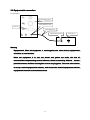
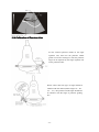
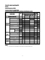
1
WED-160
Full Digital Ultrasonic Diagnostic System
(Version: V1.4)
Operation/Technical Manual
SHENZHEN WELL.D MEDICAL ELECTRONICS CO. LTD.
Preface........................................................................................................................................................... 1
Statement................................................................................................................................................. 1
Manufacturer's warranty.......................................................................................................................... 1
Matters need Attention.............................................................................................................................2
Safety labels.............................................................................................................................................2
Safety classification................................................................................................................................. 3
General tips for equipment operation...................................................................................................... 3
General Safety Message...........................................................................................................................4
Contraindication...................................................................................................................................... 5
1 Summary.....................................................................................................................................................7
1.1 Brief Introduction.............................................................................................................................. 7
1.2 Range Of Application........................................................................................................................ 7
1.3 Appearance........................................................................................................................................ 8
1.4 Technical Specificatio........................................................................................................................9
1.5 Electric principle block diagram......................................................................................................10
1.6 Standard configurations................................................................................................................... 10
1.7 Optional pieces................................................................................................................................ 10
2 Installation................................................................................................................................................. 11
2.1 Operation environmental requirements............................................................................................11
2.2 Connecting probe and main unit...................................................................................................... 11
2.3 Equipotential connection................................................................................................................. 12
3 Operating procedures............................................................................................................................. 13
3.1 Introduction of keyboard................................................................................................................. 13
3.2 Display interface.............................................................................................................................. 13
3.3 Operation Procedures.......................................................................................................................13
4 Setting.........................................................................................................................................................15
4.1 General.............................................................................................................................................15
4.2 Hospital Information........................................................................................................................15
4.3 Typical Case.....................................................................................................................................15
4.4 Obstetrical Algorithm...................................................................................................................... 16
4.5 Dicom Setting:................................................................................................................................. 18
4.6 Save Setting..................................................................................................................................... 19
4.7 Other Setting....................................................................................................................................20
4.8 Exit the System................................................................................................................................ 20
5 Adjust Image Parameters............................................................................................................................ 21
5.1 Switch Display Mode.......................................................................................................................21
5.2 Freeze Image....................................................................................................................................22
5.3 Adjust Gain...................................................................................................................................... 22
5.4 Adjust Displaying Depth..................................................................................................................22
5.5 Adjust Probe Scanning Angle.......................................................................................................... 22
5.6 Adjust Dynamic Scope.....................................................................................................................23
5.7 Focus Adjustment............................................................................................................................ 23
5.8 Probe Working Frequency............................................................................................................... 23
5.9 Switch the Probe.............................................................................................................................. 23
5.10 Frame Relative Adjustment........................................................................................................... 24
5.11 M Speed Adjustment......................................................................................................................24
-I-
5.12 Sound Power Adjust.......................................................................................................................24
6 Image Processing and Adjustment..............................................................................................................25
6.1 Tissue Harmonic Image................................................................................................................... 25
6.2 Pseudo-color Treatment................................................................................................................... 25
6.3 Image Enhancement.........................................................................................................................25
6.4 Full Screen Displaying Image..........................................................................................................25
6.5 Brightness and Contrast Adjustment................................................................................................25
6.6 GAMA Calibration.......................................................................................................................... 26
6.7 Image Turnover................................................................................................................................26
6.8 Histogram........................................................................................................................................ 26
6.9 Local Amplification......................................................................................................................... 27
7 Note............................................................................................................................................................ 28
7.1 Note in Full Screen.......................................................................................................................... 28
7.2 Body Sign Setting............................................................................................................................ 28
7.3 Input Arrow......................................................................................................................................29
7.4 Clear Screen.....................................................................................................................................29
8 Images and films.........................................................................................................................................30
8.1 Save Static Images........................................................................................................................... 30
8.2 Film Replay..................................................................................................................................... 30
8.3 Saving Films.................................................................................................................................... 30
8.4 Loading Films..................................................................................................................................31
8.5 Data Backup.....................................................................................................................................31
8.6 Import Patient Information.............................................................................................................. 31
9 Case Management and Report.................................................................................................................... 33
9.1 Open/Close Files..............................................................................................................................33
9.2 Inquire Files..................................................................................................................................... 33
9.3 Add New File...................................................................................................................................33
9.4 Modify Files.....................................................................................................................................34
9.5 Delete File........................................................................................................................................35
9.6 About Report....................................................................................................................................35
9.7 Report Form.....................................................................................................................................36
9.8 Print Report......................................................................................................................................36
10 Measurement.............................................................................................................................................38
10.1 General...........................................................................................................................................38
10.2 Routine Measurement.................................................................................................................... 38
10.2.1 Distance Measurement........................................................................................................38
10.2.2 Area Measurement.............................................................................................................. 38
10.2.3 Circumference Measurement.............................................................................................. 39
10.2.4 Angle Measurement............................................................................................................ 40
10.2.5 Volume Measurement......................................................................................................... 40
10.2.6 Other Measurements........................................................................................................... 41
10.3 Heart Measurement under Mode B................................................................................................41
10.4 Measurement under Mode M.........................................................................................................42
10.4.1 Overview.............................................................................................................................42
10.4.2 Heart Rate Measurement.....................................................................................................43
10.4.3 Time Measurement............................................................................................................. 43
- II -
11 Operating Instruction for Function of Puncture Guide......................................................................44
11.1 Fixation of Puncture Frame and Probe...........................................................................................44
11.2 Selection of Puncture Needles....................................................................................................... 45
11.3 Call out the Puncture Guding Line................................................................................................ 45
11.4 Calibration of Puncture Line..........................................................................................................46
11.5 Insertion of Puncture Needle..........................................................................................................47
11.6 After Puncture................................................................................................................................ 48
12 Check and Maintenance.......................................................................................................................49
12.1 Service life..................................................................................................................................... 49
12.2 Main unit maintenance...................................................................................................................49
12.3 Probe maintenance.........................................................................................................................50
12.4 Correct usage of probe...................................................................................................................51
13 Simple Trouble Shooting...................................................................................................................... 52
13.1 Check............................................................................................................................................. 52
13.2 Simple trouble shooting................................................................................................................. 52
14 Transportation and Storage................................................................................................................. 53
14.1 Environment requirements on transportation and storage............................................................. 53
14.2 Transportation................................................................................................................................ 53
14.3 Storage........................................................................................................................................... 53
Appendix A Acoustic output reporting table.............................................................................................54
- III -
Preface
Statement
�
This publication, including pictures and illustrations, is property of Shenzhen Well.D Medical
Electronics Co., Ltd. and under protection of international copyright law.
�
Information in this document is not annotated to change. The manufacture shall not state nor
observe any warranty basing on this point, and definitely give up any implied warranty basing on
any special purpose of selling or making benefit.
�
Without previous written permission from the producer, this document must not be photocopied,
reproduced or translated into other languages.
�
We preserve the right of revision on this document without still further notice.
�
Some pictures in this manual, which are schematic diagrams for indication only, may disaccord
with the real object, and then the real object should be regarded as the final.
�
is trade marks of Shenzhen Well.D Medical Electronics Co., Ltd. Any abuse of these trade
marks without permission will be sued to assume legal responsibility according to laws.
Manufacturer's warranty
�
Shenzhen Well.D Medical Electronics Co., Ltd. assumes the responsibility for equipment security,
reliability and performance only under the preconditions that the disassembly, assembly and
maintenance of the equipment are all performed by its assigned professional and the equipment is
used strictly in compliance with the operation manual.
�
Shenzhen Well.D Medical Electronics Co., Ltd. ensures a guarantee period within a year and a
half since the delivery day and promises there is no problem with the new equipment in material
and technology. Within the guarantee period, Shenzhen Well.D Medical Electronics Co., Ltd. will
maintain the equipment and replace the parts of non-man-made damages free of charge. But will
not repair or replace the equipment surface if it is damaged.
�
This guarantee is only available for failures occurred when the equipment is operated in
compliance with the operation manual. And the guaranteed equipment can only be used in the
prescribed range given in manual.
�
This guarantee excludes losses or damages caused by external reasons such as thunder struck,
earthquake, theft, unsuitable use or abuse and refitting the equipment.
�
Shenzhen Well.D Medical Electronics Co., Ltd. shall not be responsible for damages caused by
other devices or arbitrary connection to other devices.
-1-
�
Shenzhen Well.D Medical Electronics Co., Ltd. shall not be responsible for losses, damages or
injuries caused by delayed service request.
�
When there is problem with the products, please contact Shenzhen Well.D Medical Electronics
Co., Ltd. and explain the equipment model, serial number, date of purchase and the problem.
Matters need Attention
�
To ensure operational safety and long-term stable equipment performance, please read this
operation manual closely and understand the equipment functions, operation and maintenance at
all points before operating the equipment, especially contents of "Warning", "Caution" and
"Note".
�
Maloperation or inobservance of the instructions given by manufacturer or its agents may result
in equipment damage or personal injury.
�
The following convention works through this manual to lay special emphasis on some
information.
�
"Warning":
Stands for neglect of it will cause severe personal injury, death or realized
property loss.
�
"Caution": Stands for neglect of it will cause slight personal injury or property damage.
�
"Note": to remind user of installation, operation or maintenance information. These
information is very significant but with no risk. Any warning against dangers shall not be
contained in NOTE.
Safety labels
Equipment labels explanation:
Type B equipment
Note!
documents
Refer
to
accompanying
Turn-on (general supply)
Disconnect (general supply)
VIDEO
Equipotential
IPX7
Protection against dripping water
USB
XGA
-2-
Packing and transportation labels explanation:
Handle carefully
Temperature limit
Upward
Piling layer limit
Keep dry
Protect against heat
Safety classification
According to the degree of safety of application in the presence of a flammable anaesthetic mixture
with air or with oxygen or nitrous oxide
oxide::
WED-160 not suitable for use in the presence of a flammable anaesthetic mixture with air or with oxygen
or nitrous oxide;
According to the mode of operation:
Continuous operation;
According to the degree of protection against harmful ingress of water as detailed in the current
edition of IEC 529
529:
Main unit is general equipment,the probe is enclosed equipment protected against the effects of
immersion,IPX7.
According to the type of protection against electric shock:
ClassⅠequipment energized from an external electrical power source.
equipment;
According to the degree of protection against electric shock:
Type B
General tips for equipment operation
◆
In operation
1.
Heat radiation holes are strictly prohibited to be covered.
2.
After closedown, do not switch on the equipment within 2 - 3 minutes.
-3-
3.
On scanning, if any abnormal case is found, stop scanning immediately and shut down
the equipment.
4.
The patient is prohibited to touch any non-applied part of the equipment.
5.
When operating, do not press the keyboard panel too much, otherwise the equipment
might be damaged.
◆
After operation
1.
Power off the equipment.
2.
Pull out the plug from power supply socket instead of pulling the cable.
3.
Clean off the couplant on the probe with soft medical sterilized cotton ball.
4.
Put the probe into the special case.
General Safety Message
Safety of the operator and patients and reliability of the equipment are taken into consideration
during designing and producing, the following safety precaution must be implemented:
1.
The equipment shall be operated by qualified operating staff or under their instructions.
2.
Do not open the equipment and change the parameters without permission. If necessary,
please turn to for Shenzhen Well.D Medical Electronics Co., Ltd. or its authorized agent
for service.
3.
The equipment has already been regulated into its optimal performance. Do not adjust
any preset control or switch unless operate as per instructions in the manual.
4.
If there is equipment failure, please shut down the equipment at once and contact for
Shenzhen Well.D Medical Electronics Co., Ltd. or their authorized agent.
5.
If it is needed to connect the equipment with other company’s' electronic or mechanical
devices , please contact Shenzhen Well. D Medical Electronics Co,. Ltd. before
connection.
6.
Equipment operation, storage and transportation environment
Environmental requirements on normal operation:
a) Environment temperature range: 0℃~+40℃
b) Relative humidity range: ≤80%
c)
Atmosphere pressure range: 70KPa~106KPa
Environment requirements on equipment storage and transportation:
d) Environment temperature range: -20℃~+55℃
e) Relative humidity range: <80%(20℃)
-4-
7.
Do not hit the fragile TFT-LCD display. If it cracks, deal carefully with it in case the liquid
crystal gets into eyes or mouths.
8.
Must not hit the inner chargeable Li-ion battery nor throw it into fire in case it trigger an
explosion ; Do not short circuit the battery output electrodes in case the battery be
damaged; and please use the original binding charger to charge the battery. More over,
because used battery will cause environment pollution, please handle the battery
correctly for recovery processing.
9.
Must not disassemble the power supply adapter. If failures happen, it should be handled
by the professional; the charging output can only be used for charging the battery of the
equipment, any improper use on other battery may cause explosion, fire and other
unexpected hazards.
10. Must not short circuit the output of the adapter, a long term short circuit shall result in
adapter damage.
11. Please use standard power cord as the input line of the network power supply for the
adapter to reduce risk.
12. Shenzhen Well.D Medical Electronics Co., Ltd. shall not take any responsibility for any
risk resulted from unauthorized re-fitment by the users.
13. To disconnect the equipment from the power supply network by unplug the adapter from
the power supply network.
14. Refer to appendix A for sound output parameters.
Contraindication
★ This equipment is not suit for contain-gas viscera, such as lung examine.。
★ Suggesting not to examine the wounded or urgent inflammation part, it could Cause
across infect.
★ Below patient forbided to use vagina and recta probe:
Vagina colpitis, such as colpitis, and sexual illness; discoverture people; vagina
abnormality; menses; vagina shrink;vagina ultrasound exame difficulty person; vagina
bleeding; placenta front sufferer.
★ Below sufferer is forbidden to puncture:
high blood pressure, coronary heart disease, cruor obstacle and haemorrhage separative
sufferer.
-5-
Warning
When WED-160 works in strong electromagnetic environment exceeding its
statement, its image might be interfered and the diagnoses could be influenced. At this
time, please stop operating until the EMC interference is removed.
Warning
When WED-160 works at the state of overlapping or paralleling with other
If it must work close to other
equipments, there might be unexpected EMC problems
problems;If
equipments, please observe carefully and check if some equipment is influenced by
unexpected EM coupling.
Warning
Replacement of non-standard probe parts may cause unexpected EMC problem.
NOTE
Accessory equipment connected to the analog and digital interfaces must be
certified according to the respective IEC standards (e.g. IEC 60950 for data processing
equipment and IEC 60601-1-1 for medical equipment). Furthermore, all configurations
shall comply with the valid version of the system standard IEC 60601-1-1. Everybody
who connects additional equipment to the signal input or signal output part configures
a medical system, and is therefore responsible that the system complies with the
requirements of the valid version of the system standard IEC60601-1. IF in doubt,
consult the technical service department or your local representative.
-6-
1 Summary
1.1 Brief Introduction
�
WED-160 is a high resolution convex/linear
array scanning ultrasound diagnostic
system ,which utilizing the PC control and digital scan converter(DSC) while adopting multiple
technologies such as digital beam forming (DBF) , real-time dynamic aperture (RDA), dynamic
receive apodization (DRA), dynamic receive focusing (DRF), dynamic frequency scan (DFS), 8-TGC
as well as frame correlation , and it can support various linear and convex array probes with a wide
�
�
�
�
�
�
�
�
range of frequency.
It provides five display modes as B、B+B、B+M、M、4B and gray scale of 256 levels.
It provides such functions as real-time ,freeze, magnification, up and down flip, left and right
flip ,edge enhancement, gamma calibration of the picture.
It allows the continuous playback and single frame play of 500 frames at most, while static
pictures of 1000 frames can be stored and recalled at most. The patient data can be saved while
saving the pictures.
It provides multiple level regulation for scanning depth, scanning angle, dynamic range, acoustic
output power, frame correlation factors ,focus number, distance between focuses, focus position.
It functions at display of date and time; annotation of name ,sex ,age of patients, doctor and
hospital’s name ;full screen annotation. It allows to switch in the Chinese and English operating
interface, to input in Chinese or English input mode, and to choose the arrow mark as well as 40
body posture marks.
It possesses abundant software such as routine measurement (including distance, area and girth,
angle and volume),heart measurement (including distance ,time, slope , heart rate)and obstetric
measurement ( to estimate the gestational weeks ,expected date of confinement and fetus weight
by BPD、CRL、GS、FL、HC、AC)
This device has the DICOM3.0 port, thus can transmit the picture in the DICOM format. It also
provides the color video output, thus it can be connected with the external display with the big
screen .In addition, the device has the high speed USB2.0 port ,thus the picture can be saved into
the portable storage device and it can be connected with an external video printer. There is a
built-in memory with high-capacity which can save a large mount of pictures.
The device has a injection moulding envelop and the usage of non-industrial frequency
transformer switching power supply, programmable parts and surface mounting technology (SMT)
make the whole unit highly compact, small in volume and light in weight.
1.2 Range Of Application
Application in abdominal, Obstetric, Cardiac, small parts sonography.
-7-
1.3 Appearance
Power switch
USB2.0 port
RS-232
Dicom3.0
XGA
Video out
Fuse
GND
-8-
Power
socket
1.4 Technical Specificatio
Probe
C3-1/60R/3.5 MHz
L3-1/7.5Mhz
C1-6/20R/5.0MHz
EC1-1/13R/6.5MHz
convex array
HF linear
micro convex
endo-vaginal
Display depth (mm)
250 (max)
≥190
≥80
≥90
≥90
Lateral
≤2
(depth≤80)
≤3
(80<depth≤130)
≤1
(depth≤60)
≤1
(depth≤40)
≤1
(depth≤40)
Axial
≤1
(depth≤80)
≤0.5
(depth≤80)
≤0.5
(depth≤40)
≤0.5
(depth≤40)
≤3
≤3
≤2
≤2
Maximal detect depth (mm)
Resolution
(mm)
Blind zone(mm)
Geometric
position
precision
Horizontal
≤0
≤3
≤4
≤2
Vertical
≤3
≤5
≤2
≤2
Monitor size
12.1 Inch TFT LCD
Display mode
B, B+B, B+M, B+M/M, M, 4B
Image gray scale
256 Scale
Cine loop
≥600 Frame
Image storage
≥1000Frame
Scan angle
Adjustable
Scan depth
40mm-250mm
Image flip
Up/down, left/right, black/ white
Image Process
Focus
Measure
Character display
GAMA, Image Smoothen, THI, Pseudo color, Histogram, Zoom
Focus Number, Focus position, Focal space
Distance, circumference, area, volume, heart, GA, FW, EDD
Date, clock, name, PID, age, Sex, hospital name, doctor,
Notation
full-screen character editor, posture mark, Position indication
USB port
USB2.0
power consumption (MAX)
100VA
Net weight
6.4kg
Size
289.16mm×304.96mm×222.02mm
-9-
1.5 Electric principle block diagram
1.6 Standard configurations
�
Mainframe
�
C3-1/60R/3.5MHz convex array
�
PS cable
�
2 pieces of fuse tube F2AL250V
�
Coupling gel 250ml
�
User Manual
�
Final examination report
�
Packing List
1.7 Optional pieces
�
C1-6/20R/5.0MHz Micro-convex probe
�
L3-1/7.5Mhz HF linear probe
�
EC1-1/13R/6.5MHz Endo-vaginal probe
�
Monitor
�
Video printer
Warning:
Please use the spare parts specified above. The manufacturer shall not bear the
responsibility of safety problem, unexpected EMC performance drop caused by
arbitrary adoption of non-specified spare parts.
- 10 -
2 Installation
2.1 Operation environmental requirements
�
Environment temperature range: 0℃ ~+40℃
�
Relative humidity range: ≤80%
�
Atmosphere pressure range: 70kPa~106kPa
�
Power supply: a.c.100V~240V,50Hz/60Hz
�
Avoid strenuous vibration during operation; Keep away from equipments with high
electric field, high magnetic field and high voltage; avoid strong sunlight on the display;
Keep the equipment well-ventilated, damp proof and dustproof.
Note:
Check according to the "packing list" after uncasing and make sure that there is no
shipping damage, and then install the equipment according to requirements and
methods described in “Installation
”.
Installation”
Warning:
If there is breakage at unpacking check, it is banned to use the device to ensure
security.
The probe should be protected from felling off or crashing and the manufacturer
manufacturer..
ng probe and main unit
2.2 Connecti
Connecting
Check the power supply too see if it is in the expected range (AC100V-240V,50Hz/60Hz), then connect
the equipment and the power socket with the cable.
Connect the probe:
①Insert the probe
- 11 -
② Locking the
probe
along
this direction
2.3 Equipotential connection
See the figure:
Equipment back panel
Equipotential terminal
VGA out
Equipotential line
Equipotential terminal
Other device
Warning
Equipotential: When this equipment is used together with other device, equipotential
should be in consideration.
When the equipment is in use, the doctor and patient are under the risk of
uncontrollable compensating current influence, which is caused by different
electric
potential between facilities and tangible current carrying parts. The most safe solution
and connect the medical equipments with the
is set up a united equipotential network
network,and
equipotential network in the treatment room.
- 12 -
3 Operating procedures
3.1 Introduction of keyboard
TGC
Keyboard
(alphanumeric
keys)
Function keys
Trackball
3.2 Display interface
Parameters
displaying area
Images
displaying area
Control Panel
System setting
3.3 Operation Procedures
1.
Press button
on left side of machine to start the device;
2.
Select examine type;
3.
Apply medical ultrasonic coupling agent on the sound window surface of probe, press it on
- 13 -
scanning place, and the real time images will be displayed on screen;
4.
Freeze the image, and start measuring operation on it;
5.
Press REPORT key and input the basic information of patient, then save;
6.
Press Print key or Click REPORT to generate and print reports.
- 14 -
4 Setting
4.1 General
Click
to open SETTING window, and click
on upper right to close.
Setting consists of six parts: Hospital Information, Typical Case, Obstetrical Algorithm,
Dicom Setting, Save Setting and Other Setting.
4.2 Hospital Information
Hospital Name, Telephone and Address refer to the information of examining hospital, wherein,
the hospital name and telephone number will be included in the reporting page automatically.
The diagnosis doctor and examine doctor in examine file can be added or deleted via Doctor List
here, and the examine result can be added or deleted via the Examine Result here.
4.3 Typical Case
It refers to the typical cases in the original systematic examine files, which can also be manually
added. Common cases encountered during examine can be added via the Typical Case here if the
case is not included originally in the system.
- 15 -
Besides the categories originally included in the system such as the obstetrics and gynecology
reporting form, liver, gall, pancreas and spleen lists etc., Case Category pull-down list also
includes a Customer Typical Case option. Choose Add New Typical Case via this pull-down
list to add new case under specific case category.
4.4 Obstetrical Algorithm
Preset the algorithm before obstetrical measurement in this window.
Beside of modifying obstetrical algorithm presets, there are three more blocks, respectively
Gestational Age Table, Fetus Growth Curve and Fetus Physiology Score.
Gestational Age Table:
Click
to enter, there are mainly two parts: preset obstetrical list and user edit
mode, respectively as shown in the figure below:
Preset Obstetrical List
User Edit Mode
Move the cursor to corresponding obstetrical list and click, there are 31 sheets of obstetrical list,
wherein, **User refers to user edit mode, shown as follow:
BPD(Tokyo Method)
AC(Hadlock Method)
HL(Hansmann Method)
BPD(Hadlock Method)
AC(Korean Method)
HL(Jeanty Method)
BPD(Korean Method)
AC(Merz Method)
HL(Osaka Method)
BPD(Hansmann Method)
AD(Hansmann Method)
OFD(Hansmann Method)
- 16 -
BPD(Merz Method)
CRL(Tokyo Method)
FL(Tokyo Method)
CRL(Korean Method)
FL(Hadlock Method)
CRL(Hansmann Method)
FL(Korean Method)
CRL(Robinson Method)
FL(Merz Method)
GS(Tokyo Method)
HC(Hadlock Method)
GS(Korean Method)
HC(Korean Method)
GS(Hansmann Method)
HC(Hansmann Method)
GS(Hellman Method)
HC(Merz Method)
AFI(Korean Method)
FTA(Osaka Method)
Fetus Growth Curve:
Click
and enter, as shown in the figure below:
The system includes totally 31 types of fetus growth curves for users’ reference, which can be
exited by pressing
.
Fetus Physiology Score:
Press
and enter, as shown in the figure below:
Choose corresponding scores in the fetal breath movement, fetal movement, fetal tone and
amniotic fluid index options according to physical development conditions of fetus and
- 17 -
press
. The final Fetus Physiology Score appears in reporting pages.
4.5 Dicom Setting:
�
DICOM Overlay Information is used to set whether characters information will be loaded or
not during saving DICOM images.
�
Window Center and Window Width is used to set up the contrast and brightness during
saving DICOM images.
�
When auto send
is checked on, the film files will be DICOM transmitted
when they are being saved.
�
The Dicom Transmitting function is added for pictures in the form of .dcm can be adjusted
with its gray scale, which is helpful to doctor diagnosis.
�
Dicom transmission refers to transmission of pictures in .dcm forms between two machines
installed with Dicom protocol, and the specific settings are included in the Dicom Setting
heare. Dicom settings are required both for transmitting end and receiving end, the receiving
tips: Can only transmit .DCM format
end number and server end number must be same. (tips:
images or movies
movies)
�
Dicom transmission requires receiving end and transmitting end, this machine can be used
as both. When it is used as receiving end, the received parameters can be set in receiving end
setting, and the local receiving function should be started; if the local receiving is disabled,
the transmitting fails. If the machine is used as transmitting end, specific settings are shown
as follow:
1)
Set the parameters on transmitting end;
- 18 -
2)
Click
3)
Click
4)
Click
, open the interface shown as follow.
and pop up the dialogue frame, then select open;
, send .dcm pictures to receiving end, and Successfully Sent
dialogue frame pops up;
5)
Click
.
4.6 Save Setting
After customer saving route is checked, the system will remind the users to select route when the
images are saved every time.
It is also possible to modify default saving route, and click
.
The images can be saved with optional form, including .bmp, jpg, .dcm and tif forms
The film forms include .seq, DCM (DICOM) and avi.
Press
after set.
- 19 -
4.7 Other Setting
�
The SHOW and HIDE on main menu, which can switch the displaying and hide
�
�
of
Machine Type can be used to set the type of machine.
Language bar can be used to set the displaying language of entire software.
�
can be used to periodically backup files etc. to prevent data lost.
4.8 Exit the System
Click
Press
Press
, and following dialogue frame appears:
to stop the device and put it into standby status;
to exit B-ultrasound interface and enter into windows operation system.
- 20 -
5 Adjust Image Parameters
5.1 Switch Display Mode
Switch to Mode B
Press key B, the current image is switched to real time single B mode (device start default mode).
Switch to Mode BB
Press key BB
BB, the current image is switched to double B mode, the screen displays both real time
and frozen images. Press key BB again to switch the real time and frozen status of the two
images.
Switch to B+M Mode
Press key BM/M
BM/M, and the current image is switched to B+M mode, the screen displays a type B
and a type M real time images at same time, and the sampling line on type B image can be moved
horizontally by trackball.
Switch to Mode B+M/M
Under B+M or M mode, press key 4B and switch the current image to B+M/M mode, the screen
displays a type B and two type M real time images, the two sampling lines on type B image can
be moved horizontally by trackball, and press key L/R to switch the line to be moved.
Switch to Mode M
Under Mode B+M
B+M, press key BM/M to switch the current image to Mode M, the screen displays
a type M image.
Switch to Mode 4B
Under Mode B and BB, press key 4B to switch the current image to 4B mode. The screen
displays four type B images including one real time mode. Continue to press key 4B to switch the
real time and frozen status of the four images.
B
BB
- 21 -
B+M
B+M/M
M
4B
5.2 Freeze Image
Press key FREEZE to switch real time and frozen status.
5.3 Adjust Gain
The Total Gain: 32dB on right side of screen is the current gain, which is adjusted via button
GAIN
GAIN. The close/middle/remote fields can be adjusted via the 8 sections of TGC on keyboard.
5.4 Adjust Displaying Depth
Under real time mode (non-4B mode), dial DEPTH up and down to change the current
displaying depth (or scale). The current depth display is on right side of screen, and the
adjustment scope changes along with probe type. Take the following tale for reference.
Probe Type
C3-1
Convex Array
probe
L3-1
High Frequency
Linear array probe
C1-6
Slight Convex
probe
EC1-1
Intracavity Probe
Depth
(mm)
90~250
40~105
50~125
50~125
Depth 90
Depth 180
Depth 240
5.5 Adjust Probe Scanning Angle
Dial ANGLE up and down in real time scanning status to adjust the size of angle, and take the
following table for reference on probe angle.
Probe Type
C3-1
convex array probe
EC1-1
Intracavity probe
C1-6
micro convex probe
Angle
60ºvisible and adjustable
120ºvisible and adjustable
90ºvisible and adjustable
- 22 -
5.6 Adjust Dynamic Scope
Dial DR up and down in real time scanning mode to alter the dynamic scope which is from 0 to
192dB, with adjustable range at 64 to 192dB
5.7 Focus Adjustment
Focus Number
PIN up and down under real time B or BB modes to switch the focus numbers, and the
Dial FOC
FOCPIN
screen displays the current focus number such as Focus Number: 2 on right side.
Focus Distance
When focus is ≥2, dial FOCSPAN up and down to select focus combination state, and change
the size of focus distance by knob PARAMADJU
PARAMADJU. The current focus distance value will be
displayed on right side of screen.
Focus Position
Dial FOCPOSI up and down in real time scanning status to move the focus vertically and change
its position.
Notice:
Images near focus indicator
have better focusing. It is possible to change the focus
number, position and distance to direct the focus to interested area.
5.8 Probe Working Frequency
Dial FREQUENCY up and down in real time scanning status to switch the frequency of
currently working probe, and the current working frequency will be displayed on upper right
corner of screen, such as Frequency: 35MHz. The frequency scopes of probes are shown as
follow:
C3-1 Convex Array probe —— 2.0MHz, 2.5MHz, 3.5MHz, 4.5MHz, 6.0MHz
C1-6 Slightly Convex probe —— 4.5MHz, 5.0MHz, 5.5MHz
EC1-1 Intracavity probe —— 4.5 MHz, 5.5MHz, 6.5MHz, 7.5MHz, 8.5 MHz
L3-1 High Frequency Linear Array probe —— 5.5 MHz, 6.5MHz, 7.5MHz, 8.5MHz, 12 MHz
5.9 Switch the Probe
When the device is connected with 2 probes, the automatically identified plug port A is default
working probe after start, and the probe type is displayed on upper right corner of screen, such as
Probe: R50A. Press key PROBE-S and switch to plug B, and then probe on plug B starts to
- 23 -
work.
Notice:
Two probes can not work together.
Stop the device before plugging in/out the probes and restart after probes are properly
connected.
5.10 Frame Relative Adjustment
Under mode B, BB and 4B, dial M Velocity/Frame Correlation and down to change frame size
which will be displayed on upper right side of screen in real time, such as Frame Relative: 0.55,
with adjustable scope as 0.20 to 0.95.
5.11 M Speed Adjustment
Under real time B+M, B+M/M and M mode, dial M Velocity/Frame Correlation up and down
to adjust the brush speed of type M images on screen. The current scanning speed will be
displayed on left side of screen, such as M speed: 6ms. There are four optional gears for the
device: 6mS, 8mS, 10mS and 12mS
5.12 Sound Power Adjust
Under real time scanning mode, continuously click
to adjust the current scanning
sound output power.
Notice:
Ultrasonic waves may do harm to human body, and long time radiation with high power
must be avoided. Be careful when using the function. The probe may heat up when the
power of ultrasonic wave is added.
- 24 -
6 Image Processing and Adjustment
6.1 Tissue Harmonic Image
During real time scanning status, continuously click
to enable/disable tissue harmonic
image. Tissue harmonic weave status will be indicated on right side of screen: Tissue Harmonic:
ON.
Able Tissue Harmonic
Disable Tissue Harmonic
Notice:
Tissue harmonic is only valid under abdomen probe.
color Treatment
6.2 Pseudoseudo-color
Click ▼ in right side of
to alter the current false color value,
there are totally 10 types of false color settings.
on the lower part is
used to set the threshold of false color, drive the slide block or click
to adjust.
6.3 Image Enhancement
Click ▼ in right side of
to select image enhancing function,
there totally 8 statuses optional.
6.4 Full Screen Displaying Image
Click
, the image is displayed in full screen, and press key R to exit.
6.5 Brightness and Contrast Adjustment
The
on right side of screen corresponds to brightness and contrast
adjustment slide blocks, drive the slide block or click
- 25 -
to adjust
6.6 GAMA Calibration
Sequentially click
� GamaAdjust to enable the function. The screen displays as
following frame:
Directly drive The slider to adjust GAMA value.
Notice:
GAMA calibration may be required to normally display images for externally connected
displayer.
6.7 Image Turnover
Turning Over Up and Down
Dial U/D up and down to turn over the image up and down.
Turning Over Lift and Right
Under B or BB mode real time scanning status, dial L/R up and down to turn the image over left
and right. The
on upper left side of screen is the image scanning direction indication.
6.8 Histogram
� Histogram to display histogram.
After image is frozen, sequentially click
- 26 -
6.9 Local Amplification
Press key ZOOM
ZOOM, and move cursor to image area, the cursor changes to 【十】. Press mouse left
and select area via trackball, then the image of the area is amplified and displayed in the center of
screen, which can be moved by trackball. Press key ZOOM again to exit local amplification.
Press ZOOM and adjust knob ParamAdju to amplify or shrink the image, and the amplified rate
is indicated on upper left side of image in real time. The range of rate is Zoom in: 1.1~Zoom in:
5.0. Press ZOOM to exit.
- 27 -
7 Note
7.1 Note in Full Screen
Press key TEXT, Input note content and then click anywhere in image area to finish noting.
Press � to delete wrongly input words.
Press Ctrl+Shift to shift input method.
In order to move the note content, click it on screen until it becomes green and cursor changes to
direction icon, and then move the content and click again.
To delete a comment content, Press right key to choose the comment content, Then press Delete.
7.2 Body Sign Setting
Sequentially press key BODY � BodyMark, and body signs are indicated on right side of screen.
Click corresponding icons to show body signs on lower right corner or screen. Following is brief
introduction to partial body signs.
1. Abdomen:
Body front side (male)
Body front side (female)
Body back side
Body left side
Body right side
2. Obstetrics
Mother body (front side)
Fetus
3. Head:
4. Heart:
5. Others:
Sequentially press BODY � Remove BodyMark to delete body signs.
- 28 -
7.3 Input Arrow
Sequentially press BODY � Arrow
Arrow, the cursor changes to +. Click anywhere in image area, and
drive the arrow to expected length, and lick again to input arrow. Repeat to make more arrows.
7.4 Clear Screen
Press CLEAR to clear the measuring mark, notes and body signs etc. information in image area.
- 29 -
8 Images and films
8.1 Save Static Images
Images can be saved in .bmp, .jpg, .dcm and .tif forms, see details in Save Settings.
Pictures in .dcm form can be adjusted with gray scale, which is helpful for doctor diagnosis.
Operation Procedures:
1.
In real time or frozen status, continuously press key GRAB to save the current image in
memory. Image preview window is indicated on right side of screen, and 5 current images
can be saved at max.
2.
Click
3.
Select examine type in examine file column, and then click
�
, and sequentially click
Save New Record or Not
Not, select YES
YES, and click
to build a new personal document;
to pop up dialogue frame
to exit case information window;
1.
After operations above, the saving route of saved image is:
D:\Data\Patient-Image\Year-month\Year-month-day\Case Number-Patient name\
For example: if a patient named Lily was examined in Nov. 10 in 2009 with her case number
as 1, then the saving route of saved image is:
D:\Data\Patient-Image\2009-11\2009-11-10\1_Lily.
Notice:
Restart the software and save image, the images saved last time will be covered.
� Double click in the menu Image Preview on right side of screen to open the image, wherein,
images in .dcm form can be adjusted with gray scale. Press FREEZE and return to current
active status.
� Right click in the menu Image Preview on right side of screen to select CLEAR and
selected image is removed. It is also possible to clear all.
8.2 Film Replay
�
�
�
After startup and entered into real time scanning status, the device collects the current
images into memory, and press FREEZE after about 30S to freeze the image.
Press knob Play/Pause to start film replay, the images will be displayed in cycle. Current
replay status is displayed on lower right corner of image, for example: Frame 0-XXX/XXX
0-XXX/XXX,
respectively represent current frame and total frame number.
Please press knob Play/Pause to suspend replay, and adjust the knob to realize single frame
reply. Press again and switch back to automatic recycling replay status. Press key FREEZE
and return to current active status.
8.3 Saving Films
Film saving forms: the films can be saved in .seq, DICOM and AVI form. See Saving Settings
for detail.
- 30 -
Film saving route:
Click
to pop up saving route selection dialogue frame.
Notice:
Please do not try other operations during film saving
8.4 Loading Films
If there are film files on hard disk, choose
in right menu, and select
corresponding file in dialogue frame popped up. Click open button to display automatically.
(Note the file type)
If film files in DICOM form are loaded, press left button in image area, and move trackball to set
image brightness
8.5 Data Backup
Start software, and plug in movable device (for example U disk). Click
select
and
to enter into following interface:
Select
to copy the Typical Case, Setting Files, Saved Images and Data
Base (patients’ information) to movable device and backup.
Notice:
In order to prevent data lost, please ensure to backup patients
patients’’ information before replacing
hard disk or upgrade/resume system.
8.6 Import Patient Information
Notice:
Before importing, ensure the patients
patients’’ information is backed up according to 8.5.
Since Welld.mdb files include all the word information of patients, these files refresh every
- 31 -
time a patient
patient’’s file is newly added or existed case is modified. So consider twice before
importing patients
patients’’ information.
1.
2.
Without replacing hard disk, all the data in windows disk D will remain unchanged after the
system is upgraded or resumed, the data in disk D will be recalled after repeated
upgrade/reset. Under such condition, the patients’ information is not required to be imported
or resumed. If it is needed, ensure there is no newly added case or case data is unchanged
after backup, and then import information (with following method). Otherwise, the modified
or newly added data will permanently lost unless you do not need them, please do remember
this!
When the hard disk is replaced or formatted, backup patients’ information needs to be
imported: First resume the system, and then copy the corresponding Data files (Typical case
or Setting), saved images and data base (patients’ information) in disk or movable memory
device to windows disk D and cover original files, then enter into B-ultrasonic system. Press
REPORT on keyboard or click
on main interface to enter into file interface, shown
as follow.
At this time, Search, Modify, Delete and Print etc. operations are available.
- 32 -
9 Case Management and Report
9.1 Open/Close Files
�
Click
on main interface and enter into file interface, shown as follow.
Patient
information
Diagnosis
report
�
Click
on upper right corner of file window to close file window.
9.2 Inquire Files
�
�
�
Open file window, the inquiry status is systematically defaulted.
The system allows inquiry to relative files via numbers, names and ID. Input inquiry content
in numbers, names and ID frames, and press the key ENTER on standard PC key board.
Beside of inquiring word documentary information, when one of the diagnosis reports is
clicked, the system will also load on the saved images correspondent to the report, and the
details are shown in the drawer menu Image Preview on right side. If the report is printed,
then the first two images are printed (in the order of saving).
9.3 Add New File
�
�
1.
If the patient’s information is not included in data base, it needs to be added.
Newly built files are mainly composed of two parts: newly built patient information and
newly built diagnosis report. The patient information must be established before diagnosis
report. See the details as follow:
Build new patient information (method 1):
1)
Click
2)
Input other relative information in patient information part.
and serial number is automatically added.
- 33 -
3)
Click
to confirm if the newly built patient information needs to be saved; click
to
cancel the new patient information.
2.
1)
Build new patient information (method 2):
Under this method, it needs to provide a port to another PC, and then the patient registered
on the other PC will be indicated in working list.
2)
Click
3)
It is mainly composed of two parts: Working List and Current Examines, the Current
Examine will sequentially indicate the waiting patients in Working List.
4)
Click
to open working list window, shown as follow:
and add the basic information of patient currently examined into Patient
Information.
3. Build new examine file: the following procedures are executed on the premise that the
newly built patient information is confirmed to be saved, otherwise they are not to be executed.
1) Fill relative information of examine file.
2)
Click
to confirm and save.
Notice:
If the examine result is included in the typical cases, then open the
on right side,
double click existed description and directly import current examine file.
Press
to import current examine file into Typical Case.
Select one item in typical cases list and right click, choose DELETE to remove it. The
typical cases originally existed in the system can not be removed.
The report date is current date, which can be modified by clicking down arrow.
�
Multiple examine files (i.e. diagnosis report) can be added to the same Patient Information.
9.4 Modify Files
�
�
If a file is included in data base, which needs to be modified with some content, then choose
Modify File.
There are mainly two parts needed for modifying a file: modifying patient information and
- 34 -
1.
1)
modifying diagnosis report. See the details as follow:
Modify Patient Information
Recall the file needs to be modified by Inquiry File.
2)
Click
3)
Modify relative content in patient information.
4)
2.
1)
2)
Click
to confirm if the modified content needs to be saved, and click
modification.
Modify Examine File:
Recall the file needs to be modified via Inquiry File.
Modify the relative content in the file.
3)
Click
.
to cancel the
, there is a dialogue frame Modify Current Diagnosis Report or Not
Not, click YES
to confirm the modification, and Successfully Modified window pops up.
9.5 Delete File
�
�
1.
1)
2)
2.
1)
2)
3)
Some files in the data base that are not needed any more, such files can be deleted.
There are two conditions in deleting files: deleting Patient Information and deleting
Diagnosis Report. The entire file is deleted when Patient Information is deleted. When
Diagnosis Report is deleted, only one of the diagnosis reports under the Patient Information
is deleted.
Delete Patient Information:
Recall the file needs to be deleted by Inquiry File.
Click
and dialogue frame Confirm to Delete Current Patient Information appears,
then click YES to confirm deletion.
Delete Diagnosis Report:
Recall the file which contains the diagnosis report needs to be deleted.
Select the diagnosis report needs to be deleted via trackball and light mouse.
Click
and dialogue frame Confirm to Delete Current Diagnosis Report appears.
Click YES to confirm deletion.
Notice:
Keys ADD, MODIFY, DELETE, SAVE and CANCEL are available when they are colored,
and invalid when they are gray.
9.6 About Report
�
�
Report form will be automatically indicated according to the examine type in Files �
Examine File
File.
Routine measurement will not be included in report page.
- 35 -
�
Report will add pages automatically according to measured items, in order to ensure the
information on report page is fully indicated.
9.7 Report Form
�
1.
2.
3.
4.
5.
6.
�
There are six parts in report, with details as follow:
Report single title: hospital name + colorful Doppler ultrasound examination report form.
Basic information of examined patient: including examine type, examiner number, name,
gender, age, room number and bed number.
Image displaying area: saved image will be displayed here, with two images at max.
Measuring value record: composed of measuring parameter and measuring value.
Clinical content: including clinical symptoms and clinical diagnosis conclusion.
Basic information of examine unit: including examine doctor, report doctor, examine date,
Tel and examine unit (stamp).
See the following report forms on following pictures for detail.
9.8 Print Report
�
�
Print
Selected the corresponding check the file before printing, then click "Print
Print" button to print.
Print
If you created a report, Press the "Print
Print" to print the report.
- 36 -
1
2
3
4
5
6
- 37 -
10 Measurement
10.1 General
�
�
�
The device can give multiple measurements to multiple items, with six measuring marks: ,
, △, □, ◇ and ○.
The device provide more than 10 measuring items, including Heart, Gynecology,
Obstetrics, Vessel, Hip, Urological
Urological,, Abdomen, Fetus Heart, Prostate Gland, Kidney,
Neck Artery, Thyroid Gland and Vein. Adjust knob EXAM to select corresponding
examine type, and press key MEASURE to enter.
Press key MEASURE
MEASURE, system automatically enter into measurement status under current
examine type and displaying mode, it also displays measuring menu.
10.2 Routine Measurement
10.2.1 Distance Measurement
1.
2.
3.
4.
5.
Choose examine type as abdomen and press key MEASURE to sequentially select
GENERIC � DISTANCE on right side of screen;
Move icon 十 to start point by trackball, and left click to confirm;
Move icon 十 by trackball and click left to confirm, at this time the cursor changes to
single arrow, and the measuring value is automatically displayed on lower left corner.
Adjust measurement: move the cursor via trackball near to start/end point, press mouse left
when the icon becomes hand form, and move trackball to adjust. Press left key to confirm
after adjustment.
If it needs to continuously measure multiple groups of distance values, move the cursor to
start point by trackball and left click to confirm. At the same time, cursor changes into cross
icon 十, and then repeat step 3.
Distance Measurement Diagram
10.2.2 Area Measurement
Trace Method:
1. Select examine type as abdomen, and press key MEASURE. Sequentially select GENERIC
2.
3.
� AREA �
on right side of screen;
Move cross icon 十 to start point by track ball and click left;
1) Drive icon 十 to another point and click left to confirm, repeat the operation and click
- 38 -
4.
right finally to end measurement, the measured value will be displayed on lower left corner;
2) Press left key and directly drive the cursor, finally release left key and press right key to
finish the measurement;
Repeat step 1 to 3 to continuously measure multiple groups of area values.
Area Measurement Diagram
(Trace)
Ellipse Method:
1. Select examine type as abdomen, and press key MEASURE. Sequentially select GENERIC
2.
3.
4.
5.
� AREA �
on right side of screen;
Move cross icon 十 to start point by track ball and click left;
Move cross icon 十 until ellipse trace appears, drive to proper position and click left to
confirm the other point. At this time the cursor is still cross icon, and drive the icon to alter
the size of ellipse and finally press left to end the measurement. Measured value is indicated
on lower left corner;
Click the nodes or arc of ellipse with cursor, move trackball and press left key to change the
size of ellipse;
If it needs to continuously measure multiple groups of area values, move the cursor to start
point and click left to confirm, the cursor becomes cross icon, and repeat step 3 and 4.
Area Measurement Diagram
(Ellipse)
10.2.3 Circumference Measurement
Trace Method:
1. Select examine type as abdomen, and press key MEASURE. Sequentially select GENERIC
- 39 -
2.
3.
4.
� CIRCUMFERENCE �
on right side of screen;
Move cross icon 十 to start point by track ball and click left;
1) Drive icon 十 to another point and click left to confirm, repeat the operation and click
right finally to end measurement, the measured value will be displayed on lower left corner;
2) Press left key and directly drive the cursor, finally release left key and press right key to
finish the measurement;
Repeat step 1 to 3 to continuously measure multiple groups of area values.
Ellipse Method:
1. Select examine type as abdomen, and press key MEASURE. Sequentially select GENERIC
2.
3.
4.
5.
� CIRCUMFERENCE �
on right side of screen;
Move cross icon 十 to start point by track ball and click left;
Move cross icon 十 until ellipse trace appears, drive to proper position and click left to
confirm the other point. At this time the cursor is still cross icon, and drive the icon to alter
the size of ellipse and finally press left to end the measurement. Measured value is indicated
on lower left corner;
Click the nodes or arc of ellipse with cursor, move trackball and press left key to change the
size of ellipse;
If it needs to continuously measure multiple groups of area values, move the cursor to start
point and click left to confirm, the cursor becomes cross icon, and repeat step 3 and 4.
10.2.4 Angle Measurement
Select examine type as abdomen, and press key MEASURE. Sequentially select GENERIC � Angle,
Click at three points in the picture with the trackball, then the angle value shaped from the three points
will be displayed on the left side of the screen automatically.
10.2.5 Volume Measurement
The volume is measured in three axis method. Continuously measure three groups of distance
values according to distance measuring method above, and the system automatically calculate the
volume and indicate it on lower left corner.
Shown as follow:
Volume Measurement
Diagram
- 40 -
10.2.6 Other Measurements
�
�
Distance narrow rate measurement: continuously measure two groups of distance values and
the system automatically calculate distance narrow rate and indicate it on lower left of
image.
Area narrow rate measurement: continuously measure two groups of area values and the
system automatically calculate area narrow rate and indicate it on lower left of image.
10.3 Heart Measurement under Mode B
�
Heart measurement under Mode B mainly consists of eight parts
,
and they are respectively:
�
The operation method of Volume, Circumference and Area in the measurement items above
is:
Left click and move the trackball to trace out the measuring are, click left again and the
measured value is automatically indicated on lower left of image. Shown as follow:
- 41 -
�
See routine measurement for the operation methods of other measurement items.
10.4 Measurement under Mode M
10.4.1 Overview
�
Heart measurement totally includes four parts under Mode M
respectively:
�
The
in
measurement
methods
of
Aorta
Root
Diameter,
, and they are
Left
Atrium
Diameter
, Aorta Valve Cusp, Mitral Valve D-E Average, Tricuspid Valve D-E Peak
Value, R-R Interval, and Mitral Valve Open Diameter in
are similar.
The specific measuring procedure is: move the cursor by trackball to start point and click
left to confirm, drive the trackball until vertical dotted line appears, and left click to end
measure.
Notice:
Move the Start/End Point: if it is needed, move cursor to start/end point, and then the
cursor changes to hand form. Left click, move the start/end point to proper position and
click again to finish measure.
Repeat Measurement: directly measure if it needs, and there is no need to reselect
measurement items on right side.
- 42 -
�
Measurement methods of Slope, Time and Heart Rate in
aorta M type sampling in
, Cardiac Cycle under
, Aorta Valve R-R Interval, and Aorta Valve E-F
Slope in
are similar.
The specific measuring procedure is: move the cursor by trackball to start point and click
left to confirm, drive the trackball until vertical line appears, and left click to end measure.
Notice:
Move the Start/End Point Line: if it is needed, move cursor to start/end point, and then the
cursor changes to hand form. Left click, move the start/end point to proper position and
click again to finish measure.
Repeat Measurement: directly measure if it needs, and there is no need to reselect
measurement items on right side.
10.4.2 Heart Rate Measurement
Heart rate is based on two periods of cardiograms, and measured in following stpes:
1. Select examine type as Heart, acquire satisfactory image and freeze under mode B/M or M,
sequentially press Measure � Generic � Heart Rate;
2. Move cursor by trackball to start point and left click to confirm until vertical line appears;
3. Move trackball until another vertical line appears, and left click in proper position to finish
measure.
10.4.3 Time Measurement
1.
2.
3.
Select examine type as Heart, acquire satisfactory image and freeze under mode B/M or M,
sequentially press Measure � Generic � Time;
Move cursor by trackball to start point and left click to confirm until vertical line appears;
Move trackball until another vertical line appears, and left click in proper position to finish
measure.
Take the process above as reference for measuring other items.
- 43 -
11 Operating Instruction for Function of Puncture Guide
Warning
Warning::
The puncture guiding line must be calibrated before performing a new puncture .If the puncture
needle is not consistent with the puncture guiding line , please do not perform the puncture
operation.
It is with risk when performing the puncture guided under ultrasound, thus it should be operated by
operators with the proper qualification and capability ,and the preparative work before operation
such as examination of clotting time ,BPC,electrocardiogram, blood pressure, sterilization of
puncture set and puncture probe and signing of operation agreement should be done strictly.
11.1 Fixation of Puncture Frame and Probe
Put the probe aside, then loosen
the fixing screw nut on the
puncture frame.
Mount the puncture frame on the
probe
- 44 -
Buckle on the puncture frame and
turn over the probe , then fasten
the fixing screw nut of the
puncture frame
11.2 Selection of Puncture Needles
The puncture needles can be divided into 3 specification numbers such as No.7, No.9 and No.12. You can
choose the proper puncture needle and needle guard based on the practical need. Please ensure that the
specification number of selected puncture needle should be consistent with the selected needle guard.
11.3 Call out the Puncture Guding Line
Under mode B real time scanning status, click
on right side of screen to call out needle guide
line. Continuously left click to adjust the angle (25°, 40°, 55°and 70°) of needle guide line until exit.
- 45 -
11.4 Calibration of Puncture Line
Fix the selected puncture needle on the angle
regulator unit, then turn the puncture needle
guard in ward for 180 degree. Thus the puncture
angle can be adjusted by the angle regulator unit
on the puncture frame
Please ensure that each type of angle should be
identical with the detent (Detent Angle: 25°、40°、
55°、70°). The puncture needle angle should also
be identical with the angle of puncture guiding
line
- 46 -
11.5 Insertion of Puncture Needle
Please ensure that the tissue for puncture is on the
puncture guiding line first. Then adjust the angle
and insert the puncture needle slowly in the
direction of needle guard
Please do not move the probe when
inserting the puncture needle for
ensuring the accuracy of the puncture
operation
- 47 -
11.6 After Puncture
Turn the puncture needle guard outward for 180
degree to make the rabbet of the puncture needle
guard outward ,then remove the probe from it
Press“Cancel”or“Clear”key to exit the puncture
guiding line
- 48 -
12 Check and Maintenance
12.1 Service life
Bases on the manufacturer’s design, production related files, this model’s use life is six years. The
Product’s material will gradually aging, if the product continually use over the designed use life, it
may bring the problem of the performance reduced and fault rate raise.
Note
Note:
The Discard the device according to local law.
Do not discard it mixing with other household garbage.
Warning
Warning:
The manufacturer shall not assume the responsibility of risks caused by using the
device beyond its service life.
12.2 Main unit maintenance
�
Instrumentation environment should accord with "Operation environmental requirement".
�
If device enclosure needs cleaning,shutdown the device first and then wipe with alcohol sponges.
�
Device should not turn on and off frequently.
�
When the device does not work for a long time, pack the device according to the instructions on
the packing. Store it properly in the warehouse. The storage environment should accord with
“Transportation and storage environmental requirements".
Caution
Caution:
Please refer to instructions prescribed by the manufacturer closely when using
detergents.
Be careful with cleaning of the display, because it is very easy to scratch and spoil.
Please wipe it with dry soft cloth.
Please do not clean the inner base of the device.
Please do not place the device in liquid.
Do not leave any detergent on the device surface.
Though there will be no chemical reaction between the device enclosure and most of
those detergents, We still suggest no detergent in cleaning lest the device surface is
spoiled.
- 49 -
12.3 Probe maintenance
�
Probe is an expensive and frangible part. Never hit it or drop it on floor. When diagnoses pauses,
put the probe in its case and press
�
key to keep it in a state of "Frozen".
See to use medical ultrasound coupling gel during diagnosis. Degree of protection against
harmful ingress of water is IPX7. For the probes, water should not immerge over the probe
acoustic window (refer to figure). Regularly check the probe enclosure to ensure it is good incase
liquid ingression spoil the inner components.
C3-1/C5-1
�
L3-1
EC1-1
C1-6
Probe and host machine once tie, be request not to take down at will , for fear probe pin and
socket's contract badness.
Note:
Probe might be damaged due to long time covered coupling gel.
Clean the probe head after every use.
Do not clean the probe with a surgical brash neither soft brash. Only soft cloth can be
used to clean it.
Do not press the probe on the patient too long to avoid discomfort.
Warning:
Must not use extender, ethylene oxide or any other organic solvent which tend to
deface the probe's protective foil.
Must not place the probe in liquid or detergent.
Must keep the equipment or probe from any type of liquid's infiltration.
Must not clean device or probe by airing or heating.
- 50 -
12.4 Correct usage of probe
In order to prolong probe's service life and obtain optimum performance,follow these instructions:
1.
Periodic inspection on probe cable, socket and acoustic window.
2.
Shutdown the device first and then connect or disconnect the probe.
3.
Do not drop probe or flint body,and never hit the probe acoustic window, otherwise probe should
be damaged.
4.
Never heat the probe.
5.
Never bend or pull probe cable, otherwise the internal connection should be broken.
6.
Use couplant only on probe header and then clean probe.
7.
Inspect probe acoustical window, enclosure and cable seriously after probe cleaning. Never use
the probe again if any crack or breakage is found.
- 51 -
13 Simple Trouble Shooting
13.1 Check
�
Check if the power supply is ok or not, main unit power cable is connected well or not. When the
supply voltage surpasses the equipment specified voltage range (AC100V-240V, 50Hz/60Hz),
must not switch on the equipment.
�
Check if the probe and main unit are connected well or not.
�
Check power cable and probe lines regularly and replace the damaged or broken one if found.
13.2 Simple trouble shooting
No.
Malfunction phenomena
Troubleshooting
1
Open power switch of the device, with 1. Examine power supply;
no signal appeared on the screen and 2. Examine supply line and plug;
the indicator light is not on.
3. Examine whether the fuse is burned-out;
4. Examine the lightness adjusting knob of
the monitor.
2
Discontinuous striae and snow appear 1. Examine power supply: strike fire interfere
on the screen.
of the other devices;
2. Environmental examination: Electric and
magnetic field interfere;
3. Examine power and probe plugs: whether
they are well connected.
3
Unclear image display
1.Adjust the brightness and contrast knobs on
the front panel;
2.Adjust the 8 level TGC and the total gain
knob on the front panel;
3.Clean the screen optical filter.
4
Unclear near field
1. Adjust the total gain knob on the front
panel and the 8 level TGC;
2. Adjust the focus position to the near field;
5
Unclear far field
1. Adjust the total gain knob on the front
panel and the 8 level TGC;
2. Adjust the focus number, space and
position to set the focus in the far field.
- 52 -
14 Transportation and Storage
14.1 Environment requirements on transportation and storage
Environment temperature range:-20℃~+55℃
Relative humidity range:<80%(20℃)
.2 Transportation
14
14.2
Signs on the packing box conform to 《Iconograph and sign of packing, storage and transportation》
(GB/T191-2008). Simple shockproof establishment is fitted within the box,which apply to aviation,
railway, highway or steamship transportation. Keep dry, avoid inversion and collision.
.3 Storage
14
14.3
Equipment should be taken out from the packing when storage time exceeds six months,power on it for
four hours, and then pack it correctly and keep it in a warehouse. The device must not be piled, and do not
place it closely against the floor, walls or roof.
Keep it well ventilated, do not expose it to strong sunlight or caustic gases.
- 53 -
Appendix A Acoustic output reporting table
B mode
Nominal frequency: 3.5MHz
Trans ducer Model: C3-1/60R/3.5MHz
Manufactured By: SHENZHEN WELL.D MEDICAL ELECTRONIC CO.,LTD.
TIS
Index label
Pra
0.85
(MPa)
P
Scan
Aaprt≤1cm2
0.98
-
115.2
-
Aaprt>1
1cm
1cm2
-
Min.of [Pα(zs), Izpta,αzs)] (mW)
-
Zs
-
(cm)
Zbp
acoustic
Zb
(cm)
Z at max. Ipi,α
(cm)
deq (Zb)
(cm)
(cm)
fawf
(MHz)
Nonscan
TIC
-
(a)
-
#
1.46
(mW)
Associated
parameters
Non-scan
MI
Maximum index value
TIB
5.30
2.91
2.91
-
-
-
#
X
(cm)
1.64
-
-
-
#
Y
(cm)
1.30
-
-
-
#
Dim of Aaprt
td
(μsec)
0.52
prr
(Hz)
3787
pr at max. Ipi
(MPa)
2.48
deq at max. Ipi
(cm)
Other
information
Ipa,α at max. MI
Operating
(W/cm2)
207.9
F-PIN
1
1
-
-
-
#
Power,%
80
80
-
-
-
#
Angle,°
30
30
Focus position
7
7
control
conditions
Notes:
(a)This probe is not intended for transcranial or neonatal cephalic use;
- 54 -
Acoustic output reporting table
M mode(Inc B mode)
Nominal frequency: 3.5MHz
Transducer Model: C3-1/60R/3.5MHz
Manufactured By: SHENZHEN WELL.D MEDICAL ELECTRONIC CO.,LTD.
TIS
Index label
Pra
0.89
(MPa)
P
Scan
Aaprt≤1cm2
1cm2
Aaprt>1
Nonscan
0.49
-
0.036-
0.11
(a)
82.0
-
4.7
#
Min.of [Pα(zs), Izpta,αzs)] (mW)
2.61
Zs
2.94
(cm)
Zbp
acoustic
Zb
(cm)
Z at max. Ipi,α
(cm)
deq (Zb)
(cm)
(cm)
fawf
(MHz)
TIC
1.51
(mW)
Associated
parameters
Non-scan
MI
Maximum index value
TIB
2.94
5.07
5.30
0.35
2.91
2.91
-
2.90
2.90
#
X
(cm)
2.35
-
2.34
2.34
#
Y
(cm)
1.30
-
1.30
1.30
#
Dim of Aaprt
Other
information
td
(μsec)
0.53
prr
(Hz)
4166
pr at max. Ipi
(MPa)
2.58
deq at max. Ipi
(cm)
Ipa,α at max. MI
(W/cm2)
0.34
203.9
F-PIN
1
1
-
1
1
#
Operating
Power,%
80%
80%
-
80%
80%
#
control
Angle,°
30
30
30
30
#
conditions
Focus position
7
7
-
7
7
#
M Speed,S
1.25
1.25
-
1.25
1.25
#
Notes:
(a)This probe is not intended for transcranial or neonatal cephalic use;
- 55 -
Acoustic output reporting table
B mode
Nominal frequency: 5.0MHz
Transducer Model: C1-6/20R/5.0MHz
Manufactured By: SHENZHEN WELL.D MEDICAL ELECTRONIC CO.,LTD.
TIS
Index label
Pra
0.64
(MPa)
P
Scan
Aaprt≤1cm2
1cm2
Aaprt>1
Nonscan
0.19
-
-
-
(a)
10.0
-
-
#
Min.of [Pα(zs), Izpta,αzs)] (mW)
-
Zs
-
(cm)
Zbp
acoustic
Zb
(cm)
Z at max. Ipi,α
(cm)
deq (Zb)
(cm)
(cm)
fawf
(MHz)
TIC
1.26
(mW)
Associated
parameters
Non-scan
MI
Maximum index value
TIB
2.98
3.87
3.87
-
-
-
#
X
(cm)
0.52
-
-
-
#
Y
(cm)
0.79
-
-
-
#
Dim of Aaprt
Other
information
td
(μsec)
0.43
prr
(Hz)
4784
pr at max. Ipi
(MPa)
1.87
deq at max. Ipi
(cm)
Ipa,α at max. MI
Operating
(W/cm2)
117.2
F-PIN
1
1
-
-
-
#
Power,%
80
80
-
-
-
#
Angle,°
90
90
Focus position
6
6
-
-
-
#
control
conditions
Notes:
(a)This probe is not intended for transcranial or neonatal cephalic use;
- 56 -
Acoustic output reporting table
M mode(Inc B mode)
Nominal frequency: 5.0MHz
Transducer Model: C1-6/20R/5.0MHz
Manufactured By: SHENZHEN WELL.D MEDICAL ELECTRONIC CO.,LTD.
TIS
Index label
Pra
0.66
(MPa)
P
Scan
Aaprt≤1cm2
1cm2
Aaprt>1
Nonscan
0.22
0.025
-
0.05
(a)
11.6
1.35
1.35
#
Min.of [Pα(zs), Izpta,αzs)] (mW)
-
Zs
-
(cm)
Zbp
acoustic
Zb
(cm)
Z at max. Ipi,α
(cm)
deq (Zb)
(cm)
(cm)
fawf
(MHz)
TIC
1.29
(mW)
Associated
parameters
Non-scan
MI
Maximum index value
TIB
2.68
3.00
0.31
3.86
3.86
3.88
-
3.88
#
X
(cm)
0.63
0.63
-
0.63
#
Y
(cm)
0.79
0.80
-
0.80
#
Dim of Aaprt
Other
information
td
(μsec)
0.43
prr
(Hz)
4807
pr at max. Ipi
(MPa)
1.93
deq at max. Ipi
(cm)
Ipa,α at max. MI
(W/cm2)
0.30
119.1
F-PIN
1
1
1
-
1
#
Operating
Power,%
80%
80%
80%
-
80%
#
control
Angle,°
90
90
90
-
90
#
conditions
Focus position
6
6
6
-
6
#
M Speed,S
1.25
1.25
1.25
-
1.25
#
Notes:
(a)This probe is not intended for transcranial or neonatal cephalic use;
- 57 -
Acoustic output reporting table
B mode
Nominal frequency: 7.5MHz
Transducer Model: L3-1/7.5MHz
Manufactured By: SHENZHEN WELL.D MEDICAL ELECTRONIC CO.,LTD.
TIS
Index label
Pra
0.54
(MPa)
P
Scan
Aaprt≤1cm2
1cm2
Aaprt>1
Nonscan
0.64
-
-
-
(a)
24.0
-
-
#
Min.of [Pα(zs), Izpta,αzs)] (mW)
-
Zs
-
(cm)
Zbp
acoustic
Zb
(cm)
Z at max. Ipi,α
(cm)
deq (Zb)
(cm)
(cm)
fawf
(MHz)
TIC
1.30
(mW)
Associated
parameters
Non-scan
MI
Maximum index value
TIB
4.9
5.70
-
-
5.70
-
-
-
#
X
(cm)
1.02
-
-
-
#
Y
(cm)
0.71
-
-
-
#
Dim of Aaprt
Other
information
td
(μsec)
0.28
prr
(Hz)
5649
pr at max. Ipi
(MPa)
3.41
deq at max. Ipi
(cm)
Ipa,α at max. MI
(W/cm2)
109.6
Operating
F-PIN
1
1
-
-
-
#
control
Power,%
80%
80%
-
-
-
#
conditions
Focus position
3
3
-
-
-
#
Notes:
(a)This probe is not intended for transcranial or neonatal cephalic use;
- 58 -
Acoustic output reporting table
M-mode(Inc B mode)
Nominal frequency: 7.5MHz
Transducer Model: L3-1/7.5MHz
Manufactured By: SHENZHEN WELL.D MEDICAL ELECTRONIC CO.,LTD.
TIS
Index label
Pra
0.56
(MPa)
P
Scan
Aaprt≤1cm2
0.60
0.029
22.1
1.06
TIC
1cm2
Aaprt>1
Nonscan
-
0.045
(a)
1.06
#
1.34
(mW)
Min.of [Pα(zs), Izpta,αzs)] (mW)
-
Zs
-
(cm)
Associated
Zbp
acoustic
Zb
(cm)
Z at max. Ipi,α
(cm)
deq (Zb)
(cm)
parameters
Non-scan
MI
Maximum index value
TIB
(cm)
fawf
(MHz)
2.47
4.90
0.20
5.70
5.70
5.70
-
5..70
#
X
(cm)
1.01
1.00
-
1.00
#
Y
(cm)
0.70
0.70
-
0.70
#
Dim of Aaprt
Other
information
td
(μsec)
0.28
prr
(Hz)
5681
pr at max. Ipi
(MPa)
3.52
deq at max. Ipi
(cm)
Ipa,α at max. MI
Operating
(W/cm2)
0.19
109.7
F-PIN
1
1
1
-
1
#
Power,%
80%
80%
80%
-
80%
#
Focus position
3
3
3
-
3
#
M Speed,S
1.25
1.25
1.25
-
1.25
#
control
conditions
Notes:
(a)This probe is not intended for transcranial or neonatal cephalic use;
- 59 -
Acoustic output reporting table
B mode
Nominal frequency: 6.5MHz
Transducer Model: EC1-1/13R/6.5MHz
Manufactured By: SHENZHEN WELL.D MEDICAL ELECTRONIC CO.,LTD.
TIS
Index label
Pra
0.64
(MPa)
P
Scan
Aaprt≤1cm2
1cm2
Aaprt>1
Nonscan
0.23
-
-
-
(a)
10.0
-
-
#
Min.of [Pα(zs), Izpta,αzs)] (mW)
-
Zs
-
(cm)
Zbp
acoustic
Zb
(cm)
Z at max. Ipi,α
(cm)
deq (Zb)
(cm)
(cm)
fawf
(MHz)
TIC
1.38
(mW)
Associated
parameters
Non-scan
MI
Maximum index value
TIB
2.48
4.66
4.66
-
-
-
#
X
(cm)
0.63
-
-
-
#
Y
(cm)
0.79
-
-
-
#
Dim of Aaprt
Other
information
td
(μsec)
0.31
prr
(Hz)
4807
pr at max. Ipi
(MPa)
2.05
deq at max. Ipi
(cm)
Ipa,α at max. MI
Operating
(W/cm2)
120.1
F-PIN
1
1
-
-
-
#
Power,%
80
80
-
-
-
#
Angle,°
120
120
Focus position
3
3
-
-
-
#
control
conditions
Notes:
(a)This probe is not intended for transcranial or neonatal cephalic use;
- 60 -
Acoustic output reporting table
M-mode(Inc B mode)
Nominal frequency: 6.5MHz
Transducer Model: EC1-1/13R/6.5MHz
Manufactured By: SHENZHEN WELL.D MEDICAL ELECTRONIC CO.,LTD.
TIS
Index label
Pra
0.65
(MPa)
P
Scan
Aaprt≤1cm2
0.23
0.014
10.2
0.62
TIC
1cm2
Aaprt>1
Nonscan
-
0.026
(a)
0.62
#
1.41
(mW)
Min.of [Pα(zs), Izpta,αzs)] (mW)
-
Zs
-
(cm)
Associated
Zbp
acoustic
Zb
(cm)
Z at max. Ipi,α
(cm)
deq (Zb)
(cm)
parameters
Non-scan
MI
Maximum index value
TIB
(cm)
fawf
(MHz)
2.36
2.34
0.26
4.66
4.66
4.61
-
4.61
#
X
(cm)
0.62
0.62
-
0.62
#
Y
(cm)
0.80
0.79
-
0.79
#
Dim of Aaprt
Other
information
td
(μsec)
0.31
prr
(Hz)
4807
pr at max. Ipi
(MPa)
2.05
deq at max. Ipi
(cm)
Ipa,α at max. MI
(W/cm2)
0.25
124.1
F-PIN
1
1
1
-
1
#
Operating
Power,%
80%
80%
80%
-
80%
#
control
Angle,°
120
120
120
-
120
#
conditions
Focus position
3
3
3
-
3
#
M Speed,S
1.25
1.25
1.25
-
1.25
#
Notes:
(a)This probe is not intended for transcranial or neonatal cephalic use;
- 61 -
Manufacturer Information
Name:Shenzhen Well.D Medical Electronics Co., Ltd.
Add: 13/F, New Energy Bldg., Nanhai Ave., Nanshan District, shenzhen 518054, China
Well.d Park, Qinglan 3 Rd., National Biopharmaceutical Industrial Base, Pingshan New Area,
Shenzhen 518118, China
Tel: 0086-755-36900018, 36900019, 36900020
Fax: 0086-755-36900018
Http://www.welld.com.cn, www.welld.net
Email:[email protected]
Authorized representative in EU
Name: Wellkang Ltd.
Add: Suite B, 29 Harley Street LONDON WIG 9QR England, United Kingdom
Tel: +44(20)88168300,88168309,79934346,79276844
Fax: +44(20)76811874
Web: www.CE-marking.com
www.CE-marking.org
E-mail: [email protected]
CE mark
www.CEmarking.org