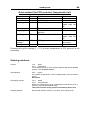
1
Biometra a Whatman company TGGE System 230 V 115 V Code-No. 024-000 Code-No. 024-090 Manual March 1999 !! Warning !! Please read these instructions carefully before using this apparatus! Biometra biomedizinische Analytik GmbH Rudolf-Wissell-Straße 30, D-37079 Göttingen P.O. Box 1544, D-37005 Göttingen Tel:++49 – (0)5 51 / 50 68 6-0 * Fax: ++49 – (0)5 51 / 50 68 6-66 e-mail: [email protected] internet: http://www.biometra.de TGGE System The TGGE method is covered by patents issued to Diagen (now QIAGEN GmbH). The polymerase chain reaction (PCR) process is covered by patents issued to Hoffmann-La Roche. Acryl-Glide is a trademark of Amresco Inc. Biometra is a trademark of Biometra GmbH. Whatman is a trademark of Whatman International Ltd. The POLAND software service established by Gerhard Steger, Department of Biophysics, University of Duesseldorf, is available by internet http://www.biophys.uniduesseldorf.de/service/polandform.html. Manual Version 2.5 Software Version 2.10, 2.11 Biometra GmbH, Rudolf-Wissell-Str. 30, 37079 Göttingen, Germany 7/24/2002 TGGE System 1 Table of Contents 1 The TGGE System .............................................................................................. 4 1.1 Introduction .................................................................................................... 4 1.2 Principle of method ........................................................................................ 4 1.3 Special features of the TGGE System ........................................................... 5 1.4 How to start with TGGE analysis ................................................................... 6 2 General Recommendations ............................................................................... 8 2.1 Safety warnings ............................................................................................. 8 2.2 Notes for use.................................................................................................. 8 3 Components of the TGGE System .................................................................... 9 3.1 Electrophoresis unit ..................................................................................... 10 3.2 Controller unit............................................................................................... 17 3.2.1 Instrument keys and ports ...................................................................... 17 3.2.2 Programming of the TGGE Controller..................................................... 18 4 Sample preparation .......................................................................................... 24 4.1 Purity of samples ......................................................................................... 24 4.2 Quantity and volumes of samples ................................................................ 25 5 Setting up polyacrylamide gels....................................................................... 25 5.1 Selecting Concentration of PAA gels ........................................................... 25 5.2 Setting up the gel solution............................................................................ 25 5.3 Some remarks corresponding to standard TGGE conditions ....................... 28 5.4 Assembling the gel sandwich....................................................................... 30 5.5 Disassembling the gel sandwich .................................................................. 32 6 Electrophoresis with the TGGE System ......................................................... 33 6.1 Electrophoresis conditions ........................................................................... 33 6.2 Preparing the electrophoresis unit ............................................................... 33 6.3 Perpendicular TGGE.................................................................................... 35 6.4 Parallel TGGE.............................................................................................. 39 6.5 Silver staining............................................................................................... 44 6.6 Ethidium bromide-staining ........................................................................... 48 6.7 Blotting......................................................................................................... 48 6.8 Autoradiography........................................................................................... 48 6.9 Elution of DNA from the TGGE gel .............................................................. 48 7 TGGE in analysis of point mutations in dsDNA ............................................ 49 7.1 ....... Theoretical background of a detection rate approximating 100% for point mutations - calculations with the POLAND program .................................... 49 7.2 The “old” Poland program (Old Server)........................................................ 51 7.2.1 About the Poland service........................................................................ 51 7.2.2 Program-specific information .................................................................. 51 7.2.3 How to use the “old” Poland program (Standard) ................................... 53 7.3 The “new” Poland program (New Server) .................................................... 55 7.3.1 About the Poland service........................................................................ 55 7.3.2 Program-specific information .................................................................. 55 7.3.3 References for Poland Service ............................................................... 56 7.3.4 HELP for Poland Service: ....................................................................... 56 TGGE System 2 7.3.5 How to use the “new” Poland program ................................................... 65 7.4 The optimized DNA fragment....................................................................... 66 7.4.1 Asymmetric GC-clamps for PCR primers used for TGGE analysis......... 68 7.4.2 Chemical clamp with Psoralen (Furo[3,2-g]coumarin, C11H6O3)............. 68 7.4.3 POLAND analysis of samples................................................................. 69 8 Optimizing parallel TGGE by perpendicular TGGE....................................... 70 8.1 Check short DNA fragments for their melting behavior ................................ 70 8.2 From perpendicular to parallel TGGE .......................................................... 71 9 TGGE / SSCP..................................................................................................... 72 9.1 Running an SSCP on the TGGE.................................................................. 72 9.2 DNA sample preparation.............................................................................. 73 9.3 Gel casting ................................................................................................... 73 9.4 Electrophoresis ............................................................................................ 75 9.5 Routine analysis........................................................................................... 75 10 TGGE in RNA analysis ..................................................................................... 76 10.1 Completely double-stranded RNA................................................................ 76 10.2 Partly double-stranded RNA, e.g. viroid RNA .............................................. 76 10.3 ...........Single-stranded RNA with single hairpin structures, m-RNA secondary structures ..................................................................................................... 76 10.4 Staining........................................................................................................ 77 11 TGGE in protein analysis ................................................................................. 78 11.1 Buffers ......................................................................................................... 78 12 Trouble-shooting .............................................................................................. 80 13 TGGE Testkit..................................................................................................... 93 13.1 Introduction .................................................................................................. 93 13.2 Protocol........................................................................................................ 93 13.2.1 Gel composition:..................................................................................... 93 13.2.2 Running buffer: ....................................................................................... 94 13.2.3 Electrophoresis parameters:................................................................... 95 13.3 Gel images................................................................................................... 95 14 Appendix ........................................................................................................... 96 14.1 Technical Data ............................................................................................. 96 14.1.1 System.................................................................................................... 96 14.1.2 Electrophoresis Chamber ....................................................................... 96 14.1.3 TGGE System Controller with integrated power pack............................. 96 14.2 Buffers ......................................................................................................... 97 14.3 Silver staining solutions: .............................................................................. 99 15 References ...................................................................................................... 101 16 Order information and spare parts................................................................ 105 17 Instructions for return shipment ................................................................... 106 TGGE System 3 18 Equipment Decontamination Certificate....................................................... 107 19 Warranty .......................................................................................................... 108 Declaration of Conformity 4 TGGE System 1 The TGGE System 1.1 Introduction Temperature Gradient Gel Electrophoresis is a new and powerful electrophoresis method for separation of nucleic acids like DNA or RNA or for analysis of proteins. The TGGE method, which is covered by patents, uses the temperature dependent change of conformation for separating molecules (for review see Reference 1). Since the introduction of the first commercial available TGGE apparatus in 1989 temperature gradient gel electrophoresis has gained high interest in scientific and clinical laboratories due to the unprecedented resolution capability and easiness of analysis. The range of scientific publications using the TGGE method is broad and covers all disciplines which use molecular biology methods: e.g. Oncology2-4, Virology5,6, Immunology7,8, RNA Viroid Research9-12, Prion Research13, Population Analysis14-15. The TGGE method has also been used for quantitative analysis in industry16-17 and for conformational analysis of proteins18-19. 1.2 Principle of method Conventional protein or nucleic acid electrophoresis separates molecules mainly according to size or charge. TGGE adds a new parameter for separation, namely the shape of the molecule. The shape is mostly determined by the secondary and tertiary structure of the molecule and can be changed by external influences like temperature, salt concentration, pH etc. The conformation both of proteins and nucleic acids depend on weak binding forces like hydrogen bonds or van der Waals bonds. Increasing the temperature above a certain limit breaks down these bonds. The molecules will adopt a so called denatured conformation in contrast to the native one. E.g. with DNA it is possible to determine the temperature which is necessary to break down hydrogen bonds along double stranded DNA. This temperature is called midpoint of transition (TM) or melting temperature and characteristic for a certain stretch of DNA (see figure 1). TGGE uses the melting temperature to identify DNA which differs in sequence among a mixture of molecules of the same size. TGGE therefore not only separates molecules but gives additional information about the sequence (DNA or RNA) or the stability of proteins. Migration branched DNA (partially denatured conf.) single stranded DNA (completely denatured) double stranded DNA T1 (cold) TM Temperature T2 (warm) Figure 1: Schematic drawing of different conformations of DNA during temperature gradient gel electrophoresis. 5 TGGE System 1.3 Special features of the TGGE System The microprocessor controlled gradient block of the TGGE System allows strictly defined linear gradients with high resolution. A run distance of 2 mm which can easily be detected by eye corresponds to a maximum temperature difference of about 0.6°C. Therefore even slightest differences of molecules can be detected by the new TGGE System. Because of the small amount of material used for separation DNA or RNA fragments appear as fine bands which can clearly be distinguished from each other. Even complex band patterns can be analyzed due to the high resolution capability of the gradient block. Comparing the TGGE method with another screening method like SSCP shows superior performance of the TGGE method20-22. The controlled temperature conditions make repetition of experiments easy and lead to reproducible gel results. The small format of the gradient block has been optimized in order to reduce sample volume and especially to save experimental time. Perpendicular and parallel TGGE are two different modes applicable with the Biometra TGGE System without need for specialized parts or equipment. Whereas perpendicular TGGE is mostly used for defining optimal separation conditions, parallel TGGE allows the analysis of multiple samples (e.g. screening). perpendicular TGGE: temperature gradient is perpendicular to the electrophoretic run direction → one sample is spread over a broad temperature range parallel TGGE: temperature gradient is parallel to run direction → multiple samples are spread over a narrow temp. range Perpendicular TGGE: T1 (cold) - Parallel TGGE T2 (warm) - T1 (cold) Gel slots Run direction + + T2 (warm) Figure 2: Schematic drawing of typical results after perpendicular TGGE (left panel) and parallel TGGE (right panel). 6 TGGE System 1.4 How to start with TGGE analysis Starting with the gene of interest one develops the right combination of PCR primers for amplification of the desired gene fragment. The polymorphic site must be located inside the amplicon and not at the far end. The POLAND software helps one to identify suitable primers and the adequate fragment length of the amplicon. This program gives a rough estimation too, what temperature parameters will fit best to the desired separation23-25. A revised version of the POLAND program can be found on the world wide web (http:// www.biophys.uni-duesseldorf.de/service/polandform.html). For example the polymorphic site is represented by allele A and allele a. The two alleles can either exist as homoduplices (AA or aa) or heteroduplices (Aa or aA) (see figure 3). A A A a a a Sequence of Allele A Sequence of Allele a A A a a A C C G T G G C A T C G T A G C a A Figure 3: Schematic drawing of double stranded DNA with polymorphism „A“ or „a“ (left panel) and corresponding DNA sequence (right panel). Each line represents double stranded DNA. The TGGE System used as perpendicular TGGE (see above, figure 2) gives the possibility to identify the different alleles by their individual melting behavior. Samples with homoduplex „AA“ or „aa“ have a distinct melting temperature (e-g. Tm1 and Tm2, see figure 4), at which double stranded DNA separates into branched DNA. At even higher temperature the branched DNA separates into individual strands. After perpendicular TGGE, a heteroallele sample like „Aa“ normally shows four different Tm values. The PCR amplification of a heteroallele sample results in four different double stranded DNA types: The two homoduplices "AA" and "aa" as well as two heteroduplices "Aa" and "aA", which have a non-pairing base at the polymorphic site. This non-pairing base will lead to a shift of the Tm to lower values (Tm3 and Tm4). Perpendicular TGGE shows at which temperature the different DNA strands will separate. For future analysis by perpendicular or parallel TGGE a narrower temperature range which includes the TM values of homo- and heteroduplices can be used. Please remember always, that wild type and mutant DNA have to be mixed before PCR to get the 4 bands with different melting behavior. Sometimes the melting difference between heteroduplices cannot be resolved but remember that 3 bands on the TGGE gel are enough to detect a mutation. 7 TGGE System T1 (cold) T2 (warm) _ Gel slot + Tm4: Heteroduplex aA Tm1: Homoduplex AA Tm3: Heteroduplex Aa Tm2: Homoduplex aa Figure 4: Schematic drawing of melting behavior of double stranded, heterozygotic DNA with allele typ „Aa“ after perpendicular TGGE. Screening of multiple samples is performed by using parallel TGGE (see above, figure 2). Parallel TGGE looks like conventional SSCP-analysis, but has a higher probability to identify possible mutants. TM4: Heteroduplex aA - T1 (cold) TM3: Heteroduplex Aa TM2: Homoduplex aa Tm1: Homoduplex AA + T2 (warm) Figure 5: Schematic drawing of a screening for double stranded, heterozygotic DNA with allele type „Aa“ after parallel TGGE. (Sometimes the melting difference between heteroduplices can not be resolved.) Different plates with pre-fixed slots for 8, 12 or 18 samples are available for screening purposes (see chapters 6.4 and 15). TGGE System 8 2 General Recommendations 2.1 Safety warnings Check the voltage of the power supply and of the control unit before use. In any case of malfunction of the power supply, controller or electrophoresis chamber, do not open the case but contact Biometra or your local distributor. First switch off the power switch of the power supply before opening the lid of the electrophoresis chamber if you want to interrupt or stop the electrophoresis run. During electrophoresis don’t touch the electrode wires or the buffer inside the electrophoresis chamber. High voltage. Danger of life! Whenever polyacrylamide gels are handled pay attention to standard laboratory safety regulations, e.g. wear lab coat, protective gloves and eye shield. Polyacrylamide is neurotoxic. 2.2 Notes for use • • • • Do not scratch the protective foil of the gradient block. In case of a damaged foil contact Biometra or your local distributor for a replacement foil. Do not use strong acids or basic solutions or organic solvents for cleaning glass plates, the electrophoresis chamber or the gradient block. Do not incubate glass plates over night in cleaning solution. Wear protective gloves during all steps of the silver staining protocols to avoid staining artifacts due to the high sensitivity of the staining protocol. 9 TGGE System 3 Components of the TGGE System The TGGE System contains all components which are necessary to get started. All kinds of TGGE applications (parallel or perpendicular TGGE, Constant Temperature GE, Time resolved TGGE) can be run with the System. For certain applications which need different numbers or sizes of sample slots the adequate parts are available and can be ordered from Biometra or your local distributor (see 6.3. Order Information). The TGGE System (Order number: 024-000) consists of: • TGGE Controller with integrated power supply, 100 program stores and control function of temperature and electrophoresis conditions • TGGE-Electrophoresis unit with 2 removable buffer chambers, Peltier-element powered gradient block and control cable • TGGE Starter Kit contains 3 plane „Bonding“ glass plates 1 glass plate with 8 slots for parallel TGGE 1 glass plate with 1 rectangular slot for perpendicular TGGE 1 glass pate with 12 slots for parallel TGGE Electrophoresis wicks (100/pkg) Polybond film (25/pkg) 1 Cover glass plate and 10 cover films 1 Acryl-Glide (100 ml) • Manual 024-001 024-002 024-003 024-021 024-022 024-023 024-025 024-015 024-030 024-031 211-319 When unpacking your System please check whether all mentioned parts are included. If individual parts are missing call Biometra or your local distributor. 10 TGGE System 3.1 Electrophoresis unit The electrophoresis unit consists of 4 parts: n 1 safety lid with 2 electric plugs (anode and cathode) o 2 removable electrophoresis chambers each with platinum wires and electric connectors (volume: max. 250 ml) ➌ housing with Peltier-element powered gradient block q 37 pin connecting cable to control unit n o ➌ Figure 6: Parts of the electrophoresis unit 11 TGGE System T1 L1 L2 L3 L4 L5 L6 T2 T 1 L 1 L 2 L 3 L 4 L 5 L 6 T1 L1 L2 L3 L4 L5 L6 T2 T 2 Figure 6b: Gradient block with temperature lines and marks for positioning the gel slots (top). Safety lid with arrow indicating the running direction of nucleic acids (middle). Gradient block covered with safety lid; setup for a perpendicular TGGE run (bottom, left); setup for a parallel TGGE run (bottom, right). 12 TGGE System The gradient block is centrally positioned in the middle of the electrophoresis unit and protected by a layer of white foil. This foil is necessary to protect the electronic parts beneath it from liquid, buffer or other harmful chemicals. If the protection foil has been scratched during use stop working and exchange the protection foil with a new one. The two opposite sides of the gradient block are marked with lettering T1 and T2. Beneath these symbols the Peltier-elements which build up the temperature gradient during electrophoresis can be found. Both sides of the gradient block can reach any preset temperature from 15°C - 80°C. The orientation of the temperature gradient, i.e. which side of the gradient shall be cold or hot, can be freely determined. Between symbols T1 and T2 six thick lines (L1 to L6) and five thin lines (not coded) are marked on the block, which represent the entire linear range of the gradient block (see figure 7). The temperature difference between two lines is identical from line to line. E.g. if T1 is 30°C and T2 is 75°C, the temperature difference is 7.4°C between two thick lines or 3.7°C between a thick and a thin line. T1 L1 L2 L3 L4 L5 L6 T2 When performing parallel TGGE the beginning and the end of the linear temperature gradient are represented by the first and the last line. Whereas when performing perpendicular TGGE the ends of each line represent the linear temperature range. When choosing a temperature gradient e.g. between 25°C and 65°C these two temperatures can actually be found at the first marked line (figure 7: 10mm distance to the block edge) and at the last marked line (figure 7: 50 mm distance) on the block. The block areas to the left and right of these lines are slightly hotter respectively cooler (see figure 7 and table 1 + 2). 13 TGGE System Linearity of block 80 Temperature (°C) 70 60 50 40 30 20 10 0 1 6 11 16 21 26 31 36 41 46 51 56 61 Distance (mm) T1 L1 L2 L3 L4 L5 L6 T2 Figure 7: upper panel: Temperature profile of gradient block measured by a micro sensor on top of the gradient plate every 1mm beginning from one edge of the block. 10 mm distance and 50 mm distance correspond to first (L1) and last marked thick line (L2) on the gradient block respectively. lower panel: Schematic drawing of the block. 14 TGGE System The maximum temperature difference between the two sides of the gradient block (T1 and T2) during electrophoresis is 45 Kelvin. That means it is possible to build up a gradient between 35°C and 80°C or between 25°C and 70°C, just to give two examples. ----------------------------------------------------------------------------------------------------------------Programming T1 and T2, the actual temperature of L1 to L6 can be calculated by the following formula: Ln = L1 + (n - 1) ∆ϑ (Ln = Temperature of line n; n = 1 .......6) L6 - L1 ∆ϑ = ------------ (∆ϑ = Temperature difference between two thick lines) 5 T1 L1 L2 L3 L4 L5 L6 T2 ----------------------------------------------------------------------------------------------------------------When leaving out the gradient function the block can be cooled down to 4°C or heated up to 80°C. Although Peltier elements reach lower respectively higher temperature values, the surrounding plastic material does not permit the temperature range of the TGGE System to be extended. The electrophoresis buffer chambers can freely be positioned around the gradient block. This makes it easy to switch between parallel or perpendicular TGGE. When the electrophoresis buffer chambers stay parallel to the lines of the gradient block (see figure 8 left panel) parallel TGGE applications can be run. By simply switching the chambers perpendicular to the lines of the gradient block (see figure 8 right panel) it is possible to run perpendicular TGGE. T1 L1 L2 L3 L4 L5 L6 T2 T1 L1 L2 L3 L4 L5 L6 T2 Figure 8: Orientation of the two electrophoresis buffer chambers (- represents cathode, + represents anode) relative to the centrally positioned gradient block. 15 TGGE System T1 15,94 16,94 17,94 18,94 19,94 20,94 21,94 22,94 23,94 24,94 25,94 26,94 27,94 28,94 29,94 30,94 31,94 32,94 33,94 34,94 35,94 36,94 37,94 38,94 Programming L1 and L6 (°C): L1 L2 L3 L4 L5 L6 T2 DELTA 20 21 22 23 24 25 26 27 28 29 30 31 32 33 34 35 36 37 38 39 40 41 42 43 57 58 59 60 61 62 63 64 65 66 67 68 69 70 71 72 73 74 75 76 77 78 79 80 61,06 62,06 63,06 64,06 65,06 66,06 67,06 68,06 69,06 70,06 71,06 72,06 73,06 74,06 75,06 76,06 77,06 78,06 79,06 80,06 81,06 82,06 83,06 84,06 7,4 7,4 7,4 7,4 7,4 7,4 7,4 7,4 7,4 7,4 7,4 7,4 7,4 7,4 7,4 7,4 7,4 7,4 7,4 7,4 7,4 7,4 7,4 7,4 27,4 28,4 29,4 30,4 31,4 32,4 33,4 34,4 35,4 36,4 37,4 38,4 39,4 40,4 41,4 42,4 43,4 44,4 45,4 46,4 47,4 48,4 49,4 50,4 34,8 35,8 36,8 37,8 38,8 39,8 40,8 41,8 42,8 43,8 44,8 45,8 46,8 47,8 48,8 49,8 50,8 51,8 52,8 53,8 54,8 55,8 56,8 57,8 42,2 43,2 44,2 45,2 46,2 47,2 48,2 49,2 50,2 51,2 52,2 53,2 54,2 55,2 56,2 57,2 58,2 59,2 60,2 61,2 62,2 63,2 64,2 65,2 49,6 50,6 51,6 52,6 53,6 54,6 55,6 56,6 57,6 58,6 59,6 60,6 61,6 62,6 63,6 64,6 65,6 66,6 67,6 68,6 69,6 70,6 71,6 72,6 Table 1: Examples for actual temperatures on the gradient block programming L1 and L6 16 TGGE System Programming T1 and T2 (°C): T1 L1 L2 L3 L4 L5 L6 T2 DELTA 20 21 22 23 24 25 26 27 28 29 30 31 32 33 34 35 36 37 38 39 40 24,05 25,05 26,05 27,05 28,05 29,05 30,05 31,05 32,05 33,05 34,05 35,05 36,05 37,05 38,05 39,05 39,96 40,87 41,78 42,69 43,6 31,43 32,43 33,43 34,43 35,43 36,43 37,43 38,43 39,43 40,43 41,43 42,43 43,43 44,43 45,43 46,43 47,18 47,92 48,67 49,41 50,16 38,81 39,81 40,81 41,81 42,81 43,81 44,81 45,81 46,81 47,81 48,81 49,81 50,81 51,81 52,81 53,81 54,39 54,97 55,56 56,14 56,72 46,19 47,19 48,19 49,19 50,19 51,19 52,19 53,19 54,19 55,19 56,19 57,19 58,19 59,19 60,19 61,19 61,61 62,03 62,44 62,86 63,28 53,57 54,57 55,57 56,57 57,57 58,57 59,57 60,57 61,57 62,57 63,57 64,57 65,57 66,57 67,57 68,57 68,82 69,08 69,33 69,59 69,84 60,95 61,95 62,95 63,95 64,95 65,95 66,95 67,95 68,95 69,95 70,95 71,95 72,95 73,95 74,95 75,95 76,04 76,13 76,22 76,31 76,4 65 66 67 68 69 70 71 72 73 74 75 76 77 78 79 80 80 80 80 80 80 7,38 7,38 7,38 7,38 7,38 7,38 7,38 7,38 7,38 7,38 7,38 7,38 7,38 7,38 7,38 7,38 7,216 7,052 6,888 6,724 6,56 Table 1: Examples for actual temperatures on the gradient block programming T1 and T2 17 TGGE System 3.2 Controller unit The controller is a highly integrated, micro processor driven unit for controlling the temperature, ramping time and ramping rate of the gradient block as well as supplying the power for the electrophoresis unit. For entering and storing run parameters the front panel of the controller offers 4 function keys and a full numerical key pad. During the run the display of the controller continuously shows the current parameters. 3.2.1 Instrument keys and ports p o n n Power on/off switch o Display p 4 function keys A, B, C, D q Alphanumerical key pad q Figure 9: Front panel of the TGGE System Controller. n o p q r n Computer port (RS 232) o Connectors for electrode cable: red = anode, black = cathode p Printer port q Interface to electrophoresis unit r Mains and fuses Figure 10: TGGE System Controller from the rear TGGE System 18 3.2.2 Programming of the TGGE Controller After switching on the controller the display shows the instruments name and the software version. Immediately afterwards the main menu appears. Main menu T1: 22.0°C T2: 22.0°C block off A? B Elpho + C programs D At the bottom line of the display 4 possible options which can be retrieved by the 4 functions keys ªA, B, C, D are shown. These 4 options change during programming relative to the chosen menu. (Temperatures T1 and T2 shown in the display are dependent on the room temperature.) ªA “?”: comments or tips about the current program step ªB “Elpho”: commands to load and start the program ªC “programs”: commands to edit new programs, change or delete existing programs ªD “+”: different options like printing of program stores or of running protocols, choice of language, choice of signal In general select by scrolling, activate by pressing enter; except when selecting program numbers or temperature values! Function key D: Options 1 print programs 2 signal 3 language A ↑ B ↓ C quit D enter 4 standard mode 5 test mode 6 void A ↑ B ↓ C quit D enter ªA ↑ and ªB ↓ allow scrolling of display. By ªC “quit” you will return to the main menu. 1: Printing of program stores. A dot matrix printer can be connected to the controller by using the port at the rear (see figure 10). 2: Choice whether a beep signal can be heard at the end of a program or when the program has reached an infinite time step 3: Choice between ªA “German” or ªB “English” 4: not occupied 5: not occupied 6: not occupied TGGE System 19 Function key C: Editing of programs program no: __ A list B del C quit D enter After entering a number of a non occupied program store, the display shows: Name: > < ABCDEFGHIJKLMNOPQRST UVWXYZ – () α σ /, <>& +. % A → B ABC C quit D enter 1:L1:__ L6: alternative: T1: T2: A ? B T1 C quit D → The temperature gradient between T1 and T2 must not exceed 45 K. In the main menu pressing ªC will offer the possibility to edit a new program. First choose a program store number. ªA “list” displays all program stores from 0 to 99 with names or the information <empty>. ªB “del” deletes the last entry. To give the program an individual name strike ªB “ABC” to jump with the cursor into the letter field. Moving inside this field is possible by the keys ªA “→” and ªB “←”. Pressing ªD “enter” the current high lighted letter is stored in the name field. This step can be repeated 8x times. Pressing ªD “enter” two times leads to the next step. It is now possible to enter for the first program step temperature values for both sides of the gradient block: Alternatively the temperatures for L1 and L6 (first and last thick lane on the gradient block) or for T1 and T2 (left and right edge of the gradient block) can be programmed. The change between programming L and T can be done by pressing ªB "T1". If a number has been entered at the field L1, you have to confirm by pressing ªD “enter”. The cursor jumps to L6. After programming L6 you will be asked "ok ?": Pressing ªB "no→L1" allows new programming of L1 and L2.Pressing ªD "yes" confirms the temperatures. 1:L1: 25.0°C L6: Alternative: T1: __ T2: A ? B delete C quit D → After entering a temperature for T1, T2, L1 or L6 the temperature can be Deleted by pressing ªB "delete". 1:L1: L6: Alternative: T1: __ T2: A ? B L1 C quit D → If programming of T1 and T2 (left and right edge of the gradient block) has been done but L1 and L6 are preferred, changing to L1 and L6 can be done by pressing ªB "L1". 1: L1: 25.0°C L6: 60.0°C T1: 21.1°C T2: 63.8°C Ok ?: A ? B no→L1 C quit D → Or After entering L6 or T2 all four temperatures (L1, L6, T1, T2) are displayed. "ok ?": Pressing ªD "yes" leads to the programming of the Electrophoresis parameters. Pressing ªB "no→L1" leads back to programming L1. Pressing ªB "T1" allows programming of T1 and T2. Pressing ªB "no→T1" leads back to programming T1. Pressing ªB "L1" allows programming of L1 and L6. 1: L1: 25.0°C L6: 60.0°C T1: 21.1°C T2: 63.8°C Ok ?: A ? B no→T1 C quit D → Depending on the decision of programming L1 and L6 or T1 and T2 the programmed temperatures will be shown in the display. After programming L1 and L6 this temperatures will be shown in the display during the next programming steps. TGGE System 1:L1: 25.0°C L6: 60.0°C time: __0m 0s El: 0V 500mA 30W A ? B V*h C quit D → 1:L1: 25.0°C L6: 60.0°C time: 30m 0s El: 0V 500mA 30W A ? B V*h C quit D→ 1:L1:__ L6: V*h: __0.00 Vh El: 0V 500mA A ? B Time C quit 20 Time: You can choose electrophoresis time. Standard values for a TGGE run are 30 – 45 min. If you enter 30 and confirm by press ing ªD “enter” you get 30 s. If you enter 30 x you will get 30 min. If you enter 30 xx you will get 30 h. V*h: Pressing ªB "V*h" replaces times by the more precise Volt x hour integration. Pressing ªB "Time" replaces V*H by time. 30W D→ 1:L1: 25.0°C L6: 60.0°C time: 30m 0s El: __ 0V 500mA 30W A ? B special C quit D → El: Three different electrophoresis parameters (voltage, current or wattage) can be set. Current and wattage are pre-set at max. values of 500 mA and 30W respectively. In the beginning we recommend to set only the Voltage. Depending on the resistance of the gel electrophoresis the controller will regulate the other two parameters automatically. 1:L1: 25.0°C L6: 60.0°C time: 30m 0s El: 250V 500mA 30W A ? B special C quit D → After confirming the voltage by pressing ªD “enter” and pressing two times ªD “→”, the following two parameters are not changed. 1: special functions ramptime: __0m 0s Ramptime = Ramping time Pressing ªB "special" gives you the choice to choose how fast the Gradient block is going to the established gradient. Normally you choose 1s which means maxi ramping speed. In this case enter “1” and confirm with ªD “enter” Pressing ªB "standard" results in the standard display. A ? B standard C quit D → 1:L1: 25.0°C L6: 60.0°C Time: 30m 0s El: 250V 500mA 30W A ? B special C quit D → 2:L1:__ L2: Pressing ªD “→” starts the programming of step 2 in this program. alternative: T1: T2: A ? B T1 C quit D→ program no: ...... pgm end: .......step(s) runtime: ....h.....m.......s By pressing ªC “quit” any change will be saved and the following messages appear: L1: 22.0°C block off After a few seconds the main menu appears. L6: 22.0°C A? B Elpho C programs D + TGGE System 21 Function key B: Start/Stop Start block function and electrophoresis Start program: __ In the main menu pressing key ªB “block” offers the possibility to start a program. A list B del C quit L1: 25.0 °C D enter L6: 60.0°C ramp: 1 time: 0m 1s El: 250 V 20mA 20W A list B block C program D + L1: 25.0 °C L6: 60.0°C hold: 1 0m 1s 0.00 Vh El: 250 V 8mA 20W A list B block C program D + Pressing ªC “program” during block run Program no: 4 Pgm is active! A copy B del C quit D display program no: 4 name: ................ A copy B C quit D display After entering a program number or choosing a program from the list (ªA “list” ) the block starts to establish the gradient. The timer for the ramping starts immediately. The electrophoresis is started as soon as the gradient block reaches the programmed temperature (gradient. The limiting factor (const. V, mA or W) is indicated by an blinking arrow.) After establishing the gradient, line two of the display changes. The timer now starts again and counts the electrophoresis running time. Additionally the volt/hour integrator starts to count. It is possible to review a program during run. After pressing ªC “program” and the corresponding store number a warning message appears. Then it is possible to display the active program (ªD “display”) or to copy it into a new store (ªA “copy”). It’ s not possible to change the currently running program By pressing ªC “quit” the main menu appears again. Stop block function and electrophoresis program: 0 TEST To stop a current running program you press ªB “Elpho” and pause? again ªD“stop". stop? By pressing ªC “quit” you return to previous display without any. A ? B pause C quit D stop changes. When pressing ªD “stop” the run will be aborted and you leave the actual running program. Pressing ª B “pause” holds the actual situation of the gradient L1: 25.0 °C L6: 60.0°C hold: 1 pause 0.00 Vh (stopping electrophoresis, holding the temperature gradient) El: 250 V 8mA 20W “pause” is blinking and shown in the display alternatively with the A ? B Elpho C programs D + time. program: 0 TEST continue? stop? A? B contin C quit stop Pressing ªB "Elpho" (in pause status): Pressing ªB "contin" will continue the program. D TGGE System 22 Function key A "?" T1: 22.0°C block off T2: 22.0°C Pressing ªA "?" in the main menu results in the following display: A? B Elpho C programs D + B: start/stop/pause C: edit/delete/copy D. special functions A? B Elpho C programs D + L1: 25.0 °C L6: 60.0°C hold: 1 0m 1s 1.16Vh El: 250 V 8mA 20W A list B block C program D + T1: 21.1 °C L1→3: 21.1 L4→6: 46.0 ← = const T2: 63.8°C 32.0 39.0 53.0 60.0 rtime: h m Pressing ªA "?" in a running program results in the following display: The actual temperatures of T1 and T2 as well as the actual temperatures of L1 to L6 are shown in the display. The limiting factor (const. V, mA or W) is indicated by an blinking arrow (←). The actual remaining electrophoresis time is shown on the bottom (right side of the display). TGGE System 23 Error messages TGGE - System check connection to thermoblock TGGE connector cable is not connected to gradient block and / or system Controller. Check connections!!!!! warning: gradient too large! max. grad. T1→T2: 45°C A ? B no→L1 C quit D enter or warning: gradient too large! max. grad. T1→T2: 45°C A ? B no→T1 C quit D enter Programmed temperature gradient too large. program no: It is possible to review a program during run. After pressing ªC “program” and the corresponding store number a warning message appears. TEST pgm is active! A copy B del C quit D display program no: __ name: not programmed! A ↑ B ↓ C quit This program number has not been programmed. D enter 1:L1: __ L6: entry required T1: T2: A ? B L1 C quit D → or 1:L1: __ L6: entry required T1: __ T2: A ? B T1 C quit D → No temperature or time has been programmed. TGGE System 24 4 Sample preparation 4.1 Purity of samples Due to the high sensitivity of the staining procedure after TGGE it is recommended to use purified DNA, RNA or protein samples. Any impurities might be misinterpreted after TGGE, thereby making the analysis of gels difficult. Nevertheless it is possible to use even crude mixtures for TGGE analysis. PCR-amplified DNA fragments can usually be analyzed without purification. But note, that the presence of high amounts of nonspecific, secondary PCR products may result in difficulties with interpretation of band pattern, melting profile, etc. For example, in parallel TGGE, nonspecific bands with a higher molecular weight than the specific PCR product may be misinterpreted as heteroduplices, or analogs with lower thermal stabilities. Therefore, before running a TGGE gel, please check the PCR product and, if necessary, purify the specific PCR product of interest, e.g., by agarose gel electrophoresis and subsequent gel extraction. Sample preparation for direct DNA analysis 1 volume of DNA/RNA samples are dissolved with 1 volume of TBE or Na-TAE loading buffer or with 0.1 volume of the total loading volume ME loading buffer (see Appendix 2). The resulting mixture is loaded directly on to the polyacrylamide gels. Secure that the slots are filled up to maximum (if necessary add loading buffer to fill up the slots to maximum). In case of low-concentration samples we recommend to prepare 5x conc. loading buffer. 0.2 volume of this concentrated loading buffer is mixed with the sample and loaded onto the polyacrylamide gel. Denaturation/Renaturation for heteroduplex analysis of DNA For heteroduplex analysis the samples are denatured and renatured prior to TGGE. Quantitative denaturation is accomplished by heating in 4 M urea. The following protocol is recommended for all DNA fragments with GC-contents of 50 - 70%. Depending on the buffer to be used for electrophoresis add one sample volume of corresponding DR buffer (denaturation/renaturation buffer) to the sample and mix. Heat at 95°C for 5 minutes (denaturation). Incubate at 50°C for 15 minutes (renaturation). The sample is then loaded directly to the gel. In order to achieve the recommended loading volumes for diagonal or perpendicular TGGE (refer to chapter 4.2), the samples should be filled up with running (or loading) buffer. • Renaturing at 50°C: - higher temperatures (higher stringency) can be chosen for high GC contents to avoid artificial hybrids - lower temperatures (e.g. 37°C) are applied for expected hybrids with multiple mismatches or for sequences with very low GC content. The renaturation temperature should be approx. 10°C below the Tm of the desired hybrid. TGGE System 25 4.2 Quantity and volumes of samples Depending on the slot size of the gel Biometra recommends the following amounts of material: 50 µl volume, approx. 50 ng DNA/RNA of interest 5 µl volume, 3 - 5 ng DNA/RNA of interest glass plate with 1 rectangular slot + 2 marker slots _____________________________________________________________________________________________________ 5 µl volume, 3 - 5 ng DNA/RNA of interest glass plate with 8 slots: _____________________________________________________________________________________________________ 3 µl volume, 1 - 3 ng DNA/RNA of interest glass plate with 12 slots: _____________________________________________________________________________________________________ 2 µl volume, approx. 1 ng DNA/RNA of interest glass plate with 18 slots: If less volumes fill up the slots with running buffer or loading buffer to the volumes listed on top! This will create better results. 5 Setting up polyacrylamide gels 5.1 Selecting Concentration of PAA gels The TGGE System represents a highly optimized system for performing flat bed polyacrylamide gel electrophoresis under defined temperature conditions. In addition to typical TGGE applications the system is ideally suited to run standard fragment separations without temperature gradient very quickly. Depending on the molecular weight of the sample we recommend the following acrylamide/bisacrylamide concentrations: Conc. 3% 5% 8% DNA fragment length > 1000 bp 500 - 1000 bp < 500 pb 5.2 Setting up the gel solution Each gel sandwich contains approx. 2.5 ml polyacrylamide solution. We therefore recommend to prepare 10 ml solution to pour 3-4 gels at the same time. Polymerized gels which are not immediately used must be stored at room temperature. To inhibit any gel drying we recommend to wrap the polymerized gels into saran foil or wet plastic bags. Wet towels can be used only for short time storage. TGGE System 26 Keep in mind that polymerized polyacrylamide gels which include urea should not be used after 2 – 4 days of storage (depending on storage conditions)! 27 TGGE System Recipe for 10 ml gel solution (3 – 4 gels) for TBE running buffer: Urea (cEnd= 7 M) Acrylamide/bis Acrylamide stock solution (30 : 0,8), 40% (w/v) 10x conc.TBE (cEnd= 0.1 x conc.) 50% Glycerol (cEnd= 2%) Water, distilled 3% Gel 4.2 g 0.75 ml 5% Gel 4.2g 1.25 ml 8% Gel 4.2 g 2.0 ml 0.1 ml 0.1 ml 0.1 ml 0.5 ml 3.5 ml 0.5 ml 3 ml 0.5 ml 2.5 ml Make sure that the urea has been completely resolved. It is possible to heat up the urea containing solution slightly (40°C – 50°C) for a short time in order to improve the solubilization of urea. De-gas the solution under gentle vacuum for 3 - 5 min. Water, distilled TEMED APS (4%) 22.5 µl 42 µl fill up to 10 ml 22.5 µl 22.5 µl 42 µl 42 µl Mix gently. Avoid air bubbles! Pour the gel solution into the glass plate sandwich immediately thereafter (see chapter 4.1.2) without air bubbles. Recipe for 10 ml gel solution (3 – 4 gels) for Na-TAE running buffer: Urea (cEnd= 8 M) Acrylamide/bis Acrylamide stock solution (30 : 0,8), 40% (w/v) 10x conc. Na-TAE, pH 8.4 (cEnd= 0.2 x conc.) 40% Glycerol (cEnd= 2%) Water, distilled 3% Gel 4.8 g 0.75 ml 5% Gel 4.8g 1.25 ml 8% Gel 4.8 g 2.0 ml 0.2 ml 0.2 ml 0.2 ml 0.5 ml 3.5 ml 0.5 ml 3.0 ml 0.5 ml 2.5 ml Make sure that the urea has been completely resolved. It is possible to heat up the urea containing solution slightly (40°C – 50°C) for a short time in order to improve the solubilization of urea. De-gas the solution under gentle vacuum for 3 - 5 min. Water, distilled TEMED APS (4%) 14 µl 45 µl fill up to 10 ml 14 µl 14 µl 45 µl 45 µl Mix gently. Avoid air bubbles! Pour the gel solution into the glass plate sandwich immediately thereafter (see chapter 4.1.2) without air bubbles. 28 TGGE System Recipe for 10 ml gel solution (3 – 4 gels) for ME (MOPS/EDTA) running buffer: Urea (cEnd= 8 M) Acrylamide/bis Acrylamide stock solution (30 : 0,8), 40% (w/v) 50x conc. ME-buffer (cEnd= 1 x conc.) 40% Glycerol (cEnd= 2%) Water, distilled 3% Gel 4.8 g 0.75 ml 5% Gel 4.8g 1.25 ml 8% Gel 4.8 g 2.0 ml 0.2 ml 0.2 ml 0.2 ml 0.5 ml 3.5 ml 0.5 ml 3.0 ml 0.5 ml 2.5 ml Make sure that the urea has been completely resolved. It is possible to heat up the urea containing solution slightly (40°C – 50°C) for a short time in order to improve the solubilization of urea. De-gas the solution under gentle vacuum for 3 - 5 min. Water, distilled TEMED APS (4%) 17 µl 76 µl fill up to 10 ml 17 µl 17 µl 76 µl 76 µl Mix gently. Avoid air bubbles! Pour the gel solution into the glass plate sandwich immediately thereafter (see chapter 4.1.2) without air bubbles. 5.3 Some remarks corresponding to standard TGGE conditions Electrophoresis buffer (running buffer): • Always membrane filtrate (e.g. 0.45µm pore size) the buffers before use! • Running buffer: always use the concentration identical with the gel condition • TBE is the most common used buffer system but the electrophoresis is not as fast as with Na-TAE buffer. It is possible to add up to 5 mM NaCl if a higher ionic strength is desired, for reversible melting processes which are required for parallel TGGE in multiple sample analysis. A higher NaCl concentration should not be used because it causes an unacceptable high electrical current. • Na-TAE is the buffer for fastest electrophoresis. • ME buffer meets all the requirements of a variety of TBE-buffers with different ionic strengths but is only stable for a very short time. (Stable for about 3 days. Do not use as the buffer becomes yellow.) • ME buffer allows Na+ concentrations up to 20 mM which greatly favors "reversible melting" and still allows short run times for TGGE electrophoresis. Mobile Cl- ions which slow down the migration velocity of nucleic acids are avoided by using the sodium salt form of MOPS. Due to their reduced mobility the large MOPS anions keep the current low. TGGE System 29 Gel conditions: • 4 M urea can be used for low GC and high degree of mismatches. This concentration increases Tm instead of the standard concentration (8 M urea) by approx. 16 - 20°C using TBE buffer or 8 – 12°C using Na-TAE buffer. • 10 M urea can be used for high GC and lowers Tm instead of the standard concentration (8 M urea) by approx. 8 - 10°C using TBE buffer or 4 – 6°C using Na-TAE buffer. • Glycerol reduces the steepness of very cooperative transition curves, broadening the profile and expanding the temperature range for detecting small Tm differences of closely related nucleic acids: 0% Glycerol increases cooperativity (>200 bp, narrower transitions) >2% Glycerol lowers cooperativity (>200 bp, broader transitions) TGGE conditions: • Voltage can be raised to 400 V if the current is below 30 mA • Current should not exeed 30 mA Running buffer: pre-run run 0.2 x conc. Na-TAE 250 V, 10-12 mA, 2-5 min. 250V, 15-20 mA, 30-60 min. 0.1 x conc. TBE 250 V, 4- 5 mA, 2-5 min. 250 V, 9-12 mA, 30-60 min. • T1 = 20°C: can be raised to obtain optimized resolution • T1 = 20°C: lowering should be avoided by using 4M urea • T2 = 60°C: higher temperatures should be avoided by using 10 M urea or/and 10 mM MOPS. • T2 = 60°C: can be lowered to obtain optimized resolution • T2 = 80°C: maximum temperature, can be used if the gel is carefully protected against evaporation. Sample preparation: • Denaturation / renaturation cycle: • Renaturing at 50°C: higher temperatures (higher stringency) can be chosen for high GC contents to avoid artificial hybrids • Renaturing at 50°C: lower temperatures (e.g. 37°C) are applied for expected hybrids with multiple mismatches or for sequences with very low GC content. The renaturation temperature should be approx. 10°C below the Tm of the desired hybrid. • Samples should be dissolved in buffers with ionic strength identical to the ionic strength of the running buffer. • If samples are dissolved in buffer different to the running buffer, the samples have to be equilibrated against the running buffer (e.g. using dialysis). Nucleic acids can be precipitated with ethanol and dissolved in denaturation/renaturation buffer or running buffer. TGGE System 30 5.4 Assembling the gel sandwich The thinness of the gel makes it necessary to cast polyacrylamide gels on a gel support film (Polybond, Order Number: 024-030). Each sandwich consists of four elements: Bonding glass plate without spacer Polybond film Polyacrylamide gel Glass plate with fixed spacer and fixed slot former (different types of slots available). Glass plates • Glass plates must be dry and free of any dirt or dust. Biometra recommends to wear powder-free gloves even during cleaning of glass plates in order to prevent any skin debris which might interfere with silver staining. • Do not use strong acidic or basic solutions or organic solvents for cleaning the glass plates. • Do not incubate glass plates over night in cleaning solutions. Pretreatment of glass plate with spacer and slot former • Glass plate with spacer and slot former must be carefully treated with Acryl-Glide solution (Order Number 211-319) or a similar hydrophobic solution. Drop about 0.5 ml of solution onto the plate and especially between the slot former. (This protection layer helps to withdraw the polyacrylamide gel from the sandwich after polymerization.) Wait 2 – 3 min. and than polish the plate with soft paper to remove any haze! • This procedure should be repeated after each run. • Do not drop Acryl-Glide onto the spacer of the glass plate! This possibly leads to leakage during polymerization. • Clean spacers with ethanol before assembling the glass plate sandwich. • If necessary treat the spacers with a small amounts of silicone grease (to protect leakage). Polybond film • Use only original pre-cut Polybond film which perfectly fits onto the gradient block. • Biometra recommends to use Polybond film only once. Repeated usage and especially staining might weaken the strength of the Polybond film. • The Polybond film has two different sides: one hydrophobic side which repels water and one hydrophilic side on which a water drop will adhere. You can test the different sides with a drop of water. (The protection paper is attached to the hydrophobic site.) • Remove the protecting paper sheet before assembling the sandwich. Handle the Polybond film only with powder-free gloves. • The hydrophobic side of the Polybond film must be orientated to the Bonding glass plate. On the hydrophilic side the polyacrylamide gel will polymerize and stick. • Press the hydrophobic side of the Polybond film firmly to the Bonding glass plate by using your thumb, a rubber or a gel casting clip. This will prevent that any polyacrylamide solution running between the Bonding glass plate and the Polybond film. 31 TGGE System Glass plate sandwich • Assemble the Acryl-Glide treated glass plate with spacer, the Polybond film and the Bonding glass plate as indicated in figure 11. n q o ➌ Figure 11: Setting up a gel sandwich for PAGE. Bonding glass plate (n), Polybond film (o) and glass plate with spacer and slot former (p) are assembled (left panel). The Polybond film is only visible at the inclined edge of the glass plate with spacer (middle panel). Fasten the clamps (q) above the spacer to increase the pressure and to ensure a leakage free sandwich. Figure 12: Pouring the polyacrylamide gel. Initially 1 ml of polyacrylamide gel solution must be poured. The gel sandwich must be hold at an angle of 45° when pouring (left panel). The solution must run along one side of the plate sandwich to avoid air bubbles. The remaining 1.5 ml of solution is poured into the plate sandwich during which time the plate sandwich is slowly brought back into a vertical position (right panel). • Fill up the plate sandwich as high as possible. The gel solution is overlayed with 200µl of Isopropyl Alcohol (2-Propanol, Isopropanol) or Isobutyl Alcohol (2Methyl-1-propanol, Isobutanol) or distilled water to produce a horizontal surface of the gel. TGGE System 32 • Polymerization of the gel must be at least for 0.5 h, better for 1 - 1.5 h or optional over night at room temperature. The sandwich should stand up vertically and must not be moved during polymerization. • Gels may be stored up to 4 days at room temperature (wrapped in wet paper towels in a plastic bag). Do not store at 4°C! 5.5 Disassembling the gel sandwich • Remove the clamps from the plate sandwich. • Remove the Bonding glass plate from the sandwich by sliding it smoothly! The gel polymerized to the Polybond film will adhere to the other glass plate. • Withdraw the Polybond film with the adhering polyacrylamide gel carefully from the other glass plate. In the area of the slot former remove the Polybond film very carefully to avoid any damage to the slots. • If slots show distortion or wrinkles don’t fill in samples because after electrophoresis bands in this lane will show distortion as well. TGGE System 33 6 Electrophoresis with the TGGE System The electrophoresis unit of the TGGE System has been designed to accommodate all TGGE and related applications like CTGE, TTGE and SSCP, without cumbersome changes. It’s easy to switch between perpendicular, parallel or diagonal TGGE (for adequate accessories see chapter 6.4). 6.1 Electrophoresis conditions The electrophoresis conditions depend on the kind of material to be separated, e.g. fragment size differences, kind of application, e.g. parallel or perpendicular TGGE, sample preparation, e.g. high salt or low salt preparation, buffer system. Any recommendations can only be used as guidelines to start with. Further improvements to the analysis is easily possible by adjusting the run conditions to the individual needs. Voltage: Current: Run Time: 100 V - 400 V; 5 mA - 25 mA; 10 min - 2 h; usually 250 V usually 10-20 mA usually 30 min 6.2 Preparing the electrophoresis unit • • • • Use the leveling eye on the electrophoresis unit and the 4 leveling feet to adjust the unit. Remove the safety lid and fill in max. 250 ml of the desired running buffer per buffer chamber (e.g. 0.1 x conc. TBE, see Appendix 13.2.). The same running buffer should only be used once. Soak the pre-cut electrode wicks (order number: 024-020) in the running buffer before use. Drop 0.3 - 0.5 ml of thermal coupling solution like 0.1% Triton or 0.1% Tween 20 on the surface of the gradient block (see figure 13 left panel). The thermal coupling solution will increase the adhesion of the Polybond film with the attached polyacrylamide gel and therefore supports temperature equilibration between gradient block and polyacrylamide gel. The whole block must be covered by the thermal coupling solution layer. No air bubbles must form. n o ➌ Figure 13: Positioning of polyacrylamide gel on gradient block. A small volume of thermal coupling solution (o) is applied to the gradient block (n)(left panel). The Polybond film with the polyacrylamide gel on top (➌) is put on the gradient block. Slightly bend the Polybond film (right panel) in order to spread the thermal coupling solution evenly. TGGE System • • • 34 To position the polyacrylamide gel on the gradient block the Polybond film should be held between thumb and middle finger and slightly bended. This leads to an even distribution of thermal coupling solution beneath the Polybond film. If air bubbles are visible beneath the polyacrylamide gel, try to squeeze them out by moving the gel slightly back and forth. If this will not succeed completely remove the gel and repeat the aforementioned steps. Don’t touch the polyacrylamide gel directly with your fingers or your gloves. Excess thermal coupling solution must be removed from the gradient block by using paper towels. 35 TGGE System 6.3 Perpendicular TGGE During perpendicular TGGE a mixture of molecules is separated over a wide temperature range. The temperature gradient is perpendicular to the electrophoresis run direction. Steps before TGGE sample preparation (refer to chapter 4.1) programming of temperature gradient and electrophoresis parameters (refer to chapter 3.2.2) Prepare in advance: polyacrylamide gel attached to Polybond film running buffer (250 ml for each chamber) pre-cut and pre-soaked electrode wicks pre-cut cover film cover glass plate (treated on both sides with Acryl-Glide) • Because of the fixed orientation of the temperature gradient the removable electrophoresis chambers must be positioned as indicated in figure 14. n Platform surrounding the gradient block o Electrophoresis buffer chamber with connector for cathode ➌ Gradient block T1 L1 L2 L 3 L 4 L 5 L 6 T2 q Electrophoresis buffer chamber with connector for anode Figure 14: Orientation of the two electrophoresis buffer chambers for perpendicular TGGE. • Fill in the running buffer (e.g. 0.1 x conc. TBE) into each electrophoresis chamber. Before you place the gel onto the gradient block be sure that the sample is ready for loading and cover film is available. • To make full use of the linear range of the gradient block the polyacrylamide gel attached to the Polybond film should be positioned as indicated in figure 15. The rectangular and the marker slots of the gel (p) are positioned at the beginning of the gradient block (the marked lines represent the beginning of the linear range of the gradient block). 36 TGGE System n ➌ ➌ o T1T1 T2T2 T1 T1 TT 22 Figure 15: Orientation of the polyacrylamide gel attached to the Polybond film (n) on the gradient block (o) during perpendicular TGGE. See the position of the gel slots (➌) relative to the marked lines of the gradient block. • Two pre-cut and pre-soaked electrode wicks must be positioned at the start and the end of the polyacrylamide gel. Wick and gel have to overlap (see figure 16). ➎ 2 - 3 mm ➍ ➋ ➊ Figure 16: Side view of the polyacrylamide gel (➋) on top of the gradient block during pre-run. Pay attention to the position of electrode wicks (➍) on top of the polyacrylamide gel. (Polybond film (➊), slot of polyacrylamide gel (➎)). • • Avoid any contact between sample slots and electrode wicks. Otherwise the samples will diffuse into the electrode wicks. Load the samples quickly at room temperature without air bubbles. Do not start the temperature gradient (the temperature gradient is established after the samples have fully entered the polyacrylamide gel). glass plate with 1 rectangular slot + 2 marker slots 50 µl volume, approx. 50 ng DNA/RNA of interest 5 µl volume, 3 – 5 ng DNA/RNA of interest If less volumes fill up the slots with running buffer or loading buffer to the volumes listed on top! This will create better results. 37 TGGE System The time between mounting the gel onto the gradient block and loading the sample must not exceed 5 minutes. • Close the safety lid of the electrophoresis chamber and start electrophoresis at 20°C or 25°C and 250 V for 2 - 5 min. Standard electrophoresis conditions are given in chapters 5.3 and 6.1. • Make sure that the orientation of the gel and the safety lid is exact as indicated in the following: T1 • • • • • L1 L2 L3 L4 L5 L6 T2 Wait at the electrophoresis chamber until the samples have fully entered the polyacrylamide gel (unlike with the former TGGE System of QIAGEN this process will only take 1-3 minutes) and have moved about 3 - 5 mm in the gel. Stop the electrophoresis run, open the safety lid. Rinse the - now empty - slots with 0.5 - 1 ml running buffer. Cover the polyacrylamide gel including the slots with the 7 x 6 cm pre-cut cover film (see figure 17). A small buffer layer must remain between cover foil and gel. Avoid air bubbles! The cover film must be positioned with the long side parallel to the buffer chambers (= perpendicular to the arrow on the safety lid). Soak any excessive buffer from the side of the gel. The gel must not swim in buffer solution. q ➎ ➌ ➋ ➊ Figure 17: The polyacrylamide gel (o) has to be covered by a pre-cut hydrophobic cover film (➌). A small buffer layer remains between gel and cover film. (Polybond film (➊), wicks (q) , slot of polyacrylamide gel (➎)) 38 TGGE System • • Bring the electrode wicks to an overlap with the cover film. The overlap between wick and cover film should be almost 2 cm (see figure 17). Avoid air bubbles. Be sure that the 2 silicone barriers are fixed to the cover glass plate before use: • Cover the sandwich with the Acryl-Glide treated cover glass plate (see figure 18). The silicone barriers have to be positioned perpendicular to the wicks and never on top of the wicks! q ➏ ➎ ➌ ➋ ➊ Figure 18: Side view of the polyacrylamide gel (➋) on top of the gradient block. Sandwich of polyacrylamide gel ➋), pre-cut cover foil (➌), electrode wicks (➍) and cover glass plate with silicone barriers (➏) during perpendicular TGGE run (Polybond film(➊), slot of polyacrylamide gel (➎)). • • Start the temperature gradient and wait until the gradient has been established (usually 0.5 - 1 minute). Start the electrophoresis run. • The Bromophenol blue dye only gives you an indication how far the samples have migrated until you have optimized the best run time. • After the electrophoresis run, switch off the controller, open the safety lid of the electrophoresis unit, remove the polyacrylamide gel and proceed further for staining the gel (chapter 6.5). It is recommended to fix the gel immediately in order to improve the analysis. 39 TGGE System 6.4 Parallel TGGE During parallel TGGE a mixture of molecules is separated over a narrow temperature range determined by perpendicular TGGE. The temperature gradient is parallel to the electrophoresis run direction. Steps before TGGE sample preparation (refer to chapter 4.1) programming of temperature gradient and electrophoresis parameters (refer to chapter 3.2.2) To prepare in advance: polyacrylamide gel attached to Polybond film running buffer (250 ml for each chamber) pre-cut and pre-soaked electrode wicks pre-cut cover foil glass plate (treated on both sides with Acryl-Glide) • For parallel TGGE the removable electrophoresis buffer chambers must be positioned as indicated in figure 19. n Platform surrounding the gradient block o Electrophoresis buffer chamber with connectors for cathode T1 L1 L2 L3 L4 L5 L6 T2 ➌ Gradient block q Electrophoresis buffer chamber with connectors for anode Figure 19: Orientation of the electrophoresis chambers for parallel TGGE. • Fill in the running buffer (e.g. 0.1 x conc. TBE) into each electrophoresis chamber. Before you place the gel onto the gradient block be sure that the sample is ready for loading and cover film is available. • The polyacrylamide gel attached to the Polybond film must be positioned as indicated in figure 20. The slots of the gel (p) should be positioned at the beginning of the gradient block. The first marked line (L1) represents the beginning of the linear range of the gradient block. 40 TGGE System n ➌ o T1 T2 T1 T2 Figure 20: Orientation of the polyacrylamide gel attached to the Polybond film (n) on the gradient block (o) during parallel TGGE. See the position of the gel slots (➌) relative to the marked lines of the gradient block. • Two pre-cut and pre-soaked electrode wicks must be positioned at the start and the end of the polyacrylamide gel. Wick and gel have to overlap (see figure 16). ➍ 2 - 3 mm ➌ ➋ ➊ Figure 16: Side view of the polyacrylamide gel (➋) on top of the gradient block during pre-run. Pay attention to the position of electrode wicks (➍) on top of the polyacrylamide gel. (Polybond film (➊), pre-cut Polybond film(➌), slot of polyacrylamide gel (➎)). • • Avoid any contact between sample slots and electrode wicks. Otherwise the samples will diffuse into the electrode wicks. Load the samples quickly at room temperature without air bubbles. Do not start the temperature gradient (the temperature gradient is established after the samples have fully entered the polyacrylamide gel). TGGE System 41 Depending on the slot size of the used gel Biometra recommends the following amounts of material: 5 µl volume, 3 - 5 ng DNA/RNA of interest glass plate with 8 slots: _____________________________________________________________________________________________________ 3 µl volume, 1 - 3 ng DNA/RNA of interest _________________________________________________________________________________ glass plate with 18 slots: 2 µl volume, approx. 1 ng DNA/RNA of interest glass plate with 12 slots: If less volumes fill up the slots with running buffer or loading buffer to the volumes listed on top! This will create better results. The time between mounting the gel onto the gradient block and loading the sample must not exceed 5 minutes. Close the safety lid of the electrophoresis chamber and start electrophoresis at 20°C or 25°C and 250 V for 2 - 5 min. Standard electrophoresis conditions are given in chapters 5.3 and 6.1. • Make sure that the orientation of the gel and the safety lid is exact as indicated in the following: • Wait at the electrophoresis chamber until the samples have fully entered the polyacrylamide gel (unlike with the former TGGE System of QIAGEN this process will only take 1-3 minutes) and have moved about 3 - 5 mm in the gel. Stop the electrophoresis run, open the safety lid. Rinse the - now empty - slots with 0.5 - 1 ml running buffer. Cover the polyacrylamide gel including the slots with the 7 x 6 cm pre-cut cover film (see figure 17). A small buffer layer must remain between cover foil and gel. Avoid air bubbles! The cover film must be positioned with the long side parallel to the buffer chambers (= perpendicular to the arrow on the safety lid). T1 • L1 L2 L3 L4 L5 L6 T2 • • • • Soak any excessive buffer from the side of the gel. The gel must not swim in buffer solution. 42 TGGE System q ➎ ➌ ➋ ➊ Figure 17: The polyacrylamide gel (o) has to be covered by a pre-cut hydrophobic cover film (p). A small buffer layer remains between gel and cover film. (Polybond film (➊), wicks (q) , slot of polyacrylamide gel (➎)) • • Bring the electrode wicks to an overlap with the cover film. The overlap between wick and cover film should be almost 2 cm (see figure 17). Avoid air bubbles. Be sure that the 2 silicone barriers are fixed to the cover glass plate before use: • Cover the sandwich with the Acryl-Glide treated cover glass plate (see figure 18). The silicone barriers have to be positioned perpendicular to the wicks and never on top of the wicks! q ➏ ➎ ➌ ➋ ➊ Figure 18: Side view of the polyacrylamide gel (➋) on top of the gradient block. Sandwich of polyacrylamide gel ➋), pre-cut cover foil (➌), electrode wicks (➍) and cover glass plate with silicone barriers (➏) during perpendicular TGGE run (Polybond film(➊), slot of polyacrylamide gel (➎)). TGGE System • • 43 Start the temperature gradient and wait until the gradient has been established (usually 0.5 - 1 minute). Start the electrophoresis run. • The Bromophenol blue dye only gives you an indication how far the samples have migrated until you have optimized the best run time. • After the electrophoresis run, switch off the controller, open the safety lid of the electrophoresis unit, remove the polyacrylamide gel and proceed further for staining the gel (chapter 6.5). It is recommended to fix the gel immediately in order to improve the analysis. TGGE System 44 6.5 Silver staining Aside from autoradiography silver staining is the most sensitive method for detecting small amounts of DNA, RNA or proteins in polyacrylamide gels. Due to the low thickness of the gels (0.5 mm) the staining procedure takes no more than 35 minutes. Other staining protocols may be used, but generally exhibit less sensitivity. This must be considered in relation to the amount of DNA loaded on the gel. All incubation steps are done in small plastic containers which are agitated on a rocking platform (e.g. order number 042-400 or 042-500). For handling several polyacrylamide gels simultaneously, Biometra offers a semiautomated instrument called Blot Processor (order number 015-000 or 015-090). Please contact Biometra or your local distributor to receive further information about the Blot Processor. Wear non-powdered protective gloves during all steps of the silver staining protocol to avoid staining artifacts due to the high sensitivity of the staining protocol. • Remove the protective plastic sheets from the gel. • Carefully remove any residual thermal coupling solution from the back of the gel (Polybond film) prior to staining • Put the polyacrylamide gel with the gel side upwards into the staining tray. Avoid air bubbles during all staining steps. • It’s recommended to prepare at least 100 ml solution for each incubation step. • If NaCl has been added to the gel running buffer, incubate the TGGE gel for 15 min in Fixation solution to remove the NaCl. 45 TGGE System Standard method: Step Fixation Silver Binding Washing Time 5 min 10 min 3 x 1 min Solutions* Fixation solution AgNO3-Solution Fresh ddH2O Developing Stopping Washing 10 min 5 min 10 min Developer Stopping Solution Rinse under fresh ddH2O Preparing storage for 1-5 h 50% glycerol Notes prepare freshly demineralised water may be ok prepare freshly Demineralised water may be ok Not absolutely necessary! Preparing the gel for storage ( 1 - 5 h at room temperature in 50% glycerol) is not necessary. Staining solutions: Fixation 10% EtOH 0.5% Acetic Acid 100 ml ethanol and 5 ml acetic acid are adjusted with distilled water to 1 liter. Silver Binding 0.19% AgNO3 1.9g AgNO3 is dissolved in 1 liter of distilled water. (Can be reused 5 times) Store dark! Developing Solution 1.5% NaOH 0.08% NaBH4 0.1% Formaldehyde Dissolve 15 g NaOH in 1 liter distilled water. Add 0.8g NaBH4 and 2.7 ml formaldehyde stock solution (37% in water). This buffer must be freshly prepared immediately before use! Stopping Solution 0.75% Na2CO3 Dissolve 7.5 g sodium carbonate in ddH2O. Total volume: 1 liter 46 TGGE System Quick method (for PCR products) (Sanguinetti et al): Step Fixation Silver Binding Washing Time 3 min 5 min 3 x 1 min Solutions* Fixation solution AgNO3-Solution Fresh ddH2O Developing Stopping 5 min 5 min Washing 10 min Developing solution Ethanol and acetic acid solution Rinse under fresh ddH2O Demineralised water may be ok 50% glycerol Not absolutely necessary Room temperature Preparing storage Drying for 1-5 h Notes prepare freshly prepare freshly Demineralised water may be ok prepare freshly Preparing the gel for storage ( 1 - 5 h at room temperature in 50% glycerol) is not necessary. Staining solutions: Fixation: 10% EtOH 0.5% Glacial Acid 100 ml ethanol and 5 ml acetic acid are adjusted with double distilled water to 1 liter. Prepare freshly ! Silver Binding 0.2% AgNO3 2.0 g AgNO3 is dissolved in 1 liter of distilled water. (Can be reused 5 times) Store dark! Developing Solution 3.0 % NaOH 0.5% Formaldehyde Dissolve 3 g NaOH and 1.35 ml formaldehyde stock solution (37% in water) in 100 ml double distilled water. This buffer must be freshly prepared immediately before use! Stopping Solution: identical with Fixation solution (10% EtOH, 0.5% Glacial Acid) 47 TGGE System Quick method using the AMRESCO SilverPAGE staining kit (Code No. 211-761) Step Fixation Sensibilisation Washing Time 15 min 10 min 10 min Solutions 2 x 100 ml Fixation solution 2 x 100 ml 30% ethanol 3 x 200 ml fresh ddH2O Silver Binding 15 min reconstituted Silver Binding Agent + Formaldehyde Rinse under fresh ddH2O Washing Developing Stopping Preparing storage Drying 0.5-1 min 1 - 2 min reconstituted Developing solution + Formaldehyde 5 min for 1-5 h 7.5% Acetic acid 50% glycerol Notes prepare freshly! Demineralised water may be ok prepare freshly! !!!!! prepare freshly! Develop to desired level! Not absolutely necessary Room temperature Preparing the gel for storage ( 1 - 5 h at room temperature in 50% glycerol) is not necessary. Staining solutions: Fixation: 30% EtOH 10% Acetic Acid 300 ml ethanol and 100 ml acetic acid are adjusted with double distilled water to 1 liter. Sensibilisation: 30% EtOH Prepare freshly 60 ml ethanol in 140 ml double distilled water. Silver Binding: Prepare Silver Binding Agent by reconstituting contents of one pouch in 1 l of ddH20. (This solution must be prepared fresh every time!) Immediately before staining, add 0.7 ml of 37% Formaldehyde to 200 ml of reconstituted Silver Binding Agent. Developing Solution: Just prior to use, prepare developing solution by reconstituting contents of one pouch of Developer I and 15 mg of Developer II in 200 ml of ddH20. (This solution must be prepared fresh every time!) Immediately before developing, add 0.7 ml of 37% Formaldehyde to 200 ml of reconstituted developing solution. Stopping Solution: 7.5% Acetic Acid 75 ml acetic acid are adjusted with double distilled water to 1 l. TGGE System 48 6.6 Ethidium bromide-staining Incubate the gel in staining solution (0.5 µg/ml ethidium bromide in 1 x conc. TBE) for 30 45 min. Analyze under UV radiation (27). 6.7 Blotting DNA from TGGE gels can be blotted onto a solid-state support either by electroblotting (Fastblot) or vacuum-blotting (Vacu-Blot System). If DNA is to be blotted after TGGE analysis, the TGGE gel must be poured onto the hydrophobic side of the gel support film (Polybond film). Otherwise, the gel cannot be detached from the gel support film! 6.8 Autoradiography TGGE gels can also be directly exposed to x-ray films is radiolabeled samples are analyzed. Direct exposure: Incubate the TGGE gel for 15 min. in Fixation solution (see 6.5 Silver staining). Optional: Silver stain the gel. Remove residual buffer from the gel. Expose to an x-ray film at room temperature. Exposure of dried TGGE gels: Incubate the TGGE gel for 15 min. in Fixation solution (see 6.5 Silver staining). Optional: Silver stain the gel. Incubate the gel in 2 - 5% glycerol for 10 minutes to prevent the gel from cracking. Incubate an appropriate sheet of cellophane (no Saran wrap!!!!!) in 2 - 5% glycerol. Layer the cellophane on the gel. Air dry at room temperature for one day or use a geldryer at 50°C for at least 3h. Exposure to an X-ray film. 6.9 Elution of DNA from the TGGE gel DNA fragments which have been separated on TGGE, for example, different alleles of one gene, can be eluted from silver-stained TGGE gel and reamplified by PCR. Using a Pasteur pipet, punture the gel and extract a µl piece containing the particular DNA duplex. Incubate in 20 µl TE buffer overnight. Use a 1 µl aliquot for reamplification. TGGE System 49 7 TGGE in analysis of point mutations in dsDNA For analysis of point mutations in dsDNA, an extremely high detection rate of greater than 95% is routinely achieved when the experiment is carefully planned. The next two chapters provide information for optimizing detection of base substitutions. 7.1 Theoretical background of a detection rate approximating 100% for point mutations - calculations with the POLAND program DNA does not melt by deannealing base pair by base pair from one end to the other, but by cooperative denaturation of long stretches, called melting domains. The length of a melting domain is 25 to several hundred base pairs. The midpoint melting temperature Tm and the length of a melting domain are mainly determined by the nucleotide sequence of the DNA. The Tm of DNA fragments differing by even small changes, such as point mutations, can differ by as much as 1.5°C. When heteroduplices, hybrids of two species of DNA fragments differing in their base composition, have been formed, the mismatches lower the Tm value significantly. Thus the heteroduplex analysis is the preferable because of the additional resolution provides (1, 28). The principle by which TGGE uses differences in Tm is that the DNA fragments are electrophoresed through a linear temperature gradient in the polyacrylamide gel. When the fragments reach the temperature at which the lowest melting domain starts to melt, they take on a branched, Y-shaped configuration, which slows down mobility in the TGGE gel matrix. The electrophoretic migration of fragments differing by single base changes is retarded by branching at different temperatures, thus they are resolved from one another during temperature gradient electrophoresis. The denaturing behavior of any DNA fragment can be predicted, if its sequence is known. For this purpose the POLAND calculation can be used. The POLAND software is available in the internet: http://www.biophys.uni-duesseldorf.de/service/polandform.html. POLAND software predicts location and Tm values of melting domains for dsDNA and dsRNA as well as their perpendicular TGGE pattern. The ability to predict the melting behavior of particular DNA fragments enables one to construct DNA fragments with optimized melting behavior, resulting in a nearly 100% detection rate for point mutations inside of this fragment. Since the end of February 1999 the POLAND program is available in two versions. Using the above internet address allows the user to select between the old POLAND request form (Standard), the old POLAND expert request form or the new POLAND request form. The following information is visible on the screen: 50 TGGE System Poland Server: ANNOUNCEMENT ___________________________________________________________________________________________ The WWW server for prediction of nucleic acid's thermal stability (called Poland server according to the author of the basic mathematics) will move during the near future to another computer. This is not a mere relocation of the program that you have used up to now, but the input/output procedure is completely rewritten for the new server. The new server produces better/nicer (?) plots and has a better/more elongated help file. But be aware of new bugs, which might be introduced during the rewriting and relocation. The old server, both the standard and the expert form, are unchanged. Both forms will be available for the near future. However, that server is running on our DEC Alpha under OpenVMS, and we run into more and more trouble to support that machine. __________________________________________________________________________________ Now, make a decision: NEW Server Poland request form OLD Server Poland request form Poland expert request form __________________________________________________________________________________ Institut für Physikalische Biologie (Department of Biophysics) Heinrich Heine-Universität Düsseldorf, Germany Feb. 26, 1999 G. Steger / M. Labensky / A. Jäger TGGE System 7.2 51 The “old” Poland program (Old Server) 7.2.1 About the Poland service The Poland program is an experimental service of the University of Düsseldorf Biophysics Department, and thus the whole set-up, access and service are subject to change. The Poland server will calculate thermal denaturation profiles and temperaturedependent uv absorbance or gel mobility of double stranded RNA or DNA, based on sequence input and parameter settings in the request form. - Details below ! The program used in these calculations was developed by Gerhard Steger, for comparing theoretical predictions to experimental data, mainly optical denaturation profiles, taken at 260 and 280 nm, and TGGE (temperature gradient gel electrophoresis) experiments. The original version was written in VAX Fortran (VMS), using the Graphics Kernel System GKS for data presentation. 7.2.2 Program-specific information Calculation is based on D. Poland's algorithm in the implementation described by Gerhard Steger. The Poland algorithm calculates the denaturation profile for double-stranded nucleic acid using nearest-neighbor stacking interactions and loop entropy functions described in the literature. An extension of the algorithm, the 'virtual stack' model, allows for the incorporation of specific mismatched sequence positions in the stability calculations, as described by Heinz Werntges. The input data required for calculation are: • the sequence (≤ 1,000 bp). Use GCG-format or plain format without spacing. Plain format accepts only 180 characters per line! • mismatched positions (optional), • the strand concentration, affecting the dissociation temperature (use the programmed standard), • parameter set selection (DNA/DNA low salt/RNA; oligo/long ds), • output format options (choose GIF format). Data sets predicted by the program comprise the following: • A perspective view on the temperature-dependent denaturation profile, that is denaturation probability vs. sequence position vs. temperature. • The temperature-dependent relative uv hypochromicities as they would be measured in optical melting, at wavelengths of 260 and 280 nm (282 nm in case of DNA), respectively ( full hypochromicity corresponds to approx. 30% of the OD at low temperature ). • The derivative form of above hypochromicities, showing the melting temperature(s) and corresponding half width(s) of the transitions(s), giving hints about the transition cooperativity. TGGE System 52 • Predicted relative gel mobility as calculated according to Lerman et al., as a graph vs. temperature for different values of the 'retardation length' parameter. This plot can be used for direct comparison with TGGE experiments; superpositions of plots generated with or without mismatched positions given are useful as a hint whether specific mismatched duplexes could be detected among homoduplexed DNA in a mixture of sample and reference double strands having undergone a denaturation-renaturation cycle, using either perpendicular or parallel TGGE. • A 'half-denaturation temperature' plot showing the half-denaturation temperature for each base. This plot can also be used to estimate the destabilizing effect of mismatches on the surrounding part of the sequence: a temperatureshift of the TGGE transition can be expected if the lowest melting part of the sequence is directly affected by the mismatch! Calculations can be done for oligonucleotides (>15 bases) or long double strands (>50 bases), respectively. In the case of oligonucleotide mode, a length-dependent correction for the strand dissociation process is applied; the temperature range is adapted as well. We do not have sufficient experimental results to stringently check for this mode to give valid results, but for the length range of about 20 nucleotides there is at least experimental evidence. Using 'oligo' mode with far longer sequences gives misleading results! Graphics output is possible in Postscript, HPGL, GIF and PBM format, numeric results are available as well. All graphics results are directly sent to the WWW client, for GIF inline images and pbm images links are provided to retrieve a copy for the external viewer (or for download to disk). 53 TGGE System 7.2.3 How to use the “old” Poland program (Standard) Poland service request form This form is an experimental service of the Biophysics Department, further informations are available here! The Poland server will calculate the thermal denaturation profile of double stranded RNA or DNA based on sequence input and parameter settings in this form. Calculation is based on D. Poland's algorithm in the implemetation described by G. Steger. Calculations can be done for oligonucleotides (>15 bases) or long double strands (>50 bases), respectively. A form allowing for 'expert' parameter settings is available, too. Graphics output is possible in Postscript, HPGL, GIF and PBM format, numeric results are available as well. Graphics results are directly sent to your WWW client. For a (more or less) detailed description of the various parameters, you may read a help page. Sequence title line: Sequence: (plain format without spacing; max. 180 chars per line) Mismatched positions: (comma-separated) Strand concentration: (default 1.0e-6 M; don't give the unit) Thermodynamic parameters: Sequence length: DNA default parameters. oligonucleotide or long double strand ? Output options: __ Click here to click here to GIF inline images. submit reset , or the form to defaults. If you have comments or suggestions on this service, you can send us mail here ! BiophysWWW / G. Steger Oct. 1996 TGGE System Working with the web-based POLAND program only need 4 steps: 1. Enter DNA sequence (≤ 1000 bases) 2. Enter mismatch position (optional) 3. Choose GIF format 4. Press submit 54 TGGE System 7.3 55 The “new” Poland program (New Server) 7.3.1 About the Poland service The Poland server will calculate thermal denaturation profiles and temperaturedependent UV absorbance or gel mobility of double stranded RNA, DNA, or RNA/DNA-hybrids based on sequence input and parameter settings in the Poland request form. -- Details of the Poland program are given below. The program used in these calculations was developed by Gerhard Steger for comparing theoretical predictions to experimental data, mainly optical denaturation profiles, taken at 260 and 280 nm, and TGGE (temperature gradient gel electrophoresis) experiments. The original version was written in VAX Fortran (VMS), using the Graphics Kernel System GKS for data presentation. 7.3.2 Program-specific information Calculation is based on D. Poland's algorithm including the modification by Fixman & Freire in the implementation described by Gerhard Steger. The Poland algorithm calculates the denaturation profile for double-stranded nucleic acid using nearestneighbor stacking interactions and loop entropy functions described in the literature. The input data required for calculation are: • • • • • • the sequence, of course (and no default here!), optional mismatched positions, the strand concentration, affecting the dissociation temperature, the method to calculate the final dissociation into single strands, the thermodynamic parameter set (DNA/DNA low salt/RNA), and the temperature range in which the calculation is performed. In case you need access to the full range of input options, more options are available to the experts. Data sets predicted and figures drawn by the program are described below; see also for OUTPUT. • • • A perspective view on the temperature-dependent denaturation profile (denaturation probability vs. sequence position vs. temperature. This plot does not include the dissociation of dsNA into single-strands; thus it shows most clearly the relative stability of the different parts of the NA. The temperature-dependent relative UV hypochromicities as measured in optical melting, at wavelengths of 260 and 280 nm (282 nm in case of DNA), respectively (full hypochromicity corresponds to approx. 30% of the OD at low temperature). The derivative form of above hypochromicities, showing the melting temperature(s) and corresponding half width(s) of the transitions(s), giving hints about the transition cooperativity. TGGE System • • 56 Predicted relative gel mobility, calculated according to Lerman et al., vs. temperature for different values of the 'retardation length' parameter Lr. This plot can be used for direct comparison with TGGE experiments; superpositions of plots generated with or without mismatched positions given are useful as a hint whether specific mismatched duplexes could be detected among homoduplexed DNA in a mixture of sample and reference double strands having undergone a denaturation-renaturation cycle, using either perpendicular or parallel TGGE. A 'half-denaturation temperature' plot showing the temperature at which each base pair has a probability of 50% to be in the open state. Similar to the threedimensional plot, this plot can be used to estimate the destabilizing effect of mismatches on the surrounding part of the sequence: a temperature-shift of the TGGE transition can be expected if the lowest melting part of the sequence is directly affected by the mismatch. Calculations can be done for oligonucleotides (>15 bases) or long double strands (>50 bases), respectively. In the case of oligonucleotide mode, a length-dependent correction for the strand dissociation process is applied. We do not have sufficient experimental results to stringently check for this mode to give valid results, but for the length range of about 20 nucleotides there is at least experimental evidence. Using 'oligo' mode with far longer sequences gives misleading results! 7.3.3 References for Poland Service Description of implemented programs Steger, G. (1994). Nucleic Acids Res. 22, 2760-2768. Thermal denaturation of double-stranded nucleic acids: prediction of temperatures critical for gradient gel electrophoresis and polymerase chain reaction. Original version of algorithm: Poland, D. (1974). Biopolymers 13, 1859-1871. Recursion relation generation of probability profiles for specific-sequence macromolecules with longrange correlations. Fixman & Freire (1977). Biopolymers 16, 2693-2704. 7.3.4 HELP for Poland Service: 7.3.4.1 POLAND The program POLAND simulates transition curves of double-stranded nucleic acids (DNA and RNA as well as DNA/RNA hybrids). Additional information available: OUTPUT, RELATED PROGRAMS, RESTRICTIONS, ALGORITHM, SUGGESTIONS, PARAMETERS TGGE System 7.3.4.2 57 OUTPUT The program writes it output in numeric format, which is converted to graphics by Tk/Tcl. Additional information available: General description, resolution, 3Dplot, GelPlot, MeltPlot, Temp50%Plot Resolution of graphics output The primary graphics output is produced as PostScript® (vector format). That format is converted to GIF® (raster format); this is a format directly displayed by your WWW browser. The resolution of the GIF images is selectable: 72 dots per inch (72 dpi) is the standard screen resolution; 150 dpi or 300 dpi are nice for printing. But be aware on NanoWeak® systems: the higher resolutions need a lot of memory and tend to crash your system. 3DPlot Probability of an open base-pair is plotted as a function of position in sequence and temperature. GelPlot Relative mobility is plotted as a function of temperature for the three different stiffness parameters. MeltPlot Relative hypochromicity and its derivative is plotted as a function of temperature at 260 nm and 282 nm (RNA 280 nm). References: for RNA: Coutts, S.M. (1971). Biochim. Biophys. Acta 232, 94-106. Thermodynamics and kinetics of GC base pairing in the isolated extra arm of serine-specific tRNA from yeast for DNA: Blake, R.D. & Haydock, P.V. (1979). Biopolymers 18, 3089-3109. Effect of sodium ion on the highresolution melting of lambda DNA Temp50%Plot Temperature is plotted at which the corresponding base stack has a probability of 50% to be in the open state. The two horizontal lines in the plot mark the temperature range of calculation; i.e., a curve coinciding with such a line is not valid. 7.3.4.3 RELATED PROGRAMS The Poland program calculates the denaturation behavior of double-stranded NA. LinAll, RNAfold, and mFold calculate the secondary structure of single-stranded (R)NA; in addition LinAll and RNAfold allow the prediction of denaturation behavior of ssRNA. TGGE System 7.3.4.4 58 Restrictions The sequence has to be shorter than 1001 nucleotides but longer than 5 nucleotides. Valid nucleotides are A, G, C, U, and T. Calculation of asymmetric or bulge loops is not possible. 7.3.4.5 Algorithm Calculation is based on Poland's algorithm including the modifications proposed by Fixman & Freire. With the original algorithm of Poland computing time is proportional to the square of the sequence length. With the modification according to Fixman & Freire computing time is proportional to 10 times the sequence length, but it works only with loop parameters according to Poland. 7.3.4.6 SUGGESTIONS Hints for combination of parameters and their values. Additional information available: RNA DNA RNA/DNA Ionic_strength_dependence RNA Thermodynamic values according to Turner et al. are ideally suited for calculation in 1 M ionic strength after correction of all DeltaS values by 1.021 and all DeltaSGC values by 0.961. These corrections are equivalent to a shift in Tm values of A:U stacks by -7 K or -2%, respectively and of G:C/G:C stacks by +7 K or +2%, respectively. Optimal (?) parameter combination for Turner et al.: -d 1.021 1.000 0.961 (DeltaS, DeltaS(A:U), and (DeltaS(G:C) factor) -n 1.e-3 (Dissociation constant ß) -c 1.e-6 (ß*c0 = 1E-8 to 1E-10) -s 1.000 (loop parameter Sigma) -l g (internal loops according to Gralla & Crothers) -t 90. 120. 0.5 (Temperature range and steps) TGGE System 59 Optimal (?) parameter combination for Pörschke et al.: -d 1.000 1.040 0.970 (DeltaS, DeltaS(A:U), and (DeltaS(G:C) factor) -n 1.e-3 (Dissociation constant ß) -c 1.e-6 (ß*c0 = 1E-9 to 1E-11) -s 1.e-6 (loop parameter Sigma) -l p (internal loops according to Poland) -a f (algorithm according to Fixman & Freire) -t 90. 120. 0.5 (Temperature range and steps) DNA Thermodynamic values according to Gotoh et al. and Klump, both, are ideally suited for calculations. The parameter set of Breslauer et al. does not fit our experiments (?). The parameter set of Allawi & SantaLucia is based on a reevaluation of all known parameter sets for DNA; i.e., this set may the optimal one. For references to the original thermodynamic parameter sets see here. Additional information available: Gotoh Breslauer et al. Klump SantaLucia et al. Allawi & SantaLucia • Gotoh Optimal (?) parameter combination for Gotoh: -d 1.000 -n 1.e-3 -c 1.e-6 -s 1.e-3 -l p -a f -t 60. 80. 0.5 (DeltaS factor) (Dissociation constant ß) (ß*c0 = 1E-9 to 1E-11) (loop parameter Sigma) (internal loops according to Poland) (algorithm according to Fixman & Freire) (Temperature range and steps) • Breslauer et al. No optimal parameter combination for Breslauer et al. • Klump Optimal (?) parameter combination for Klump: -d 1.000 -n 1.e-3 -c 1.e-6 -s 1.e-3 -l p -a f -t 70. 90. 0.5 (DeltaS factor) (Dissociation constant ß) (ß*c0 = 1E-9 to 1E-11) (loop parameter Sigma) (internal loops according to Poland) (algorithm according to Fixman & Freire) (Temperature range and steps) TGGE System • SantaLucia et al. • Allawi & SantaLucia This is the "unified parameter set"! RNA/DNA Thermodynamic values according to Sugimoto et al. (1995) in 1 M NaCl. The top strand is RNA, the bottom strand is DNA (5'-r-3'/3'-d-5'); the input sequence is the RNA strand. Ionic strength dependence Following values may be used for correction of calculated Tm-values: Tm,2 - Tm,1 ---------- = f(G:C)*I(G:C) + (1-f(G:C))*I(A:U) log(c2/c1) with Tm c f(G:C) I(X:Y) DNA I(A:T) I(G:C) RNA I(A:U) I(G:C) = transition (midpoint, melting) temperature = ionic strength (=concentration of Na ions) = G:C content = dependence of ionic strength of base pair type X:Y = 18.3 °C (Owen, Hill, & Lapage (1969). Biopolymers 7, 503-516.) = 11.3 °C (Frank-Kamenetskii (1971). Biopolymers 10, 2623-2624.) = 20.0 °C (Steger, Müller & Riesner (1980). = 8.4 °C Biochim. Biophys. Acta 606, 274-284.) 60 TGGE System 7.3.4.7 61 PARAMETERS Additional information available: BASE_STACKING_(thermodynamic_parameters) ENTROPY_CORRECTION_of_base_stacking LOOP_PARAMETERS_(thermodynamic_parameters) TEMPERATURE_RANGE_OF_CALCULATION MISMATCHED_POSITIONS_in_original_sequence CONCENTRATION_and_DISSOCIATION_CONSTANT STIFFNESS_of_nucleic_acid THERMODYNAMIC PARAMETER SETS for BASE STACKING You can select between five different thermodynamic parameter sets of base stacking (for loop parameters see below): Additional information available: RNA DNA RNA/DNA • RNA • for RNA in 1 M NaCl Freier, S.M., Kierzek, R., Jaeger, J.A., Sugimoto, N., Caruthers, M.H., Neilson, T. & Turner, D.H. (1986). Proc. Natl. Acad. Sci. USA 83, 9373-9377. Improved free-energy parameters for predictions of RNA duplex stability. • for RNA in 1 M NaCl Pörschke, D., Uhlenbeck, O.C. & Martin, F.H. (1973). Biopolymers 12, 1313-1335. Thermodynamics and kinetics of the helix-coil transition of oligomers containing GC base pairs. • DNA • for DNA in 0.019 M NaCl Gotoh, O. (1983). Adv. Biophys. 16, 1-52. Prediction of melting profiles and local helix stability for sequenced DNA. •for DNA • in 1 M NaCl Breslauer, K.J., Frank, R., Bloecker, H. & Marky, L.A. (1986). Proc. Natl. Acad. Sci. USA 83, 37463750. Predicting DNA duplex stability from the base sequence. • • for DNA in 0.1 M NaCl Klump, H.H. (1987). Canad. J. Chem. 66, 804-809. Energetics of order/order transitions in nucleic acids. TGGE System 62 Klump, H. (1990). in Landolt-Börnstein, New Series, Group VII Biophysics, Vol. 1 Nucleic Acids, Subvol. c Spectroscopic and Kinetic Data, Physical Data I, (W. Saenger, ed.), Springer-Verlag Berlin, p. 244-245. Calorimetric studies on DNAs and RNAs. • for DNA in 1 M NaCl SantaLucia, J. Jr., Allawi, H.T. & Seneviratne, P.A. (1996). Biochemistry 35, 3555-3562. Improved nearest-neighbor parameters for predicting DNA duplex stability. for DNA in 1 M NaCl Allawi, H.T. & SantaLucia, J. Jr. (1997). Biochemistry 36, 10581-10594. Thermodynamics and NMR of Internal G·T Mismatches in DNA. • RNA/DNA • for RNA/DNA hybrids in 1 M NaCl Sugimoto, N., Nakano, S., Katoh, M., Matsumura, A., Nakamuta, H., Ohmichi, T., Yoneyama, M. & Sasaki, M. (1995). Biochemistry 34, 11211-11216. Thermodynamic parameters to predict stability of RNA/DNA hybrid duplexes. • ENTROPY CORRECTION of base stacking (Option not available by WWW) DeltaS values of base stacking may be corrected by factors in order to simulate deviating ionic strengths. The Delta S values of the thermodynamic parameters are multiplied with these factors. The first is used for correction of all Delta S values, the second only for A:U stacks, the third only for G:C stacks. Different values are used as defaults in dependence on the chosen thermodynamic parameter set. • LOOP PARAMETERS (thermodynamic parameters) (Option not available by WWW; i.e., Sigma is fixed to 1.e-3, and loop entropy is calculated according to Poland.) Loops which appear during denaturation by internal base stack opening may be calculated by three different methods: • -l p ==> DeltaS(loop) = SIGMA*(loop+1)**-1.75 (according to Poland or Fixman & Freire) Use only with stacking parameters according to Pörschke et al. (-p p), Gotoh (-p g), or Klump (-p k). • -l g ==> DeltaS(loop) = SIGMA*DeltaS(loop) (according to Gralla & Crothers) Use only with stacking parameters according to Turner et al. (-p t). • -l t ==> DeltaS(loop) = SIGMA*DeltaS(loop) (according to Turner et al.) Use only with stacking parameters according to Turner et al. (-p t). TGGE System 63 Therefore, Sigma influences the cooperativity and the half width of each transition. With '-a f' you can change the default algorithm (only in case '-l p'). With the original algorithm of Poland (default), computing time is proportional to the square of the sequence length. With '-a f' the modified algorithm of Fixman & Freire is used which results in computing time proportional to 10 times the sequence length but works only with loop parameters according to Poland (-l p) up to a sequence length of 1000 base pairs. References: Poland, D. (1974) Biopolymers 13, 1859-1871. Recursion Relation Generation of Probability Profiles for Specific-Sequence Macromolecules with Long-Range Correlations. Fixman and Freire (1977) Biopolymers 16, 2693-2704. Theory of DNA melting curves. Gralla, J. & Crothers, D.M. (1973) J. Mol. Biol. 78, 301-319. Free energy of imperfect nucleic acid helices. III. Small internal loops resulting from mismatches. Freier, S.M., Kierzek, R., Jaeger, J.A., Sugimoto, N., Caruthers, M.H., Neilson, T. & Turner, D.H. (1986) Proc. Natl. Acad. Sci. USA 83, 9373-9377. Improved free-energy parameters for predictions of RNA duplex stability. • TEMPERATURE RANGE OF CALCULATION The temperature range for calculations has to be adapted to the other parameters; thus see the topic Suggestions. In principal not more than 110 temperature points are allowed for a single calculation. • MISMATCHED POSITIONS in original sequence Mismatches are given as a comma-separated list of sequence positions; f.e. -m 2,3,111 specifies mismatched 'base pairs' at positions 2, 3, and 111. If the mismatch is longer than a 'base pair', the position of each base has to be given separately. The sequence position of the mismatched base pair(s) may be given in any order. Calculation of asymmetric or bulge loops is not possible; these have to be modeled by larger mismatches (internal loops). • CONCENTRATION and DISSOCIATION CONSTANT -c 1.e-6 Concentration of single strands C0 -n 1.e-3 Dissociation constant ß ß*c0 influences temperature Tm and half width of the second order transition, i.e. the strand separation. The dissociation constant ß has to be in the range 1. => ß => 1.E-5., and the strand concentration has to be in the range 1. => C0 => 1.E-13. If case of short oligonucleotides, ß might be calculated according to Benight, A.S. & Wartell, R.M. (1983). Biopolymers 22, 1409-1425. Influence of basepair changes and cooperativity parameters on the melting curves of short DNAs. and Benight, A.S., Wartell, R.M. & Howell, D.K. (1981). Nature 289, 203-205. Theory agrees with experimental thermal denaturation of short DNA restriction fragments. TGGE System 64 • STIFFNESS of nucleic_acid (Lr) Stiffness of nucleic acid or gel pore size is given f.e. with -l 40 90 200. References: Lerman, L.S., Fischer, S.G., Hurley, I., Silverstein, K. & Lumelsky, N. (1984). Ann. Rev. Biophys. Bioeng. 13, 399-423. Fischer, S.G. & Lerman. L.S. (1982). Proc. Natl. Acad. Sci. USA 80, 1579-1583. Riesner, D., Henco, K. & Steger, G. (1991). In: Advances in Electrophoresis, Vol. 4 (Chrambach, A., Dunn, M.J. & Radola, B.J., eds.) VCH Verlagsgesellschaft, Weinheim, pp. 169-250. Temperatur-gradient gel electrophoresis: A method for the analysis of conformational transitions and mutations in nucleic acids and proteins ___________________________________________________________________ Institut für Physikalische Biologie (Department of Biophysics) Heinrich Heine-Universität Düsseldorf, Germany Feb. 26, 1999 G. Steger / M. Labensky / A. Jäger 65 TGGE System 7.3.5 How to use the “new” Poland program Poland service request form Sequence title line: Sequence: (plain format; no numbers; max. 1000 nts; min. 5 nts) Mismatched positions: (comma-separated numbers) Thermodynamic parameters: Dissociation constant ß: DNA (100 mM NaCl, Klump). Oligonucleotide (ß is function of seq.length) Long double strand (default: ß=1.0E-3/M) Strand concentration: (default: 1.0E-6 M) Temperature range: Which graphics do you want: Graphics size: (GIF format) Click here to Click here to submit Reset Low temperature limit: High temperature limit: Temperature step (default: 110.0°C) size: (default: 2.0°C) (default: 40.0°C) Tm(p=50%) plot 3d plot Mobility plot Melting curve 72x72 dpi , or the form to defaults. Institut für Physikalische Biologie (Department of Biophysics) Heinrich Heine-Universität Düsseldorf, Germany Feb. 26, 1999 G. Steger / M. Labensky / A. Jäger Diff. melting curve TGGE System 7.4 66 The optimized DNA fragment The optimized fragment for detection of point mutations in dsDNA is derived by PCR amplification and has a length of 200 / 300 to max. 800 / 900 bp. It consists of 1 - 2 melting domains derived from the native sequence plus a synthetic stabilizing region (GC-clamp). The GC clamp is highly stable because of 3 hydrogen bridges between G and C whereas there are only 2 hydrogen bridges between A and T. This clamp may either consist of a 40 bp artificial stretch of GC base pairs (29) or a covalent chemical clamp (Psoralen = Furo[3,2-g]coumarin, C11H6O3). Psoralen is the better choice for temperature gradients at high temperatures because Psoralen intercalates with the double helix and after UV-treatment it links both strands covalently (irreversible binding). Both clamps are introduced into the DNA fragment by a 5' overhang of one of the PCR amplification primers. (For easier reading of the following text both kinds of clamps will re referred to as GC-clamps" further on.) The melting properties of a DNA fragment are best described by the two-dimensional "TempPlot diagram" (fig. 21). In this diagram the Tm value is given on the y-axis and the base pair number on the x-axis. The optimal fragment for TGGE analysis shows a "stair type" profile with 2 or 3 "steps", respectively (fig. 21, lower diagram). The highest "step" (the melting domain with the highest melting Temperature Tm) is the artificial GC-clamp. Length and midpoint melting temperature of the 1 - 2 lower "steps" (melting domains with lower Tms) are determined by the original sequence of the DNA under study. Figure 21: Constructing the optimized DNA fragment TGGE System 67 When constructing an optimized fragment, start with the "TempPlot" of an approximately 1000 bp sequence. For PCR amplification, select a fragment consisting of 1 - 2 melting domains. Put the GC-clamp at the more stable end or at any end, if the fragment contains one melting domain. Figure 22 schematically demonstrates how fragments with the utmost longest part of the sequence could be selected according to the "TempPlot" diagram. The primers used for PCR amplification have to meet the following rules: • Use non complementary primer sequences. Do not allow base-pairings of the last 3 bases at the 3'-end, neither with any other bases in the primer itself, nor with the counterpart primer. • Select primers 20 - 25 bp in length • Be sure that there are no additional primer annealing sites in the DNA sequence. Figure 22: Schematic "TempPlot" diagram of a DNA sequence TGGE System 68 Fragments which have not been "clamped" may also be analyzed on TGGE, but they must contain at least two melting domains. In this case, the most stable melting domain may act as a "natural GC-clamp", provided that electrophoresis is terminated before this second domain reaches its respective Tm. Under these experimental conditions nucleotide changes within this highest melting domain will not lead to a shift of bands on the TGGE gel, and hence, will not be detectable. In conclusion, the absence of GC-clamps will still allow nearly 100% detection rate for mutations in the low melting temperature domain(s), but virtually no ability to detect mutations in the domain with the highest Tm. 7.4.1 Asymmetric GC-clamps for PCR primers used for TGGE analysis The 5’ end or the 3’ end of the primer for the end of the segment at which a clamp is optimal must carry a GC-clamp. The length of the clamp depends on the sequence of the sample. The denaturing behavior of the modified sample can be tested using the POLAND software. short GC-clamp (23 bp): cccgc cgcgc cccgc cgccc gcc long GC-clamp (40 bp)44: cgccc gccgc gcccc gcgcc cggcc cgccg ccccc gcccg long GC-camp (39 bp)45: ccccg ccccc gccgc ccccc ccgcg cccgg cgccc ccgc 7.4.2 Chemical clamp with Psoralen (Furo[3,2-g]coumarin, C11H6O3) The 5’ end of the primer for the end of the segment at which a clamp is optimal (POLAND program) must carry a appropriately linked psoralen moity at the end adjacent to T or A, preceding the genomic sequence. The optimal primer sequence may be 5’(Pso)pTaPpnpnp.....3’, given the preference of psoralen for binding between TpA and ApT pairs13,46,47. Crosslinking of the PCR product is done e.g. in a flat-bottom microtiter plate using a 365 nm UV source. Working with small volumes it may be necessary to minimize evaporation by cross-linking at 4 – 10°C. The yield is not affected by temperature. The distance of the sample from the UV source affects the yield. 15 min at 0.5 cm distance of the sample from an 8 W UV lamp is sufficient. 69 TGGE System 7.4.3 POLAND analysis of samples Unoptimized DNA fragment Domains not distinctly different Second order line is flat Optimized DNA fragment (GC clamp attached) Domains distinctly different Two distinct second order line plateaus Optimized DNA fragment with mismatch Mismatch position Mismatch changes melting behavior TGGE System 70 8 Optimizing parallel TGGE by perpendicular TGGE 8.1 Check short DNA fragments for their melting behavior All short DNA fragments (100 - 150 bp) should be checked first by a perpendicular TGGE gel. This is not only a good place to start for practical optimization of parallel TGGE, but also verifies the reversible melting behavior of the DNA fragment (fig. 23). "Reversible melting" can only occur if the DNA fragment consists of at least two separate melting domains (fig 23 a, c, d). Reversible melting behavior must be verified since it is required for successful parallel TGGE analysis. Thus, be sure to check all fragments on a perpendicular TGGE gel which do not contain 40 bp GCclamp or which have not been evaluated with the aid of the POLAND program. Figure 23: Short DNA fragments without GC-clamp TGGE System 71 8.2 From perpendicular to parallel TGGE Perpendicular TGGE routinely uses a standard temperature gradient from 20 - 60°C in combination with buffers that contain minimum 8 M urea. Using this same standard gradient in parallel TGGE is possible, but time-consuming, since a longer running time is required to move the sample from the slot (top of the gel) to the effective range of separation (middle or lower part of the gel where the domains begin to melt). In other words, much of the time consumed by electrophoresis is nonproductive since melting will not begin to occur until the DNA fragment has migrated a considerable distance. Parallel TGGE can be easily be optimized by the information acquired from a preliminary perpendicular TGGE gel. Figure 24: From perpendicular to parallel TGGE Steps: 1. Determine the temperature range of effective separation. This temperature interval is defined by two temperatures, Tlow and Thigh. If possible, use two different DNA fragments, one wild-type and one mutated, and perform heteroduplex analysis. In this case, Tlow is determined by the highest temperature where all duplices remain double-stranded (no retardation in the gel), Thigh is determined by the melting temperature (Tm) of the most stable homoduplex (see fig. 24a). In the event that only a wild-type sequence is available, determine Thigh by the melting temperature Tm of this sequence, and define Tlow = (Thigh - 10°C). Note: The temperature range of effective separation will have to be determined for each new DNA fragment analyzed. 2. Using the information from step 1, select a temperature gradient for parallel TGGE which overlaps the range of effective separation. Program the temperature of Tlow for L1 (first thick lane on the gradient block, close to T1) and program the temperature of Thigh + 5°C for L6 (last thick lane on the gradient block, close to T2) for the parallel TGGE run (fig. 24b). TGGE System 72 9 TGGE / SSCP 9.1 Running an SSCP on the TGGE TGGE can be used in combination with SSCP ("single strand conformation polymorphism") to improve (often dramatically) the frequency of detecting SSCP markers. TGGE/SSCP is non-radioactive, because it utilizes silver staining detection. SSCP relies upon the separation of single-stranded DNA or RNA which have formed hairpin secondary structures. Different conformations exhibit different electrophoretic mobilities. The conformation which a particular single-stranded molecule adopts is sequence-dependent, and mutations are detected by their influence upon the secondary structure, and hence, the altered electrophoretic mobility. Figure 25: Effect of conformational differences As indicated in figure 25, conformational differences between two different basesubstituted fragments can be achieved only within a limited temperature range (Tlow T high): At temperatures below Tlow, both fragments adopt the characteristic hairpin structure. On the other hand, at temperatures higher than T high, neither fragment will form the hairpin, and both will have the same mobility. Only at temperatures within the range of Tlow - T high will the base-substituted DNA be distinguishable from the wild-type DNA. 73 TGGE System By virtue of the temperature gradient which TGGE imposes upon the gel, one particular area of the gel will provide the appropriate temperature range (i.e., Tlow Thigh) to allow formation of the hairpin, and hence, visualization of differences in mobility. 9.2 DNA sample preparation Add 3 µl of 95% formamide/10 mM EDTA to a 3µl aliquot of the PCR-amplified sample. Heat to 90°C for 5 min. Immediately chill on ice!!!!!!!!! Note: For denaturation, please refer to the above mentioned protocol. Do not use NaOH!!!!! 9.3 Gel casting Recipe for 10 ml gel solution (3 – 4 gels) for SSCP-ME running buffer: Acrylamide/bis Acrylamide Stock solution (30 : 0.8), 40% (w/v) 50x conc. SSCP-ME-buffer (cEnd= 1 x conc.) 40% Glycerol (cEnd= 2%) Water, distilled 3% Gel 0.75 ml 5% Gel 1.25 ml 8% Gel 2.0 ml 0.2 ml 0.2 ml 0.2 ml 0.5 ml 2 ml 0.5 ml 1.5 ml 0.5 ml 1 ml Make sure that the urea has been completely resolved. It is possible to heat up the urea containing solution slightly (40°C – 50°C) for a short time in order to improve the solubilization of urea. De-gas the solution under gentle vacuum for 3 - 5 min. Water, distilled TEMED APS 17 µl 76 µl fill up to 10 ml 17 µl 17 µl 76 µl 76 µl Mix gently. Avoid air bubbles! Pour the gel solution into the glass plate sandwich immediately thereafter (see chapter 4.1.2) without air bubbles. Cast the TGGE gels according to the instructions given under "Setting up polyacrylamide gels". TGGE System 74 TGGE System 75 9.4 Electrophoresis Perpendicular TGGE with a 12 or 18 slot gel (as normally used for parallel TGGE) is the fastest approach to determine the optimal conditions for SSCP. Proceed as described for parallel TGGE. Establish the temperature gradient from 5°C (cathode, black) to 30°C (anode, red) and use SSCP-ME buffer for the run. Run at 200 V for 2 - 5 hours. Covering the gel with a protective cellophane sheet is not absolutely necessary because the gel will not dessicate at the temperatures used in this protocol. Silver stain the gel. 9.5 Routine analysis When the temperature range of SSCP separation has been determined, routine analysis can be performed in a gel with constant temperature. TGGE System 10 76 TGGE in RNA analysis TGGE is a perfect tool to analyze RNA for secondary structures. Applications have been published on the differentiation of plant pathogen variants (1, 33, 34 35, 36, 37), analysis of intermediates of plant pathogens (1, 11, 30, 31, 32, 33), and also on the analysis of hairpin structures in m-RNA (1, 38). The technique of choice is perpendicular TGGE. Depending on the species of RNA (M-RNA, r-RNA etc.) which is to be analyzed, standard protocols and conditions described in this manual are generally applicable, but may require minor modifications. 10.1 Completely double-stranded RNA The Tm values of a dsRNA sequence will be about 20°C higher in comparison to the corresponding ds-DNA sequence. For dsRNA with low GC-content, start with the standard protocols using ME-buffer. For very GC-rich sequences, raise the temperature in the TGGE gel (40°C - 80°C) or lower the ionic strength in the electrophoresis buffer and gel by using another buffering system (electrophoresis buffer: 0.1 x conc. TBE: 8.9 mM Tris, 8.9 mM boric acid. 0.24 mM EDTA; gel: 0.1 x conc. TBE buffer, 5% polyacrylamide, 8 M urea; temperature gradient 35 - 60°C; published for analysis of dsCARNA5 (1, 33, 34, 36, 37) and reovirus RNA (34)). Note: The reduction in ionic strength will lower the Tm!!! 10.2 Partly double-stranded RNA, e.g. viroid RNA Use the suggested protocols provided in this manual for ME-buffer or refer to the buffer and gel systems published in various papers (electrophoresis buffer: 0.2 x conc. TBE: 17.8 mM Tris, 17.8 mM boric acid, 0.4 mM EDTA; gel: 0.2 x conc. TBE, 5% polyacrylamide, no urea (1, 32, 33, 34, 37)). 10.3 Single-stranded RNA with single hairpin structures, m-RNA secondary structures Use standard protocols given in this manual for ME-buffer or refer to other buffer and gel systems described in the literature(38) (electrophoresis buffer: 10 mM sodium phosphate, pH = 6.0, with or without 1 mM MgCl2; gel: 8% polyacrylamide, 10 mM sodium phosphate, pH = 6.0, with or without 1 mM MgCl2). TGGE System 77 10.4 Staining For detection of the RNA, silver-staining is recommended. For identification of double-stranded virus RNA from crude plant extracts, a protocol based on immunoblotting has been published (35). TGGE System 11 78 TGGE in protein analysis TGGE can be successful applied to investigation of protein/Structural transitions, and also thermostability of protein-nucleic acid interactions. In comparison to conventional methods such as spectroscopy, hydrodynamics or calometry, TGGE analysis offers several advantages: • Only minimal amounts of sample material are required. • TGGE may be carried out by using crude protein extracts. • The effect of additives that influence the protein stability may easily be investigated. 11.1 Buffers In contrast to nucleic acids, which can generally all be analyzed with standard buffer conditions, each protein requires its own special buffer system. This buffer has to fulfill the same requirements as those for native gel electrophoresis of proteins: • The protein has to be in its native conformation at low temperature (e.g. room temperature). • The protein has to carry a net charge. • The protein has to be soluble in the used buffer. • The protein has to migrate as a honogeneous band under native conditions. This single one prerequisite has to be tested on a non denaturing gel before testing the denaturation behavior on TGGE. Additionally: • The pH value of buffer should be virtually independent of the temperature. • In order to avoid excessive current in TGGE gel above 100 mA, the ionic strength of the electrophoresis buffer should not exceed 30 mM. 79 TGGE System Temperature dependence of the pH-value of different electrophoresis buffers: Buffer pH (20°C)* ∆pH / ∆T 50°C** ----------------------------------------------------------------------------------------------------------------15 mM glycine / NaOH 30 mM H3BO3 / NaOH 40 mM Borax + 20 mM H3BO3 25 mM glycine / NaOH 30 mM Borax + 75 mM H3BO3 89 mM Tris / H3BO3 25 mM Tris / glycine 375 mM Tris / HCl 61 mM NaH2PO4 / 10 mM Na2HPO4 25 mM Na2HPO4 / 25 mM KH2PO4 30 mM Na2HPO4 / 8.7 mM KH2PO4 125 mM Tris / HCl 23 mM Na2HPO4 / 132 mM NaH2PO4 690 mM glycine + 240 mM H3BO3 48 mM Tris / H3PO4 48 mM KOH / acetic acid 20 mM sodium acetate / acetic acid 48 mM KOH / acetic acid 690 mM glycine / acetic acid 11.9 10.0 9.1 9.4 8.6 8.3 8.3 8.2 7.5 7.3 6.8 6.8 6.0 5.6 5.5 4.8 4.5 3.6 3.5 - 1.75 - 0.52 - 0.44 - 1.66 - 0.33 - 0.68 - 1.05 - 1.29 + 0.08 - 0.07 - 0.04 - 1.39 + 0.10 - 0.59 - 0.18 + 0.07 + 0.03 + 0.09 - 0.17 ___________________________________________________________________ * The pH-value of 20°C is that with the optimum buffer capacity. ** ∆pH is given for an incease in temperature of 50°C (∆T = 50°C). In the literature, different buffer systems have been reported for the following proteins: Dehydrogenases (41), ß-lactamase (1), tet-repressor from E. coli (33, 42), alphaamylases (1, 34, 39), and serine proteases (40). 80 TGGE System 12 Trouble-shooting The following trouble-shooting guide may be helpful in solving any problem that you may encounter. If you need further assistance, please do not hesitate to contact your local Biometra distributor or Biometra directly. In any case where you recognize a failure which is marked in the list by an exclamation mark, please stop working with the instrument and call the local representative for replacing faulty parts. Problem Preparing the gel solution Urea can not be dissolved in gel solution Gel does not polymerize Cause 1. Dissolving urea is an endothermic process and requires energy in form of heat. 1. Heat up the acrylamide/urea solution – but not more than 40°C – 50°C. Mix the solution. 1. Old chemicals. 1. Prepare acrylamide/bisacrylamide solution freshly. Prepare 4% APS freshly and freeze in small aliquots. 2. Check all reagents that have been included in the gel solution and mix thoroughly. 3. Degas solution before adding TEMED and APS. 2. Gel solution prepared incorrectly. 3. To much oxygen in the gel solution. Gel polymerizes to fast Solutions 1. To much TEMED and APS has been added to the gel solution. 2. The gel solution has been heated in order to dissolve the urea. 1. Check the concentrations of TEMED and APS. Use the amounts given in the standard protocol. 2. Allow the gel solution to cool down to room temperature before adding TEMED and APS. (Note: the gel solution should not be warmed up to more than 50°C.) 81 TGGE System Preparing the gel sandwich Gel solution leaks out of sandwich 1. Gel sandwich has been set up incorrectly 2. Clips not correct positioned. 3. Scratches on the spacers or old clips. 1. Clean spacers with methanol. 2. Fasten the clips above the spacer to increase the pressure. Polybond film with gel can not easily removed from the sandwich 1. Gel sticks to the glass plate with spacer 1. Glass plate with spacer must be treated with Acryl-Glide before use. Gel does not stick to the Polybond film 1. Gel has been poured onto the hydrophobic side of the Polybond film. 1. Pour the gel onto the hydrophilic side if you want to link the gel covalently to the Polybond film. Hydrophobic face of the Polybond film must face the bonding plate. (Check the Polybond film with a drop of water for the hydrophilic and hydrophobic side.) Front (top) of gel shows a zig zag line 1. Gel has not been overlaid with solution. 1. Overlay the gel solution in the sandwich with 200µl Isopropyl- or Isobutyl-Alcohol. (Alternatively bidistilled water can be used.) Air bubbles in the gel 1. The glass plate has not been cleaned carefully. Air bubbles between slots 1. The glass plate needs treatment with AcrylGlide 1. Treat glass plate with spacer with Acryl-Glide before each use. (Clean spacers with EtOH!) Slots are distorted 1. Gel sticks to the slot former and/or glass plate. 2. Polybond film with gel have been removed beginning from the bottom or to quick. 1. Treat glass plate with spacer with Acryl-Glide before each use. 3. Use silicone grease along the glass spacer, but never on the sample slots!!!! 1. Clean the glass plate and slot formers with ddH2O and EtOH before use. Avoid the intensive use of organic solvents. They will dissolve the glue of the spacer and slot formers and thus remove them from the glass plate. 2. Sandwich was hold 2. Hold the sandwich at an angle of vertical during pouring. 45° during pouring. 3. Solution poured to quick 3. Pour the gel solution slowly along or in the middle of the one side of the glass plate. sandwich. 4. The glass plate needs 4. Treat glass plate with spacer with treatment with AcrylAcryl-Glide Glide 2. Remove slowly the Polybond film with gel from the glass plate with spacer beginning from the top. 82 TGGE System Problem Cause Solutions Electrophoresis unit and Controller Scratches in the white cover film of the gradient block 1. Cover film of the 1. Remove the cover film and gradient block damaged. replace by a new one. Gel running No or minimal current (< 5 mA), Marker dyes stop in the gel. 1. Safety lid is not seated properly. 2. Assembly of the TGGE system is incorrect. 3. Gel is drying out. White opaque areas can be seen inside the gel. 4. Electrodes are dirty or damaged. 5. Wicks to dry and/or placed not correct. 6. Programmed voltage to low. Extremely high current (> 50mA) 1. High ionic strength in electrophoresis buffer, wrong buffer concentration. 1. Position the safety lid correctly. 2. Check the assembly of the TGGE system and check plug connections. 3. Carefully protect the gel against evaporation. Take special care of the slots. After the sample has migrated into the gel, cover the gel with cover film and additionally with the special Cover glass plate (with 2 silicone barriers). 4. Check/clean the electrodes inside the buffer chambers. 5. Immerse the electrophoresis wicks in the buffer and place them properly on the gel. 6. Increase voltage. 1. Check the composition of electrophoresis buffer and gel. Silver staining artifacts Cloudy yellow or brown staining Brown spots in the gel 1. Wash extensively after silver binding step. 2. Use rocking table for staining protocols. 3. Place gel up side in the staining tray – use sufficient solution. 1. Un-dissolved crystals of 1. Dissolve urea completely before urea remain in the gel gel pouring. 2. Contaminated chemicals 2. Use fresh stock solutions. Filter prior to use. Wear only nonpowdered gloves during handling the gel. 3. Gel casting glass plate 3. Clean gel casting plate and the or Cover film have not Cover film carefully before they been cleaned properly. come in direct contact with the gel. Wear gloves when handling the gel. 83 TGGE System Problem Cause Solutions Gel is stained completely black 1. Chloride ions are 1. a) Check if tap water has been or looks like a "silver mirror" contaminating the used for one of the buffers (tap staining solutions, the water always contains chloride electrophoresis buffer or ions). Prepare and use only fresh the gel solution. During solutions with ddH2O. b) Check, if tap water has been used for the staining protocol washing the gel twice after silver chloride (AgCl) is incubation with 0.1% AgNO3 in precipitated in the gel dest. H2O. Use ddH2O instead. c) (gel looks "milky" and is If deionized water is used, its then reduced to elemental silver integrity should be checked (no chlorid ions): Take approximately 1 ml of the deionized water or the buffer you want to check and add some drops of 0.1% AgNO3. If you see a milky precipitation (silver chloride, AgCl), the solution is contaminated with chloride ions. Use distilled water for preparing the buffers and washing gel. 2. High amounts of 2. Desalt the sample prior to TGGE. chloride ions contaminate the sample. Gel has a heavy background No DNA bands are visible in the gel Gel fades out 1. Heavy background, caused by smearing of the sample: High amounts of proteins or polysaccharides (also stained by the silver) may contaminate the sample. 2. Old chemicals, especially acrylamide/bis solution 3. Silver-staining protocol has been carried out incorrectly. 1. Check the purity of the sample prior to TGGE. 1. No DNA, or amount of DNA sample is below the level of detection. 2. Too much DNA, inverse silver-staining. 1. Check the amounts of DNA. 1. Stopping solution (0.75% Na2CO3) was not sufficient. 1. Incubate the gel in the staining buffer for 10 min. 2. Use fresh stock solutions. 3. Follow the silver staining protocol as exactly as described. Use an excess of freshly distilled water when washing the gel twice. Do not prolong the incubation in Developing solution. 2. If bands contain high amounts of DNA, the silver-staining may result in an inverse staining: the background is darker than the band itself. Reduce amounts of DNA. 84 TGGE System Problem Cause Solutions Band pattern in general Bands are diffuse 1. Reduce amount of DNA/RNA 1. Sample volume is too large. 2. Sample is contaminated. 2. Purify the sample in order to reduce contaminating proteins or polysaccharides 3. The DNA has 3. Proceed with silver staining undergone diffusion protocol immediately after inside the gel, because electrophoresis. the DNA has not been fixed after running the gel. Interpretation of the TGGE band pattern DNA bands are diffuse 1. Sample volume is too large. 2. Too much DNA in the sample, gel is overloaded. 3. Temperature gradient has not been stable during electrophoresis. 4. The gel has been shifted during electrophoresis, thus the temperature gradient inside the gel has not been stable. 5. The DNA has undergone diffusion inside the gel, because of extremely prolonged electrophoresis time at low voltage. 6. The DNA has undergone diffusion inside the gel, because the DNA has not been fixed after running the gel. 1. Load the correct sample volumes. Only in parallel TGGE: 7. The band has migrated to a temperature which causes an irreversible transition of the DNA into single strands. 7. Check the melting behavior of your DNA fragment in a perpendicular TGGE gel. If perpendicular TGGE shows a sigmoidal (S-shaped) curve, determine the effective range of separation. Set up new conditions for parallel TGGE. If parallel TGGE does not show a sigmoidal (S-shaped) curve, check all items listed under "No S-shaped curve in perpendicular TGGE". 2. Check the amount of DNA. 3. Check the thermal coupling solution used. Use 0.1% Triton or Tween 20. 4. Check the volume of thermal coupling solution used. The gel should not change the position during the run. 5. Run the electrophoresis at 200 300 V (depending on the buffer used). 6. Incubate the gel directly after the run in Fixation solution (of the silver staining protocol, 10% EtOH/0.5% acetic acid). Silver stain. 85 TGGE System Problem Band pattern is disturbed or distorted Cause 1. Air bubbles in the gel. 2. 3. 4. 5. No S-shaped curve in perpendicular TGGE (fig. 25) Solutions 1. Run a new gel without air bubbles. Gel has been punctured 2. Load the sample carefully. Don't (pipette tip) during touch the gel by the pipette tip. loading of the sample. The edges of the gel 3. Carefully protect the gel against have dried out during evaporation. Cover the gel electrophoresis. The additionally with the special Cover front with the bands glass plate (with 2 silicone "smiles". barriers). Wrong composition of 4. Check the composition of the gel gel and / or and electrophoresis buffer. electrophoresis buffer. A "salt front" is in the gel. During electrophoresis run this salt front is indicated by an abnormal mobility of the marker dyes. The dye bands look extremely sharp, sometimes the bromophenolblue band moves at the same position as the xylene cyanol blue band. High amounts of salt 5. Desalt the sample before loading. ions in the sample. Symptoms like described under 4. 1. Wrong buffer concentration, ionic strength in the gel is too high. DNA has been denatured. 2. Amount of urea in the gel is not sufficient. The DNA has not been denatured. 1. Check the composition of the gel and the electrophoresis buffer. 2. Check the amount of urea added to the gel. The standard protocol requires 8 M urea when dsDNA fragment (GC-contnent 55 - 75%) is analyzed on TGGE. 3. Check the denaturation / 3. Ineffective renaturation renaturation protocol used to form of the DNA, only ssDNA heteroduplices. has been loaded onto the gel. 4. Consider adding a stabilizing 4. DNA fragment is not clamp to the fragment. Evaluate stabilized by a GC-rich the optimized DNA fragment for part of the sequence. TGGE analysis by calculating the Irreversible melting in a melting pattern of the sequence one-step transition into with the POLAND program. completely singlestranded DNA. 5. Check the gradient block. 5. Unstable temperature Purge any air bubbles from the gradient or no gradient block. temperature gradient at all. TGGE System Figure 25: No S-shaped curve in perpendicular TGGE 86 87 TGGE System Problem PCR product derived from a putative heterozygous locus exhibits no heteroduplex bands in parallel TGGE (fig. 26 a and b). Cause Solutions 1. Electrophoresis time is too short. 1. 2. The temperature gradient range used in the experiment is not sufficient. The temperature at the cathode (-) is too high. The melting domain containing the point mutation has already been denatured, when the DNA enters the gel. Masking of homoduplex bands. Only heteroduplex bands are visible in the gel. The DNA fragments have already passed the "effective range of separation". Homo- and heteroduplices have been separated, but the fastetst running homoduplex bands have also passed "Tdiss", the temperature of total denaturation. Due to the irreversible transition into completely ssDNA, the homoduplex bands become diffuse, sometimes nearly invisible. The sample loaded onto the gel only contains two different kinds of only homoduplices with nearly identical Tm. The point mutation is located in one of the most stable melting domains (parts) of the fragment. 2. Run a perpendicular TGGE at first. Estimate the effective temperature range of separation and the running time required for the DNA fragment to reach this range in a parallel TGGE run. See 1 3. See 1 4. Force heteroduplex formation by heating and reannealing the PCR sample prior to electrophoresis. 5. Calculate the melting map of your DNA fragment by the POLAND program. Construct a new fragment, which contains the site of mutation in one of the melting domains with the lowest Tm values. (See "Theoretical backbone of a detection rate approximately 100%".) Check for the possibility that only one species of DNA was in your sample: ineffective or nonexistent PCR amplification of a particular allele. Loss of heterozygosity in a cell line, etc.... Check all items listed under "No Sshaped curve in perpendicular TGGE". 3. 4. 5. 6. 6. Only one species of DNA has been loaded onto the gel. 7. Wrong composition of buffer and/or gel, unstable or nonexistent temperature gradient. No sigmoidal (S-shaped) curve at all in the 7. TGGE System corresponding perpendicular TGGE. 88 TGGE System Figure 26 a: No heteroduplex bands in parallel TGGE 89 TGGE System Figure 26 b: No heteroduplex bands in parallel TGGE. 90 91 TGGE System Problem Gel visualizes more bands than expected (fig 27). Cause Solutions 1. PCR artifacts: 1. Recheck the sample by running a Nonspecific high non-denaturing polyacrylamide molecular weight gel or a perpendicular TGGE gel. products are sometimes Silver stain this gel. Hot start obtained by PCR PCR. Adjust cycle parameters to amplification of genomic finesse primer annealing. DNA samples. Due to the extremely sensitive silver-staining they become visible on a TGGE gel, although the PCR product seemed to be "clean" on an agarose gel stained by ethidium bromide. 2. Check on a perpendicular gel for 2. ssDNA due to ssDNA which does not show the asymmetric PCR or sigmoidal inflection. Change the ineffective renaturation PCR conditions in order to during the formation of achieve even amplification of both heteroduplices is single strands and force visualized on the gel by heteroduplex formation by heating a band of orange to and reannealing the PCR sample brown-red color. prior to electrophoresis. 3. Check the number of sigmoidal (S-shaped) curves on a 3. DNA simply contains perpendicular TGGE. more species than expected. 4. Always use glass plates slot formers that are not damaged. 4. The slot formers on the glass plates are damaged. "Ghost bands" are caused by these imperfect slot formers. TGGE System Figure 27: More bands than expected. 92 93 TGGE System 13 TGGE Testkit 13.1 Introduction The TGGE Testkit contains samples for 4 parallel and 4 perpendicular TGGE runs with Na-TAE buffer. The samples contain a wild type mixture with one DNA double strand, a mutant mixture with one DNA double strand and a heteroduplex mixture with four different DNA double strands: two homoduplices and two heteroduplices. Wild type: Mutant: Heteroduplex: Loading buffer: min. 20 µl min. 20 µl min. 120 µl min. 180 µl (with blue marker for Na-TAE buffer) 13.2 Protocol 13.2.1 Gel composition: 8% PAA 8 M Urea 0.2 x Na-TAE 2% Glycerol Recipe for 10 ml gel solution (3 – 4 gels) for Na-TAE running buffer: Urea (cEnd= 8 M) Acrylamide/bis Acrylamide stock solution (30 : 0,8), 40% (w/v) 10x conc. Na-TAE, pH 8.4 (cEnd= 0.2 x conc.) 40% Glycerol (cEnd= 2%) Water, distilled 8% Gel 4.8 g 2.0 ml 0.2 ml 0.5 ml 2.5 ml Make sure that the urea has been completely resolved. It is possible to heat up the urea containing solution slightly (40°C – 50°C) for a short time in order to improve the solubilization of urea. De-gas the solution under gentle vacuum for 3 - 5 min. Water, distilled TEMED APS (4%) fill up to 10 ml 14 µl 45 µl Mix gently. Avoid air bubbles! Pour the gel solution into the glass plate sandwich immediately thereafter without air bubbles. TGGE System 94 13.2.2 Running buffer: 0.2 x Na-TAE Buffer composition: Na-TAE, pH 8.4, 1 M Sodium acetate Stock solution (10 x conc.) 10 mM EDTA 400 mM TRIS pH = 8.4 (titrate with Acetic Acid, never use HCl!) Na-TAE Running Buffer 0.2x conc. Na-TAE, pH 8.4 TBE buffer: (not recommended for the TGGE Testkit) Using TBE as running buffer results in less sharp bands, longer running time and lower melting temperatures! Perpendicular gel: pre-run: 4 min + run: 40 min. Using the 30°C - 70°C temperature gradient allows no optimization of parameters for parallel TGGE as the melting curve starts just before the DNA is completely molten into single strands. Parallel gel: pre-run: 4 min + run: 10 min. Using the 40°C - 60°C temperature gradient gives no resolution, as all double strands are molten and only single strands exist. The temperature gradient has to be reduced to 20°C - 50°C. 20 min running time necessary. 95 TGGE System 13.2.3 Electrophoresis parameters: perpendicular gel (figure 28): Sample volume: Temperature range (T1-T2): Pre-run: Run: 20µl Heteroduplex (Hd) + 5µl load. buffer + 25µl run. buffer 30°C - 70°C 4 min., 250 V, 20°C 40 min., 250 V, 30°C - 70°C parallel gel (figure 29): Sample volume: Temperature range (T1-T2): Pre-run: Run: 2 µl sample + 0.5 µl load. buffer + 2.5 µ run. buffer 40°C - 60°C 4 min., 300 V, 25°C 10 min., 300 V, 40°C - 60°C 13.3 Gel images This is what you should see on your gels if you proceed according to the protocol: M Hd Figure 28: perpendicular gel M Wt Mt Hd = Marker = Wild type = Mutant = Heteroduplex M M Mt Wt Hd Mt Figure 29: parallel gel Wt Hd M TGGE System 14 Appendix 14.1 Technical Data 14.1.1 System Working temperature Humidity 4°C – 35°C 10 % - 80% 14.1.2 Electrophoresis Chamber Temperature range of gradient block Maximum linear temperature range (above 20°C, room temperature) Start of linear gradient (parallel) Start of linear gradient (perpendicular) Temperature Accuracy Temperature Uniformity 5°C – 80°C 45 K, for example 30°C – 75°C or 25°C – 70°C first marked line end point of each line ± 0.3°C ± 0.3°C/ 2 mm Gradient block size Gel size (W x L) Run distance Size (LxWxH) Weight 6 cm x 6 cm 7.4 cm x 8.0 cm 6.2 cm (parallel), 6.2 cm (perpendicular) 22.5 cm x 22.5 cm x 23 cm 4.2 kg 14.1.3 TGGE System Controller with integrated power pack Program stores 100 Display LCD Languages German, English Mains voltage 115V / 230 V Frequency 50-60 Hz Wattage max. 30 VA Fuses 2x 3.15 AT (115 V) / 2x 1.6 AT (230 V) Interfaces 1 parallel port (Centronics for printer) 1 serial port (RS232) Size (LxWxH) Weight 31 cm x 22 cm x 11.5 cm 3.8 kg Integrated power pack Voltage Current Power max. 400 V max. 500 mA max. 30 W 96 TGGE System 14.2 Buffers Loading buffers: Loading buffer TBE: 0.1x conc. TBE (up to 1x conc. TBE is possible) 0.1% Triton-X 100 0.01% Bromophenol Blue dye 0.01% Xylene Cyanol dye Loading buffer Na-TAE: 0.2x conc. Na-TAE, pH 8.4 0.1% Triton-X 100 0.01% Bromophenol Blue dye 0.01% Xylene Cyanol dye Loading buffer ME: 10 x conc. MOPS 10 mM EDTA 0.05% Bromophenol Blue dye 0.05% Yxlene Cyanol dye pH = 8.0 Denaturation / Renaturation (DR) Loading buffers: DR Loading buffer TBE: 0.1x conc. TBE (up to 1x conc. TBE is possible) 0.1% Triton-X 100 0.01% Bromophenol Blue dye 0.01% Xylene Cyanol dye 7 M Urea DR Loading buffer Na-TAE: 0.2x conc. Na-TAE, pH 8.4 0.1% Triton-X 100 0.01% Bromophenol Blue dye 0.01% Xylene Cyanol dye 8 M Urea DR Loading buffer ME: 20 x conc. MOPS 20 mM EDTA 0.01% Bromophenol Blue dye 0.01% Yxlene Cyanol dye 8 M Urea pH = 8.0 97 TGGE System Running buffers: TBE 890 mM Boric Acid Stock solution (10 x conc.) 20 mM EDTA 890 mM TRIS Do not titrate to adjust pH! TBE Running buffer 0.1 x conc. TBE (up to 1x conc. TBE is possible) Na-TAE, pH 8.4, 1 M Sodium acetate Stock solution (10 x conc.) 10 mM EDTA 400 mM TRIS pH = 8.4 (titrate with Acetic Acid, never use HCl!) Na-TAE Running Buffer 0.2x conc. Na-TAE, pH 8.4 ME (MOPS/EDTA) 1 M MOPS Stock solution (50 x conc.) 50 mM EDTA pH = 8.0 ME-Running Buffer 1 x conc. ME, pH = 8.0 SSCP buffer: SSCP-ME buffer 1 M MOPS Stock solution (50 x conc) 250 mM EDTA (Free Acid) pH = 8.0 SSCP-ME Running buffer 1 x conc. SSCP-ME, pH = 8.0 Others: TE buffer 10 mM Tris/HCl 0.1 mM EDTA pH = 8.0 TEMED Solution of N,N,N’,N’tetramethylethylendiamine APS 4% Ammonium persulfate Glycerol 40% Glycerol 50% 40% glycerol in water 50% glycerol in water 98 TGGE System 99 14.3 Silver staining solutions: Standard method: Fixation 10% EtOH 0.5% Acetic Acid 100 ml ethanol and 5 ml acetic acid are adjusted with distilled water to 1 liter. Prepare freshly ! Silver Binding 0.19% AgNO3 1.9g AgNO3 is dissolved in 1 liter of distilled water. (Can be reused for 5 gels.) Store dark! Developing Solution 1.5% NaOH 0.08% NaBH4 0.1% Formaldehyde Dissolve 15 g NaOH in 1 liter distilled water and add 0.8g NaBH4. Immediately before developing add 2.7 ml formaldehyde stock solution (37% in water). This solution must be prepared fresh every time! Stopping Solution 0.75% Na2CO3 Dissolve 7.5 g sodium carbonate in ddH2O. Total volume: 1 liter Quick method (for PCR products): Fixation: 10% EtOH 0.5% Glacial Acid 100 ml ethanol and 5 ml acetic acid are adjusted with double distilled water to 1 liter. Silver Binding 0.2% AgNO3 2.0 g AgNO3 is dissolved in 1 liter of distilled water. (Can be reused for 5 gels.) Store dark! Developing Solution 3.0 % NaOH 0.5% Formaldehyde Dissolve 3 g NaOH and 1.35 ml formaldehyde stock solution (37% in water) in 100 ml double distilled water. This solution must be prepared fresh every time!) Stopping Solution: identical with Fixation solution (10% EtOH, 0.5% Glacial Acid) TGGE System 100 Quick method using the AMRESCO SilverPAGE staining kit (Code No. 211-761) Fixation: 30% EtOH 10% Acetic Acid 300 ml ethanol and 100 ml acetic acid are adjusted with double distilled water to 1 liter. Sensibilisation: 30% EtOH Prepare freshly 60 ml ethanol in 140 ml double distilled water. Silver Binding: Prepare Silver Binding Agent by reconstituting contents of one pouch in 1 l of ddH20. (This solution must be prepared fresh every time!) Immediately before staining, add 0.7 ml of 37% Formaldehyde to 200 ml of reconstituted Silver Binding Agent. Developing Solution: Just prior to use, prepare developing solution by reconstituting contents of one pouch of Developer I and 15 mg of Developer II in 200 ml of ddH20. (This solution must be prepared fresh every time!) Immediately before developing, add 0.7 ml of 37% Formaldehyde to 200 ml of reconstituted developing solution. Stopping Solution: 7.5% Acetic Acid 75 ml acetic acid are adjusted with double distilled water to 1 l. TGGE System 15 101 References 1. Riesner, D., Henco, K. and Steger, G. (1990): Temperature-Gradient Gel Electrophoresis: A method for the analysis of conformational transitions and mutations in nucleic acids and protein. Page 169-250 In Chrambach, A., Dunn, M.J., Radola, B.J.: Advances in Electrophoresis, Vol. 4, VCH Verlagsgesellschaft Weinheim 2. Kappes, S. et al.(1995): p53 mutations in ovarian tumors, detected by temperature-gradient gel electrophoresis, direct sequencing and immunohistochemistry. Int. J. Cancer 64: 52-59 3. Milde-Langosch, K. et al. (1995): Presence and persistence of HPV and p53 mutation in cancer of the cervix uteri and the vulva. Int. J. Cancer 63: 639645 4. Horn, D. et al.(1996): Three novel mutations of the NF1 gene detected by temperature gradient gel electrophoresis of exons 5 and 8. Electrophoresis 17: 1559-1563 5. Wieland, U. et al.(1996): Quantification of HIV-1 proviral DNA and analysis of genomic diversity b ypolymerase chain reaction and temperature gradient gel electrophoresis. J. Virology Methods 57: 127-139 6. Kuhn, J.E. et al. (1995): Quantitation of human cytomegalovirus genomes in the brain of AIDS patients. Journal of Medical Virology 47: 70-82 7. Linke, B. et. al. (1995): Identification and structural analysis of rearranged immunoglobulin heavy chain genes in lymphomas and leukemia. Leukemia 9: 840-847. 8. Menke M.A. et al. (1995): Temperature gradient gel electrophoresis for analysis of a polymerase chain reaction-based diagnostic clonality assay in the early stages of cutaneous T-cell lymphomas. 9. Hecker, R. et al. (1988): Analysis of RNA structure by temperature-gradient gel electrophoresis: viroid replication and processing. Gene 72: 59-74 10. Baumstark, T. and Riesner, D. (1995): Only one of four possible secondary structures of the central conserved region of potato spindle tuber viroid is a substrate for processing in a potato nuclear extract. Nucleid Acids Research 23: 4246-4254 11. Loss, P., Schmitz, M., Steger, G. and Riesner, D. (1991): Formation of a thermodynamically metastable structure containing hairpin II is critical for the potato spindle tuber viroid. EMBO Journal 10: 719-728 12. Riesner, D. (1998): Nucleic acid structures. In: Antisense Technology. Practical Approach Series. Oxford University Press. p1-24 (in press) TGGE System 102 13. Wiese, U. et al. (1995): Scanning for mutations in the human prion protein open reading frame by temporal temperature gradient gel electrophoresis. Electrophoresis 16: 1851-1860 14. Nubel, U. et al. (1996): Sequence heterogenities of genes encoding 16S rRNAs in Paenibacillus polymyxa detected by temperature gradient gel electrophoresis. 15. Lessa, E.P. and Applebaum, G. (1993): Screening techniques for detecting allelic variation in DNA sequences. Molecular Ecology 2: 119-129 16. Richter, A., Plobner, L., Schumacher, J. 1997: Quantitatives PCR-Verfahren zur Bestimmung der Plasmidkopienzahl in rekombinanten Expressionssystemen. BIOforum 20: 545-547 17. Henco, K. and Heibey, M. (1990): Quantitative PCR – the determination of template copy numbers by temperature gradient gel electrophoresis. Nucleic Acids Research 18: 6733-6734 18. Birmes, A. et al. (1990): Analysis of the conformational transition of proteins by temperature-gradient gel electrophoresis. Electrophoresis 11: 795-801 19. Arakawa, T. et al. (1993): Analysis of the heat-induced denaturation of proteins using temperature gradient gel electrophoresis. Analytical Biochemistry 208: 255-259 20. Chen, X. et al. (1995): High resolution SSCP by optimization of the temperature by transverse TGGE. Nucleic Acids Research 23: 4524-4525 21. Scholz, R.B. et al. (1993): Rapid screening for Tp53 mutations by temperature gradient gel electrophoresis: a comparison with SSCP analysis. Human Molecular Genetics 2: 2155-2158 22. Elphinstone and Baverstock, P.R. (1997): Detecting mitochondrial genotypes by temperature gradient gel electrophoresis and heteroduplex analysis. BioTechniques 23: 982-986 23. Poland, D. (1974): Recursion relation generation of probability profiles for sequence-specific macromolecules with long-range correlations. Biopolymers 13:1859-1871 24. Lerman, L.S. and Silverstein, K. (1987): Computational simulation of DNA-melting and its application to denaturing gradient-gel electrophoresis. Meth. Enzymol. 155: 482-501 25. Steger, G. (1994): Thermal denaturation of double-stranded nucleic acids: prediction of temperatures critical for gradient electrophoresis and polymerase chain reaction. TGGE System 103 26. Schumacher, J, Randels, J.W. and Riesner,D. (1983): A two dimensional electrophoretic technique for detection of circular viroids and virusoids. Anal. Biochem. 135, 288 - 295 27. Sambrook, J., Fritsch, E.F. and Maniatis,T. (1989): Molecular cloning, Cold Spring Habor Laboratory press 28. Steger, G. and Riesner, D. (1992): Temperaturgradienten-Gelelektrophorese: eine Methode zur Analyse von Konformationsübergängen und Mutationen in Nukleinsäuren und Proteinen. In Radola, B.J. (ed) Handbuch der elektrophorese, VCH Verlagsgesellschaft, Weinheim 29. Sheffield, V.C., Cox, D.R. and Lerman, R.M. (1989): Attachment of a 40-basepair G+C-rich sequence (GC-clamp) to genomic DNA fragments by the polymerase chain reactiob results in improved detection of single-base changes. Proc. Natl. Acad. Sci. USA 86, 232 - 236 30. Hecker R., Wang Z., Steger G. and Riesner D. (1988): Analysis of RNA structure by temperature-gradient gel electrophoresis: viroid replication and processing. Gene 72, 59-74 31. Jiang L., Chen W., Tain L.P. and Liu Y. (1991): Temperature-gradient gel electrophoresis of apple scar skin viroid. Acta Microbiol. Sin. 30, 278-283 32. Riesner D., Hecker R. and Steger G. (1988): Structure of viroid replication intermediates as studied by thermodynamics and temperature-gradient gel electrophoresis. In Sarma R.H. and Sarma M.H. (eds.) Structure & Expression, Vol. I: From Proteins to Ribosomes, Adenine press, 261-285 33. Riesner D., Steger G., Zimmat R., Owens R.A., Wagenhöfer M., Hillen W., Vollbach S. and Henco K. (1989): Temperature-gradient gel electrophoresis of nuleic acids: Analysis of confor-mational transitions, sequence variations, and protein-nucleic acid interactions. Electrophoresis 10, 377-389 34. Rosenbaum V. and Riesner D. (1987): Temperature-gradient gel electrophoresis: thermodynamic analysis of nucleic acids and proteins in purified form and in cellular extract. Biohys. Chem. 26, 235-246 35. Schönborn J., Oberstraß J., Breyel E., Tittgen J., Schumacher J., Lukacs N. (1991): Monoclonall antibodies to double-stranded RNA as probes of RNA structure in crude nucleic acid extracts. Nucleic Acids Res. 19, 2993-3000 36. Po Tien, Steger G., Rosenbaum V., Kaper J. and Riesner D. (1987): Doublestranded cucumovirus associated RNA5: experimental analysis of necrogenic and non-necrogenic variants by temperature-gradient gel electrophoresis. Nucleic Acids Res. 15, 5069-5083 TGGE System 104 37. Zimmat R., Gruner R., Hecker R., Steger G. and Riesner D. (1991): Analysis of mutations in viroid RNA by non-denaturing and temperature-gradient gel electrophoresis. In R.H. Sarma and M.H. Sarma (eds.) Structure & Methods, Vol. 3:, DNA & RNA, Adenine Press, 339-357 38. Rosenbaum V., Klahn T., Lundberg Holmgren E., von Gabain A. and Riesner D. (1992): Co-existing structures of an mRNA stability determinat: The 5‘ region of the Escherichia coli and Serratia marcescens ompA mRNA, J.Mol.Biol., in press 39. Birmes A., Sättler A., Maurer S.O. and Riesner D. (1990): „Analysis of the conformational transitions of proteins by temperature-gradient gel electrophoresis“. Electrophoresis 11, 795-801 40. Sättler A., Kanka S., Schrörs W. and Riesner D. (1992): „Random mutagenesis of the weak calcium binding side in SubtilisinCarlsberg and screening for thermal stability by temperature-gradient gel electrophoresis“. Accepted for: 1st International Symposium of Subtilisin Enzymes, EMBL, Hamburg 41. Thatcher D. and Hodson B. (1981): „Denaturation of proteins and nucleic acids by thermal-gradient electrophoresis“. Biochem. J. 197, 105-109 42. Wagenhöfer M., Hansen D. and Hillen W. (1988): „Thermal denaturation of engineered tet repressor proteins and their complexes with tet operator and tetracycline studie by temperature-gradient gel electrophoresis“. Analytical Biochem. 175, 422-432 43. Sanguinetti C.J., Neto E.D. and Simpson A.J.G. (1994): BioTechniques 17, 915 44. Kappes, S., Milde-Langosch, K., Kressin, P., Passlack, B., Dockhorn-Dwornczak, B., Röhlke, P. and Löning, T. (1995): "p53 Mutations in ovarian tumors, detected by temperature-gradient gel electrophoresis, direct sequencing and immunohistochemistry". Int. J. Cancer 64, 52 - 59 45. Kluwe, L., MacCollin, M., Tatagiba, M., Thomas, S., Hazim, W., Haase, W. and Mautner, V.-F. (1998): "Phenotypic variability associated with 14 splicesite mutations in the NF2 gene". American Journal of Medical Genetics 77, 228 - 233 46. Lerman, L. S. and Beldjord, C. (1998). "Comprehensive mutation detection with denaturing gradient gel electrophoresis". In R.G.H. Cotton, E. Edkins and S. Forrest (eds.) Mutation Detection, A Practical Approach, Oxford University Press, 35 - 62 47. Gamper, H., Piette, J. and Hearst, J.E. (1984): Photochem. Photobiol. 40, 29 ff 105 TGGE System 16 Order information and spare parts Biometra offers a wide range of accessories parts and consumables related to Temperature Gradient Gel Electrophoresis. Item Order No. TGGE System; 230 V, electrophoresis unit with Peltier element-powered gradient block and 2 removable electrophoresis buffer chambers, system controller with integrated power supply, Starter Kit and manual TGGE System, 115 V, dito TGGE System Controller with integrated power supply, 230 V TGGE System Controller with integrated power supply, 115 V TGGE electrophoresis unit with Peltier element powered gradient block, 2 removable electrophoresis buffer chambers and connector cable TGGE connector cable (controller to electrophoresis unit) TGGE Starter Kit with 3 Bonding glass plates, 3 types of glass plates with slots, precut electrode wicks, pre-cut Polybond film and cover film 024-000 TGGE Testkit, samples for 4 parallel and 4 perpendicular TGGE runs (20 µl wild type, 20 µl mutant, 120 µl Heteroduplex, 180 µl loading buffer with blue-marker for Na-TAE buffer) Accessories TGGE removable electrophoresis buffer chambers, TGGE electrode wicks, pre-cut 8 x 7 cm, 2 pcs 100 pcs TGGE bonding plate, 9 x 9 cm, w/o spacer TGGE glas plate 9 x 9 cm, 8 slots 4x3x0.4 mm, approx. 5 µl TGGE glas plate 9 x 9 cm, 1 slot (rectangular) 40x3x0.4 mm, approx. 50 µl TGGE glas plate 9 x 9 cm, 1 slot (diagonal) 62x3x0.4 mm, approx. 75 µl TGGE glas plate 9 x 9 cm, 12 slots 3x2x0.4 mm, TGGE glas plate 9 x 9 cm, 18 slots 2x2x0.4 mm, TGGE glas plate 9 x 9 cm, 0.5 mm spacer, no slots approx. 3 µl approx. 2 µl TGGE Polybond film, pre-cut 8.8 x 8.8 cm, 25/pkg TGGE Cover glass plate with silicone barriers + 10 cover films, pre-cut 7 x 6 cm TGGE cover film, pre-cut 7 x 6 cm 25/pkg TGGE Polybond film, pre-cut 8.8 x 8.8 cm, 100/pkg TGGE cover film, pre-cut 7 x 6 cm 100/pkg TGGE Slotformer („Slot forming units“), 10 x „multi“, 9 x „long“ Gel casting clips, 3/pkg Consumables Acrylamide/bis Acrylamide, 40% (30:0.8), 500 ml Ammonium Persulfate, 4x 25 g EDTA (Tetrasodium Salt, Dihydrate), 500 g Glycerol, 1 l Sodium Acetate, anhydrous, 500 g TEMED, 25 ml Acryl-Glide, 100 ml Silver PAGE, Silver staining kit for 20 stains TRIS, 1 kg 024-090 024-001 024-091 024-002 024-033 024-003 024-050 024-010 024-015 024-021 024-022 024-023 024-024 024-025 024-026 024-027 024-030 024-031 024-032 024-034 024-035 024-121 010-007 210-254 210-486 210-245 210-854 210-602 210-761 211-319 211-761 220-826 Consumables from Biometra are not available outside Germany. Please source from another supplier. TGGE System 17 106 Instructions for return shipment If you would like to send the unit back to us, please read the following return instructions. Should you have any problems with the TGGE System, please contact your local Biometra dealer or our service department: Biometra biomedizinische Analytik GmbH Service Department Rudolf-Wissell-Straße 30 D-37079 Göttingen Phone:++49 – (0)5 51 / 50 68 6-0 Fax: ++49 – (0)5 51 / 50 68 6-66 • Return only defective devices. For technical problems which are not definitively recognisable as device faults please contact the Technical Service Department at Biometra. • Use the original box or a similarly sturdy one. • Label the outside of the box with “CAUTION! SENSITIVE INSTRUMENT!” • Please enclose a precise description of the fault, which also reveals during which procedures the fault occurred, if possible. • Important: Clean all parts of the instrument from residues, and of biologically dangerous, chemical and radioactive contaminants. Please include a written confirmation ( use the “Equipment Decontamination Declaration” following on the next page) that the device is free of biologically dangerous and chemical or radioactive contaminants in each shipment. If the device is contaminated, it is possible that Biometra will be forced to refuse to accept the device. • The sender of the repair order will be held liable for possible losses resulting from insufficient decontamination of the device. • Please enclose a note which contains the following: a) Sender’s name and address, b) Name of a contact person for further inquiries with telephone number. 107 TGGE System 18 Equipment Decontamination Certificate To enable us to comply with german law (i.e. §28 StrlSchV, §17 GefStoffV and §19 ChemG) and to avoid exposure to hazardous materials during handling or repair, will you please complete this form, prior to the equipment leaving your laboratory COMPANY / INSTITUTE _____________________________________________________________ ADDRESS ________________________________________________________________________ TEL NO ______________________________ FAX NO _____________________________ E-MAIL ___________________________________________________________________________ EQUIPMENT If on loan / evaluation Model Serial No ______________ __________________ ______________ __________________ ______________ __________________ ______________ __________________ Start Date: ________ Finish Date ________ Hazardous materials used with this equipment _________________________________________________________________________________ _________________________________________________________________________________ _________________________________________________________________________________ Has the equipment been cleaned and decontaminated? YES / NO (delete) Method of cleaning / decontamination _________________________________________________________________________________ _________________________________________________________________________________ _________________________________________________________________________________ NAME _______________________________ POSITION ___________________________ (HEAD OF DIV./ DEP./ INSTITUTE / COMPANY) SIGNED _____________________________ DATE _______________________________ PLEASE RETURN THIS FORM TO BIOMETRA GMBH OR YOUR LOCAL BIOMETRA DISTRIBUTOR TOGETHER WITH THE EQUIPMENT. PLEASE ATTACH THIS CERTIFICATE OUTSIDE THE PACKAGING. INSTRUMENTS WITHOUT THIS CERTIFICATE ATTACHED WILL BE RETURNED TO SENDER. TGGE System 19 108 Warranty This Biometra instrument has been carefully built, inspected and quality controlled before dispatch. Hereby Biometra warrants that this instrument conforms to the specifications given in this manual. This warranty covers defects in materials or workmanship for 12 month as described under the following conditions: This warranty is valid for 12 month from date of shipment to the customer from Biometra or an authorized distributor. This warranty will not be extended to a third party without a written agreement of Biometra. This warranty covers only the instrument and all original accessories delivered with the instrument. This warranty is valid only if the instrument is operated as described in the manual. Biometra will repair or replace each part which is returned and found to be defective. This warranty does not apply to wear from normal use, failure to follow operating instructions, negligence or to parts altered or abused.