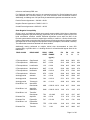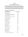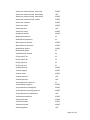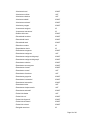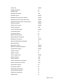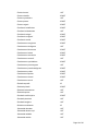
1
LABORATORY PROCEDURE
BD Phoenix™ NMIC/ID Panels
BD Phoenix™ NMIC Panels
BD Phoenix™ NID Panels
INTENDED USE
The BD Phoenix™ Automated Microbiology System is intended for the in vitro rapid
identification (ID) and quantitative determination of antimicrobial susceptibility by minimal
inhibitory concentration (MIC) of Gram Negative aerobic and facultative anaerobic bacteria
belonging to the family Enterobacteriaceae and non-Enterobacteriaceae.
SUMMARY AND EXPLANATION OF THE TEST
Micromethods for the biochemical identification of microorganisms were reported as early as
19181. Several publications reported on the use of the reagent-impregnated paper discs and
micro-tube methods for differentiating enteric bacteria1-9. The interest in miniaturized
identification systems led to the introduction of several commercial systems in the late 1960s,
and they provided advantages in requiring little storage space, extended shelf life,
standardized quality control, and ease of use.
Many of the tests used in the Phoenix ID panels are modifications of the classical methods.
These include tests for fermentation, oxidation, degradation and hydrolysis of various
substrates. In addition to these, the Phoenix system utilizes chromogenic and fluorogenic
substrates as well as single carbon source substrates in the identification of organisms10,11.
The modern broth microdilution test used today has origins in the tube dilution test used in 1942
by Rammelkamp and Maxon to determine in vitro antimicrobial susceptibility testing of bacterial
isolates from clinical specimens12. The broth dilution technique involves exposing bacteria to
decreasing concentrations of antimicrobial agents in liquid media by serial two-fold dilutions.
The lowest concentration of an antimicrobial agent in which no visible growth occurs is defined
as the minimal inhibitory concentration (MIC).
The introduction in 1956 of a microtitrator system, using calibrated precision spiral wire loops
and droppers for making accurate dilutions rapidly allowed Marymont and Wentz to develop a
serial dilution antimicrobial susceptibility test (AST)13. The microtitrator system was accurate
and allowed the reduction in volumes of antimicrobial agents. The term microdilution appeared
in 1970 to describe the MIC tests performed in volumes of 0.1 mL or less of antimicrobial
solution14.
The Phoenix AST test is a modified miniaturized version of the micro-broth doubling dilution
technique. Susceptibility testing in the Phoenix system is performed through determination of
bacterial growth in the presence of various concentrations of the antimicrobial agent tested.
PRINCIPLES OF THE PROCEDURE
A maximum of 100 identification and antimicrobial susceptibility tests can be performed in the
Phoenix instrument at a time using Phoenix ID/AST combination panels. A sealed and
self-inoculating molded polystyrene tray, with 136 micro-wells containing dried reagents,
serves as the Phoenix disposable. The combination panel includes an ID side with dried
substrates for bacterial identification, an AST side with varying concentrations of antimicrobial
agents, and growth and fluorescent controls at appropriate well locations. The Phoenix system
utilizes an optimized colorimetric redox indicator for AST, and a variety of colorimetric and
fluorometric indicators for ID. The AST Broth is cation-adjusted (e.g., Ca++ and Mg++) to
optimize susceptibility testing performance.
Page 1 of 30
The Phoenix panel is comprised of a 51 well ID side and an 85 well AST side. The ID side
contains 45 wells with dried biochemical substrates and 2 fluorescent control wells. The AST
side contains 84 wells with dried antimicrobial agents and 1 growth control well. Panels are
available as ID only (Phoenix™ NID Panels and Phoenix™ PID Panels), AST only (Phoenix™
NMIC Panels and Phoenix™ PMIC Panels), or ID/AST combination (Phoenix™ NMIC/ID
Panels and Phoenix™ PMIC/ID Panels). Unused wells are reserved for future se.
Phoenix panels are inoculated with a standardized inoculum. Organism suspensions must be
prepared only with the BBL™ CrystalSpec™ or BD PhoenixSpec™ nephelometer. Once
inoculated, panels are placed into the instrument and continuously incubated at 35°C. The
instrument tests panels every 20 minutes: on the hour; at 20 minutes past the hour; and again
at 40 minutes past the hour up to 16 hours if necessary. Phoenix panels are read only by the
instrument. Phoenix panels cannot be read manually.
Bacterial Identification: The ID portion of the Phoenix panel utilizes a series of conventional,
chromogenic, and fluorogenic biochemical tests to determine the identification of the organism.
Both growth-based and enzymatic substrates are employed to cover the different types of
reactivity in the range of taxa. The tests are based on microbial utilization and degradation of
specific substrates detected by various indicator systems. Acid production is indicated by a
change in the phenol red indicator when an isolate is able to utilize a carbohydrate substrate.
Chromogenic substrates produce a yellow color upon enzymatic hydrolysis of either
p-nitrophenyl or p-nitroanilide compounds. Enzymatic hydrolysis of fluorogenic substrates
results in the release of a fluorescent coumarin derivative. Organisms that utilize a specific
carbon source reduce the resazurin-based indicator. In addition, there are other tests that
detect the ability of an organism to hydrolyze, degrade, reduce, or otherwise utilize a substrate.
A complete list of taxa that comprises the Phoenix ID Database is provided in Table A.
Reactions employed by various substrates and the principles employed in the Phoenix ID
reactions are described in Table B.
Antimicrobial Susceptibility Testing: The Phoenix AST method is a broth based
microdilution test. The Phoenix system utilizes a redox indicator for the detection of organism
growth in the presence of an antimicrobial agent15. Continuous measurements of changes to
the indicator as well as bacterial turbidity are used in the determination of bacterial growth.
Each AST panel configuration contains several antimicrobial agents with a range of two-fold
doubling dilution concentrations. Organism identification is used in the interpretation of the MIC
values of each antimicrobial agent producing Susceptible, Intermediate, or Resistant (SIR)
result classifications.
A complete list of taxa for which the Phoenix system can provide AST results is provided in
Table A. The list of antimicrobial agents and concentrations available for susceptibility testing in
the Phoenix system is provided under Performance Characteristics.
There are antimicrobial agents for use with the Phoenix System that are not proven to be
effective for treating infections for all organisms listed in the taxa. For interpreting and reporting
results of antimicrobial agents that have been shown to be active against organism groups both
in vitro and in clinical infections refer to the individual pharmaceutical antimicrobial agent
labeling. Alternatively, refer to the most recent CLSI M100 Performance Standard, Table 1
“Suggested Groupings of US FDA-Approved Antimicrobial Agents That Should Be Considered
for Routine Testing and Reporting on Organisms by Clinical Microbiological Laboratories”16.
The components required for testing using the Phoenix system include: 1) Phoenix panels with
panel closures, 2) Phoenix ID Broth, 3) Phoenix AST Broth, 4) Phoenix AST Indicator solution,
5) Phoenix Inoculation Station, 6) Phoenix Panel Caddy, 7) BBL CrystalSpec or BD
PhoenixSpec nephelometer, 8) 25 µL pipettor and sterile tips, and 9) Miscellaneous lab
supplies (listed under Materials Required But Not Provided).
Prior to inoculation the Phoenix panel is placed on the Inoculation Station with the inoculation
ports at the top for filling. Separate inocula are added manually to the ID and AST ports. The
Page 2 of 30
inocula flow down the panel in serpentine fashion, filling the panel wells as the liquid front
progresses toward the pad. The pad absorbs excess inoculum. Closures are manually inserted
in the fill ports. An air admittance port is located in the divider area of the panel lid to ensure
adequate oxygen tension in the panel for the duration of the test.
INGREDIENTS
For a listing of biochemical substrates used in the Phoenix panel refer to Table B. The package
insert enclosed in the panel box provides a listing of the specific antimicrobial agents and
concentrations found in the panel.
PRECAUTIONS
For in vitro Diagnostic Use.
All patient specimens and microbial cultures are potentially infectious and should be treated
with universal precautions. Please refer to CDC manual Bio-safety in Microbiological and
Biomedical Laboratories, 4th Edition, 1999, as well as other recommended literature.
Prior to discarding, sterilize specimen containers and other contaminated materials by
autoclaving.
Panels, once inoculated, should be handled carefully until placed in the instrument.
STORAGE AND HANDLING
Phoenix Panels: Panels are individually packaged and must be stored unopened at room
temperature (15 - 25°C). Do not refrigerate or freeze. Visually inspect the package for holes or
cracks in the foil package. Do not use if the packaging or panel appears to be damaged. If
stored as recommended, the panels will retain expected reactivity until the date of expiration.
Phoenix ID Broth: Tubes are packaged as 100 tube packs. Visually inspect the tubes for
cracks, leaks, etc. Do not use if there appears to be a leak, tube or cap damage or visual
evidence of contamination (i.e., haziness, turbidity). Store Phoenix ID Broth tubes at 2-25°C.
Expiration dating is shown on the tube label.
Phoenix AST Broth: Tubes are packaged as 100 tube packs. Visually inspect the tubes for
cracks, leaks, etc. Do not use if there appears to be a leak, tube or cap damage or visual
evidence of contamination (i.e., haziness, turbidity). Store Phoenix AST Broth tubes at 2-25°C.
Expiration dating is shown on the tube label.
Phoenix AST Indicator Solution: The indicator solution is individually pouched and packaged
as a package of 10 dropper bottles. Visually inspect the bottle for cracks, leaks, etc. Do not use
if there appears to be a leak, bottle or cap damage or any change from a dark blue color. Store
Phoenix AST Indicator Solution at 2-8°C. Each bottle contains enough solution to test up to 100
panels. Expiration dating is shown on the box, pouch, and bottle label and is for unopened
bottles. An opened bottle is stable for up to 14 days if stored at 2-8°C. Be sure the bottle is
held vertically when dispensing the AST Indicator Solution.
SPECIMEN COLLECTION AND PROCESSING
The Phoenix system is not for use directly with clinical specimens. Only pure culture isolates of
aerobic and/or facultatively anaerobic Gram Negative organisms are acceptable for testing.
The test isolate must be a pure culture. It is recommended that cultures be no more than 24
hours old unless additional incubation is required to achieve sufficient growth.
Isolates must be tested with a Gram stain test to assure the appropriate selection of Phoenix
panel type. Once the Gram stain reaction is confirmed, select the appropriate Phoenix panel for
Page 3 of 30
inoculation (e.g., NMIC/ID panel for use with Gram Negative organisms). Selection of the
incorrect panel type could lead to incorrect results.
For AST testing in the Phoenix system, isolates recovered from non-selective media are
recommended. It is recommended that media containing antibiotics not be used for organisms
to be tested in the Phoenix system. Selective media may inhibit some strains of bacteria;
therefore, caution must be used when selecting isolated colonies from these media.
For ID and AST testing, refer to Table C for recommended media.
When swabs are used, only cotton-tipped applicators should be used to prepare the inoculum
suspensions. Some polyester swabs may cause problems with inoculation of the panels.
The usefulness of the Phoenix system or any other diagnostic procedure performed on clinical
specimens is directly influenced by the quality of the specimens themselves. It is strongly
recommended that laboratories employ methods discussed in the Manual of Clinical
Microbiology17 for specimen collection, transport, and placement on primary isolation media.
Inoculum for use on the Phoenix system is prepared by the CLSI-recommended direct colony
suspension method18. Due to variations in inoculum concentrations prepared with McFarland
standards, use of the BBL CrystalSpec or BD PhoenixSpec nephelometer is required for
adjusting the test inoculum prior to use in the Phoenix system.
It is highly recommended that the purity of the inoculum be checked by preparing a purity plate.
See “Purity Check” below.
MATERIALS REQUIRED
Materials Provided:
·
Phoenix Panels
·
Phoenix ID Broth
·
Phoenix AST Broth
·
Phoenix AST Indicator Solution
·
Phoenix Inoculation Station
·
Phoenix Panel Caddy
·
BBL CrystalSpec™ or BD PhoenixSpec™ Nephelometer and Standards
·
25 µL pipettor and sterile tips
·
50 µL pipettor and sterile tips
·
2 Pipette stands
Materials Required But Not Provided:
·
Gram stain reagents
·
Sterile cotton swabs
·
Nonselective culture plated media (e.g., Trypticase™ Soy Agar with 5% Sheep Blood)
·
Incubators
·
Biohazard disposable container
·
Markers, etc
Page 4 of 30
PHOENIX TEST PROCEDURE
Note: The Phoenix instrument should always be powered on. If it is not, power on the
instrument and allow 2 hours for the instrument to warm up before loading panels.
Prepare the Phoenix instrument to receive new panels as described in the BD Phoenix System
User’s Manual (“Operation, Daily System Maintenance”).
Care should be exercised when handling Phoenix panels. You should handle panels by the
sides only to avoid marking, smudging or obscuring the front or back of the panel in any way.
Accession barcode labels affixed to a Phoenix panel should:
·
Not be of fluorescent material
·
Not cover any Phoenix panel reaction wells
·
Not cover the Phoenix panel sequence number barcode
Broth and Panel Preparation:
1.
Confirm the Gram stain reaction of the isolate before proceeding with the inoculum
preparation for use in the Phoenix instrument. Once the Gram stain reaction is confirmed,
select the appropriate Phoenix panel for inoculation. Selection of the incorrect panel type
could lead to incorrect results.
2. Examine the pouch, and do not use the panel if the pouch is punctured or opened.
Remove the panel from the pouch. Discard the desiccant. Do not use the panel if there is
no desiccant or if the desiccant pouch is torn. Note: Panels must be used within 2 hours
of being removed from the pouch.
3. Place the panel on the Inoculation Station with ports at the top and pad on the bottom.
4. Label a Phoenix ID Broth tube with the patient’s specimen number. Using aseptic
technique, pick colonies of the same morphology with the tip of a sterile cotton swab (do
not use a polyester swab) or a wooden applicator stick from one of the recommended
media. See Table C.
5. Suspend the colonies in the Phoenix ID Broth (4.5 mL).
6. Cap the tube and vortex for 5 seconds.
7. Allow approximately ten seconds for air bubbles to surface. Tap the tube gently to aid in
eliminating bubbles.
8. Confirm default settings for inoculum density before inoculating panels. Insert the tube into
the BBL CrystalSpec or BD PhoenixSpec Nephelometer. Make sure the tube is inserted as
far as it will go. Note: Only the BD PhoenixSpec Nephelometer can be used to make
inoculum densities of 0.25 McFarland. (Refer to the BBL CrystalSpec Nephelometer or
BD PhoenixSpec product insert for correct usage instructions and calibration verification.)
9. If the inoculum density is set to 0.5 McFarland for the panel type being run, then a range of
0.50-0.60 is acceptable. If the inoculum density is set to 0.25 for the panel type being run,
then a range of 0.20-0.30 is acceptable. If the density of organisms is low, you can add
colonies from the isolate. Re-vortex the sample and reread to confirm that the correct
density has been achieved. If the density of organisms exceeds 0.6 McFarland, follow the
steps below to dilute the broth. It is very important to accurately fill the wells in the panel.
Note: The standardized bacterial suspension in ID broth must be used within 60
minutes of preparation.
a Using a marker, mark the broth level in the over-inoculated Phoenix ID Broth tube.
Page 5 of 30
b Using a sterile pipette, aseptically add fresh Phoenix ID Broth to the inoculum. Only
Phoenix ID broth may be used to dilute the inoculum.
c Vortex the tube and allow to sit for 10 seconds.
d Place the tube in the nephelometer and remeasure the turbidity of the suspension.
•
If the reader is greater than 0.6, repeat steps b-d.
•
If the reading is 0.5-0.6, go to Step e.
e Using a sterile pipette, aseptically remove excess broth to the original level indicated by
the mark on the tube created in Step a.
Remove excess broth to avoid overfilling the panel. Also, do not removed too much
broth, as there may be insufficient broth to adequately fill the panel.
f Broth may now be used to inoculate the Phoenix AST Broth and/or the Phoenix panel.
10. If you are performing identification only, proceed to Step 15 and continue the procedure.
11. Label a Phoenix AST Broth tube (8.0 mL) with the patient’s specimen number. Holding the
AST Indicator Solution bottle vertically, add one free-falling drop of AST indicator solution
to the AST broth tube. Invert to mix. DO NOT VORTEX. Note: Allow AST Indicator
Solution to warm to room temperature before dispensing into AST broth. The
unused portion of the indicator should be returned to 2-8ºC as soon as possible. Do
not store at room temperature for more than 2 hours. Opened bottles should be
discarded after 14 days from initial opening. If volume other than one drop is added
inadvertently, discard the tube and use a fresh tube of AST broth. After the addition
of the Indicator to AST broth, the mixed solution can be stored in the dark, at room
temperature, for as long as 8 hours. Tubes must be used within 2 hours after the
addition of the indicator solution if exposed to light.
12. If an inoculum density of 0.50 – 0.60 was used, transfer 25 µL of the bacterial suspension
from the ID tube into the AST broth tube. If an inoculum density of 0.20 – 0.30 was used,
transfer 50 µL (use 2 shots if utilizing a 25 µL pipettor) of the bacterial suspension from the
ID tube into the AST broth tube. Note: Panels must be inoculated within 30 minutes of
the time that the AST inoculum is prepared.
13. Cap the AST tube and invert several times to mix. Do not vortex.
14. Wait a few seconds for air bubbles to surface. Tap the tube gently to aid in eliminating
bubbles.
15. Pour the ID tube inoculum into the fill port on the ID side of the panel (51-well side). Allow
the fluid to traverse down the tracks before moving the panel. If using an AST (only) panel,
DO NOT inoculate the ID side of the panel. Retain the ID or AST tube for a purity check.
16. Pour the AST tube inoculum into the fill port on the AST side of the panel (85-well side).
Allow the fluid to traverse down the tracks before moving the panel.
17. Before placing panel closure, check for residual droplets of inoculum on the edge of the fill
ports. If a droplet is present, remove the droplet with absorbent material. The used
absorbent material must be discarded along with your biohazard waste.
18. Snap on the panel closure. Make sure that the closure is fully seated.
Visually inspect panels to be sure each of the wells is full. Look at both sides of the panel.
Make certain that the wells are not overfilled. If any of the wells are unfilled or overfilled,
inoculate a new panel. Note: Panels must be loaded into the instrument within 30
minutes of inoculation. Panels must be kept in the inoculation station after
inoculation until the excess fluid has been completely absorbed by the pad. Panels
Page 6 of 30
should stay vertical in the transport caddy until loaded into the instrument.
Inoculated panels should be handled with care. Avoid knocking or jarring the panel.
Purity Check
1. Using a sterile loop, recover a small drop from the inoculum fluid tube either before or after
inoculating the panel.
2. Inoculate an agar plate (any appropriate medium) for a purity check.
3. Discard inoculum fluid tube and cap in a biohazard disposal container.
4. Incubate the plate for 24-48 hours at 35°C under appropriate conditions.
ID Inoculum Density Flexibility
You may run the ID portion of a panel in the opposite mode from what is configured by
darkening well A17 on the back of the panel before placing the panel in the instrument. This
allows you to run a panel at an inoculum density of 0.20 – 0.30 even if you are configured for a
density of 0.5 for that particular panel type. Likewise, you can run a panel at an inoculum
density of 0.50 – 0.60 if you are configured for a density of 0.25.
There is no way to alter the density setting during Panel Login. To use a panel in the opposite
density mode, using a black Sharpie™ (permanent marker) blacken the entire well. See the
BD Phoenix System User’s Manual (“Operation, ID Inoculum Density Flexibility”) for position of
well A17.
For instructions for panel login and loading, refer to the BD Phoenix System User’s Manual
(“Panel Login” and “Inserting Panels in the Instrument”).
Current Instrument
Inoculum Density
Configuration
Inoculum
Concentration
Desired for Test
Panel
Amount of ID Inoculum to
Add to AST Broth**
Well A-17
0.50
0.25
50 µL
Blackened
0.25
0.50
25 µL
Blackened
** If also running AST
USER QUALITY CONTROL
In order to ensure appropriate set up procedure and acceptable performance of the system, the
following organisms are recommended for testing. The user is advised to review the individual
AST panel formats to determine if all test strains need to be tested for routine laboratory Quality
Control. Refer to the Package Insert that accompanies the Phoenix panels for expected ID and
AST results for QC organisms.
For instructions for QC panel login and loading, refer to the BD Phoenix System User’s Manual
(“Panel Login” and “Inserting Panels in the Instrument”).
ID (NMIC/ID and NID panels):
Escherichia coli ATCC™ 25922
Pseudomonas aeruginosa ATCC™ 27853
AST (NMIC/ID, NMIC panels):
Page 7 of 30
Escherichia coli ATCC™ 25922
Pseudomonas aeruginosa ATCC™ 27853
Escherichia coli ATCC™ 35218
Klebsiella pneumoniae ATCC™ 700603
For the most reliable results, it is recommended that the QC organisms be subcultured at least
twice on two consecutive days onto TSA II with 5% Sheep Blood Agar before use in the
Phoenix system.
Compare recorded results to those listed in the Package Insert. If discrepant results are
obtained, review test procedures as well as confirm purity of the quality control strain used
before contacting BD Diagnostics Technical Services Department. Unacceptable QC results
are documented as “Fail” and acceptable QC results are documented as “Pass” on the QC
Report.
RESULTS
Organism identification will appear on the Phoenix Report Form with a probability percentage
from the Phoenix database based on the substrate reaction profile. Results from each
substrate will appear as +, -, V or X for each reaction. The MIC results and Interpretive
Categorical Results (SIR) will be shown for the appropriate organism/antimicrobial agent
combinations.
Special messages will be shown when the BDXpert System detects results that are of
particular clinical interest.
Further information concerning results obtained from the Phoenix system can be found in the
BD Phoenix System User’s Manual (“Obtaining Results”).
Messages
Error messages may appear if the system detects unexpected reactivity due to inappropriate
procedure or instrument malfunction. For a complete listing of error codes and their meaning
refer to the BD Phoenix System User’s Manual (“System Alerts”, “Needs Attention” and
“Troubleshooting”).
Special Notes
In general, the Phoenix System provides a MIC for all organisms at any of the concentrations
defined on a specific panel. For certain antimicrobic/organism combinations a specific
minimum or maximum MIC is reported even if there is a lower or higher concentration on the
panel. These MIC values are applied by the software and are reported out as less than or equal
to (</=) for the minimum MIC or greater than (>) for the maximum MIC. The table below
provides the range for these special antimicrobic/organism combinations.
Antimicrobial Agent
Amikacin
Aztreonam
Organism(s)
Applied Range (µg/mL)
Morganella morganii
2-64
Proteus penneri
2-64
Proteus vulgaris
2-64
Providencia species
2-64
Providencia stuartii
2-64
Page 8 of 30
Cefotaxime
Providencia species
2-64
Cefotetan
Proteus mirabilis
4-64
Gentamicin
Escherichia coli
1-16
Piperacillin
Morganella morganii
4-128
Achromobacter species
4-128
Achromobacter species
Serratia marcescens
2/4 – 128/4
4/4 – 128/4
Serratia species
4/4-128/4
Tetracycline
Morganella morganii
1-16
Ticarcillin
Achromobacter species
4-128
Alcaligenes species
4-128
Brevundimonas species
4-128
Chryseobacterium species
4-128
Delftia acidoverans
4-128
Myroides species
4-128
Ochrobactrum anthropi
4-128
Providencia species
4-128
Ralstonia species
4-128
Salmonella species
4-128
Serratia species
4-128
Shewanella species
4-128
Shingobacterium species
4-128
Wautersia species
4-128
Ticarcillin/
Clavulanate
Citrobacter freundii
Morganella morganii
4/2 – 128/2
4/2 – 128/2
Tobramycin
Enterobacter aerogenes
0.5-16
Trimethoprim
Enterobacter aerogenes
1-16
Proteus mirabilis
1-16
Piperacillin/
Tazobactam
LIMITATIONS OF THE PROCEDURE
See the package insert shipped with the panel for specific organism/antimicrobial limitations.
General
A Gram stain test is required for the selection of the appropriate Phoenix panel types. Accurate
identification and/or AST results may not be made without this test.
Use only well-isolated bacterial colonies from one of the recommended primary isolation
media. See Table C. Media containing esculin should not be used. Use of mixed colonies
could result in inaccurate identification and/or AST interpretations.
Page 9 of 30
If the instrument inoculum density is configured to 0.5 (for the panel type being used), an
inoculum density of 0.50 – 0.60 must be met. Only the BBL CrystalSpec or BD Phoenix Spec
Nephelometer can be used to measure the inoculum density.
If the instrument inoculum density is configured to 0.25 (for the panel type being used), an
inoculum density of 0.20 – 0.30 must be met. Only the BD PhoenixSpec Nephelometer can be
used to measure inoculum density for this range.
Phoenix panels can be read only by the Phoenix instrument. Visual interpretation of the
Phoenix panels is not possible. Any attempt to manually interpret results from the panel may
lead to misidentification and/or inaccurate AST interpretations.
Identification
The unique panel environment combined with the shortened incubation time may result in
Phoenix panel reactions varying from those obtained using conventional biochemical media.
Antimicrobial Susceptibility Testing
After the addition of Phoenix AST Indicator Solution to the AST broth tubes, mix by inversion.
DO NOT VORTEX. Vortexing may cause air bubbles to form in the AST broth, which can result
in inappropriate filling of the Phoenix panel during inoculation.
Because of the low probability of occurrence or special growth requirements, some organisms
included in the ID taxa are not included in the AST database. These organisms will display the
message “Organism not included in the AST database, perform alternate method.”
For some organism/antimicrobial combinations, the absence of resistant strains precludes
defining any result categories other than “susceptible.” For strains yielding results suggestive of
a “nonsusceptible” category, organism identification and antimicrobial susceptibility test results
should be confirmed. Subsequently, the isolates should be saved and submitted to a reference
laboratory that will confirm the result using the CLSI reference dilution method.
PERFORMANCE CHARACTERISTICS
Gram Negative Identification
In two internal studies, the performance of the Phoenix Gram Negative identification was
evaluated. The 0.5 inoculum density configuration and the 0.25 inoculum density configuration
were tested with 721 strains (0.5) and 784 strains (0.25), respectively. Enteric and non-enteric
results were evaluated against commercial and non-commercial methods.
The Phoenix Gram Negative identification performance is outlined below:
Species Level
McFarland
Agreement
No Agreement
No ID
0.5
95.6%
3.6%
0.8%
0.25
98.1%
1.4%
0.5%
An internal study was performed to simulate inter-site reproducibility. The identification results
obtained using the Phoenix system were compared with expected results. This performance
testing demonstrated intra-site and inter-site reproducibility of at least 95% or greater.
Confirmatory ESBL Test
To determine the accuracy of the Phoenix Confirmatory ESBL test, accuracy testing was
performed at multiple sites using Clinical and Challenge isolates. The results from the ESBL
test resident on the Phoenix panels were compared to the results obtained from the CLSI
Page 10 of 30
reference confirmatory ESBL test.
For Challenge organisms this result is an expected result and for Clinical isolates this result
was obtained from concurrent testing in the CLSI reference broth microdilution method.
Additionally, a challenge set of 30 previously characterized organisms was tested at one site.
Positive Percent Agreement = 183/189 = 96.8%
Negative Percent Agreement = 780/812 = 96.1%
Overall Percent Agreement = 963/1001 = 96.2%
Gram Negative Susceptibility
Clinical, stock, and challenge isolates were tested across multiple clinical sites to determine
Essential Agreement (EA) and Category Agreement (CA) of the Phoenix system to the CLSI
broth microdilution reference method. Essential Agreement occurs when the MIC of the
Phoenix system and the reference method agree exactly or is within ± 1 dilution of each other.
Category Agreement occurs when the Phoenix system results agree with the reference method
with respect to the CLSI categorical interpretative criteria (susceptible, intermediate, resistant).
The table below summarizes the data from these studies.
Additionally, testing performed at multiple clinical sites demonstrated at least 95%
reproducibility or greater within ± 1 doubling dilution for all antimicrobial agents listed in the
table below.
DRUG CLASS
DRUG NAME
DRUG
CODE
DRUG
RANGE
EA
N
EA
%
CA
N
CA
%
(µg/mL)
5-Fluoroquinolone
Ciprofloxacin
CIP
0.25-4
2853
98.8
2853
95.1
5-Fluoroquinolone
Gatifloxacin
GAT
0.25-8
2213
98.8
2213
95.8
5-Fluoroquinolone
Levofloxacin
LVX
0.25-8
2934
98.5
2934
95.8
5-Fluoroquinolone
Moxifloxacin
MXF
0.12-8
2202
98.3
2202
97.6
5-Fluoroquinolone
Norfloxacin
NOR
0.25-16
2792
97.5
2792
94.3
5-Fluoroquinolone
Ofloxacin
OFX
0.25-8
2926
98.5
2926
94.6
Aminoglycoside
Amikacin
AN
0.5-64
2598
94.7
2598
96.7
Aminoglycoside
Gentamicin
GM
0.25-16
2751
96.2
2751
96.3
Aminoglycoside
Tobramycin
NN
0.12-16
2658
93.3
2658
95.3
B-Lac/B-Lac. Inh
Amoxicillin/
Clavulanate
AMC
0.5/0.25- 32/16
2249
96.7
2249
90.9
Ampicillin/
Sulbactam
SAM
0.5/0.25- 32/16
1305
97.2
1305
87.5
Ticarcillin/
Clavulanate
TIM
1/2-128/2
1527
92.5
1527
89.7
B-Lactam Pen
Ampicillin
AM
0.5-32
1712
97.0
1712
94.6
B-Lactam Pen
Piperacillin
PIP
0.5-128
1781
94.3
1781
93.8
B-Lac/B-Lac. Inh
Piperacillin/
Tazobactam
TZP
0.5/4- 128/4
1546
93.2
1546
94.9
Ticarcillin
TIC
1-128
2882
94.7
2882
92.7
B-Lac/B-Lac. Inh
B-Lac/B-Lac. Inh
B-Lactam Pen
Page 11 of 30
Carbapenem
Imipenem
IPM
1-16
2680
97.2
2680
96.8
Carbapenem
Meropenem
MEM
0.25-16
2905
97.6
2905
98.3
Cephem
Cefazolin
CZ
0.5-32
1331
96.7
1331
94.4
Cephem
Cefepime
FEP
0.5-64
1789
95.2
1789
92.9
Cephem
Cefotaxime
CTX
0.5-64
2268
95.0
2268
92.7
Cephem
Cefotetan
CTT
2-64
1175
96.6
1175
96.7
Cephem
Cefoxitin
FOX
0.5-64
1397
96.9
1397
93.3
Cephem
Ceftazidime
CAZ
0.5-64
1796
96.5
1796
94.4
Cephem
Ceftriaxone
CRO
0.5-64
1872
95.8
1872
90.9
Cephem
Cefuroxime
CXM
1-64
1068
96.3
1068
93.3
Cephem
Cephalothin
CF
1-64
2025
96.4
2025
89.0
Folate Antagonist
Trimethoprim
TMP
0.5-16
1856
95.5
1856
98.7
Folate Antagonist
TrimethoprimSulfamethoxazole
SXT
0.5/9.5- 16/304
2212
96.0
2212
97.7
Monobactam
Aztreonam
ATM
0.5-64
1470
96.2
1470
96.2
Nitrofurantoin
Nitrofurantoin
FM
8-512
2130
95.8
2130
84.4
Quinolone
Nalidixic Acid
NA
2-32
2103
96.2
2103
98.6
Tetracycline
Tetracycline
TE
0.5-16
2837
95.5
2837
92.3
REFERENCES
1. Bronfenbrenner, J., and Schlesigner, M.J. 1918. “A Rapid Method for the Identification of
Bacteria Fermenting Carbohydrates,” Am. J. Public Health. 8:922-923.
2. Arnold, W.M., Jr., and Weaver, R.H. 1948. “Quick Microtechniques for Identification of
Cultures - I. Indole production,” J. Lab. Clin. Med. 33:1334-1337.
3. Bachmann, B., and Weaver, R.H. 1951. “Rapid Microtechnics for Identification of Cultures
- V. Reduction of Nitrates to Nitrites,” Am. J. Clin. Pathol. 21:195-196.
4. Hannan, J., and Weaver, R.H. 1948. “Quick Microtechniques for the Identification of
Cultures - II. Fermentations,” J. Lab. Clin. Med. 33:1338-1341.
5. Hartman, P.A. 1968. Paper strip and disc methods, p. 123-132. Miniaturized
microbiological methods. Academic Press, New York.
6. Sanders, A.C., Faber, J.E., and Cook, T.M. 1957. “A Rapid Method for the Characterization
of Enteric Pathogen Using Paper Discs,” Appl. Microbiol. 5:36-40.
7. Synder, M.L. 1954. “Paper Discs Containing Entire Culture Medium for the Differentiation
of Bacteria,” Pathol. Bacteriol. 67:217-226.
8. Soto, O.B. 1949. “Fermentation Reactions with Dried Paper Discs Containing
Carbohydrate and Indicator,” Puerto Rican J. Publ. Hlth. Trop. Med. 25:96-100.
9. Weaver, R.H. 1954. “Quicker Bacteriological Results,” Am. J. Med. Technol.
20:14-26.
10. Kämpfer, P., Rauhoff, O., and Dott, W. 1991. “Glycosidase Profiles of Members of the
Family Enterobacteriaceae,” J. Clin. Microbiol. 29:2877-2879.
Page 12 of 30
11. Manafi, M., Kneifel, W., and Bascomb, S. 1991. “Fluorogenic and Chromogenic Substrates
Used in Bacterial Diagnostics,” Microbiol. Rev. 55:335-348.
12. Rammelkamp, C.H. and Maxon, T. 1942. “Resistance of Staphylococcus aureus to the
Action of Penicillin,” Proc. Soc. Biol. and Med. 51:386-389.
13. Marymont, J.H. and Wentz, R.M. 1966. “Serial Dilution Antibiotic Sensitivity Testing with
the Microtitrator System,” Am. J. Clin. Pathol. 45:548-551.
14. Gavan, T.L., and Town, M.A. 1970. “A Microdilution Method for Antibiotic Susceptibility
Testing: An Evaluation,” Am. J. Clin. Pathol. 53:880-885.
15. Lancaster, M.V. and Fields, R.D. 1996. Antibiotic and Cytotoxic Drug Susceptibility Assays
Using Resazurin and Poising Agents. U.S. Patent #5,501,959.
16. CLSI. M100-S15 Performance Standards for Antimicrobial Susceptibility Testing; Fifteenth
Informational Supplement. January, 2005.
17. Murray, Patrick R., et al. ed., Manual of Clinical Microbiology, 8th Edition, ASM Press,
Washington, D.C., 2003.
18. CLSI. M7-A6 Methods for Dilution Antimicrobial Susceptibility Tests for Bacteria That Grow
Aerobically; Approved Standard—Sixth Edition. January, 2003.
Manufactured by
Becton, Dickinson and Company
7 Loveton Circle
Sparks, MD 21152 USA (800) 638-8663
Made in USA
TECHNICAL INFORMATION
Approved by:_________________________
Date Effective:________________________
Supervisor:___________________________Date:____________________
Director:_____________________________ Date:____________________
Reviewed:____________________________
9/2006
Phoenix, BDXpert, BBL CrystalSpec, PhoenixSpec, Trypticase and BD are trademarks of
Becton, Dickinson and Company. ATCC is a trademark of American Type Culture Collection.
Sharpie is a trademark of Sanford. CHROMagar is a trademark of Dr. A. Rambach.
Page 13 of 30
Table A
Taxa for ID/AST Determination
There are antimicrobial agents for use with the Phoenix system that are not proven to be
effective for treating infections for all organisms listed in this section. For interpreting and
reporting results of antimicrobial agents that have shown to be active against organism groups
both in vitro and in clinical infections refer to the individual pharmaceutical antimicrobial agent
labeling. Alternatively, refer to the most recent CLSI M100 Performance Standard, Table 1
“Suggested Groupings of US FDA-Approved Antimicrobial Agents That Should be Considered
for Routine Testing and Reporting on Organisms by Clinical Microbiological Laboratories.”
Gram Negative (0.5 McFarland)
Gram Negative Taxa1
ID, AST, ID/AST
Achromobacter piechaudii
AST
Achromobacter species
ID/AST
Achromobacter xylosoxidans ssp. denitrificans
AST
Achromobacter xylosoxidans ssp. xylosoxidans
AST
Acinetobacter baumannii
ID/AST
Acinetobacter baumannii/calcoaceticus complex
ID/AST
Acinetobacter calcoaceticus
AST
Acinetobacter haemolyticus
ID/AST
Acinetobacter johnsonii
AST
Acinetobacter junii
AST
Acinetobacter lwoffii
ID/AST
Acinetobacter radioresistens
AST
Acinetobacter species
ID/AST
Actinobacillus lignieresii
ID
Actinobacillus suis
ID
Actinobacillus ureae
ID
Aeromonas allosaccharophila
AST
Aeromonas caviae
ID/AST
Aeromonas eucrenophila
AST
Aeromonas hydrophila
ID/AST
Aeromonas jandaei
AST
Aeromonas media
AST
Aeromonas salmonicida
AST
Aeromonas salmonicida ssp. achromogenes
AST
Page 14 of 30
Aeromonas salmonicida ssp. masoucida
ID/AST
Aeromonas salmonicida ssp. pectinolytica
AST
Aeromonas salmonicida ssp. salmonicida
ID/AST
Aeromonas salmonicida ssp. smithia
ID/AST
Aeromonas schubertii
ID/AST
Aeromonas sobria
ID/AST
Aeromonas trota
AST
Aeromonas veronii
ID/AST
Alcaligenes faecalis
ID/AST
Bergeyella zoohelcum
ID
Bordetella bronchiseptica
ID
Brevundimonas diminuta
ID/AST
Brevundimonas vesicularis
ID/AST
Burkholderia cepacia
ID/AST
Burkholderia gladioli
ID
Cardiobacterium hominis
ID
CDC group EF-4a
ID
CDC group EF-4b
ID
CDC group EO-2
ID
CDC group Vb-3
ID
Cedecea davisae
ID/AST
Cedecea lapagei
ID/AST
Cedecea neteri
ID/AST
Cedecea species 3
AST
Cedecea species 5
AST
Chromobacterium violaceum
ID
Chryseobacterium gleum
ID/AST
Chryseobacterium indologenes
ID/AST
Chryseobacterium meningosepticum
ID/AST
Chryseobacterium scophthalmum
AST
Citrobacter amalonaticus
ID/AST
Citrobacter braakii
ID/AST
Citrobacter farmeri
ID/AST
Citrobacter freundii
ID/AST
Citrobacter gillenii
AST
Page 15 of 30
Citrobacter koseri
ID/AST
Citrobacter murliniae
AST
Citrobacter rodentium
AST
Citrobacter sedlakii
ID/AST
Citrobacter werkmanii
ID/AST
Citrobacter youngae
ID/AST
Comamonas terrigena
ID
Comamonas testosteroni
ID
Delftia acidovorans
ID/AST
Edwardsiella hoshinae
ID/AST
Edwardsiella ictaluri
ID/AST
Edwardsiella tarda
ID/AST
Eikenella corrodens
ID
Empedobacter brevis
ID
Enterobacter aerogenes
ID/AST
Enterobacter amnigenus
AST
Enterobacter amnigenus biogroup 1
ID/AST
Enterobacter amnigenus biogroup 2
ID/AST
Enterobacter asburiae
ID/AST
Enterobacter cancerogenus
ID/AST
Enterobacter cloacae
ID/AST
Enterobacter cowanii
AST
Enterobacter dissolvens
AST
Enterobacter gergoviae
ID/AST
Enterobacter hormaechei
ID/AST
Enterobacter intermedius
ID/AST
Enterobacter kobei
AST
Enterobacter nimipressuralis
AST
Enterobacter sakazakii
ID/AST
Escherichia blattae
AST
Escherichia coli
ID/AST
Escherichia fergusonii
ID/AST
Escherichia hermannii
ID/AST
Escherichia vulneris
ID/AST
Ewingella americana
ID
Page 16 of 30
Hafnia alvei
ID/AST
Kingella denitrificans
ID
Kingella kingae
ID
Klebsiella granulomatis
AST
Klebsiella oxytoca
ID/AST
Klebsiella pneumoniae ssp. ozaenae
ID/AST
Klebsiella pneumoniae ssp. pneumoniae
ID/AST
Klebsiella pneumoniae ssp. rhinoscleromatis
ID/AST
Kluyvera ascorbata
ID/AST
Kluyvera cryocrescens
ID/AST
Kluyvera georgiana
AST
Leclercia adecarboxylata
ID/AST
Leminorella grimontii
ID
Leminorella richardii
ID
Mannheimia haemolytica
ID
Methylobacterium extorquens
ID
Moellerella wisconsensis
ID/AST
Moraxella (Branhamella) catarrhalis
ID
Moraxella species
ID
Morganella morganii
ID/AST
Myroides odoratus/odoratimimus
ID/AST
Ochrobactrum anthropi
ID/AST
Oligella ureolytica
ID
Oligella urethralis
ID
Pantoea agglomerans
ID/AST
Pantoea ananatis
AST
Pantoea dispersa
AST
Pantoea stewartii ssp. indologenes
AST
Pantoea stewartii ssp. stewartii
AST
Pasteurella aerogenes
ID
Pasteurella multocida
ID
Pasteurella pneumotropica
ID
Photobacterium damselae
ID
Plesiomonas shigelloides
ID
Pragia fontium
ID
Page 17 of 30
Proteus hauseri
AST
Proteus mirabilis
ID/AST
Proteus myxofaciens
AST
Proteus penneri
ID/AST
Proteus vulgaris
ID/AST
Providencia alcalifaciens
ID/AST
Providencia heimbachae
AST
Providencia rettgeri
ID/AST
Providencia rustigianii
ID/AST
Providencia stuartii
ID/AST
Pseudomonas aeruginosa
ID/AST
Pseudomonas alcaligenes
AST
Pseudomonas fluorescens
ID/AST
Pseudomonas luteola
ID/AST
Pseudomonas mendocina
ID/AST
Pseudomonas monteilii
AST
Pseudomonas oryzihabitans
ID/AST
Pseudomonas pertucinogena
AST
Pseudomonas pseudoalcaligenes
ID/AST
Pseudomonas putida
ID/AST
Pseudomonas species
ID/AST
Pseudomonas stutzeri
ID/AST
Pseudomonas veronii
AST
Rahnella aquatilis
ID
Ralstonia pickettii
ID/AST
Ralstonia solanacearum
AST
Ralstonia species
AST
Raoultella ornithinolytica
ID/AST
Raoultella planticola
AST
Raoultella terrigena
AST
Rhizobium radiobacter
ID
Salmonella aberdeen
AST
Salmonella abortus-equi
AST
Salmonella adelaide
AST
Salmonella aderike
AST
Page 18 of 30
Salmonella agona
AST
Salmonella alachua
AST
Salmonella anatum
AST
Salmonella arizonae
AST
Salmonella avana
AST
Salmonella bahrenfeld
AST
Salmonella blockley
AST
Salmonella bongori
AST
Salmonella braenderup
AST
Salmonella bredeney
AST
Salmonella bunn
AST
Salmonella california
AST
Salmonella carrau
AST
Salmonella cerro
AST
Salmonella champaign
AST
Salmonella chittagong
AST
Salmonella cholerasuis
AST
Salmonella choleraesuis ssp. arizonae
ID/AST
Salmonella choleraesuis ssp. choleraesuis
ID/AST
Salmonella choleraesuis ssp. diarizonae
AST
Salmonella choleraesuis ssp. houtenae
AST
Salmonella choleraesuis ssp. indica
AST
Salmonella choleraesuis ssp. salamae
AST
Salmonella cubana
AST
Salmonella dakar
AST
Salmonella daressalaam
AST
Salmonella derby
AST
Salmonella dessau
AST
Salmonella DT
AST
Salmonella dublin
AST
Salmonella duesseldorf
AST
Salmonella enteritidis
AST
Salmonella fresno
AST
Salmonella gallinarum
ID/AST
Salmonella give
AST
Page 19 of 30
Salmonella haardt
AST
Salmonella hadar
AST
Salmonella hamburg
AST
Salmonella hartford
AST
Salmonella heidelberg
AST
Salmonella illinois
AST
Salmonella infantis
AST
Salmonella inverness
AST
Salmonella java
AST
Salmonella javiana
AST
Salmonella kentucky
AST
Salmonella kirkee
AST
Salmonella kunduchi
AST
Salmonella kvittingfoss
AST
Salmonella lansing
AST
Salmonella litchfield
AST
Salmonella liverpool
AST
Salmonella london
AST
Salmonella luciana
AST
Salmonella manhattan
AST
Salmonella mbandaka
AST
Salmonella meleagridis
AST
Salmonella memphis
AST
Salmonella michigan
AST
Salmonella minneapolis
AST
Salmonella minnesota
AST
Salmonella montevideo
AST
Salmonella muenchen
AST
Salmonella muenster
AST
Salmonella newington
AST
Salmonella newport
AST
Salmonella nottingham
AST
Salmonella ohio
AST
Salmonella onderstepoort
AST
Salmonella oranienburg
AST
Page 20 of 30
Salmonella panama
AST
Salmonella paratyphi A
ID/AST
Salmonella paratyphi B
AST
Salmonella poona
AST
Salmonella pullorum
ID/AST
Salmonella quinhon
AST
Salmonella rubislaw
AST
Salmonella saintpaul
AST
Salmonella schwarzengrund
AST
Salmonella seftenberg
AST
Salmonella species
ID/AST
Salmonella tallahassee
AST
Salmonella thompson
AST
Salmonella typhi
ID/AST
Salmonella typhimurium
AST
Salmonella virginia
AST
Salmonella westerstede
AST
Salmonella worthington
AST
Serratia entomophilia
AST
Serratia ficaria
ID/AST
Serratia fonticola
ID/AST
Serratia grimesii
AST
Serratia liquifaciens
ID/AST
Serratia marcescens
ID/AST
Serratia odorifera
AST
Serratia odorifera 1
ID/AST
Serratia odorifera 2
ID/AST
Serratia plymuthica
ID/AST
Serratia proteamaculans ssp. proteamaculans
AST
Serratia proteamaculans ssp. quinovora
AST
Serratia rubidaea
ID/AST
Shewanella algae
AST
Shewanella putrefaciens
ID/AST
Shigella boydii
ID/AST
Shigella dysenteriae
ID/AST
Page 21 of 30
Shigella flexneri
ID/AST
Shigella sonnei
ID/AST
Shigella species
ID/AST
Sphingobacterium multivorum
ID/AST
Sphingobacterium spiritivorum
ID/AST
Sphingobacterium thalpophilum
ID/AST
Sphingomonas paucimobilis
ID
Stenotrophomonas maltophilia
ID/AST
Suttonella indologenes
ID
Tatumella ptyseos
ID
Vibrio alginolyticus
ID
Vibrio cholerae
ID
Vibrio fluvialis
ID
Vibrio hollisae
ID
Vibrio metschnikovii
ID
Vibrio mimicus
ID
Vibrio parahaemolyticus
ID
Vibrio vulnificus
ID
Wautersia gilardii
AST
Wautersia paucula
ID/AST
Weeksella virosa
ID
Yersinia aldovae
AST
Yersinia bercovieri
AST
Yersinia enterocolitica
ID/AST
Yersinia frederiksenii
ID/AST
Yersinia intermedia
ID/AST
Yersinia kristensenii
ID/AST
Yersinia mollaretii
AST
Yersinia pseudotuberculosis
ID/AST
Yersinia rohdei
AST
Yersinia ruckeri
ID/AST
Yokenella regensburgei
ID
1
Not all species encountered during clinical performance evaluations.
Page 22 of 30
Gram Negative (0.25 McFarland)
Gram Negative Taxa1
ID, AST, ID/AST
Achromobacter species
ID/AST
Acinetobacter baumannii/calcoaceticus complex
ID/AST
Acinetobacter haemolyticus
ID/AST
Acinetobacter lwoffii
ID/AST
Actinobacillus lignieresii
ID
Actinobacillus suis
ID
Actinobacillus ureae
ID
Aeromonas caviae
ID/AST
Aeromonas hydrophila
ID/AST
Aeromonas salmonicida ssp. masoucida
ID/AST
Aeromonas salmonicida ssp. salmonicida
ID/AST
Aeromonas salmonicida ssp. smithia
ID/AST
Aeromonas schubertii
ID/AST
Aeromonas sobria
ID/AST
Aeromonas veronii
ID/AST
Alcaligenes faecalis
ID/AST
Bergeyella zoohelcum
ID
Bordetella bronchiseptica
ID
Brevundimonas diminuta
ID/AST
Brevundimonas vesicularis
ID/AST
Burkholderia cepacia
ID/AST
Burkholderia gladioli
ID
Cardiobacterium hominis
ID
CDC group EF-4a
ID
CDC group EF-4b
ID
CDC group EO-2
ID
CDC group Vb-3
ID
Cedecea davisae
ID/AST
Cedecea lapagei
ID/AST
Cedecea neteri
ID/AST
Chromobacterium violaceum
ID
Page 23 of 30
Chryseobacterium gleum
ID/AST
Chryseobacterium indologenes
ID/AST
Chryseobacterium meningosepticum
ID/AST
Citrobacter amalonaticus
ID/AST
Citrobacter braakii
ID/AST
Citrobacter farmeri
ID/AST
Citrobacter freundii
ID/AST
Citrobacter koseri
ID/AST
Citrobacter sedlakii
ID/AST
Citrobacter werkmanii
ID/AST
Citrobacter youngae
ID/AST
Comamonas terrigena
ID
Comamonas testosteroni
ID
Delftia acidovorans
ID/AST
Edwardsiella hoshinae
ID/AST
Edwardsiella ictaluri
ID/AST
Edwardsiella tarda
ID/AST
Eikenella corrodens
ID
Empedobacter brevis
ID
Enterobacter aerogenes
ID/AST
Enterobacter amnigenus biogroup 1
ID/AST
Enterobacter amnigenus biogroup 2
ID/AST
Enterobacter asburiae
ID/AST
Enterobacter cancerogenus
ID/AST
Enterobacter cloacae
ID/AST
Enterobacter gergoviae
ID/AST
Enterobacter hormaechei
ID/AST
Enterobacter intermedius
ID/AST
Enterobacter sakazakii
ID/AST
Escherichia coli
ID/AST
Escherichia fergusonii
ID/AST
Escherichia hermannii
ID/AST
Escherichia vulneris
ID/AST
Ewingella americana
ID
Hafnia alvei
ID/AST
Page 24 of 30
Klebsiella oxytoca
ID/AST
Klebsiella pneumoniae ssp. ozaenae
ID/AST
Klebsiella pneumoniae ssp. pneumoniae
ID/AST
Klebsiella pneumoniae ssp. rhinoscleromatis
ID/AST
Kluyvera ascorbata
ID/AST
Kluyvera cryocrescens
ID/AST
Leclercia adecarboxylata
ID/AST
Leminorella grimontii
ID
Leminorella richardii
ID
Mannheimia haemolytica
ID
Moellerella wisconsensis
ID/AST
Morganella morganii
ID/AST
Myroides odoratus/odoratimimus
ID/AST
Ochrobactrum anthropi
ID/AST
Oligella ureolytica
ID
Oligella urethralis
ID
Pantoea agglomerans
ID/AST
Pasteurella aerogenes
ID
Pasteurella multocida
ID
Pasteurella pneumotropica
ID
Photobacterium damselae
ID
Plesiomonas shigelloides
ID
Pragia fontium
ID
Proteus mirabilis
ID/AST
Proteus penneri
ID/AST
Proteus vulgaris
ID/AST
Providencia alcalifaciens
ID/AST
Providencia rettgeri
ID/AST
Providencia rustigianii
ID/AST
Providencia stuartii
ID/AST
Pseudomonas aeruginosa
ID/AST
Pseudomonas fluorescens
ID/AST
Pseudomonas luteola
ID/AST
Pseudomonas mendocina
ID/AST
Pseudomonas oryzihabitans
ID/AST
Page 25 of 30
Pseudomonas putida
ID/AST
Pueudomonas stutzeri
ID/AST
Rahnella aquatilis
ID
Ralstonia pickettii
ID/AST
Raoultella ornithinolytica
ID/AST
Rhizobium radiobacter
ID
Salmonella choleraesuis ssp. arizonae
ID/AST
Salmonella choleraesuis ssp. choleraesuis
ID/AST
Salmonella gallinarum
ID/AST
Salmonella paratyphi A
ID/AST
Salmonella pullorum
ID/AST
Salmonella species
ID/AST
Salmonella typhi
ID/AST
Serratia ficaria
ID/AST
Serratia fonticola
ID/AST
Serratia liquifaciens
ID/AST
Serratia marcescens
ID/AST
Serratia odorifera 1
ID/AST
Serratia odorifera 2
ID/AST
Serratia plymuthica
ID/AST
Serratia rubidaea
ID/AST
Shewanella putrefaciens
ID/AST
Shigella boydii
ID/AST
Shigella dysenteriae
ID/AST
Shigella flexneri
ID/AST
Shigella sonnei
ID/AST
Sphingobacterium multivorum
ID/AST
Sphingobacterium spiritivorum
ID/AST
Sphingobacterium thalpophilum
ID/AST
Sphingomonas paucimobilis
ID
Stenotrophomonas maltophilia
ID/AST
Suttonella indologenes
ID
Tatumella ptyseos
ID
Vibrio alginolyticus
ID
Vibrio cholerae
ID
Page 26 of 30
Vibrio fluvialis
ID
Vibrio hollisae
ID
Vibrio metschnikovii
ID
Vibrio mimicus
ID
Vibrio parahaemolyticus
ID
Vibrio vulnificus
ID
Wautersia paucula
ID/AST
Weeksella virosa
ID
Yersinia enterocolitica
ID/AST
Yersinia frederiksenii
ID/AST
Yersinia intermedia
ID/AST
Yersinia kristensenii
ID/AST
Yersinia pseudotuberculosis
ID/AST
Yersinia ruckeri
ID/AST
Yokenella regensburgei
ID
1
Not all species encountered during clinical performance evaluations.
Page 27 of 30
Table B
List of Reagents and Principles Employed in the Phoenix
System
Substrate Name
Code
L-PHENYLALANINE-AMC
4MU-N-ACETYL-BD-GLUCOSAM
INIDE
A_LPHET
A_NAG
L-GLUTAMIC ACID-AMC
L-TRYPTOPHAN-AMC
L-PYROGLUTAMIC ACID-AMC
A_LGTA
A_LTRY
A_LPYR
L-PROLINE-AMC
L-ARGININE-AMC
ARGININE-ARGININE-AMC
GLYCINE-AMC
L-LEUCINE-AMC
LYSINE-ALANINE-AMC
GLUTARYL-GLYCINE-ARGININE
-AMC
A_LPROB
A_LARGH
A_ARARR
A_GLYB
A_LLEUH
A_LYALD
A_GUGAH
GLYCINE-PROLINE-AMC
COLISTIN
A_GLPRB
C_CLST
POLYMYXIN B
D-MANNITOL
CITRATE
ACETATE
ADONITOL
MALONATE
ALPHA-KETOGLUTARIC ACID
C_PXB
C_DMNT
C_CIT
C_ACT
C_ADO
C_MLO
C-KGA
TIGLIC ACID
FLUORESCENT POSITIVE
CONTROL
C_TIG
FLR_CTL
FLUORESENT POSTIVE
CONTROL
FLR_CTL
L-PROLINE-NA
N_LPROT
GAMMA-L-GLUTAMYL-NA
N_LGGH
BIS (PNP) PHOSPHATE
P_BPHO
PNP-BD-GLUCOSIDE
P_BDGLU
Principle
Enzymatic hydrolysis of the amide or
glycosidic bond results in the release of a
fluorescent coumarin or
4-methylumbelliferone derivative.
Resistance to the antimicrobial agents results
in a reduction of resazurin based indicator.
Utilization of a carbon source results in a
reduction of the resazurin-based indicator.
Control to standardize fluorescent substrate
results.
Enzymatic hydrolysis of the colorless amide
substrate releases yellow p-nitroaniline.
Enzymatic hydrolysis of the colorless aryl
substituted glycoside releases yellow
p-nitrophenol.
Page 28 of 30
Substrate Name
Code
Principle
BETA-ALLOSE
N-ACETYL-GALACTOSAMINE
R_BALL
R_NGA
N-ACETYL-GLUCOSAMINE
R_NGU
SORBITOL
R_DSBT
SUCROSE
R_DSUC
GALACTURONIC ACID
R_GRA
MALTULOSE
R_MTU
L-RHAMNOSE
R_LRHA
BETA-GENTIOBIOSE
R_BGEN
DEXTROSE
R_DEX
D-GALACTOSE
R_DGAL
D-FRUCTOSE
R_DFRU
D-GLUCONIC ACID
R_DGUA
D-MELIBIOSE
R_DMLB
L-ARABINOSE
R_LARA
METHYL-B-GLUCOSIDE
R_MBGU
ORNITHINE
S_ORN
Utilization of ornithine results in pH rise and
change in fluorescent indicator.
UREA
S_URE
Hydrolysis of urea and the resulting ammonia
change results in pH rise and change in
fluorescent indicator.
ESCULIN
T_ESC
Hydrolysis of esculin results in a black
precipitate in the presence of ferric ion.
Utilization of carbohydrate results in lower pH
and change in indicator (phenol red).
Page 29 of 30
Table C
Recommended Media and Approved Use
Recommended Media
Trypticase™ Soy Agar with 5% Sheep Blood
Bromthymol Blue (BTB) Lactose Agar
BBL™ CHROMagar™ Orientation
Chocolate Agar
Columbia Agar with 5% Horse Blood
Columbia Agar with 5% Sheep Blood
Cystine-Lactose-Electrolyte-Deficient (CLED) Agar
Dey/Egley (D/E) Neutralizing Agar
Eosin Methylene Blue
Hektoen Enteric Agar
MacConkey Agar
Trypticase™ Soy Agar without Blood
Trypticase™ Soy Agar with Lecithin and Tween™
80
Xylose Lysine Desoxycholate Agar
Approved Use
ID
AST
Yes
Yes
Yes
Yes
Yes
Yes
Yes
Yes
Yes
Yes
Yes
Yes
Yes
Yes
Yes
No
Yes
Yes
Yes
No
Yes
Yes
Yes
No
Yes
No
Yes
No
Page 30 of 30