1
Antibody Microarrays User Manual
I. Introduction & Protocol Overview continued
microarray you wish to review, as the composition of each lot may vary slightly,
depending on antibody availability at the time of printing.)
The arrayed antibodies can be purchased individually from BD Biosciences
Pharmingen or BD Biosciences Transduction Labs. If you need specific information about one or more antibodies on the microarray, please visit
www.bdbiosciences.com and navigate to the appropriate technical support
pages.
Sensitivity and species cross-reactivity of the Ab Microarray
Because no two antibodies have exactly the same binding affinity (KA), the lower
detection limit of the Ab Microarray must be defined for each antibody. But on
average, we find that the Ab Microarray can detect as little as 20 pg/ml of a given
target protein.
Although we have not thoroughly tested each microarray for its cross-species
reactivity, it is likely that many of the epitopes recognized by arrayed antibodies
are conserved among closely—or even distantly—related species. In practice,
however, it is difficult to predict how a given microarray will perform for different
species. We recommend you review the array-specific antibody list (available at
www.clontech.com) to decide which Ab Microarray is suitable for your experimental system.
Overview of the Ab Microarray Procedure
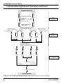
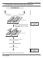
The Ab Microarray Protocol, outlined in Figure 1, is a fluorescence-based
procedure in which solid-phase antibody is used to capture fluorescently-labeled
antigen. The entire procedure, from sample preparation to array scanning, takes
just one day to complete. Note that Ab Microarrays do not measure absolute
concentrations. Instead, they provide a relative measure of protein abundance;
that is, the protein levels in one sample (Sample A) are compared to those of a
second sample (Sample B).
Measuring protein abundances with Ab Microarrays consists of five main steps:
1.
2.
3.
4.
5.
Extracting protein
Labeling protein with Cy5 and Cy3 dyes
Removing unbound dye
Incubating labeled protein with Ab Microarrays
Scanning microarrays to measure bound antigen
In Step 1, total protein is extracted from 50–200 mg of cells or whole tissue. The
samples are first centrifuged to produce a pellet. Then, after a single freeze-thaw
cycle, each pellet is resuspended in Extraction/Labeling Buffer. (Before
resuspension, whole tissue must first be homogenized using a mortar & pestle,
as described in Section V.) Extraction/Labeling Buffer contains non-denaturing
BD Biosciences Clontech
4
www.bdbiosciences.com
Protocol # PT3648-1
Version # PR23092