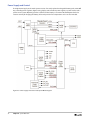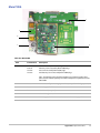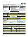
1
Service Manual i ii SonoSite, Inc. 21919 30th Drive SE Bothell, WA 98021-3904 USA Telephone: 1-888-482-9449 or 1-425-951-1200 Fax: 1-425-951-1201 SonoSite Ltd Alexander House 40A Wilbury Way Hitchin Herts SG4 OAP UK T: +44-1462-444800 F: +44-1462-444801 Caution: Federal (United States) law restricts this device to sale by or on the order of a physician. Edge, SiteLink, SonoCalc, SonoHD2, SonoMB, SonoMBe, SonoSite, and the SonoSite logo are registered (in some jurisdictions) and unregistered trademarks owned by SonoSite, Inc. DICOM is the registered trademark of the National Electrical Manufacturers Association for its standards publications relating to digital communications of medical information. Non-SonoSite product names may be trademarks or registered trademarks of their respective owners. The SonoSite ultrasound system(s) referenced in this document may be covered by one or more of the following U.S. patents: 5722412, 5817024, 5893363, 6135961, 6203498, 6364839, 6371918, 6383139, 6416475, 6447451, 6471651, 6569101, 6648826, 6575908, 6604630, 6817982, 6835177, 6962566, 7169108, 7449640, 7534211, 7549961, 7588541, 7591786, 7604596, 7643040, 7686766, 7694814, 7727153, 7740586, 7804970, 7809400, 7819807, 7841575, 7849250, 7867168, 7883276, D456509, D461895, D509900, D538432, D544962, D558351, D559390, D591423, D592750, D592760, D625014, D625015, and by the following counterpart foreign patents: AU727381, AU730822, CA2372152, CA2372158, CA2373065, CN ZL 97113678.5, CN ZL 98106133.8, CN ZL 98108973.9, CN ZL 200830007734.8, DE60021552.0, DE60029777.2, DE60034670.6, DE69730563.5, DE6980539.6, DE69831698.3, DE60 2004 23 816.3-08, FR0815793, FR0875203, FR0881492, FR1175713, FR1180970, FR1589878, GB0875203, GB0881492, GB1175713, GB1180970, GB1180971, GB1589878, IT0815793, IT0881492, IT1175713, IT1589878, KR528102, KR532359, NO326202, NO326814, NZ542968, RCD000897368-0001, SP0815793, SP0881492, SP1589878. Patents pending. P15644-01 12/2011 Copyright 2011 by SonoSite, Inc. All rights reserved. iii iv Table of Contents Introduction Audience . . . . . . . . . . . . . . . . . . . . . . . . . . . . . . . . . . . . . . . . . . . . . . . . . . . . . . . . . . . . . . . . . . . . . . . . . . . .1 Contact Information . . . . . . . . . . . . . . . . . . . . . . . . . . . . . . . . . . . . . . . . . . . . . . . . . . . . . . . . . . . . . . . . .1 Conventions, symbols, and terms . . . . . . . . . . . . . . . . . . . . . . . . . . . . . . . . . . . . . . . . . . . . . . . . . . . .1 Labeling symbols . . . . . . . . . . . . . . . . . . . . . . . . . . . . . . . . . . . . . . . . . . . . . . . . . . . . . . . . . . . . . . . . . . . .2 Specifications Specifications . . . . . . . . . . . . . . . . . . . . . . . . . . . . . . . . . . . . . . . . . . . . . . . . . . . . . . . . . . . . . . . . . . . . . . . .5 Dimensions . . . . . . . . . . . . . . . . . . . . . . . . . . . . . . . . . . . . . . . . . . . . . . . . . . . . . . . . . . . . . . . . . . . . . . . . . 5 Environmental limits . . . . . . . . . . . . . . . . . . . . . . . . . . . . . . . . . . . . . . . . . . . . . . . . . . . . . . . . . . . . . . . . 5 Electrical specifications . . . . . . . . . . . . . . . . . . . . . . . . . . . . . . . . . . . . . . . . . . . . . . . . . . . . . . . . . . . . . 5 Battery specifications . . . . . . . . . . . . . . . . . . . . . . . . . . . . . . . . . . . . . . . . . . . . . . . . . . . . . . . . . . . . . . . 6 Compatible accessories and peripherals . . . . . . . . . . . . . . . . . . . . . . . . . . . . . . . . . . . . . . . . . . . . . .6 Safety Electrical safety . . . . . . . . . . . . . . . . . . . . . . . . . . . . . . . . . . . . . . . . . . . . . . . . . . . . . . . . . . . . . . . . . . . . . .9 Electrical safety classification . . . . . . . . . . . . . . . . . . . . . . . . . . . . . . . . . . . . . . . . . . . . . . . . . . . . . . . . 11 Equipment safety . . . . . . . . . . . . . . . . . . . . . . . . . . . . . . . . . . . . . . . . . . . . . . . . . . . . . . . . . . . . . . . . . . . .11 Battery safety . . . . . . . . . . . . . . . . . . . . . . . . . . . . . . . . . . . . . . . . . . . . . . . . . . . . . . . . . . . . . . . . . . . . . . . .12 Clinical safety . . . . . . . . . . . . . . . . . . . . . . . . . . . . . . . . . . . . . . . . . . . . . . . . . . . . . . . . . . . . . . . . . . . . . . . .13 Hazardous materials . . . . . . . . . . . . . . . . . . . . . . . . . . . . . . . . . . . . . . . . . . . . . . . . . . . . . . . . . . . . . . . . .13 Electromagnetic compatibility . . . . . . . . . . . . . . . . . . . . . . . . . . . . . . . . . . . . . . . . . . . . . . . . . . . . . . .14 Electrostatic discharge . . . . . . . . . . . . . . . . . . . . . . . . . . . . . . . . . . . . . . . . . . . . . . . . . . . . . . . . . . . . . . 14 Separation distance . . . . . . . . . . . . . . . . . . . . . . . . . . . . . . . . . . . . . . . . . . . . . . . . . . . . . . . . . . . . . . . . . 15 Guidance and manufacturer’s declaration . . . . . . . . . . . . . . . . . . . . . . . . . . . . . . . . . . . . . . . . . . . 16 Standards . . . . . . . . . . . . . . . . . . . . . . . . . . . . . . . . . . . . . . . . . . . . . . . . . . . . . . . . . . . . . . . . . . . . . . . . . . .19 Electrical safety standards . . . . . . . . . . . . . . . . . . . . . . . . . . . . . . . . . . . . . . . . . . . . . . . . . . . . . . . . . . . 19 EMC standards classification . . . . . . . . . . . . . . . . . . . . . . . . . . . . . . . . . . . . . . . . . . . . . . . . . . . . . . . . 20 Acoustic standards . . . . . . . . . . . . . . . . . . . . . . . . . . . . . . . . . . . . . . . . . . . . . . . . . . . . . . . . . . . . . . . . . . 20 Biocompatibility standards . . . . . . . . . . . . . . . . . . . . . . . . . . . . . . . . . . . . . . . . . . . . . . . . . . . . . . . . . . 20 Airborne equipment standards . . . . . . . . . . . . . . . . . . . . . . . . . . . . . . . . . . . . . . . . . . . . . . . . . . . . . . 20 DICOM standard . . . . . . . . . . . . . . . . . . . . . . . . . . . . . . . . . . . . . . . . . . . . . . . . . . . . . . . . . . . . . . . . . . . . 20 HIPAA standard . . . . . . . . . . . . . . . . . . . . . . . . . . . . . . . . . . . . . . . . . . . . . . . . . . . . . . . . . . . . . . . . . . . . . 20 System Overview About the System . . . . . . . . . . . . . . . . . . . . . . . . . . . . . . . . . . . . . . . . . . . . . . . . . . . . . . . . . . . . . . . . . . . .21 Theory of Operation . . . . . . . . . . . . . . . . . . . . . . . . . . . . . . . . . . . . . . . . . . . . . . . . . . . . . . . . . . . . . . . . .22 Description of Operating Modes . . . . . . . . . . . . . . . . . . . . . . . . . . . . . . . . . . . . . . . . . . . . . . . . . . . . . 23 Additional System Feature Performances . . . . . . . . . . . . . . . . . . . . . . . . . . . . . . . . . . . . . . . . . . . . 25 Front End Overview . . . . . . . . . . . . . . . . . . . . . . . . . . . . . . . . . . . . . . . . . . . . . . . . . . . . . . . . . . . . . . . . . 26 PW Doppler Processing . . . . . . . . . . . . . . . . . . . . . . . . . . . . . . . . . . . . . . . . . . . . . . . . . . . . . . . . . . . . . 28 CW Doppler Processing . . . . . . . . . . . . . . . . . . . . . . . . . . . . . . . . . . . . . . . . . . . . . . . . . . . . . . . . . . . . . 29 Back End Overview . . . . . . . . . . . . . . . . . . . . . . . . . . . . . . . . . . . . . . . . . . . . . . . . . . . . . . . . . . . . . . . . . . 30 Control Subsystem . . . . . . . . . . . . . . . . . . . . . . . . . . . . . . . . . . . . . . . . . . . . . . . . . . . . . . . . . . . . . . . . . . 31 Power Supply and Control . . . . . . . . . . . . . . . . . . . . . . . . . . . . . . . . . . . . . . . . . . . . . . . . . . . . . . . . . . 32 ECG Module . . . . . . . . . . . . . . . . . . . . . . . . . . . . . . . . . . . . . . . . . . . . . . . . . . . . . . . . . . . . . . . . . . . . . . . . 34 DICOM . . . . . . . . . . . . . . . . . . . . . . . . . . . . . . . . . . . . . . . . . . . . . . . . . . . . . . . . . . . . . . . . . . . . . . . . . . . . . 34 IMT . . . . . . . . . . . . . . . . . . . . . . . . . . . . . . . . . . . . . . . . . . . . . . . . . . . . . . . . . . . . . . . . . . . . . . . . . . . . . . . . . 34 v Troubleshooting System and Subsystem Diagnosis . . . . . . . . . . . . . . . . . . . . . . . . . . . . . . . . . . . . . . . . . . . . . . . . . . . .35 System Repair . . . . . . . . . . . . . . . . . . . . . . . . . . . . . . . . . . . . . . . . . . . . . . . . . . . . . . . . . . . . . . . . . . . . . . .35 Test Equipment . . . . . . . . . . . . . . . . . . . . . . . . . . . . . . . . . . . . . . . . . . . . . . . . . . . . . . . . . . . . . . . . . . . . . .35 Failure (Assert) Codes . . . . . . . . . . . . . . . . . . . . . . . . . . . . . . . . . . . . . . . . . . . . . . . . . . . . . . . . . . . . . . . .36 Verifying a System Assert Code . . . . . . . . . . . . . . . . . . . . . . . . . . . . . . . . . . . . . . . . . . . . . . . . . . . . . . 36 DICOM . . . . . . . . . . . . . . . . . . . . . . . . . . . . . . . . . . . . . . . . . . . . . . . . . . . . . . . . . . . . . . . . . . . . . . . . . . . . . 37 Maintenance Periodic Maintenance . . . . . . . . . . . . . . . . . . . . . . . . . . . . . . . . . . . . . . . . . . . . . . . . . . . . . . . . . . . . . . . .39 Cleaning and disinfecting . . . . . . . . . . . . . . . . . . . . . . . . . . . . . . . . . . . . . . . . . . . . . . . . . . . . . . . . . . . .39 Cleaning and disinfecting the ultrasound system . . . . . . . . . . . . . . . . . . . . . . . . . . . . . . . . . . . . 40 Cleaning and disinfecting transducers . . . . . . . . . . . . . . . . . . . . . . . . . . . . . . . . . . . . . . . . . . . . . . . 41 Cleaning and disinfecting the battery . . . . . . . . . . . . . . . . . . . . . . . . . . . . . . . . . . . . . . . . . . . . . . . . 42 Cleaning the footswitch . . . . . . . . . . . . . . . . . . . . . . . . . . . . . . . . . . . . . . . . . . . . . . . . . . . . . . . . . . . . . 42 Cleaning and disinfecting ECG cables . . . . . . . . . . . . . . . . . . . . . . . . . . . . . . . . . . . . . . . . . . . . . . . . 43 Performance Testing Overview . . . . . . . . . . . . . . . . . . . . . . . . . . . . . . . . . . . . . . . . . . . . . . . . . . . . . . . . . . . . . . . . . . . . . . . . . . . .45 Recommend Test Equipment . . . . . . . . . . . . . . . . . . . . . . . . . . . . . . . . . . . . . . . . . . . . . . . . . . . . . . . .45 Setting Up Performance Tests . . . . . . . . . . . . . . . . . . . . . . . . . . . . . . . . . . . . . . . . . . . . . . . . . . . . . . . .45 Basic Operational Tests . . . . . . . . . . . . . . . . . . . . . . . . . . . . . . . . . . . . . . . . . . . . . . . . . . . . . . . . . . . . . .46 2D Performance Tests . . . . . . . . . . . . . . . . . . . . . . . . . . . . . . . . . . . . . . . . . . . . . . . . . . . . . . . . . . . . . . . .46 2D Performance / Image Quality . . . . . . . . . . . . . . . . . . . . . . . . . . . . . . . . . . . . . . . . . . . . . . . . . . . . . 46 Axial Measurement Accuracy . . . . . . . . . . . . . . . . . . . . . . . . . . . . . . . . . . . . . . . . . . . . . . . . . . . . . . . . 47 Lateral Measurement Accuracy . . . . . . . . . . . . . . . . . . . . . . . . . . . . . . . . . . . . . . . . . . . . . . . . . . . . . . 47 Penetration . . . . . . . . . . . . . . . . . . . . . . . . . . . . . . . . . . . . . . . . . . . . . . . . . . . . . . . . . . . . . . . . . . . . . . . . . 48 Additional Performance Tests . . . . . . . . . . . . . . . . . . . . . . . . . . . . . . . . . . . . . . . . . . . . . . . . . . . . . . . .48 Color Doppler (Color) . . . . . . . . . . . . . . . . . . . . . . . . . . . . . . . . . . . . . . . . . . . . . . . . . . . . . . . . . . . . . . . 48 Color Power Doppler (CPD) . . . . . . . . . . . . . . . . . . . . . . . . . . . . . . . . . . . . . . . . . . . . . . . . . . . . . . . . . . 49 M Mode Imaging . . . . . . . . . . . . . . . . . . . . . . . . . . . . . . . . . . . . . . . . . . . . . . . . . . . . . . . . . . . . . . . . . . . 49 Tissue Harmonic Imaging . . . . . . . . . . . . . . . . . . . . . . . . . . . . . . . . . . . . . . . . . . . . . . . . . . . . . . . . . . . 49 Pulsed Wave (PW) Doppler Imaging . . . . . . . . . . . . . . . . . . . . . . . . . . . . . . . . . . . . . . . . . . . . . . . . . 50 Continuous Wave (CW) Doppler Imaging . . . . . . . . . . . . . . . . . . . . . . . . . . . . . . . . . . . . . . . . . . . . 50 Image Quality Verification Test/Livescan . . . . . . . . . . . . . . . . . . . . . . . . . . . . . . . . . . . . . . . . . . . . . 50 Printer . . . . . . . . . . . . . . . . . . . . . . . . . . . . . . . . . . . . . . . . . . . . . . . . . . . . . . . . . . . . . . . . . . . . . . . . . . . . . . 51 Battery Charging . . . . . . . . . . . . . . . . . . . . . . . . . . . . . . . . . . . . . . . . . . . . . . . . . . . . . . . . . . . . . . . . . . . . 51 Video Output . . . . . . . . . . . . . . . . . . . . . . . . . . . . . . . . . . . . . . . . . . . . . . . . . . . . . . . . . . . . . . . . . . . . . . . 51 Replacement Parts Display . . . . . . . . . . . . . . . . . . . . . . . . . . . . . . . . . . . . . . . . . . . . . . . . . . . . . . . . . . . . . . . . . . . . . . . . . . . . . .53 Control Panel . . . . . . . . . . . . . . . . . . . . . . . . . . . . . . . . . . . . . . . . . . . . . . . . . . . . . . . . . . . . . . . . . . . . . . . .54 Main PCBA . . . . . . . . . . . . . . . . . . . . . . . . . . . . . . . . . . . . . . . . . . . . . . . . . . . . . . . . . . . . . . . . . . . . . . . . . .55 Miscellaneous Parts . . . . . . . . . . . . . . . . . . . . . . . . . . . . . . . . . . . . . . . . . . . . . . . . . . . . . . . . . . . . . . . . . .56 Transducer Nest Frame Assembly . . . . . . . . . . . . . . . . . . . . . . . . . . . . . . . . . . . . . . . . . . . . . . . . . . . .60 Ordering Replacement Parts . . . . . . . . . . . . . . . . . . . . . . . . . . . . . . . . . . . . . . . . . . . . . . . . . . . . . . . . .60 Service Event Reporting Service Event Report Form . . . . . . . . . . . . . . . . . . . . . . . . . . . . . . . . . . . . . . . . . . . . . . . . . . . . . . . . . . .62 Service Event Report Instructions . . . . . . . . . . . . . . . . . . . . . . . . . . . . . . . . . . . . . . . . . . . . . . . . . . . . .63 Returning Products to SonoSite . . . . . . . . . . . . . . . . . . . . . . . . . . . . . . . . . . . . . . . . . . . . . . . . . . . . . .64 Shipping Instructions . . . . . . . . . . . . . . . . . . . . . . . . . . . . . . . . . . . . . . . . . . . . . . . . . . . . . . . . . . . . . . . 64 vi Chapter 1: Introduction Before servicing the Edge Ultrasound System, please read this manual. The ultrasound system has multiple configurations and feature sets. All are described in this service manual but not every option may apply to your system. System features depend on your system configuration, transducer, and exam type. Refer to the Edge Ultrasound System User Guide for additional information regarding safety, system controls, operation, capabilities, and specifications. This chapter also defines labeling symbols, specifications, and standards. Audience The intended audience of this manual is properly trained field and in-house service personnel. Contact Information Questions and comments are encouraged. SonoSite is interested in your feedback regarding the service manual. If you encounter difficulty with the system, use the information in this manual to help correct the problem. If the problem is not covered here, contact SonoSite Technical Support as follows: Technical Support (USA, Canada) 1-877-657-8118 Technical Support fax: 1-425-951-6700 Technical Support e-mail: [email protected] SonoSite website: www.sonosite.com (Select Resources > Support & Service) International Technical Support: Contact your local representative or call (USA) +425-951-1330 European Service Center +44-(0)1462-444-800 e-mail: [email protected] Japan Service Center +81-3-5304-5337 Conventions, symbols, and terms The user guide follows these conventions: • A WARNING describes precautions necessary to prevent injury or loss of life. • A Caution describes precautions necessary to protect the products. • Numbered steps in procedures must be performed in order. • Items in bulleted lists do not require performance in sequence. Chapter 1: Introduction 1 Labeling symbols The following symbols are used on the products, packaging, and containers. Table 1: Labeling Symbols Symbol Definition Alternating Current (AC) Class 1 device indicating manufacturer’s declaration of conformance with Annex VII of 93/42/EEC Class 1 device requiring verification by the Notified Body of sterilization or measurement features, or to a Class IIa, IIb, or III device requiring verification or auditing by the Notified Body to applicable Annex(es) of 93/42/EEC Attention, see the user guide Follow instructions for use. Device complies with relevant Australian regulations for electronic devices. LOT Batch code, date code, or lot code type of control number Biological risk Device complies with relevant Brazilian regulations for electro-medical devices. Canadian Standards Association. The “C” and “US” indicators next to this mark signify that the product has been evaluated to the applicable CSA and ANSI/UL Standards, for use in Canada and the US, respectively. REF Catalog number Collect separately from other household waste (see European Commission Directive 93/86/EEC). Refer to local regulations for disposal. Corrugated recycle Dangerous voltage Date of manufacture 2 Chapter 1: Introduction Table 1: Labeling Symbols (Continued) Symbol Definition Manufacturer Direct Current (DC) Do not get wet. Do not stack over 2 high. Do not stack over 5 high. Do not stack over 10 high. Electrostatic sensitive devices Device complies with relevant FCC regulations for electronic devices. Fragile GEL STERILE R STERILE EO Gel Sterilized using irradiation Sterilized using ethylene oxide Hot Device emits a static (DC) magnetic field. Non-ionizing radiation Paper recycle Chapter 1: Introduction 3 Table 1: Labeling Symbols (Continued) Symbol Definition SN Serial number type of control number Temperature limitation Atmospheric pressure limitation Humidity limitation Submersible. Protected against the effects of temporary immersion. Water-Tight Equipment. Protected against the effects of extended immersion. Handle transducer with care. Follow manufacturer’s instructions for disinfecting time. Disinfect transducer. Type BF patient applied part (B = body, F = floating applied part) Defibrillator proof type CF patient applied part Underwriter’s Laboratories labeling Pollution Control Logo. (Applies to all parts/products listed in the China RoHS disclosure table. May not appear on the exterior of some parts/products because of space limitations.) China Compulsory Certificate mark (“CCC Mark”). A compulsory safety mark for compliance to Chinese national standards for many products sold in the People’s Republic of China. WARNING: Connect Only Accessories and Peripherals Recommended by SonoSite 4 Chapter 1: Introduction WARNING: Connect Only Accessories and Peripherals Recommended by SonoSite Chapter 2: Specifications This chapter contains information regarding system specifications and accessory compatibility. The information applies to the ultrasound system, transducers, accessories, and peripherals. Specifications Dimensions System • Length: 13 in. (33 cm) • Width: 12.4 in. (31.5 cm) • Height: 2.5 in. (6.3 cm) Display • Length: 9.7 in. (24.6 cm) • Height: 7.3 in. (18.5 cm) • Diagonal: 12.1 in. (30.7 cm) Environmental limits Note: The temperature, pressure, and humidity limits apply only to the ultrasound system, transducers, and battery. Operating (system, battery, and transducer) 10–40°C (50–104°F), 15–95% R.H. 700 to 1060hPa (0.7 to 1.05 ATM) Mode of Operation: Continuous 35°C or below Non-Continuous above 35°C (30 minutes on /30 minutes off) Shipping and storage (system and transducer) -35–65°C (-31–149°F), 15–95% R.H. 500 to 1060hPa (0.5 to 1.05 ATM) Shipping and storage (battery) -20–60°C (-4–140°F), 15–95% R.H. (For storage longer than 30 days, store at or below room temperature.) 500 to 1060hPa (0.5 to 1.05 ATM) Electrical specifications Power Supply Input: 100-240 VAC, 50/60 Hz, 2.0 A Max @ 100 VAC Power Supply Output #1: 15 VDC, 5.0 A Max Power Supply Output #2: 12 VDC, 2.3 A Max Combined output not exceeding 75 watts. Chapter 2: Specifications 5 Battery specifications The battery is comprised of six lithium-ion cells plus electronics, a temperature sensor, and battery contacts. Run time is up to two hours, depending on imaging mode and display brightness. This chapter contains electrical, and clinical safety information required by regulatory agencies. The information applies to the ultrasound system, transducer, accessories, and peripherals. Compatible accessories and peripherals SonoSite has tested the Edge ultrasound system with the following accessories and peripherals and has demonstrated compliance to the requirements of IEC60601-1-2:2007. You may use these SonoSite accessories and third-party peripherals with the Edge ultrasound system. WARNING: Use of the accessories with medical systems other than the Edge ultrasound system may result in increased emissions or decreased immunity of the medical system. WARNING: Use of accessories other than those specified may result in increased emissions or decreased immunity of the ultrasound system. Accessories and peripherals compatible with Edge ultrasound system 6 Description Part Number Maximum Cable Length C8 transducer P08010 6.0 ft/1.8 m C11x transducer P07678 6.5 ft/2.0 m C60x transducer P07680 6.0 ft/1.8 m D2x transducer P05165 6.0 ft/1.8 m HFL38x transducer P07682 6.0 ft/1.8 m HFL50x transducer P07693 6.0 ft/1.8 m ICTx transducer P07690 6.0 ft/1.8 m L25x transducer P07691 8.0 ft/2.4 m L38xi transducer P12742 6.0 ft/1.8 m L52x transducer (Vet) V00033 7.9 ft/2.4 m P10x transducer P07696 6.5 ft/2.0 m P21x Transducer P07698 6.5 ft/2.0 m SLAx transducer P07699 8.0 ft/2.4 m TEEx Transducer P05183 7.5 ft/2.3 m Bar code scanner P14166 4.8 ft/1.5 m Battery for PowerPack P13123 — Battery Pack P15051 — PowerPack P13122 — Black & white printer P13745 — Chapter 2: Specifications Accessories and peripherals compatible with Edge ultrasound system (Continued) Black & white printer power cable — 3.3 ft/1 m Black & white printer USB cable — 10.8 ft/3.3 m Color printer P13983 — Color printer power cable — 3.3 ft/1 m Color printer video cable — 6.0 ft/ 1.8 m ECG lead wires P14202 24 in/ 0.6 m ECG module P08501 5.8 ft/1.8 m Edge Dock P15078 — Edge Stand P15800 — Footswitch P14689 9.8 ft/3.0 m Petite mouse P14451 6 ft /1.8 m Power cord (system) P00848 (USA) 10 ft/3 m Power Supply/Battery Charger P09823 6.8 ft/ 2 m PowerPark P12822 — Triple Transducer Connect P15922 — USB wireless adapter P12046 — Chapter 2: Specifications 7 8 Chapter 2: Specifications Chapter 3: Safety This chapter contains electrical and clinical safety information required by regulatory agencies. The information applies to the ultrasound system, transducers, accessories, and peripherals. Electrical safety This system meets EN60601-1, Class I/internally-powered equipment requirements and Type BF and Type CF isolated patient-applied parts safety requirements. This system complies with the applicable medical equipment requirements published in the Canadian Standards Association (CSA), European Norm Harmonized Standards, and Underwriters Laboratories (UL) safety standards. See “Standards” on page 19. For maximum safety observe the following warnings and cautions. WARNING: To avoid the risk of injury, do not operate the system in the presence of flammable gasses or anesthetics. Explosion can result. WARNING: To avoid the risk of electrical shock or injury, do not open the system enclosures. All internal adjustments and replacements, except battery replacement, must be made by a qualified technician. WARNING: To avoid the risk of electrical shock: • This equipment must be connected only to a supply mains with protective earth. • Use only properly grounded equipment. Shock hazards exist if the power supply is not properly grounded. Grounding reliability can be achieved only when equipment is connected to a receptacle marked “Hospital Only” or “Hospital Grade” or equivalent. The grounding wire must not be removed or defeated. • When using the system in an environment where the integrity of the protective earth conductor arrangement is in doubt, operate the system on battery power only and disconnect the power supply. • Do not let the bar code scanner or external mouse touch the patient. • Do not touch any of the following: • The power supply and the patient at the same time • The ungrounded signal input/output connectors on the back of the ultrasound system • The system battery contacts (inside the battery compartment) • The system transducer connector when the transducer or Triple Transducer Connect (TTC) is disconnected • The system transducer connector on the TTC if no transducers are connected • Do not connect the system power supply or docking system to a multiple portable socket outlet (MPSO) or extension cord. • Before using the transducer, inspect the transducer face, housing, and cable. Do not use the transducer if the transducer or cable is damaged. • Always disconnect the power supply from the system before cleaning the system. • Do not use any transducer that has been immersed beyond the specified cleaning or disinfection level. See Chapter 6, “Maintenance” • Use only accessories and peripherals recommended by SonoSite, including the power supply. Connection of accessories and peripherals not recommended by SonoSite could result in electrical shock. Contact SonoSite or your local representative for a list of accessories and peripherals available from or recommended by SonoSite. Chapter 3: Safety 9 WARNING: To avoid the risk of electrical shock and fire hazard: • Inspect the power supply, AC power cords, cables, and plugs on a regular basis. Ensure that they are not damaged. • The power cord set that connects the power supply of the ultrasound system or the stand to mains power must only be used with the power supply or docking system, and cannot be used to connect other devices to mains power. 10 WARNING: To prevent injury to the operator/bystander, the transducer must be removed from patient contact before the application of a high-voltage defibrillation pulse. WARNING: To avoid possible electrical shock or electromagnetic interference, verify proper operation and compliance with relevant safety standards for all equipment before clinical use. Connecting additional equipment to the ultrasound system constitutes configuring a medical system. SonoSite recommends verifying that the system, all combinations of equipment, and accessories connected to the ultrasound system comply with JACHO installation requirements and/or safety standards such as AAMI-ES1, NFPA 99 OR IEC Standard 60601-1-1 and electromagnetic compatibility standard IEC 60601-1-2 (Electromagnetic compatibility), and are certified according to IEC Standard 60950 (Information Technology Equipment (ITE)). Caution: Do not use the system if an error message appears on the image display: note the error code; call SonoSite or your local representative; turn off the system by pressing and holding the power key until the system powers down. Caution: To avoid increasing the system and transducer connector temperature, do not block the airflow to the ventilation holes on the side of the system. Chapter 3: Safety Electrical safety classification Class I equipment The ultrasound system is classified as Class I equipment when powered from the external power supply or mounted on the stand because the external power supply is a Class 1 protectively earthed power supply. The stand has no protective earth. Ground bond testing is not applicable to the ultrasound system or the stand. Note: AC powered peripherals that may be used with the system are Class I and are individually protectively earthed. Ground bond testing may be conducted on each AC powered peripheral. Internally powered equipment Ultrasound system not connected to the power supply (battery only) Type BF applied parts Ultrasound transducers Type CF applied parts ECG module/ECG leads IPX-7 (watertight equipment) Ultrasound transducers IPX-8 (watertight equipment) Footswitch Non AP/APG Ultrasound system power supply, docking system, and peripherals. Equipment is not suitable for use in the presence of flammable anaesthetics. Equipment safety To protect your ultrasound system, transducers, and accessories, follow these precautions. Caution: Excessive bending or twisting of cables can cause a failure or intermittent operation. Caution: Improper cleaning or disinfecting of any part of the system can cause permanent damage. For cleaning and disinfecting instructions, see Chapter 6, “Maintenance.” Caution: Do not submerge the transducer connector in solution. The cable is not liquid-tight beyond the transducer connector/cable interface. Caution: Do not use solvents such as thinner or benzene, or abrasive cleaners on any part of the system. Caution: Remove the battery from the system if the system is not likely to be used for some time. Caution: Do not spill liquid on the system. Chapter 3: Safety 11 Battery safety To prevent the battery from bursting, igniting, or emitting fumes and causing personal injury or equipment damage, observe the following precautions. WARNING: The battery has a safety device. Do not disassemble or alter the battery. WARNING: Charge the batteries only when the ambient temperature is between 0° and 40°C (32° and 104°F). WARNING: Do not short-circuit the battery by directly connecting the positive and negative terminals with metal objects. WARNING: Do not touch battery contacts. WARNING: Do not heat the battery or discard it in a fire. WARNING: Do not expose the battery to temperatures over 60°C (140°F). Keep it away from fire and other heat sources. WARNING: Do not charge the battery near a heat source, such as a fire or heater. WARNING: Do not leave the battery in direct sunlight. WARNING: Do not pierce the battery with a sharp object, hit it, or step on it. WARNING: Do not use a damaged battery. WARNING: Do not solder a battery. WARNING: The polarity of the battery terminals are fixed and cannot be switched or reversed. Do not force the battery into the system. WARNING: Do not connect the battery to an electrical power outlet. WARNING: Do not continue recharging the battery if it does not recharge after two successive six hour charging cycles. WARNING: Do not ship a damaged battery without instructions from SonoSite Technical Support. (See “Technical Support (USA, Canada)” on page 1.) WARNING: If the battery leaks or emits an odor, remove it from all possible flammable sources. WARNING: Periodically check to make sure that the battery charges fully. If the battery fails to charge fully, replace it. Caution: To avoid the battery becoming damaged and causing equipment damage, observe the following precautions: • Do not immerse the battery in water or allow it to get wet. • Do not put the battery into a microwave oven or pressurized container. • If the battery emits an odor or heat, is deformed or discolored, or in any way appears abnormal during use, recharging or storage, immediately remove it and stop using it. If you have any questions about the battery, consult SonoSite or your local representative. • Store the battery between -20°C (-4°F) and 60°C (140°F). • Use only SonoSite batteries. • Do not use or charge the battery with non-SonoSite equipment. Only charge the battery with the system. 12 Chapter 3: Safety Clinical safety WARNING: Non-medical (commercial) grade peripheral monitors have not been verified or validated by SonoSite as being suitable for diagnosis. WARNING: To avoid the risk of a burn hazard, do not use the transducer with high frequency surgical equipment. Such a hazard may occur in the event of a defect in the high frequency surgical neutral electrode connection. WARNING: Do not use the system if it exhibits erratic or inconsistent behavior. Discontinuities in the scanning sequence are indicative of a hardware failure that must be corrected before use. WARNING: Some transducer sheaths contain natural rubber latex and talc, which can cause allergic reactions in some individuals. Refer to 21 CFR 801.437, User labeling for devices that contain natural rubber. WARNING: Perform ultrasound procedures prudently. Use the ALARA (as low as reasonably achievable) principle and follow the prudent use information concerning MI and TI. WARNING: SonoSite does not currently recommend a specific brand of acoustic standoff. If an acoustic standoff is used, it must have a minimum attentuation of .3dB/cm/MHz. WARNING: Some SonoSite transducers are approved for intraoperative applications if a market-cleared sheath is used. WARNING: To avoid injury or reduce the risk of infection to the patient, observe the following: • Follow Universal Precautions when inserting and maintaining a medical device for interventional and intraoperative procedures. • Appropriate training in interventional and intraoperative procedures as dictated by current relevant medical practices as well as in proper operation of the ultrasound system and transducer is required. During vascular access, the potential exists for serious complications including without limitation the following: pneumothorax, arterial puncture, guidewire misplacement, and risks normally associated with local or general anesthesia, surgery, and post-operative recovery. WARNING: To avoid device damage or patient injury, do not use the P10x, P17x, or P21x needle guide bracket on patients with pacemakers or medical electronic implants. The needle guide bracket for the P10x, P17x, and P21x transducers contains a magnet that is used to ensure the bracket is correctly oriented on the transducer. The magnetic field in direct proximity to the pacemaker or medical electronic implant may have an adverse effect. Hazardous materials WARNING: Products and accessories may contain hazardous materials. Ensure that products and accessories are disposed of in an environmentally responsible manner and meet federal and local regulations for disposing hazardous materials. WARNING: The liquid crystal display (LCD) contains mercury. Dispose of the LCD properly in accordance with local regulations. Chapter 3: Safety 13 Electromagnetic compatibility The ultrasound system has been tested and found to comply with the electromagnetic compatibility (EMC) limits for medical devices to IEC 60601-1-2:2001. These limits are designed to provide reasonable protection against harmful interference in a typical medical installation. WARNING: Caution: The Edge ultrasound system should not be used adjacent to or stacked with other equipment. If such use occurs, verify that the Edge ultrasound system operates normally in that configuration. Medical electrical equipment requires special precautions regarding EMC and must be installed and operated according to these instructions. Portable and mobile RF communications equipment can affect the ultrasound system. Electromagnetic interference (EMI) from other equipment or interference sources could result in performance disruption of the ultrasound system. Evidence of disruption may include image degradation or distortion, erratic readings, equipment ceasing to operate, or other incorrect functioning. If this occurs, survey the site to determine the source of disruption, and take the following actions to eliminate the source(s). • • • • • • • • • • • • Caution: Turn equipment in the vicinity off and on to isolate disruptive equipment. Relocate or re-orient interfering equipment. Increase distance between interfering equipment and your ultrasound system. Manage use of frequencies close to ultrasound system frequencies. Remove devices that are highly susceptible to EMI. Lower power from internal sources within facility control (such as paging systems). Label devices susceptible to EMI. Educate clinical staff to recognize potential EMI-related problems. Eliminate or reduce EMI with technical solutions (such as shielding). Restrict use of personal communicators (cell phones, computers) in areas with devices susceptible to EMI. Share relevant EMI information with others, particularly when evaluating new equipment purchases which may generate EMI. Purchase medical devices that comply with IEC 60601-1-2 EMC Standards. To avoid the risk of increased electromagnetic emissions or decreased immunity, use only accessories and peripherals recommended by SonoSite. Connection of accessories and peripherals not recommended by SonoSite to the ultrasound system may result in malfunction of the ultrasound system or other medical electrical devices in the area. Contact SonoSite or your local representative for a list of accessories and peripherals available from or recommended by SonoSite. See the SonoSite accessories user guide. Electrostatic discharge Caution: 14 Chapter 3: Safety Electrostatic discharge (ESD), or static shock, is a naturally occurring phenomenon. ESD is common in conditions of low humidity, which can be caused by heating or air conditioning. ESD is a discharge of the electrical energy from a charged body to a lesser or non-charged body. The degree of discharge can be significant enough to cause damage to a transducer or an ultrasound system. The following precautions can help reduce ESD: anti-static spray on carpets, anti-static spray on linoleum, and anti-static mats. Separation distance Recommended separation distances between portable and mobile RF communications equipment and the Edge ultrasound system The Edge ultrasound system is intended for use in an electromagnetic environment in which radiated radio frequency (RF) disturbances are controlled. The customer or the user of the Edge ultrasound system can help prevent electromagnetic interference by maintaining a minimum distance between portable and mobile RF communications equipment (transmitters) and the Edge ultrasound system as recommended below, according to the maximum output power of the communications equipment. Rated maximum output power of transmitter Watts Separation distance according to frequency of transmitter m 150 kHz to 80 MHz d=1.2 P 80 MHz to 800 MHz d=1.2 P 800 MHz to 2.5 GHz d=2.3 0.01 0.12 0.12 0.23 0.1 0.38 0.38 0.73 1 1.2 1.2 2.3 10 3.8 3.8 7.3 100 12 12 23 P For transmitters rated at a maximum output power not listed above, the recommended separation distance (d) in meters (m) can be estimated using the equation applicable to the frequency of the transmitter, where P is the maximum output power rating of the transmitter in watts (W) according to the transmitter manufacturer. Note: At 80 MHz and 800 MHz, the separation distance for the higher frequency range applies. These guidelines may not apply in all situations. Electromagnetic propagation is affected by absorption and reflection from structures, objects, and people. Chapter 3: Safety 15 Guidance and manufacturer’s declaration WARNING: Other equipment, even equipment that complies with CISPR emission requirements, can interfere with the Edge ultrasound system. The Edge ultrasound system contains an IEEE 802.11 transmitter that utilizes the ISM frequency band from 2.412 to 2.4835 GHz and implements two methods of transmission: • IEEE 802.11b with Complementary Code Keying (CCK), Differential Quaternary Phase Shift Keying (DQPSK), and Differential Binary Phase Shift Keying (DBPSK) at 16 dB • IEEE 802.11g with Orthogonal Frequency Division Multiplexing (OFDM) at 13 dBm Guidance and Manufacturer’s Declaration - Electromagnetic Emissions The Edge ultrasound system is intended for use in the electromagnetic environment specified below. The customer or the user of the Edge ultrasound system should assure that it is used in such an environment. Emissions Test Compliance Electromagnetic Environment RF emissions ClSPR 11 Group 1 The Edge ultrasound system uses RF energy only for its internal function. Therefore, its RF emissions are very low and are not likely to cause any interference in nearby electronic equipment. RF emissions ClSPR 11 Class A Harmonic emissions IEC 61000-3-2 Class A The Edge ultrasound system is suitable for use in all establishments other than domestic and those directly connected to the public low-voltage power supply network which supplies buildings used for domestic purposes.± Voltage fluctuations/flicker emissions IEC 61000-3-3 Complies Guidance and Manufacturer’s Declaration - Electromagnetic Immunity The Edge ultrasound system is intended for use in the electromagnetic environment specified below. The customer or the user of the Edge ultrasound system should assure that it is used in such an environment. IEC 60601 Test Level Compliance Level Electromagnetic Environment Electrostatic Discharge (ESD) IEC 61000-4-2 ±6.0KV contact ±8.0KV air ±6.0KV contact ±8.0KV air Floors should be wood, concrete or ceramic tile. If floors are covered with synthetic material, the relative humidity should be at least 30%. Electrical fast Transient burst IEC 61000-4-4 ±2KV for power supply lines ±1KV for input/output lines ±2KV for power supply lines ±1KV for input/output lines Mains power quality should be that of a typical commercial or hospital environment. Immunity Test 16 Chapter 3: Safety Guidance and Manufacturer’s Declaration - Electromagnetic Immunity (Continued) The Edge ultrasound system is intended for use in the electromagnetic environment specified below. The customer or the user of the Edge ultrasound system should assure that it is used in such an environment. IEC 60601 Test Level Compliance Level Electromagnetic Environment Surge IEC 61000-4-5 ±1KV line(s) to line(s) ±2KV line(s) to earth ±1KV line(s) to line(s) ±2KV line(s) to earth Mains power quality should be that of a typical commercial or hospital environment. Voltage dips, short interruptions and voltage variations on power supply input lines IEC 61000-4-11 >5% UT >5% UT (>95% dip in UT) for 0.5 cycle 40% UT (>95% dip in UT) for 0.5 cycle 40% UT (60% dip in UT) for 5 cycles 70% UT (60% dip in UT) for 5 cycles 70% UT (30% dip in UT) for 25 cycles >5% UT (30% dip in UT) for 25 cycles >5% UT (>95% dip in UT) for 5s (>95% dip in UT) for 5s Mains power quality should be that of a typical commercial or hospital environment. If the user of the Edge ultrasound system requires continued operation during power mains interruptions, it is recommended that the Edge ultrasound system be powered from an uninterruptible power supply or a battery. Power Frequency Magnetic Field IEC 61000-4-8 3 A/m 3 A/m Power frequency magnetic fields should be at levels characteristic of a typical location in a typical commercial or hospital environment. Conducted RF IEC 61000-4-6 3 Vrms 150 kHz to 80 MHz 3 Vrms Portable and mobile RF communications equipment should be used no closer to any part of the Edge ultrasound system including cables, than the recommended separation distance calculated from the equation applicable to the frequency of the transmitter. Immunity Test Recommended Separation Distance d = 1.2 P Chapter 3: Safety 17 Guidance and Manufacturer’s Declaration - Electromagnetic Immunity (Continued) The Edge ultrasound system is intended for use in the electromagnetic environment specified below. The customer or the user of the Edge ultrasound system should assure that it is used in such an environment. Immunity Test IEC 60601 Test Level Compliance Level Electromagnetic Environment Radiated RF IEC 61000-4-3 3 Vim 80 MHz to 2.5 GHz 3 V/m d = 1.2 P 80 MHz to 800 MHz d = 2.3 P 800 MHz to 2,5 GHz Where P is the maximum output power rating of the transmitter in watts (W) according to the transmitter manufacturer and d is the recommended separation distance in meters (m). Radiated RF IEC 61000-4-3 (continued) Field strengths from fixed RF transmitters, as determined by an electromagnetic Site surveya, should be less than the compliance level in each frequency rangeb. Interference may occur in the vicinity of equipment marked with the following symbol: Note: UT is the AC mains voltage prior to application of the test level. At 80 MHz and 800 MHz, the higher frequency range applies. These guidelines may not apply in all situations. Electromagnetic propagation is affected by absorption and reflection from structures, objects and people. a. Field strengths from fixed transmitters such as base stations for radio (cellular/cordless) telephones and land mobile radios, amateur radio, AM and FM radio broadcast and TV broadcast cannot be predicted theoretically with accuracy. To assess the electromagnetic environment due to fixed RF transmitters, an electromagnetic site survey should be considered. If the measured field strength in the location in which the SonoSite ultrasound system is used exceeds the applicable RF compliance level above, the SonoSite ultrasound system should be observed to verify normal operation. If abnormal performance is observed, additional measures may be necessary, such as re-orienting or relocating the SonoSite ultrasound system. b. Over the frequency range 150 kHz to 80 MHz, field strengths should be less than 3 V/m. FCC Caution: Changes or modifications not expressly approved by the party responsible for compliance could void the user’s authority to operate the equipment. This device complies with part 15 of the FCC Rules. Operation is subject to the following two conditions: 18 • This device may not cause harmful interference. • This device must accept any interference received, including interference that may cause undesired operation. Chapter 3: Safety Immunity testing requirements The Edge ultrasound system complies with the essential performance requirements specified in IEC 60601-1-2 and IEC 60601-2-37. Results of immunity testing show that the Edge ultrasound system meets these requirements and is free from the following: • Noise on a waveform or artifacts or distortion in an image or error of a displayed numerical value that cannot be attributed to a physiological effect and that may alter the diagnosis • Display of incorrect numerical values associated with the diagnosis to be performed • Display of incorrect safety related indications • Production of unintended or excessive ultrasound output • Production of unintended or excessive transducer assembly surface temperature • Production of unintended or uncontrolled motion of transducer assemblies intended for intra-corporeal use Standards Electrical safety standards AAMI/ANSI ES 60601-1:2005, Medical Electrical Equipment—Part 1: General Requirements for Basic Safety and Essential Performance. CAN/CSA C22.2, No. 60601-1, Canadian Standards Association, Medical Electrical Equipment—Part 1. General Requirements for Safety. CAN/CSA C22.2, No. 60601-1:08, Medical Electrical Equipment—Part 1: General Requirements for Basic Safety and Essential Performance. IEC 60601-1:1988, International Electrotechnical Commission, Medical Electrical Equipment—Part 1. General Requirements for Safety. IEC 60601-1:2005, Medical Electrical Equipment—Part 1: General Requirements for Basic Safety and Essential Performance. IEC 60601-1-1:2000, Medical Electrical Equipment—Part 1-1. General Requirements for Safety-Section 1-1. Collateral Standard. Safety Requirements for Medical Electrical Systems. IEC 60601-2-37:2001, International Electrotechnical Commission, Particular Requirements for the Safety of Ultrasonic Medical Diagnostic and Monitoring Equipment. IEC 60601-2-37:2007, Medical Electrical Equipment—Part 2-37: Particular Requirements for the Basic Safety and Essential Performance of Ultrasonic Medical Diagnostic and Monitoring Equipment. IEC 61157, International Electrotechnical Commission, Standard Means for the Reporting of the Acoustic Output of Medical Diagnostic Ultrasonic Equipment. EMC standards classification CISPR 11, International Electrotechnical Commission, International Special Committee on Radio Interference. Industrial, Scientific, and Medical (ISM) Equipment—Radio-Frequency Disturbance Characteristics—Limits and Methods of Measurement. Classification for the ultrasound system, docking system, accessories, and peripherals when configured together: Group 1, Class A. IEC 60601-1-2:2007, Medical Electrical Equipment—Part 1-2: General Requirements for Safety—Collateral Standard: Electromagnetic Compatibility—Requirements and Tests. Acoustic standards NEMA UD 2-2004, Acoustic Output Measurement Standard for Diagnostic Ultrasound Equipment. NEMA UD 3-2004, Standard for Real-Time Display of Thermal and Mechanical Acoustic Output Indices on Diagnostic Ultrasound Equipment, American Institute of Ultrasound in Medicine. Chapter 3: Safety 19 Biocompatibility standards AAMI/ANSI/ISO 10993-1, Biological evaluation of medical devices—Part 1: Evaluation and testing (2009). AAMI/ANSI/ISO 10993-5, Biological evaluation of medical devices—Part 5: Tests for In Vitro cytotoxicity (2009). AAMI/ANSI/ISO 10993-10, Biological evaluation of medical devices—Part 10: Tests for irritation and delayed-type hypersensitivity (2002). AAMI/ANSI/ISO 10993-11, Biological evaluation of medical devices—Part 11: Tests for systemic toxicity (2006). AAMI/ANSI/ISO 10993-12, Biological evaluation of medical devices—Part 12: Sample preparation and reference materials (2007). Airborne equipment standards RTCA DO-160E, Radio Technical Commission for Aeronautics, Environmental Conditions and Test Procedures for Airborne Equipment, Section 21.0 Emission of Radio Frequency Energy, Category B. 118. DICOM standard NEMA PS 3.15, Digital Imaging and Communications in Medicine (DICOM)—Part 15: Security and System Management Profiles. HIPAA standard Health Insurance and Portability and Accountability Act, Pub.L. No. 104-191. 45 CFR 160, General Administrative Requirements. 45 CFR 164, Security and Privacy. 20 Chapter 3: Safety Chapter 4: System Overview About the System The SonoSite Edge high-resolution ultrasound system is a portable, full featured, general purpose, software controlled, diagnostic ultrasound system using all digital architecture. The system is used to acquire and display high-resolution, real-time ultrasound data in 2D, M Mode, Pulsed Wave (PW) Doppler, Continuous Wave (CW) Doppler, Color Power Doppler (CPD), and color Doppler (Color) or in a combination of these modes. The system has an electrocardiography (ECG) display feature and supports a 3-lead ECG cable assembly to collect data for M Mode and Doppler measurements. The system provides measurement capabilities for anatomical structures and fetal biometry that provide information used for clinical diagnostic purposes. The system has a PW and CW Doppler audio output feature, cine review, image zoom, labeling, biopsy, measurements and calculations, image storage and review, printing, and recording capabilities. The system includes the optional ability to measure the intima-media thickness (IMT) of the carotid artery using digital ultrasound images. The IMT measurement of the carotid artery may be used adjunctively with other medical data obtained by a physician to help assess the cardiovascular health of a patient. The system includes optional Digital Imaging and Communications (DICOM) capabilities as well as general computer communication capabilities to provide the acceptance, transfer, display, storage, and digital processing of ultrasound images and loops. Security support is also provided to facilitate HIPAA compliance. The system/transducer is capable of exceeding a TI or an MI of 1.0 in certain operating modes or mode combinations. The system displays the current output level in terms of one of two bioeffects indices (“Mechanical Index [MI]” and “Thermal Index [TI]”) in accordance with the AIUM/NEMA Standard for Real Time Display of Thermal and Mechanical Acoustic Output Indices on Diagnostic Ultrasound Equipment. Chapter 4: System Overview 21 Theory of Operation The Edge ultrasound system has seven (7) major functional groups: • • • • • • • Transducer Acquisition Subsystem Processing Subsystem Display Subsystem Control Subsystem User Interface Subsystem Power Subsystem is a system block diagram that shows the relationship of the functional groups. External video to monitor, printer Acquisition RF Bus Processing AQ Bus Display subsystem subsystem subsystem Video User interface IrDA Control Bus Control subsystem Serial Bus Power subsystem Battery pack assembly Display power Transducer Logic power Pulser voltage Power adapter External power Figure 4.1 SonoSite Edge High-Resolution Ultrasound System Block Diagram The Transducer elements convert the pulser voltage to acoustic energy during the transmit portion of the ultrasound acquisition cycle. The elements convert the acoustic echo to voltage in the receive portion of the acquisition. The voltage developed on the transducer elements is sensed by the acquisition subsystem. The system transducers have 64 to 192 elements. The Acquisition Subsystem consists of the beamformer and interface to the transducer. The beamformer controls the timing of the transmit pulses to focus the acoustic beam. The beamformer amplifies the low-level received echos and controls the receive focusing. The system beamformer transmits on up to 128 elements and receives on 64 elements. The Processing Subsystem includes capabilities for interfacing with the beamformer and performing high speed processing. The processing subsystem demodulates, filters, detects, and compresses the signal supplied by the beamformer into display information. 22 Chapter 4: System Overview The Display Subsystem converts the detected ultrasound data into picture elements (pixels). The software user interface graphics are combined with the ultrasound information and converted to a video stream. The external video port supports NTSC and PAL format. The Control Subsystem consists of the central processing unit, program and video memory, permanent image storage and retrieval memory, external communication interface ports, and connection to the user interface keys. The control software includes the acoustic power and intensity software subsystem, power group monitors, and a beamformer monitor. This software guarantees a level of patient safety by ensuring the system is operating within acoustic power and intensity limits. The User Interface Subsystem represents the software interface and form factor. The software interface is the interaction between the user and the screen layout components. The form factor is the type of physical buttons, location, and grouping of the buttons and the device size, shape, and weight. Dedicated controls are for high usage activities and grouped according to the user workflow. The Power Subsystem provides the system power and protects the hardware from destructive and/or unsafe conditions by detecting failures in the system through hardware and software monitors. Detection of a fault results in disabling of the pulser supply, and signaling of an error to the Control Group. The power subsystem includes the battery pack and battery charging electronics. Description of Operating Modes 2D Mode 2D mode is a two dimensional image of the amplitude of the echo signal. It is used for location and measurement of anatomical structures and for spatial orientation during operation of other modes. In 2D, a two-dimensional cross-section of a 3-dimensional soft tissue structure such as the heart is displayed in real time. Ultrasound echoes of different intensities are mapped to different gray scale or color values in the display. The outline of the 2D cross-section may be a rectangle, parallelogram, trapezoid, sector, or a full circle, depending on the particular transducer used. 2D mode can be used in combination with any other modes. M Mode M Mode is also known as “T-M mode” or “time-motion” mode. It is used primarily for cardiac measurements such as valve timing and septal wall thickness when accurate timing information is required. Ultrasound echoes of different intensities are mapped to different gray scale values in a scrolling display. M Mode displays time motion information of the ultrasound data derived from a stationary beam. Depth is arranged along the vertical axis with time along the horizontal axis. M Mode can be used alone but is normally used in conjunction with a 2D image for spatial reference. The 2D image has a graphical line (M-line) superimposed on the 2D image indicating where the M Mode beam is located. Chapter 4: System Overview 23 Color Doppler (Color) In color Doppler, a real-time, two-dimensional cross-section of blood flow is displayed. The 2D cross-section may be presented as a rectangle, parallelogram, trapezoid, sector, or a full circle, depending on the particular transducer used. The 2D cross-section is presented as a full color display, with various colors being used to represent the velocity, both positive and negative, of the blood flow echoes. Often, to provide spatial orientation, the full color blood flow cross-section is overlaid on top of the gray scale cross-section of soft tissue structure (2D echo). For each pixel in the overlay, the decision of whether to display VCD, gray scale (echo) information or a blended combination is based on the relative strength of echoes from the soft-tissue structures and from the red blood cells. A high pass filter (wall filter) is used to remove the signals from stationary or slowly moving structures. Tissue motion is discriminated from blood flow by assuming that blood is moving faster than the surrounding tissue, although additional parameters may also be used to enhance the discrimination. The remaining signal after wall filtering may be averaged over time (persistence) to present a steady state image of blood flow distribution. Variance information may also be displayed to provide information when large variance is observed in the velocity information. Color Power Doppler (CPD) In CPD, a real-time two-dimensional cross-section of blood flow is displayed. The 2D cross-section may be presented as a rectangle, parallelogram, trapezoid, sector, or a full circle, depending on the particular transducer used. The 2D cross-section is presented as a full color display, with various colors being used to represent the power in blood flow echoes. Often, to provide spatial orientation, the full color blood flow cross-section is overlaid on top of the gray scale cross-section of soft tissue structure (2D echo). For each pixel in the overlay, the decision of whether to display CPD, gray scale (echo) information or a blended combination is based on the relative strength of echoes from the soft-tissue structures and from the red blood cells. A high pass filter (wall filter) is used to remove the signals from stationary or slowly moving structures. Tissue motion is discriminated from blood flow by assuming that blood is moving faster than the surrounding tissue, although additional parameters may also be used to enhance the discrimination. The power in the remaining signal after wall filtering may be averaged over time (persistence) to present a steady state image of blood flow distribution. Continuous Wave (CW) Doppler CW provides a real-time representation of blood flow and is displayed as a velocity-versus-time sweeping output. Velocity (or frequency) is presented as the vertical axis with time along the horizontal axis. The magnitude of the detected signal is represented as different gray scale values. CW Doppler mode provides the clinician with the ability to obtain blood flow velocities focused about a user specified focal region. A continuous transmit waveform of ultrasound energy with a known frequency is transmitted and focused by the system; on the receive side, the transducer receive echoes are continuously amplified, focused about the focal region and converted to a base band quadrature signal. The signal is analyzed by a quadrature phase detector that establishes two receive channels to allow detection of flow direction. These two channels are then analyzed by a fast complex Fourier transform (FFT) circuit to establish the spectrum of frequencies present in the echoes. The data are displayed as spectrum frequencies with respect to time. CW can be used alone but is normally used in conjunction with a 2D image for spatial reference. The 2D image has a graphical line (D-line) superimposed on the 2D image indicating where the M-mode beam is located. 24 Chapter 4: System Overview Pulsed Wave (PW) Doppler PW provides a real-time representation of blood flow and is displayed as a velocity-versus-time sweeping output. Velocity (or frequency) is presented as the vertical axis with time along the horizontal axis. The magnitude of the detected signal is represented as different gray scale values. The ultrasound data is derived from a single area, the sample volume, on a stationary beam. PW Doppler mode provides the clinician with the ability to obtain blood flow velocities about a spatial sample volume. A burst of ultrasound with a known spectrum is transmitted by the system; on the receive side, the transducer receive echoes are amplified and range gated at the appropriate depth. The signal is analyzed by a quadrature phase detector that establishes two receive channels to allow detection of flow direction. These two channels are then analyzed by a fast complex Fourier transform (FFT) circuit to establish the spectrum of frequencies present in the echoes. The data are displayed as spectrum frequencies with respect to time. PW can be used alone but is normally used in conjunction with a 2D image for spatial reference. The 2D image has a graphical line (D-line) superimposed on the 2D image indicating where the M-mode beam is located. The sample volume position (depth) and size are also indicated on the D-Line. Additional System Feature Performances Broadband Imaging This ultrasound acquisition system uses high resolution broadband technology in the transmit pulsers, transducer, and receivers. The receive path can capture and process signals over a wide spectrum, from below 2.0 MHz to beyond 10 MHz. For each application, the transmit pulse is designed to produce an appropriate bandwidth. For example, in 2D grayscale imaging, a wide band pulse is used to support good axial resolution. For Doppler modes, a narrower band pulse is used, which improves the spectral resolution of the detected Doppler signal. In addition to transmit pulse control, programmable digital signal processing is used in the receive path to further refine the bandwidth used to produce the final image. Digital filters are applied to the digitized received signal to limit and shape the spectral bandwidth used to generate the displayed output. Tissue Specific Imaging In this feature, parameters for signal and image processing are optimized to maximize the image quality or to obtain the best compromise of resolution and penetration for different specific clinical applications. These parameters include: the order of received filters, the bandwidth, the dynamic range, the compression curve, the gain setting and parameters for compounding frequency band, etc. For example, different system parameter setups are used for abdominal or peritoneal scanning. This feature is for ease of use for the operator by automatically setting up system control parameters rather than manually adjusting settings for best performance. Biopsy Guidance The system can display a pair of biopsy guidelines that represent the anticipated path of the biopsy needle. The image of an anatomical target, biopsy guidelines, a scan plane marker, and a biopsy needle are displayed to assist in guiding the biopsy needle to the target. The system also provides needle guidance for vascular access procedures. For additional information, see the biopsy user guides. Measurement and Calculation Capabilities The system offers a variety of measurements and calculations, specific to exam type and transducer. A list of them, and author references, are in the system user guide. Measurement accuracy is also discussed. Chapter 4: System Overview 25 Continuous Wave Doppler Audio Output The system provides for audio output of the CW velocity information. This can be presented as stereo information, with flow moving towards the transducer on one channel and flow away on the other, or as a mono output with the single audio output representing the summation of the flow directions. Pulsed Wave Doppler Audio Output The system provides for audio output of the PW velocity information. This can be presented as stereo information, with flow moving towards the transducer on one channel and flow away on the other, or as a mono output with the single audio output representing the summation of the flow directions. Electrocardiograph (ECG) Display ECG is provided to measure the electrical signal generated by the heart. A three lead interface: Right Arm (RA), Left Arm (LA) and Left Leg (LL), is provided on the system. The ECG signal is displayed as an amplitude-versus-time sweeping output. Amplitude is presented on the vertical axis with time along the horizontal axis. Front End Overview The Front End is designed to support various imaging modalities such as 2D, M-Mode, Spectral Doppler and Color Doppler. From the Front End's perspective, all modes can be grouped into a few basic types: Single mode, simultaneous modes and triggered modes. All these modes are built from similar, basic transmit and receive sequences controlled within the Front End. A generic top level block diagram of a typical Front End is in the figure below. Transducer Receive Section T/R Sw A/D Delay Weight TGC Amplifier Pulser X Beamformed Data Delay Waveform T/R Sw A/D Delay Weight TGC Amplifier Pulser X Delay Waveform Control Transmit Section Figure 4.2 Front End Block Diagram The transmit section consists of a waveform generator, delay block, and high power high voltage driver to excite the transducer element. Multiple elements are driven with delays determined by the time of flight in the medium from the elements to the point in space where the beam is to be focused. The longer the time of flight is to the focal point the smaller the delay is for a given transmit element to allow all to arrive at the focal point at the same time. 26 Chapter 4: System Overview The number of elements driven is determined by element sensitivity off axis and depth of field considerations. The waveform is selected to drive the transducer at a certain center frequency, bandwidth, and power and is optimized for the given mode. The receive section consists of a transmit/receive switch to protect the receiver from the transmit voltage, a variable gain receiver to amplify and condition the return echoes, an A/D to digitize the data, a delay block to focus the return signals and a weight block to scale the return echoes for each channel. All the signals are then summed together to generate the beam-formed receive data. The analog gain varies with depth to compensate for signal attenuation through the medium. The delays and weights are independent for each channel. The delay and weight for the receive channel can typically be changed dynamically to keep the receive beam in continuous focus. The delay is simply set by the time of flight in the medium from the point of interest to the element, which starts at skin-line and proceeds to the deepest depth of interest. The control section drives the data to the various data path elements on a line by line basis, controls the timing of the transmit and receive sections and controls the tagged information and timing of the data to the rest of the system. Chapter 4: System Overview 27 PW Doppler Processing Doppler processing includes both audio processing which presents Doppler signal in the form of stereo audio and spectral processing which generates data for display of Doppler spectrum in the form of a scrolling spectrogram. Doppler power spectrum is estimated performing Discrete Fourier Transforms on short, overlapped segments of wall filtered Doppler signal. Doppler audio data is generated from wall filtered data by phase shifting the in-phase component.PW Doppler Processing Function Block Diagram Peak & Mean Temporal Averaging QBP RF Wall Filter Post Gain Baseline shift Compress Resample Back End Display Interpolate Window FFT |.| Audio Output indicates IQ pairs I Hilbert Phase Shifter Delay Q Figure 4.3 PW Doppler Processing Function Block Diagram 28 Chapter 4: System Overview + + + + - + Audio Gain 2x16 bits @ PRF rate Audio Gain CW Doppler Processing CW Doppler data will be presented to the signal processor as complex (I/Q) data from the analog front end of the external DSP. The 16-bit data will be presented as consecutive samples at a data rate varying from 1.5 kHz to 64 kHz for the complex pair. Most of CW processing is similar to that of PW except for the QBP function. In place of QBP will be a low pass decimating filter that operates on incoming I/Q data. The Doppler Processing block must allow storage of 128 undetected I/Q pairs in to allow the system to measure and correct for phase mismatch. Measuring and correcting will need to be accomplished in system software. MuxMux CW Asic PCB PCB CW Asic DSP DSP Amplifier Amplifier Other Channel with same Delay Other Channel with same Delay Freq LO I Freq LO Gen Delay LO Gen Delay Master Clk LO Q Q LO I LO Q I Master Clk Q I Q QSum I Sum Q Q Q A/D A/D QSum Wall Filter/ Wall Filter/ I-V I I-V I Sum A/D Filter I A/D Filter I I Q I A/D A/D Q I Q Complex Complex BP PRF BP PRF FilterFilter I Q Doppler To To Doppler Processing Processing I Sample Clock Sample Clock I Mux Q I Q Mux Amplifier Other Channel with same Delay Freq Amplifier Other Channel withLO Delay same Delay LO I Gen LO Q Master Clk Freq Delay LO I LO Gen LO Q Master Clk Figure 4.4 CW Doppler Processing Function Block Diagram Chapter 4: System Overview 29 Back End Overview The Back End subsystem is responsible for the conversion of raw acquisition data into a raster image ready for display. The Back End subsystem also contains the video data path that supports generation of video comprising of the ultrasound image as well as graphics annotation. Video generation of both standard composite interlaced video and progressive scan video is supported. Most functionality is within the ASIC but the memory resources for acquisition memory, and display memory are found in external memory components. The conversion from PC type video to TV type video is also performed externally. Control is received initially from the CPU to setup each functional block and afterward the hardware is completely data driven. This control takes the form of programming setup registers inside the blocks and setting up scan conversion tables. Each block provides temporary storage as required to buffer data and keep their respective processing pipeline full and operating. Also note that the block diagrams show only the data path, but each block is responsible for generating any necessary memory addresses for their respective input data stream. The Edge Back End subsystem is shown in the figure below. Figure 4.5 Back End Subsystem Block Diagram The Back End Subsystem performs processing encompassing three main data domains, acquisition data, raster data, and video data. Support for acquisition data includes the input buffer, flash suppression, frame average, and external ACQ memory. Cine buffer management is performed by the acquisition controller. Conversion from acquisition data to raster data is performed by the graphics overlay, scan conversion engine, sweeping engine, and 3D engine. Raster data is stored in an external DISPLAY memory. Also supporting raster operations is the graphics support block that provides acceleration hardware for pixel operations from the CPU and graphics overlay engine. Video data is processed as progressive scan and supplied externally on a digital bus. In addition, interlaced video is supplied in both composite and S-video formats. The progressive video path includes buffers, priority logic, and LUTs. External video in signals are input and multiplexed onto the external video out path to allow for external sources to display information on connected displays, VCRs, or printers. 30 Chapter 4: System Overview Control Subsystem The Edge Control Subsystem is shown in the figure below. INFRASTRUCTURE SYSPWR, SYSCLKS, SYSRST MASS STORAGE EXPANSION FAST ETHERNET ETHERNET SYSTEM FLASH SYSTEM RAM PCI BUS 2 DUAL UART, USB PCI BRIDGE U/I, DEBUG USB DATA ADDRESS CONTROL SYSTEM BUS SYSTEM TIMERS DMA FLOATING POINT IIC CONTROLLER SH CONTROL SH MUX CONTROL SH BUS cache INTERRUPTS PROCESSOR JTAG 8 GPIO SCANHEAD ULTRASOUND DATA PATH VIDEO AUDIO ECG IIC BUS DATA CONTROL Figure 4.6 Control Subsystem Block Diagram The core control subsystem contains the processor, the system bus, the system memory resources of FLASH and RAM, the interrupt logic, system timers, a DMA engine, and a floating point unit. Support for the ultrasound subsystem consists of a scanhead interface, scanhead mux control. Communication interfaces consists of an Ethernet interface, USB port, two general purpose serial bus interfaces, and the IIC bus. The EDGE control architecture is an open architecture. It supports functionality extension through the incorporation of the PCI bridge to the PCI bus. Functionality may be added by adding to the PCI Bus. Chapter 4: System Overview 31 Power Supply and Control The Edge Power Supply and Control System consists of an easily replaced rechargeable battery pack; an On/Off Key; a standby power regulator; digital, analog, display and transducer power supplies; a power monitor and a power control system. Operating current is drawn from the battery or an external AC/DC Adapter which also contains circuitry for charging the battery. A fan and provision for a temperature sensor are also included. Figure 4.7 Power Supply and Control Subsystem Block Diagram 32 Chapter 4: System Overview Battery Pack (VBAT) A rechargeable lithium-ion battery pack will be used to operate the unit in battery mode. The pack will include a capacity monitoring circuit and any required pack protection circuitry. A one-wire, bidirectional, serial interface (BDATA) will be used to read and write the pack data. Battery Charger The charge circuitry is in the external AC/DC Adapter as shown in the following block diagram. Output Cable POWER SUPPLY / BATTERY CHARGER +15VDC Voltage Source Current Limited Voltage Source Universal AC Input Current Sense Ferrite / Common Mode Choke 3 turns on Fair Rite PN: 263162510 2 Line Neutral Ground Charge Control 0.1 UF 10 UF R2 SYSTEM 5 5 VCHG 2 2 VBAT+ Ferrite RATE/SYNC 3 3 20K Voltage Sense 2K .C57 .01uF VSENSE PWR_RETURN 4 4 VBAT- System Power Supply 1 1 Cable Shield Figure 4.8 Battery Charging Subsystem Block Diagram Chapter 4: System Overview 33 ECG Module The ECG module allows a representation of the heart electrical activity to be displayed in real time with ultrasound images acquired and displayed on the system video display. The ECG module interfaces to the patient through three (3) ECG leads: Right Arm ECG lead (RA), Left Arm ECG lead (LA), and Left Leg ECG lead (LL). The ECG received signal from the ECG electrodes are isolated, amplified, and filtered by the ECG module before it is sent to the system for further processing and display. The ECG module and cable are an integrated assembly. The module receives power from the system. Patient isolation is provided by the ECG module, allowing the connection and signals to the system to be system-ground referenced. The isolation between the patient and the system meets the requirements of IEC 601-1 for Type CF equipment. The ECG function accepts input from an external serial A/D and performs gain, filter, DC Offset and trigger functions. The resultant data is output at either the 200Hz sample rate or decimates the data by 2 or 4 and outputs the data into acquisition memory. The data is assumed to be signed. The ECG trigger function is implemented by a simple edge sensitive trigger along with SW monitoring the ECG data and triggering the FE after a user defined delay from the detected R wave. An interrupt is provided that will interrupt the processor after a set delay from the detected level and slope. A simple block diagram of the HW is shown below. Figure 4.9 ECG Block Diagram DICOM The system features Digital Imaging and Communications (DICOM) capability to provide the acceptance, transfer, display, storage, and digital processing of single ultrasound images as well as loops of ultrasound images. IMT The system includes the ability to measure the intima-media thickness (IMT) of the carotid artery using digital ultrasound images. The intima is that region of the arterial wall from and including the endothelial surface at the lumen to the luminal margin of the media. The media layer extends from the intima to the adventitia of the vessel wall. The adventitia is normally quite echogenic on ultrasound images when compared to the media. The IMT measurement of the carotid artery may be used adjunctively with other medical data obtained by a physician to help assess the cardiovascular health of a patient. 34 Chapter 4: System Overview Chapter 5: Troubleshooting This chapter contains information to help you correct problems with system operation. System and Subsystem Diagnosis This section covers basic diagnostic and troubleshooting procedures you may follow if the system does not operate properly. To diagnose system failures, consult the referenced diagnostic figures that follow or SonoSite Technical Support. Table 5.1: Troubleshooting Subassemblies and Diagnostic Figures Subassemblies Diagnostic Figures or Table DICOM Table 5.2 Display TBA Battery TBA Control Panel TBA System Repair The system is repairable through subassembly replacement or through replacement of parts as recommended by SonoSite. Component level repair of Printed Circuit Board Assemblies is performed only at the SonoSite repair facility. Replacement of board level components by unauthorized service facilities voids the SonoSite warranty. Test Equipment Test equipment is not required for this troubleshooting section. Troubleshooting test aids include an external monitor and a spare battery. Chapter 5: Troubleshooting 35 Failure (Assert) Codes The system displays an “assert screen” for hardware and software issues related to Main PCBA failures. Main PCBA failures typically result in “assert codes” that are output to the display. If an assert screen appears, note the assert information and contact SonoSite Technical Support to clarify the failure. Figure 5.1 shows an assert screen. The assert information required is the information listed on the “C:” line and the “D:” line. Record all information on the “C:” line and “D:” line and contact Technical Support Figure 5.1 Assert Screen Verifying a System Assert Code System asserts are caused by hardware and/or software faults. Hardware asserts typically require main PCBA replacement. Software asserts can be reset and the system may recover. A simple method to identify the cause of the assert is identified here: Assert Cause 36 1 2 3 Record the assert code. Press and release the Power button to power the system down. Press the Power button again to power on the system. • If the system powers on normally, it has recovered from the fault (software assert) and you may use the system. • If the assert condition remains, corrective action must be taken; usually replacement of the main PCBA is required. Contact SonoSite Technical Support for assistance and to obtain repair parts. If the Power button is not functional, all sources of power must be removed to allow the system to power down. I.e., disconnect AC power and remove the battery. Chapter 5: Troubleshooting DICOM Table 5.2: DICOM Troubleshooting Error Message Tiller Error Code Cause Troubleshooting Socket communication failed TSOCKET_CONNECT_FAILURE Invalid network configuration. Wrong port number. Application is not running. Printer is offline. Using Ping, verify that the Printer/Archiver is connected. • If Ping fails, check the devices IP address, Edge IP address, Subnet mask, and Gateway IP address. • If Ping is OK, use Verify to check if device is available. If Verify fails: a) Check the Printer/Archiver’s Port configuration on the Edge. b) Ensure that the Printer is online and the Archiver’s application is running. Archiver transaction failed TDICARCH_OPEN_FAILURE Wrong Capture Type selected Verify that the Archiver supports the selected Capture Type setting, e.g., US Image, SC Image or US-Ret Image. Printer transaction failed TDICPRNT_OPEN_FAILURE Wrong Image settings Verify that the printer supports the selected Image settings. E.g,. Color (RGB) or Grayscale (Monochrome) DICOM network communication failed TDNETWORK_OPEN_FAILURE Device does not recognize Edge, rejects association Verify that Edge AE Title or IP address is correctly configured on the Printer/Archiver. Note: Some devices require that the Imaging modality (Edge) be recognized in order to accept images. This requires configuration on the device. Internal failure detected TDNETWORK_READ_FAILURE Invalid DICOM Attribute Check Edge Printer DICOM settings for correctness (e.g., film size, format) Chapter 5: Troubleshooting 37 38 Chapter 5: Troubleshooting Chapter 6: Maintenance This chapter contains information to help you properly care for the system, transducers, and accessories. Periodic Maintenance No periodic or preventive maintenance is required for the system, transducers, or accessories other than cleaning and disinfecting the transducer after every use. (See “Cleaning and disinfecting transducers” on page 41.) There are no internal adjustments or alignments required and there are no internal components that require periodic testing, calibration, adjustment, or alignment. Performance tests are described in Chapter 7, “Performance Testing” of this manual. Performing maintenance procedures not described in this manual may void the product warranty. Local regulations may require electrical safety testing. Contact SonoSite Technical Support for any maintenance questions. (See “Technical Support (USA, Canada)” on page 1.) Cleaning and disinfecting Use the recommendations in this section when cleaning or disinfecting the ultrasound system, transducer, and accessories. Use the cleaning recommendations in the peripheral manufacturer’s instructions when cleaning or disinfecting peripherals. For recommended cleaners and disinfectants, see the disinfectant list available on the CD included with the ultrasound system and on www.sonosite.com. WARNING: Disinfectants and cleaning methods listed are recommended by SonoSite for compatibility with product materials, not for biological effectiveness. Refer to the disinfectant label instructions for guidance on disinfection efficacy and appropriate clinical uses. WARNING: The level of disinfection required for a device is dictated by the type of tissue it contacts during use. To avoid infection, ensure that the disinfectant type and the solution strength and duration are appropriate for the equipment. For information, see the disinfectant label instructions and the recommendations of the Association for Professionals in Infection Control and Epidemiology (APIC) and the FDA. WARNING: To prevent contamination, the use of sterile transducer sheaths and sterile coupling gel is recommended for clinical applications of an invasive or surgical nature. Do not apply the transducer sheath and gel until you are ready to perform the procedure. Caution: Some transducer sheaths contain natural rubber latex and talc, which can cause allergic reactions in some individuals. Refer to 21 CFR 801.437, User labeling for devices that contain natural rubber. Chapter 6: Maintenance 39 Cleaning and disinfecting the ultrasound system The exterior surface of the ultrasound system and the accessories can be cleaned and disinfected using a recommended cleaner or disinfectant. WARNING: To avoid electrical shock, before cleaning, disconnect the system from the power supply or remove from the mini-dock or docking system. WARNING: To avoid infection always use protective eyewear and gloves when performing cleaning and disinfecting procedures. WARNING: To avoid infection, ensure that the solution expiration date has not passed. Caution: Do not spray cleaners or disinfectant directly on the system surfaces. Doing so may cause solution to leak into the system, damaging the system and voiding the warranty. Caution: Do not use strong solvents such as thinner or benzene, or abrasive cleansers, since these will damage the exterior surfaces. Caution: Use only recommended cleaners or disinfectants on system surfaces. Immersion-type disinfectants are not approved for use on system surfaces. Caution: When you clean the system, ensure that the solution does not get inside the system controls or the battery compartment. Caution: Do not scratch the LCD screen. To clean the LCD screen Dampen a clean, non-abrasive, cotton cloth with an ethanolic-based cleaner, and wipe the screen clean. Apply the cleaner to the cloth rather than the surface of the screen. To clean and disinfect system surfaces 1 Turn off the system. 2 Disconnect the system from the power supply, or remove it from the mini-dock or docking system. 3 Clean the exterior surfaces using a soft cloth lightly dampened in a mild soap or detergent cleaning solution to remove any particulate matter or body fluids. Apply the solution to the cloth rather than the surface. 4 Mix the disinfectant solution compatible with the system, following disinfectant label instructions for solution strengths and disinfectant contact duration. 5 Wipe surfaces with the disinfectant solution. 6 Air dry or towel dry with a clean cloth. 40 Chapter 6: Maintenance Cleaning and disinfecting transducers To disinfect the transducer and its cable, use the immersion method or the wipe method. WARNING: To avoid electrical shock, before cleaning, disconnect the transducer from the system. WARNING: To avoid injury, always use protective eyewear and gloves when performing cleaning and disinfecting procedures. WARNING: To avoid infection, ensure that the solution expiration date has not passed. Caution: Transducers must be cleaned after every use. Cleaning transducers is necessary prior to effective disinfection. Ensure that you follow the manufacturer's instructions when using disinfectants. Caution: Do not use a surgeon's brush when cleaning transducers. Even the use of soft brushes can damage a transducer. Use a soft cloth. Caution: Using a non-recommended cleaning or disinfection solution, incorrect solution strength, or immersing a transducer deeper or for a longer period of time than recommended can damage or discolor the transducer and void the transducer warranty. Caution: Do not allow cleaning solution or disinfectant into the transducer connector. Caution: Do not allow disinfectant to contact metal surfaces. Use a soft cloth lightly dampened in a mild soap or compatible cleaning solution to remove any disinfectant that remains on metal surfaces. Caution: Attempting to disinfect a transducer or transducer cable using a method other than the one included here can damage the transducer and void the warranty. To clean and disinfect a transducer (wipe method) 1 Disconnect the transducer from the system. 2 Remove any transducer sheath. 3 Clean the surface using a soft cloth lightly dampened in a mild soap or detergent cleaning solution to remove any particulate matter or body fluids. Apply the solution to the cloth rather than the surface. 4 Rinse with water or wipe with water-dampened cloth; then wipe with a dry cloth. 5 Mix the disinfectant solution compatible with the transducer, following disinfectant label instructions for solution strengths and disinfectant contact duration. 6 Wipe surfaces with the disinfectant solution. 7 Air dry. 8 Examine the transducer and cable for damage such as cracks, splitting, or fluid leaks. If damage is evident, discontinue use of the transducer, and contact SonoSite or your local representative. Chapter 6: Maintenance 41 To clean and disinfect a transducer (immersion method) 1 Disconnect the transducer from the system. 2 Remove any transducer sheath. 3 Clean the surface using a soft cloth lightly dampened in a mild soap or compatible cleaning solution to remove any particulate matter or body fluids. Apply the solution to the cloth rather than the surface. 4 Rinse with water or wipe with water-dampened cloth, and then wipe with a dry cloth. 5 Mix the disinfectant solution compatible with the transducer, following disinfectant label instructions for solution strengths and disinfectant contact duration. 6 Immerse the transducer into the disinfection solution not more than 12-18 inches (31-46 cm) from the point where the cable enters the connector. Follow the instructions on the disinfectant label for the duration of the transducer immersion. 7 Using the instructions on the disinfectant label, rinse to the point of the previous immersion, and then air dry or towel dry with a clean cloth. 8 Examine the transducer and cable for damage such as cracks, splitting, or fluid leaks. If damage is evident, discontinue use of the transducer, and contact SonoSite or your local representative. Cleaning and disinfecting the battery Caution: To avoid damaging the battery, do not allow cleaning solution or disinfectant to come in contact with the battery terminals. To clean and disinfect a battery (wipe method) 1 Remove the battery from the system. 2 Clean the surface using a soft cloth lightly dampened in a mild soap or detergent cleaning solution. Apply the solution to the cloth rather than the surface. 3 Wipe the surfaces with the disinfection solution. Sani-Cloth HB, Sani-Cloth Wipes, or 70% isopropyl alcohol is recommended. 4 Air dry. Cleaning the footswitch Caution: To avoid damaging the footswitch, do not sterilize. It is not intended for use in a sterile environment. To clean the footswitch 1 Dampen a non-abrasive cloth with one of the following products: • Isopropyl alcohol • Soap and water • Cidex • Sodium Hypochlorite 5.25% (Bleach) diluted 10:1 2 Wring out cloth until slightly wet and then gently rub soiled area until clean. 42 Chapter 6: Maintenance Cleaning and disinfecting ECG cables Caution: To avoid damaging the ECG cable, do not sterilize. To clean and disinfect the ECG cable (wipe method) 1 Remove the cable from the system. 2 Clean the surface using a soft cloth lightly dampened in a mild soap or detergent cleaning solution. Apply the solution to the cloth rather than the surface. 3 Wipe the surfaces with any of the following products: • Bleach (sodium hypochlorite) • Cidex disinfectants • Green soap 4 Air dry or towel dry with a clean cloth. Chapter 6: Maintenance 43 44 Chapter 6: Maintenance Chapter 7: Performance Testing Overview WARNING: Critical Test Function — A failure of the system functions tested in this section could affect safety or effectiveness of the system adversely. While performing the steps in this section, verify that the images on the system display and on the external monitor are acceptable. To obtain 2D images, SonoSite recommends using the Gammex 403GS Soft Tissue Phantom or the Gammex 413A Multipurpose Phantom. A .7db/cm phantom is recommend but not required. Some features and capabilities are optional and therefore may be unavailable to test. Recommend Test Equipment • • • • • SonoSite ultrasound system under test C60x/5-2 MHz transducer P21x/5-1 MHz transducer Gammex 403 GS Multipurpose Phantom, 413A Soft Tissue Phantom, or equivalent. Acoustic gel Setting Up Performance Tests Set up Performance Tests 1 2 3 Attach the C60x/5-2 MHz transducer to the system. Select Gen for optimization and OB for exam type. Couple the transducer to the phantom, adjusting gain settings and transducer for a proper phantom image (e.g., pins are high-level echoes positioned in straight lines; cysts are sonolucent, edges are sharp, and graphite particles of the phantom are mid-grays). Chapter 7: Performance Testing 45 Basic Operational Tests Basic System Operation Tests 1 2 3 4 5 6 7 8 Verify that the correct transducer name appears in the upper right corner of the system display. Verify proper date and time. Verify that the scan plane orientation mark in the image located near the skinline corresponds to element #1 on the transducer. To test, put your finger on the probe and run it across the transducer face. Your finger touching the transducer face should appear at the orientation mark on the display image format. Verify that all of the keyboard keys are functional. Verify that all controls operate smoothly over their full range and that the system responds properly. Verify that as the Gain controls are increased and decreased, there is a corresponding increase and decrease in echo intensity. Capture a Cineloop buffer. Exercise the Cineloop controls and verify proper operation. Close the lid and verify the unit goes into sleep mode. Open the lid and verify the unit returns to normal operation. Verify the airflow from the vent on the left side of the system is blowing out. 2D Performance Tests 2D Performance / Image Quality Test 2D Performance and Image Quality 1 2 3 4 5 6 7 46 Use a C60x/5-2 MHz transducer in 2D mode. Adjust the position of the C60x/5-2 MHz transducer on the phantom. With the array pointing down and the orientation mark to the operator’s left, element #1 corresponds with the left side of the array. Use the 2D system controls to obtain a clear image that shows both the horizontal and vertical rows of pins. Verify that the ultrasound image appears uniform in both the axial and lateral direction, with no dropouts or intensity variations. Verify that the cystic structure at the focal zone is clearly differentiated from the surrounding tissue and is echo-free, while solid tissue with numerous echo sources, appears solid. Press the Freeze key and then save the image. Press the Freeze key again to return to live imaging. Chapter 7: Performance Testing Axial Measurement Accuracy Note: Measurements must be performed while the image is frozen. Set Up Axial Measurement Accuracy 1 2 3 4 5 6 Test Axial Measurement Accuracy 1 2 Acquire the image. Press the Freeze key. Press the Caliper key. The caliper appears on the image display. (See the Edge Ultrasound System User Guide, if necessary, for caliper operation.) Use the touchpad to position one of the calipers. Press the Select key to fix the caliper and enable the other caliper. Use the touchpad to move the other caliper. The results update as you move the caliper, and the measurement is complete when you finish moving the calipers. (Press the Select key to alternate the active caliper, and adjust the measurement with the touchpad.) Measure the distance, center to center, of any two pins that are 5-12 cm apart vertically. Verify that the distance measured is within the tolerance listed in Table 7.1. Lateral Measurement Accuracy Set Up Lateral Perform “Set Up Axial Measurement Accuracy” on page 47. Measurement Accuracy Test Lateral Measurement Accuracy 1 2 3 Measure the distance, center to center, of any two pins that are 4-10 cm apart horizontally. Verify that the distance measured is within the tolerance listed in Table 7.1. Press the Freeze key to return the system to live 2D mode. Table 7.1: System Measurement Accuracy Measurements Tolerance Axial Distance +/- 2% Lateral Distance +/- 2% Chapter 7: Performance Testing 47 Penetration The penetration measurement is an integral part of the quality assurance program. Penetration is defined as the deepest depth at which an ultrasound system can provide adequate image quality of small anatomical structures. Penetration measurements should be performed and the results retained for comparison to future measurements. Penetration measurements should remain fairly consistent over time assuming use of the same system settings and scanhead. Degradation of the penetration measurement in excess of 1cm may indicate a transducer or system electronics issue. Loss of measured penetration may also be caused by degradation (dessication) of the ultrasound phantom. Ultrasound phantoms used for penetration measurements must also be part of a quality assurance program to maintain their integrity. Follow all of the phantom manufacturer recommendations for use, storage, and maintenance of the phantom. Test Penetration 1 2 3 4 5 6 Use the same scanhead and system settings as previous measurements if possible. Adjust the system controls to obtain a clear image that shows the limits of echo penetration. Press the Freeze key and then save the image. Measure from the center of the skinline to the deepest vertical position—where the scatter echoes start to break up and tissue definition is lost. Record and retain the results for future reference. Scanhead type and system settings (exam type, depth, resolution mode, etc.) should also be recorded to ensure proper comparison with future tests. Press the Freeze key again to return to live imaging. Additional Performance Tests Color Doppler (Color) Test Color 1 2 3 4 5 6 7 48 Connect any transducer. Press the Color key. “Color” should be annotated in the top left corner of the display. A Region of Interest (ROI) box is displayed on top of the grayscale image. Use the touchpad to move the Color ROI. Verify that the ROI moves to the new position on the display. Adjust the Depth control for minimum depth in the image. Adjust the Gain control so that color speckles just appear inside the ROI box. Gently tap the face of the transducer and observe that the ROI box fills with color information. Press the Freeze key and then save the image. Press the Freeze key again to return to live imaging. Chapter 7: Performance Testing Color Power Doppler (CPD) Test CPD 1 2 3 4 5 6 Connect any transducer. Press the Color key. A Region of Interest (ROI) box is displayed on top of the grayscale image. Press the Color softkey to switch to CPD. “CPD” should be annotated in the top left corner of the display. Adjust the Depth control for minimum depth in the image. Adjust the Gain control so that color speckles just appear inside the ROI box. Gently tap the face of the transducer and observe that the ROI box fills with color information. M Mode Imaging Test M Mode Imaging 1 2 3 4 5 6 7 Attach a C60x transducer and acquire an image. Press the M Mode key for the M Mode sample line. Position the M Mode sample line over the image using the touchpad. Press the M Mode key again to turn on M Mode. Select the desired sweep speed from the on-screen menu (slow, med, or fast). The on-screen menu will show the selected sweep speed. Press the Freeze key to freeze the image. Save the image. Press the Freeze key again to return to live imaging. Press the 2D key to return to 2D imaging. Tissue Harmonic Imaging Test THI Imaging 1 2 3 4 5 6 Attach the C60x transducer and acquire an image. Set the depth to maximum and note the depth at which echo information is lost. Press the THI key on the control panel so it displays THI on the display. Tissue Harmonic Imaging in now active. Observe a decrease in dot size and a significant loss in penetration due to the higher frequency. Image resolution increases. Press the Freeze key and then save the image. Press the Freeze key again to return to live imaging. Press the THI key again to turn off Tissue Harmonic Imaging. Chapter 7: Performance Testing 49 Pulsed Wave (PW) Doppler Imaging Test PW Doppler Imaging 1 2 3 4 5 6 7 Attach the P21x transducer. Press the Doppler key for the Doppler sample gate. Press the Doppler key again for the Doppler spectral trace. Place a large drop of ultrasound gel on the transducer lens. Adjust the Gain control as necessary and then gently tap the top of the gel and observe a reflection on the spectral trace and the sound from the speakers. Press the Freeze key and then save the image. Press the Freeze key again to return to live imaging. Press the 2D key to return to 2D imaging. Continuous Wave (CW) Doppler Imaging Test CW Doppler Imaging 1 2 3 4 5 6 7 8 9 Attach the P21x transducer. Press the Patient key. Select the Cardiac exam type. Press the Done softkey. Press the Doppler key for the Doppler sample gate. Press the PW softkey to switch to CW Mode. Press the Doppler key again for the Doppler spectral trace. Place a large drop of ultrasound gel on the transducer lens. Adjust the Gain control as necessary and then gently tap the top of the gel and observe a reflection on the spectral trace and the sound from the speakers. 10 Press the Freeze key and then save the image. Press the Freeze key again to return to live imaging. 11 Press the 2D key to return to 2D imaging. Image Quality Verification Test/Livescan • Products with replaced subassemblies, or products that have been otherwise disassembled, must undergo an Image Quality Verification Test/Livescan. • The Image Quality Verification Test/Livescan should be performed after successfully completing all applicable performance tests listed prior in this chapter. • The test is completed before returning the system to service. • A certified sonographer must perform the test. • The Livescan test performed is at the discretion of the Sonographer and will represent their acceptance of a successful service event. • Review all saved images and verify that the images are displayed properly. 50 Chapter 7: Performance Testing Printer The printer test is an optional test that requires a video printer and minidock to be connected to the system under test. Skip this test if a printer and minidock are not available. Test Printer Operation 1 2 3 4 5 Connect the minidock to the system under test. Connect the video output of the minidock to the printer. Verify proper printer type is configured in the system Setups page. Press the print button and verify that the printer begins to print an image. After the image begins to emerge from the printer, press the print button again. The printer should ignore the second print command. Verify the proper content of the printed image. Battery Charging Test Battery Charging Operation 1 2 3 4 Remove the system from the docking system and insert a battery into the system. Press the Power key to turn the system on. Allow the battery to discharge. The battery indicator icon on the display, below the Transducer Type indicator, will extinguish from left to right as the battery discharges. Note: The Power and Sleep delays in the Setup page should be selected to “Off” to properly perform this test. The battery may take 1–2 hours to discharge. Reattach the system to the Docking System and attach the AC power cord to the power connector. Note that the battery indicator indicates that the battery is charging. The sections of the battery indicator will light sequentially from left to right as the battery charges. Video Output The video output test is an optional test that requires a minidock and external video monitor to be connected to the system under test. Skip this test if a minidock and external monitor are not available. Test Video Output 1 2 3 Connect the minidock to the system under test. Connect the video output of the minidock to an external video monitor. Turn on the system power and verify that the video on the external monitor matches the video on the system display. If the video does not appear similar, or there is no display on the external monitor, see Chapter 5, “Troubleshooting” for troubleshooting procedures. Chapter 7: Performance Testing 51 52 Chapter 7: Performance Testing Appendix A: Replacement Parts The following tables contain all the field-replaceable parts for the EDGE ultrasound system. Quantities are one unless otherwise noted. Display Table A.1: Display Part Number Description P15637 Service Assembly, Display, Edge P15638 Warranty Service Assembly, Display, Edge V15637 Vet Service Assembly, Display, Edge V15638 Vet Warranty Service Assembly, Display, Edge Appendix A: Replacement Parts 53 Control Panel Table A.2: Control Panel 54 Part Number Description P15630 Warranty Service Assembly Control Panel, English, Edge P15631 Warranty Service Assembly Control Panel, French, Edge P15632 Warranty Service Assembly Control Panel, German, Edge P15633 Warranty Service Assembly Control Panel, Italian, Edge P15634 Warranty Service Assembly Control Panel, Spanish, Edge P15635 Warranty Service Assembly Control Panel, Russian, Edge P15636 Warranty Service Assembly Control Panel, Portuguese, Edge P15618 Service Assembly Control Panel, English, Edge P15621 Service Assembly Control Panel, French, Edge P15622 Service Assembly Control Panel, German, Edge P15623 Service Assembly Control Panel, Italian, Edge P15624 Service Assembly Control Panel, Spanish, Edge P15625 Service Assembly Control Panel, Russian, Edge P15626 Service Assembly Control Panel, Portuguese, Edge Appendix A: Replacement Parts Main PCBA 1 5 4 3 2 Table A.3: Main PCBA Item Part Number Description 1 P15615 P15617 V15615 V15617 Service Assembly Main PCBA, Edge Warranty Service Assembly Main PCBA, Edge Vet Service Assembly Main PCBA, Edge Vet Warranty Service Assembly Main PCBA, Edge Note: This part does not include the transducer nest frame assembly. Those parts must be ordered separately if needed to complete the replacement of the Main PCBA. 2 P08850 Service Assembly, Power Supply, M-Turbo (compatible with Edge system) 3 P09541 Power Supply Shield 4 P10168 2GB SD Card (4 required) 5 P07442 SD Card Daughter-card Not shown P09542 Power Supply Shield Cover. Attaches to Item 3 Power Supply Shield Appendix A: Replacement Parts 55 Miscellaneous Parts Table A.4: Miscellaneous Parts Part Number Description Cables (images are not to scale) P02308 FFC, 12 Position Jumper, 0.5 Pitch, 3” Length (3” Flat Flex Cable) This cable is used in two locations on the Control Panel PCB 56 P14729 Cable Assy, Main to Control Panel PCB, Video P14747 Cable, Main To Control Panel PCB, Backlight P15410 FFC, 12 Position, 0.5MM Pitch, Opposite Side (8” Flat Flex Cable) Appendix A: Replacement Parts Table A.4: Miscellaneous Parts Part Number Description Other Electrical Parts P02312 Fan Assembly P03872 Speaker Assembly P14727 USB Extension PCB Assy P14742 Compression Connector Insulator, EDGE (Used with P15723) P15273 Connector, Compression 166 Wires/Inch, 33.350 (Used with P14742) Appendix A: Replacement Parts 57 Table A.4: Miscellaneous Parts Part Number Description Mechanical/Cosmetic Parts - Bottom Enclosure Contact SonoSite Technical Support if it is necessary to replace the bottom enclosure. Ordering the bottom enclosure requires special handling due to the serial number label. P09542 58 Power Supply Shield Appendix A: Replacement Parts Table A.4: Miscellaneous Parts Part Number Description P14750 Foot (4x) P15305 Hinge Cover P16156 Service Assembly, EDGE Top Enclosure Appendix A: Replacement Parts 59 Transducer Nest Frame Assembly 7 1 6 2 5 3 4 Figure A.1 Nest Frame Parts Table A.5: Nest Frame Assembly Find Number Part Number Description 1 P07750 Nest Frame Assembly 2 P00364 Connector, Interposer (8x) 3 P03833 Shield, Perimeter, Short (2x) 4 P03834 Shield, Perimeter, Long (2x) 5 P00924 Screw, Shoulder, Thrust Plate (4x) 6 P00353 Wear Plate not shown P00646 Spring, Thrust Plate, .047 wire (4x) 7 P08200 Socket Head Cap Screw, M2.5-.45x10mm (4x) Ordering Replacement Parts To order parts, contact SonoSite Technical Support as indicated in “Contact Information” on page 1. 60 Appendix A: Replacement Parts Appendix B: Service Event Reporting The Service Event Report provides information about product failures to the manufacturer and to authorized service facilities, which provide approved warranty services for SonoSite products. For all repairs completed, complete the form and email a copy of it to [email protected] or mail to the following address: SonoSite, Inc. Technical Support 21919 30th Drive SE Bothell, Washington 98021 USA To contact SonoSite Technical Support, see“Contact Information” on page 1. Appendix B: Service Event Reporting 61 Service Event Report Form Service Event Report Choose a Service Type Choose a Part Status Instructions on reverse Service Type (check one) Out of Box Failure Warranty Service Out of Warranty Service Enter complete contact information For SonoSite Use O Parts Status (check one) No parts necessary for this repair. Service Event Report for your information. I need parts for this repair (list the parts below and attach Purchase Order) I need parts to replenish my stock (list the parts used below and attach Purchase Order) Will not replenish stock. Please give me a RMA for the return of the faulty parts. No parts necessary. Please issue a RMA for repair at SonoSite. Service Request Order Number RMA Number Work Order Service Provider Name: Provider Reference: Company: Date Reported: Address: Phone Number: Fax Number: E-mail address: Enter product information for the system being repaired Device Description Ref Number: Serial Number: Name: Lot Number: ARM/SHDB Version: Configuration: Problem Found Service Performed Put the system configuration here. You can find this on the System Information Page Enter DETAILED problem description here. Performed By: Parts Removed Part Name Enter details for parts being removed from the system. Parts Installed Part Name Date: Part Number Serial Number Lot Number Enter details for parts being installed into the system. Part Number Serial Number Lot Number Tests Performed (attach test data) Test: 62 Appendix B: Service Event Reporting Rev Replace Test: Rev Replace Service Event Report Instructions Instructions for completing the Service Event Report Sections highlighted in yellow must be completed for SonoSite to accept the Service Event Report. If additional information is required for certain circumstances you will be advised. Forward the completed form to: Email: [email protected] Fax: +1-425-951-6700 Service Type x Out of Box Failure: the item has arrived from SonoSite with failures. x Warranty Service: the item has failed after arrival and is covered by either the included warranty or a valid extended warranty. x Out of Warranty Service: the item has failed and it is no longer covered by a warranty. Parts Status x Check One. Service Provider x Name: the name of the technician performing the work. x Provider Reference: a unique number used by the Provider to track Service Event Reports. Any format is acceptable. x Company: the name of the Distributor or authorized repair facility. x Address: the address replacement parts will be shipped to. x Date Reported: the date the failure was reported to SonoSite. x Phone Number: the phone number to contact the service technician. x Fax Number: the fax number to contact the service technician. x Email Address: the email address to contact the service technician. Device Description: x Name: the description of the failed product. x Ref Number: the reference number from the part number label of the failed product. x Serial Number: the serial number from the part number label of the failed product. x Lot Number: if applicable, the Lot Number from the device identification label. x ARM/SHDB Version: the software level of the failed device. Typically found on the system information screen. x Configuration: for configurable devices, the optional features enabled. Event Description x A description of the problem in the words of the user. Typically what the user reports to the repair facility. Diagnosis x A description of what the repair technician found. Include a list of the suspect parts. Service Performed x A description of the work performed to repair the system. Typically only completed if it is repaired from stock repair parts. Parts Removed x Part Name: the name of the failed/suspect part to be replaced. x Part Number: the part number of the failed/suspect part. x Serial Number: the serial number from the failed/suspect part. x Lot Number: the lot number if applicable. x Rev: the revision of the failed/suspect part if available. x Replaced By: the person replacing the part. Parts Installed x The same information as the Parts Removed except from the parts installed if work has already been performed. If you are waiting for parts to be ordered, leave this section blank. Tests Performed x The results of any testing performed, if testing has already been performed. Appendix B: Service Event Reporting 63 Returning Products to SonoSite You will be asked to provide the following information: • Contact name and phone number • Product name • Serial number • Description of the problem Shipping Instructions Please contact SonoSite to get a return material authorization number (RMA). Contact SonoSite before returning any product. The shipping address for all returned products is: SonoSite, Inc. Attn: Technical Support RMA ___________________ 21919 30th Drive SE Bothell, Washington 98021 USA 64 Appendix B: Service Event Reporting INDEX Numerics 2D performance tests axial measurement accuracy . . . . . . . . . . . . . . . . . . . . . . . . . . . . . . . . . . . . . . . . . . . . image quality . . . . . . . . . . . . . . . . . . . . . . . . . . . . . . . . . . . . . . . . . . . . . . . . . . . . . . . . lateral measurement accuracy . . . . . . . . . . . . . . . . . . . . . . . . . . . . . . . . . . . . . . . . . . penetration . . . . . . . . . . . . . . . . . . . . . . . . . . . . . . . . . . . . . . . . . . . . . . . . . . . . . . . . . 49 49 49 50 A ALARA . . . . . . . . . . . . . . . . . . . . . . . . . . . . . . . . . . . . . . . . . . . . . . . . . . . . . . . . . . . . . . . . . 13 assert code . . . . . . . . . . . . . . . . . . . . . . . . . . . . . . . . . . . . . . . . . . . . . . . . . . . . . . . . . . . . . 38 assistance, customer . . . . . . . . . . . . . . . . . . . . . . . . . . . . . . . . . . . . . . . . . . . . . . . . . . . . . 1 B battery clean . . . . . . . . . . . . . . . . . . . . . . . . . . . . . . . . . . . . . . . . . . . . . . . . . . . . . . . . . . . . . . . safety . . . . . . . . . . . . . . . . . . . . . . . . . . . . . . . . . . . . . . . . . . . . . . . . . . . . . . . . . . . . . . specifications . . . . . . . . . . . . . . . . . . . . . . . . . . . . . . . . . . . . . . . . . . . . . . . . . . . . . . . . battery charging test . . . . . . . . . . . . . . . . . . . . . . . . . . . . . . . . . . . . . . . . . . . . . . . . . . . . . biological safety . . . . . . . . . . . . . . . . . . . . . . . . . . . . . . . . . . . . . . . . . . . . . . . . . . . . . . . . . 44 12 6 53 13 C cables clean and disinfect ECG . . . . . . . . . . . . . . . . . . . . . . . . . . . . . . . . . . . . . . . . . . . . . . . . cautions, definition . . . . . . . . . . . . . . . . . . . . . . . . . . . . . . . . . . . . . . . . . . . . . . . . . . . . . . . clean battery . . . . . . . . . . . . . . . . . . . . . . . . . . . . . . . . . . . . . . . . . . . . . . . . . . . . . . . . . . . . . ECG cable . . . . . . . . . . . . . . . . . . . . . . . . . . . . . . . . . . . . . . . . . . . . . . . . . . . . . . . . . . . footswitch . . . . . . . . . . . . . . . . . . . . . . . . . . . . . . . . . . . . . . . . . . . . . . . . . . . . . . . . . . LCD screen . . . . . . . . . . . . . . . . . . . . . . . . . . . . . . . . . . . . . . . . . . . . . . . . . . . . . . . . . . system . . . . . . . . . . . . . . . . . . . . . . . . . . . . . . . . . . . . . . . . . . . . . . . . . . . . . . . . . . . . . transducers . . . . . . . . . . . . . . . . . . . . . . . . . . . . . . . . . . . . . . . . . . . . . . . . . . . . . . . . . . cleaners, recommended . . . . . . . . . . . . . . . . . . . . . . . . . . . . . . . . . . . . . . . . . . . . . . . . . . . 45 1 44 45 44 42 42 43 41 D disinfect battery . . . . . . . . . . . . . . . . . . . . . . . . . . . . . . . . . . . . . . . . . . . . . . . . . . . . . . . . . . . . . ECG cable . . . . . . . . . . . . . . . . . . . . . . . . . . . . . . . . . . . . . . . . . . . . . . . . . . . . . . . . . . . system . . . . . . . . . . . . . . . . . . . . . . . . . . . . . . . . . . . . . . . . . . . . . . . . . . . . . . . . . . . . . transducers . . . . . . . . . . . . . . . . . . . . . . . . . . . . . . . . . . . . . . . . . . . . . . . . . . . . . . . . . . disinfectants, recommended . . . . . . . . . . . . . . . . . . . . . . . . . . . . . . . . . . . . . . . . . . . . . . . 44 45 42 43 41 E electrical safety . . . . . . . . . . . . . . . . . . . . . . . . . . . . . . . . . . . . . . . . . . . . . . . . . . . . . . . . . . . . . . specifications . . . . . . . . . . . . . . . . . . . . . . . . . . . . . . . . . . . . . . . . . . . . . . . . . . . . . . . . electromagnetic compatibility . . . . . . . . . . . . . . . . . . . . . . . . . . . . . . . . . . . . . . . . . . . . . equipment safety . . . . . . . . . . . . . . . . . . . . . . . . . . . . . . . . . . . . . . . . . . . . . . . . . . . . . . . . error message . . . . . . . . . . . . . . . . . . . . . . . . . . . . . . . . . . . . . . . . . . . . . . . . . . . . . . . . . . . 9 5 14 11 10 65 F Failure Reporting . . . . . . . . . . . . . . . . . . . . . . . . . . . . . . . . . . . . . . . . . . . . . . . . . . . . . . . . 63 H humidity limits . . . . . . . . . . . . . . . . . . . . . . . . . . . . . . . . . . . . . . . . . . . . . . . . . . . . . . . . . . 5 I image quality verification test . . . . . . . . . . . . . . . . . . . . . . . . . . . . . . . . . . . . . . . . . . . . . . . . 52 review . . . . . . . . . . . . . . . . . . . . . . . . . . . . . . . . . . . . . . . . . . . . . . . . . . . . . . . . . . . . . . 52 L LCD screen clean . . . . . . . . . . . . . . . . . . . . . . . . . . . . . . . . . . . . . . . . . . . . . . . . . . . . . . . . . . . . . . . 42 M main PCBA failures . . . . . . . . . . . . . . . . . . . . . . . . . . . . . . . . . . . . . . . . . . . . . . . . . . . . . . . . . . . . . 38 P performance tests 2D . . . . . . . . . . . . . . . . . . . . . . . . . . . . . . . . . . . . . . . . . . . . . . . . . . . . . . . . . . . . . . . . . battery . . . . . . . . . . . . . . . . . . . . . . . . . . . . . . . . . . . . . . . . . . . . . . . . . . . . . . . . . . . . . CPD . . . . . . . . . . . . . . . . . . . . . . . . . . . . . . . . . . . . . . . . . . . . . . . . . . . . . . . . . . . . . . . . CW . . . . . . . . . . . . . . . . . . . . . . . . . . . . . . . . . . . . . . . . . . . . . . . . . . . . . . . . . . . . . . . . M-Mode . . . . . . . . . . . . . . . . . . . . . . . . . . . . . . . . . . . . . . . . . . . . . . . . . . . . . . . . . . . . overview . . . . . . . . . . . . . . . . . . . . . . . . . . . . . . . . . . . . . . . . . . . . . . . . . . . . . . . . . . . . printer . . . . . . . . . . . . . . . . . . . . . . . . . . . . . . . . . . . . . . . . . . . . . . . . . . . . . . . . . . . . . . PW . . . . . . . . . . . . . . . . . . . . . . . . . . . . . . . . . . . . . . . . . . . . . . . . . . . . . . . . . . . . . . . . . THI . . . . . . . . . . . . . . . . . . . . . . . . . . . . . . . . . . . . . . . . . . . . . . . . . . . . . . . . . . . . . . . . . Velocity Color . . . . . . . . . . . . . . . . . . . . . . . . . . . . . . . . . . . . . . . . . . . . . . . . . . . . . . . . video output . . . . . . . . . . . . . . . . . . . . . . . . . . . . . . . . . . . . . . . . . . . . . . . . . . . . . . . . periodic maintenance . . . . . . . . . . . . . . . . . . . . . . . . . . . . . . . . . . . . . . . . . . . . . . . . . . . . pressure limits . . . . . . . . . . . . . . . . . . . . . . . . . . . . . . . . . . . . . . . . . . . . . . . . . . . . . . . . . . . printer test . . . . . . . . . . . . . . . . . . . . . . . . . . . . . . . . . . . . . . . . . . . . . . . . . . . . . . . . . . . . . . . . Product Failures . . . . . . . . . . . . . . . . . . . . . . . . . . . . . . . . . . . . . . . . . . . . . . . . . . . . . . . . . 48 53 51 52 51 47 53 52 51 50 53 41 5 53 63 R replacement parts list . . . . . . . . . . . . . . . . . . . . . . . . . . . . . . . . . . . . . . . . . . . . . . . . . . . . . . . . . . . . . . . . . ordering . . . . . . . . . . . . . . . . . . . . . . . . . . . . . . . . . . . . . . . . . . . . . . . . . . . . . . . . . . . . Return Material Authorization number (RMA) . . . . . . . . . . . . . . . . . . . . . . . . . . . . . . . . . Returning Products . . . . . . . . . . . . . . . . . . . . . . . . . . . . . . . . . . . . . . . . . . . . . . . . . . . . . . 66 55 62 66 66 S safety battery . . . . . . . . . . . . . . . . . . . . . . . . . . . . . . . . . . . . . . . . . . . . . . . . . . . . . . . . . . . . . biological . . . . . . . . . . . . . . . . . . . . . . . . . . . . . . . . . . . . . . . . . . . . . . . . . . . . . . . . . . . electrical . . . . . . . . . . . . . . . . . . . . . . . . . . . . . . . . . . . . . . . . . . . . . . . . . . . . . . . . . . . . electromagnetic compatibility . . . . . . . . . . . . . . . . . . . . . . . . . . . . . . . . . . . . . . . . . . equipment . . . . . . . . . . . . . . . . . . . . . . . . . . . . . . . . . . . . . . . . . . . . . . . . . . . . . . . . . . Service Event Report . . . . . . . . . . . . . . . . . . . . . . . . . . . . . . . . . . . . . . . . . . . . . . . . . . . . . Shipping Instructions . . . . . . . . . . . . . . . . . . . . . . . . . . . . . . . . . . . . . . . . . . . . . . . . . . . . . shipping specifications . . . . . . . . . . . . . . . . . . . . . . . . . . . . . . . . . . . . . . . . . . . . . . . . . . . . storage specifications equipment . . . . . . . . . . . . . . . . . . . . . . . . . . . . . . . . . . . . . . . . . . . . . . . . . . . . . . . . . . subassembly replacement . . . . . . . . . . . . . . . . . . . . . . . . . . . . . . . . . . . . . . . . . . . . . . . . . system clean and disinfect . . . . . . . . . . . . . . . . . . . . . . . . . . . . . . . . . . . . . . . . . . . . . . . . . . . . measurement accuracy . . . . . . . . . . . . . . . . . . . . . . . . . . . . . . . . . . . . . . . . . . . . . . . . overview . . . . . . . . . . . . . . . . . . . . . . . . . . . . . . . . . . . . . . . . . . . . . . . . . . . . . . . . . . . . 12 13 9 14 11 63 66 5 5 37 42 49 23 T temperature limits . . . . . . . . . . . . . . . . . . . . . . . . . . . . . . . . . . . . . . . . . . . . . . . . . . . . . . . theory of operation . . . . . . . . . . . . . . . . . . . . . . . . . . . . . . . . . . . . . . . . . . . . . . . . . . . . . . transducer clean and disinfect . . . . . . . . . . . . . . . . . . . . . . . . . . . . . . . . . . . . . . . . . . . . . . . . . . . . disinfect . . . . . . . . . . . . . . . . . . . . . . . . . . . . . . . . . . . . . . . . . . . . . . . . . . . . . . . . . . . . 5 24 43 43 U user guide, conventions used . . . . . . . . . . . . . . . . . . . . . . . . . . . . . . . . . . . . . . . . . . . . . . 1 V video output tests . . . . . . . . . . . . . . . . . . . . . . . . . . . . . . . . . . . . . . . . . . . . . . . . . . . . . . . 53 W warnings, definition . . . . . . . . . . . . . . . . . . . . . . . . . . . . . . . . . . . . . . . . . . . . . . . . . . . . . . 1 67 68 P15644-01 *P15644-01*