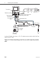
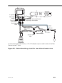
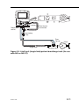
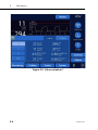
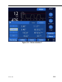
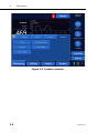
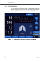
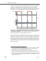
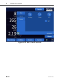
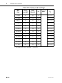
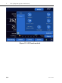
1
624131/00 © 2009 HAMILTON MEDICAL AG. All rights reserved. Printed in Switzerland. No part of this publication may be reproduced or stored in a database or retrieval system, nor transmitted, in any form or by any means, electronic, mechanical, by photocopying, recording, or otherwise, without the prior written permission of HAMILTON MEDICAL. This manual may be revised or replaced by HAMILTON MEDICAL at any time and without notice. You should ensure that you have the most current applicable version of this manual; if in doubt, contact HAMILTON MEDICAL AG Marketing Department. Nothing in this manual shall limit or restrict in any way HAMILTON MEDICAL’s right to revise or otherwise change or modify the equipment (including its software) described herein, without notice. In the absence of an express, written agreement to the contrary, HAMILTON MEDICAL has no obligation to furnish any such revisions, changes, or modifications to the owner or user of the equipment (including software) described herein. The equipment must be operated and serviced only by trained professionals. HAMILTON MEDICAL’s sole responsibility with respect to the equipment and its use is as stated in the Limited Warranty provided in this manual. ASV is a trademark of HAMILTON MEDICAL in some countries. Other product and company names mentioned herein may be the trademarks of their respective owners. HAMILTON MEDICAL will make available on request circuit diagrams, component parts lists, descriptions, calibration instructions, or other information that will assist the user’s appropriately trained personnel to repair those parts of the equipment designated by HAMILTON MEDICAL to be repairable. Manufacturer HAMILTON MEDICAL AG Via Crusch 8 CH-7402 Bonaduz Switzerland Phone: (+41) 81 660 60 10 Fax: (+41) 81 660 60 20 [email protected] www.hamilton-medical.com ii Distributor in USA HAMILTON MEDICAL, Inc. 4990 Energy Way P.O. Box 30008 Reno, NV 89520 Phone: (775) 858-3200 Toll-free: (800) 426-6331 Fax: (775) 856-5621 [email protected] 624131/04 HAMILTON-C2 software information The software version for the HAMILTON-C2 is visible in the System -> Info window. The software version on the screen should match the version on the title page of this manual. See Section 3.3.1 for details. Definitions1 CAUTION Indicates a potentially hazardous situation which, if not avoided, could result in minor or moderate injury. NOTE: Emphasizes information of particular importance. 1. Caution as defined by ISO 624131/04 iii General cautions and notes Intended use The HAMILTON-C2 ventilator is intended to provide positive pressure ventilatory support to adults and pediatrics. Intended areas of use: • In the intensive care ward or in the recovery room • During secondary transport from one hospital to another • During transfer of ventilated patients within the hospital The HAMILTON-C2 ventilator is a medical device intended for use by qualified, trained personnel under the direction of a physician and within the limits of its stated technical specifications. Caution (USA only): • Federal law restricts this device to sale by or on the order of a physician. • Not intended to be used during secondary transport from one hospital to another. The intended patient population ranges from pediatric patients with 30 cm height (3 kg ideal body weight) up to adults with 250 cm height (139 kg ideal body weight). The minimum tidal volume delivered shall be larger or equal to 20 ml. This does exclude the application on neonatal patients. General operation notes • The displays shown in this manual may not exactly match what you see on your own ventilator. • Familiarize yourself with this operator’s manual before using the ventilator on a patient. • Displayed information that is ghosted is not active and may not be selected. • Dashes displayed in place of monitored data indicate that valid values are not yet available or do not apply. iv 624131/04 • If a ventilator control does not respond when selected by touch or by the turn of a knob, the control is not active in this particular instance or the function is not implemented. Monitoring and alarms • The HAMILTON-C2 is not intended to be a comprehensive vital sign monitor for patients on life-support equipment. Patients on life-support equipment should be appropriately monitored by qualified medical personnel and suitable monitoring devices. The use of an alarm monitoring system does not give absolute assurance of warning for every form of malfunction that may occur with the ventilator. Alarm messages may not exactly pinpoint a problem; the exercise of clinical judgment is necessary. • An alternative means of ventilation shall be available whenever the ventilator is in use. If a fault is detected in the ventilator or its life-support functions are in doubt, disconnect the HAMILTON-C2 from the patient and immediately start ventilation with such a device (for example, a resuscitation bag), using PEEP and/or increased oxygen concentration when appropriate. The ventilator must be removed from clinical use and serviced by a HAMILTON MEDICAL authorized service engineer. • It is recommended that additional independent monitoring devices be used during mechanical ventilation. The operator of the ventilator must still maintain full responsibility for proper ventilation and patient safety in all situations. • Do not silence the audible alarm when leaving the patient unattended. • Do not use the exhaust port of the expiratory valve for spirometry. Due to the HAMILTON-C2’s base flow, the exhaust gas output is larger than the patient’s actual exhaled volume. • Do not put a vessel filled with a liquid on the ventilator. If a liquid enters the product, a fire and/or electric shock may occur. 624131/04 v Fire and other hazards • To reduce the risk of fire or explosion, do not place the ventilator in a combustible or explosive environment (for example, around flammable anesthetics or other ignition sources). Do not use it with any equipment contaminated with oil or grease. • To reduce the risk of fire, do not use high-pressure gas hoses that are worn or contaminated with combustible materials like grease or oil. • To reduce the risk of fire, use only breathing circuits intended for use in oxygen-enriched environments. Do not use antistatic or electrically conductive tubing. • In case of fire, immediately secure the patient’s ventilatory needs, switch off the ventilator, and disconnect it from its gas and electrical sources. Service and testing • To ensure proper servicing and to prevent possible physical injury, only HAMILTON MEDICAL authorized service personnel should attempt to service the ventilator. • To reduce the risk of electrical shock, diconnect electrical power from the ventilator before servicing. Be aware that battery power remains even after the mains is disconnected. Be aware that if the power switch is off, some parts still carry high voltage. • Do not attempt service procedures other than those specified in the service manual. • Use replacement parts supplied by HAMILTON MEDICAL only. • Any attempt to modify the ventilator hardware or software without the express written approval of HAMILTON MEDICAL automatically voids all warranties and liabilities. • The preventive maintenance program requires a general service every 5000 hours or yearly, whichever comes first. vi 624131/04 • To ensure the ventilator’s safe operation, always run the tests and calibrations prescribed in Section 3 before using the ventilator on a patient. If the ventilator fails any tests, remove it from clinical use immediately. Do not use the ventilator until necessary repairs are completed and all tests have passed. • The manufacturer considers itself responsible for the safety, reliability, and performance of the ventilator only if: appropriately trained personnel carry out assembly operations, extensions, readjustments, modifications or repairs; the electrical installation of the relevant room complies with the appropriate requirements; and the ventilator system is used in accordance with the operator’s manual. Electromagnetic susceptibility The HAMILTON-C2 complies with the IEC 60601-1-2 EMC (Electro Magnetic Compatibility) Collateral Standard. It is intended for use in the electromagnetic environment described in Table A-13 through Table A-15. Do not use the HAMILTONC2 in an environment with magnetic resonance imaging (MRI) equipment. Units of measure Pressures are indicated on the HAMILTON-C2 in cmH2O or mbar. Hectopascals (hPa) are used by some institutions instead. Since 1 mbar equals 1 hPa, which equals 1.016 cmH2O, the units may be used interchangeably. Disposal Dispose of all parts removed from the device according to your institution’s protocol. Follow all local, state, and federal regulations with respect to environmental protection, especially when disposing of the electronic device or parts of it (for example, oxygen cell, batteries). 624131/04 vii Year of manufacture The year of manufacture is shown on the serial number label on the HAMILTON-C2 ventilation unit. viii 624131/04 1 General information. . . . . . . . . . . . . . . . . . . . . . . . 1-1 1.1 1.2 1.3 1.4 2 Preparing for ventilation . . . . . . . . . . . . . . . . . . . . 2-1 2.1 2.2 2.3 2.4 2.5 2.6 2.7 2.8 2.9 2.10 2.11 2.12 2.13 2.14 3 Introduction . . . . . . . . . . . . . . . . . . . . . . . . . . . . . . . . . . . 1-2 Functional description . . . . . . . . . . . . . . . . . . . . . . . . . . . 1-4 1.2.1 System overview . . . . . . . . . . . . . . . . . . . . . . . . . . 1-4 1.2.2 Gas supply and delivery . . . . . . . . . . . . . . . . . . . . . 1-5 1.2.3 Gas monitoring with the Flow Sensor. . . . . . . . . . . 1-7 Physical description . . . . . . . . . . . . . . . . . . . . . . . . . . . . . 1-8 1.3.1 Breathing circuits and accessories. . . . . . . . . . . . . . 1-8 1.3.2 Ventilator unit . . . . . . . . . . . . . . . . . . . . . . . . . . . 1-11 1.3.3 Screen . . . . . . . . . . . . . . . . . . . . . . . . . . . . . . . . . 1-18 Symbols used on device labels and packaging . . . . . . . . 1-20 Introduction . . . . . . . . . . . . . . . . . . . . . . . . . . . . . . . . . . . 2-2 Installing the patient tubing support arm . . . . . . . . . . . . . 2-4 Installing the humidifier . . . . . . . . . . . . . . . . . . . . . . . . . . 2-5 Installing the patient breathing circuit . . . . . . . . . . . . . . . 2-6 Installing a pneumatic nebulizer . . . . . . . . . . . . . . . . . . . 2-14 Installing the optional Aeroneb Pro nebulizer . . . . . . . . . 2-15 Using an expiratory filter . . . . . . . . . . . . . . . . . . . . . . . . 2-16 Connecting to primary power source . . . . . . . . . . . . . . . 2-17 2.8.1 Connecting to AC power . . . . . . . . . . . . . . . . . . . 2-17 2.8.2 Connecting to DC power . . . . . . . . . . . . . . . . . . . 2-18 About the batteries . . . . . . . . . . . . . . . . . . . . . . . . . . . . 2-18 Connecting the oxygen supply . . . . . . . . . . . . . . . . . . . . 2-20 Connecting to an external patient monitor or other device . . . . . . . . . . . . . . . . . . . . . . . . . . . . . . . . . . . . . . 2-21 Starting up the ventilator . . . . . . . . . . . . . . . . . . . . . . . . 2-22 Shutting down the ventilator . . . . . . . . . . . . . . . . . . . . . 2-23 Display navigation guidelines . . . . . . . . . . . . . . . . . . . . . 2-23 Tests, calibrations and utilities . . . . . . . . . . . . . . . 3-1 3.1 3.2 3.3 Introduction . . . . . . . . . . . . . . . . . . . . . . . . . . . . . . . . . . . 3-2 Running the preoperational check . . . . . . . . . . . . . . . . . . 3-3 System functions . . . . . . . . . . . . . . . . . . . . . . . . . . . . . . . 3-5 3.3.1 Info: Viewing device-specific information . . . . . . . . 3-5 3.3.2 Tests & calib: Running sensor calibrations and the tightness test . . . . . . . . . . . . . . . . . . . . . . . . . . . . . 3-6 3.3.3 Sensors on/off: Enabling/disabling oxygen monitoring . . . . . . . . . . . . . . . . . . . . . . . . . . . . . 3-10 3.3.4 Date & time: Setting date and time . . . . . . . . . . . 3-11 ix Table of contents 3.4 3.5 4 Ventilator settings . . . . . . . . . . . . . . . . . . . . . . . . . 4-1 4.1 4.2 4.3 4.4 4.5 5 Introduction . . . . . . . . . . . . . . . . . . . . . . . . . . . . . . . . . . . 4-2 Patient setup. . . . . . . . . . . . . . . . . . . . . . . . . . . . . . . . . . . 4-3 Modes window: Setting the ventilation mode . . . . . . . . . . 4-5 Controls windows: Setting controls including apnea backup ventilation. . . . . . . . . . . . . . . . . . . . . . . . . . . . . . . 4-6 4.4.1 Adjusting and confirming control settings without mode change . . . . . . . . . . . . . . . . . . . . . . . . . . . . . 4-7 4.4.2 Adjusting and confirming control settings after mode change . . . . . . . . . . . . . . . . . . . . . . . . . . . . 4-10 4.4.3 About apnea backup ventilation . . . . . . . . . . . . . . 4-12 4.4.4 Table of control settings, mode additions and ranges . . . . . . . . . . . . . . . . . . . . . . . . . . . . . . . . . 4-16 Alarms windows . . . . . . . . . . . . . . . . . . . . . . . . . . . . . . . 4-21 4.5.1 Limits 1 and Limits 2: Setting alarm limits . . . . . . . 4-21 4.5.2 Loudness: Adjusting alarm loudness . . . . . . . . . . . 4-24 4.5.3 Buffer: Viewing alarm information . . . . . . . . . . . . 4-25 4.5.4 Table of alarm limit settings and ranges . . . . . . . . 4-25 Monitoring . . . . . . . . . . . . . . . . . . . . . . . . . . . . . . . 5-1 5.1 5.2 5.3 5.4 x Utilities . . . . . . . . . . . . . . . . . . . . . . . . . . . . . . . . . . . . . . 3-12 3.4.1 Configuration: Configuring the ventilator . . . . . . . 3-12 3.4.2 Data transfer: Copying event log data to a USB memory device . . . . . . . . . . . . . . . . . . . . . . . . . . . 3-13 Alarm tests . . . . . . . . . . . . . . . . . . . . . . . . . . . . . . . . . . . 3-15 3.5.1 High pressure . . . . . . . . . . . . . . . . . . . . . . . . . . . . 3-15 3.5.2 Low minute volume . . . . . . . . . . . . . . . . . . . . . . . 3-15 3.5.3 Low oxygen alarm. . . . . . . . . . . . . . . . . . . . . . . . . 3-15 3.5.4 Disconnection on patient side . . . . . . . . . . . . . . . . 3-16 3.5.5 Loss of external power . . . . . . . . . . . . . . . . . . . . . 3-16 3.5.6 Exhalation obstructed . . . . . . . . . . . . . . . . . . . . . . 3-17 3.5.7 Apnea. . . . . . . . . . . . . . . . . . . . . . . . . . . . . . . . . . 3-17 Introduction . . . . . . . . . . . . . . . . . . . . . . . . . . . . . . . . . . . 5-2 Values window: Viewing numeric patient data . . . . . . . . . 5-3 Graphics window: Selecting second screen graphic . . . . . . 5-7 About graphic types . . . . . . . . . . . . . . . . . . . . . . . . . . . . . 5-9 5.4.1 Waveforms . . . . . . . . . . . . . . . . . . . . . . . . . . . . . . . 5-9 5.4.2 Dynamic Lung. . . . . . . . . . . . . . . . . . . . . . . . . . . . . 5-9 5.4.3 Vent Status . . . . . . . . . . . . . . . . . . . . . . . . . . . . . . 5-10 5.4.4 ASV target graphics panel. . . . . . . . . . . . . . . . . . . 5-10 5.4.5 Trends . . . . . . . . . . . . . . . . . . . . . . . . . . . . . . . . . 5-10 5.5 6 Intelligent Panels . . . . . . . . . . . . . . . . . . . . . . . . . . 6-1 6.1 6.2 6.3 6.4 7 6-2 6-3 6-3 6-4 6-4 6-5 6-6 6-8 Introduction . . . . . . . . . . . . . . . . . . . . . . . . . . . . . . . . . . . How to respond to an alarm . . . . . . . . . . . . . . . . . . . . . . Alarm buffer . . . . . . . . . . . . . . . . . . . . . . . . . . . . . . . . . . Events window: Reviewing the event log . . . . . . . . . . . . . Alarm troubleshooting table. . . . . . . . . . . . . . . . . . . . . . . 7-2 7-5 7-6 7-8 7-9 Special functions . . . . . . . . . . . . . . . . . . . . . . . . . . 8-1 8.1 8.2 8.3 8.4 9 Introduction . . . . . . . . . . . . . . . . . . . . . . . . . . . . . . . . . . . Dynamic Lung panel. . . . . . . . . . . . . . . . . . . . . . . . . . . . . 6.2.1 Tidal volume (Vt) . . . . . . . . . . . . . . . . . . . . . . . . . . 6.2.2 Compliance (Cstat) . . . . . . . . . . . . . . . . . . . . . . . . 6.2.3 Patient triggering: Muscle . . . . . . . . . . . . . . . . . . . 6.2.4 Resistance: Bronchial tree. . . . . . . . . . . . . . . . . . . . Vent Status panel. . . . . . . . . . . . . . . . . . . . . . . . . . . . . . . ASV target graphics panel . . . . . . . . . . . . . . . . . . . . . . . . Responding to alarms . . . . . . . . . . . . . . . . . . . . . . 7-1 7.1 7.2 7.3 7.4 7.5 8 5.4.6 Loops . . . . . . . . . . . . . . . . . . . . . . . . . . . . . . . . . . 5-12 Table of monitored parameters . . . . . . . . . . . . . . . . . . . 5-13 Standby . . . . . . . . . . . . . . . . . . . . . . . . . . . . . . . . . . . . . . 8-2 100% O2 . . . . . . . . . . . . . . . . . . . . . . . . . . . . . . . . . . . . . . . . . . . . . . . 8-5 Manual breath/inspiratory hold . . . . . . . . . . . . . . . . . . . . 8-5 Nebulizer . . . . . . . . . . . . . . . . . . . . . . . . . . . . . . . . . . . . . 8-6 Maintenance . . . . . . . . . . . . . . . . . . . . . . . . . . . . . . 9-1 9.1 9.2 9.3 9.4 9.5 Introduction . . . . . . . . . . . . . . . . . . . . . . . . . . . . . . . . . . . 9-2 Cleaning, disinfection and sterilization . . . . . . . . . . . . . . . 9-2 9.2.1 General guidelines for cleaning . . . . . . . . . . . . . . . 9-6 9.2.2 General guidelines for chemical disinfection. . . . . . 9-7 9.2.3 General guidelines for autoclave, ETO or plasma sterilization . . . . . . . . . . . . . . . . . . . . . . . . . . . . . . 9-7 Preventive maintenance . . . . . . . . . . . . . . . . . . . . . . . . . . 9-8 9.3.1 Servicing the air intake and fan filters. . . . . . . . . . 9-10 9.3.2 Replacing the batteries. . . . . . . . . . . . . . . . . . . . . 9-12 9.3.3 Charging and calibrating the batteries . . . . . . . . . 9-13 9.3.4 Replacing the oxygen cell. . . . . . . . . . . . . . . . . . . 9-14 Storage . . . . . . . . . . . . . . . . . . . . . . . . . . . . . . . . . . . . . 9-15 Repacking and shipping . . . . . . . . . . . . . . . . . . . . . . . . . 9-15 xi Table of contents A Specifications . . . . . . . . . . . . . . . . . . . . . . . . . . . . .A-1 A.1 A.2 A.3 A.4 A.5 A.6 A.7 A.8 A.9 A.10 A.11 A.12 A.13 B Modes of ventilation . . . . . . . . . . . . . . . . . . . . . . . B-1 B.1 B.2 B.3 B.4 B.5 B.6 B.7 B.8 B.9 xii Physical characteristics. . . . . . . . . . . . . . . . . . . . . . . . . . . A-2 Environmental requirements . . . . . . . . . . . . . . . . . . . . . . A-3 Pneumatic specifications . . . . . . . . . . . . . . . . . . . . . . . . . A-3 Electrical specifications . . . . . . . . . . . . . . . . . . . . . . . . . . A-4 Control settings . . . . . . . . . . . . . . . . . . . . . . . . . . . . . . . A-5 Monitored parameters . . . . . . . . . . . . . . . . . . . . . . . . . . A-9 Alarms . . . . . . . . . . . . . . . . . . . . . . . . . . . . . . . . . . . . . A-13 Configuration specifications . . . . . . . . . . . . . . . . . . . . . A-15 Ventilator breathing system specifications . . . . . . . . . . . A-17 Other technical data . . . . . . . . . . . . . . . . . . . . . . . . . . . A-18 Standards and approvals . . . . . . . . . . . . . . . . . . . . . . . . A-21 EMC declarations (IEC 60601-1-2). . . . . . . . . . . . . . . . . A-22 Warranty. . . . . . . . . . . . . . . . . . . . . . . . . . . . . . . . . . . . A-28 Introduction . . . . . . . . . . . . . . . . . . . . . . . . . . . . . . . . . . . B-2 The biphasic concept. . . . . . . . . . . . . . . . . . . . . . . . . . . . . B-5 Mandatory modes. . . . . . . . . . . . . . . . . . . . . . . . . . . . . . . B-9 B.3.1 S)CMV+ mode . . . . . . . . . . . . . . . . . . . . . . . . . . . . B-9 B.3.2 PCV+ mode . . . . . . . . . . . . . . . . . . . . . . . . . . . . . B-12 Spontaneous modes (SPONT and NIV) . . . . . . . . . . . . . . . B-14 SIMV modes . . . . . . . . . . . . . . . . . . . . . . . . . . . . . . . . . . B-20 B.5.1 SIMV+ mode. . . . . . . . . . . . . . . . . . . . . . . . . . . . . B-21 B.5.2 PSIMV+ and NIV-ST modes . . . . . . . . . . . . . . . . . B-24 Adaptive support ventilation (ASV) . . . . . . . . . . . . . . . . . B-32 DuoPAP (Duo Positive Airway Pressure) and . . . . . . . . . . B-32 B.7.1 Introduction . . . . . . . . . . . . . . . . . . . . . . . . . . . . . . B-32 B.7.2 The many faces of DuoPAP . . . . . . . . . . . . . . . . . . B-33 B.7.3 Pressure support in DuoPAP breaths. . . . . . . . . . . . B-33 B.7.4 Synchronization . . . . . . . . . . . . . . . . . . . . . . . . . . . B-34 B.7.5 Controls of DuoPAP . . . . . . . . . . . . . . . . . . . . . . . . B-35 APRV (Airway Pressure Release Ventilation) . . . . . . . . . . . B-36 B.8.1 Introduction . . . . . . . . . . . . . . . . . . . . . . . . . . . . . . B-36 B.8.2 Initialization of APRV . . . . . . . . . . . . . . . . . . . . . . . B-37 B.8.3 Sustained high pressure recruitment manoeuvres . . B-38 B.8.3 Controls of APRV . . . . . . . . . . . . . . . . . . . . . . . . . . B-39 SAFETY mode and ambient state. . . . . . . . . . . . . . . . . . . B-40 C ASV (adaptive support ventilation) . . . . . . . . . . . C-1 C.1 C.2 C.3 C.4 C.5 C.6 C.7 D Introduction . . . . . . . . . . . . . . . . . . . . . . . . . . . . . . . . . . . C-2 ASV use in clinical practice . . . . . . . . . . . . . . . . . . . . . . . . C-4 Detailed functional description of ASV . . . . . . . . . . . . . . C-17 C.3.1 Definition of normal minute ventilation . . . . . . . . C-17 C.3.2 Targeted minute ventilation . . . . . . . . . . . . . . . . . C-17 C.3.3 Lung-protective rules strategy . . . . . . . . . . . . . . . C-19 C.3.4 Optimal breath pattern . . . . . . . . . . . . . . . . . . . . C-22 C.3.5 Dynamic adjustment of lung protection . . . . . . . . C-26 C.3.6 Dynamic adjustment of optimal breath pattern . . C-27 Minimum work of breathing (Otis’ equation) . . . . . . . . . C-29 ASV technical data . . . . . . . . . . . . . . . . . . . . . . . . . . . . . C-31 Initialization of ventilation . . . . . . . . . . . . . . . . . . . . . . . C-33 References . . . . . . . . . . . . . . . . . . . . . . . . . . . . . . . . . . . C-34 Clinical application of noninvasive ventilation . .D-1 D.1 D.2 D.3 D.4 D.5 D.6 D.7 D.8 D.9 D.10 D.11 Introduction . . . . . . . . . . . . . . . . . . . . . . . . . . . . . . . . . . . D-2 Benefits of noninvasive ventilation, . . . . . . . . . . . . . . . . . D-3 Required conditions for use . . . . . . . . . . . . . . . . . . . . . . . D-4 Contraindications. . . . . . . . . . . . . . . . . . . . . . . . . . . . . . . D-5 Potential adverse reactions . . . . . . . . . . . . . . . . . . . . . . . . D-5 Selecting a patient interface . . . . . . . . . . . . . . . . . . . . . . . D-6 Control settings . . . . . . . . . . . . . . . . . . . . . . . . . . . . . . . . D-7 Alarms . . . . . . . . . . . . . . . . . . . . . . . . . . . . . . . . . . . . . . . D-8 Monitored parameters . . . . . . . . . . . . . . . . . . . . . . . . . . . D-8 Additional notes about using noninvasive ventilation . . . . D-9 References . . . . . . . . . . . . . . . . . . . . . . . . . . . . . . . . . . . D-10 E Low-pressure oxygen. . . . . . . . . . . . . . . . . . . . . . . E-1 F Pneumatic diagram . . . . . . . . . . . . . . . . . . . . . . . . F-1 G Parts and accessories . . . . . . . . . . . . . . . . . . . . . . .G-1 H Communications interface. . . . . . . . . . . . . . . . . . .H-1 H.1 H.2 H.3 Introduction . . . . . . . . . . . . . . . . . . . . . . . . . . . . . . . . . . . H-2 Patient monitor . . . . . . . . . . . . . . . . . . . . . . . . . . . . . . . . H-3 Patient data management system (PDMS) or other computer system . . . . . . . . . . . . . . . . . . . . . . . . . . . . . . . H-5 xiii Table of contents H.4 I Connector pin assignments . . . . . . . . . . . . . . . . . . . . . . . H-6 Configuration . . . . . . . . . . . . . . . . . . . . . . . . . . . . . I-1 I.1 I.2 I.3 I.4 I.5 I.6 Introduction . . . . . . . . . . . . . . . . . . . . . . . . . . . . . . . . . . . I-2 Accessing configuration . . . . . . . . . . . . . . . . . . . . . . . . . . I-2 General: Selecting the language, units of measure and oxygen source. . . . . . . . . . . . . . . . . . . . . . . . . . . . . . . . . . I-3 I.3.1 Language: Selecting the default language. . . . . . . . I-3 I.3.2 Units: Selecting the default unit of measure for pressure display . . . . . . . . . . . . . . . . . . . . . . . . . . . I-4 I.3.3 More: Selecting the oxygen source and enabling the communications interface . . . . . . . . . . . . . . . . . I-5 Graphics window . . . . . . . . . . . . . . . . . . . . . . . . . . . . . . . I-6 I.4.1 MMP: Selecting the default main monitoring parameter display . . . . . . . . . . . . . . . . . . . . . . . . . . I-6 Settings window . . . . . . . . . . . . . . . . . . . . . . . . . . . . . . . . I-7 I.5.1 Use settings: Selecting the default startup settings . I-7 Vent Status: Configuring the Vent Status panel. . . . . . . . I-10 Glossary. . . . . . . . . . . . . . . . . . . . . . . . . . . .Glossary-1 Index . . . . . . . . . . . . . . . . . . . . . . . . . . . . . . . . Index-1 xiv List of figures 1-1 1-2 1-3 1-4 1-5 1-6 1-7 1-8 2-1 2-2 2-3 2-4 2-5 2-6 2-7 2-8 2-9 2-10 2-11 2-12 2-13 3-1 3-2 3-3 3-4 3-5 3-6 4-1 4-2 4-3 4-4 4-5 4-6 4-7 4-8 4-9 5-1 Gas delivery in the HAMILTON-C2 . . . . . . . . . . . . . . . . . . . . . . . 1-5 Flow Sensor variable orifice . . . . . . . . . . . . . . . . . . . . . . . . . . . . 1-7 HAMILTON-C2 with accessories . . . . . . . . . . . . . . . . . . . . . . . . . 1-9 Front view . . . . . . . . . . . . . . . . . . . . . . . . . . . . . . . . . . . . . . . . 1-11 Rear view . . . . . . . . . . . . . . . . . . . . . . . . . . . . . . . . . . . . . . . . . 1-14 Left side view . . . . . . . . . . . . . . . . . . . . . . . . . . . . . . . . . . . . . . 1-16 Right side view . . . . . . . . . . . . . . . . . . . . . . . . . . . . . . . . . . . . . 1-17 Default (basic) screen . . . . . . . . . . . . . . . . . . . . . . . . . . . . . . . . 1-18 Installing the patient tubing support arm and humidifier . . . . . . 2-4 Patient breathing circuit for use with inspiratory heater wire. . . . 2-8 Patient breathing circuit for use without heater wires . . . . . . . . . 2-9 Patient breathing circuit for use with HME . . . . . . . . . . . . . . . . 2-10 LiteCircuit (single-limb) patient breathing circuit (for use with NIV or NIV-ST) . . . . . . . . . . . . . . . . . . . . . . . . . . . . . . . . . . . . . 2-11 Installing the expiratory valve . . . . . . . . . . . . . . . . . . . . . . . . . . 2-12 Installing the Flow Sensor . . . . . . . . . . . . . . . . . . . . . . . . . . . . . 2-13 Installing a pneumatic nebulizer . . . . . . . . . . . . . . . . . . . . . . . . 2-14 Installing the Aeroneb Pro nebulizer . . . . . . . . . . . . . . . . . . . . 2-15 Power cord retaining clip . . . . . . . . . . . . . . . . . . . . . . . . . . . . . 2-17 Power source symbols and battery charge indicator . . . . . . . . . 2-19 Oxygen inlet fittings . . . . . . . . . . . . . . . . . . . . . . . . . . . . . . . . . 2-21 Power switch . . . . . . . . . . . . . . . . . . . . . . . . . . . . . . . . . . . . . . 2-22 Info window. . . . . . . . . . . . . . . . . . . . . . . . . . . . . . . . . . . . . . . . 3-5 Tests & calib window . . . . . . . . . . . . . . . . . . . . . . . . . . . . . . . . . 3-6 Sensors on/off window. . . . . . . . . . . . . . . . . . . . . . . . . . . . . . . 3-10 Date & time window . . . . . . . . . . . . . . . . . . . . . . . . . . . . . . . . 3-11 Configuration window . . . . . . . . . . . . . . . . . . . . . . . . . . . . . . . 3-12 Data transfer window. . . . . . . . . . . . . . . . . . . . . . . . . . . . . . . . 3-14 Patient setup window. . . . . . . . . . . . . . . . . . . . . . . . . . . . . . . . . 4-4 Modes window . . . . . . . . . . . . . . . . . . . . . . . . . . . . . . . . . . . . . 4-5 Basic (Controls) window . . . . . . . . . . . . . . . . . . . . . . . . . . . . . . . 4-8 More window . . . . . . . . . . . . . . . . . . . . . . . . . . . . . . . . . . . . . . 4-9 Apnea window. . . . . . . . . . . . . . . . . . . . . . . . . . . . . . . . . . . . . 4-10 Basic window during mode change (ASV mode change). . . . . . 4-11 Limits 1 window. . . . . . . . . . . . . . . . . . . . . . . . . . . . . . . . . . . . 4-22 Limits 2 window. . . . . . . . . . . . . . . . . . . . . . . . . . . . . . . . . . . . 4-23 Loudness window . . . . . . . . . . . . . . . . . . . . . . . . . . . . . . . . . . 4-24 HAMILTON-C2 screen . . . . . . . . . . . . . . . . . . . . . . . . . . . . . . . . 5-3 624131/04 xv List of figures 5-2 5-3 5-4 5-5 5-6 5-7 5-8 6-1 6-2 6-3 6-4 6-5 6-6 7-1 7-2 7-3 7-4 7-5 7-6 8-1 8-2 8-3 9-1 9-2 9-3 9-4 A-1 B-1 B-4 B-5 B-6 Values window 1. . . . . . . . . . . . . . . . . . . . . . . . . . . . . . . . . . . . 5-4 Values window 2. . . . . . . . . . . . . . . . . . . . . . . . . . . . . . . . . . . . 5-5 Values window 3. . . . . . . . . . . . . . . . . . . . . . . . . . . . . . . . . . . . 5-6 Graphics window . . . . . . . . . . . . . . . . . . . . . . . . . . . . . . . . . . . 5-8 Pressure waveform display. . . . . . . . . . . . . . . . . . . . . . . . . . . . . 5-9 Trends display . . . . . . . . . . . . . . . . . . . . . . . . . . . . . . . . . . . . . 5-11 Loops display. . . . . . . . . . . . . . . . . . . . . . . . . . . . . . . . . . . . . . 5-12 Ventilator screen with Dynamic Lung panel . . . . . . . . . . . . . . . . 6-2 Dynamic Lung panel . . . . . . . . . . . . . . . . . . . . . . . . . . . . . . . . . 6-3 Compliance shown by the Dynamic Lung . . . . . . . . . . . . . . . . . 6-4 Patient triggering shown by the Dynamic Lung muscle . . . . . . . 6-4 Rinsp shown by the bronchial tree of the Dynamic Lung . . . . . . 6-5 Vent Status panel . . . . . . . . . . . . . . . . . . . . . . . . . . . . . . . . . . . 6-6 Visual alarm indications . . . . . . . . . . . . . . . . . . . . . . . . . . . . . . . 7-4 Safety ventilation screen . . . . . . . . . . . . . . . . . . . . . . . . . . . . . . 7-4 Technical fault screen . . . . . . . . . . . . . . . . . . . . . . . . . . . . . . . . 7-5 Alarm buffer with active alarms. . . . . . . . . . . . . . . . . . . . . . . . . 7-7 Alarm buffer with inactive alarms . . . . . . . . . . . . . . . . . . . . . . . 7-7 Events window . . . . . . . . . . . . . . . . . . . . . . . . . . . . . . . . . . . . . 7-8 Special function keys . . . . . . . . . . . . . . . . . . . . . . . . . . . . . . . . . 8-3 Activate Standby window . . . . . . . . . . . . . . . . . . . . . . . . . . . . . 8-3 Standby window . . . . . . . . . . . . . . . . . . . . . . . . . . . . . . . . . . . . 8-4 Removing the filter cover. . . . . . . . . . . . . . . . . . . . . . . . . . . . . 9-10 Removing the air intake filters . . . . . . . . . . . . . . . . . . . . . . . . . 9-11 Removing battery 2 . . . . . . . . . . . . . . . . . . . . . . . . . . . . . . . . . 9-12 Replacing the oxygen cell . . . . . . . . . . . . . . . . . . . . . . . . . . . . 9-15 HAMILTON-C2 dimensions . . . . . . . . . . . . . . . . . . . . . . . . . . . . A-2 Conventional pressure-controlled ventilation in a passive patient. Flow to patient during inspiration (I); flow from patient during exhalation (E) only. . . . . . . . . . . . . . . . . . . . . . . . . . . . . . . . . . . B-6 Conventional pressure-controlled ventilation in an active patient when the trigger is off. Pressure increases when the patient tries to exhale (E) and pressure decreases when the patient tries to inspire (I), as valves are closed. . . . . . . . . . . . . . . . . . . . . B-7 Biphasic PCV+ in an active patient when trigger is off. The patient can freely inspire and exhale during any phase of ventilation (+). . . . . . . . . . . . . . . . . . . . . . . . . . . . . . . . . . . . . . . B-8 (S)CMV+ basic controls . . . . . . . . . . . . . . . . . . . . . . . . . . . . . . . B-9 (S)CMV+ more controls . . . . . . . . . . . . . . . . . . . . . . . . . . . . . . B-10 Breath delivery by the adaptive volume controller . . . . . . . . . . B-11 xvi 624131/04 B-2 B-3 B-7 B-8 B-9 B-10 B-11 B-12 B-13 B-14 B-15 B-16 B-17 B-18 B-19 B-20 B-21 B-22 B-23 B-24 B-25 B-26 B-27 B-28 B-29 B-30 B-31 B-32 B-33 B-34 B-35 C-1 C-2 C-3 C-4 C-5 PCV+ basic controls . . . . . . . . . . . . . . . . . . . . . . . . . . . . . . . . . B-12 PCV+ more controls . . . . . . . . . . . . . . . . . . . . . . . . . . . . . . . . . B-13 SPONT basic controls . . . . . . . . . . . . . . . . . . . . . . . . . . . . . . . . B-15 SPONT more controls . . . . . . . . . . . . . . . . . . . . . . . . . . . . . . . . B-16 SPONT apnea controls . . . . . . . . . . . . . . . . . . . . . . . . . . . . . . . B-17 NIV basic controls. . . . . . . . . . . . . . . . . . . . . . . . . . . . . . . . . . . B-18 NIV more controls . . . . . . . . . . . . . . . . . . . . . . . . . . . . . . . . . . B-19 NIV apnea controls . . . . . . . . . . . . . . . . . . . . . . . . . . . . . . . . . . B-20 Breath timing in SIMV+ . . . . . . . . . . . . . . . . . . . . . . . . . . . . . . B-21 SIMV+ basic controls . . . . . . . . . . . . . . . . . . . . . . . . . . . . . . . . B-22 SIMV+ more controls . . . . . . . . . . . . . . . . . . . . . . . . . . . . . . . . B-23 SIMV+ apnea controls . . . . . . . . . . . . . . . . . . . . . . . . . . . . . . . B-24 Breath timing in PSIMV+ and NIV-ST . . . . . . . . . . . . . . . . . . . . B-25 PSIMV+ basic controls . . . . . . . . . . . . . . . . . . . . . . . . . . . . . . . B-26 PSIMV+ more controls . . . . . . . . . . . . . . . . . . . . . . . . . . . . . . . B-27 PSIMV+ apnea controls . . . . . . . . . . . . . . . . . . . . . . . . . . . . . . B-28 NIV-ST basic controls . . . . . . . . . . . . . . . . . . . . . . . . . . . . . . . . B-29 NIV-ST more controls . . . . . . . . . . . . . . . . . . . . . . . . . . . . . . . . B-30 NIV-ST apnea controls . . . . . . . . . . . . . . . . . . . . . . . . . . . . . . . B-31 DuoPAP pressure curve. . . . . . . . . . . . . . . . . . . . . . . . . . . . . . . B-32 Pressure support in DuoPAP/APRV . . . . . . . . . . . . . . . . . . . . . . B-34 DuoPAP basic controls . . . . . . . . . . . . . . . . . . . . . . . . . . . . . . . B-35 DuoPAP more controls . . . . . . . . . . . . . . . . . . . . . . . . . . . . . . . B-35 DuoPAP apnea controls . . . . . . . . . . . . . . . . . . . . . . . . . . . . . . B-36 APRV breath timing . . . . . . . . . . . . . . . . . . . . . . . . . . . . . . . . . B-37 APRV basic controls . . . . . . . . . . . . . . . . . . . . . . . . . . . . . . . . . B-39 APRV more controls . . . . . . . . . . . . . . . . . . . . . . . . . . . . . . . . . B-39 APRV apnea controls . . . . . . . . . . . . . . . . . . . . . . . . . . . . . . . . B-40 Display SAFETY mode and ambient state . . . . . . . . . . . . . . . . . B-41 Clinical use of ASV. The numbers in parentheses are step numbers, which are explained in the next subsections. . . . . . . . . C-5 ASV basic controls . . . . . . . . . . . . . . . . . . . . . . . . . . . . . . . . . . . C-6 ASV more controls . . . . . . . . . . . . . . . . . . . . . . . . . . . . . . . . . . . C-7 Hypothetical example of high %MinVol setting incompatible with the lung-protective rules strategy. The open circle denotes the actual target, the closed triangle (never shown on the ventilator) denotes the (energetically) optimal target according to Otis’ equation. The HAMILTON-C2 will alarm and inform the user that the ASV target cannot be achieved. . . . . . . . . . . . C-12 ASV target graphics panel . . . . . . . . . . . . . . . . . . . . . . . . . . . . C-13 624131/04 xvii List of figures C-6 C-7 C-8 C-9 C-10 C-11 C-12 C-13 C-14 G-1 G-2 G-3 H-1 H-2 H-3 H-4 I-1 I-2 I-3 I-4 xviii ASV monitored data window . . . . . . . . . . . . . . . . . . . . . . . . . C-14 Normal minute ventilation as a function of IBW. For patients between 3 and 5 kg, minute ventilation is 0.3 l/min/kg. For patients above 30 kg, minute ventilation is calculated as 0.1 l/min/kg. For patients with IBW between these points, the values indicated by the dotted line are used. Minute ventilation for a 15 kg patient thus is calculated as 0.2 l/min x 15 kg = 3 l/min. . . . . . . . . . . . . . . . . . . . . . . . . . . . . . . . . . . . . . . . . . . C-17 MinVol = 7 l/min. All possible combinations of Vt and f which result in a minute ventilation of 7 l/min lie on the bold line. . . . C-18 Lung-protective rules strategy to avoid high tidal volumes and pressures (A), low alveolar ventilation (B), dynamic hyperinflation or breath stacking (C), and apnea (D). . . . . . . . . . . . . C-20 Anatomy of the ASV target graphics panel. The rectangle shows the safety limits; the circle shows the target breath pattern.. . . . . . . . . . . . . . . . . . . . . . . . . . . . . . . . . . . . . . . . . . C-23 Example of a situation after the three initial breaths. The cross marks the actual measured values for Vt and rate.. . . . . . . . . . C-25 Lung-protective limits are changed dynamically and according to the respiratory system mechanics. However, the limits derived from the operator input are never violated. . . . . . . . . . . . . . . . C-26 Changes of target values in broncho-constriction. For clarity, the safety limits are omitted. For clinical examples, see Belliato 2000. . . . . . . . . . . . . . . . . . . . . . . . . . . . . . . . . . . . . . . . . . . . C-28 Three different relationships between rate and WOB are plotted for a hypothetical lung: (+) purely resistive load causes WOB to rise with rate, (x) purely elastic load creates highest load at low rates, (o) the total lung shows a clear minimum which can be calculated according to the equation below. . . . . . . . . . . . . . . C-29 Ventilator parts and accessories — standard trolley . . . . . . . . . . G-5 Ventilator parts and accessories — standard trolley . . . . . . . . . . G-6 Universal transport trolley parts . . . . . . . . . . . . . . . . . . . . . . . . . G-8 HAMILTON-C2 connected to a patient monitor . . . . . . . . . . . . . H-4 HAMILTON-C2 connected to a computer system . . . . . . . . . . . . H-5 RS-232 connector pinout. . . . . . . . . . . . . . . . . . . . . . . . . . . . . . H-7 RS-232 cable (PN 157354) wiring diagram. . . . . . . . . . . . . . . . . H-7 Language configuration window . . . . . . . . . . . . . . . . . . . . . . . . . I-3 Units configuration window . . . . . . . . . . . . . . . . . . . . . . . . . . . . I-4 More configuration window . . . . . . . . . . . . . . . . . . . . . . . . . . . . I-5 MMP configuration window . . . . . . . . . . . . . . . . . . . . . . . . . . . . I-6 624131/04 I-5 I-6 I-7 I-8 I-9 Use settings configuration window. . . . . . . . . . . . . . . . . . . . . . . I-7 Mode Ctrls configuration window . . . . . . . . . . . . . . . . . . . . . . . I-8 Alarms configuration window . . . . . . . . . . . . . . . . . . . . . . . . . . I-9 Vent Status configuration window . . . . . . . . . . . . . . . . . . . . . . I-10 Vent Status intelligent panel . . . . . . . . . . . . . . . . . . . . . . . . . . I-11 624131/04 xix List of figures xx 624131/04 List of tables 1 1-1 1-2 2-1 3-1 4-1 4-2 4-3 4-4 5-1 6-1 6-2 7-1 7-2 9-1 9-2 A-1 A-2 A-3 A-4 A-5 A-6 A-7 A-8 A-9 A-10 A-11 A-12 A-13 A-14 A-15 Compatible parts and accessories . . . . . . . . . . . . . . . . . . . . . . . 1-10 Symbols used on device labels and packaging . . . . . . . . . . . . . 1-20 Breathing circuit parts according to Pat. height. . . . . . . . . . . . . . 2-7 When to perform tests and calibrations . . . . . . . . . . . . . . . . . . . 3-2 Settings during back up mode PCV+ . . . . . . . . . . . . . . . . . . . . 4-14 Setting during back up mode SIMV+ . . . . . . . . . . . . . . . . . . . . 4-15 Control settings, mode additions and ranges . . . . . . . . . . . . . . 4-16 Alarm limit settings and ranges . . . . . . . . . . . . . . . . . . . . . . . . 4-25 Monitored parameters . . . . . . . . . . . . . . . . . . . . . . . . . . . . . . . 5-11 Dynamic Lung normal values . . . . . . . . . . . . . . . . . . . . . . . . . . . 6-5 Vent Status parameters . . . . . . . . . . . . . . . . . . . . . . . . . . . . . . . 6-7 Alarm indications in HAMILTON-C2 . . . . . . . . . . . . . . . . . . . . . . 7-3 Alarms and other messages . . . . . . . . . . . . . . . . . . . . . . . . . . . . 7-9 Decontamination methods for HAMILTON-C2 parts. . . . . . . . . . . . . . . . . . . . . . . . . . . . . . . . . . 9-4 Preventive maintenance schedule . . . . . . . . . . . . . . . . . . . . . . . . 9-8 Physical characteristics . . . . . . . . . . . . . . . . . . . . . . . . . . . . . . . . A-2 Environmental requirements. . . . . . . . . . . . . . . . . . . . . . . . . . . . A-3 Pneumatic specifications. . . . . . . . . . . . . . . . . . . . . . . . . . . . . . . A-3 Electrical specifications . . . . . . . . . . . . . . . . . . . . . . . . . . . . . . . . A-4 Control setting ranges and resolutions . . . . . . . . . . . . . . . . . . . . A-5 Controls active in HAMILTON-C2 ventilation modes . . . . . . . . . A-7 Monitored parameter ranges, resolutions and accuracies . . . . . . . . . . . . . . . . . . . . . . . . . . . . . . . . . . . . . . A-8 Real-time curves . . . . . . . . . . . . . . . . . . . . . . . . . . . . . . . . . . . . A-10 Adjustable alarm ranges and resolutions. . . . . . . . . . . . . . . . . . A-11 Configuration specifications . . . . . . . . . . . . . . . . . . . . . . . . . . . A-13 Ventilator breathing system specifications . . . . . . . . . . . . . . . . A-15 Other technical data. . . . . . . . . . . . . . . . . . . . . . . . . . . . . . . . . A-16 Guidance and manufacturer's declaration – electromagnetic emissions . . . . . . . . . . . . . . . . . . . . . . . . . . . . . . . . . . . . . . . . . A-20 Guidance and manufacturer's declaration – electromagnetic immunity . . . . . . . . . . . . . . . . . . . . . . . . . . . . . . . . . . . . . . . . . A-21 Recommended separation distances between portable and mobile RF communications equipment and the HAMILTON-C2 ventilator . . . . . . . . . . . . . . . . . . . . . . . . . . . . . . . . . . . . . . . . . A-25 624131/04 xxi List of tables B-1 B-2 B-2 C-1 C-5 C-6 G-1 G-2 H-1 H-2 Classification of HAMILTON-C2 ventilation modes. . . . . . . . . . . . B-3 Control parameters for initialization of APRV. . . . . . . . . . . . . . . B-38 SAFETY mode settings. . . . . . . . . . . . . . . . . . . . . . . . . . . . . . . . B-42 Blood gas results and other conditions with possible ASV adjustments . . . . . . . . . . . . . . . . . . . . . . . . . . . . . . . . . . . . . . C-10 Interpretation of breathing pattern at 100% MinVol setting . . C-14 Interpretation of breathing pattern at much lower than 100% MinVol setting. . . . . . . . . . . . . . . . . . . . . . . . . . . . . . . . . . . . . C-15 Interpretation of breathing pattern at much higher than 100% MinVol setting. . . . . . . . . . . . . . . . . . . . . . . . . . . . . . . . . . . . . C-15 ASV technical data . . . . . . . . . . . . . . . . . . . . . . . . . . . . . . . . . C-31 Initial breath pattern . . . . . . . . . . . . . . . . . . . . . . . . . . . . . . . . C-33 Ventilator parts and accessories. . . . . . . . . . . . . . . . . . . . . . . . . G-2 Universal transport trolley parts . . . . . . . . . . . . . . . . . . . . . . . . . G-7 Interfacing hardware for patient monitors . . . . . . . . . . . . . . . . . H-4 Requirements for interfacing PDMSs . . . . . . . . . . . . . . . . . . . . . H-6 xxii 624131/04 C-2 C-4 C-3 1 1 General information 1.1 Introduction 1-2 1.2 Functional description 1-4 1.2.1 System overview 1-4 1.2.2 Gas supply and delivery 1-5 1.2.3 Gas monitoring with the Flow Sensor 1-7 Physical description 1-8 1.3.1 Breathing circuits and accessories 1-8 1.3 1.4 624131/04 1.3.2 Ventilator unit 1-11 1.3.3 Screen 1-18 Symbols used on device labels and packaging 1-20 1-1 1 1.1 General information Introduction The HAMILTON-C2 is designed for intensive care ventilation of adult and pediatric patients. Ventilation modes. This full-functioned intensive care ventilator offers a complete range of modes. PCV+, PSIMV+, and SPONT are conventional pressure-controlled modes. (S)CMV+ and SIMV+, delivered by an adaptive volume controller, combine the attributes of pressure-controlled with volume-targeted ventilation. DuoPAP and APRV are two related forms of pressure ventilation designed to support spontaneous breathing on two alternating levels of CPAP. ASV® (adaptive support ventilation) guarantees that the patient receives the selected minute ventilation with the optimal breath pattern (lowest pressure and volume, optimal rate to minimize work of breathing and intrinsic PEEP). NIV (noninvasive ventilation) and NIV-ST (spontaneous/timed noninvasive ventilation) provide pressure support ventilation through a mask or other noninvasive interface. Patient-triggered breaths are flow triggered. Monitoring. The HAMILTON-C2 offers a variety of monitoring capabilities. It displays monitored parameters as numbers. You can also see this data graphically, as a combination of real-time waveforms (curves), Loops, Trends and special Intelligent Panels. These Intelligent Panels include the Dynamic Lung, which shows the lung’s activity, and the Vent Status, which indicates the patient’s level of ventilator dependency. Additionally, when the ASV mode is active, the ASV target graphics panel and the ASV monitored data window may be displayed. The ASV target graphics panel shows how the ASV controller moves towards its target, while the ASV monitored data window provides this information in a numeric form. The HAMILTON-C2’s monitored data is based on pressure and flow measurements collected by the HAMILTON MEDICAL proximal Flow Sensor, between the Y-piece and the patient, and on FiO2 measurements by the integral oxygen monitor. Alarms. The HAMILTON-C2’s operator-adjustable and nonadjustable alarms help ensure your patient’s safety. 1-2 624131/04 User interface. The ventilator’s ergonomic design, including a 10.4 in. color touchscreen, a press-and-turn knob, and keys, lets you easily access the ventilator settings and monitored parameters. You can tilt the graphical user interface up to 45°. Customizability. You can customize the HAMILTON-C2 so that it starts up with institution-defined settings. Power. The HAMILTON-C2 uses as its primary power source ac mains (100 to 240 V AC, 50/60 Hz) or a DC supply (+12 to +24 V). If the primary power source fails, the ventilator power source automatically switches to backup batteries. The standard battery (battery 1) powers the HAMILTON-C2 typically for 2.5 h, and the optional, hot-swappable battery (battery 2) doubles the running time. Mounting variations for the HAMILTON-C2 include a standard trolley, a transport trolley, and a shelf mount. Both types of trolley have space for oxygen cylinders. The transport trolley lets you mount infusion pumps and other devices commonly used at the bedside. Nebulization function. The nebulization function lets your HAMILTON-C2 power a pneumatic nebulizer connected to the nebulizer outlet. The communications interface provides an RS-232 port for connection to a remote monitor, patient data management system (PDMS), or other computer system. 624131/04 1-3 1 General information 1.2 Functional description The following paragraphs describe the operation of the HAMILTON-C2 ventilator from a hardware perspective. 1.2.1 System overview The HAMILTON-C2 is an electronically controlled pneumatic ventilation system with an integrated air compressing system. It is powered by ac or dc with battery backup to protect against power failure or unstable power and to facilitate intrahospital transport. The HAMILTON-C2’s pneumatics deliver gas, and its electrical systems control pneumatics, monitor alarms, and distribute power. The user provides inputs to the HAMILTON-C2 microprocessor system through a touchscreen, keys, and a press-and-turn knob. These inputs become instructions for the HAMILTONC2’s pneumatics to deliver a precisely controlled gas mixture to the patient. The HAMILTON-C2 receives inputs from the proximal Flow Sensor and other sensors within the ventilator. Based on this monitored data, the HAMILTON-C2 adjusts gas delivery to the patient. Monitored data is also displayed by the graphic user interface. The HAMILTON-C2’s microprocessor system controls gas delivery and monitors the patient. The gas delivery and monitoring functions are cross-checked by an alarm controller. This crosschecking helps prevent simultaneous failure of these two main functions and minimizes the possible hazards of software failure. A comprehensive system of visual and audible alarms helps ensure the patient’s safety. Clinical alarms can indicate an abnormal physiological condition. Technical alarms, triggered by the ventilator’s self-tests, including ongoing background checks, can indicate a hardware or software failure. In the case of some technical alarms, a special safety mode ensures a basic minute ventilation while giving the user time for corrective 1-4 624131/04 actions. When a condition is critical enough to possibly compromise safe ventilation, the HAMILTON-C2 is placed into the ambient state. The ambient and expiratory valves are opened, letting the patient inspire room air through the ambient valve and exhale through the expiratory valve. The HAMILTON-C2 has several means to ensure that safe patient or respiratory pressures are maintained. The maximum working pressure is ensured by the high pressure alarm limit. If the set high pressure limit is reached, the ventilator cycles into exhalation. The ventilator pressure cannot exceed 60 cmH2O. 1.2.2 Gas supply and delivery The HAMILTON-C2 uses room air and low- or high-pressure oxygen (Figure 1-1). Air enters through a fresh gas intake port and is compressed together with the oxygen by the blower. Oxygen enters through a high1- or low-pressure2 inlet. Highpressure oxygen* HAMILTON-C2 O2 mixer valve Lowpressure oxygen* Air Blower Inspiratory valve Breathing circuit Patient Expiratory valve To room air Flow Sensor *Only one oxygen source, high- or low-pressure, is required. Figure 1-1. Gas delivery in the HAMILTON-C2 1. High pressure oxygen: Maximal Pressure 600kPa / Maximal Flow 120l/min 2. Low Pressure oxygen: Maximal Pressure 600kPa / Maximal Flow 15 l/min 624131/04 1-5 1 General information Within the ventilator, the gas enters the HAMILTON-C2’s pneumatic system. If high-pressure oxygen is supplied, a mixer valve provides for the operator-set concentration. If low-pressure oxygen is supplied, the delivered oxygen concentration is determined by the flow of the source oxygen. Gas is supplied to the patient via the inspiratory valve. The microprocessor controls the size of the inspiratory valve opening and the length of time it is open to meet the user settings. The HAMILTON-C2 delivers gas to the patient through the inspiratory limb breathing circuit parts, which may include an inspiratory filter, flex tubes, the humidification system, water traps, the Y-piece, and the Flow Sensor. An internal pneumatic nebulizer supplies the nebulizer flow. Gas exhaled by the patient passes through the expiratory limb breathing circuit parts, including flex tubes, the Flow Sensor, the Y-piece, a water trap, and an expiratory valve cover and membrane. Gas is vented through the expiratory valve cover such that no exhaled gas comes into contact with any internal components of the HAMILTON-C2. Measurements taken at the Flow Sensor are used in the pressure, flow, and volume measurements. An oxygen cell (sensor) monitors the oxygen concentration of the gas to be delivered to the patient. This galvanic cell generates a voltage proportional to the partial pressure of oxygen in the delivered gas. This oxygen measurement is compensated for changes in pressure. The operations of the inspiratory and expiratory valves are coordinated to maintain system pressure levels. 1-6 624131/04 1.2.3 Gas monitoring with the Flow Sensor The HAMILTON-C2 accurately measures flow, volume, and pressure in the patient’s airway with the HAMILTON MEDICAL Flow Sensor. This proximal Flow Sensor lets the HAMILTON-C2 sense even weak patient breathing efforts. Between its highly sensitive flow trigger and fast response time, the HAMILTONC2 helps minimize the patient’s work of breathing. The Flow Sensor contains a thin, diamond-shaped membrane within the outer housing and has a pressure port on either side. The membrane allows bidirectional flow through its variable orifice (Figure 1-2). Figure 1-2. Flow Sensor variable orifice The area of the orifice changes depending on the flow rate. It opens progressively as the flow increases, creating a pressure drop across the orifice. The pressure difference is measured by a high-precision differential pressure sensor inside the ventilator. The pressure difference varies with flow (relationship determined during Flow Sensor calibration), so the patient’s flow is determined from the pressure drop. The HAMILTON-C2 calculates volume from the flow measurements. The Flow Sensor is highly accurate even in the presence of secretions, moisture, and nebulized medications. The HAMILTON-C2 continuously flushes the sensing tubes with mixed gases (rinse flow) to prevent blockage. 624131/04 1-7 1 General information 1.3 Physical description 1.3.1 Breathing circuits and accessories Figure 1-3 shows the HAMILTON-C2 with its breathing circuit and accessories. Contact your HAMILTON MEDICAL representative for details on breathing circuits and accessories supplied by HAMILTON MEDICAL. See Table 1-1 for information on other compatible breathing circuits and accessories. NOTE: To ensure proper ventilation operation, use only parts and accessories specified in Table 1-1. 1-8 624131/04 Q W E Y R T Figure 1-3. HAMILTON-C2 with accessories Q Graphic user interface W Support arm E Breathing circuit (see figures 2-6 through 2-9 for details) R Humidifier T Standard trolley (option) Y Breathing circuit connections 624131/04 1-9 1 General information m Table 1-1. Compatible parts and accessories Part Use... Patient breathing circuit • HAMILTON MEDICAL patient breathing circuits • Other circuits that meet the ventilator breathing system specifications in Appendix A. Circuits must comply with ISO 5367. Mask • HAMILTON MEDICAL face masks • Other face or nasal masks, except those incorporating an expiratory valve Inspiratory filter • HAMILTON MEDICAL inspiratory bacteria filter • Other filters that have a 22 mm female conical inlet connector and a 22 mm male conical outlet connector, and that meet the ventilator breathing system specifications in Appendix A Humidification device • Fisher & Paykel humidifier (for example, MR810 or MR850) • Any active humidifier with a flow capability of up to 120 l/min that is approved for the intended use. Humidifiers must comply with ISO 8185. • Heat and moisture exchanger (HME). HMEs must comply with ISO 9360. Flow Sensor HAMILTON MEDICAL parts only (marked with the HAMILTON "H") Expiratory valve membrane and cover HAMILTON MEDICAL parts only Nebulizer • Internal nebulizer: Pneumatic nebulizer specified for 8 l/min • External nebulizer: Pneumatic (small-volume) nebulizer powered by an external gas source, or a standalone ultrasonic or electronic (piezo) micropump nebulizer such as the Aerogen® Aeroneb® Pro nebulizer system Oxygen cell HAMILTON MEDICAL parts only 1-10 624131/04 1.3.2 Ventilator unit Figure 1-4 through Figure 1-6 show the controls, indicators, and other important parts of the ventilator unit. When a key is pressed and the selected function is active, the LED beside the key is lit. Q W E R T Y U I O P r { } e w q Figure 1-4. Front view Item Description Q Touchscreen W Alarm lamp. Entire lamp lights when an alarm is active (red = high-priority alarm, yellow = medium- or low-priority alarm). In addition, a red LED in the middle is continuously lit when alarm silence is active. This red LED flashes when an alarm silence is inactive but an alarm is active. 624131/04 1-11 1 General information Item Description E Battery charge indicator. Lights to show that the batteries can be charged. It is lit whenever the ventilator is connected to ac power or to > 20 V dc, whether or not power is switched on. R Power/standby switch. Powers the ventilator on and off and accesses standby. To put the ventilator into standby, press and quickly release the switch, then select Activate Standby. (For details on standby, see Section 8.1.) To switch off ventilator power, press the switch quickly to access standby, then press the switch again for > 3 s; or, if there is a technical fault, press and hold the switch for > 10 s. 1-12 T Screen lock/unlock key. Prevents inadvertent touchscreen entries. Y 100% O2 key. Delivers 100% oxygen for 2 min if highpressure is connected. Pushing a second time ends the 100% oxygen enrichment period. U Manual breath/inspiratory hold key. Triggers a mandatory breath when pressed and released during exhalation. Triggers an inspiratory hold when held down during any breath phase. For details see Section 8.3. I Nebulizer on/off key. Activates pneumatic nebulizer, during the inspiration phase if high-pressure oxygen is connected. The indicator is lit whenever nebulization is active. Nebulization stops automatically after 30 min. You can switch it off earlier by pressing the key again. For details, see Section 8.4. O Alarm silence key. Silences the main ventilator audible alarm for 2 min. Pushing a second time cancels the alarm silence. The red LED beside the key flashes when an alarm is active but unsilenced. It is continuously lit while the alarm silence is active. 624131/04 Item Description P Press-and-turn (P&T) knob. Selects and adjusts ventilator settings and selects monitored data. A green ring around the knob is lit when power is switched on. { Expiratory valve cover and membrane } From patient port. The expiratory limb of the patient breathing circuit and the expiratory valve are connected here. q To patient port.The inspiratory filter and the inspiratory limb of the patient breathing circuit are connected here. w Flow sensor connection. Always attach the blue tube to the blue connector and the clear tube to the silver connector. The blue tube should always be toward the patient. e Pneumatic nebulizer output connector r Oxygen cell with cover 624131/04 1-13 1 General information Q W E R O T I U Y Figure 1-5. Rear view Item 1-14 Description Q Serial number label W RS-232 connector E Ethernet connector (Reserved for future use) R Fresh air intake and cooling fan vents T AC power cord with retaining clip Y DC power connector U AC power receptacle 624131/04 Item Description I Low-pressure oxygen connector O High-pressure oxygen DISS or NIST inlet fitting 624131/04 1-15 1 General information Q W Figure 1-6. Left side view Item 1-16 Description Q Graphical user interface tilt assembly W Expiratory valve cover exhaust port 624131/04 W Q Figure 1-7. Right side view Item Description Q Battery door W USB connector. For software update and event log NOTE: The USB connector is intended for passive memory devices only. 624131/04 1-17 1 General information 1.3.3 Screen You can directly access all the windows for mode, controls, alarms, and monitoring from the screen during normal ventilation. The default screen is shown (Figure 1-8). O I P { E Q U W Y T E R E Figure 1-8. Default (basic) screen Item 1-18 Description Q Active mode. If NIV or NIV-ST is active, Noninvasive is also displayed. W Main controls. The most important controls. Open the Controls window via the Controls button to show all ventilator controls. E Window buttons (tabs). Open the associated windows. 624131/04 Item Description R Input power. Shows all available power sources. The framed symbol indicates the current source (AC = mains, DC = DC power supply, 1 = battery 1, 2 = battery 2 (optional). The green part of each battery symbol shows the level of battery charge, while the red shows the level of discharge. T Graphic display. Shows the pressure/time waveform (curve) plus one additional user-selected graphic, including another real-time waveform or an Intelligent Panel. Y Trigger symbol. Indicates the patient is triggering a breath. U Main monitoring parameters (MMP). You can view other numeric parameters from the monitored parameter windows. I Message bar. Displays alarm messages. If an alarm is active, view the alarm buffer by touching the message bar. See Section 7 for further information. O Maximum Pressure setting P Pressure limitation. Maximum Pressure - 10 cmH2O or Pasvlimit setting in ASV. { Inactive alarm indicator. Indicates that there is information about inactive alarms in the alarm buffer. View the alarm buffer by touching the inactive alarm indicator. 624131/04 1-19 1 1.4 General information Symbols used on device labels and packaging Table 1-2. Symbols used on device labels and packaging Symbol Definition Power on/off switch Manufacturer Date of manufacture Type B applied part (classification of medical electrical equipment, type B, as specified by IEC 60601-1) Consult operator’s manual. Refer to the operator’s manual for complete information. This label on the device points the user to the operator’s manual for complete information. In the operator’s manual, this symbol cross-references the label. 0197 CE Marking of Conformity, seal of approval guaranteeing that the device is in conformance with the Council Directive 93/42/EEC concerning medical devices Indicates the degree of protection against electric shock according to IEC 60601-1. Class II devices have double or reinforced insulation, as they have no provision for protective grounding. IPX1 Indicates the degree of protection provided by enclosure according to IEC 60601-1 Canadian Standards Association and National Recognized Test Laboratory approval C US Dispose according to Council Directive 2002/96/EC or WEEE (Waste Electrical and Electronic Equipment) 1-20 624131/04 Table 1-2. Symbols used on device labels and packaging (continued) Symbol Definition Serial number This way up Fragile, handle with care Keep dry Temperature limitations Humidity limitations Atmospheric pressure limitations at transport and storage Stacking limitations Recyclable materials 624131/04 1-21 1 1-22 General information 624131/04 2 2 624131/04 Preparing for ventilation 2.1 Introduction 2-2 2.2 Installing the patient tubing support arm 2-4 2.3 Installing the humidifier 2-5 2.4 Installing the patient breathing circuit 2-6 2.5 Installing a pneumatic nebulizer 2.6 Installing the optional Aeroneb Pro nebulizer 2-16 2.7 Using an expiratory filter 2-17 2.8 Connecting to primary power source 2-18 2.8.1 Connecting to AC power 2-18 2.8.2 Connecting to DC power 2-19 2.9 About the batteries 2-19 2.10 Connecting the oxygen supply 2-21 2.11 Connecting to an external patient monitor or other device 2-23 2.12 Starting up the ventilator 2-23 2.13 Shutting down the ventilator 2-24 2.14 Display navigation guidelines 2-24 2-14 2-1 2 Preparing for ventilation 2.1 Introduction CAUTION • Additional equipment connected to medical electrical equipment must comply with the respective IEC or ISO standards. Furthermore, all configurations shall comply with the requirements for medical electrical systems (see IEC 60601-1-1 or clause 16 of edition 3 of IEC 60601-1, respectively). Anybody connecting additional equipment to medical electrical equipment configures a medical system and is therefore responsible that the system complies with the requirements for medical electrical systems. Also be aware that local laws take priority over the above mentioned requirements. If in doubt, consult your local representative or Technical Support. • To prevent possible patient injury, do not block the holes at the back of the ventilator. These holes are vents for the fresh air intake and the cooling fan. • To prevent back pressure and possible patient injury, do not attach a spirometer, tube, or other device to the exhaust port of the exhalation valve housing. • To prevent interrupted operation of the ventilator or any accessories, use only accessories or cables that are expressly stated in this manual. • To prevent interrupted operation of the ventilator due to electromagnetic interference, avoid using it adjacent to or stacking other devices on it. If adjacent or stacked use is necessary, verify the ventilator’s normal operation in the configuration in which it will be used. • To prevent possible personal injury and equipment damage, make sure the ventilator is secured to the trolley or shelf with the quick-locking mechanism. 2-2 624131/04 CAUTION • To prevent possible equipment damage, avoid tipping over the ventilator when crossing thresholds. • To prevent possible equipment damage, lock the trolley’s wheels when parking the ventilator. • Before using the ventilator for the first time, HAMILTON MEDICAL recommends that you clean its exterior and sterilize its components as described in Section 8. • To electrically isolate the ventilator circuits from all poles of the supply mains simultaneously, disconnect the mains plug. 624131/04 2-3 2 2.2 Preparing for ventilation Installing the patient tubing support arm CAUTION To prevent possible patient injury due to accidental extubation, check the support arm joints and secure as necessary. Install the patient tubing support arm on either side of the HAMILTON-C2 trolley (Figure 2-1). The arm snaps into place. Q W Figure 2-1. Installing the patient tubing support arm and humidifier Q Support arm mount W Humidifier slide bracket 2-4 624131/04 2.3 Installing the humidifier CAUTION • To prevent possible patient injury and possible water damage to the ventilator, make sure the humidifier is set to appropriate temperature and humidification settings. • To prevent possible patient injury and equipment damage, do not turn the humidifier on until the gas flow has started and is regulated. Starting the heater or leaving it on without gas flow for prolonged periods may result in heat build-up, causing a bolus of hot air to be delivered to the patient. Circuit tubing may melt under these conditions. Turn the heater power switch off before stopping gas flow. Install a humidifier to the HAMILTON-C2 using the slide bracket on the trolley column (Figure 2-1). Prepare the humidifier as described in the manufacturer’s operation manual. 624131/04 2-5 2 2.4 Preparing for ventilation Installing the patient breathing circuit CAUTION • To minimize the risk of bacterial contamination or physical damage, handle bacteria filters with care. • To prevent patient or ventilator contamination, always use a bacteria filter between the ventilator and the inspiratory limb of the patient breathing circuit. • To reduce the risk of fire, use only breathing circuits intended for use in oxygen-enriched environments. Do not use antistatic or electrically conductive tubing. NOTE: • For optimal ventilator operation, use HAMILTON MEDICAL breathing circuits or other circuits that meet the specifications given in Appendix A. When altering the HAMILTON MEDICAL breathing circuit configurations (for example, when adding accessories or components), make sure not to exceed these inspiratory and expiratory resistance values of the ventilator breathing system, as required by IEC 60601-2-12: adult, 6 cmH2O at 60 l/min and pediatric, 6 cmH2O at 30 l/min). • Any bacteria filter, HME, or additional accessories in the expiratory limb may substantially increase flow resistance and impair ventilation. • To ensure that all breathing circuit connections are leak-tight, perform the tightness test every time you install a circuit or change a circuit part. • Regularly check the water traps and the breathing circuit hoses for water accumulation. Empty as required. 2-6 624131/04 Install the breathing circuit as follows: 1. Select the correct breathing circuit parts for your patient from Table 2-1. Table 2-1. Breathing circuit parts according to Pat. height Pat. height IBW (kg) Trach tube ID (mm) Breathing circuit tube OD (mm) 30 to 150 cm (11 to 59 in.) 3 to 48 3 to 7 15 > 130 cm (51 in.) > 30 5 22 Flow Sensor Pediatric/ adult 2. Assemble the patient breathing circuit. Figure 2-2 through Figure 2-5 show four typical circuit configurations; for ordering information, contact your HAMILTON MEDICAL representative. Follow the specific guidelines for the different parts. 3. Properly position the breathing circuit after assembly. Make sure the hoses will not be pushed, pulled, or kinked during patient’s movement, nebulization, or other procedures. 624131/04 2-7 2 Preparing for ventilation To patient From patient Nebulizer outlet Expiratory valve membrane Expiratory valve cover Flow Sensor connectors Inspiratory filter Expiratory limb Water trap Inspiratory limb Flow Sensor Y-piece Heater wire Humidifier In place of the flex tube shown, a 15 x 22 adapter may be used to attach the Flow Sensor to the ET tube. Figure 2-2. Patient breathing circuit for use with inspiratory heater wire 2-8 624131/04 To patient Nebulizer outlet Flow Sensor connectors Inspiratory filter From patient Expiratory valve membrane Expiratory valve cover Flow Sensor Expiratory limb Inspiratory limb Thermometer Y-piece Water trap Humidifier In place of the flex tube shown, a 15 x 22 adapter may be used to attach the Flow Sensor to the ET tube. Figure 2-3. Patient breathing circuit for use without heater wires 624131/04 2-9 2 Preparing for ventilation Nebulizer outlet To patient Flow Sensor connectors Expiratory valve membrane Expiratory valve cover From patient Flow Sensor Expiratory limb Inspiratory limb Y-piece HME In place of the flex tube shown, a 15 x 22 adapter may be used to attach the Flow Sensor to the HME or ET tube. Figure 2-4. Patient breathing circuit for use with HME 2-10 624131/04 To patient Flow Sensor connectors Nebulizer outlet Inspiratory filter Flow Sensor Single limb Mask Whisper valve Figure 2-5. LiteCircuit (single-limb) patient breathing circuit (for use with NIV or NIV-ST) 624131/04 2-11 2 Preparing for ventilation Expiratory valve membrane: Holding the expiratory valve housing (Figure 2-6) upside-down, seat the silicone membrane onto the housing. The metal plate goes toward the ventilator. Position the housing and twist clockwise until it locks into place. Q W E Figure 2-6. Installing the expiratory valve 2-12 624131/04 Q Expiratory valve housing W Expiratory valve membrane E Metal plate toward ventilator Flow Sensor: Insert a Flow Sensor between the Y-piece of the breathing circuit and the patient connection (Figure 2-7). The blue tube is closest to the patient. Connect the blue and colorless tubes to the Flow Sensor connectors in the front panel. The blue tube goes to the blue connector. The colorless tube goes to the silver connector. Position the Flow Sensor upright to prevent kinking and moisture buildup. NOTE: To prevent inaccurate Flow Sensor readings, make sure the Flow Sensor is correctly installed: • The blue Flow Sensor tube must be toward the patient. • The Flow Sensor tubings must be upright. • The Flow Sensor tubings must not be kinked. • The Flow Sensor tubings must be secured with clamp (included with Flow Sensor). 624131/04 2-13 2 Preparing for ventilation Q W E Figure 2-7. Installing the Flow Sensor Q Colorless tube away from patient W Blue tube towards patient E Flow sensor 2.5 Installing a pneumatic nebulizer The nebulization feature provides a stable driving pressure to power a pneumatic nebulizer connected to the nebulizer outlet, optimally specified for 8 l/min flow. Connect the nebulizer and accessories as shown in Figure 2-8. Table 1-1 has information about compatible nebulizers. 2-14 624131/04 NOTE: • Do not use an expiratory filter or HME in the patient’s breathing circuit during nebulization. Nebulization can cause an expiratory side filter to clog, substantially increasing flow resistance and impairing ventilation. • Connect the nebulizer in the inspiratory limb per your institution’s policy and procedures. Connecting the nebulizer between the Flow Sensor and the endotracheal tube increases dead space ventilation. • To prevent the expiratory valve from sticking due to nebulized medications, use only medications approved for nebulization and regularly check and clean or replace the expiratory valve membrane. Inspiratory limb Nebulizer Connector Tube Figure 2-8. Installing a pneumatic nebulizer 624131/04 2-15 2 2.6 Preparing for ventilation Installing the optional Aeroneb Pro nebulizer NOTE: Connect only approved piezo nebulizers to the HAMILTON-C2 ventilator. The Aerogen Aeroneb Pro nebulizer system is available as an option for the HAMILTON-C2. Attach it to the mounting bracket (Figure 2-9). Consult the operating instructions supplied with the nebulizer for further installation and operating information. Figure 2-9. Installing the Aeroneb Pro nebulizer 2-16 624131/04 2.7 Using an expiratory filter CAUTION The use of an expiratory filter may lead to a significant increase in expiratory circuit resistance. Excessive expiratory circuit resistance may compromise ventilation and increased patient work of breathing and/or AutoPEEP. An expiratory filter is not required on the HAMILTON-C2, but you may use one according to your institution’s protocol. An expiratory filter is not required, because the expiratory valve design prevents internal ventilator components from contact with the patient’s exhaled gas. If you do use an expiratory filter, place it on the patient side of the expiratory valve cover. Remove any expiratory filter or HME during nebulization. Monitor closely for increased expiratory circuit resistance. An Exhalation obstruction alarm may also indicate excessive expiratory circuit resistance. If the Exhalation obstruction alarm occurs repeatedly, remove the expiratory filter immediately. If you otherwise suspect increased expiratory circuit resistance, remove the expiratory filter or install a new filter to eliminate it as a potential cause. NOTE: Monitored parameters for increased expiratory resistance are not specific to the breathing circuit and may indicate increased patient airway resistance and/or increased resistance of the artificial airway (if used). Always check the patient and confirm adequate ventilation. 624131/04 2-17 2 Preparing for ventilation 2.8 Connecting to primary power source Either ac or dc can supply the primary power to the HAMILTON-C2. 2.8.1 Connecting to AC power NOTE: • To prevent unintentional disconnection of the power cord, make sure it is well seated into the ventilator’s socket and secured with the power cord retaining clip (Figure 2-10). • The HAMILTON-C2 does not require protective earth grounding, because it is a class II device, as classified according to IEC 60601-1. Connect the HAMILTON-C2 to an outlet that supplies AC power between 100 and 240 V, 50/60 Hz. Always check the reliability of the ac outlet. The ac power symbol in the bottom right-hand corner of the screen is displayed with a frame around it. Q Figure 2-10. Power cord retaining clip Q Power cord retaining clip 2-18 624131/04 2.8.2 Connecting to DC power CAUTION Connect the HAMILTON-C2 to the 12 to 24 V DC onboard power circuit of an ambulance vehicle only! If the HAMILTON-C2 is connected to a DC power source the DC power symbol in the bottom right-hand corner of the screen is displayed with a frame around it. 2.9 About the batteries NOTE: • The backup batteries are intended for short-term use only. They are not intended to be a primary power source. • HAMILTON MEDICAL recommends that the ventilator’s batteries be fully charged before you ventilate a patient. If the batteries are not fully charged and AC power fails, always pay close attention to the level of battery charge. Two backup batteries, one standard and the other optional, protect the HAMILTON-C2 from low, or failure of, the primary power source. When the primary power source (either AC mains or a DC power supply) fails, the ventilator automatically switches to operation on backup battery with no interruption in ventilation. An alarm sounds to signal the switchover. You must silence the alarm to confirm notification of the power system change; this resets the alarm. If the optional battery (battery 2) is available and adequately charged, the ventilator switches to this battery first. When battery 2 is depleted or not installed, the ventilator switches to the standard battery (battery 1). The batteries power the ventilator until the primary power source is again adequate or until the battery is depleted. Each battery powers the ventilator typically for 2.5 h. 624131/04 2-19 2 Preparing for ventilation As a further safeguard, the HAMILTON-C2 provides a low battery alarm. It also has a capacitor-driven backup buzzer that sounds continuously for at least 2 min when battery power is completely lost. The ventilator charges the batteries whenever the ventilator is connected to either AC or > 20 V DC, with or without the ventilator power switch on. The battery charge indicator (Figure 2-11) lights show that the batteries are being charged. Q W E R Figure 2-11. Power source symbols and battery charge indicator Q Battery charge indicator W Crossed-out battery 1 means standard battery not available E AC mains symbol (or DC) R Frame indicates current power source 2-20 624131/04 The power source symbols in the bottom right-hand corner of the screen show the available power sources. A frame around a symbol indicates the current ventilator power source. Green indicates the level of battery charge. Check the battery charge level before putting the ventilator on a patient and before unplugging the ventilator for transport or other purposes. A green symbol indicates a fully charged battery. A red and green symbol indicates a partially charged battery. If battery symbol 1 is crossed out, the standard battery is discharged or defective. If battery symbol 2 is not shown, the optional battery is not installed. If a battery is not fully charged, recharge it by connecting the ventilator to the primary power source for a minimum of 4 h, until the battery charge level is 80 to 100%. Alternatively, the battery can also be charged with the external charger. Section 9.3.2 describes how to replace the batteries. 2.10 Connecting the oxygen supply CAUTION • Always check the status of the oxygen cylinders or other supply before using the ventilator during transport. • Make sure oxygen cylinders are equipped with pressure-reducing valves. • To minimize the risk of fire, do not use high-pressure gas hoses that are worn or contaminated with combustible materials like grease or oil. 624131/04 2-21 2 Preparing for ventilation NOTE: • To prevent damage to the ventilator, connect only clean, dry medical-grade oxygen. • Before starting ventilation, make sure the appropriate oxygen source, either high-pressure oxygen (HPO mode) or low-pressure oxygen (LPO mode), was selected during configuration, see Appendix I. Oxygen for the HAMILTON-C2 can come from a high- or lowpressure source. High-pressure oxygen (Flow: 120 l/min, Pressure: 2.8 to 6 bar/280 to 600 kPa/41 to 87 psi), provided by a central gas supply or a gas cylinder, is supplied through DISS or NIST male gas fittings. With the optional cylinder holder, you can mount oxygen cylinders to the trolley. If you use gases from cylinders, secure the cylinders to the trolley with the accompanying straps. Low-pressure oxygen (Flow: 15 l/min, Pressure: 6 bar/600 kPa/87 psi) is provided by a concentrator or liquid cylinder. For information about connecting low-pressure oxygen, see Appendix E. Connect the oxygen hose to the HAMILTON-C2’s high-pressure or low-pressure oxygen inlet fitting, shown in Figure 2-12. Q W Figure 2-12. Oxygen inlet fittings Q Oxygen high-pressure inlet fitting W Oxygen low pressure fitting 2-22 624131/04 2.11 Connecting to an external patient monitor or other device NOTE: All devices connected to the HAMILTON-C2 must be for medical use and meet the requirements of standard IEC 60601-1. You can connect your ventilator to a patient monitor, a PDMS, or a computer via the RS-232 port. See Appendix G for details on the communications interface. 2.12 Starting up the ventilator 1. Switch on the ventilator power switch (Figure 2-13). The ventilator will run a self-test. Q Figure 2-13. Power switch Q Power switch 2. After a short time, you will see the patient setup window (Figure 4-1). Set up the ventilator as described in Section 4.2. 624131/04 2-23 2 Preparing for ventilation CAUTION To ensure the ventilator’s safe operation, always run the preoperational check before using the ventilator on a patient. If the ventilator fails any tests, remove it from clinical use immediately. Do not use the ventilator until necessary repairs are completed and all tests have passed. 3. Run the preoperational check (Section 3.2). NOTE: If the HAMILTON-C2 is new, be sure it has been properly configured for default language, alarms, and others (see Appendix I). 2.13 Shutting down the ventilator To shut the HAMILTON-C2 down, press and quickly release the power switch to access standby, then press the switch again for > 3 s; or, if there is a technical fault, press and hold the switch for > 10 s. NOTE: The ventilator remains connected to power when the power switch is switched off. This permits the batteries to charge. To totally disconnect the ventilator from power, unplug it from the mains power outlet or disconnect it from the dc supply. 2.14 Display navigation guidelines Use the touchscreen and the press-and-turn knob to access the HAMILTON-C2 ventilation parameters and monitored data. You typically use a select - activate or select - activate - adjust activate procedure. 2-24 624131/04 To open a window, touch the window tab to select and activate it; or turn the knob to select the window tab (it is framed in yellow) and then press the knob to activate your selection. Selected Activated To close a window, touch the window tab or the X in the upper left-hand corner to select and activate it; or turn the knob to select the X (it is framed in yellow) and then press the knob to activate your selection. To adjust a control, touch the control to select and activate it; or turn the knob to select the control (it is framed in yellow) and then press the knob to activate your selection. The activated control turns red. Turn the knob to increment or decrement the value. Press the knob or touch the control to confirm the adjustment and deactivate. Selected Activated To scroll through a log using the scroll Scroll bar or arrows, touch the scroll bar to select bar and activate it; or turn the knob to select the scroll bar (it is framed in yellow) and then press it to activate your selection. Your selection becomes red when activated. Now turn the knob to scroll through the log. Touch the scroll bar or press the knob to deactivate. 624131/04 2-25 2 2-26 Preparing for ventilation 624131/04 3 3 Tests, calibrations and utilities 3.1 Introduction 3-2 3.2 Running the preoperational check 3-3 3.3 System functions 3-5 3.3.1 Info: Viewing device-specific information 3-5 3.4 3.3.2 Tests & calib: Running sensor calibrations and the tightness test 3-6 3.3.3 Sensors on/off: Enabling/disabling oxygen monitoring 3-11 3.3.4 Date & time: Setting date and time 3-12 Utilities 3-12 3.4.1 Configuration: Configuring the ventilator 3-13 3.4.2 Data transfer: Copying event log data to a USB memory device 3-14 3.5 624131/04 Alarm tests 3-16 3.5.1 High pressure 3-16 3.5.2 Low minute volume 3-16 3.5.3 Low oxygen alarm 3-16 3.5.4 Disconnection on patient side 3-17 3.5.5 Loss of external power 3-17 3.5.6 Exhalation obstructed 3-18 3.5.7 Apnea 3-18 3-1 3 Tests, calibrations and utilities 3.1 Introduction The tests and calibrations described in this section help verify the safety and reliability of the HAMILTON-C2. Perform the HAMILTON-C2’s tests and calibrations as described in Table 3-1. If a test fails, troubleshoot the ventilator as indicated or have the ventilator serviced. Make sure the tests pass before you return the ventilator to clinical use. Table 3-1. When to perform tests and calibrations When to perform Test or calibration Before placing a new patient on the ventilator Preoperational check CAUTION To ensure the ventilator’s safe operation, always run the full preoperational check before using the ventilator on a patient. If the ventilator fails any tests, remove it from clinical use immediately. Do not use the ventilator until necessary repairs are completed and all tests have passed. After installing a new or decontaminated breathing circuit or component (including a Flow Sensor) Tightness test, Flow Sensor calibration After installing a new oxygen cell or when a related alarm occurs Oxygen cell calibration As desired Alarm tests 3-2 624131/04 3.2 Running the preoperational check CAUTION To prevent possible patient injury, disconnect the patient from the ventilator before running this test. Make sure another source of ventilatory support is available. When to perform: Before placing a new patient on the ventilator. Required materials: Use the setup below appropriate to your patient type. To ensure that the ventilator also functions according to specifications on your patient, we recommend that your test circuit be equivalent to the circuit used for ventilation. Adult patients • Breathing circuit, 22 mm ID with 22F connectors • Flow Sensor, pediatric/adult • Demonstration lung, 2 l, with adult ET tube between Flow Sensor and lung (PN 151815 or equivalent) Pediatric patients • Breathing circuit, 15 mm ID with 22F connectors • Flow Sensor, pediatric/adult • Demonstration lung, 0.5 l, with pediatric ET tube between Flow Sensor and lung (PN 151816 or equivalent) 624131/04 3-3 3 Tests, calibrations and utilities Procedure: Do or observe... Verify... Notes 1. Connect ventilator to ac or dc power and oxygen supply. Assemble the patient breathing circuit. Breathing circuit is assembled correctly. See Figure 2-2 through Figure 2-5. 2. Switch on power. When ventilator is The buzzer sounds only switched on, buzzer briefly in the beginning. sounds and the red alarm lamp flashes. After the selftest is passed the alarm lamp flashes red again. 3. Make sure the ventilator is in standby, and select Preop check from the Patient setup window. 4. Open System -> Tests & calib window (Figure 3-2). Select and run the Tightness test, then the Flow Sensor calibration. Follow all prompts. These tests pass. For details on running these tests and calibrations, refer to Section 3.3.2. 5. If necessary, run O2 cell calibration. Close window. These tests pass. See Section 3.3.2.4. 6. Generate an alarm (for example, by disconnecting mains power). Corresponding alarm message in message bar (for example, Loss of external power). During standby, patient alarms are suppressed. 7. Resolve the alarm situation (for example, reconnect mains power). Alarm is reset. Corrective action: If the ventilator does not pass the preoperational check, have it serviced. 3-4 624131/04 3.3 System functions You can run tests and calibrations, view device-specific information, and perform other ventilator system functions from the System window. NOTE: The audible alarm is silenced during the calibration functions and for 30 s thereafter. 3.3.1 Info: Viewing device-specific information Open the System -> Info window (Figure 3-1) to view device-specific information. Figure 3-1. Info window 624131/04 3-5 3 Tests, calibrations and utilities 3.3.2 Tests & calib: Running sensor calibrations and the tightness test Open the System -> Tests & calib window (Figure 3-2) to access the tests and calibrations. Figure 3-2. Tests & calib window 3-6 624131/04 3.3.2.1 Tightness test with the LiteCircuit NOTE: • Make sure another source of ventilatory support is available during this test. The patient must be disconnected from the ventilator during it. • To cancel the tightness test while it is in progress, select Tightness again. Description: This test checks for leakage in the patient breathing circuit and determines the circuit’s compliance compensation factor. The ventilator is pressurized to 50 cmH2O. The circuit is considered tight if this pressure can be maintained. If there is a leak, the pressure falls in proportion to the size of leak. Procedure: 1. Set the ventilator up as for normal ventilation, complete with the LiteCircuit. 2. Disconnect the Whisper valve together with Flow Sensor from the circuit. 3. Activate Tightness test from the Tests&calib window. 4. The message line displays Tighten system. Block the opening with a clean gauze-covered finger. 5. Wait and VERIFY that the message line displays Tightness test OK. If the message line displays Tightness test failed, check the circuit connections. Replace leaking parts and repeat the tightness test. 6. Reconnect the Whisper valve with Flow Sensor. 7. Repeat the Tightness test as described above (steps 3 to 5). 8. Wait and VERIFY that the message line displays Tightness test failed. If the message line displays Tightness test OK, check the Whisper valve, and repeat the tightness test. 624131/04 3-7 3 Tests, calibrations and utilities 9. Reconnect the patient. 3.3.2.2 Tightness test NOTE: • Make sure another source of ventilatory support is available during this test. The patient must be disconnected from the ventilator during it. • To cancel the tightness test while it is in progress, select Tightness again. Description: This test checks for leakage in the patient breathing circuit and determines the circuit’s compliance compensation factor. The ventilator is pressurized to 50 cmH2O. The circuit is considered tight if this pressure can be maintained. If there is a leak, the pressure falls in proportion to the size of leak. Procedure: 1. Set the ventilator up as for normal ventilation, complete with the breathing circuit. 2. Activate Tightness test from the Tests&calib window. 3. Disconnect patient is now displayed. Disconnect the breathing circuit at the patient side of the Flow Sensor. Do not block the open end of the Flow Sensor. 4. Tighten patient system is now displayed. Block the opening (a finger covered with an alcohol pad may be used). 5. Connect patient is now displayed. Reconnect the patient. 6. VERIFY that there is a green check mark in the box beside Tightness. Corrective action: Troubleshoot any alarms as described in Section 7. 3-8 624131/04 3.3.2.3 Flow Sensor calibration NOTE: • Make sure another source of ventilatory support is available during this calibration. The patient must be disconnected from the ventilator during it. • To cancel the Flow Sensor calibration while it is in progress, select Flow Sensor again. • Circuit resistance compensation measured during calibration. Description: This calibration checks and resets the calibration points specific to the Flow Sensor in use. Procedure: 1. Set the ventilator up as for normal ventilation, complete with breathing circuit and Flow Sensor. 2. Activate Flow Sensor test from the Tests&calib window. 3. If you have not already disconnected the patient, the message line displays Disconnect patient. Disconnect the patient now. 4. Follow the instructions displayed in the message line, turning the Flow Sensor as indicated. NOTE: If you are using a LiteCircuit, block the opening of the whisper valve with a clean gauze-covered finger. 5. VERIFY that the message line displays the green tick. If the message line displays the red cross, rerun the test. If the second attempt fails, install a new Flow Sensor. 6. Reconnect the patient, as indicated. 624131/04 3-9 3 Tests, calibrations and utilities 3.3.2.4 Oxygen cell calibration NOTE: • There is no need to disconnect the patient from the ventilator when performing the oxygen cell calibration. • The oxygen cell calibration requires that a HAMILTON MEDICAL oxygen cell be installed and that the ventilator’s oxygen monitoring be enabled. To check for an oxygen cell, see Section 9.3.4. To determine whether oxygen monitoring is enabled, check the System -> Sensors on/off window. • If using the low-pressure-mode disconnect all O2supplies during calibration. After reconnecting the oxygen concentration is realised at 21 %. Description: During this 2-min calibration of the oxygen cell, the HAMILTON-C2 delivers an increased oxygen concentration (if oxygen is connected in the high pressure mode) or 21% oxygen (if oxygen is connected in the low pressure mode or disconnected). It tests the cell and resets the calibration points specific to the cell in use. Procedure: 1. From the Tests & calib window, select O2 cell. 2. VERIFY that there is a green check mark in the box beside O2 cell. Corrective action: Troubleshoot any alarms as described in Section 7. 3-10 624131/04 3.3.3 Sensors on/off: Enabling/disabling oxygen monitoring CAUTION The HAMILTON-C2’s oxygen monitoring function can be disabled. To prevent possible patient injury due to nonfunctional alarms and monitoring, however, HAMILTON MEDICAL recommends that oxygen monitoring always be enabled. Open the System -> Sensors window (Figure 3-3). Select or deselect O2 monitoring, as desired. Figure 3-3. Sensors on/off window 624131/04 3-11 3 Tests, calibrations and utilities 3.3.4 Date & time: Setting date and time NOTE: Make sure the date and time are set correctly so that event log entries have accurate time and date stamps. Open the System -> Date & time window (Figure 3-4). Select and adjust a parameter. Repeat as necessary. Apply the changes. Figure 3-4. Date & time window 3.4 Utilities You can configure the ventilator and transfer event log data to a USB memory device from the Utilities window. 3-12 624131/04 3.4.1 Configuration: Configuring the ventilator Open the Utilities -> Configuration window (Figure 3-5). Select Configuration while pressing the unlabled button to access the configuration mode, described in Appendix I. Figure 3-5. Configuration window 624131/04 3-13 3 Tests, calibrations and utilities 3.4.2 Data transfer: Copying event log data to a USB memory device You can save the event and service logs to a USB memory device. The device must have a FAT or FAT32 format and it must not have an operating system or a security system installed. To save the logs, place the ventilator into standby and insert a memory device into the USB connector (Figure 1-7). Open the Utilities -> Data transfer window (Figure 3-6), and select Copy to USB. Remove the memory device when File transfer successful is displayed. A foler named "C2_sn<Serial Number>" will be created containing all eventlog and servicelog files. NOTE: • The USB connector is intended for passive memory devices only. • If you remove the memory device before the files are successfully transferred, you must reinitialize the USB port by powering the ventilator off and on again. • The USB device must be USB 1.1 compatible. << 3-14 624131/04 Figure 3-6. Data transfer window 624131/04 3-15 3 Tests, calibrations and utilities 3.5 Alarm tests The HAMILTON-C2 performs a self-check during start-up and continuously during operation. Alarm functionality is verified by this self-check. You may also want to run alarm tests, which demonstrate the alarms’ operation. Before performing the alarm tests, set the HAMILTON-C2 up as for normal ventilation, complete with breathing circuit and 2 l demonstration lung assembly with ET tube. 3.5.1 High pressure 1. Make sure a 2 l demonstration lung assembly is connected to the ventilator. 2. Put the ventilator into the PCV+ mode. 3. Set the Pressure alarm limit to 15 cmH2O above the measured Ppeak. 4. Squeeze the demonstration lung hard during inspiration. 5. VERIFY that the High pressure alarm is activated, the ventilator cycles into exhalation, and pressure falls to the PEEP/CPAP level. 3.5.2 Low minute volume 1. Let the ventilator deliver 10 breaths with no alarms. 2. Adjust the minimum ExpMinVol alarm limit so it is higher than the measured value. 3. VERIFY that the Low minute volume alarm is activated. 3.5.3 Low oxygen alarm 1. Set the Oxygen control to 50%. 2. Wait for 2 min. 3. Disconnect the oxygen supply. 3-16 624131/04 4. VERIFY that the Oxygen concentration displayed in the monitoring window decreases. VERIFY that the Low oxygen alarm activates. 5. Wait 30 s or until the oxygen concentration falls below 40%. 6. Reconnect the oxygen supply. 7. VERIFY that the Low oxygen alarm resets. The Low oxygen alarm should reset when the measured oxygen exceeds 45%. 3.5.4 Disconnection on patient side 1. Disconnect the demonstration lung. 2. VERIFY that the Disconnection on patient side alarm is activated. 3. Reconnect the demonstration lung. 4. VERIFY that the alarm resets and that the HAMILTON-C2 automatically resumes ventilation. 3.5.5 Loss of external power 1. With the HAMILTON-C2 connected to ac power, start it up. 2. Disconnect the power cord. 3. VERIFY that the Loss of external power alarm is activated and that the HAMILTON-C2 is powered by its backup batteries. 4. Reconnect the HAMILTON-C2 to ac power. 5. VERIFY that the alarm resets and that the HAMILTON-C2 is again powered by ac. 624131/04 3-17 3 Tests, calibrations and utilities 3.5.6 Exhalation obstructed 1. Block the expiratory valve exhaust port. 2. Observe the pressure rise. 3. VERIFY that the Exhalation obstructed alarm is activated. 3.5.7 Apnea 1. Put the ventilator into SPONT mode. Make sure apnea backup ventilation is disabled. 2. Wait for the set apnea time. 3. VERIFY that the Apnea alarm is activated. 4. Squeeze the demonstration lung. 5. VERIFY that the Apnea alarm resets. 3-18 624131/04 4 4 Ventilator settings 4.1 Introduction 4-2 4.2 Patient setup 4-3 4.3 Modes window: Setting the ventilation mode 4-5 4.4 Controls windows: Setting controls including apnea backup ventilation 4.5 4-6 4.4.2 Adjusting and confirming control settings without mode change 4-10 4.4.2 Adjusting and confirming control settings after mode change 4-10 4.4.3 About apnea backup ventilation 4-12 4.4.4 Table of control settings, mode additions and ranges 4-16 Alarms windows 4-21 4.5.1 Limits 1 and Limits 2: Setting alarm limits 4-21 4.5.2 Loudness: Adjusting alarm loudness 4-24 4.5.3 Buffer: Viewing alarm information 4-25 4.5.4 Table of alarm limit settings and ranges 4-25 624131/04 4-1 4 4.1 Ventilator settings Introduction CAUTION • To prevent possible patient injury, make sure the ventilator is set up for the appropriate patient type with the appropriate breathing circuit parts as described in Section 2. Make sure the Flow Sensor calibration is performed before you use the ventilator. • To ensure the ventilator’s safe operation, always run the preoperational check before using the ventilator on a patient. If the ventilator fails any tests, remove it from clinical use immediately. Do not use the ventilator until necessary repairs are completed and all tests have passed. • It is the clinician’s responsibility to ensure that all ventilator settings are appropriate, even when "automatic" features such as ASV or standard settings are used. This section tells you how to set up the HAMILTON-C2 for ventilation on an individual patient. Prepare the ventilator as instructed in Section 2. You must be familiar with using the touchscreen and knob to select, activate, and confirm parameters. For details, see Section 2.14. 4-2 624131/04 4.2 Patient setup After you initiate ventilation, the patient setup window (Figure 4-1) is shown. The default settings are shown. Select, adjust, and activate the desired items. Make sure the ventilator is configured with the appropriate breathing circuit parts, as described in Section 2.4. 1. If you haven’t already done so, run the Preop check (Section 3.2). 2. Select New patient to start up with default settings, or select Last patient to start up with the last active ventilation parameters in use. 3. If you selected New patient, adjust the Gender and Pat. height settings. The ideal bodyweight (IBW) is automatically calculated and displayed1. 4. Select Start ventilation to start ventilation. 1. 624131/04 The IBW, based on Pennsylvania Medical Center (adults) and Traub SL. Am J Hosp Pharm 1980 (pediatric patients), is calculated as follows: IBW: Ideal Body Weight [kg] BH: Body Height [cm] BH 70 cm IBW = 0.125 x BH – 0.75 70 < BH 128 IBW = 0.0037 x BH2 – 0.4018 x BH + 18.62 BH 129 Male IBW = 0.9079 x BH – 88.022 Female IBW = 0.9049 x BH – 92.006 4-3 4 Ventilator settings Figure 4-1. Patient setup window 4-4 624131/04 4.3 Modes window: Setting the ventilation mode NOTE: For details on modes, consult Appendix C (for adaptive support ventilation, ASV), Appendix D (clinical application details for noninvasive ventilation), or Appendix B (for all other modes). The active ventilation mode is displayed at the top right-hand corner of t he screen. Change the mode as follows: 1. Open the Modes window (Figure 4-2). 2. Select a mode. Q W Figure 4-2. Modes window Q Active mode W New selected mode 624131/04 4-5 4 Ventilator settings 3. Confirm the mode. The controls window (Figure 4-3) opens automatically. Review and confirm the control settings (see Section 4.4.2). If the control settings are not confirmed, the window automatically closes after a period of time. The new mode selection will not be valid, and the previous settings remain in effect. 4.4 Controls windows: Setting controls including apnea backup ventilation NOTE: • In addition to control settings, the Basic window displays breath timing parameters determined from timing control settings; see Figure 4-3. If ASV is active the Basic window also shows calculated MinVol and IBW; see Figure 4-6. • If you intend to use the adaptive support ventilation (ASV) or the noninvasive ventilation modes (NIV or NIV-ST), we recommend that you consult Appendix C or Appendix D for more details. You set controls from the three Controls windows: Basic, More, and Apnea. You enable the sigh function through the More window. You set apnea backup through the Apnea window. Table 4-2 is an alphabetical list of the control settings with their ranges. For control setting ranges and standard settings, see Table A-5. For control settings applicable to the different ventilation modes, see Table A-6. 4-6 624131/04 4.4.1 Adjusting and confirming control settings without mode change NOTE: You can also change PEEP/CPAP, Oxygen, and one additional control setting from the basic screen without opening the Controls window. Change the control settings at any time as follows: 1. Open the Controls -> Basic window (Figure 4-3). 2. Select a parameter and adjust the value. The change takes effect immediately. Repeat for any other desired parameters. 3. Open the Controls -> More window (Figure 4-4), and select and adjust parameters as desired. 4. If applicable, open the Controls -> Apnea window (Figure 4-5). Select or deselect Backup as desired. The backup mode and settings are displayed. See Section 4.4.3 for further details on how apnea backup functions. 624131/04 4-7 4 Ventilator settings W Q Figure 4-3. Basic (Controls) window Q Control settings applicate to the mode W Timing parameters, determined from the timing settings (if control breaths are permitted in the selected mode): •I:E: Ratio of inspiratory time to expiratory time. Applies to mandatory breaths. •TE: Duration of expiratory phase •TI: Duration of expiratory phase 4-8 624131/04 Figure 4-4. More window 624131/04 4-9 4 Ventilator settings Q Figure 4-5. Apnea window Q Apnea backup control settings (ghosted to show they cannot be modified) 4.4.2 Adjusting and confirming control settings after mode change After you select a different mode, the Basic window automatically opens (Figure 4-6). You must review and confirm these proposed settings or the mode change will not be accepted. Review and confirm the control settings as follows: 4-10 624131/04 1. Select a parameter and adjust the value. The change takes effect as soon as you confirm the mode change. Repeat for any other desired parameters. 2. Open the Controls -> More window (Figure 4-4), and select and adjust parameters as desired. 3. If applicable, open the Controls -> Apnea window (Figure 4-5). Select or deselect Backup as desired. The backup mode and settings are displayed. See Section 4.4.3 for further details on how apnea backup functions. 4. Confirm the entire selection. Q Figure 4-6. Basic window during mode change (ASV mode change) Q Calculated IBW and MinVol 624131/04 4-11 4 Ventilator settings 4.4.3 About apnea backup ventilation CAUTION HAMILTON MEDICAL recommends that apnea backup ventilation be enabled whenever a mode that allows spontaneous breathing is selected. For safety reasons, apnea backup is enabled by default. The HAMILTON-C2 provides apnea backup ventilation, a mechanism that minimizes possible patient injury due to apnea or cessation of respiration. Apnea can occur in all modes except (S)CMV+, PCV+, and ASV. When the HAMILTON-C2 is in such a mode and no inspiratory efforts are detected or control breaths are delivered during an operator-set interval, it declares apnea. If apnea backup ventilation is enabled, ventilation continues. When apnea backup ventilation is enabled. Apnea backup provides ventilation after the apnea time passes with no breath attempts detected. (You set the Apnea time in the Alarms window.) When this occurs, the HAMILTON-C2 automatically and immediately switches into apnea backup ventilation. It annunciates a low-priority alarm, displays Apnea ventilation, and provides ventilation at the following settings: If the original support mode is... the HAMILTON-C2 enters this and ventilates using backup mode... these settings SIMV+ SIMV+ Increased rate SPONT, PSIMV+ SIMV+ Startup control settings1 DuoPAP/APRV SIMV+ NIV, NIV-ST PCV+ see Table 4-1 1. The start-up setting is the default for a new patient for the first application of that specific control. If the control setting is later changed, the new setting overrides the standard setting. 4-12 624131/04 If the patient triggers two consecutive breaths, the HAMILTONC2 reverts to ventilation in the original support mode and at the original settings, and it displays Apnea ventilation ended. Once apnea backup ventilation is enabled or disabled, it retains this status in all applicable modes. Apnea backup ventilation requires no clinician intervention, although you can freely change the mode during apnea backup ventilation, either switching to a new mode or accepting the backup mode as the new mode. When apnea backup ventilation is disabled, the high-priority Apnea alarm is annunciated when apnea occurs. 624131/04 4-13 4 Ventilator settings Table 4-1. Settings during backup mode PCV+ IBW (kg) P control1 (cmH2O) Rate2 (b/min) I:E 3 to 5 15 30 1:2 6 to 8 15 25 1:2 9 to 11 15 20 1:2 12 to 14 15 20 1:2 15 to 20 15 20 1:2 21 to 23 15 15 1:2 24 to 29 15 15 1:2 30 to 39 15 14 1:2 40 to 59 15 12 1:2 60 to 89 15 10 1:2 90 to 99 18 10 1:2 100 20 10 1:2 1. The maximum of the set Psupport/Pinsp or the value as listed. 2. The maximum of the last set value (in any ventilation mode) or if the user has never set any value, the setting is based on IBW. 4-14 624131/04 Table 4-2. Settings during backup mode SIMV+ Rate2 (b/min) I:E 3 to 5 35 1:2 6 to 8 35 1:2 9 to 11 35 1:2 12 to 14 35 1:2 15 to 20 35 1:2 21 to 23 35 1:2 24 to 29 35 1:2 30 to 39 35 1:2 40 to 59 35 1:2 60 to 89 35 1:2 90 to 99 35 1:2 100 35 1:2 IBW (kg) Vt1 1. The last set value (in any ventilation mode) or if the user has never set any value, the setting is calculated based on Vt/kg configuration. 2. The maximum of the last set value (in any ventilation mode) or if the user has never set any value, the setting is based on IBW, but always 35 b/min. 624131/04 4-15 4 Ventilator settings 4.4.4 Table of control settings, mode additions and ranges Table 4-3. Control settings, mode additions and ranges Parameter Definition Range Apnea (back up) A function that provides ventilation after the adjustable apnea time passes without breath attempts. Applies in SIMV+, PSIMV+, SPONT, NIV, and NIV-ST, APRV, DuoPAP. On or Off ETS Expiratory trigger sensitivity. The percent of peak inspiratory flow at which the ventilator cycles from inspiration to exhalation. Increasing the ETS setting results in a shorter inspiratory time, which may be beneficial in patients with obstructive lung disease. The ETS setting lets you match the inspiratory time of pressure-supported breaths to the patient’s neural timing. Applies to spontaneous breaths. 5 to 70% (of inspiratory peak flow) Flowtrigger The patient’s inspiratory flow that triggers the ventilator to deliver a breath. The HAMILTONC2 generates a continuous and constant base flow from the inspiratory outlet to the expiratory outlet during the later part of exhalation. Base flow is essential for flow trigger. This base flow ranges from 4 to 20 l/min, as follows: • For Flowtrigger values 2 l/min: 4 l/min • For Flowtrigger values > 2 l/min: 2 x Flowtrigger setting Applies to all breaths. Off, 1 to 10 l/min ((S)CMV+ and PCV+ modes) 1 to 10 l/min (other modes) NOTE: If autotriggering occurs, first check the patient, breathing circuit, and other settings as possible causes before decreasing the trigger sensitivity. 4-16 624131/04 Table 4-3. Control settings, mode additions and ranges (continued) Parameter Definition Range Gender Sex of patient. Used to compute ideal body weight (IBW). Male, Female I:E Ratio of inspiratory time to expiratory time. Applies to mandatory breaths. Applies in (S)CMV+ and PCV+. 1:9.0 to 4.0:1 Loudness Alarm loudness. 1 to 10 %MinVol Percentage of minute volume to be delivered. The HAMILTON-C2 uses the %MinVol, Pat. height, and Gender settings to calculate the target minute ventilation. The %MinVol for a normal patient might be 100% (100 ml/min/ kg body weight for adults and 300 ml/min/kg body weight for pediatric patients); for a COPD patient, 90%; for an ARDS patient, 120%; and for other patients, 110%. Add 20% if body temperature > 38.5 °C (101.3 °F) and 5% per 500 m (1640 ft) above sea level. Applies in ASV (see Appendix C). 25 to 350% Oxygen Oxygen concentration to be delivered. Applies to all breaths. Not active when lowpressure oxygen is used. 21 to 100% Pasvlimit Maximum pressure to be applied. For the ASV controller to function correctly, Pasvlimit must be at least 15 cmH2O above PEEP/CPAP. Changing Pasvlimit or the Pressure alarm limit automatically changes the other: Pressure is always 10 cmH2O greater than Pasvlimit. Applies only in ASV mode. 5 to 60 cmH2O Pat. height Patient height. It determines the ideal bodyweight (IBW), which is used in calculations for ASV and start-up settings. 30 to 250 cm (12 to 100 in.) 624131/04 4-17 4 Ventilator settings Table 4-3. Control settings, mode additions and ranges (continued) Parameter Definition Range Pcontrol Pressure (additional to PEEP/CPAP) to be applied during the inspiratory phase. Applies in PCV+. 5 to 60 cmH2O (above PEEP/ CPAP) PEEP/CPAP PEEP (positive end expiratory pressure) and CPAP (continuous positive airway pressure), baseline pressures applied during the expiratory phase. Applies to all breaths. 0 to 35 cmH2O P high Applies in DuoPAP and APRV. Absolute pressure, includes PEEP. 0 to 60 cmH2O Pinsp Pressure (additional to PEEP/CPAP) to be applied during the inspiratory phase. Applies in PSIMV+ and NIV-ST. 5 to 60 cmH2O (above PEEP/ CPAP) P low Applies in APRV. 0 to 35 cmH2O 4-18 624131/04 Table 4-3. Control settings, mode additions and ranges (continued) Parameter Definition Range P-ramp Pressure ramp. Time required for inspiratory pressure to rise to the set (target) pressure. The P-ramp setting lets you fine-tune the initial flow output during a pressure-controlled or pressure-supported breath to match the ventilator flow to the patient’s demand. Short P-ramp settings (0 to 50 ms) provide higher initial flow rates and result in faster attainment of the target pressure. This may benefit patients with elevated respiratory drive. Setting the P-ramp too low, especially in combination with a small ET tube (high resistance), may result in a noticeable pressure overshoot during the early stage of inspiration and a Pressure limitation alarm. Setting the P-ramp too high may prevent the ventilator from attaining the set inspiratory pressure. A square (rectangular) pressure profile is the goal. Lower P-ramp values have been correlated with reduced work of breathing in certain patients. Applies to all breaths. 0 to 200 ms NOTE: To prevent possible pressure overshoot in pediatric applications, it is recommended that P-ramp be set to at least 75 ms. Psupport Pressure support. Pressure (additional to PEEP/ CPAP) to be applied during the inspiratory phase. Pressure support helps the patient counteract the flow resistance of the breathing circuit and endotracheal tube. It compensates for the decreasing tidal volume and rising respiratory rate of a spontaneously breathing patient. 0 to 60 cmH2O (above PEEP/ CPAP) Applies to spontaneous breaths in SPONT, NIV, and SIMV+. 624131/04 4-19 4 Ventilator settings Table 4-3. Control settings, mode additions and ranges (continued) Parameter Definition Range Rate Respiratory frequency or number of breaths per minute. Applies in (S)CMV+, PCV+. 4 to 80 b/min. Applies in PSIMV+, NIV-ST. 5 to 80 b/min. Applies in SIMV+, DuoPAP. 1 to 80 b/min. Sigh Breaths delivered at a regular interval (every 50 breaths) at a pressure up to 10 cmH2O higher than nonsigh breaths, as allowed by the Pressure alarm limit. During sigh breaths, the Pressure and Vt alarm limits remain in effect to help protect the patient from excessive pressures and volumes. Applies in all modes except DuoPAP and APRV. On or Off T high Applies in DuoPAP and APRV. 0.1 to 40 s TI Time to deliver the required gas (time to reach the operator-set Vt or Pcontrol value). Applies in SIMV+, PSIMV+, and NIV-ST. 0.3 to 12 s TI max Maximum inspiratory time. Applies in NIV and NIV-ST. 1.0 to 3.0 s T low Applies in APRV. 0.2 to 40 s Vt Tidal volume delivered during inspiration. Applies in (S)CMV+ and SIMV+. 20 to 2000 ml 4-20 624131/04 4.5 Alarms windows You can set alarm limits, adjust the alarm loudness, and view active alarms through the Alarms windows. 4.5.1 Limits 1 and Limits 2: Setting alarm limits CAUTION To prevent possible patient injury, make sure the alarm limits are appropriately set before you place the patient on the ventilator. NOTE: If the ventilator is in the ASV, (S)CMV+, or SIMV+ mode, be sure the Pressure alarm is appropriately set. This alarm provides a safety pressure limit for the HAMILTON-C2 to appropriately adjust the inspiratory pressure necessary to achieve the target tidal volume. The maximum available inspiratory pressure is 10 cmH2O below the Pressure limit, indicated by a blue line on the pressure waveform display. Set Pressure to a safe value (e.g., 45 cmH2O, which limits the pressure target to a maximum of 35 cmH2O). If Pressure is set too low, there may not be enough margin for the HAMILTON-C2 to adjust its inspiratory pressure in order to deliver the target tidal volume. You can access the Alarms window and change alarm settings at any time, without affecting ventilation. Table 4-3 is an alphabetical list of the settings and definitions. Review and adjust the alarm limits as follows: 1. Open the Alarms -> Limits 1 window (Figure 4-7). 2. Select a parameter and adjust the value. Repeat for any other desired parameters. Open the Limits 2 window (Figure 4-8) and repeat as desired. 624131/04 4-21 4 Ventilator settings W Q Figure 4-7. Limits 1 window Q Red bar indicates the monitored value is out of range. W Actual monitored value 4-22 624131/04 Figure 4-8. Limits 2 window 624131/04 4-23 4 Ventilator settings 4.5.2 Loudness: Adjusting alarm loudness NOTE: • If the alarm loudness was set to < 5 before the ventilator was powered off, the loudness setting will default to 5 when the HAMILTON-C2 is powered on. • If you decrease the alarm loudness during the night shift, do not forget to return it to its daytime setting! Adjust the alarm loudness as follows: 1. Open the Alarms -> Loudness window (Figure 4-9). 2. Adjust the Loudness value as desired. Test the loudness as desired. 3. Repeat the process as required. Figure 4-9. Loudness window 4-24 624131/04 4.5.3 Buffer: Viewing alarm information See Section 7.3 for a description of the alarm buffer. 4.5.4 Table of alarm limit settings and ranges Table 4-4. Alarm limit settings and ranges Parameter Definition Range Apnea time The maximum time allowed from the beginning of one inspiration to the beginning of the next inspiration. If the patient does not trigger a breath during this time, an alarm is annunciated. Apnea backup ventilation will begin, if enabled. 15 to 60 s ExpMinVol (low and high) Low and high expiratory minute volume. If either limit is reached, a high-priority alarm is annunciated. Low: Off, 0.1 to 50 l/min (NIV, NIV-ST); 0.1 to 50 l/min (other modes) High: 0.1 to 50 l/min fTotal (low and high) Low and high monitored total breath rate (fTotal), including both spontaneous and mandatory breaths. If either limit is reached, a medium-priority alarm is annunciated. 0 to 99 b/min Oxygen (low and high) Low and high monitored oxygen concentration (Oxygen). If either limit is reached, a high-priority alarm is annunciated. Applies only when low-pressure oxygen is used. Low: 18 to 97% 624131/04 High: 18 to 103% 4-25 4 Ventilator settings Table 4-4. Alarm limit settings and ranges (continued) Parameter Definition Range Pressure High monitored pressure at the patient airway (Ppeak). If Pressure is reached, a high-priority alarm is annunciated. In addition, when pressure reaches Pressure minus 10 cmH2O, pressure is limited: no further pressure is applied. If Pressure is reached, the ventilator immediately stops gas flow to the patient and opens the expiratory valve to reduce pressure to the PEEP/CPAP level. The ventilator is designed to limit patient airway pressure to 60 cmH2O, but if pressure climbs to 75 cmH2O, the ambient valve opens, releasing pressure to the ambient level. An exception is sigh breaths, when the ventilator may apply inspiratory pressure 3 cmH2O below the Pressure alarm limit. In ASV, changing the Pasvlimit control setting or the Pressure alarm limit automatically changes the other: Pressure is always 10 cmH2O greater than Pasvlimit. 15 to 70 cmH2O Vt (low and high) Low and high expiratory tidal volume, for two consecutive breaths. If either limit is reached, a medium-priority alarm is annunciated. Off, 10 to 3000 ml 4-26 624131/04 5 Monitoring 5 5.1 Introduction 5-2 5.2 Values window: Viewing numeric patient data 5-3 Graphics window: Selecting second screen graphic 5-7 About graphic types 5-9 5.4.1Waveforms 5-9 5.4.2Dynamic Lung 5-9 5.3 5.4 5.5 624131/04 5.4.3Vent Status 5-10 5.4.4ASV target graphics panel 5-10 5.4.5Trends 5-10 5.4.6Loops 5-12 Table of monitored parameters 5-13 5-1 5 Monitoring 5.1 Introduction CAUTION • To ensure that oxygen monitoring is always fully functional, replace an exhausted or missing oxygen cell as soon as possible or use an external monitor that complies with EN ISO 21647. • The ventilator’s oxygen monitoring function can be disabled. To prevent possible patient injury due to nonfunctional alarms and monitoring, however, HAMILTON MEDICAL recommends that oxygen monitoring always be enabled. • In case of malfunction of the ventilator’s builtin monitoring and in order to maintain an adequate level of patient monitoring at all times, it is recommended that additional independent monitoring devices be used. The operator of the ventilator must still maintain full responsibility for proper ventilation and patient safety in all situations. During ventilation, you can view patient data on the HAMILTON-C2 screen (Figure 5-1). You can configure the screen layout with different waveforms or with Intelligent Panel graphics to suit your institution’s needs. You can access the Monitoring window at any time without affecting breath delivery. 5-2 624131/04 Q W E R T Y Figure 5-1. HAMILTON-C2 screen Q Main monitoring parameters (MMP) W P max E Pressure limitation: (Pressure - 10 cmH2O) or Pasvlimit R Airway pressure (pPaw) waveform (standard graphic 1) T Patient trigger indicator Y Dynamic Lung (selectable graphic 2) 5.2 Values window: Viewing numeric patient data You can view numeric patient data from the Values window. Table 5-1 describes the monitored parameters. 1. Open the Monitoring -> Values window (Figure 5-2). 2. Select 1, 2 (Figure 5-3), or 3 (Figure 5-4), or if the ventilator is in the ASV mode, ASV monitored data (Figure C-6). 624131/04 5-3 5 Monitoring Figure 5-2. Values window 1 5-4 624131/04 Figure 5-3. Values window 2 624131/04 5-5 5 Monitoring Figure 5-4. Values window 3 5-6 624131/04 5.3 Graphics window: Selecting second screen graphic The HAMILTON-C2 displays a pressure/time waveform by default, but through the Graphics window you can select the second graphic to be displayed at the bottom of the screen. Section 5.4 describes the graphic types. 1. Open the Monitoring -> Graphics window (Figure 5-5) either with the window tab or by touching the graphic at the bottom of the screen. 2. Select the desired type of graphic: Flow: Flow/time waveform Volume: Volume/time waveform Dynamic Lung (shown in Figure 6-2) Vent Status (shown in Figure 6-6) ASV target graphics panel, if the ventilator is in the ASV mode (shown in Figure C-5) Trends (shown in Figure 5-7) Loops (shown in Figure 5-8) 624131/04 5-7 5 Monitoring Figure 5-5. Graphics window 5-8 624131/04 5.4 About graphic types 5.4.1 Waveforms NOTE: The ventilator uses an autoscaling function, so the scales of individual waveforms may differ, based on the range of values to be displayed. For example, the flow scale may vary from one flow/time waveform to another. The HAMILTON-C2 plots pressure, volume, and flow against time. A blue pressure limitation line shows the maximum "safe" pressure, which is 10 cmH2O below the set Pressure alarm limit. The Pressure limit is shown as a red line. When the ventilator is in the ASV, (S)CMV+, or SIMV+ mode, it uses Pressure as a safety boundary for its inspiratory pressure adjustment. The ventilator does not apply inspiratory pressures higher than this pressure limitation value. An exception is sigh breaths, when the ventilator may apply inspiratory pressures 3 cmH2O below the Pressure alarm limit. Q W Figure 5-6. Pressure waveform display Q Red Pressure alarm line W Blue Pressure limitation line 5.4.2 Dynamic Lung The Dynamic Lung panel visualizes tidal volume, lung compliance, patient triggering, and resistance in real-time. For more information, see Section 6. 624131/04 5-9 5 Monitoring 5.4.3 Vent Status The Vent Status panel visualizes parameters related to oxygenation, CO2 elimination, and patient activity, and it indicates the patient’s level of ventilator dependency and when discontinuing ventilation should be considered. For more information, see Section 6. 5.4.4 ASV target graphics panel The ASV target graphics panel (Figure C-5), which is accessible only in the ASV mode, shows how the adaptive lung controller moves toward its targets. It shows both the target and actual parameters for tidal volume, frequency, pressure, and minute ventilation. See Appendix C for detailed information on ASV, including how to interpret the data in the target graphics panel. 5.4.5 Trends You can choose to show monitored parameters as 1-, 6-, 12-, or 24-hour trends. You will see the trend displays (Figure 5-7), including all data since you switched on the ventilator for the past 1, 6, 12, or 24 hours. From the time you switch on power to the HAMILTON-C2, the HAMILTON-C2 continually stores the monitored parameters in its memory, so you have access to any of this data, even after standby. If the HAMILTON-C2 is switched off, the data of the last patient appears from the memory when power is switched on again. The freeze and cursor measurement function may also be used to examine points on trend waveforms. When trends are frozen, the time axis shows elapsed time relative to the present and the corresponding value of the monitored parameter. 5-10 624131/04 All monitoring parameters can be trended. The following parameters are trended in combination: - Ppeak/PEEP - fTotal/fControl -ExpMinVol/MVSPONT E W Q Figure 5-7. Trend display Q Current time W Mean or median value (green) E Elapsed time relative to present 624131/04 5-11 5 Monitoring 5.4.6 Loops The Hamilton-C2 can display a dynamic loop based on the following parameter combinations: - Pressure-Volume - Pressure-Flow - Flow-Volume E W Q Figure 5-8. Loop display Q P max W Pressure limitation E Dynamic loop waveform 5-12 624131/04 5.5 Table of monitored parameters NOTE: The HAMILTON-C2 automatically measures inspiratory resistance (Rinsp), compliance (Cstat), and AutoPEEP breath by breath, during mandatory and spontaneous breaths in all modes, without interruption in ventilation. To obtain these measurements, the HAMILTON-C2 uses a statistical technique called the least squares fitting (LSF) method. This method is applied on a breath-bybreath basis, without the need for special inspiratory flow patterns and occlusion maneuvers, provided that the patient is relaxed or nearly relaxed. Actively breathing patients can create artifact or noise, which can affect the accuracy of these measurements, however. The more active the patient, the less accurate the measurements. To minimize patient participation during these measurements, you may want to increase Psupport by 10 cmH2O. After completion, return this control to its former setting. Table 5-1 is an alphabetical list of the HAMILTON-C2’s monitored parameters. These can be viewed in the Values windows (Figure 5-2 through Figure 5-4). The display of monitored parameters is updated every breath. 624131/04 5-13 5 Monitoring Table 5-1. Monitored parameters Parameter (unit) Definition AutoPEEP (cmH2O) The difference between the set PEEP and the calculated total PEEP within the lungs. AutoPEEP is the abnormal pressure generated by air "trapped" in the alveoli due to inadequate lung emptying. Ideally, it should be zero. AutoPEEP is calculated using the LSF method applied to the entire breath. When AutoPEEP is present, volutrauma or barotrauma might develop. In active patients, AutoPEEP may present an extra workload to the patient. AutoPEEP or air trapping may result from an expiratory phase that is too short, which may be observed under these conditions: • Delivered tidal volume too large • Expiratory time too short or respiratory rate too high • Circuit impedance too high or expiratory airway obstruction • Peak expiratory flow too low Cstat (ml/cmH2O) Static compliance of the respiratory system, including lung and chest wall compliances. It is calculated using the LSF method. Cstat can help diagnose changes in elastic characteristics of the patient’s lungs. NOTE: Actively breathing patients can create artifact or noise, which can affect the accuracy of these measurements, however. To minimize patient participation during these measurements, you may want to increase Psupport by 10 cmH2O. After completion, return this control to its former setting. Exp Flow (l/min) 5-14 Peak expiratory flow. 624131/04 Table 5-1. Monitored parameters (continued) Parameter (unit) Definition ExpMinVol (l/min) Expiratory minute volume. The moving average of the monitored expiratory volume per minute over the last 8 breaths. fControl (b/min) Mandatory breath frequency, in ASV. The moving average of machine-delivered breaths per minute over the last 8 total breaths. fSpont (b/min) Spontaneous breath frequency. The moving average of spontaneous breaths per minute over the last 8 total breaths. An increased fSpont may indicate that the patient is compensating for a low compliance. This may indicate ventilatory fatigue due to imposed work of breathing. fTotal (b/min) Total breathing frequency. The moving average of the patient’s total breathing frequency over the last 8 breaths, including both mandatory and spontaneous breaths. When the patient triggers or the user initiates a breath, fTotal may be higher than the Rate setting. NOTE: Respiratory rate monitoring on the HAMILTON-C2 requires breath delivery followed by detection of expiratory flow at the proximal Flow Sensor. I:E Inspiratory:expiratory ratio. Ratio of the patient’s inspiratory time to expiratory time for every breath cycle. This includes both mandatory and spontaneous breaths. I:E may differ from the set I:E ratio if the patient breathes spontaneously. Insp Flow (l/min) Peak inspiratory flow, spontaneous or mandatory. Leak (%) Leakage percent. The percentage of the delivered inspiratory volume (VTI) that is not returned during exhalation, averaged over the past 8 breaths. Leak can indicate leaks on the patient side of the Flow Sensor (endotracheal tube, chest tube, mask). It does not include leakage between the ventilator and Flow Sensor. 624131/04 5-15 5 Monitoring Table 5-1. Monitored parameters (continued) Parameter (unit) Definition MVSpont (l/min) Spontaneous expiratory minute volume. The moving average of the monitored expiratory volume per minute for spontaneous breaths, over the last 8 mandatory and spontaneous breaths. Oxygen (%) Oxygen concentration of the delivered gas. It is measured by the oxygen cell in the inspiratory pneumatics. This parameter is not displayed if the oxygen supply is not connected; if the oxygen cell is not installed, is defective, or is not a genuine HAMILTON MEDICAL part; or if oxygen monitoring is disabled. PEEP/CPAP (cmH2O) Monitored PEEP (positive end expiratory pressure)/CPAP (continuous positive airway pressure). The airway pressure at the end of exhalation. Measured PEEP/CPAP may differ slightly from set PEEP/CPAP, especially in actively breathing patients. Pinsp (cmH2O) Inspiratory pressure, the automatically calculated target pressure (additional to PEEP/CPAP) applied during the inspiratory phase. Available in ASV and in the Vent Status panel. Pinsp is as follows: ASV, (S)CMV+, SIMV+: Automatically calculated target pressure Pressure-controlled mode (PCV+): Pcontrol setting PSIMV+, NIV-ST: Pinsp setting SPONT, NIV: Psupport setting APRV, DuoPAP: Phigh setting Pmean (cmH2O) Mean airway pressure. The absolute pressure, averaged over the breath cycle. Pmean is an important indicator of the possible impact of applied positive pressure on hemodynamics and surrounding organs. Ppeak (cmH2O) Peak airway pressure. The highest pressure during the previous breath cycle. It is influenced by airway resistance and compliance. It may differ noticeably from alveolar pressure if airway flow is high. 5-16 624131/04 Table 5-1. Monitored parameters (continued) Parameter (unit) Definition RCexp (s) Expiratory time constant. The rate at which the lungs empty, as follows: Actual TE% emptying 1 x RCexp 63% 2 x RCexp 86.5% 3 x RCexp 95% 4 x RCexp 98% RCexp is calculated as the ratio between VTE and flow at 75% of the VTE. ASV uses RCexp in its calculations. In adults, an RCexp value above 1.2 s indicates airway obstruction, and a value below 0.5 s indicates a severe restrictive disease. Use RCexp to set optimal TE (Goal: TE 3 x RCexp): • In passive patients: Adjust rate and I:E. • In active patients: Increase Psupport and/or ETS to achieve a longer TE. These actions may reduce the incidence of AutoPEEP. Rinsp (cmH2O/(l/s)) Resistance to inspiratory flow caused by the endotracheal tube and the patient’s airways, during inspiration. It is calculated using the LSF method applied to the inspiratory phase. NOTE: Actively breathing patients can create artifact or noise, which can affect the accuracy of these measurements, however. To minimize patient participation during these measurements, you may want to increase Psupport by 10 cmH2O. After completion, return this control to its former setting. TE (s) 624131/04 Expiratory time. In mandatory breaths, TE is measured from the start of exhalation until the set time has elapsed for the switchover to inspiration. In spontaneous breaths, TE is measured from the start of exhalation, as dictated by the ETS setting, until the patient triggers the next inspiration. TE may differ from the set expiratory time if the patient breathes spontaneously. 5-17 5 Monitoring Table 5-1. Monitored parameters (continued) Parameter (unit) Definition TI (s) Inspiratory time. In mandatory breaths, TI is measured from the start of breath delivery until the set time has elapsed for the switchover to exhalation. In spontaneous breaths, TI is measured from the patient trigger until the flow falls to the ETS setting, for the switchover to exhalation. TI may differ from the set inspiratory time if the patient breathes spontaneously. VTE (ml) Expiratory tidal volume. The volume exhaled by the patient. It is determined from the Flow Sensor measurement, so it does not show any volume added due to compression or lost due to leaks in the breathing circuit. If there is a gas leak at patient side, the displayed VTE may be less than the tidal volume the patient actually receives. VTI (ml) Inspiratory tidal volume. The volume delivered to the patient. It is determined from the Flow Sensor measurement. If there is a gas leak at the patient side, the displayed VTI may be larger than the displayed VTE. 5-18 624131/04 6 624131/04 Intelligent Panels 6 6.1 Introduction 6-2 6.2 Dynamic Lung panel 6-3 6.2.1 Tidal volume (Vt) 7-3 6.2.2 Compliance (Cstat) 6-4 6.2.3 Patient triggering: Muscle 6-4 6.2.4 Resistance: Bronchial tree 6-5 6.3 Vent Status panel 6-6 6.4 ASV target graphics panel 6-8 6-1 6 6.1 Intelligent Panels Introduction You can lay out the ventilator screen to display any of the three types of Intelligent Panel, which are described in the following subsections. Figure 6-1 shows the screen with the Dynamic Lung panel. Figure 6-1. Ventilator screen with Dynamic Lung panel 6-2 624131/04 6.2 Dynamic Lung panel The Dynamic Lung panel (Figure 6-2) visualizes tidal volume, lung compliance, patient triggering, and resistance in realtime. The lungs expand and contract in synchrony with actual breaths. Numeric values for resistance (Rinsp) and compliance (Cstat) are also displayed. If all values are in a normal range, the panel is framed in green. Q W E R Figure 6-2. Dynamic Lung panel Q "Normal" lungs (reference) W Numeric parameters E Bronchial tree R Patient trigger 6.2.1 Tidal volume (Vt) The Dynamic Lung expands and contracts to show tidal volume (Vt) in real-time. It moves in synchrony with actual breaths, based on the proximal Flow Sensor signal. The lung size shown is relative to "normal" size for the patient’s height (IBW), based on a "normal" value of 10 ml/kg. A disconnection alarm is visualized by a deflated lung. An Exhalation obstructed alarm is visualized by an inflated lung. 624131/04 6-3 6 Intelligent Panels 6.2.2 Compliance (Cstat) The Dynamic Lung shows compliance (Cstat) breath by breath relative to "normal" values for the patient’s height (Figure 6-3). As the figure shows, the shape of the lungs changes with compliance. The numeric value is also displayed. Low compliance Normal compliance High compliance Figure 6-3. Compliance shown by the Dynamic Lung 6.2.3 Patient triggering: Muscle The muscle in the Dynamic Lung shows patient triggering (Figure 6-4). Q Figure 6-4. Patient triggering shown by the Dynamic Lung muscle Q Muscle 6-4 624131/04 6.2.4 Resistance: Bronchial tree The bronchial tree in the Dynamic Lung shows resistance (Rinsp) breath by breath relative to "normal" values for the patient’s height (Figure 6-4). The numeric value is also displayed. Normal resistance Moderately high resistance High resistance Figure 6-5. Rinsp shown by the bronchial tree of the Dynamic Lung Table 6-1. Dynamic Lung normal values Parameter Definition of normal value Tidal volume (Vt) 10 ml/kg IBW (calculated from Pat. height) Compliance (Cstat) For Pat. height between 30 and 135 cm (11 and 53 in.): 0.000395 * Pat. height2.38 For Pat. height > 135 cm (53 in.): -0.0028 * Pat. height2 + 1.3493 * Pat. height - 84.268 Resistance (Rinsp) For Pat. height 210 cm (83 in.): (1.993 - 0.0092 * Pat. height) * 10.2 + 5 For Pat. height > 210 cm (83 in.): 0.5 + 5 624131/04 6-5 6 6.3 Intelligent Panels Vent Status panel The Vent Status panel (Figure 6-6) visualizes six parameters related to the patient’s ventilator dependency, including oxygenation, CO2 elimination, and patient activity. A floating indicator (floater) moving up and down within the column shows the value for a given parameter. When the indicator is in the light blue (weaning) zone, a timer starts, showing how long that value has been in the weaning zone. When all values are in the weaning zone, the Vent Status panel is framed in green, indicating that weaning should be considered. The panel is updated breath by breath. Table 6-2 describes the parameters shown in the Vent Status panel. You can configure the weaning zone ranges in the configuration mode. Table A-10 lists the weaning zone ranges and defaults. Q W E T R Figure 6-6. Vent Status panel Q Group title W Monitored graphic value (floater) E Light blue weaning zone with user-configurable limits R Numeric monitored value T Elapsed time value has been in weaning zone 6-6 624131/04 Table 6-2. Vent Status parameters Parameter (unit) Definition Oxygen (%) Oxygen setting. See Table 4-3. PEEP (cmH2O) PEEP/CPAP setting. See Table 4-3. MinVol (l/min) Normal minute ventilation (defined in Appendix C.3.1). Pinsp (cmH2O) Inspiratory pressure, the target pressure (additional to PEEP/CPAP) applied during the inspiratory phase (see Table 5-1). RSB (1/(l*min))1 Rapid shallow breathing index. The total breathing frequency (fTotal) divided by the exhaled tidal volume (VTE). Because a patient with dyspnea typically takes faster, shallower breaths than a nondyspneic patient, RSB is high in the dyspneic patient and low in the nondyspneic patient. RSB is often used clinically as an indicator to judge whether a ventilated patient is ready for weaning. RSB has significance for spontaneously breathing patients only and is shown only if 80% of the last 25 breaths are spontaneous. %fSpont (%) Spontaneous breath percentage. The moving average of the percentage of spontaneous breaths over the last 8 total breaths. 1. Weaning zone defaults are based on a normal of < 100/(l*min) for adult patients. 624131/04 6-7 6 6.4 Intelligent Panels ASV target graphics panel The ASV target graphics panel (Figure C-5), which is accessible only in the ASV mode, shows how the adaptive lung controller moves toward its targets. It shows both the target and actual parameters for tidal volume, frequency, pressure, and minute ventilation. See Appendix D for detailed information on ASV, including how to interpret the data in the target graphics panel. 6-8 624131/04 7 624131/04 Responding to alarms 7 7.1 Introduction 7-2 7.2 How to respond to an alarm 7-5 7.3 Alarm buffer 7-6 7.4 Events window: Reviewing the event log 7-9 7.5 Alarm troubleshooting table 7-10 7-1 7 7.1 Responding to alarms Introduction The HAMILTON-C2’s alarms notify the operator of problems. These alarm types, including their audiovisual characteristics and required actions, are summarized in Table 7-1. You can view active or inactive alarms, as applicable, in the alarm buffer (Figure 7-4). Information about the alarm is also stored in an event log (see Section 7.4). When a low-, medium-, or high-priority alarm occurs, ventilation typically continues. When the condition that caused the alarm is corrected, the HAMILTON-C2 automatically resets the alarm. By contrast, a technical fault alarm indicates a potentially more serious equipment problem. In less serious cases, the ventilator enters the safety ventilation mode, which ensures a basic minute ventilation while giving the user time for corrective actions. A constant blower speed helps maintain the default inspiratory pressure. The expiratory valve opens as needed to switch system pressure levels between PEEP and inspiratory pressure. Patient sensing is nonfunctional during safety ventilation. You must switch off ventilator power to exit safety ventilation. If the technical fault alarm is serious enough to possibly compromise safe ventilation, the ventilator enters the ambient state. The inspiratory valve is closed and the ambient and expiratory valves are opened, letting the patient breathe room air unassisted. You must switch off ventilator power to exit the ambient state. 7-2 624131/04 624131/04 7-3 Yellow Red Yellow, with alarm message Red, with Safety ventilation: xxxxxx (Figure 7-2) or Technical fault: xxxxxx (Figure 7-3) Low-priority alarm Technical fault Same as for high-priority alarm, if technically possible. At the minimum a continuous buzzer tone. The buzzer cannot be silenced. Two sequences of beeps. This is not repeated. A sequence of 3 beeps, repeated periodically. If the audible alarm is not silenced during the first minute, the continuous-tone buzzer also sounds. A sequence of 5 beeps, repeated until the alarm is reset. If the audible alarm is not silenced during the first minute, the continuous-tone buzzer also sounds. Audio Action required The ventilator enters the safety mode, or, if it cannot safely ventilate, the ambient state. Provide alternative ventilation. Turn off the ventilator. Have the ventilator serviced. Operator awareness is required. The patient needs prompt attention. The patient’s safety is compromised. The problem needs immediate attention. 1. If more than one alarm is active, the associated alarm messages alternate in the message bar. Yellow Yellow, with alarm message Mediumpriority alarm Red Alarm lamp Red, with alarm message (Figure 7-1) Message bar1 High-priority alarm Alarm type Table 7-1. Alarm indications in HAMILTON-C2 7 Responding to alarms Q W E Q Message bar W Alarm lamp E Alarm silence key Figure 7-1. Visual alarm indications Figure 7-2. Safety ventilation screen 7-4 624131/04 Figure 7-3. Technical fault screen 7.2 How to respond to an alarm CAUTION • To prevent possible patient injury when alarms are active, check the patient for adequate ventilation. Identify and remove the cause of the alarms. Readjust the alarm limits only when they are inappropriately set for the current conditions. • To prevent possible patient injury arising from an equipment malfunction, HAMILTON MEDICAL recommends that you immediately remove any ventilator with a technical fault from use, record the technical fault code, and have the ventilator serviced. 624131/04 7-5 7 Responding to alarms NOTE: • Be aware that an alarm may result from either a clinical condition or an equipment problem. • Be aware that one alarm condition can induce multiple alarms. Normally only one or two indicate the root cause of the alarm; the rest are resultant. Your search for the causes of the alarm condition should be assisted by, but not limited to, the alarm messages displayed. Respond to an alarm as follows: 1. Approach the patient immediately. Secure sufficient and effective ventilation for the patient. You may silence the alarm if possible. 2. Correct the alarm condition from the alarm messages, referring to Table 7-2. For low-, medium-, and high-priority alarms, when the alarm triggering condition is corrected, the ventilator automatically resets the alarm. For a technical fault alarm, switch off ventilator power first; then correct the problem. 7.3 Alarm buffer The alarm buffers show up to six alarm messages: • If there are currently active alarms, the alarm buffer shows the most recent active alarms (Figure 7-4). The associated alarm messages also alternate in the message bar. Active alarms are in boxes with rounded corners. • If no alarms are active, the alarm buffer shows the most recent inactive alarms (Figure 7-5). Inactive alarms are in boxes with square corners. Open the Alarms -> Buffer window with the Buffer tab, by touching the message bar in the upper left-hand corner, or by touching the inactive alarm indicator (Figure 7-5). The most recent alarm is at the top. You can clear the alarm messages for all inactive alarms with the Reset button. Closing the buffer does not erase its contents. 7-6 624131/04 Q W E R Figure 7-4. Alarm buffer with active alarms Q Active alarms alternate in message bar. Touch to open alarm buffer. W Low- or medium-priority alarm (yellow background) E High-priority alarm (red background) R Box with rounded corners 624131/04 7-7 7 Responding to alarms Q W E T R Figure 7-5. Alarm buffer with inactive alarms Q Indicates there is information about inactive alarms in the alarm buffer. W High-priority alarm (red background) E Low- or medium-priority alarm (yellow background) R Press Reset button to clear information about inactive alarms. T Box with right-angle corners 7-8 624131/04 7.4 Events window: Reviewing the event log The Events window shows the event log, or data about clinically relevant ventilator occurrences since the HAMILTON-C2 was powered on, including alarms, setting changes, calibrations, maneuvers, and special functions. The date, time, and description are included. NOTE: A more extensive log including technical and configuration information is available to service engineers. Figure 7-6. Events window 624131/04 7-9 7 Responding to alarms 7.5 Alarm troubleshooting table Table 7-2 is an alphabetical list of the alarm messages displayed by the HAMILTON-C2, along with their definitions and suggested corrective actions. These corrective actions are sequenced to correct the most probable malfunction or to present the most efficient corrective action first. The proposed actions, however, may not always correct the particular problem. Table 7-2. Alarms and other messages Alarm Definition Action needed Apnea High priority. No patient trigger within the operator-set Apnea time in SPONT, SIMV+, PSIMV+, NIV, or NIV-ST mode. Check the patient. Consider switching to a mandatory mode or increasing the mandatory rate. Apnea ventilation Low priority. No breath delivered for the operator-set apnea time. Apnea backup is on. Apnea backup ventilation has started. The ventilator is in the corresponding backup mode. Check the control settings for the backup mode. Apnea ventilation ended Low priority. Backup mode was reset, and HAMILTON-C2 is again ventilating in its original support (pre-apnea) mode. No action required. 7-10 624131/04 Table 7-2. Alarms and other messages (continued) Alarm ASV: Cannot meet target Definition Action needed Low priority. The operator-set %MinVol cannot be delivered, possibly because of setting conflicts. Check the patient. Check the control settings. Consider decreasing the %MinVol setting or increasing Pasvlimit to an appropriate level. Consider suctioning or other therapy. NOTE: Display the ASV target graphics panel to help troubleshoot this alarm. Battery 1 calibration required Low priority. Battery 1 requires calibration. You may continue to use the battery. Calibrate the battery. Battery 2 calibration required Low priority. Battery 2 requires calibration. You may continue to use the battery. Calibrate the battery. Battery low High priority. The ventilator is running on its battery, and the battery can support < 10 min ventilator operation. Connect the ventilator to its primary power source. Install charged batteries. Battery 1: temperature high High priority. The battery temperature is higher than expected. Remove the ventilator from the sun or other heat source. Install a new battery. Battery 2: temperature high High priority. The battery temperature is higher than expected. Remove the ventilator from the sun or other heat source. Install a new battery. Battery power loss High priority. No battery is present. Insert a battery. 624131/04 7-11 7 Responding to alarms Table 7-2. Alarms and other messages (continued) Alarm Definition Battery 1: Wrong battery Low priority. The battery in use is not a HAMILTON-C2 Li-Ion battery. Change the battery. Use a HAMILTON-C2 Li-Ion battery. Battery 2: Wrong battery Low priority. The battery in use is not a HAMILTON-C2 Li-Ion battery. Change the battery. Use a HAMILTON-C2 Li-Ion battery. Check Flow Sensor High priority. Flow Sensor measurements are out of expected range. The ventilator switches over to PCV+ mode and displays ventilator pressure (Pvent) instead of Paw. The ventilator automatically returns to its previous mode when the measurements are within the expected range. Check the Flow Sensor and the sensing lines. Try to calibrate the Flow Sensor. Install a new Flow Sensor. Check Flow Sensor tubing High priority. The Flow Sensor sensing lines are disconnected or occluded. The ventilator switches over to PCV+ mode and displays ventilator pressure (Pvent) instead of Paw. The ventilator automatically returns to its previous mode when the measurements are within the expected range. Check the Flow Sensor and the sensing lines. Try to calibrate the Flow Sensor. Install a new Flow Sensor. Device temperature high High priority. The internal temperature of the ventilator is higher than expected. Remove the ventilator from the sun or other heat source. Check the cooling fan filter and fan. Have the ventilator serviced. Disconnection High priority. Peak pressure lower than expected in NIV or NIV-ST mode. Troubleshoot as per Disconnection on patient side or Disconnection on ventilator side alarm. 7-12 Action needed 624131/04 Table 7-2. Alarms and other messages (continued) Alarm Definition Action needed Disconnection on patient side High priority. VTE < 1/8 delivered VTI, and delivered VTI > 50 ml. Check the patient. Check the breathing circuit for a disconnection between the patient and the Flow Sensor, or for other large leaks (for example, ET tube, bronchopleural fistula). CAUTION A fan failure could result in oxygen enrichment inside the ventilator and a subsequent fire hazard. Disconnection on ventilator side High priority. VTI measured at the airway < 1/2 delivered VTI, and delivered VTI > 50 ml. Check the breathing circuit for a disconnection between the ventilator and the Flow Sensor or for other large leaks (for example, breathing circuit, humidifier). Reconnect and calibrate the Flow Sensor. Exhalation obstructed High priority. End-expiratory pressure (set PEEP/CPAP + 5 cmH2O). Check the patient. Check the expiratory limb for occlusion. Check the expiratory valve membrane and cover. Check the Flow Sensor tubes for occlusion. Adjust breath timing controls to increase the expiratory time. Have the ventilator serviced. 624131/04 7-13 7 Responding to alarms Table 7-2. Alarms and other messages (continued) Alarm Definition Action needed Exhalation port occluded High priority. The baseline circuit flow is < 5 l/min for a period of 1 min. Active only in NIV and NIV-ST. Check the LiteCircuit for leakage. External Flow Sensor failed High priority. The external flow sensor doesn’t work properly. Change the flow sensor. Fan failure Medium priority. The cooling fan is malfunctioning. Disconnect the ventilator from the patient. Have the ventilator serviced. High frequency Medium priority. The measured fTotal > the set alarm limit. Check the patient for adequate ventilation (VTE). Check the alarm limits. If the ventilator is in ASV, refer to Appendix C.2. High minute volume High priority. The measured ExpMinVol > the set alarm limit. Check the patient. Check and adjust the ventilator settings, including alarms. High oxygen High priority. Measured Oxygen is > the set alarm limit (low-pressure oxygen) or the operator-set Oxygen + 5% (high-pressure oxygen). Calibrate the oxygen cell. Install a new oxygen cell. 7-14 624131/04 Table 7-2. Alarms and other messages (continued) Alarm Definition Action needed High pressure High priority. The measured inspiratory pressure > the set Pressure alarm limit. The ventilator immediately closes the inspiratory valve to stop gas flow to the patient and opens the expiratory valve to reduce pressure to the PEEP/CPAP level. The ventilator attempts to limit patient airway pressure to 60 cmH2O, but if pressure climbs to 75 cmH2O, the ventilator enters the ambient state. This alarm cannot be silenced. Check the patient. Adjust the Pressure alarm limit. Check the breathing circuit and Flow Sensor tubes for kinks and occlusions. Provide alternative ventilation once the ventilator enters the ambient state. High pressure during sigh Low priority. A sigh cannot be fully delivered, because excessive inspiratory pressure (Pressure -3 cmH2O) would be required. The sigh is partially delivered. Check the patient. Check the breathing circuit. Adjust the Pressure alarm limit. Consider disabling the sigh function. Instrument maybe contaminated Low priority. It ist possible that the HAMILTON-C2 has been contaminated. Decontaminate the instrument. IRV Low priority. The set I:E ratio is above 1:1, leading to inverse ratio ventilation. Check the timing control settings. Loss of external power High priority, reset when silenced. The HAMILTON-C2 is running on battery power due to loss of its primary power source. Silence the alarm. Check integrity of connection to primary power source. Check battery status. If you have spare batteries, prepare to swap if necessary. Prepare for possible power loss. Obtain alternative ventilation. 624131/04 7-15 7 Responding to alarms Table 7-2. Alarms and other messages (continued) Alarm Definition Action needed Loss of PEEP Medium priority. Paw < (PEEP/ CPAP - 3 cmH2O) for more than 10 s and PEEP/CPAP cmH2O Check the patient. Check the breathing circuit for leaks. Replace the breathing circuit, if necessary. Low frequency Medium priority. Measured fTotal < the set alarm limit. Check the patient. Adjust the low fTotal alarm limit. If the ventilator is in ASV, check the %MinVol and Pat. height settings. Consider suctioning, check for a kinked ET tube, or consider the possibility of acute asthma. Low minute volume High priority. Measured ExpMinVol < the set alarm limit. Check the patient. Check the breathing circuit. Check and adjust the ventilator settings, including alarms. If the ventilator is in ASV, check the %MinVol and Pat. height settings. Consider suctioning, check for a kinked ET tube, or consider the possibility of acute asthma. Low oxygen High priority. Measured Oxygen is < the set alarm limit (low-pressure oxygen) or the operator-set Oxygen - 5% (high-pressure oxygen). Check the patient. Check the oxygen supply. Provide an alternative source of oxygen, if necessary. Calibrate the oxygen cell. Install a new oxygen cell. Vt high Medium priority. The delivered Vt > 1.5 the set Vt high alarm limit. Pressure is reduced by 3 mbar for next breath. Reduce the Psupport setting. Adjust the high Vt high alarm limit. 7-16 624131/04 Table 7-2. Alarms and other messages (continued) Alarm Definition Action needed V t low Medium priority. Measured VTE < the set limit for 2 consecutive breaths. Check the patient. Check and adjust the ventilator settings, including alarm limits. Check for leaks and disconnects. If the ventilator is in ASV, consider suctioning, check for a kinked ET tube, or consider the possibility of acute asthma. O2 cell cal needed Low priority. Oxygen cell calibration data is not within expected range, or cell is new and requires calibration. Calibrate the oxygen cell. O2 cell defective High priority. The oxygen cell is depleted. Install a new oxygen cell. CAUTION To ensure that oxygen monitoring is always fully functional, replace an exhausted or missing oxygen cell as soon as possible or use an external monitor that complies with ISO 21647. O2 cell missing 624131/04 Low priority. There is no signal from the oxygen cell. Install an oxygen cell or use an external monitor, according to ISO 21647. 7-17 7 Responding to alarms Table 7-2. Alarms and other messages (continued) Alarm Definition Action needed CAUTION To ensure that oxygen monitoring is always fully functional, replace an exhausted or missing oxygen cell as soon as possible or use an external monitor that complies with ISO 21647. NOTE: To prevent leakage within the ventilator, make sure an oxygen cell is installed at all times, even if you use an external monitor or disable oxygen monitoring. O2 cell not systemcompatible Low priority. The incorrect type of oxygen cell is installed. Install an oxygen cell intended for the HAMILTON-C2 (PN 396200). Oxygen supply failed High priority. Oxygen source flow lower than expected. Check the patient. Check the oxygen supply. Provide an alternative source of oxygen, if necessary. Pressure limit has changed Low priority. You have changed either the Pressure or Pasvlimit in ASV. Changing either setting automatically changes the other: Pressure is always 10 cmH2O greater than Pasvlimit. Make sure the pressure limit is high enough so that sufficient pressure can be applied for adequate breath delivery. Pressure limitation Medium priority at first, then low priority after silenced. Inspiratory pressure, including PEEP/CPAP, is 10 cmH2O below Pressure. The ventilator limits applied pressure, so the target pressure or volume may not be achieved. Check the patient for adequate ventilation. Check ventilator settings and alarm limits. 7-18 624131/04 Table 7-2. Alarms and other messages (continued) Alarm Definition Action needed Pressure not released High priority. Airway pressure has exceeded the Pressure limit, and the pressure was not released via the expiratory valve after 5 s. The ventilator enters the ambient state. Provide alternative ventilation. Check expiratory valve and breathing circuit. Have the ventilator serviced. Preventive maintenance required Low priority. According to its operating hours, the ventilator requires preventive maintenance. Have the ventilator serviced. Replace HEPA filter Low priority. The air inlet HEPA filter shows increased resistance. Replace the HEPA filter. Real time clock failure Low priority. Date and time not set. Set date and time. Safety ventilation: xxxxxx Technical fault. A hardware or software malfunction was detected. The ventilator switches to the safety mode. Provide alternative ventilation. Have the ventilator serviced. CAUTION To prevent possible patient injury arising from an equipment malfunction, HAMILTON MEDICAL recommends that you immediately remove any ventilator with a technical fault from use, record the code, and have the ventilator serviced. Technical event: xxxxxx 624131/04 Low, medium, or high priority. A hardware or software malfunction was detected. A technical alarm cannot typically be corrected by the operator. Ventilation continues. Have the ventilator serviced. 7-19 7 Responding to alarms Table 7-2. Alarms and other messages (continued) Alarm Technical fault: xxxxxx Definition Action needed Technical fault. A hardware or software malfunction was detected. The ventilator switches to the ambient state. Provide alternative ventilation. Have the ventilator serviced. CAUTION To prevent possible patient injury arising from an equipment malfunction, HAMILTON MEDICAL recommends that you immediately remove any ventilator with a technical fault from use, record the code, and have the ventilator serviced. Turn Flow Sensor 7-20 Low priority. The Flow Sensor connections are reversed. Ventilation continues, but the ventilator corrects for the reversed signal. Reverse the ends of the Flow Sensor. The blue sensing line is close to the patient and must be attached to the blue connector. The colorless sensing line is close to the ventilator and must be attached to the white connector. 624131/04 8 8 624131/04 Special functions 8.1 Standby 8-2 8.2 100% O2 8-5 8.3 Manual breath/inspiratory hold 8-5 8.4 Nebulizer 8-6 8-1 8 8.1 Special functions Standby CAUTION • To prevent possible patient injury due to lack of ventilatory support, secure alternative ventilation for the patient before entering the standby mode. You must confirm that no patient is attached before entering standby. • To prevent possible patient injury or damage to breathing circuit from overheated gas after reconnection from standby, turn off the humidifier when entering the standby mode. NOTE: • To keep the batteries fully charged, make sure the ventilator is connected to ac power while in standby mode. • When in standby, the HAMILTON-C2 does not automatically resume ventilation when the patient is reconnected. Instead you must manually restart ventilation. • Patient alarms are suppressed during standby. Standby is a waiting mode that lets you maintain ventilator settings while the HAMILTON-C2 is not performing any ventilatory functions. To put the ventilator into standby, press and quickly release the power/standby switch (Figure 8-1) while the ventilator is powered on. The Activate standby window (Figure 8-2) opens. Select Activate standby. 8-2 624131/04 Q W Q Power/standby switch E W 100% O2 E Manual breath/inspiratory hold R Nebulizer on/off R Figure 8-1. Special function keys Figure 8-2. Activate Standby window 624131/04 8-3 8 Special functions The Standby window (Figure 8-3) opens. During standby, the window shows the elapsed time since standby was started. To end standby, either select Start ventilation, or press and quickly release the power switch. Ventilation resumes with the previous settings. Figure 8-3. Standby window 8-4 624131/04 8.2 100% O2 The 100% O2 function delivers 100% oxygen for 2 min. This is useful for pre-oxygenation before tracheal suctioning or for other clinical applications. To start oxygen enrichment, press the 100% O2 key. The ventilator starts delivering 100% oxygen. Afterwards the ventilator resets the concentration to the previous operator-set value. To terminate delivery of 100% O2 before the 2-min period press the key again. The HAMILTON-C2 resumes ventilation at the set oxygen concentration. CAUTION • Prolonged exposure to high oxygen concentrations may cause irreversible blindness and pulmonary fibrosis in pediatrics. NOTE: Oxygen alarms are suppressed during the 100% O2 function. 8.3 Manual breath/inspiratory hold This function lets you deliver a manually triggered breath or perform an inspiratory hold maneuver. To deliver a manual breath only, press and release the key during exhalation. Do not press the key quickly and repeatedly. The manual breath uses the mandatory breath settings (standard or operator-set). If you try to initiate a manual breath during the early stage of inspiration or the early stage of exhalation, the breath will not be delivered. 624131/04 8-5 8 Special functions To perform an inspiratory hold, hold the key down during any breath phase. If the ventilator is in exhalation, it delivers a mandatory breath, then performs a hold maneuver until the key is released, up to 15 s additional to the set inspiratory time. If the ventilator is in inspiration, it performs a hold maneuver at the end of inspiration, lasting until the key is released, for up to 15 s additional. 8.4 Nebulizer CAUTION • Do not use an expiratory filter or HME in the patient’s breathing circuit during nebulization. Nebulization can cause an expiratory side filter to clog, substantially increasing flow resistance and impairing ventilation. • To prevent the expiratory valve from sticking due to nebulized medications, use only medications approved for nebulization and regularly check and clean the expiratory valve. NOTE: • The pneumatic nebulizer is inactive when low-pressure oxygen is used. • Delivered ventilation is compensated for the contribution of the internal nebulizer so that the expected volume and pressure are delivered. The HAMILTON-C2’s pneumatic nebulization function powers a standard inline nebulizer for delivery of prescribed medications in the ventilator circuit. When nebulization is active, the nebulizer flow is synchronized with the inspiratory phase of each breath for 30 min. Nebulization can be activated in all modes of ventilation. To start nebulization, press the nebulizer on/off key. To terminate nebulization before the set time, press the key again. 8-6 624131/04 For effective nebulization, use a pneumatic nebulizer jar as specified in Table 1-1. Section 2.5 describes how to install the nebulizer. 624131/04 8-7 8 8-8 Special functions 624131/04 9 9 Maintenance 9.1 Introduction 9-2 9.2 Cleaning, disinfection and sterilization 9-2 9.2.1 General guidelines for cleaning 9-6 9.2.2 General guidelines for chemical disinfection 9-7 9.2.3 General guidelines for autoclave, ETO or plasma sterilization 9-7 Preventive maintenance 9-8 9.3 624131/04 9.3.1 Servicing the air intake and fan filters 9-10 9.3.2 Replacing the batteries 9-12 9.3.3 Charging and calibrating the batteries 9-13 9.3.4 Replacing the oxygen cell 9-14 9.4 Storage 9-15 9.5 Repacking and shipping 9-15 9-1 9 9.1 Maintenance Introduction Follow these maintenance procedures to ensure the safety and reliability of the HAMILTON-C2. All the procedures in this manual are intended to be performed by the operator. For further maintenance, contact your service representative. 9.2 Cleaning, disinfection and sterilization CAUTION • To minimize the risk of bacterial contamination or physical damage, handle bacteria filters with care. • To prevent patient exposure to sterilizing agents and to prevent premature deterioriation of parts, sterilize parts using the techniques recommended in this section only. • To reduce the risk of electrical shock, disconnect electrical power from the ventilator before cleaning. NOTE: • Do not reuse single-use breathing circuit parts and other accessories, including Flow Sensors. They must be discarded after single use. • Do not attempt to sterilize the interior of the ventilator. • Do not attempt to sterilize the whole ventilator with ETO gas. • Exposure to sterilizing agents may reduce the useful life of certain parts. Using more than one sterilization technique on a single part may damage a part. 9-2 624131/04 NOTE: Because sanitation practices vary among institutions, HAMILTON MEDICAL cannot specify specific practices that will meet all needs or be responsible for the effectiveness of these practices. This manual provides general guidelines only and with validated cleaning, disinfection, and sterilization methods only. It is the user’s responsibility to ensure the validity and effectiveness of the actual methods used. The following subsections provide general guidelines for cleaning and decontaminating parts. Table 9-1 tells you the specific methods that are applicable to each HAMILTON-C2 part. For parts not supplied by HAMILTON MEDICAL, follow the manufacturers’ guidelines. Do not attempt cleaning procedures unless specified by HAMILTON MEDICAL or the original manufacturer. After cleaning and decontaminating parts, perform any required tests and calibrations described in Section 3. 624131/04 9-3 9 Maintenance Table 9-1. Decontamination methods for HAMILTON-C2 parts Part (material) How to decontaminate Remarks Ventilator exterior, including housing, gas supply hoses, and power cord Wipe with an appropriate bactericidal agent after each patient use Do not use alcohol as a disinfectant. It does not harm the ventilator but it has not been proven to be an effective bactericidal or bacteriostatic. Do not clean the ventilator interior. This can damage internal parts. Touchscreen Dampen a soft cloth with isopropyl alcohol or a nonabrasive glass cleaner and wipe the screen. Avoid using cleaners other than glass cleaners. Do not use any vinegar-based solutions. Avoid using gritty cloths. Handle the touchscreen with care. To facilitate cleaning the touchscreen during ventilation, use the screen lock key. Flow Sensor, reusable Chemically disinfect or gas (ETO or plasma) sterilize Mild alkaline agents can be used for cleaning. Hard brushes or other materials may damage the flap or connector and must not be used. Use disinfectants recommended for plastic materials. Disinfectants including ASP CIDEX®, Schülke & Mayr Gigasept® FF, or Henkel-Ecolab Incidur® have been tested according to the manufacturers’ guidelines. After cleaning, visually inspect the Flow Sensor body, tubings, and internal flap. Discard the Flow Sensor if there is any sign of damage or if it cannot be calibrated. Do not steam sterilize. Expiratory valve membrane (silicone rubber) Steam autoclave Inspect the membrane for damage; replace if necessary. Replace after 40 autoclave cycles at 134° C or 273° F. 9-4 624131/04 Table 9-1. Decontamination methods for HAMILTON-C2 parts (continued) Part (material) How to decontaminate Expiratory valve housing (PA12 polyamide plastic) Steam autoclave Other breathing circuit parts or accessories Follow the manufacturer’s guidelines 624131/04 Remarks Inspect the membrane for damage; replace if necessary. Replace after 40 autoclave cycles at 134° C or 273° F. 9-5 9 Maintenance 9.2.1 General guidelines for cleaning NOTE: • To prevent damage to breathing circuit parts, do not clean with hard brushes, pointed instruments, or rough materials. • To prevent damage to breathing circuit parts, follow the soap manufacturer’s guidelines. Exposure to soap solution that is stronger than recommended can shorten the useful life of some products. Soap residue can cause blemishes or fine cracks, especially on parts exposed to elevated temperatures during sterilization. Clean the HAMILTON-C2 parts as follows: 1. Disassemble parts. Breathing circuits must be disassembled completely. 2. Wash parts in warm water and soap or mild detergent solution. 3. Rinse parts thoroughly with clean, warm water. 4. Air dry. 5. Inspect all parts, and replace if damaged. 6. If you will sterilize or disinfect the part, continue with the appropriate sterilization/disinfection procedure (Section 9.2.2 or Section 9.2.3). If you will not sterilize or disinfect the part, reassemble and reinstall parts, and perform any required tests. 9-6 624131/04 9.2.2 General guidelines for chemical disinfection NOTE: Table 9-1 lists materials of construction for the HAMILTON-C2 parts. To prevent premature deterioration of parts, make sure the disinfecting chemical is compatible with the part material. Disinfect the HAMILTON-C2 parts as follows: 1. Disassemble. 2. Clean (Section 9.2.1). 3. Disinfect with a mild bactericidal chemical solution compatible with the part’s materials of construction. Specific disinfectants given in Section 9-1 have been tested according to the manufacturers’ guidelines. Other brand names with similar active ingredients may also be suitable. 4. Reassemble and reinstall parts, and perform any required tests. 9.2.3 General guidelines for autoclave, ETO or plasma sterilization Autoclave, ETO or plasma sterilize the HAMILTON-C2 parts as follows: 1. Clean (Section 9.2.1). 2. Reassemble. 3. Inspect. NOTE: Sterilize using a validated sterilization procedure (e.g. autoclave at 134 °C or 273 °F for 10 min, ETO sterilize at 55 °C or 131 °F or plasma sterilize at 37 to 50 °C or 99 to 122 °F). 4. Perform any required tests. 624131/04 9-7 9 Maintenance 9.3 Preventive maintenance Perform preventive maintenance on your HAMILTON-C2 according to the schedule in Table 9-2. The following subsections provide details for some of these preventive maintenance procedures. NOTE: • HAMILTON MEDICAL recommends that you document all maintenance procedures. • Dispose of all parts removed from the device according to your institution’s protocol. Follow all local, state, and federal regulations with respect to environmental protection, especially when disposing of the electronic device or parts of it (for example, oxygen cell, batteries). Table 9-2. Preventive maintenance schedule Interval Between patients and according to hospital policy Part/accessory Procedure Breathing circuit (including mask, inspiratory filter, Flow Sensor, nebulizer jar, expiratory valve cover and membrane) Replace with sterilized or new single-use parts. Run the tightness test and the Flow Sensor calibration (Section 3.3.2). Entire ventilator Run the preoperational check (Section 3.2). Every 2 days or according to hospital policy Breathing circuit Empty any water from breathing tubes or water traps. Every month (or more often, if required) Air intake dust filters and fan filter 9-8 Inspect parts for damage. Replace as necessary. Check for dust and lint. If needed, clean or replace (Section 9.3.1). 624131/04 Table 9-2. Preventive maintenance schedule (continued) Interval Part/accessory Procedure Every 6 months (while ventilator is in storage) Batteries Recharge batteries by plugging ventilator into ac power source for at least 4 h. Yearly or every 5000 h, whichever comes first, or as necessary Oxygen cell Replace if depleted (Section 9.3.4). NOTE: Oxygen cell life specifications are approximate. The actual cell life depends on operating environment. Operation at higher temperatures or higher oxygen concentrations shortens cell life. Air intake HEPA filter Replace. Ventilator Perform service-related preventive maintenance.1 LCD backlight Replace.1 Every 20,000 h Turbine Replace.1 Every 5 years (30,000 h) Ventilator Perform service-related preventive maintenance.1 1. Must be done by a HAMILTON MEDICAL authorized service engineer according to instructions in the HAMILTON-C2 service manual. 624131/04 9-9 9 Maintenance 9.3.1 Servicing the air intake and fan filters Service the air intake and fan filters as follows (Figure 9-1): 1. Remove the filter cover (E). 2. Remove the two air intake dust filters (Q). Pull up the retaining clip and pull out the HEPA filter. Install a new HEPA filter as required. Install new dust filters or wash the existing filters in a mild soap solution, rinse, dry, and reinstall. 3. Remove the fan filter (W). Install a new fan filter or wash the existing filter in a mild soap solution, rinse, dry and reinstall. 4. Reinstall the filter cover. Q W E Figure 9-1. Removing the filter cover Q Air intake dust filters W Fan filter E Filter cover 9-10 624131/04 Q R W E Figure 9-2. Removing the air intake filters Q Air intake HEPA filter retaining clip W Air intake HEPA filter E Cooling Fan air intake filter R Air intake dust filter 624131/04 9-11 9 Maintenance 9.3.2 Replacing the batteries NOTE: • To ensure that the ventilator always has battery backup, keep locked battery 1 in the ventilator at all times during ventilator operation. Battery 2 may be hot swapped while the ventilator is operating. • Because of damage of the battery door lock the batteries in the correct position. Open the battery door (E). To remove battery 1 (Q), turn the retaining screw until the battery is released. To remove battery 2 (W), press on the tab, and slide the battery out of its housing. Replace with a newly charged battery (see Figure 9-3). Q E W Figure 9-3. Removing battery 2 Q Battery 1 W Battery 2 E Door 9-12 624131/04 9.3.3 Charging and calibrating the batteries You can charge and calibrate the HAMILTON-C2’s "smart" batteries with a HAMILTON MEDICAL supplied charger/calibrator (PN 369104). Calibrating the batteries allows the ventilator to accurately read the remaining battery charge. Charge and calibrate the batteries following the instructions supplied with the charger/calibrator. CAUTION The charger/calibrator item is not a Medical device. It has to be used outside of the patient room. 624131/04 9-13 9 Maintenance 9.3.4 Replacing the oxygen cell NOTE: • Replace the oxygen cell with genuine HAMILTON MEDICAL parts only; otherwise, oxygen measurement will not function. • To prevent leakage within the ventilator, make sure an oxygen cell is installed at all times, even if you use an external monitor or disable oxygen monitoring. • To prevent a permanent alarm use special HAMILTON MEDICAL oxygen cells only. To remove the oxygen cell, pull off its cover, then disconnect and remove the cell (see Figure 9-4). Install the new cell; then apply the oxygen cell cover. Run the oxygen cell calibration (see Section 3.3.2.4). CAUTION To reduce the risk of explosion, do not burn the oxygen cell or force the cell open. 9-14 624131/04 Q W Figure 9-4. Replacing the oxygen cell Q Oxygen cell W Cover 9.4 Storage To maintain the battery charge and to prolong the life of the batteries, keep the ventilator connected to its primary power source. Have the batteries recharged every 6 months, depending on storage conditions (see specifications in Appendix A). 9.5 Repacking and shipping If you must ship the ventilator, use the original packing materials. If these materials are not available, contact your HAMILTON MEDICAL representative for replacement materials. 624131/04 9-15 9 9-16 Maintenance 624131/04 APPENDIX A 624131/04 A Specifications A.1 Physical characteristics A-2 A.2 Environmental requirements A-3 A.3 Pneumatic specifications A-3 A.4 Electrical specifications A-4 A.5 Control settings A-5 A.6 Monitored parameters A-9 A.7 Alarms A-13 A.8 Configuration specifications A-15 A.9 Ventilator breathing system specifications A-17 A.10 Other technical data A-18 A.11 Standards and approvals A-21 A.12 EMC declarations (IEC 60601-1-2) A-22 A.13 Warranty A-28 A-1 A Specifications A.1 Physical characteristics Table A-1. Physical characteristics Weight 9.5 kg (21 lb) 38.5 kg (85 lb) with standard trolley Dimensions See Figure A-1 33 cm (13.0 in.) 43 cm (16.9 in.) 140 cm (55.1 in.) 31 cm (12.2 in.) 46 cm (18.1 in.) 25 cm (9.8 in.) 66 cm (28.0 in.) Figure A-1. HAMILTON-C2 dimensions A-2 624131/04 A.2 Environmental requirements Table A-2. Environmental requirements Temperature Operating: 5 to 40 °C (41 to 104 °F) Storage: -20 to 60 °C (-4 to 140 °F) Relative humidity Operating/storage: 10 to 95%, noncondensing Altitude Up to 4000 m (13,120 ft) above sea level A.3 Pneumatic specifications Table A-3. Pneumatic specifications High-pressure oxygen inlet Pressure: 2.8 to 6 bar / 280 to 600 kPa / 41 to 87 psi Flow: 40 to 120 l/min STPD Connector: DISS (CGA 1240) or NIST Low-pressure oxygen inlet Peak pressure: 6 bar/600 kPa/87 psi Flow: 15 l/min Connector: Quick-coupling system, compatible with Colder Products Company® (CPC) PMC Series Air supply Integrated turbine Gas mixing system Delivered flow: 240 l/min peak flow, 120 l/min continuous flow, 4 to 20 l/min continuous base flow Delivered pressure: 0 to 60 cmH2O Inspiratory outlet (To patient port) Connector: ISO 15 mm female/22 mm male conical Expiratory outlet (From patient port) Connector (on expiratory valve): ISO 15 mm female/ 22 mm male conical 624131/04 A-3 A Specifications A.4 Electrical specifications Table A-4. Electrical specifications Input power 100 to 240 V ac ±10%, 50/60 Hz or 12 to 24 V dc ±10% Power consumption 50 W typical, 150 W maximum Batteries Electrical specifications for battery 1 or 2: 14.4 V dc, 6.6 Ah, 3.5 A Type: Lithium-ion, supplied by HAMILTON MEDICAL only Operating time with one battery in use (with turbine in use and with these settings: C = 15 ml/cmH2O, Rate = 10 b/min, Pinsp = 10 cmH2O, PEEP/CPAP = 5 cmH2O): 2 h minimum, 2.5 h typical. This operating time applies to a new, fully charged battery not exposed to extreme temperatures. The actual operating time depends on battery age and on how the battery is used and recharged. Recharge time for battery 1 and/or 2: 4 h minimum while ventilator is connected to either dc between 20 and 27 V or ac. Storage: -20 to 60 °C, 95% relative humidity. Storage place should be free from vibration, dust, direct sunlight, moisture, and corrosive gases, and with a recommended temperature range < 21 °C. Extended exposure to temperatures above 45 °C could degrade battery performance and life. NOTE: Battery life specifiations are approximate. The actual battery life depends on ventilator settings, battery age, and level of battery charge. To ensure maximum battery life, maintain a full charge and minimize the number of complete discharges. A-4 624131/04 A.5 Control settings Table A-5 is an alphabetical list of the HAMILTON-C2’s control settings, ranges, and resolutions. Table A-6 lists the control settings that apply to the various ventilation modes. Table A-5. Control setting ranges and resolutions Setting Range Default settings Resolution Apnea (backup) On, Off -- On ETS (expiratory trigger sensitivity) 5 to 70 % (of inspiratory peak flow) 5% 25 %1 Flowtrigger Off, 1 to 10 l/min ((s)CMV+ and PCV+ modes 1 to 10l/min (other modes) 1 l/min 5 l/min1 Gender Male, Female -- Male1 I:E (DuoPAP) 1:9 to 4:1 (1:599 to 149:1) -- 1:2 Loudness (alarm) 1 to 10 1 5 %MinVol (% minute volume) 25 to 350 % 5% 100 %1 Mode (S)CMV+, PCV+, SIMV+, PSIMV+, SPONT, ASV, NIV, NIV-ST, DuoPAP, APRV -- ASV1 Oxygen 21 to 100 % 1% 50 %1 Pasvlimit 5 to 60 cmH2O 1 cmH2O 40 cmH2O1 624131/04 A-5 A Specifications Table A-5. Control setting ranges and resolutions (continued) Setting Range Default settings Resolution Pat. height (patient height) 30 to 250 cm (3 to 139 kg IBW) 2 cm 174 cm1 Pcontrol (control pressure, added to PEEP/CPAP) 5 to 60 cmH2O 1 cmH2O 15 cmH2O PEEP/CPAP 0 to 35 cmH2O 1 cmH2O 5 cmH2O1 Pinsp (inspiratory pressure, added to PEEP/CPAP) 5 to 60 cmH2O 1 cmH2O Table C-6 P-ramp 0 to 200 ms 25 ms 50 ms1 Psupport (pressure support, added to PEEP/CPAP) 0 to 60 cmH2O 1 cmH2O 15 cmH2O Phigh (DuoPAP/ APRV) 0 to 60 cmH2O 1 cmH2O 15 cmH2O1 Plow (APRV) 0 to 35 cmH2O 1 cmH2O 5 cmH2O1 Rate 4 to 80 b/min: (S)CMV+, PCV+ 5 to 80 b/min: PSIMV+, NIV-ST 1 to 80 b/min: SIMV+, DuoPAP 1 b/min Table C-6 Sigh On, Off -- Off Thigh (DuoPAP/ APRV) 0.1 to 40 s 0.1 s 2 s for DuoPAP, for APRV see Table B-2 TI (inspiratory time) 0.3 to 12 s 0.1 s 1 s in ASV, 2 s in all other modes A-6 624131/04 Table A-5. Control setting ranges and resolutions (continued) Setting Range Resolution Default settings TI max (maximum inspiratory time, spontaneous breaths) (NIV and NIV-ST) 1.0 to 3.0 s 0.1 s 1.5 s Tlow (APRV) 0.2 to 40 s 0.1 s see Table B-2 Vt (tidal volume) 20 to 2000 ml 10 ml for < 1000 ml 50 ml for 1000 ml 700 ml 1. Configurable in the operating range. 624131/04 A-7 624131/04 A-8 ETS Spontaneous breaths ASV- %MinVol specific Pasvlimit General Baseline pressure -- -- Mandatory breaths Timing Mode Closedloop mode ASV -- -- Pcontrol PCV+ -- I:E -- Vt (S)CMV+ Mandatory modes PEEP/CPAP Pinsp APRV T low --- Pat. height Gender Oxygen P-ramp Flowtrigger -P low -TI max ETS P high T high DuoPAP -- Pinsp NIV-ST DuoPAP/APRV P support Vt TI PSIMV+ SIMV+ Rate SIMV modes Table A-6. Controls active in HAMILTON-C2 ventilation modes -- -- -- NIV ETS TI max PEEP/CPAP -- P support SPONT Pressure support modes A Specifications A.6 Monitored parameters Table A-7 is an alphabetical list of monitored parameter ranges, resolutions, and accuracies, including those of the Vent Status panel. Table A-8 lists the ranges of the real-time curves and loops. Pressure, flow, and volume measurements are based on readings from the Flow Sensor, and they are expressed in BTPS (body temperature and pressure saturated). You can choose to show all monitored parameters as 1-, 6-, 12-, or 24-hour trends. Table A-7. Monitored parameter ranges, resolutions and accuracies Parameter Range Resolution Accuracy Pressure (Ppeak, Pmean, PEEP/CPAP) 0 to 80 cmH2O 0.1 cmH2O for < 10 cmH2O 1 cmH2O for 10 cmH2O ± (2 % of full scale reading + 4 % of actual reading) AutoPEEP 0 to 80 cmH2O 0.1 cmH2O for < 10 cmH2O 1 cmH2O for 10 cmH2O -- 0 to 999 l/min 0.1 l/min for < 100 l/min 1 l/min for 100 l/min ±10 % of actual reading or ±20 ml/s, whichever is greater 0.1 l/min for < 100 l/min 1 l/min for 100 l/min ±10 % of actual reading or ±20 ml/s, whichever is greater Flow Insp Flow Exp Flow 0 to 999 l/min Volume A-9 624131/04 A Specifications Table A-7. Monitored parameter ranges, resolutions and accuracies (continued) Parameter Range Resolution Accuracy VTE, VTI 0 to 9000 ml 1 ml ±10 % of actual reading or ±10ml, whichever is greater ExpMinVol, MVSpont 0.0 to 99.9 l/min 0.01 l/min for < 3.0 l/min 0.1 l/min for 3.0 l/min ±10 % of actual reading or ±0.3 l/min, whichever is greater Leak 0 to 100 % 1% ±10 % of actual reading (for leak volumes between 100 and 200 ml) I:E 1:99 to 9.9:1 1 for 1:99 to 1:10 0.1 for 1:9.9 to 9.9:1 -- fTotal, fSpont 0 to 999 b/min 1 b/min ±1 b/min TI, TE 0.00 to 99.9 s 0.01 s for < 10.0 s 0.1 s for 10.0 s ±0.1 s Time Other calculated and displayed parameters Cstat A-10 0 to 200 ml/cmH2O 0.1 ml/cmH2O for < 100 ml/cmH2O 1 ml/cmH2O for 100 ml/cmH2O -- 624131/04 Table A-7. Monitored parameter ranges, resolutions and accuracies (continued) Parameter Range Resolution Accuracy RCexp 0.0 to 99.9 s 0.01 s for < 10.0 s 0.1 s for 10.0 s -- Rinsp 0 to 999 cmH2O/l/s 1 cmH2O/l/s -- 18 to 104% 1% ±(volume fraction of 2.5 % + 2.5 % of actual reading) Oxygen 21 to 40 % 1% -- PEEP 0 to 8 cmH2O 1 cmH2O -- MinVol 0 to 350 % of normal minute ventilation expressed in l/min 5% -- Pinsp 0 to 50 cmH2O 1 cmH2O -- 1 1/(l*min) -- 1% -- Oxygen Oxygen Vent Status panel RSB %fSpont 10 to 400 1/(l*min) 100 to 0% Other calculated and displayed parameters IBW 624131/04 3 to 139 kg 1 kg -- A-11 A Specifications Table A-8. Real-time curves and loops Parameter Range Real time curves Volume (V) 0 to 3200 ml Flow -300 to 300 l/min Airway pressure (Paw) -10 to 60 cmH2O Time 0 to 15 s Loops Pressure/Volume x: 0 to 3200 ml y: -10 to 60 cmH2O Volume/Flow x: 0 to 3200 ml y: -300 to 300 l/min Pressure/Flow x: -300 to 300 l/min y: -10 to 60 cmH2O A-12 624131/04 A.7 Alarms Table A-9 is an alphabetical list of the adjustable alarm ranges and resolutions. Table 7-2 describes other, nonadjustable alarms. Table A-9. Adjustable alarm ranges and resolutions Parameter Operating range Resolution Default settings Apnea time 15 to 60 s 5s 20 s 1 ExpMinVol (low) Off, 0.1 to 50 l/ min (NIV, NIV-ST) 0.1 to 50 l/min (other modes) 0.1 l/min for < 1 l/ min 0.5 l/min for 1 l/ min and 10 l/ min 1 l/min for 10 l/ min 0.5 * Rate * Vt ExpMinVol (high) 0.1 to 50 l/min 0.1 l/min for < 1 l/ min 0.5 l/min for 1 l/ min and 10 l/ min 1 l/min for 10 l/ min 1.5 * Rate * Vt fTotal (low) 0 to 99 b/min 1 b/min 0 b/min 1 fTotal (high) 0 to 99 b/min 1 b/min 40 b/min 1 Oxygen (low) 18 to 97% 1% 45 %1 Oxygen (high) 18 to 103% 1% 50 %1 Pressure 15 to 70 cmH2O 1 cmH2O 40 cmH2O 1 624131/04 A-13 A Specifications Table A-9. Adjustable alarm ranges and resolutions (continued) Parameter Operating range Resolution Default settings Vt (low) Off, 10 to 3000 ml 5 ml for < 500 ml 10 ml for 500 to 1000 ml 50 ml for > 1000 ml 0.5 * Vt Vt (high) Off, 10 to 3000 ml 5 ml for < 500 ml 10 ml for 500 to 1000 ml 50 ml for > 1000 ml 1.5 * Vt 1. The default setting is configurable. A-14 624131/04 A.8 Configuration specifications Table A-10 lists the configurable parameters, ranges, and resolutions. Table A-10. Configuration specifications Parameter Range Resolution Default setting General Language English, Chinese, Czech, Danish, Dutch, French, German, Greek, Hungarian, Indonesian, Italian, Japanese, Korean, Norwegian, Polish, Portuguese, Russian, Serbian, Slovak, Spanish, Swedish, Turkish -- English 1 Units hPa, mbar, cmH2O -- cmH2O 1 More HPO mode, LPO mode -- HPO 1 Communications interface GALILEO identifier ON. HAMILTON-C2 identifier ON. -- GALILEO1 624131/04 A-15 A Specifications Table A-10. Configuration specifications (continued) Parameter Range Resolution Default setting Graphics MMP Settings Pmean, PEEP/CPAP, Ppeak, ExpMinVol, VTI, VTE, VLeak, fTotal, fSpont, Oxygen, Cstat, Rinsp -- All mode, control, and alarm settings plus alarm loudness setting Ppeak 1, ExpMinVol 1, VTE 1, fTotal 1 Table A-5 6 to 12 ml/kg 1 ml/kg 10 ml/kg 1 Oxygen 21 to 100 % 1% 21 to 40 % 1, 2 PEEP 0 to 35cmH2O 1 cmH2O 0 to 10cmH2O 1, 3 %MinVol 25 to 350 % 1% 50 to 150 % 1 Pinsp 0 to 60cmH2O 1 cmH2O 0 to 8 cmH2O 1 RSB 0 to 150 1/(l*min) 1 1/(l*min) 10 to 100 1/(l*min) 1 %fSpont 0 to 100 % 1% 75 to 100 % 1, 4 Vt/IBW Vent Status 1. The default setting is configurable. 2. The low Oxygen setting is always 21%. 3. The low PEEP setting is always 0 cmH2O. 4. The high %fSpont setting is allways 100%. A-16 624131/04 A.9 Ventilator breathing system specifications Table A-11 lists specifications for the HAMILTON-C2 ventilator breathing system. . Table A-11. Ventilator breathing system specifications Parameter Specification Resistance1 Adult circuit (19 mm ID, flow of 60 l/min): Inspiratory limb: < 6 cmH2O/60 l/min Expiratory limb: < 6 cmH2O/60 l/min Pediatric circuit (15 mm ID, flow of 30 l/min): Inspiratory limb: < 6 cmH2O/30 l/min Expiratory limb: < 6 cmH2O/30 l/min Coaxial circuit (flow of 60l/min): Inspiratory limb: < 2.05 cmH2O/60 l/min Expiratory limb: < 2.3 cmH2O/60 l/min Compliance1 Adult circuit (19 mm ID): Approximately 2 ml/cmH2O Pediatric circuit (15 mm ID): Approximately 1.9 ml/cmH2O Coaxial circuit: Approximately 0.64 ml/cmH2O Volume1 Adult circuit (19 mm ID): Approximately 2.4 l Pediatric circuit (15 mm ID): Approximately 1.8 l Flow Sensor: 9 ml (single-use) or 11 ml (reusable) Bacteria filter Particle size: Captures particles of 0.3 m (micron) with > 99.99% efficiency Resistance: < 4 cmH2O at 60 l/min Flow Sensor dead space < 9 ml 1. The inspiratory limb includes ambient valve, Flow Sensor, inspiratory filter, inspiratory tubes, and humidifier. It does not include the heating wire. The expiratory limb includes expiratory tubes, water trap, expiratory valve, and Flow Sensor. 624131/04 A-17 A Specifications A.10 Other technical data Table A-12 lists other ventilator technical data. Table A-12. Other technical data Parameter Specification Patient ideal body weight (determined from Pat. height setting) 3 to 139 kg (6.6 to 306 lb)1 Inspiratory pressure 0 to 60 cmH2O Maximum limited pressure 60 cmH2O Maximum working pressure 0 to 60 cmH2O (a combination of PEEP/ CPAP and Pinsp). Ensured through pressure limiting. Maximum inspiratory flow 240 l/min (150 l/min with 100% O) Tidal volume/target tidal volume 20 to 2000 ml Minute volume capability Up to 60 l/min Inspiratory time (spontaneous breaths) 0.2 to 3 s Minimum expiratory time 20% of cycle time; 0.2 s to 0.8 s Inspiratory valve response time < 13 ms Automatic expiratory base flow 4 to 20 l/min For Flowtrigger 2 l/min: 4 l/min For Flowtrigger > 2 l/min: 2 x Flowtrigger Means of inspiratory triggering Flow (Flowtrigger control setting) Oxygen mixer accuracy ± (volume fraction of 2.5% + 2.5% of actual reading) A-18 624131/04 Table A-12. Other technical data (continued) Parameter Specification Measuring and display devices Pressure and volume measurements: Type: Differential pressure transducer, variable orifice Sensing position: Patient Y-piece Measurements: See Table A-7 Time measurements: Type: Microprocessor Sensing position: Inside ventilator Measurements: See Table A-7 Oxygen measurement: Type: Galvanic cell Sensing position: Inspiratory pneumatics Measurement: Delivered oxygen concentration, range: 18 to 103% Response time: < 12 s to reach 90% of final oxygen concentration Initialization time (time from switching on until operating performance): < 40 s Display of settings, alarms, and monitored data: Type: TFT color Size: 640 x 480 pixels, 10.4 in. (264 mm) diagonal Oxygen cell life 624131/04 1 year or 5000 h nominal. Actual cell life depends on operating environment. Operation at higher temperatures or higher oxygen concentrations shortens cell life. A-19 A Specifications Table A-12. Other technical data (continued) Parameter Specification Alarm loudness 50 to 65dB(A) at 1 m Tests and special functions Tightness test, oxygen cell calibration, Flow Sensor calibration, 100% O2, manual breath, inspiratory hold maneuver, nebulization (30 min, 8 l/min), communications interface, compensation of breathing circuit resistance and compliance 1. Actual patient weight can be much greater (e.g., 300 kg or 661 lb) A-20 624131/04 A.11 Standards and approvals The HAMILTON-C2 was developed in accordance with pertinent international standards. The ventilator is manufactured within an ISO 13485 and ISO 9001, Council Directive 93/42/EEC, Annex II, Article 1 certified quality management system. The ventilator meets the Essential Requirements of Council Directive 93/42/EEC. It is a class IIb device. The ventilator meets relevant parts of the following standards: • IEC 60601-1: Medical electrical equipment, Part 1: General requirements for safety. The device classification is: Class II, Type B applied part (ventilator breathing system, VBS), ordinary enclosed equipment without protection against ingress of liquids, continuous operation • IEC 60601-1-2: Medical electrical equipment: General requirements for safety - Collateral standard: Electromagnetic compatibility. Requirements and tests • IEC 60601-2-12: Medical electrical equipment: Particular requirements for the safety of lung ventilators - Critical care ventilators • CAN/CSA-C22.2 No. 601.1: Medical electrical equipment: General requirements for safety • UL 60601-1: Medical electrical equipment: General requirements for safety 624131/04 A-21 A Specifications A.12 EMC declarations (IEC 60601-1-2) The HAMILTON-C2 ventilator is intended for use in the electromagnetic environment specified in Table A-13, Table A-14, and Table A-15. The customer or the user of the HAMILTON-C2 ventilator should ensure that it is used in such an environment. The HAMILTON-C2 is intended for use in an electromagnetic environment in which radiated RF disturbances are controlled. The customer or the user of the HAMILTON-C2 can help prevent electromagnetic interference by maintaining a minimum distance between portable and mobile RF communications equipment (transmitters) and the HAMILTON-C2 as recommended in Table A-15, according to the maximum output power of the communications equipment. Table A-13. Guidance and manufacturer's declaration – electromagnetic emissions Emissions test Compliance Electromagnetic environment guidance RF emissions CISPR 11 Group 1 The HAMILTON-C2 ventilator uses RF energy only for its internal function. Therefore, its RF emissions are very low and are not likely to cause any interference in nearby electronic equipment. RF emissions CISPR 11 Class A Harmonic emissions IEC 61000-3-2 Class A Voltage fluctuations/ flicker emissions IEC 61000-3-3 Complies The HAMILTON-C2 ventilator is suitable for use in all establishments other than domestic and those directly connected to the public low-voltage power supply network that supplies buildings for domestic purposes. A-22 624131/04 Table A-14. Guidance and manufacturer's declaration – electromagnetic immunity1 Electromagnetic environment – guidance IEC 60601-1-2 test level Compliance level Electrostatic discharge (ESD) IEC 61000-4-2 ±6 kV contact ±8 kV air ±6 kV contact ±8 kV air Floors should be wood, concrete, or ceramic tile. If floors are covered with synthetic material, the relative humidity should be at least 30%. Electrical fast transient/burst IEC 61000-4-4 ±2 kV for power supply lines ±1 kV for input/output lines ±2 kV for power supply lines ±1 kV for input/output lines Mains power quality should be that of a typical commercial or hospital environment. Surge IEC 61000-4-5 ±1 kV line(s) to line(s) ±1 kV line(s) to line(s) Mains power quality should be that of a typical commercial or hospital environment. Voltage dips, short interruptions, and voltage variations on power supply input lines IEC 61000-4-11 <5% UT (>95% dip in UT) for 0.5 cycle 40% UT (60% dip in UT) for 5 cycles 70% UT (30% dip in UT) for 25 cycles <5% UT (>95% dip in UT) for 5 s <5% UT (>95% dip in UT) for 0.5 cycle 40% UT (60% dip in UT) for 5 cycles 70% UT (30% dip in UT) for 25 cycles <5% UT (>95% dip in UT) for 5 s Mains power quality should be that of a typical commercial or hospital environment. If the user requires continued operation during power mains interruptions, it is recommended that the ventilator be powered from an uninterruptible power supply or a battery. Immunity test 624131/04 A-23 A Specifications Table A-14. Guidance and manufacturer's declaration – electromagnetic immunity1 (continued) Immunity test Power frequency (50/60 Hz) magnetic field IEC 61000-4-8 IEC 60601-1-2 test level 3 A/m Compliance level 3 A/m Electromagnetic environment – guidance The power frequency magnetic field should be at levels characteristic of a typical location in a typical commercial or hospital environment. Portable and mobile RF communications equipment should be used no closer to any part of the HAMILTON-C2 ventilator, including cables, than the recommended separation distance calculated from the equation applicable to the frequency of the transmitter. Recommended separation distance: Conducted RF IEC 61000-4-6 3 Vrms 3 Vrms d = 0.35 P 10 Vrms d = 1.2 P 150 kHz to 80 MHz outside ISM bands2 10 Vrms 150 kHz to 80 MHz in ISM bands4 A-24 624131/04 Table A-14. Guidance and manufacturer's declaration – electromagnetic immunity1 (continued) Immunity test Radiated RF IEC 61000-4-3 IEC 60601-1-2 test level 20 V/m 80 MHz to 2.5 GHz Electromagnetic environment – guidance Compliance level 20 V/m d = 0.6 P 80 MHz to 800 MHz d = 1.15 P 80 MHz to 2.5 GHz where P is the maximum output power rating of the transmitter in watts (W) according to the transmitter manufacturer and d is the recommended separation distance in meters (m)3. Field strengths from fixed RF transmitters, as determined by an electromagnetic site survey4, should be less than the compliance level in each frequency range5. Interference may occur in the vicinity of equipment marked with the symbol 1. UT is the ac mains voltage prior to application of the test level. 2. The ISM (industrial, scientific and medical) bands between 150 kHz and 80 MHz are 6.765 MHz to 6.795 MHz; 13.553 MHz to 13.567 MHz; 26.957 MHz to 27.283 MHz; and 40.66 MHz to 40.70 MHz. 3. The compliance levels in the ISM frequency bands between 150 kHz and 80 MHz and in the frequency range 80 MHz to 2.5 GHz are intended to decrease the likelihood that mobile/ portable communications equipment could cause interference if it is inadvertently brought into patient areas. For this reason, an additional factor of 10/3 is used in calculating the recommended separation distance for transmitters in these frequency ranges. 624131/04 A-25 A Specifications 4. Field strengths from fixed transmitters, such as base stations for radio (cellular/cordless) telephones and land mobile radios, amateur radio, AM and FM radio broadcast and TV broadcast cannot be predicted theoretically with accuracy. To assess the electromagnetic environment due to fixed RF transmitters, an electromagnetic site survey should be considered. If the measured field strength in the location in which the HAMILTON-C2 ventilator is used exceeds the applicable RF compliance level above, the HAMILTON-C2 ventilator should be observed to verify normal operation. If abnormal performance is observed, additional measures may be necessary, such as re-orienting or relocating the HAMILTON-C2 ventilator. 5. Over the frequency range 150 kHz to 80 MHz, field strengths should be less than 3 V/m. A-26 624131/04 Table A-15. Recommended separation distances between portable and mobile RF communications equipment and the HAMILTON-C2 ventilator1 Rated maximum output power of transmitter (W) Separation distance according to frequency of transmitter (m)2,3,4,5 150 kHz to 80 MHz outside ISM bands 150 kHz to 80 MHz in ISM bands d = 0.35 P d = 1.2 P 0.01 0.035 0.12 0.06 0.12 0.1 0.11 0.38 0.19 0.37 1 0.35 1.2 0.6 1.15 10 1.1 3.8 1.9 3.65 100 3.5 12 6 11.5 80 MHz to 800 MHz 80 MHz to 2.5 GHz d = 0.6 P d = 1.15 P 1. These guidelines may not apply in all situations. Electromagnetic propagation is affected by absorption and reflection from structures, objects, and people. 2. For transmitters rated at a maximum output power not listed above, the recommended separation distance d in meters (m) can be determined using the equation applicable to the frequency of the transmitter, where P is the maximum output power rating of the transmitter in watts (W) according to the transmitter manufacturer. 3. At 80 MHz and 800 MHz, the separation distance for the higher frequency range applies. 4. The ISM (industrial, scientific and medical) bands between 150 kHz and 80 MHz are 6.765 MHz to 6.795 MHz; 13.553 MHz to 13.567 MHz; 26.957 MHz to 27.283 MHz; and 40.66 MHz to 40.70 MHz. 5. An additional factor of 10/3 is used in calculating the recommended separation distance for transmitters in the ISM frequency bands between 150 kHz and 80 MHz and in the frequency range 80 MHz to 2.5 GHz to decrease the likelihood that mobile/portable communications equipment could cause interference if it is inadvertently brought into patient areas. 624131/04 A-27 A Specifications A.13 Warranty LIMITED WARRANTY THE WARRANTY DESCRIBED IN THIS AGREEMENT IS IN LIEU OF ANY AND ALL OTHER WARRANTIES, EXPRESS OR IMPLIED, INCLUDING IMPLIED WARRANTIES OF MERCHANTABILITY AND FITNESS FOR A PARTICULAR PURPOSE. HOWEVER, IMPLIED WARRANTIES ARE NOT DISCLAIMED DURING THE PERIOD OF THIS LIMITED WARRANTY. HAMILTON MEDICAL guarantees its products to be shipped free from defects in material and workmanship. The warranty does not include disposable items. Disposable items and consumable products are considered to be of single use or of limited use only and must be replaced regularly as required for proper operation of the product following the operator’s manual. HAMILTON MEDICAL and the manufacturer shall have no obligations nor liabilities in connection with the product other than what is specified herein, including without limitation, obligations and/ or liabilities for alleged negligence, or for strict liability. In no event shall the company be liable for incidental or consequential damages, either direct or contingent. A-28 624131/04 This Limited Warranty shall be void and not apply: A. If the product has not been installed and connected by an authorized local representative of HAMILTON MEDICAL in accordance with the instructions furnished by HAMILTON MEDICAL and by a HAMILTON MEDICAL representative; B. If replacements and/or repairs have not been performed by authorized or properly trained personnel. C. If no evidence is present that the occurrence of damage/ repair happened within the certified warranty period; D. If the serial number has been altered, effaced or removed and there is no bill of sale or evidence to verify the product’s purchase date; E. If the defects arise from misuse, negligence, or accidents or from repair, adjustment, modification or replacement made outside HAMILTON MEDICAL’s factories or other than an authorized service center or authorized service representative; F. If the product has been modified, or in any nature altered without prior written authorization from HAMILTON MEDICAL. Replacements and/or repairs furnished under this Limited Warranty do not carry a new warranty, but carry only the unexpired portion of the original Limited Warranty. The warranty of repaired and/or replaced components does not exceed the Limited Warranty of the device. To obtain service under this Limited Warranty, claimant must promptly notify the country’s sales partner of HAMILTON MEDICAL regarding the nature of the problem, serial number and the date of purchase of the Product. Except as stated above, HAMILTON MEDICAL shall not be liable for any damages, claims or liabilities including, but not limited to, personal bodily injury, or incidental, consequential, or special damages. 624131/04 A-29 A Specifications A.14 Miscellaneous The general terms and conditions of HAMILTON MEDICAL shall be applicable. This agreement shall be governed by and contrued in accordance with the laws of Switzerland and may be enforced by either party under the jurisdiction of the court of Chur, Switzerland. A-30 624131/04 APPENDIX B Modes of ventilation B B.1 Introduction B-2 B.2 The biphasic concept B-5 B.3 Mandatory modes B-9 B.3.1 (S)CMV+ mode or APVcmv B-9 B.3.2 PCV+ mode B-12 B.4 Spontaneous modes (SPONT and NIV) B-14 B.5 SIMV modes B-20 B.5.1 SIMV+ mode or APVsimv B-21 B.5.2 PSIMV+ and NIV-ST modes B-24 B.6 Adaptive support ventilation (ASV) B-31 B.7 DuoPAP (Duo positive airway pressure) B-32 B.7.1 Introduction B-32 B.7.2 The many faces of DuoPAP B-33 B.7.3 Pressure support in DuoPAP breaths B-33 B.7.4 Synchronization B-34 B.7.5 Controls of DuoPAP B-35 APRV (Airway pressure release ventilation) B-36 B.8.1 Introduction B-36 B.8.2 Initialization of APRV B-37 B.8.3 Sustained high pressure recruitment manoeuvres B-38 B.8.4 Controls of APRV B-39 SAFETY mode and ambient state B-40 B.8 B.9 624131/04 B-1 B B.1 Modes of ventilation Introduction This section discusses the principles of operation for the HAMILTON-C2 ventilation modes. It lays the groundwork by describing the biphasic concept, which is at the heart of the device’s pneumatic design and which is vital to understanding how the HAMILTON-C2 ventilates in all modes. The HAMILTON-C2 has a full range of ventilation modes that provide full and partial ventilatory support. Table B-1 classifies these modes according to a scheme developed by Branson et al1. The table classifies modes based on primary breath type and characteristics of mandatory breaths in that mode. Table A-6 lists the controls active in all modes. Volume modes in the HAMILTON-C2 are delivered by an adaptive volume controller. Combining the advantages of pressure-controlled ventilation with volume-targeted ventilation, the adaptive volume controller ensures that the target tidal volume is delivered but without undue application of pressure, even when lung characteristics change. The operation of the adaptive volume controller is described as part of the (S)CMV+ mode description, Section B.3.1. The HAMILTON-C2 modes have these general characteristics: • Mandatory breaths. See Table B-1 for information on mandatory breaths as they apply to the various modes. Not listed in the table are operator-initiated mandatory (manual) breaths, which are pressure controlled and time cycled. Mandatory breaths have a decelerating flow waveform. • Spontaneous breaths. Spontaneous breathing is allowed in all modes at any time. Additionally, -in all modes except (S)CMV+, PVC+ and APRV- spontaneous breaths are pressure supported and time cycled if the users set flow trigger threshold is passed. In the modes (S)CMV+ and PCV+, a spontaneous effort of the patient activating the flow trigger, results in a pressure controlled and time cycled breath. 1. Branson RD, Hess DR, Chatburn RL. Respiratory Care Equipment. Philadelphia: Lippincott Williams & Wilkins Publishers, 1999;359-93. B-2 624131/04 • Triggering. Breaths can be patient (flow) triggered in all modes, based on an operator-set flow sensitivity. All modes permit operator-initiated manual breaths. • Baseline. A positive baseline pressure (PEEP/CPAP) may be set for all breaths in all modes. • Pressure rise time. An operator-set pressure ramp (Pramp) defines the time required for inspiratory pressure to rise to the set (target) pressure. • Negative pressures. There are no negative pressures generated during exhalation. • FiO2. FiO2 can be set in all modes except when oxygen is provided by a low-pressure supply. Table B-1. Classification of HAMILTON-C2 ventilation modes Mandatory breaths Mode name PCV+ Breathing pattern1 PC-CMV Control type2 Setpoint Trigger3 F, T Limit4 P Cycle5 T Operational logic: Every breath is pressure controlled and mandatory. PSIMV+ PC-IMV Setpoint F, T P T, F Operational logic: Mandatory breaths are pressure controlled. SPONT PC-CSV Setpoint F P F Operational logic: Every breath is spontaneous. (S)CMV+ PC-CMV Adaptive F, T V, P T Operational logic: Every breath is volume targeted and mandatory. SIMV+ PC-IMV Adaptive F, T V, P T Operational logic: Mandatory breaths are volume targeted. T = time, F = flow, V = volume, P = pressure, N/A = not available 624131/04 B-3 B Modes of ventilation Table B-1. Classification of HAMILTON-C2 ventilation modes (continued) Mandatory breaths Mode name ASV Breathing pattern1 PC-IMV Control type2 Optimum Trigger3 F, T Limit4 V, P Cycle5 T Operational logic: See Appendix C for a complete description. NIV PC-CSV Setpoint F P F Operational logic: Every breath is spontaneous. Leakage is compensated for. NIV-ST PC-IMV Setpoint F, T P T, F Operational logic: Mandatory breaths are pressure controlled. Leakage is compensated for. DuoPAP PC-IMV Setpoint F, T P F, T Operational logic: Mandatory breaths are pressure controlled. Leakage is compensated for. APRV PC-APRV Setpoint T P T Every breath is pressure controlled and mandatory. T = time, F = flow, V = volume, P = pressure, N/A = not available 1. A designator that combines the primary control variable (PC = pressure control) for the mandatory breaths (or in CSV, for the spontaneous breaths) with the breath sequence (CMV = continuous mandatory ventilation – all breaths are mandatory, IMV = intermittent mandatory ventilation – spontaneous breaths between mandatory breaths, CSV = continuous spontaneous ventilation – all breaths are spontaneous). The control variable is the independent variable that the ventilator manipulates to cause inspiration. 2. The way pressure and volume are controlled within or between breaths. Setpoint means the ventilator output automatically matches a constant, unvarying, operator preset input value (like the production of a constant inspiratory pressure or tidal volume from breath to breath). Optimum is a control scheme that uses automatic adjustment of setpoints to optimize other variables as respiratory mechanics change. Adaptive control means one setpoint (e.g., the pressure limit) of the ventilator is automatically adjusted over several breaths to maintain another setpoint (e.g., the target tidal volume) as the mechanics of the respiratory system change. 3. A trigger variable starts inspiration. B-4 624131/04 4. A limit variable can reach and maintain a preset level before inspiration ends but it does not end inspiration. 5. A cycle variable is a measured parameter used to end inspiration. B.2 The biphasic concept It is widely accepted that early spontaneous breathing is beneficial for many ventilated patients, provided the device lets the patient inspire and exhale whenever the respiratory muscles contract and relax. In other words, the ventilator needs to be in synchrony with the patient’s muscle contractions, regardless of how the ventilator’s controls are set. Accordingly, the HAMILTON-C2’s pneumatics were designed to permit the patient’s free spontaneous breathing. The ventilator never forces the patient into a preset breathing pattern but always yields to spontaneous breathing. This is achieved through a special valve control system independent of any trigger mechanism. This concept is called "biphasic," because gas can flow into and out of the patient at any time. The biphasic concept applies in all HAMILTON-C2 ventilation modes. Implementation of the biphasic concept improves patient breathing comfort1, as spontaneous breathing is encouraged2, less sedation is required even with prolonged inspiratory phases3, and there is a free delivery of flow to the patient at any time. The decelerating inspiratory waveform improves gas distribution, oxygenation, and lowers peak pressures2,3,4,5,6. 1. Cinnella G, Conti G, Lofaso F, Lorino H, Harf A, Lemaire F, Brochard L, Effects of assisted ventilation on the work of breathing: volume-controlled versus pressure-controlled ventilation. Am J Respir Care Med 1996 Mar;153(3):1025-33 2. Kuhlen R, Putensen C, Editorial: Maintaining spontaneous breathing efforts during mechanical ventilatory support, Int Care Med 1999;25:1203-5 3. Sydow M, Burchardi H, Ephraim E, Zielmann S, Crozier TA, Long-term effects of two different ventilatory modes on oxygenation in acute lung injury. Comparison of airway pressure release ventilation and volumecontrolled inverse ratio ventilation. Am J Respir Crit Care Med 1994 Jun;149(6):1550-6 624131/04 B-5 B Modes of ventilation Figure B-1 through Figure B-3 illustrate this concept. Figure B-1 shows a passive patient ventilated by pressure-controlled ventilation. Gas flows into the patient when pressure rises and gas flows out of the patient when inspiratory pressure falls. Pressure Time Flow I E I E Figure B-1. Conventional pressure-controlled ventilation in a passive patient. Flow to patient during inspiration (I); flow from patient during exhalation (E) only. Figure B-2 shows a partially active patient during conventional pressure-controlled ventilation when the trigger is disabled. If respiratory activity is present during the machine-determined inspiratory phase, gas flows only into the patient. Gas flow out of the patient is impossible due to the closed expiratory valve (see Flow curve). 4. Al-Saady N, Bennett ED, Decelerating inspiratory flow waveform improves lung mechanics and gas exchange in patients on intermittent positive pressure ventilation. Int Care Med 1985;11(2):68-75 5. Tharatt R St, Allen RP, Albertson TE, Pressure controlled inverse ratio ventilation in severe adult respiratory failure, Chest 1988 Oct;94(4):755-62 6. Davis K Jr, Branson RD, Campbell RS, Porembka DT, Comparison of volume and pressure control ventilation: is flow waveform the difference? J Trauma 1996 Nov;41(5):808-14 B-6 624131/04 Pressure Time Flow I E I E Figure B-2. Conventional pressure-controlled ventilation in an active patient when the trigger is off. Pressure increases when the patient tries to exhale (E) and pressure decreases when the patient tries to inspire (I), as valves are closed. During the machine-determined expiratory phase, gas flows only out of the patient. Gas flow to the patient is impossible due to the closed inspiratory valve (see Flow curve). 624131/04 B-7 B Modes of ventilation Figure B-3 shows a partially active patient in the HAMILTONC2’s biphasic PCV+ mode. Note that inspiration and exhalation are possible at any time, thereby offering the best synchronization possible between patient and machine. PCV+ acts like an artificial atmosphere to the patient: the machine varies the airway pressure to guarantee a minimal ventilation and the patient contributes whatever they can. Pressure Time Flow + + + I E I + E Figure B-3. Biphasic PCV+ in an active patient when trigger is off. The patient can freely inspire and exhale during any phase of ventilation (+). B-8 624131/04 B.3 Mandatory modes The mandatory ventilation modes, (S)CMV+ and PCV+, deliver time-cycled mandatory breaths. B.3.1 (S)CMV+ mode or APVcmv The (S)CMV+ (synchronized controlled mandatory ventilation) mode provides volume-targeted mandatory breaths using an adaptive volume controller. The adaptive volume controller delivers the set target volume (Vt) at the lowest possible pressure, depending on lung conditions. The control settings active in the (S)CMV+ mode are shown in Figure B-4 through Figure B-5. The tidal volume (Vt) setting defines the delivered volume. The Rate and I:E control settings determine the breath timing. Breaths can be triggered by the ventilator, patient, or user. Figure B-4. (S)CMV+ basic controls 624131/04 B-9 B Modes of ventilation Figure B-5. (S)CMV+ more controls The adaptive volume controller works by comparing the userset tidal volume with the average of delivered and exhaled tidal volumes. The controller in turn adjusts the inspiratory pressure that will be applied during the next breath in order to obtain the target volume. The inspiratory pressure is adjusted in steps, to a maximum of 2 cmH2O per breath. The controller adjusts the total inspiratory pressure applied (including PEEP) so it is between (PEEP + 5 cmH2O) and (Pressure - 10 cmH2O), to a maximum of 60 cmH2O (Figure B-6). The ventilator recalculates the minimal inspiratory pressure needed to achieve the target volume as lung characteristics change. This continuous reassessment of the patient’s dynamic lung status helps guarantee the required ventilation while preventing hypoventilation or barotrauma. B-10 624131/04 Volume Trigger window Target Vt Time Rate I:E Flow TE min (dependent on Rate Time Flowtrigger Pressure Pressure 10 cmH2O Pressure Pressure limit range PEEP/CPAP PEEP + 5 cmH2O I E Time I E I E I Figure B-6. Breath delivery by the adaptive volume controller 624131/04 B-11 B Modes of ventilation B.3.2 PCV+ mode The PCV+ (pressure-controlled ventilation) mode provides pressure-controlled mandatory breaths. The mode’s biphasic nature allows free breathing at both the PEEP and the Pcontrol pressure levels. The control settings active in the PCV+ mode are shown in Figure B-7 through Figure B-8. The pressure control (Pcontrol) setting defines the applied pressure. The Rate and I:E control settings determine the breath timing. Breaths can be triggered by the ventilator, patient, or user. Figure B-7. PCV+ basic controls B-12 624131/04 Figure B-8. PCV+ more controls 624131/04 B-13 B B.4 Modes of ventilation Spontaneous modes (SPONT and NIV) The spontaneous or pressure support modes, SPONT and NIV (noninvasive ventilation), deliver spontaneous breaths and user-initiated manual (mandatory) breaths. SPONT is designed for an intubated patient, while NIV is designed for use with a mask or other noninvasive patient interface. See Appendix D for clinical application information on the noninvasive modes. In SPONT and NIV, the ventilator functions as a demand flow system. The patient’s spontaneous breathing efforts can also be supported with the set pressure support. When pressure support is set to zero, the ventilator functions like a conventional CPAP system. The control settings active in the SPONT mode are shown in Figure B-9 through Figure B-11. The control settings active in the NIV mode are shown in Figure B-12 through Figure B-14. The pressure support (Psupport) setting defines the applied pressure. The patient determines the breath timing. Breaths can be triggered by the patient or user. B-14 624131/04 Figure B-9. SPONT basic controls 624131/04 B-15 B Modes of ventilation Figure B-10. SPONT more controls B-16 624131/04 Figure B-11. SPONT apnea controls 624131/04 B-17 B Modes of ventilation Figure B-12. NIV basic controls B-18 624131/04 Figure B-13. NIV more controls 624131/04 B-19 B Modes of ventilation Figure B-14. NIV apnea controls B.5 SIMV modes The HAMILTON-C2’s SIMV (synchronized intermittent mandatory ventilation) modes, SIMV+, PSIMV+, and NIV-ST, guarantee breath delivery at the user-set Rate. Both mandatory and spontaneous breaths may be delivered in the SIMV modes. Because the SIMV modes are mixed modes, with attributes of both a mandatory and a spontaneous pressure support mode, the user sets the parameters specific to the applicable mandatory mode and to the spontaneous mode. B-20 624131/04 B.5.1 SIMV+ mode or APVsimv The SIMV+ mode combines attributes of the (S)CMV+ and SPONT modes, delivering volume-targeted, time-cycled mandatory breaths and pressure-supported, flow-cycled spontaneous breaths. As with the (S)CMV+ mode, the SIMV+ mode ensures that the set target volume is delivered during the mandatory breaths. Each SIMV+ breath interval, timv, can be thought of as having a trigger window, ttrigger, during which the ventilator waits for a patient trigger (Figure B-15). If the patient triggers a breath during this time, the ventilator immediately delivers a mandatory breath with the target volume. If the patient does not trigger a breath, then the ventilator automatically delivers a mandatory breath at the end of ttrigger. After the mandatory breath is delivered, the patient is free to take any number of spontaneous breaths for the remainder of timv. Flow timv timv Vt Vt Flowtrigger t Patient-triggered mandatory breath Patient-triggered spontaneous breath ttrigger Machine-triggered mandatory breath ttrigger Figure B-15. Breath timing in SIMV+ The control settings active in the SIMV+ mode are shown in Figure B-16 through Figure B-18. The SIMV+ mode requires that you set the parameters needed for both mandatory and spontaneous breath types. As for (S)CMV+ breaths, the tidal volume (Vt) setting defines the delivered volume of mandatory breaths. The Rate and TI control settings define the breath timing. For spontaneous breaths, the expiratory trigger sensitivity (ETS) setting defines the percentage of peak flow that cycles the HAMILTON-C2 into exhalation. Breaths can be triggered by the ventilator, patient, or user. 624131/04 B-21 B Modes of ventilation Figure B-16. SIMV+ basic controls B-22 624131/04 Figure B-17. SIMV+ more controls 624131/04 B-23 B Modes of ventilation Figure B-18. SIMV+ apnea controls B.5.2 PSIMV+ and NIV-ST modes The PSIMV+ (pressure-controlled SIMV) and NIV-ST (spontaneous/timed noninvasive ventilation) modes deliver pressure-controlled, time-cycled mandatory breaths and pressuresupported, flow-cycled spontaneous breaths. PSIMV+ combines attributes of the PCV+ and SPONT modes, while NIV-ST combines attributes of the PCV+ and NIV modes. SIMV+, like SPONT, is designed for an intubated patient, while NIV-ST, like NIV, is designed for use with a mask or other noninvasive patient interface. See Appendix D for clinical application information on the noninvasive modes. As with the PCV+ mode, PSIMV+ and NIV-ST both deliver a preset pressure, but do not guarantee a fixed tidal volume, especially during changes in respiratory system compliance, airway resistance, AutoPEEP, or the patient’s respiratory activity. B-24 624131/04 If the patient triggers a breath during a portion of the breath interval we’ll call timv, the ventilator immediately delivers a spontaneous breath (Figure B-19). If the patient does not trigger an inspiration during this time, the ventilator initiates a mandatory breath at the end of timv. P timv timv timv Pcontrol/Psupport PEEP t Machine-triggered mandatory breath Patient-triggered breath Figure B-19. Breath timing in PSIMV+ and NIV-ST The control settings active in the PSIMV+ mode are shown in Figure B-20 through Figure B-22. The control settings active in the NIV-ST mode are shown in Figure B-23 through Figure B-25. Both modes require that you set the parameters needed for both mandatory and spontaneous breath types. The inspiratory pressure (Pinsp) setting defines the applied pressure for both mandatory and spontaneous breaths. The Rate and TI (inspiratory time) control settings define the breath timing. For spontaneous breaths, the expiratory trigger sensitivity (ETS) setting defines the percentage of peak flow that cycles the HAMILTON-C2 into exhalation. Breaths can be triggered by the ventilator, patient, or user. 624131/04 B-25 B Modes of ventilation Figure B-20. PSIMV+ basic controls B-26 624131/04 Figure B-21. PSIMV+ more controls 624131/04 B-27 B Modes of ventilation Figure B-22. PSIMV+ apnea controls B-28 624131/04 Figure B-23. NIV-ST basic controls 624131/04 B-29 B Modes of ventilation Figure B-24. NIV-ST more controls B-30 624131/04 Figure B-25. NIV-ST apnea controls B.6 Adaptive support ventilation (ASV) See Appendix C for detailed information on this mode. 624131/04 B-31 B Modes of ventilation B.7 DuoPAP (Duo positive airway pressure) B.7.1 Introduction DuoPAP is a related form of pressure ventilation designed to support spontaneous breathing on two alternating levels of CPAP. In these mode, the ventilator switches automatically and regularly between two operator-selected levels of positive airway pressure or CPAP (Phigh). The patient may breathe freely at either level. In DuoPAP pressure support can be added to these spontaneous breaths. Cycling between the levels is triggered by DuoPAP timing settings or by patient effort. Pressure/time curve for this mode is shown in Figure B-26. The control settings active in the DuoPAP mode are shown in Figure B-28 until Figure B-30. In DuoPAP (Figure B-26), the switchover between the two levels is defined by pressure settings Phigh and PEEP/CPAP and time settings Thigh and Rate. Like PEEP/CPAP, Phigh is relative to atmospheric pressure. Pressure Psupport Phigh PEEP/CPAP Time T high 1/Rate Figure B-26. DuoPAP pressure curve B-32 624131/04 B.7.2 The many faces of DuoPAP With different patients and with different combinations of control settings, DuoPAP can be made to resemble a variety of conventional ventilation modes. At conventional settings and in the absence of spontaneous breathing, DuoPAP resemble PCV+. As you decrease the rate, keeping Thigh short relative to the time at the lower pressure level, the modes look more like PSIMV+, with spontaneous breaths following mandatory breaths. If you set the breath cycle time to a total of 7.5 to 15 s with just enough time at the low level to allow full or near-full exhalation, these mode looks like APRV. By setting PEEP/CPAP and Phigh equal to one another and adjusting other parameters, the mode can be made to resemble SPONT. B.7.3 Pressure support in DuoPAP breaths Pressure support can be set to assist spontaneous breaths in DuoPAP, whether they occur at the PEEP/CPAP or Phigh level. Psupport is set relative to PEEP/CPAP the target pressure becomes PEEP/CPAP. That means that spontaneous breaths at the Phigh level are supported only when this target pressure is greater than Phigh. Figure B-27 (a) shows the situation where breaths at both the PEEP and Phigh level are pressuresupported. Figure B-27 (b) shows the situation where only breaths at the PEEP/CPAP level are pressure-supported. 624131/04 B-33 B Modes of ventilation Pressure Spontaneous breath pressure supported Psupport Phigh PEEP/CPAP Time a. All spontaneous breaths pressure supported Pressure Spontaneous breath pressure supported Psupport Phigh PEEP/CPAP Time b. Only spontaneous breaths at PEEP/CPAP pressure supported Figure B-27. Pressure support in DuoPAP B.7.4 Synchronization To adapt easily to the patient’s spontaneous breathing pattern, the change-overs from low to high pressure level and vice versa are synchronized with the patient’s spontaneous breathing. The frequency of the change-over is kept constant, even with patient synchronization, by defining a trigger time window with a fixed time constant. B-34 624131/04 B.7.5 Controls of DuoPAP Figure B-28. DuoPAP basic controls Figure B-29. DuoPAP more controls 624131/04 B-35 B Modes of ventilation Figure B-30. DuoPAP apnea controls B.8 APRV (Airway pressure release ventilation) B.8.1 Introduction APRV produces alveolar ventilation as an adjunct to CPAP. Set airway pressure Phigh is transiently released to a lower level Plow, after which it is quickly restored to reinflate the lungs. For a patient who has no spontaneous breathing efforts, APRV is similar to pressure-controlled inverse ratio ventilation. APRV allows spontaneous breathing at any time during the respiratory cycle. Vt for APRV breath depends on lung compliance, respiratory resistance, the magnitude and duration of the pressure release and the magnitude of the patient’s spontaneous breathing efforts. Figure B-31 shows the breath timing and pressure settings in APRV. B-36 624131/04 Pressure Phigh P low Time T low T high Figure B-31. APRV breath timing B.8.2 Initialization of APRV NOTE: When applying long Thigh phases without patient activity, you may adjust the apnea time alarm setting to avoid switching to apnea backup ventilation. When switching to APRV the first time, timing and pressure settings proposed are based on Table B-2. Settings for Phigh, Thigh and Tlow will be stored when switching back to another mode, but recalled when returning to APRV again. The initialization occurs as in Table B-2 shown or last set value in APRV. Table B-2. Control parameters for initialization of APRV1 Phigh (mbar) IBW (kg) 624131/04 Thigh (s) Tlow (s) 3 to 5 20 1.7 0.3 6 to 8 20 2.1 0.3 9 to 11 20 2.6 0.4 B-37 B Modes of ventilation Table B-2. Control parameters for initialization of APRV1 Phigh (mbar) IBW (kg) Thigh (s) Tlow (s) 12 to 14 20 2.6 0.4 15 to 17 20 2.6 0.4 18 to 20 20 2.6 0.4 21 to 23 20 3.5 0.5 24 to 26 20 3.5 0.5 27 to 29 20 3.5 0.5 30 to 39 20 3.5 0.5 40 to 49 20 4.4 0.6 50 to 59 20 4.4 0.6 60 to 69 20 5.4 0.6 70 to 79 20 5.4 0.6 80 to 89 20 5.4 0.6 90 to 99 23 5.4 0.6 100 25 5.4 0.6 1. When switching to APRV a second time (repeatedly) the former settings are kept. B.8.3 Sustained high pressure recruitment manoeuvres One approach to lung recruitment has been that of sustained high pressure recruitment manoeuvres. APRV can be set to apply elevated pressures for up to 40 seconds. B-38 624131/04 B.8.4 Controls of APRV Figure B-32. APRV basic controls Figure B-33. APRV more controls 624131/04 B-39 Figure B-34. APRV apnea controls B.9 SAFETY mode and ambient state In case of some technical failure the HAMILTON-C2 switches to SAFETY mode. This gives the user time for corrective actions, such as organizing a replacement ventilator. The turbine runs constant to create Pinsp (Table B-2). The expiratory valve switches system pressure levels between PEEP and inspiratory pressure. Patient sensing is nonfunctional during safety ventilation. You must switch off ventilator power to exit safety ventilation. 624131/04 B-40 If the technical fault alarm is serious enough to possibly compromise safe ventilation, the ventilator enters the ambient state. The inspiratory valve is closed and the ambient and expiratory valves are opened, letting the patient breathe room air unassisted. You must switch off ventilator power to exit the ambient state. Figure B-35. Display SAFETY mode 624131/04 B-41 B Modes of ventilation Table B-3. Safety mode settings IBW (kg) Pinsp (cmH2O) Rate (b/min) I:E 3 to 5 15 30 1:2 > 21 % 6 to 8 15 25 1:2 > 21 % 9 to 11 15 20 1:2 > 21 % 12 to 14 15 20 1:2 > 21 % 15 to 20 15 20 1:2 > 21 % 21 to 23 15 15 1:2 > 21 % 24 to 29 15 15 1:2 > 21 % 30 to 39 15 14 1:2 > 21 % 40 to 59 15 12 1:2 > 21 % 60 to 89 15 10 1:2 > 21 % 90 to 99 18 10 1:2 > 21 % 100 20 10 1:2 > 21 % PEEP1 O2 1. Set PEEP plus circuit resistance (+ 5 cmH2O). B-42 624131/04 APPENDIX C C ASV (adaptive support ventilation) C.1 Introduction C-4 C.2 ASV use in clinical practice C-4 Step 1: Preoperational procedures C-7 Step 2: Preparing the HAMILTON-C2 before connecting a patient C-7 Step 3: Compensation for changes in apparatus dead space C-9 C.3 Step 4: Adjusting ventilation: maintaining adequate ventilation C-10 Step 5: Alarm settings review and special ASV alarms C-11 Step 6: Monitoring ASV C-12 Step 7: Weaning C-16 Detailed functional description of ASV C-17 C.3.1 Definition of normal minute ventilation C-17 C.3.2 Targeted minute ventilation C-17 C.3.3 Lung-protective rules strategy C-19 C.3.4 Optimal breath pattern C-22 C.3.5 Dynamic adjustment of lung protection C-26 C.3.6 Dynamic adjustment of optimal breath pattern C-27 624131/04 C.4 Minimum work of breathing (Otis’ equation) C-29 C.5 ASV technical data C-31 C.6 Initialization of ventilation C-33 C.7 References C-34 C-1 C C.1 ASV (adaptive support ventilation) Introduction In 1977, Hewlett et al. introduced mandatory minute volume (MMV)."The basic concept is that the system is supplied with a metered, preselected minute volume of fresh gas, from which the patient breathes as much as he is able, the remainder being delivered to him via a ventilator. Thus the patient is obliged to breathe, one way or the other, a Mandatory Minute Volume MMV" (Hewlett 1977). Since then, many ventilators have included versions of MMV under different names. However, all commercially available MMV algorithms have clear limitations, which lead to certain risks for the patient (Quan 1990). These include rapid shallow breathing, inadvertent PEEP creation, excessive dead space ventilation, and inadvertent wrong user settings due to very complicated use. Adaptive support ventilation (ASV) was designed to minimize those risks and limitations. ASV maintains an operator-preset, minimum minute ventilation independent of the patient‘s activity. The target breathing pattern (tidal volume and rate) is calculated using Otis’ equation, based on the assumption that if the optimal breath pattern results in the least work of breathing, it also results in the least amount of ventilatorapplied inspiratory pressure when the patient is passive. Inspiratory pressure and machine rate are then adjusted to meet the targets. A lung protection strategy ensures ASV’s safety. In contrast to MMV, ASV attempts to guide the patient using a favorable breathing pattern and avoids potentially detrimental patterns like rapid shallow breathing, excessive dead space ventilation, breath stacking (inadvertent PEEP), and excessively large breaths. C-2 624131/04 Contrary to what may be believed, ASV does not eliminate the need for a physician or clinician. However, ASV alleviates the need for tedious tasks and laborious readjustments of the ventilator; thus, it is a modern tool for the clinician. As such, ASV does not make clinical decisions. ASV executes a general command from the clinician and the clinician can modify it. This command can be summarized as follows, where the modifiable parts are in bold: Maintain a preset minimum minute ventilation, take spontaneous breathing into account, prevent tachypnea, prevent AutoPEEP, prevent excessive dead space ventilation, fully ventilate in apnea or low respiratory drive, give control to the patient if breathing activity is okay, and do all this without exceeding an applied pressure of Pasvlimit. This appendix explains in practical terms how to use ASV at the patient’s bedside and provides a detailed functional description. Since Otis’ equation (Otis 1950) is the cornerstone of the optimal-breath pattern calculation, this equation is included and described. A table of detailed technical specifications and pertinent references is also given. NOTE: This appendix describes ASV as it is implemented in the HAMILTON MEDICAL HAMILTON-C2 ventilator. It does not replace the clinical judgment of a physician and should not be used for clinical decision making. 624131/04 C-3 C C.2 ASV (adaptive support ventilation) ASV use in clinical practice ASV does not require a special sequence of actions. It is used in much the same way as are conventional modes of ventilation. Figure C-1 summarizes how to use ASV, while the subsequent subsections explain it in detail. Figure C-2 shows the control settings active in the ASV mode. C-4 624131/04 Prepare ventilator for clinical use (1) Set %MinVol and Pasvlimit (2) Set all other alarm limits appropriately (5) Ventilate patient for a period of time fSpont > fTarget + 10 b/min OR PaCO2 > 45 mmHg? YES Escalation %MinVol + 20% (%MinVol not higher than 300%) NO NO fSpont = 0 AND PaCO2 < 45 mmHg (45 mmHg with COPD) ? Patient is stable* > 60 min (longer for hard-to-wean patients)? YES NO YES De-escalation %MinVol - 10% (%MinVol not lower than 70%) FiO2 < 40% AND PEEP < 8 cmH2O ? NO YES NO Pinsp ≤ 10 cmH2O? Push to wean %MinVol - 10% (%MinVol not lower than 70%) YES Consider extubation * Stable means fControl = 0 b/min AND PaCO2 ≤ 45 mmHg (50 mmHg with COPD) Figure C-1. Clinical use of ASV. The numbers in parentheses are step numbers, which are explained in the next subsections. 624131/04 C-5 C ASV (adaptive support ventilation) Figure C-2. ASV basic controls C-6 624131/04 Figure C-3. ASV more controls Step 1: Preoperational procedures Prepare the HAMILTON-C2 for clinical use according to Section 2. This includes, but is not limited to, performing the preoperational procedures and testing indicated. Step 2: Preparing the HAMILTON-C2 before connecting a patient Prepare the ventilator for use on a patient, as follows: 1. Remove the demonstration lung, when a demonstration lung is used, and silence the alarm. 2. Activate ASV in the Modes window and then Confirm the mode change. The Controls window automatically opens. 624131/04 C-7 C ASV (adaptive support ventilation) 3. Make the following control settings: Pat. height. The Pat. height setting is used to determine IBW, which ASV uses in its calculations. %MinVol. A logical starting point is a %MinVol that will result in the same minute volume as a previous mode, if applicable. The %MinVol for a normal patient might be 100%; for a COPD patient, 90%; for an ARDS patient, 120%; and for other patients, 110%. Add 20% if body temperature > 38.5 °C (101.3 °F) and 5% per 500 m (1640 ft) above sea level. Pasvlimit. Enter the maximum pressure to be applied as Pasvlimit. For the ASV controller to function correctly, Pasvlimit must be at least 15 cmH2O greater than PEEP/ CPAP. NOTE: Changing Pasvlimit or the Pressure alarm limit automatically changes the other: Pressure is always 10 cmH2O greater than Pasvlimit. This prevents nuisance alarms when the ASV controller delivers a sigh breath, for example. Flowtrigger. Suggested setting is 2 l/min; or you can leave the previous setting, if applicable. ETS. A suggested setting is 25% (40% for a COPD patient); or you can you can leave this unchanged, if applicable. Other settings. Set PEEP/CPAP and Oxygen values according to clinical requirements. You can leave the Pramp setting at its standard value unless clinical judgment calls for adjustment. To set it, see Section 4. 4. Confirm the settings. 5. Connect the patient to the ventilator if applicable. This will initiate three test breaths. C-8 624131/04 Step 3: Compensation for changes in apparatus dead space The HAMILTON-C2 calculates the (anatomical or "series") dead space based on the IBW calculated from the Pat. height input. Dead space is calculated as 2.2 ml per kg (1 ml per lb). This dead space is a nominal value that is valid, on average, for intubated patients whose endotracheal tube is connected to the Y-piece of the ventilator by a standard catheter mount. If this dead space is altered by an artificial airway configuration such as a the use of a heat and moisture exchange filter (HME) or nonstandard tubing, modify the Pat. height setting accordingly to take into account the added or removed dead space. Consider the following when compensating dead space: • A shorter-than-standard endotracheal or tracheostomy tube has a minor effect and probably does not require compensation. • Varying the size of endotracheal tube probably has a minor effect and probably does not require compensation. • A much longer-than-normal catheter mount may be significant and may require compensation. • A bacterial filter or an HME may have a significant effect. The volume of these devices, for an adult, is on average 50 to 60 ml, but may be as high as 95 ml (Mallinckrodt Hygroster). A simple rule of thumb is to add 10% to the IBW (by adjusting the Pat. height control) if using an HME. NOTE: Changes in alveolar dead space due to ventilation/ perfusion mismatch must be compensated via the %MinVol control. 624131/04 C-9 C ASV (adaptive support ventilation) Step 4: Adjusting ventilation: maintaining adequate ventilation Once ASV is started, the HAMILTON-C2 calculates an optimal breath pattern and associated target values for tidal volume and rate according to the rules in Section C.4. ASV then adjusts the inspiratory pressure (Pinsp) and machine rate (fControl) to achieve the targets. Once the set targets are reached, the result of the ventilation needs to be assessed. All HAMILTON-C2 monitored parameters can be used for this purpose. However, to assess respiratory acid-base status, it is recommended that arterial blood gases be measured and minute ventilation be adjusted accordingly. Table C-1 provides examples of how to adjust the %MinVol setting. CAUTION It is inappropriate to adjust the IBW (through the Pat. height control) to change minute volume. Always use the %MinVol control to adjust ventilation. Table C-1. Blood gas results and other conditions with possible ASV adjustments Condition C-10 %MinVol change Remarks Normal arterial blood gases None -- High PaCO2 Increase %MinVol Pay attention to inspiratory pressures Low PaCO2 Decrease %MinVol Pay attention to mean pressures and oxygenation status 624131/04 Table C-1. Blood gas results and other conditions with possible ASV adjustments (continued) Condition %MinVol change Remarks High respiratory drive Consider increase in %MinVol Consider sedation, analgesia, or other treatments Low O2 saturation None Consider increase in PEEP/CPAP and/ or Oxygen Step 5: Alarm settings review and special ASV alarms To monitor the breathing pattern, you must review the alarm settings periodically and set them according to clinically acceptable values. As described below, ASV changes the breathing pattern according to the respiratory system mechanics and within the boundaries resulting from the operator’s settings for ASV. However, you can closely monitor ASV’s actions through the alarm system, since the alarm settings work totally independently of ASV. It is possible to select a %MinVol that is incompatible with the lung-protective rules that govern ASV (for a detailed description, see Section C.3.3). For example, the operator might want a high ventilation for a COPD patient in spite of severe pulmonary obstruction. In such a case, ASV tries to achieve the maximum possible ventilation and alarms that ASV: Cannot meet target. Such a case is shown in Figure C-4, where a high ventilation (300% at 70 kg) was set by the operator for a patient with severely obstructed lungs (Raw (total airway resistance) = 40 cmH2O/l/s). The high ventilation moves the minimum minute volume curve to the right while the obstructive disease causes the safety limit of rate to shift to the left. These two effects cause the minute volume curve to lie outside the safety limits as determined by the lung-protective rules strategy (see functional description below). ASV thus chooses the safest point closest to the userset minute volume. 624131/04 C-11 C ASV (adaptive support ventilation) 2000 Vt (ml) 1500 1000 500 0 20 40 60 f (b/min) Figure C-4. Hypothetical example of high %MinVol setting incompatible with the lung-protective rules strategy. The open circle denotes the actual target, the closed triangle (never shown on the ventilator) denotes the (energetically) optimal target according to Otis’ equation. The HAMILTON-C2 will alarm and inform the user that the ASV target cannot be achieved. Step 6: Monitoring ASV ASV interacts with the patient continuously. Whenever the patient’s respiratory mechanics change, ASV adapts to this change. Whenever the patient’s breathing activity changes, ASV adapts. To let you view the current status, the HAMILTONC2 provides the ASV target graphics panel (Figure C-5) and the ASV monitored data window (target graphics panel). Table C-2 through Table C-4 give an overview of typical ventilatory patterns and their possible interpretation from a technical point of view. Figure C-1 is a flow chart to guide you through the ASV adjustment/weaning process. C-12 624131/04 Q W E R T U Y Q Current measured point, formed by intersection of measured tidal volume and rate. W E R T Target point, formed by intersection of target tidal volume and target rate. Numerical value of target minute volume. Safety frame in which target point may move. Pinsp = inspiratory pressure set by ventilator, fControl = machine rate, fSpont = spontaneous breath rate. Y Horizontal axis for rate (f). Vertical axis for tidal volume (V). U Minute volume curve. Figure C-5. ASV target graphics panel 624131/04 C-13 C ASV (adaptive support ventilation) Figure C-6. ASV monitored data window Table C-2. Interpretation of breathing pattern at 100% MinVol setting Pinsp fControl fSpont > 10 > 10 0 Fully controlled, mechanical ventilation. To start weaning, consider reducing %MinVol. > 10 0 Acceptable Supported spontaneous breathing. Consider reducing %MinVol. <8 0 Acceptable Unsupported breathing. Consider extubation. > 10 0 High C-14 Interpretation Dyspnea. Consider increasing %MinVol and other clinical treatments. Check for autotriggering. 624131/04 Table C-3. Interpretation of breathing pattern at much higher than 100% MinVol setting Pinsp fControl fSpont Interpretation > 10 > 10 0 Fully controlled mechanical ventilation. Check arterial blood gases. To start weaning, consider reducing %MinVol. > 10 0 Acceptable Supported spontaneous breathing. Check reason for increased ventilation requirement. Consider reducing %MinVol. <8 0 Acceptable Unsupported breathing. Check reason for increased ventilation requirement. Consider reducing %MinVol and extubation. > 10 0 High Dyspnea. Check reason for increased ventilation requirement. Consider other mode of ventilation and clinical treatment. Check for autotriggering. Table C-4. Interpretation of breathing pattern at much lower than 100% MinVol setting Pinsp fControl fSpont >10 > 10 0 >10 0 Acceptable Enforced weaning pattern. Monitor arterial blood gases and patient respiratory effort. Consider decreasing or increasing %MinVol accordingly. <8 0 Acceptable Unsupported breathing. Consider extubation. >10 0 High 624131/04 Interpretation Danger of hypoventilation. Check arterial blood gases and consider increasing %MinVol. Dyspnea. Consider increasing %MinVol and other clinical treatments. Check for autotriggering. C-15 C ASV (adaptive support ventilation) Step 7: Weaning Weaning patients from the ventilator is a clinical task that requires tremendous experience and involves more than just ventilation issues. ASV always allows patients to take spontaneous breaths. Episodes of spontaneous breathing can occur and are supported by ASV even within a period of fully controlled ventilation. In other words, weaning can start with ASV so early that it may go unrecognized clinically. It is therefore important to monitor the spontaneous efforts of the patient over time. If the patient tolerates minimum respiratory support after a period of time with Pinsp < 8 cmH2O fControl = 0 weaning can be considered achieved, if minimum fSpont is acceptable ExpMinVol is acceptable What is "acceptable" must be defined by the clinician. It may be necessary to reduce the %MinVol setting to 70% or even lower to "motivate" the patient to resume spontaneous breathing. If a patient can sustain minutes or even hours with a low %MinVol setting, it does not mean that weaning is complete. In fact, the %MinVol setting must always be interpreted in conjunction with the level of Pinsp needed to achieve the set minute ventilation. Only if Pinsp and fControl are at their minimal values can weaning be assumed to be complete. C-16 624131/04 C.3 Detailed functional description of ASV C.3.1 Definition of normal minute ventilation ASV defines normal minute ventilation according to the graph in Figure C-7. Normal minute ventilation l/min/kg 3 kg 5 kg 0.3 0.2 0.1 3 15 kg 30 IBW Figure C-7. Normal minute ventilation as a function of IBW. For patients between 3 and 5 kg, minute ventilation is 0.3 l/min/kg. For patients above 30 kg, minute ventilation is calculated as 0.1 l/min/kg. For patients with IBW between these points, the values indicated by the dotted line are used. Minute ventilation for a 15 kg patient thus is calculated as 0.2 l/min x 15 kg = 3 l/min. For example, for an IBW of 70 kg, normal minute ventilation corresponds to 7 l/min. C.3.2 Targeted minute ventilation When selecting ASV, it is necessary to select an appropriate minute ventilation for the patient. Minute ventilation is set with the %MinVol control, which, together with the Pat. height control, determines the total minute ventilation in liters per minute. 624131/04 C-17 C ASV (adaptive support ventilation) A %MinVol setting of 100% corresponds to a normal minute ventilation, as defined above. A setting less than 100% or higher than 100% corresponds to a minute ventilation lower or higher than normal. From the %MinVol, the target minute ventilation (in l/min) is calculated as: IBW (in kg) x NormMinVent (in l/min/kg) x (%MinVol/100) where NormMinVent is the normal minute ventilation from Figure C-7. For example, with a %MinVol = 100 and an IBW = 70 kg, a target MinVol of 7 l/min is calculated. This target can be achieved with a number of combinations of tidal volume (Vt) and respiratory rate (f). This is shown in Figure C-8, where all possible combinations of Vt and f lie on the bold line, the target minute volume curve. 2000 Vt (ml) 1500 1000 500 0 20 40 60 f (b/min) Figure C-8. MinVol = 7 l/min. All possible combinations of Vt and f which result in a minute ventilation of 7 l/min lie on the bold line. C-18 624131/04 C.3.3 Lung-protective rules strategy Not all combinations of Vt and f shown in Figure C-8 are safe for the patient. The high tidal volumes would overdistend the lungs and the small tidal volumes may not produce alveolar ventilation at all. Another risk lies in inadequate respiratory rates. High rates could lead to dynamic hyperinflation or breath stacking and thus inadvertent PEEP. Low rates may lead to hypoventilation and apnea. It is therefore necessary to limit the number of possible combinations of Vt and f. In limiting the possible combinations of Vt and f, ASV uses a double strategy: • The operator input for ASV determines the absolute boundaries. • Internal calculations based on patient measurements further narrow the limits to counteract possible operator errors and to follow changes of respiratory system mechanics. The effect of the strategy is shown in Figure C-9 and explained in the subsequent subsections. 624131/04 C-19 C ASV (adaptive support ventilation) 2000 A Vt (ml) 1500 D 1000 C 500 B 0 20 40 f (b/min) 60 Figure C-9. Lung-protective rules strategy to avoid high tidal volumes and pressures (A), low alveolar ventilation (B), dynamic hyperinflation or breath stacking (C), and apnea (D) A: High tidal volume limit The tidal volume applied by ASV is limited (see A in Figure C-9) by two operator settings: Pasvlimit and Pat. height. The operator is required to set the Pasvlimit before connecting a patient to the HAMILTON-C2. It was recommended by a group of physicians (Slutsky 1994) that the plateau pressure not exceed 35 cmH2O. For example, a normal 70 kg normal (post-operative) patient would have a compliance of about 50 ml/cmH2O. With a PEEP level of 5 cmH2O and a Pasvlimit of 35 cmH2O, the effective pressure swing would be 30 cmH2O. This in turn would lead to an effective Vt of equal to or less than 1500 ml. If the patient‘s lungs stiffen, say to a compliance of 30 ml/cmH2O, the maximum tidal volume becomes 900 ml. If the operator sets the Pasvlimit to a very high pressure, say 50 cmH2O, the target volume is limited by the second criterion: 22 x IBW. For the 70 kg sample patient, a maximum target volume of 1540 ml results. C-20 624131/04 B: Low tidal volume limit The minimum target Vt in ASV (see B in Figure C-9) is determined by the IBW calculated from the Pat. height, which corresponds to 4.4 ml/kg. Thus, in a 70 kg patient, the minimum target Vt is 308 ml. The danger with low tidal volumes is insufficient alveolar ventilation. The determining parameter for alveolar ventilation is dead space (VDaw). Tidal volume must always be larger than VDaw. It is widely accepted that a first approximation of dead space can be obtained by the following simple equation (Radford 1954): VDaw = 2.2 * IBW (1) The lower limit for tidal volume is based on this equation and calculated to be at least twice the dead space. In other words, the minimum Vt is 4.4 x IBW. C: High rate limit The maximum rate (see C in Figure C-9) is derived from the operator-set %MinVol and the calculated IBW, which is calculated from the operator-set Pat. height. The equation used to calculate the maximum rate is as follows: fmax = target MinVol / minimum Vt (2) For example, the 70 kg patient described above would have a maximum rate of 22 b/min, when %MinVol is set to 100%. However, if the operator chooses an excessively high %MinVol of, say, 350%, the maximum rate becomes 77 b/min. To protect the patient against such high rates, ASV employs a further safety mechanism, which takes into account the patient’s ability to exhale. A measure of the ability to exhale is the expiratory time constant (RCexp) (Marini 1989, Brunner 1995). In order to achieve a nearly complete exhalation to the equilibrium point of the respiratory system (90% of the maximum potential volume change), an expiratory time of at least 2 x RCexp is 624131/04 C-21 C ASV (adaptive support ventilation) theoretically required. For this reason, ASV calculates the maximum rate based on the principle of giving a minimum inspiratory time equal to 1 x RCexp and a minimum expiratory time equal to 2 x RCexp, which results in the following equations: fmax = 60 / (3 x RCexp) = 20 / RCexp fmax 60 b/min (3) For example, the 70 kg patient with a respiratory system compliance of 50 ml/cmH2O (equal to 0.05 l/cmH2O), an airway resistance including endotracheal tube of 5 cmH2O/l/s, and a resistance of the expiratory hose and valve of another 5 cmH2O/l/s, would have an RCexp of 0.05 l/cmH2O x (5+5) cmH2O/l/s = 0.5 s and thus a maximum rate of 40 b/min. Since this value is higher than the one calculated above, the lower of the two values is in effect, i.e., 22 b/min. D. Low rate limit The lowest target rate (see D in Figure C-9) is between 5 and 15 b/min, depending on IBW. This low rate in turn limits the maximum tidal volume to 1400 ml in the example of the 70 kg patient above, when %MinVol is set to 100%. C.3.4 Optimal breath pattern Although the lung-protective rules strategy limits possible combinations of Vt and f, ASV prescribes an explicit target combination. In fact, Figure C-9 shows considerable room for selection within the dotted rectangle. The selection process is an exclusive feature of ASV. The basic assumption is that the optimal breath pattern is identical to the one a totally unsupported patient would choose naturally, provided that patient is capable of maintaining the pattern. C-22 624131/04 According to textbooks of physiology, the choice of breathing pattern is governed by either work of breathing or the force needed to maintain a pattern. ASV uses the original equation by Otis (Otis 1950) and calculates the optimal rate based on operator entries of %MinVol and the IBW (based on the Pat. height setting) as well as on the measurement of RCexp (see Section C.4). For example, with the 70 kg patient, a setting of 100 %MinVol, and a measured RCexp of 0.5 s, the optimal rate is 15 b/min according to Otis’ equation. Once the optimal rate is determined, the target Vt is calculated as: Vt = target MinVol / optimal rate (4) In the example of the 70 kg patient, the target Vt becomes 467 ml (see Section C.4 for details). Figure C-10 summarizes the calculations done in the previous subsections and shows the position of the target breathing pattern as well as the safety limits imposed by the lungprotective rules strategy. 2000 Vt (ml) 1500 1000 500 0 20 40 60 f (b/min) Figure C-10. Anatomy of the ASV target graphics panel. The rectangle shows the safety limits; the circle shows the target breath pattern. 624131/04 C-23 C ASV (adaptive support ventilation) C.3.4.1 Initial breaths: How ASV starts The question is, how to achieve the target values in a given patient if it is not known whether or not the patient can breathe spontaneously. For this purpose, ASV employs a synchronized intermittent mandatory pressure ventilation mode. Every breath triggered by the patient is pressure-supported and flow-cycled, i.e., the transition to exhalation is made based on flow. In contrast, if the patient does not trigger the breath, the delivery of the breath is pressure-preset and time-cycled. The following controls can be set by the operator: • Pat. height • %MinVol • PEEP/CPAP • Oxygen • P-ramp • ETS • Flowtrigger The following controls are adjusted automatically by ASV and thus cannot be adjusted by the operator: • SIMV rate: to change total respiratory rate • Inspiratory pressure level: to change inspiratory volume • Inspiratory time: to allow gas flow into the lungs • Startup breath pattern To safely start ASV, the operator inputs the Pat. height setting, which is used to calculate the IBW. Three initial test breaths are delivered. The resulting rate and tidal volume are measured and compared with the target values. ASV then responds according to the differences between the actual and target Vt as well as the actual and target rates. C-24 624131/04 C.3.4.2 Approaching the target Figure C-11 shows a possible scenario after the three initial test breaths. The actual breath pattern, which is plotted as a cross, shows clear deviation from the target. The task of ASV is now to move the cross as close to the circle as possible. 2000 Vt (ml) 1500 1000 500 0 20 40 60 f (b/min) Figure C-11. Example of a situation after the three initial breaths. The cross marks the actual measured values for Vt and rate. To achieve the target, the following strategy is used: • If actual Vt < target Vt, the inspiratory pressure is increased. • If actual Vt > target Vt, the inspiratory pressure is decreased. • If actual Vt = target Vt, the inspiratory pressure is left unchanged. • If actual rate < target rate, the SIMV rate is increased. • If actual rate > target rate, the SIMV rate is decreased. • If actual rate = target rate, the SIMV rate is left unchanged. As a result, the cross in Figure C-11 moves toward the circle. The actual Vt is calculated as the average of inspiratory and expiratory volumes of the last 8 breaths. This definition compensates in parts for leaks in the breathing circuit, including the endotracheal tube. 624131/04 C-25 C ASV (adaptive support ventilation) C.3.5 Dynamic adjustment of lung protection The operator preset values are not changed by ASV, and the corresponding safety limits remain as defined above. However, if the respiratory system mechanics change, the safety limits change accordingly and as defined in Section C.3.3. The safety limits are updated on a breath-by-breath basis. For example, if the lungs stiffen, the high Vt limit is lowered proportionally, and the high Rate limit is increased according to Otis’s equation (see Appendix C.3.4). This dynamic adjustment ensures that ASV applies a safe breathing pattern at all times. In graphical terms, the dotted rectangle changes as shown in Figure C-12. 2000 Vt (ml) 1500 1000 500 0 20 40 60 f (b/min) Figure C-12. Lung-protective limits are changed dynamically and according to the respiratory system mechanics. However, the limits derived from the operator input are never violated. C-26 624131/04 C.3.6 Dynamic adjustment of optimal breath pattern Once calculated, the optimal breath pattern is revised with each breath according to the RCexp measurement. Otis’ equation is applied and a new target breathing pattern is calculated. Under steady-state conditions, the targets do not change. However, if the patient‘s respiratory system mechanics change, the target values also change. For example, if the bronchi of our normal 70 kg patient (being ventilated at 15 b/min and with a Vt of 467 ml) constrict due to asthma, the expiratory resistance increases to values higher than 5 cmH2O/l/s. For this reason, more time is needed during exhalation for the lungs to reach the end-expiratory equilibrium position. Technically speaking, RCexp has increased and this increase requires a longer expiratory time. For a given minute ventilation, this calls for an increase in Vt and a decrease in rate (longer expiratory time). Otis’ equation yields the following new targets: f = 11 b/min and Vt = 636 ml. Figure C-13 shows the change. Note also that the increase in resistance results in a decrease in the volume/pressure ratio (V/ P). The changes in RCexp and dynamic compliance affect the safety limits accordingly and with each breath (see previous subsection). 624131/04 C-27 C ASV (adaptive support ventilation) 2000 Vt (ml) 1500 1000 500 0 20 40 60 f (b/min) Figure C-13. Changes of target values in bronchoconstriction. For clarity, the safety limits are omitted. For clinical examples, see Belliato 2000. C-28 624131/04 C.4 Minimum work of breathing (Otis’ equation) Otis’ basic question was: how do mammals choose their breathing pattern and on what parameters does it depend (Otis 1950)? The same question was investigated years before by Rohrer and a very similar result was obtained (Rohrer 1925). The hypothesis was that the breath pattern with the least work of breathing (WOB) is chosen by mammals. Figure C-14 below shows the relationship between rate and WOB graphically, for resistive load, elastic load, and total load to breathing. 0.25 o o xoo xo oooo x oo oooo oo o xx oo o o ooooooooooooooo xx xx ++ ++++ xx x x x x x x xx x +++++++ +++ x x x xxx x xxx x x x +++++ + + + + +++ + WOB (Joule/s) 0.2 0.15 0.1 0.05 0 0 20 40 60 f (b/min) Figure C-14. Three different relationships between rate and WOB are plotted for a hypothetical lung: (+) purely resistive load causes WOB to rise with rate, (x) purely elastic load creates highest load at low rates, (o) the total lung shows a clear minimum which can be calculated according to the equation below. The following equation was found to represent the rate where WOB is minimum: 1 + 2a RCe MinVol – f Vd Vd – 1f = ------------------------------------------------------------------------------------------------------------------a RCe where a is a factor that depends on the flow waveform. For sinusoidal flows, a is 22/60. 624131/04 C-29 C ASV (adaptive support ventilation) The corresponding tidal volume is calculated as: Vt = MinVol/f Example: A 70 kg male patient with normal lungs (Rtotal = 5 cmH2O/l/s, expiratory resistance hose and valve = 5 cmH2O/l/ s, Crs = 50ml/cmH2O) may have a measured RCexp of 0.5 s, an estimated VDaw of 154 ml, and an operator-set %MinVol of 100%. With these values, the target MinVol becomes MinVol = 100% x 70 kg x 0.1 l/min/kg = 7 l/min Next, Otis’ equation is applied with the following parameters: MinVol = 7 l/min VDaw = 154 ml RCexp = 0.5s a = 22/60 f = 10 b/min (determined using Table C-6) The result is a new rate f(1) f(1) = 15 b/min This rate is again inserted into Otis’ equation, the calculation is performed again, and the next estimate for rate f(2) is obtained. This procedure is repeated until the difference between subsequent results for rate (f) becomes lower than 0.5 b/min. In the present example, one iteration step is sufficient, i.e., ftarget = 15 b/min Finally, the target tidal volume is obtained by dividing MinVol by f: Vtarget = 7000 ml/min / 15 b/min = 467 ml C-30 624131/04 C.5 ASV technical data Table C-5 lists technical data related to ASV. Underlined parameters are operator-set in the ASV mode. Table C-5. ASV technical data ASV-related operator settings %MinVol 25 to 350% Pat. height 30 to 250 cm Internal calculations IBW In kg, calculated based on Pat. height and Gender (see Section 4.2) MinVol (target) In l/min, target minute volume is calculated as: IBW (in kg) x NormMinVent (in l/kg/min) x %MinVol/100 where NormMinVent is the normal minute ventilation from Figure C-7. fTotal In b/min, calculated on the basis of Otis’ equation VDaw 2.2 ml/kg IBW Vt (target) MinVol/ f(target) ASV monitor 624131/04 Target values (numerical) MinVol, Vt, fTotal Current achieved values (numerical) MinVol, Vt, fTotal Status of patient (numerical) fSpont, fControl, Pinsp Graphics display (curve) f versus Vt, target value, actual value, safety boundaries C-31 C ASV (adaptive support ventilation) Table C-5. ASV technical data (continued) Alarms All HAMILTON-C2 alarms are functional except apnea alarms See Section 6 Special ASV: Check hi press limit, Initialization failed, ASV: Cannot meet target Performance specifications Response time (90% of steady state) < 1 min (typical) Overshoot/undershoot < 20% Maximum pressure change per breath 2 cmH2O Lung-protective rules C-32 Maximum Vt Depends on Pasvlimit and volume/pressure ratio (V/P) However, normally MinVol/5, but always < 22 ml/kg x IBW Minimum Vt 4.4 x IBW Maximum machine rate 22 b/min x %MinVol/100 but always < 60 b/min Minimum target rate 5 to 15 b/min (see Table C-6) Maximum Pinsp Pasvlimit Minimum Pinsp 5 cmH2O above PEEP/CPAP Minimum inspiratory time (TI) 0.5 s or RCexp, whichever is longer Maximum inspiratory time (TI) 2s Minimum expiratory time (Te) 2 x RCexp Maximum expiratory time (Te) 12 s I:E range 1:4 to 1:1 624131/04 C.6 Initialization of ventilation When ASV is started, the HAMILTON-C2 delivers three test breaths in the synchronized intermittent mandatory pressure ventilation mode. The HAMILTON-C2 automatically selects the values for SIMV rate, inspiratory time (TI), inspiratory pressure (Pinsp), and minimum target rate, based on the calculated IBW, which is determined from the operator-set Pat. height and Gender settings, and according to . Table C-6. Initial breath pattern IBW (kg) P insp (cmH2O) TI (s) SIMV rate (b/min) Minimum target rate (b/min) 3 to 5 15 0.4 30 15 6 to 8 15 0.6 25 12 9 to 11 15 0.6 20 10 12 to 14 15 0.7 20 10 15 to 20 15 0.8 20 10 21 to 23 15 0.9 15 7 24 to 29 15 1 15 7 30 to 39 15 1 14 7 40 to 59 15 1 12 6 60 to 89 15 1 10 5 90 to 99 18 1.5 10 5 100 20 1.5 10 5 624131/04 C-33 C C.7 ASV (adaptive support ventilation) References • Hewlett AM, Platt AS, Terry VG. Mandatory minute volume. A new concept in weaning from mechanical ventilation. Anaesthesia 1977, 32:163-9. • Radford EP Jr. Ventilation standards for use in artificial respiration. N Engl J Med 1954, 251:877-83. • Otis AB, Fenn WO, Rahn H. Mechanics of breathing in man. J Appl Physiol 1950, 2:592-607. • Marini JJ, Crooke PS, Truwit JD. Determinants and limits of pressure-preset ventilation: a mathematical model of pressure control. J Appl Physiol 1989, 67: 1081-92. • Slutsky AS. Consensus conference on mechanical ventilation- January 28-30, 1993 at Northbrook, Illinois, USA. Int Care Med 1994, 20:64-79. • Lourens MS, Van den Berg BV, Aerts JGJ, Verbraak AFM, Hoogsteden HC, Bogtaard JM. Expiratory time constants in mechanically ventilated patients with and without COPD. Int Care Med 2000, 26:1612-8. • Quan SF, Parides GC, Knoper ST. Mandatory Minute Volume (MMV) Ventilation: An Overview. Resp Care 1990, 35:898-905. • Belliato M, Maggio M, Neri S, Via G, Fusilli N, Olivei M, Iotti G, Braschi A. Evaluation of the adaptive support ventilation (ASV) mode in paralyzed patients. Int Care Med 2000, 26, Suppl. 3:S327 • Sulzer CF, Chioléro R, Chassot PG, Mueller XM, Revelly JP. Adaptive Support Ventilation for fast tracheal extubation after cardiac surgery. Anesthesiology 2001 Dec;95(6):133945. • Tassaux D, Dalmas E, Gratadour P, Jolliet P. Patientventilator interactions during partial ventilatory support: A preliminary study comparing the effects of adaptive support ventilation with synchronized intermittent mandatory ventilation plus inspiratory pressure support. Crit Care Med 2002;30(4):801-7. C-34 624131/04 APPENDIX D D Clinical application of noninvasive ventilation D.1 Introduction D-2 D.2 Benefits of noninvasive ventilation, D-3 D.3 Required conditions for use D-4 D.4 Contraindications D-5 D.5 Potential adverse reactions D-5 D.6 Selecting a patient interface D-6 D.7 Control settings D-7 D.8 Alarms D-8 D.9 Monitored parameters D-8 D.10 Additional notes about using noninvasive ventilation D.11 References 624131/04 D-9 D-10 D-1 D D.1 Clinical application of noninvasive ventilation Introduction NOTE: • Noninvasive ventilation in critically ill patients should only be used by properly trained and experienced personnel. • As a precaution, you must be prepared to intubate the patient and start invasive ventilation at any time while noninvasive ventilation is in use. • The use of a mask may increase dead space. Always heed the mask manufacturer’s instructions when using noninvasive ventilation. The noninvasive ventilation mode (NIV) and the spontaneous/ timed noninvasive ventilation mode (NIV-ST) are HAMILTONC2’s implementation of noninvasive positive pressure ventilation (NPPV). NPPV may use as its patient interface a mask, mouthpiece, or helmet-type interface, rather than an invasive conduit such as an endotracheal tube. Used for years in home care and subacute care settings, NPPV can also benefit intensive care ventilation patients by decreasing the need for intubation and promoting early extubation. Benefits such as reduced mortality (COPD patients), reduced ventilation time (COPD and ARF patients), and reduced complication rates (of ventilator-associated pneumonias) have been clearly demonstrated1,2. 1. Mehta S et al. Noninvasive ventilation. Am J Respir Crit Care Med 2001 Feb;163(2):540-77. 2. Hess DR. The evidence for noninvasive positive-pressure ventilation in the care of patients in acute respiratory failure: a systematic review of the literature. Respiratory Care 2004 Jul;49(7):810-25. D-2 624131/04 Intended for actively breathing patients, noninvasive ventilation is provided through a nonvented or nonported mask interface. Because this open breathing circuit permits air to leak around the mask or through the mouth, the ventilator achieves and maintains the prescribed pressure by adjusting the inspiratory flow. If the leak is large, the ventilator’s inspiratory flow can be large -- up to 180 l/min -- thus compensating at least in part for most leaks. The NIV modes were also designed to minimize nuisance leak-related alarms. NIV is an adaptation of the HAMILTON-C2’s SPONT mode, while NIV-ST is an adaptation of the HAMILTON-C2’s PSIMV+ mode. The primary difference between SPONT and NIV or PSIMV+ and NIV-ST is that SPONT and PSIMV+ are designed for an intubated patient, while the NIV modes are designed for use with a mask or other noninvasive patient interface. See Appendix B for technical details about the HAMILTON-C2’s noninvasive modes. D.2 Benefits of noninvasive ventilation1, 2 Noninvasive ventilation offers these short-term benefits: • Relieves respiratory symptoms • Optimizes patient comfort • Reduces work of breathing • Improves or stabilizes gas exchange • Improves patient-ventilator synchrony • Minimizes risks associated with aspiration, intubation, injury to the mucus membranes and teeth, and circulatory reactions 1. Mehta S et al. Noninvasive ventilation. Am J Respir Crit Care Med 2001 Feb;163(2):540-77. 2. Hess DR. The evidence for noninvasive positive-pressure ventilation in the care of patients in acute respiratory failure: a systematic review of the literature. Respiratory Care 2004 Jul;49(7):810-25. 624131/04 D-3 D Clinical application of noninvasive ventilation Noninvasive ventilation offers these long-term benefits: • Improves sleep duration and quality • Maximizes quality of life • Enhances functional status • Prolongs survival D.3 Required conditions for use CAUTION • To prevent possible patient injury, do not use noninvasive ventilation on patients with no or irregular spontaneous breaths. Noninvasive ventilation was intended to provide supplemental ventilatory support to patients with regular spontaneous breaths. • To prevent possible patient injury, do not attempt to use noninvasive ventilation on intubated patients. Be sure that the following requirements are met when using noninvasive ventilation: • The patient must not be intubated. • The patient must be able to trigger the ventilator and must have regular spontaneous breaths. • The patient must be conscious. • The patient must be able to maintain an adequate airway. • The clinician’s instructions must be strictly followed. • The patient must be monitored by external monitors. • Intubation must be possible at any time. • The mask should fit face structures well. D-4 624131/04 D.4 Contraindications • Intolerance of interface • Inability to trigger breath • Facial or brain injury • Recent upper airway or esophageal surgery • Hemodynamic instability • Gastric distension • Inability to protect airway D.5 Potential adverse reactions • Skin breakdown from interface (pressures sores) • Aspiration • Conjunctivitis • Gastric insufflation • Claustrophobic reaction • Potential hemodynamic instability 624131/04 D-5 D Clinical application of noninvasive ventilation D.6 Selecting a patient interface The quality and performance of the patient interface largely determine the effectiveness of noninvasive ventilation. Either a face (oronasal) mask that covers the mouth and nose, a nasal mask that covers the nose only, a mouthpiece, or a helmettype interface may be used with noninvasive ventilation. In general, a face mask is more efficient than a nasal mask, but a nasal mask is better tolerated. Consider the following additional advantages and disadvantages when selecting a patient interface: Type Advantage Disadvantage Face mask • Little patient cooperation required • Little leakage • Ability to sleep • Verbal communication not possible • Gastric distension • Greater dead space Nasal mask • Comfort • Verbal communication possible • Little dead space • Patient cooperation required • Oral leakage Mouthpiece • Simple to use • Inexpensive • Nasal air leakage • Greater dead space In general a mask used with the noninvasive modes should meet these requirements: • It must be of the non-vented/non-ported design • Gas leakage should be controllable at low mask application pressures • The material in contact with the face should be soft, biocompatible, and nonallergenic • It should be easy to install and remove • It should remain properly positioned when the patient moves their head If you try using a nasal mask, but there is significant gas leakage through the open mouth, switch to a face mask. D-6 624131/04 D.7 Control settings CAUTION When ventilating with a mask, avoid high airway pressures. High pressures may cause gastric distension. Peak pressures exceeding 33 cmH2O may increase the risk of aspiration due to gastric insufflation1. When ventilating with such pressures, consider using an invasive mode. In case of a significant leak, the inspiratory flow may never fall below ETS, thus not allowing the ventilator to cycle into exhalation and resulting in endless inspiration. For this reason, the TI max setting was added, providing an alternative way to cycle into exhalation. When inspiration lasts longer than TI max, the HAMILTON-C2 cycles into exhalation. It is the most comfortable for the patient when the ventilator cycles based on the ETS setting rather than TI max, however. Make sure the TI max setting is sufficiently long to give ETS the chance to cycle the ventilator. Adjusting the TI max setting increases or decreases the allowable inspiratory time. Increasing ETS above the default 25% allows the ventilator to cycle to terminate inspiration at a higher flow, in order to accommodate larger leaks. Other controls require special attention. Carefully observe the patient/ventilator interaction. The leakage in these mode may reduce the actual applied PEEP/CPAP and give rise to autotriggering. Adjust Psupport or Pinsp to obtain appropriate tidal volumes. Adjust PEEP/CPAP further, considering oxygenation and AutoPEEP. 1. Bach JR, Alba AS, Saporito LR. Intermittent positive pressure ventilation via the mouth as an alternative to tracheostomy for 257 ventilator users. Chest 1993;103:174-182. 624131/04 D-7 D D.8 Clinical application of noninvasive ventilation Alarms Volume alarms are less meaningful in noninvasive than in other modes, because of the unpredictable gas leakage in these modes. These alarms are based on the returned expiratory gas volume measured at the Flow Sensor; this value may be significantly lower than the delivered tidal volume, because the delivered tidal volume is the sum of the displayed VTE and the leakage volume. To avoid nuisance volume alarms, set the low Vt and ExpMinVol alarms to a low level. Because the noninvasive modes are pressure modes, however, do pay attention to the pressure-related alarms. If the defined PEEP and inspiratory pressure can be maintained, the ventilator is compensating the gas leak sufficiently. D.9 Monitored parameters NOTE: Due to the changing and unpredictable amount of leakage, these numeric monitoring parameters cannot be used for reliable analysis of patient conditions: ExpMinVol, RCexp, Rinsp, Insp Flow, AutoPEEP, and Cstat. Close monitoring of the clinical parameters and patient comfort is therefore of critical importance. Due to the leakage at the patient interface, displayed exhaled volumes in the noninvasive modes may be substantially smaller than the delivered volumes. The Flow Sensor, a bidirectional device proximal to the patient, measures both the delivered volume and the exhaled tidal volume, then displays the percentage difference as Leak. Use Leak to assess the fit of the mask or other noninvasive patient interface. While a leak at the patient interface influences the tidal volume measurement, leaks in the breathing circuit itself do not influence the tidal volume measurement. Besides all the other clinical parameters, TI, Ppeak, PEEP/CPAP, I:E, fTotal, Pmean, and fSpont can be used to assess the patient’s ventilatory status. D-8 624131/04 D.10 Additional notes about using noninvasive ventilation Due to some unique characteristics of noninvasive ventilation, consider the following points when using it. As with any mode of ventilatory support, monitor the patient closely to evaluate the adequacy of the prescribed therapy. IntelliTrig (intelligent trigger) function. With its IntelliTrig function, the HAMILTON-C2 can automatically adapt to changing breath patterns and system leaks to achieve optimum synchronization between patient and device. To achieve this synchronization, IntelliTrig compensates any leaks and resistances between the ventilator and the patient, and with each breath it measures the leakage at the patient interface (mask). With this information IntelliTrig adapts the trigger mechanism so that leakage and the changing breath pattern do not influence the operator-set trigger sensitivity (flow trigger). Maintaining PEEP and preventing autotriggering. Significant leakage may be present in noninvasive ventilation, which may serve to reduce the actual applied PEEP/CPAP and give rise to autotriggering. If you cannot achieve the set PEEP/ CPAP, check the mask fit. If the mask fit cannot be improved, select an alternative treatment method. The HAMILTON-C2 maintains PEEP with the expiratory valve in combination with a compensating base flow delivered by the inspiratory valve through the breathing circuit. The Loss of PEEP alarm alerts you to uncompensated leaks (that is, when the measured PEEP/CPAP is 3 cmH2O lower than the set PEEP/CPAP). Checking mask fit and position. For noninvasive ventilation to function as intended, the mask must fit well and remain in place. It is desirable to maintain a good seal and minimize leakage. 624131/04 D-9 D Clinical application of noninvasive ventilation Check the mask position regularly and adjust as necessary. If the mask slides away from the mouth and nose (patient disconnection), reinstall and secure it. React promptly and appropriately to any alarms. The ventilator’s Leak parameter provides one indicator of mask fit. You can also check the proper fit of the mask by verifying that the patient can trigger and flow-cycle inspiration and by verifying that Ppeak = (PEEP/CPAP + Psupport/Pinsp) ±3 cmH2O CO2 rebreathing in noninvasive ventilation. CO2 rebreathing per breath may increase in noninvasive ventilation. This is not typically critical, because significant there is also generally significant leakage in noninvasive ventilation. CO2 rebreathing may occur, because there is not the usual dead space reduction from an endotracheal tube or tracheostomy, and because the mask or other noninvasive interface creates additional dead space. Consider this additional dead space when prescribing a specific type of noninvasive patient interface. Despite the use of a noninvasive interface, the dead space ventilation per minute may decrease if the therapy results in an increase in tidal volume and decrease in respiratory rate. D.11 References • Hess DR. The evidence for noninvasive positive-pressure ventilation in the care of patients in acute respiratory failure: a systematic review of the literature. Respir Care 2004 Jul;49(7):810-25. • Mehta S et al. Noninvasive ventilation. Am J Respir Crit Care Med 2001 Feb;163(2):540-77. • Arroliga AC. Noninvasive positive pressure ventilation in acute respiratory failure: does it improve outcome? Cleveland Clin J Med. 2001 Aug;68(8):677-80. • Hill NS. Noninvasive ventilation in chronic obstructive pulmonary disease. Clin Chest Med. 2000 Dec;21(4):78397. D-10 624131/04 • AARC. Consensus statement: Noninvasive positive pressure ventilation. Respir Care 1997;42(4):365-9. • Evans TW et al. Noninvasive positive pressure ventilation in acute respiratory failure: Report of an international consensus conference in intensive care medicine, Paris, France, 13 - 14 April 2000. Reanimation 2001;10:112-25. 624131/04 D-11 D D-12 Clinical application of noninvasive ventilation 624131/04 APPENDIX E E Low-pressure oxygen CAUTION • To reduce the risk of fire, use only hose systems approved for medical purposes and for use with oxygen between the oxygen source and ventilator. • To reduce the risk of fire, do not use a low-pressure oxygen source that delivers a flow greater than 15 l/min. • To reduce the risk of fire, ensure adequate ventilation at the rear of the ventilator. • To reduce the risk of fire, switch off the oxygen source when the ventilator is not in a ventilating mode. • To prevent possible patient injury when the ventilator is sourced from an oxygen concentrator, never operate the concentrator with a humidifier. Any humidifier system supplied with the concentrator must be drained or removed before using the ventilator. • The ventilator’s Oxygen control is not active when low-pressure oxygen is used. To prevent possible patient injury, use low-pressure oxygen only in cases where the low-pressure source can provide an adequate level of oxygenation. • To prevent possible patient injury, ensure that an emergency backup oxygen supply (for example, a cylinder) is available in case the low-pressure oxygen source fails. • To calibrate the O2-cell disconnect all O2 supplies. Calibration is done at 21% 624131/04 E-1 E Low-pressure oxygen NOTE: To prevent possible malfunction of the oxygen’s control system, do not supply both high- and low-pressure oxygen to the ventilator simultaneously. O2 concentrator at the same time can cause the NOTE: • Make sure the ventilator is configured for low-pressure oxygen. • The 100% O2 function is inactive when low-pressure oxygen is used. • The pneumatic nebulizer is inactive when low-pressure oxygen is used. The HAMILTON-C2 ventilator may be connected to an independent low-pressure oxygen source such as an oxygen concentrator or liquid oxygen cylinder. The oxygen source must be approved for medical purposes and suitable for supplying the patient directly, as the ventilator cannot regulate the oxygen concentration; the ventilator’s Oxygen control is inactive. The low-pressure oxygen source must provide a flow not greater than 15 l/min and a pressure not greater than 6 bar/600 kPa/ 87 psi. When using low-pressure oxygen, connect it to the ventilator as described in Section 2.10. Configure the ventilator for lowpressure oxygen (Section I.3.3). Follow the instructions for use of the low-pressure oxygen source. To modify the delivered gas oxygen concentration, adjust the concentrator’s flow and the ventilator’s minute ventilation. Set the ventilator’s low and high Oxygen alarms appropriately. E-2 624131/04 APPENDIX F F Pneumatic diagram O2 highpressure inlet O2 lowpressure inlet Air intake Low-pressure O2 filter O2 mixer valve Nebulizer restrictor Air filter QO2 sensor Tblower sensor Blower Nebulizer valve Noise dampening Heat exchanger Paw sensor Pflow sensor Inspiratory valve Autozero valve Oxygen cell Autozero valve E-Valve Expiratory valve From proximal Flow Sensor 624131/04 Pfilter sensor Nebulizer Standard From patient rinse flow Pambient sensor Tinst sensor Pvent_ Pvent_monitor control sensor sensor Autozero valve Qvent sensor Ambient valve To patient F-1 F F-2 Pneumatic diagram 624131/04 APPENDIX G Parts and accessories G Table G-1 through Table G-2 and Figure G-1 through Figure G-2 show the operator-replaceable HAMILTON-C2 parts. For additional parts and accessories, contact your HAMILTON MEDICAL representative. 624131/04 G-1 G Parts and accessories Table G-1. Ventilator parts and accessories Item no. (Figure G-1) G-2 Description Part no. Q Support arm, HAMILTON-C2, for standard trolley 160153 W Patient breathing set (A0-C2), HME, HAMILTONC2, adult, reusable (A0)1 260080 Patient breathing set (A1-C2), single water trap, HAMILTON-C2, adult, reusable (for use with inspiratory limb heater wire)1 260081 Patient breathing set (A2-C2), double water trap, HAMILTON-C2, adult, reusable (for use without heater wires)1 260082 Patient breathing set (P0-C2), HME, HAMILTONC2, pediatric, reusable1 260083 Patient breathing set (P1-C2), single water trap, HAMILTON-C2, pediatric, reusable (for use with inspiratory limb heater wire)1 260084 Patient breathing set (P2-C2), double water trap, HAMILTON-C2, pediatric, reusable (for use without heater wires)1 260085 LiteCircuit Standard, adult/pediatric, single-patient use (package of 15)1 151817 LiteCircuit 850, for use with Fisher & Paykel humidifier MR850, adult/pediatric, single-use (package of 8)1 151819 Patient breathing set (RT200), for use with Fisher & Paykel humidifier MR850, adult, single-use (package of 10) (RT200) 260039 Coaxial patient breathing set, pedriatic to adult, single-use (package of 20) 260086 624131/04 Table G-1. Ventilator parts and accessories (continued) Item no. (Figure G-1) E R T Description Part no. Flow Sensor, pediatric/adult, single-patient use (package of 10) 279331 Flow Sensor, pediatric/adult, reusable (package of 10) 1 155362 Demonstration lung assembly with endotracheal tube, 2 l, with 15 mm male x 22 mm male connector (adult) 151815 Demonstration lung assembly with endotracheal tube, 0.5 l, with 15 mm male x 22 mm male connector (pediatric)1 151816 Inspiratory filter 279204 Expiratory valve assembly, reusable (Includes Membrane, expiratory valve, reusable) 160245 Membrane, expiratory valve, reusable 160218 Y Humidifier U Standard trolley Humidifier support 160151 Oxygen gas cylinder mounting kit 160152 Quick-lock for tubing support arm 160154 I Oxygen cell, HAMILTON-C2 396200 O Battery (standard/optional) 369102 P Power cord { Filter set (Includes 5 sets. Each set includes 2 air intake dust filters and 1 fan filter.) 160215 } Filter, air intake (HEPA) 160216 624131/04 G-3 G Parts and accessories Table G-1. Ventilator parts and accessories (continued) Item no. (Figure G-1) Description Part no. Cable, HAMILTON-C2 serial connector to computer, 2.5 m (8.2 ft). Shielded on male (ventilator) side only.1 160336 Hose, high-pressure oxygen supply (white), 4 m1 281431 Operator’s manual, English1 624131 Operator’s manual, French1 624132 Operator’s manual, German1 624133 Operator’s manual, Spanish1 624136 Battery charger/calibrator1 369104 1. Not shown G-4 624131/04 } { P Q Rear view Q W T E O I Side view U R Y Figure G-1. Ventilator parts and accessories — standard trolley 624131/04 G-5 G Parts and accessories U Figure G-2. Ventilator parts and accessories — standard trolley G-6 624131/04 Table G-2. Universal transport trolley parts Item no. (Figure) Description Part no. Q Universal transport trolley1 160157 W Support arm, HAMILTON-C2 to universal transport trolley 160160 E Support arm, Philips to universal transport trolley 160161 R Cylinder mount, for 10 l cylinder 160158 T Cylinder mount, for two 3 l cylinders2 160159 1. This trolley is released for the CE market only. 2. Not shown. 624131/04 G-7 G Parts and accessories Q W E R Figure G-3. Universal transport trolley parts G-8 624131/04 APPENDIX H Communications interface H H.1 Introduction H-2 H.2 Patient monitor H-3 H.3 Patient data management system (PDMS) or other computer system H-5 Connector pin assignments H-6 H.4 624131/04 H-1 H H.1 Communications interface Introduction The communications interface lets the HAMILTON-C2 send monitored data, ventilator settings, and alarms to a patient monitor, a patient data management system (PDMS), or other computer system through an RS-232 connector. NOTE: • All devices connected to the HAMILTON-C2 must be for medical use and meet the requirements of IEC 60601-1. • This interface includes an EMI-protective cover for the connector. When the connector is not in use, make sure the cover is installed. H-2 624131/04 H.2 Patient monitor CAUTION To prevent possible patient injury when using a patient monitor, check the patient and the ventilator whenever the monitor reports a ventilator alarm. Not all monitors provide detailed alarm message information. NOTE: • Your monitor may not recognize and report all modes and parameters (for example, ASV mode, peak pressure monitoring parameter). It also may not recognize some specific alarms, but report them as general alarms. In such cases, HAMILTON MEDICAL recommends that you read the data directly from the HAMILTON-C2 screen. • Silencing the HAMILTON-C2’s audible alarm does not automatically silence the audible alarm of the remote patient monitor. • To connect your HAMILTON-C2 to a monitor other than those described below, contact the monitor manufacturer. With the communications interface, the HAMILTON-C2 ventilator can send data to various patient monitors. Using the HAMILTON-C2 with a patient monitor requires the hardware shown in Figure H-1. Interfacing hardware specific to the manufacturers’ monitors is listed in Table H-1. Order this interfacing hardware directly from the monitor manufacturer. 624131/04 H-3 H Communications interface Monitor HAMILTON-C2 9M RS232C RS-232 cable (shielded and grounded on monitor side only) Monitor module (for use with HAMILTON MEDICAL ventilators) Figure H-1. HAMILTON-C2 connected to a patient monitor Table H-1. Interfacing hardware for patient monitors Manufacturer Interfacing hardware required Notes Spacelabs Medical (GE Medical Systems) Flexport converter and cable for HAMILTON MEDICAL ventilators -- GE Marquette Medical Systems Octanet and cable for HAMILTON MEDICAL ventilators Tram-net is not compatible Schiller Cable for HAMILTON MEDICAL ventilators -- Dräger Medical MIB II Protocol Converter or MIB II Duo Protocol Converter and GALILEO MIB interface cable For use with Infinity Modular Monitors (formerly Siemens Medical) Nihon Kohden BSM-4100/5100 series bedside monitor QI-407P interface -- H-4 624131/04 H.3 Patient data management system (PDMS) or other computer system CAUTION The computer connected to the HAMILTON-C2 should be for medical use and meet the requirements of IEC 60601-1. Alternatively, a battery-powered laptop computer may be used. Do not connect other types of personal computer, because such computers do not fulfill the requirements of the standard. Consult a technical specialist or safety inspector in your hospital for more information. The HAMILTON-C2 can transmit data from the ventilator to a PDMS or other computer system through its RS-232 connector. Data from the ventilator can ultimately be manipulated using software such as Microsoft® Excel. This is a useful tool for data management and clinical studies. This application requires the hardware shown in Figure H-2. It also requires the DataLogger software and manual; contact your HAMILTON MEDICAL representative. For more information about the communications protocol, contact HAMILTON MEDICAL. HAMILTON-C2 9F RS232C 9M PDMS or other computer system RS-232 cable, 9M x 9F, shielded and grounded on monitor side only (PN 157354). See Figure H-4 for cable wiring diagram. Figure H-2. HAMILTON-C2 connected to a computer system 624131/04 H-5 H Communications interface Table H-2. Requirements for interfacing PDMSs Manufacturer Interfacing hardware required Centricity® Critical Care Clinisoft care station Centricity Ethernetbox and cables Formerly known as Datex-Ohmeda S/S CCIMS Capsule Technologie PDMS DataCaptor Device Interfaces (DDIs) RS-232 to XML, HL7 iMDsoft® MetaVision Clinical Information System Consult iMDsoft representative -- H.4 Notes Connector pin assignments Figure H-3 shows the location of the RS-232 connector and its pinout. H-6 624131/04 1 GND 2 RxD 3 TxD 4 DTR 5 GND (signal ground) 6 DSR 7 RTS 8 CTS 9 -- Shield Chassis ground Figure H-3. RS-232 connector pinout 9M connector 9F connector Figure H-4. RS-232 cable (PN 157354) wiring diagram 624131/04 H-7 H H-8 Communications interface 624131/04 APPENDIX I I Configuration I.1 Introduction I-2 I.2 Accessing configuration I-2 I.3 General: Selecting the language, units of measure and oxygen source I-3 I.3.1 Language: Selecting the default language I-3 I.4 I.5 I.6 624131/04 I.3.2 Units: Selecting the default unit of measure for pressure display I-4 I.3.3 More: Selecting the oxygen source and enabling the communications interface I-5 Graphics window I-6 I.4.1 MMP: Selecting the default main monitoring parameter display I-6 Settings window I-7 I.5.1 Use settings: Selecting the default start-up settings I-7 Vent Status: Configuring the Vent Status panel I-10 I-1 I I.1 Configuration Introduction During configuration, you set up the ventilator with a default language, main monitoring parameter display, startup settings for a new patient, and unit of measure for pressure. You also specify that oxygen will come from either a high- or low-pressure source, and you enable the communications interface. I.2 Accessing configuration Open the Configuration -> Utilities window with the Utilities tab. Simultaneously press the blank key and select Configuration. Select the desired configuration function as described in the next subsections. Q Q Blank key I-2 624131/04 I.3 General: Selecting the language, units of measure and oxygen source I.3.1 Language: Selecting the default language Open the General -> Language window (Figure I-1) and select the desired language for screen display. Select the naming of the volume controlled pressure adaptive modes (S)CVM+/SIMV+ or APVcmv/APVsimv. Figure I-1. Language configuration window 624131/04 I-3 I Configuration I.3.2 Units: Selecting the default unit of measure for pressure display Open the General -> Units window (Figure I-2) and select the unit of measure for pressure display. Figure I-2. Units configuration window I-4 624131/04 I.3.3 More: Selecting the oxygen source and enabling the communications interface Open the General -> More window (Figure I-3). Select the applicable oxygen source, HPO mode for high-pressure oxygen or LPO mode for low-pressure oxygen. Enable or disable Communications interface (see Appendix H) as desired. Figure I-3. More configuration window 624131/04 I-5 I Configuration I.4 Graphics window I.4.1 MMP: Selecting the default main monitoring parameter display Open the Graphics -> MMP window (Figure I-4). Select a parameter position from the left-hand side of the window, then select the desired parameter to be displayed in that position on the screen. Repeat for the remaining parameters. Q W Figure I-4. MMP configuration window Q Parameters selected for display W Select from these parameters I-6 624131/04 I.5 Settings window Through the Settings window, you define the default startup settings for a new patient. I.5.1 Use settings: Selecting the default startup settings Open the Settings -> Use settings window (Figure I-5), and make the desired settings from the Modes, Controls, and Alarms windows. You can view these settings in the Mode Ctrls (Figure I-6) and Alarms (Figure I-7) configuration windows. Open the Use settings window and select Use current settings. The next time you ventilate a New patient, the configured settings will be used by default. Figure I-5. Use settings configuration window 624131/04 I-7 I Configuration Figure I-6. Mode Ctrls configuration window I-8 624131/04 Figure I-7. Alarms configuration window 624131/04 I-9 I I.6 Configuration Vent Status: Configuring the Vent Status panel The Vent Status window (Figure I-8) lets you configure the weaning zone ranges of the Vent Status intelligent panel (Figure I-9) according to your institution’s protocol. Table A-10 lists the weaning zone ranges and defaults. To change the settings, open the Vent Status window, then select a parameter and adjust the value. Repeat for any other desired parameters. Figure I-8. Vent Status configuration window I-10 624131/04 Figure I-9. Vent Status intelligent panel 624131/04 I-11 I I-12 Configuration 624131/04 Glossary A Ampere, a unit of current. AC Alternating current. alarm buffer Contains information on the four most recent alarm occurrences. alarm lamp Lamp atop the HAMILTON-C2 that lights in a color corresponding to the active alarm. alarm silence key Silences alarm sound for 2 min. ambient state An emergency state, in which the ventilator opens the ambient and expiratory valves and closes the inspiratory valves. This lets the patient breathe room air unassisted by the ventilator. apnea Cessation of breathing. Apnea time The maximum time allowed without a breath trigger, an alarm setting. APRV Airway Pressure Release Ventilation. ASV Adaptive support ventilation, a positive pressure ventilation mode intended to adapte with the patient as they progress from full mechanical ventilation to spontaneous breathing. ASV target graphics panel ASV graphical data representation, an Intelligent Panel. ASV monitored data window ASV numeric patient data, an Intelligent Panel. ATPD Ambient temperature and pressure, dry. AutoPEEP Unintended positive end-expiratory pressure, a monitored parameter. Backup Apnea backup ventilation. backup buzzer The buzzer designed to sound for at least 2 min as a back+up to the alarm speaker. 624131/04 Glossary-1 Glossary base flow A continuous and constant gas flow from the inspiratory outlet to the expiratory outlet. It is essential for flow trigger. b/min Breaths per minute. breathing circuit Includes the inspiratory-expiratory tubing, humidifier, filters, and water traps. bronchial tree A part of the Dynamic Lung that shows resistance. BTPS Body temperature, barometric pressure at sea level, saturated with water vapor. C Compliance. CE A certification mark that indicates compliance with the Medical Device Directive, 93/42/EEC. cm Centimeter, a unit of length. cmH2O Centimeters of water, a unit of pressure. 1 cmH2O is approximately equal to 1 mbar, which equals 1 hPa. CMV Controlled mandatory ventilation. communications interface An option that lets you monitor the patient from a remote workstation. COPD Chronic obstructive pulmonary disease. CPAP Continuous positive airway pressure. CSA Canadian Standards Association. Cstat Static compliance, a monitored parameter. DC Direct current dB(A) Decibel, a unit of acoustic power. DISS Diameter index safety standard, a standard for high-pressure gas inlet fittings. DuoPAP Duo Positive Airway Pressure. Dynamic Lung An Intelligent Panel that visualizes tidal volume, lung compliance, patient triggering, and resistance in realtime. Glossary-2 624131/04 E Exhalation. EMC Electromagnetic compatibility. EMI Electromagnetic interference. EN European Norm, a European standard. ET Endotracheal. ETO Ethylene oxide. ETS Expiratory trigger sensitivity, a control setting. event log A record of clinically relevant ventilator occurrences, including alarms, setting changes, calibrations, maneuvers, and special functions since the ventilator was powered on. Exp Flow Peak expiratory flow, a monitored parameter. ExpMinVol Expiratory minute volume, a monitored parameter and alarm setting. In the Vent Status panel, ExpMinVol is the percentage of normal minute ventilation, based on IBW. f Respiratory rate. fControl Mandatory breath frequency, a monitored parameter. It is displayed in the ASV target graphics panel and the monitored data window. FiO2 Fraction of inspired oxygen. Flowtrigger The patient’s inspiratory effort that causes the ventilator to deliver a breath, a control setting. fSpont Spontaneous breathing frequency, a monitored parameter. fTotal Total breathing frequency, a monitored parameter and alarm setting. ft Foot, a unit of length. Gender Sex of patient, a control setting. HEPA High efficiency particle air filter HME Heat and moisture exchanger (artificial nose). 624131/04 Glossary-3 Glossary hPa Hectopascal, a unit of pressure. 1 hPa is equal to 1 mbar, which is approximately equal to 1 cmH2O. HPO High-pressure oxygen. Hz Hertz, or cycles per second, a unit of frequency. I Inspiration. IBW Ideal bodyweight. ICU Intensive care unit. ID Inner diameter. IEC International Electrotechnical Commission. I:E Inspiratory:expiratory ratio, a setting, timing parameter, and monitored parameter. Ratio of inspiratory time to expiratory time. in. Inch, a unit of length. Insp Flow Peak inspiratory flow, a monitored parameter. inspiratory hold A respiratory maneuver in which gas is retained in the patient’s airways, often for X-raying purposes. Intelligent Panel A type of graphic display on the HAMILTON-C2. The Intelligent Panels include the Dynamic Lung, Vent Status, ASV target graphics panel, and ASV monitored data window panels. IntelliTrig Intelligent trigger, a feature that ensures that the set trigger sensitivity can trigger a breath independent from leakage and breath pattern. IRV Inverse ratio ventilation ISO International Organization for Standardization, a worldwide federation of national standards bodies. kg Kilogram, a unit of mass. kPa Kilopascal, a unit of pressure. l Liter, a unit of volume. l/min Liters per minute, a unit of flow. Glossary-4 624131/04 lb Pound, a unit of weight. Leak Leakage percent, a monitored parameter. Loops Special graphic type. Loudness Alarm loudness, a control setting. LPO Low-pressure oxygen. LSF Least squares fitting, a mathematical procedure for finding the best fitting curve to a given set of points by minimizing the sum of the squares of the offsets of the points from the curve. m Meter, a unit of length. mandatory breath A breath for which either the timing or size is controlled by the ventilator. That is, the machine triggers and/or cycles the breath. manual breath A user-triggered mandatory breath started by pressing the manual breath key. mbar Millibar, a unit of pressure. 1 mbar equals 1 hPa, which is approximately equal to 1 cmH2O. %MinVol Percentage of minute ventilation, a control setting in ASV mode. MinVol Minute volume, a calculated and monitored parameter used in ASV mode. Based on the operator-set %MinVol, the ventilator calculates the target MinVol in l/min, then measures and displays it in the ASV target graphics panel. ml Milliliter, a unit of volume. ms Millisecond, a unit of time. MVSpont Spontaneous expiratory minute volume, a monitored parameter. NIST Noninterchangeable screw thread, a standard for highpressure gas inlet fittings. NIV Noninvasive ventilation, a ventilation mode. 624131/04 Glossary-5 Glossary NIV-ST Spontaneous/timed noninvasive ventilation, a ventilation mode. NPPV Noninvasive positive pressure ventilation. O2 Oxygen. Oxygen Oxygen concentration of the delivered gas, a control setting, monitored parameter, and, in LPO mode, an alarm setting. Pasvlimit Maximum pressure to be applied in ASV, a control setting. Pat. height A control setting. It is used to compute the patient’s ideal body weight (IBW) in calculations for ASV and start-up settings. Paw Airway pressure. Pcontrol Pressure control, a control setting in PCV+ mode. Pressure (additional to PEEP/CPAP) to be applied during the inspiratory phase. PCV+ Pressure controled ventilation PEEP/CPAP PEEP (positive end-expiratory pressure) and CPAP (continuous positive airway pressure), a control setting and monitored parameter. PEEP and CPAP are constant pressures applied during both the inspiratory and expiratory phases. Phigh High pressure in APRV and DuoPAP mode Pinsp Inspiratory pressure, the target pressure (additional to PEEP/CPAP) to be applied during the inspiratory phase. It is operator-set in the PSIMV+ and NIV-ST and a displayed parameter in the Vent Status panel and the ASV target graphics panel. Plow Low pressure in APRV mode Pressure Maximum pressure allowed in the patient breathing circuit, an alarm setting. Pmean Mean airway pressure, a monitored parameter. PN Part number. Ppeak Peak airway pressure, a monitored parameter. Glossary-6 624131/04 P-ramp Pressure ramp, a control setting. The time required for the inspiratory pressure to rise to the set (target) pressure. pressure control Maintenance of a consistent transrespiratory pressure waveform despite changing respiratory system mechanics. psi Pounds per square inch, a unit of pressure. PSIMV+ Pressure-controlled synchronized intermittent mandatory ventilation mode. Psupport Pressure support, a control setting valid during spontaneous breaths in SPONT, SIMV+, and NIV modes. Psupport is pressure (additional to PEEP/CPAP) to be applied during the inspiratory phase. Rate Breath frequency or number of breaths per minute, a control setting. RCexp Expiratory time constant, a monitored parameter. Rinsp Inspiratory flow resistance, a monitored parameter. s Second, a unit of time. safety mode An emergency state that ensures a basic minute ventilation while giving the user time for corrective actions in case of some technical fault alarms. The default inspiratory pressure is maintained, the expiratory valve opens as needed to switch system pressure levels between PEEP and inspiratory pressure, and patient sensing is nonfunctional. (S)CMV+ Synchronized controlled mandatory ventilation mode. sigh Breaths delivered to deliberately increase tidal volume at a regular interval. If enabled, a sigh breath is delivered every 50 breaths with an additional 10 cmH2O. SIMV+ Synchronized intermittent mandatory ventilation mode. SPONT Spontaneous (pressure support) mode of ventilation. spontaneous breath A breath for which both the timing and size are controlled by the patient. That is, the patient both triggers and cycles the breath. 624131/04 Glossary-7 Glossary standby The ventilator is in a waiting state, during which time there is no breath delivery. STPD Standard temperature and pressure, dry. Defined as gas at 0 °C (273 °K), barometric pressure at sea level and dry. TE Expiratory time, a monitored parameter. technical fault A type of alarm, resulting because HAMILTON-C2’s ability to ventilate safely is questionable. TF Technical fault. Thigh Maximum time in APRV and DuoPAP mode TI Inspiratory time, a control setting and monitored parameter. TI max Maximum inspiratory time, a control setting in NIV and NIV-ST modes. timv SIMV breath interval. ttrigger Trigger window in SIMV modes. Tlow Minimum time in APRV mode Trends Special graphic type. V Volt, a unit of electric potential or volume. VA Volt-ampere, a unit of electric power. VDaw Airway dead space. ventilator breathing system (VBS) A breathing system bounded by the low-pressure gas input port(s), the gas intake port(s), and the patient connection port, together with the fresh-gas inlet and exhaust port(s), if fresh-gas inlet or exhaust ports are provided, as described in ISO 4135:2001. Vent Status panel An Intelligent Panel that visualizes six parameters related to the patient’s ventilator dependency, including oxygenation, CO2 elimination and patient activity. Vt Tidal volume, a control setting, an alarm setting and a monitored parameter in the Vent Status panel. Glossary-8 624131/04 VTE Expiratory tidal volume, a monitored parameter. It is the integral of all negative flow measurements during exhalation. VTI Inspiratory tidal volume, a monitored parameter. 624131/04 Glossary-9 Glossary Glossary-10 624131/04 Index 1 Numerics 100% O2 function, details 8-5 100% O2 key, description 1-12 A Accessories general information 1-8–1-9 part numbers G-1–G-5 specifications for compatible 1-10 Adaptive support ventilation. See ASV Aeroneb Pro ultrasonic nebulizer system, how to install 2-15 Airway pressure, mean. See Pmean Alarm buffer 7-6–7-7 Alarm lamp, description 1-11 Alarm loudness window 4-21 Alarm silence key, description 1-12 Alarm tests 3-15–3-17 Alarms alarm buffer 7-6–7-7 audible, specifications A-18 how to respond to 7-1–7-18 how to set 4-18–4-20 loudness, how to adjust 4-21 messages, list 7-9–7-19 See also name of specific alarm settings and definitions 4-22–4-23 settings and ranges, adjustable A-11–A-12 visual and audible indications 7-3 All events log 7-8 Ambient state 7-2 Apnea alarm 7-9 Apnea backup ventilation description 4-12–4-13 how to enable/disable 4-11 mode and control settings 4-12 Apnea time alarm setting, definition 4-22 Apnea ventilation alarm 7-9 Apnea ventilation ended alarm 7-9 624131/04 APRV (airway pressure release ventilation) B-36–B-40 controls B-39–B-40 discription B-37–B-38 introduction B-36 ASV (adaptive support ventilation) C-1– C-34 adjustment to maintain adequate ventilation C-10–C-11 alarm settings C-11–C-12 detailed functional description C-17–C-28 how to monitor patient C-12–C-15 initialization of ventilation C-33 introduction C-2–C-3 Otis’ equation C-29–C-30 references C-34 target graphics panel C-13 use in clinical practice C-4–C-16 weaning C-16 ASV: Cannot meet target alarm 7-10 ASV monitored data window C-14 Autoclave sterilization, general guidelines 9-7 AutoPEEP monitored parameter, definition 5-11 B Backup setting, definition 4-13 Backup ventilation. See Apnea backup ventilation Base flow, specifications A-16 Batteries, backup description 2-18–2-20 how to replace 9-11–9-12 specifications A-4 Battery 1 calibration required alarm 7-10 Battery 1 failed alarm 7-10 Battery 1 over temp alarm 7-10 Battery 1 temperature high alarm 7-11 Index-1 Index Battery 2 calibration required alarm 7-10 Battery 2 failed alarm 7-10 Battery 2 over temp alarm 7-11 Battery 2 temperature high alarm 7-11 Battery low alarm 7-10 Biphasic ventilation concept, description B-5–B-8 Breath hold function 8-5 Breathing circuit how to install 2-6–2-13 ordering information G-2 specifications A-15 specifications for compatible 1-10 Buffer, alarm 7-6–7-7 C Calibrations. See Tests, calibrations, and utilities or name of specific calibration Cell, oxygen. See Oxygen cell Check Flow Sensor alarm 7-11 Check Flow Sensor tubing alarm 7-11 Chemical disinfection, general guidelines 9-7 Circuit, breathing. See Breathing circuit Cleaning, disinfection, and sterilization 9-2–9-5 Cleaning, general guidelines 9-6 Clinical application of noninvasive ventilation D-1–D-11 Communications interface option H-1– H-7 RS232 connector, location 1-14 RS-232 interface H-1–H-5 Compliance, static. See Cstat Configuration I-1–I-11 how to access I-2 how to enable communications interface I-5 how to establish start-up settings for new patient I-7–I-9 how to select default language I-3 how to select low- or high-pressure oxygen source I-5 Index-2 how to select main monitoring parameter (MMP) display I-6 how to set units of measure I-4 how to set weaning zone ranges for Vent Status panel I-10–I-11 specifications A-13–A-14 Connectors electrical power, location 1-14 gas supply, location 1-14 RS232, location 1-14 USB, location 1-17 Connectors, specifications A-3 Control settings definitions 4-13–4-17 how to adjust and confirm 4-7–4-11 See also name of specific setting or Ventilator settings Controls and indicators 1-11–1-13 See also name of specific control or indicator Cstat (static compliance) monitored parameter, definition 5-11 Curves. See Waveforms D Data Logger software, using to communicate with a computer H-5 Data transfer to USB memory device 3-13–3-14 Date & time function 3-11 Device temperature high alarm 7-11 Dimensions, ventilator A-2 Disconnection alarm 7-12 Disconnection on patient side alarm 7-12 Disconnection on ventilator side alarm 7-12 Disinfection, chemical, general guidelines 9-7 Dynamic Lung panel 6-3–6-5 how to interpret compliance 6-4 how to interpret resistance (Rinsp) 6-5 624131/04 how to interpret tidal volume (Vt) 6-3 patient triggering 6-4 DuoPAP (duo positive airway pressure) B-32–B-36 controls B-35–B-36 discription B-33–B-35 introduction B-32 E Electrical specifications A-4 EMC declarations (IEC/EN 60601-1-2) A-20–A-25 Environmental specifications A-3 ETS (expiratory trigger sensitivity) setting, definition 4-13 Event log 7-8 Exhalation obstructed alarm 7-12 Exhalation port occluded alarm 7-12 Exp Flow (peak expiratory flow) monitored parameter, definition 5-11 Expiratory filter, using with the HAMILTON-G5 2-16 Expiratory flow. See Exp Flow Expiratory minute volume. See ExpMinVol Expiratory tidal volume. See VTE Expiratory time constant. See RCexp Expiratory time monitored parameter. See TE Expiratory trigger sensitivity. See ETS Expiratory valve cover and membrane how to install 2-12 location 1-13 maintenance 9-5 ExpMinVol (expiratory minute volume) alarm setting, definition 4-22 ExpMinVol (expiratory minute volume) monitored parameter, definition 5-12 ExpMinVol (Vent Status) parameter, definition 6-7 F Fan failure alarm 7-13 Fan filter. See Filter, fan 624131/04 fControl (mandatory breath rate) monitored parameter, definition 5-12 Filter air intake (dust and HEPA), how to clean or replace 9-10–9-11 expiratory, using with the HAMILTON-G5 2-16 fan, how to clean or replace 9-10– 9-11 inspiratory particle size and efficiency A-15 specifications for compatible 1-10 Flex arm. See Support arm Flow Sensor calibration 3-8 description 1-7 how to install 2-13 location of connection 1-13 maintenace 9-4 part numbers G-2 Flowtrigger setting, definition 4-14 Frequency mandatory breath. See fControl spontaneous breath. See fSpont total breath. See fTotal Frequency. See also Rate From patient port, location 1-13 fSpont (spontaneous breath frequency) monitored parameter, definition 5-12 %fSpont (Vent Status) parameter, definition 6-7 fTotal (total respiratory rate) alarm setting, definition 4-22 fTotal (total respiratory rate) monitored parameter, definition 5-12 Function test. See Preoperational check G Gas mixing system, specifications A-3 Gas supply or Gas fitting. See Oxygen supply or fitting Gender setting, definition 4-14 General information 1-1–1-21 Glossary Glossary-1–Glossary-8 Index-3 Index Graphics how to select second screen graphic 5-7–5-8 types 5-9–5-10 H High frequency alarm 7-13 High minute volume alarm 7-13 High oxygen alarm 7-13 High pressure alarm 7-13 High pressure during sigh alarm 7-13 High tidal volume alarm 7-14 High-pressure oxygen supply, how to connect 2-20–2-21 Hold, inspiratory, how to perform 8-5 Humidifier how to install 2-5 specifications for compatible 1-10 I I:E (inspiratory:expiratory ratio) monitored parameter, definition 5-12 I:E (inspiratory:expiratory ratio) setting, definition 4-14 Insp Flow (peak inspiratory flow) monitored parameter, definition 5-12 Inspiratory filter. See Filter, inspiratory Inspiratory flow resistance. See Rinsp Inspiratory tidal volume. See VTI Inspiratory time monitored parameter. See TI Inspiratory time setting. See TI Installation Aeroneb Pro nebulizer 2-15 breathing circuit 2-6–2-13 expiratory valve cover and membrane 2-12 first-time, notes 2-3 Flow Sensor 2-13 humidifier 2-5 pneumatic nebulizer 2-14 support arm 2-4 to electrical supply 2-17–2-18 to oxygen supply 2-20–2-21 Intelligent Panels 6-1–6-8 Index-4 Dynamic Lung 6-3–6-5 Vent Status 6-6–6-7 IntelliTrig (intelligent trigger) function D-9 Interface (communications) connector, location 1-14 Internal battery. See Batteries, backup Intrinsic PEEP. See AutoPEEP IRV alarm 7-14 K Keypad, description 1-11–1-12 Knob, press-and-turn (P&T), description 1-13 L Label, serial number 1-14 Lamp, alarm, description 1-11 Language, how to configure I-3 Leak (leakage percent) monitored parameter, definition 5-12 Leakage percent. See Leak Least squares fitting (LSF) method 5-10 Logs. See Event log or Alarm buffer Loops 5-12 Loss of external power alarm 7-14 Loss of PEEP alarm 7-14 Loudness setting, definition 4-14 Loudness, alarm, how to adjust 4-21 Low frequency alarm 7-14 Low minute volume alarm 7-15 Low oxygen alarm 7-15 Low tidal volume alarm 7-15 Low-pressure oxygen E-1–E-2 LSF (least squares fitting) method 5-10 M Main monitoring parameters (MMP) I-6 how to configure display I-6 location 1-19 Maintenance 9-1–9-14 preventive 9-8–9-14 schedule 9-8–9-9 See also name of specific part Mandatory breath rate. See fControl 624131/04 Manual breath function, details 8-5 Manual breath key, description 1-12 Mask ventilation. See Noninvasive ventilation (NIV) Masks advantages and disadvantages of types in NIV D-6 specifications for compatible 1-10 Maximum inspiratory time. See TI max Maximum pressure alarm. See Pressure Mean airway pressure. See Pmean Messages, alarm, list 7-9–7-19 Minute volume (expiratory) alarm setting or monitored parameter. See ExpMinVol Minute volume setting. See %MinVol %MinVol (% minute volume) setting, definition 4-14 MMP. See Main monitoring parameters Modes of ventilation B-1–D-9 control settings active in all modes A-7 how to set 4-5 See also name of specific mode Monitored parameters accuracies A-8–A-10 definitions 5-11–5-15 ranges and resolutions A-8–A-10 See also name of specific parameter Monitoring 5-1–5-15 Monitoring windows, how to access 5-2 Mouthpiece, advantages and disadvantages for NIV D-6 MVSpont (spontaneous minute volume) monitored parameter, definition 5-13 N Nebulization function, details 8-6 Nebulizer Aeroneb Pro, how to install 2-15 pneumatic, how to install 2-14 specifications for compatible 1-10 624131/04 Nebulizer on/off key, description 1-12, 1-13 NIV mode, description B-14, B-18–B-20 NIV. See Noninvasive ventilation NIV-ST mode, description B-20, B-24, B-29–B-31 No battery alarm 7-15 Noninvasive ventilation (NIV) adverse reactions D-5 alarms D-8 benefits of D-3 checking mask fit and position D-9 CO2 rebreathing D-10 contraindications D-5 control settings D-7 maintaining PEEP and preventing autotriggering D-9 monitored parameters D-8 required conditions for use D-4 selecting a patient interface D-6 Numeric patient data, how to view 5-3– 5-6 O O2 cell cal needed alarm 7-15 O2 cell defective alarm 7-16 O2 cell missing alarm 7-16 O2 cell not system-compatible alarm 7-16 Operating hours, versions, options, and versions, how to view 3-5 Otis’ equation C-29–C-30 Oxygen alarm setting, definition 4-22 Oxygen cell calibration 3-9 maintenance 9-9 part number G-3 Oxygen fittings, location 1-14 Oxygen monitored parameter, definition 5-13 Oxygen monitoring, how to enable/disable 3-10 Oxygen setting, definition 4-15 Oxygen supply failed alarm 7-16 Index-5 Index Oxygen supply, how to connect 2-20– 2-21 Oxygen (Vent Status) parameter, definition 6-7 P Parts and accessories G-1–G-5 Pasvlimit (ASV pressure limit) setting, definition 4-15 Pat. height (patient height) setting, definition 4-15 Patient breathing circuit. See Breathing circuit Pcontrol (pressure control) setting, function and range 4-15 PCV+ mode, description B-12–B-13 Peak expiratory flow. See Exp Flow Peak inspiratory flow monitored parameter. See Insp Flow Peak proximal airway pressure. See Ppeak PEEP (Vent Status) parameter, definition 6-7 PEEP/CPAP monitored parameter, definition 5-13 PEEP/CPAP setting, definition 4-15 Piezo nebulizer. See Aeroneb Pro nebulizer system Pinsp parameter, definition 5-13 Pinsp (Vent Status) parameter, definition 6-7 Pressure (maximum pressure) alarm setting, definition 4-23 Pmean (mean airway pressure) monitored parameter, definition 5-13 Pneumatic specifications A-3 Port exhaust, location from patient, location 1-13 See also Connector to patient, location 1-13 Power specifications A-4 Power switch, description 1-16 Index-6 Ppeak (peak proximal airway pressure) monitored parameter, definition 5-13 P-ramp (pressure ramp) setting, definition 4-16 Preoperational check 3-3–3-4 Preparing for ventilation 2-1–2-24 Press-and-turn (P&T) knob, description 1-13 Pressure control setting. See Pcontrol Pressure limit has changed alarm 7-17 Pressure limitation alarm 7-17 Pressure not released alarm 7-17 Pressure ramp. See P-ramp Pressure support setting. See Psupport Pressure. See names of specific pressure Pressure-controlled ventilation modes. See PCV+ or PSIMV+ Preventive maintenance 9-8–9-15 schedule 9-8–9-9 Preventive maintenance required alarm 7-17 PSIMV+ mode, description B-20, B-24– B-28 Psupport (pressure support) setting, function and range 4-16 R Rate mandatory breath. See fControl spontaneous breath. See fSpont total respiratory. See fTotal Rate setting, definition 4-17 RCexp (expiratory time constant) monitored parameter, definition 5-14 Repacking and shipping 9-14 Replace HEPA filter alarm 7-17 Resistance, inspiratory flow. See Rinsp Responding to alarms 7-1–7-18 Rinsp (inspiratory flow resistance) monitored parameter, definition 5-14 RS-232 interface H-1–H-5 connector, pin assignments H-7 RSB (Vent Status) parameter, definition 6-7 624131/04 S SAFETY mode and ambient state B-40 Safety ventilation alarm 7-18 Safety ventilation mode, description 7-2 Schedule of preventive maintenance 9-8–9-9 (S)CMV+ mode, description B-9–B-11 Screen. See name of specific screen Sensor, Flow. See Flow Sensor Sensors on/off function 3-10 Service. See Maintenance Settings. See Ventilator settings, Control settings, or name of specific setting Setup, ventilator 2-1–2-24 Shipping 9-14 Sigh setting, definition 4-17 Silence (alarm) key, description 1-12 SIMV+ mode, description B-20–B-24 Software, DataLogger, using to communicate with a computer H-5 Special functions 8-1–8-6 Specifications A-1–A-27 alarms, settings and ranges, adjustable A-11–A-12 ASV C-31–C-32 audible alarm A-18 backup batteries A-4 base flow A-16 breathing circuit A-15 configuration A-13–A-14 connectors A-3 dimensions, ventilator A-2 electrical A-4 environmental A-3 gas mixing system A-3 inspiratory filter, particle size and efficiency A-15 monitored parameters accuracies A-8–A-10 ranges and resolutions A-8– A-10 pneumatic A-3 standards and approvals A-19 624131/04 ventilator settings, ranges, and resolutions A-5–A-6 ventilator weight A-2 SPONT mode, description B-14–B-17 Spontaneous breath frequency. See fSpont Spontaneous minute volume. See MVSpont Standards and approvals A-19 Standby, details 8-2 Startup, ventilator 2-22 Static compliance. See Cstat Steam autoclaving, general guidelines 9-7 Sterilization, steam autoclave, general guidelines 9-7 Storage, requirements 9-14 Support arm, how to install 2-4 Switch, power, description 1-16 Switched to PCV+ alarm 7-18 Symbols, definitions 1-20–1-21 Synchronized controlled mandatory ventilation mode. See (S)CMV+ Synchronized intermittent mandatory ventilation mode. See SIMV+ or PSIMV+ T TE (expiratory time) monitored parameter, definition 5-14 Technical event alarm 7-18 Technical fault alarm 7-19 description 7-2 Tests, calibrations, and utilities 3-1– 3-17 alarm tests 3-15–3-17 data transfer to USB memory device 3-13–3-14 date and time function 3-11 Flow Sensor calibration 3-8 oxygen cell calibration 3-9 preoperational check 3-3–3-4 tightness test 3-7 Index-7 Index viewing operating hours, options, and versions 3-5 when to run 3-2 TI (inspiratory time) monitored parameter, definition 5-15 TI (inspiratory time) setting definition 4-17 TI max (maximum inspiratory time) setting, definition 4-17 Tidal volume setting or alarm. See Vt Tightness test 3-7 Time constant, expiratory. See RCexp Time, expiratory (monitored parameter). See TE Time, inspiratory (monitored parameter). See TI Timing parameters, displayed in Controls window 4-8 To patient port, location 1-13 Total respiratory rate. See fTotal Trends 5-10–5-11 Trigger symbol, description 1-19 Troubleshooting, alarms 7-9–7-19 Turn the Flow Sensor alarm 7-19 U expiratory minute. See ExpMinVol expiratory tidal (monitored parameter). See VTE inspiratory tidal (monitored parameter). See VTI leakage. See VLeak spontaneous minute (monitored parameter). See MVSpont tidal. See Vt Volume limit reached alarm 7-19 Volume ventilation modes. See (S)CMV+ or SIMV+ Vt (tidal volume) alarm setting, definition 4-23 Vt (tidal volume) setting, definition 4-17 VTE (expiratory tidal volume) monitored parameter, definition 5-15 VTI (inspiratory tidal volume) monitored parameter, definition 5-15 W Warranty A-24–A-27 Waveforms, description 5-9 Weight, ventilator A-2 Window. See name of specific window Ultrasonic nebulizer. See AeroNeb Pro ultrasonic nebulizer system Units of measure, how to configure I-4 USB connector how to transfer data via 3-13 location 1-17 USB power out of tolerance alarm 7-19 V Valve, expiratory. See Expiratory valve Vent outlet temperature high alarm 7-19 Vent Status panel 6-6–6-7 list of parameters 6-7 Ventilator settings 4-1–4-23 ranges and resolutions A-5–A-6 See also name of specific setting Volume alarm, how to adjust 4-21 Index-8 624131/04