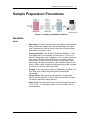
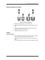
1
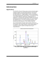
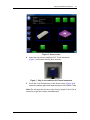
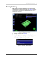
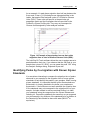
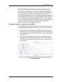
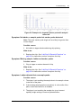
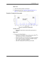
ProfilerPro Glycan Profiling Assay User Guide For LabChip GXII Touch Copyright 2014 PerkinElmer, Inc. All rights reserved. PerkinElmer is a registered trademark of PerkinElmer, Inc. All other trademarks are the property of their respective owners. PN CLS140161, Rev. C 2 Contents Introduction............................................................................................................. 3 Applications ........................................................................................................ 3 Specifications ......................................................................................................... 5 Assay Specifications .......................................................................................... 5 Kit Contents ....................................................................................................... 5 Additional Items Required .................................................................................. 7 Safety Warnings and Precautions.......................................................................... 8 Sample Preparation Procedures ............................................................................ 9 Denature ............................................................................................................ 9 Digestion .......................................................................................................... 10 Labeling ........................................................................................................... 11 Reconstitution .................................................................................................. 12 Chip Preparation Procedures............................................................................... 13 Preparing the Buffer Tube ................................................................................ 13 Preparing the Ladder Tube ............................................................................... 13 Preparing the Marker Tube ............................................................................... 14 Preparing the Chip ........................................................................................... 14 Inserting a Chip into the LabChip GXII Touch Instrument ................................. 15 Running the Assay ........................................................................................... 17 Storing the Chip ............................................................................................... 20 Chip Cartridge Cleaning ................................................................................... 20 Analyzing Data ...................................................................................................... 21 Assignment of Size and Identification of Peaks in the Electropherogram .......... 21 Identifying Peaks by Co-migration with Known Glycan Standards .................... 22 Results .................................................................................................................. 24 Troubleshooting ................................................................................................... 26 Frequently Asked Questions ................................................................................ 35 LabChip Kit Essential Practices........................................................................... 38 General ............................................................................................................ 38 Chips ................................................................................................................ 39 Samples ........................................................................................................... 43 Chip Well Aspiration Using a Vacuum ................................................................. 44 Customer Technical Support ............................................................................... 45 Licenses and Rights of Use ................................................................................. 46 PN CLS140161, Rev. C ProfilerPro Glycan Profiling Assay User Guide Introduction 3 Introduction Applications The ProfilerPro Glycan Profiling system provides a high throughput method for analysis of the glycosylation patterns of monoclonal antibodies (MAb) and other glycosylated proteins. The system provides methods for: 1) deglycosylation, where N-linked glycans are released enzymatically using PNGase F; 2) fluorescent labeling of the released glycans in presence of released proteins; 3) separation by microchip based capillary electrophoresis (CE); and 4) detection and analysis using the LabChip GXII Touch instrument. Fast separation time - less than 45 seconds per sample - makes this method valuable for use in high throughput screening of antibody glycosylation patterns in early stage development. The microchip-based CE analysis gives resolution sufficient to determine the relative abundance of the major N-glycan types found on antibodies, which contain primarily neutral N-linked glycans consisting of complex, hybrids, and high-mannose type oligosaccharides. Figure 1. Glycosylation profile of a therapeutic MAb. Relative abundance of the major glycan types can be reliably quantified. PN CLS140161, Rev. C ProfilerPro Glycan Profiling Assay User Guide Introduction 4 Features • Fast separation time - less than 45 seconds per sample, about 1.5 hours for 96 samples. • Software determines the relative amounts of each glycan present. • All-included reagent set provides predictable, reproducible results. • Reagents provided in 96-well plate format for convenience and ease of automation. • Automatic sampling from a 96-well plate PN CLS140161, Rev. C ProfilerPro Glycan Profiling Assay User Guide Specifications 5 Specifications Assay Specifications Table 1. Assay Specifications Amount of Sample Required 8 µL with concentration range of 1.25-7.5 mg/mL (10- 60 µg of MAb total) Reproducibility of %Area CV < 10% for a peak >= 2.5% of total glycan Limit of Detection 1 ng of G0f standard (smallest amount of labeled G0f standard that can be detected) Deglycosylation >95% of all N-linked glycans will be released from MAb Usable Size Range Appropriate for neutral glycans found on MAbs, some charged glycans may run outside of our usable range Sizing Reproducibility CV < 2.5% Sample Prep, Chip Prep, and Analysis Time < 8 hours for a 96-well plate Number of Samples per Chip Prep Up to 192 samples before chip needs to be reloaded with fresh gel Kit Contents The ProfilerPro Glycan Profiling system consists of: Table 2. Glycan Release and Labeling Kit Part Number 760523 Item Plate Quantity Denaturing Plate Glycoprotein denaturing plate One 96-well plate PNGase F Plate Oligosaccharides releasing plate One 96-well plate Dye Plate Oligosaccharides labeling plate One 96-well plate Note: Store plates at -20°C PN CLS140161, Rev. C ProfilerPro Glycan Profiling Assay User Guide Specifications 6 Table 3. Glycan LabChip Reagent Kit Part Number 760525 Reagent Vial Quantity Glycan Gel Matrix Red 2 vials, 1.8 mL each Glycan Marker (dry) Green 4 vials Glycan Marker Diluent White 1 vial, 0.65 mL Glycan Ladder (dry) Yellow 4 vials Glycan Ladder Diluent Purple 1 vial, 0.75 mL Note: Store reagents at 4°C. Table 4. Consumable Items Item Supplier and Catalog Number Quantity Ladder Tubes Genemate, Cat. # C-3258-1 10, 0.2 mL Detection Window VWR, Cat. # 21912-046 Cleaning Cloth 1 Swab ITW Texwipe®, Cat. # TX758B 3 Buffer Tubes E&K Scientific, Cat. # 697075NC 10, 0.75 mL Table 5. High Resolution LabChips Item Catalog Number High Resolution Chip for use with GXII Touch HT Cat. # 760524 High Resolution Chip for use with GXII Touch 24 Cat. # CLS138951 PN CLS140161, Rev. C ProfilerPro Glycan Profiling Assay User Guide Specifications 7 Additional Items Required • GX Plate Adapter for Deep-Well PCR Plates (Cat # 126209). Note: The PerkinElmer Dye Plate can be read directly in the GXII Touch instrument using the Plate Adapter. Alternatively, after reconstituting with water (Milli-Q® or equivalent), labeled samples can be transfered to a standard 96-well PCR plate prior to placing them into the instrument. • PerkinElmer Hard-Shell thin-wall 96-well skirted PCR plate (blue), Cat. # 6008870 (recommended). • Heating device: any PCR thermocycler is best. A heat block that accepts a 96-well PCR plate will also work. An incubator may work but is less desirable. • Adhesive plate seals (PCR type with a minimum thermal range of -20°C to 80°C. For example, a Beckman Coulter Cat. # 538619 or Axygen Cat. # PCR-AS-200). • Centrifuge with rotor that accepts a 96-well PCR plate. • Optional: Plate shaker (for example, a VarioMag Magnetic Shaker, Cat. # 51110, 2000 rpm, amplitude = 2 mm). PN CLS140161, Rev. C ProfilerPro Glycan Profiling Assay User Guide Safety Warnings and Precautions 8 Safety Warnings and Precautions WARNING! For Research Use Only. Not recommended or intended for diagnosis of disease in humans or animals. Do not use internally or externally in humans or animals. CAUTION : We recommend that this product and components be handled only by those who have been trained in laboratory techniques and that it is used in accordance with the principles of good laboratory practice. As all chemicals should be considered as potentially hazardous, it is advisable when handling chemical reagents to wear suitable protective clothing, such as laboratory overalls, safety glasses, and gloves. Care should be taken to avoid contact with skin or eyes. In case of contact with skin or eyes, wash immediately with water. WARNING! Denaturing Buffer, Ladder, and Gel Matrix contain SDS. Avoid inhalation and contact with skin and eyes. PN CLS140161, Rev. C ProfilerPro Glycan Profiling Assay User Guide Sample Preparation Procedures 9 Sample Preparation Procedures Figure 2. Sample preparation procedures. Denature Hints • Spin-down: All plates must be spun down before removing the seal to collect any reagent that may be attached to the plate seal. Plates have to be kept frozen at all times. Remove plate from freezer only prior to use. • Cutting the plates: The 96-well Denaturing, PNGase- F, and Dye Plates can be cut vertically into sections of 24, 48, or 72 wells if running fewer than 72 samples. To cut a plate, first stack it on top of a 96-well skirted PCR plate for support. Using a sharp blade, carefully score and cut the top and sides of the plate along the segmented groove, then carefully break off the section. After cutting, inspect the plate wells along the cut edge for cracks. Do not use wells with cracks. • Storage: If not using a whole 96-well Denaturing, PNGase F, or Dye Plate, after cutting the plate freeze the remainder immediately. • Partial Plates: When working with partial or cut plates it's helpful to stack the plate on top of a 96-well skirted PCR plate for support, especially when spinning. • Plate Seals: Use plate seals with a minimum thermal range of -20°C to 80°C. For all plate sealing, use of a rubber roller is recommended. PN CLS140161, Rev. C ProfilerPro Glycan Profiling Assay User Guide Sample Preparation Procedures 10 Procedure 1 Thaw and spin Denaturing Plate at 1200 g for 1 minute. 2 Carefully remove the plate seal. 3 Add 8 µL of sample (monoclonal antibody) with concentration range of 1.25-7.5 mg/mL (10 µg to 60 µg total protein) to Denaturing Plate. Mix by pipetting up and down or with a plate shaker. 4 a Samples should be at least one-step purified. Crude sample media or buffers that contain sugars may interfere with the labeling efficiency of the desired glycans. b For a control blank sample add 8 µL of 18 Megohm, 0.22-µm filtered water (Milli-Q® or equivalent) instead of sample and follow all digestion and labeling steps. Seal plate carefully with an adhesive plate seal. Note: Ensure the plate is completely sealed to prevent evaporation. 5 Spin Denaturing Plate at 1200 g for 1 minute. 6 Incubate Denaturing Plate for 10 minutes at 70°C using a PCR machine (with lid temperature at 70°C), heat block, or incubator. • Break point: If needed, after digestion samples may be frozen at -20°C and labeled the next day. Overnight digestion is not recommended. • Incubators: Use of incubators or ovens for incubation is not recommended. If using an incubator for the labeling step, be careful of acetic acid fumes when opening the incubator door. 1 Thaw PNGase F Plate. 2 Spin both PNGase F Plate and Denaturing Plate at 1200 g for 1 minute. 3 Carefully remove plate seals. 4 Transfer all (~11 µL) denatured sample to PNGase F Plate. Mix by pipetting up and down or with a plate shaker. Digestion Hints Procedure PN CLS140161, Rev. C ProfilerPro Glycan Profiling Assay User Guide Sample Preparation Procedures 11 5 Seal plate carefully with an adhesive plate seal. Note: Ensure the plate is completely sealed to prevent evaporation. 6 Spin PNGase F Plate at 1200 g for 1 minute. 7 Incubate for 1 hour at 37°C using a PCR machine (with lid temperature at 37°C), heat block, or incubator. • Can more than 8 µL of digested sample be labeled? Sample buffer conditions resulting in optimal glycan peak intensity was observed when labeling 8 µL of the digested sample. Labeling more or less will result in lower signal intensity. • Break point: If needed, after labeling, dried samples may be sealed and stored frozen at -20°C and reconstituted the next day. Overnight labeling is not recommended. • Mixing: It is best to use a plate shaker with max speed. • Incubators: Carefully inspect samples to ensure they are completely dry. It's likely that using an incubator or oven will increase the drying time to more than 2 hours. 1 Thaw Dye Plate. 2 Spin both Dye Plate and PNGase F Plate at 1200 g for 1 minute. 3 Carefully remove plate seals. 4 Transfer 8 µL of digested sample to the Dye Plate. Mix by pipetting up and down or with a plate shaker. 5 Spin Dye Plate at 1200 g for 1 minute. 6 Incubate the unsealed plate for 2 hours at 55°C (or until sample is completely dry) using a PCR machine (with lid open) or heat block. Labeling Hints Procedure Caution: The dye contains acetic acid. Be wary of fumes. Incubating under a fume hood is recommended. Sample must be completely dry to ensure sufficient labeling. PN CLS140161, Rev. C ProfilerPro Glycan Profiling Assay User Guide Sample Preparation Procedures 12 Reconstitution 1 Add 100 µL of water (Milli-Q® or equivalent) to dried samples. 2 Seal plate carefully with an adhesive plate seal. Note: Ensure the plate is completely sealed to prevent spilling during mixing. 3 Mix samples on a plate shaker at maximum speed for at least 1 minute. Check to be sure the pellet has completely dissolved. 4 Spin plate at 1200 g for 1 minute. Note: After spinning there may be a white pellet at the bottom of the wells. Be careful not to disturb the pellet as it may cause a chip sipper clog. 5 PN CLS140161, Rev. C Carefully remove the plate seal. The plate is ready to run on LabChip GXII Touch. ProfilerPro Glycan Profiling Assay User Guide Chip Preparation Procedures 13 Chip Preparation Procedures Note: Equilibrate the chip and all components of the Glycan Reagent kit to room temperature for 20-30 minutes before use. Preparing the Buffer Tube 1 Add 750 µL of water (Milli-Q® or equivalent) to the 0.75 mL Buffer Tube. Ensure there are no air bubbles in the Buffer Tube. 2 Insert the Buffer Tube into the buffer slot on the LabChip GXII Touch instrument. Buffer Tube Ladder Tube Sample Well A1 Ladder Tube Buffer Tube Figure 3. Locations of the Buffer Tube and Ladder Tube in the GXII Touch instrument. Preparing the Ladder Tube 1 Add 145 µL of Glycan Ladder Diluent (purple cap the Ladder tubes. 2 Vortex at highest speed for about 30 seconds and spin down. Make sure the ladder is completely reconstituted. 3 Transfer 120 µL of prepared ladder to the 0.2 mL Ladder Tube. Ensure there are no air bubbles in the Ladder Tube. 4 Insert the Ladder Tube into the ladder slot on the LabChip GXII Touch instrument. PN CLS140161, Rev. C ) to one of ProfilerPro Glycan Profiling Assay User Guide Chip Preparation Procedures 14 Preparing the Marker Tube Notes: Prepare marker solution just before loading the chip into the LabChip GXII Touch and starting the assay. Do not prepare marker solution in advance as the marker signal degrades over time. 1 Prepare marker solution by adding 125 µL of Marker Diluent (white cap ) to one of the Marker tubes. 2 Vortex at the highest speed for about 30 seconds and spin down. Make sure marker is completely reconstituted. Preparing the Chip 1 Use a pipette tip attached to a vacuum line to thoroughly aspirate all fluid from the chip wells (see Figure 4). For more details on how to set up a vacuum line see page 44. 2 Each active chip well (1, 2, 3, 4, 7, 8, 9 and 10) should be rinsed and completely aspirated twice with water (Milli-Q® or equivalent). Do not allow active wells to remain dry. 3 If any water spilled onto the top or bottom of the chip surfaces during rinsing, aspirate using a vacuum line. DO NOT run the tip over the central region of the detection window. Use the provided Detection Window Cleaning Cloth to clean the chip detection window. Figure 4. Using a vacuum to aspirate the chip wells is more effective than using a pipette. See page 44 for more details. 4 PN CLS140161, Rev. C Using a reverse pipetting technique, add 75 µL of Gel Matrix (red cap ) to chip wells 2, 3, 7, 8 and 9, and 120 µL to well 10 (Figure 6). ProfilerPro Glycan Profiling Assay User Guide Chip Preparation Procedures 15 Figure 5. Reagent placement. Note: Be sure to periodically clean the O-rings on the top plate of the chip interface on the LabChip GXII Touch instrument. Use the provided lint-free swab dampened with water (Milli-Q ® or equivalent) or 70% isopropanol solution in DI water to clean the Orings using a circular motion. Allow the O-rings to dry before inserting a chip. Inserting a Chip into the LabChip GXII Touch Instrument 1 Check that the sample plate, Buffer Tube, and Ladder Tube are placed on the instrument properly. 2 Remove the chip from the chip storage container and inspect the chip window. Clean BOTH sides of the chip window with the PerkinElmer-supplied clean-room cloth dampened with a 70% isopropanol solution in DI water. 3 Touch the Unload Chip button on the Home screen. PN CLS140161, Rev. C ProfilerPro Glycan Profiling Assay User Guide Chip Preparation Procedures 16 Figure 6. Home screen. 4 Insert the chip into the LabChip GXII Touch instrument (Figure 7) and close the chip door securely. Figure 7. Chip in the LabChip GXII Touch instrument. 5 Touch the Load Plate button on the Home screen (Figure 6) to retract the sample plate and send the sipper to the Buffer Tube. Note: Do not keep the chip door open for any length of time. Dye is sensitive to light and can be photobleached. PN CLS140161, Rev. C ProfilerPro Glycan Profiling Assay User Guide Chip Preparation Procedures 17 Running the Assay Note: Chips can be primed independently from running assays. Select the assay of choice from the insert (see Figure 9).Touch the Prime button on the Home screen. Make sure the Buffer Tube is placed on the instrument. Figure 8. Chip priming screen. 1 Touch the Run button (see Figure 8). 2 Select the appropriate assay type (see Figure 9), plate name, well pattern, and whether to read wells in columns or rows. Select number of times each well is sampled under Adv. Settings (Figure 10). Touch the green arrow. Figure 9. The Assay Choices menu. PN CLS140161, Rev. C ProfilerPro Glycan Profiling Assay User Guide Chip Preparation Procedures 18 Figure 10. Selecting wells. 3 PN CLS140161, Rev. C In the Setup Run tab, select the operator name, the option to read barcode, the destination of the file, the inclusion of sample names, expected peaks, and excluded peaks and the filename convention. Select Auto Export to export results tables automatically (Figure 11). Touch the green arrow. ProfilerPro Glycan Profiling Assay User Guide Chip Preparation Procedures 19 Figure 11. Run setup screen. 4 Touch Start to begin the run. Figure 12. Starting a run. PN CLS140161, Rev. C ProfilerPro Glycan Profiling Assay User Guide Chip Preparation Procedures 20 Storing the Chip After use, the chip must be cleaned and stored in the chip container. 1 Place the chip into the plastic storage container. The sipper should be submerged in the fluid reservoir. 2 Remove the reagents from each well of the chip using vacuum. 3 Each active well (1, 2, 3, 4, 7, 8, 9 and 10) should be rinsed and aspirated twice with water (Milli-Q® or equivalent). 4 Add 120 µL water (Milli-Q® or equivalent) to the active wells. 5 Cover the wells with Parafilm® to prevent evaporation and store the chip at room temperature until next use. The chip must be used to its lifetime (to the total number of 400 samples) within 30 days of analyzing the first plate of samples. Chip Cartridge Cleaning 1 2 PN CLS140161, Rev. C Daily a Inspect the inside of the chip cartridge and O-rings for debris. b Use the provided lint-free swab dampened with water (MilliQ® or equivalent) to clean the O-rings using a circular motion. If the O-rings stick to the chip or a pressure leak is detected, perform the more extensive monthly cleaning procedure. Monthly a To reduce pressure leaks at the chip interface, clean the Orings frequently. Remove the O-rings from the top plate of the chip interface on the LabChip GXII Touch instrument. Soak O-rings in water (Milli-Q ® or equivalent) for a few minutes. Clean the O-ring faces by rubbing between two fingers. Wear gloves. b To reduce the occurrence of current leaks, clean the chip interface frequently. Clean the top plate of the chip interface using the provided lint free swab dampened with water (MilliQ® or equivalent). c Allow the O-rings and chip interface to air dry. Reinsert the O-rings into the chip cartridge. ProfilerPro Glycan Profiling Assay User Guide Analyzing Data 21 Analyzing Data Assignment of Size and Identification of Peaks in the Electropherogram The LabChip GX Touch software analyzes the electropherograms, assigning a size (among other things) to each observed peak. It may not be intuitively obvious what this size means given that glycan molecules with the same mass can sometimes have very different apparent electrophorectic mobility. During the labeling process, the glycans are derivatized with a charged fluorophore. The electrophorectic mobility of each glycan in our specific separation medium is dependent on its charge, mass, and structure - where structural elements such as monosaccharide composition (mannose, glucose, galactose), glycosidic linkage position (C2, C3, C4, C6), and anomercity (- vs -) have been shown to affect electrophorectic mobility. Considering these complexities, the size that the software assigns does not have a direct physical meaning, like the size of a DNA molecule in base pairs or the size of a protein in Daltons. Rather, the sizes assigned to glycan peaks by the LabChip GX Touch software relate the migration time of each peak to the migration time of the glucose peaks in the glycan ladder. It is a way of normalizing the migration time of each peak so that results can be more easily compared across different runs, different instruments, different reagent lots, etc. Figure 13. The Ladder is a mixture of (1-6)-linked glucose oligosaccharides with a various number of monomeric glucose units. Lower Marker (LM) and ladder peaks used for sizing (L) annotated with RGU units. The Lower Marker is maltohexaose which runs with a size of 6.6 RGU. PN CLS140161, Rev. C ProfilerPro Glycan Profiling Assay User Guide Analyzing Data 22 As an example, if a peak has a migration time half way between the 9-mer and 10-mer (1-6)-linked glucose oligosaccharides in the ladder, that peak will be assigned a size of 9.5 Relative Glucose Units (RGU). These units are specific to the particular gel separation matrix and the fluorophore that are provided in the ProfilerPro Glycan Profiling kits. They may not correspond to Glucose Units assigned by other analysis methods. Figure 14. Example of two standard curves that relate migration time to size in Relative Glucose Units (RGU). The LabChip GX Touch software allows the user to assign names to peaks based on their size. If you observe that the G0f peak in your sample has a size of 9.3 RGU, you can name the peak “G0f” using the Analysis Settings dialog, Expected Glycan tab. Identifying Peaks by Co-migration with Known Glycan Standards It is sometimes interesting to compare the migration time of peaks observed in your samples to the migration time of known glycan standards, to help identify the peaks in your samples. When running glycan standards, it is important that the buffer the standards are presented in is closely matched to the buffer your samples are presented in. If the buffers are not well matched, the migration time of the standards may not correspond to the migration time of your sample. A simple method to achieve matched buffers is to label your standards using the reagents from the Glycan Release and Labeling kit. Prepare the standards the same way as you prepared your test samples. Any dilutions of the standards prior to addition in the Denaturing Plate should be done with water (Milli-Q® or equivalent). PN CLS140161, Rev. C ProfilerPro Glycan Profiling Assay User Guide Analyzing Data 23 Figure 15. Example showing a mix of glycan standards compared to an IgG sample, run in well-matched buffers. Blue = IgG, Red = Mix of six glycan standards (Man5, G0, G0f, G1f, G2, G2f). The standards were run at about 50 ng/µL concentration. PN CLS140161, Rev. C ProfilerPro Glycan Profiling Assay User Guide Results 24 Results In the Glycan Ladder, the Lower Marker should be the tallest peak. Following the Lower Marker peak are 7 ladder peaks with Relative Glucose Units (RGUs) of 7 to 13. Figure 16. Glycan Ladder: Lower Marker (LM) and ladder peaks (L) annotated with RGU units. For monoclonal antibody samples the Lower Marker should migrate before the major Nglycan peaks (Man5, G0, G0f, G1f', G1f, and G2f). Figure 17. MAb14: Major N-glycan peaks are annotated with percent area. PN CLS140161, Rev. C ProfilerPro Glycan Profiling Assay User Guide Results 25 Figure 18. MAb17: Major N-glycan peaks are annotated with percent area. PN CLS140161, Rev. C ProfilerPro Glycan Profiling Assay User Guide Troubleshooting 26 Troubleshooting Note: Some of the data examples shown in this section were generated with assays other than the assay described in this user guide. How to Test for Complete Digestion PerkinElmer recommends testing for complete deglycosylation by PNGase F using the Protein Express assay. Compare the digested sample (unlabeled) to the undigested sample. Follow the protocol for the Protein Express Kit for reduced conditions. Note: It may be necessary to dilute the digested sample depending on the starting concentration. A starting concentration of 2 mg/mL or below is recommended for the Protein Express assay. Under reduced conditions you will see a shift from the heavy chain (HC) to the non-glycosylated heavy chain (NGHC) for the digested sample. Figure 19. Blue: control (undigested), Red: Digested MAb. Symptom: Digestion is incomplete. Possible causes: 1 The samples were incompletely denatured. 2 There was an insufficient amount of PNGase F in the reaction. What to do: 1 PN CLS140161, Rev. C Repeat the sample preparation being careful to spin down the Denaturing Plate. Small amounts of denaturing buffer attached to the plate seal may significantly decrease the amount or concentration of reagents needed to sufficiently denature samples. Additionally verify that the samples are being denatured at 70°C for 10 minutes. ProfilerPro Glycan Profiling Assay User Guide Troubleshooting 27 2 If the sample tested was on the high end of the Glycan assay concentration range, then dilute the sample down to 5 mg/mL and repeat sample preparation. Some MAbs may be more difficult to deglycosylate than others. In this event, a greater amount of PNGase F to MAb and longer incubation time may be needed to completely deglycosylate samples. 3 Repeat the sample preparation being careful to spin down the PNGase F Plate. Small amounts of digestion buffer attached to the plate seal may significantly decrease the available amount of PNGase F. Verify that the digestion plate is incubated at 37°C for 1 hour or longer. Symptom: Peaks ahead of the marker, so marker is miscalled. If the sample contains peaks that run faster than the marker, the software may identify them as the marker incorrectly. This can be corrected by manually selecting the proper marker peak. Hover over the peak with the mouse, right click, and select Force Lower Marker. To identify the correct peak as the marker, click Turn Off Analysis in the Analysis menu and overlay the sample with a ladder. Look for the peak in the sample that aligns with the marker in the ladder (should be the tallest peak). Turn the analysis back on and select the marker peak. Figure 20. Miscalled marker. Figure 21. Sample and ladder overlayed with analysis turned off. PN CLS140161, Rev. C ProfilerPro Glycan Profiling Assay User Guide Troubleshooting 28 How to include peaks ahead of the marker in the analysis: By default, the software will only include peaks that migrate after the marker into the analysis. If there are peaks before the marker that you would like to include in the analysis you can do so by manually including them. First, be sure that the marker is assigned to the proper peak (see prior section for guidance). Peaks that are found to the left of the marker are labeled with “?” by the software. Hover over the peak you wish to include with the mouse, right click, and select Include peak. Once included into the analysis all analysis features can be applied to the peak. Symptom: Marker is miscalled in the ladder If the ladder peaks are compressed and/or broad the software may be miscalling the marker peak. There are two ways to correct this: 1 Manually select the proper marker peak by hovering over the peak with the mouse, right click, and select Force Lower Marker. It is often helpful to click Turn Off Analysis in order to identify the proper peak. 2 If the marker peak height is lower than the ladder peaks, the software often miscalls the marker peak. In this case, remove the chip from the instrument, clean out well 4, and replace with fresh marker. Figure 22. Compressed and broadened ladder peaks due to miscalled marker. PN CLS140161, Rev. C ProfilerPro Glycan Profiling Assay User Guide Troubleshooting 29 Figure 23. Example of a degraded marker peak with analysis turned off. Symptom: No ladder or sample peaks but marker peaks detected. Note: The lower marker peak height will most likely be greater than normal height. Possible causes: 1 Air bubble in sipper introduced during chip priming. What to do: 1 Reprime the chip. See “LabChip Kit Essential Practices” on page 38 for instructions on how to reprime the chip. Symptom: Missing sample, ladder and marker peaks. Possible causes: 1 Clog in sipper or marker channel of chip. What to do: 1 Reprime the chip. See “LabChip Kit Essential Practices” on page 38 for instructions on how to reprime the chip. Symptom: Ladder detected but no sample peaks. Possible causes: 1 The sipper is not reaching the sample due to low sample volume in the well of the plate. 2 If the missing sample peaks occurred only in a few wells of the plate, check those wells for air bubbles. 3 The sipper is not reaching the sample due to an incorrect capillary height setting or incorrect plate definition. PN CLS140161, Rev. C ProfilerPro Glycan Profiling Assay User Guide Troubleshooting 30 4 If the plate has been uncovered for some time, sample evaporation might have occurred. 5 Debris from the sample or sample prep is clogging the sipper. 6 The samples did not get labeled completely due to incomplete drying. What to do: 1 Add more sample to the well. 2 Manually insert a larger volume pipette tip (~100 µL) into the sample well and dislodge the bubble. Rerun these sample wells. 3 Check the plate definitions. 4 Check the sample wells, especially around the edge of the plate where evaporation is fastest, and make a fresh plate if volumes are low. 5 If you suspect there may be debris in your samples, spin the sample plate down in a centrifuge (e.g., 3000 rcf for 5 minutes). Unclog the sipper by repriming the chip. See “LabChip Kit Essential Practices” on page 38 for instructions on how to reprime the chip. 6 Make sure the correct plate type is selected. If using the Plate Type “96-Deep Well PCR with Adapter”, verify the sample volume is at least 100 µL. 7 To ensure complete drying, repeat the sample preparation procedures allowing the drying reaction to extend beyond 2 hours until the samples are completely dry. Symptom: No ladder peaks but sample peaks and marker peaks are present. Possible causes: 1 Low or no ladder volume in the Ladder Tube. What to do: 1 Add more ladder to the Ladder Tube and restart the run. Recommended standard ladder volume is 120 µL (minimum volume is 100 µL). Symptom: No marker peaks but sample peaks are present. Possible causes: 1 PN CLS140161, Rev. C No marker added to chip well 4. ProfilerPro Glycan Profiling Assay User Guide Troubleshooting 2 31 If there is marker solution in chip well 4, the problem may be due to a marker channel clog. What to do: 1 This may be due to not filling marker well or chip remaining idle on instrument for extended period of time. Add or replenish the marker solution in the chip using the following procedure: • Touch the Unload Chip button on the Home screen to open the chip door. • Return the chip to the chip container ensuring the sipper is immersed in fluid. • Thoroughly aspirate all fluid from chip well 4 using a vacuum line. • Ensure that chip well 4 is rinsed and completely aspirated twice with water (Milli-Q® or equivalent). • Add Marker Solution (green cap ) to chip well 4. • Reinsert the chip back into the instrument. • Restart the run. 2 Perform a marker channel unclogging procedure by repriming the chip. See “LabChip Kit Essential Practices” on page 38 for instructions on how to reprime the chip. Symptom: Ladder traces show up in the lanes following the ladders (delayed sip). Figure 24. Small ladder peaks in sample well caused by delayed sip. Possible causes: 1 Separation channel overloaded with sample. 2 Partial clog in the separation channel. PN CLS140161, Rev. C ProfilerPro Glycan Profiling Assay User Guide Troubleshooting 32 What to do: 1 Lower the starting sample concentration. 2 Reprime the chip. See “LabChip Kit Essential Practices” on page 38 for instructions on how to reprime the chip. Symptom: Unexpected sharp peaks. Figure 25. Unexpected sharp peak. Possible causes: • Dust or other particulates introduced through sample or reagents. What to do: 1 PN CLS140161, Rev. C Do one or all of the following: • Replace the 18 megohm, 0.22-µm filtered water (Milli-Q® or equivalent) water used for chip preparation. • Replace the buffer used for sample and reagent preparation. • Use a 0.22-micron filter for all water and buffers used for chip, sample, and reagent preparation. • Spin down sample plate to pellet any particulates. ProfilerPro Glycan Profiling Assay User Guide Troubleshooting 33 Symptom: Humps in several electropherograms which do not correspond to sample data. Figure 26. Humps in several electropherograms. Possible causes: 1 Electrode 7 is dirty and has contaminated the Gel mixture in well 7. 2 High concentrations of detergent in the sample buffer can sometimes cause humps in the electropherogram. What to do: 1 Before restarting the run, clean electrode 7. Remove the chip and follow the electrode cleaning procedure. We recommend using the provided swab and isopropanol to manually clean electrode 7. 2 Lower the detergent concentration in the sample. Symptom: Peaks migrating much faster or slower than expected. Note: Some migration time variance between chips or within a plate is considered normal chip performance. All chips are QC tested at PerkinElmer prior to shipment. Possible causes: 1 Particulates from the samples may be clogging the separation channel (this will slow down migration). 2 Gel was not primed properly into the chip. PN CLS140161, Rev. C ProfilerPro Glycan Profiling Assay User Guide Troubleshooting 34 What to do: 1 If fast or slow migration is observed repeatedly on a new chip, contact technical support to arrange return of the chip to PerkinElmer. Please send a data file showing the failure along with the return request. 2 Minimize the loading of particulates in the sample by performing a centrifuge spin of the sample plate (e.g. 3000 rcf for 5 minutes) and/or ensuring the Sip 4 mm plate type is selected in the Select Wells screen before starting a new run. The debris may be flushed out of the chip by washing and re-priming the chip. See “LabChip Kit Essential Practices” on page 38 for instructions on how to wash and reprime the chip. 3 Check the O-rings on the top surface of the chip interface and clean if necessary. PN CLS140161, Rev. C ProfilerPro Glycan Profiling Assay User Guide Frequently Asked Questions 35 Frequently Asked Questions Kit Specification 1 What is in the Lower Marker? The Lower Marker consists of dye-labeled maltohexaose. 2 What is in the Ladder? The ladder is a dye-labeled glucose homopolymer standard consisting of (1-6)-linked glucose oligosaccharides with variable number of monomeric glucose units (1 - 23 or more). Assay Preparation 1 Can samples be digested overnight at 37°C using this assay? No, overnight digestion of samples may lead to poor percent area reproducibility and sample resolution. 2 Can samples be labeled overnight at 55°C using this assay? No, samples should be labeled until dry. Extended incubation of dry labeled samples at 55°C may lead to glycan degradation. 3 Can an evaporator be used to label the glycans instead of incubating at 55°C for 2 hours? No, the labeling efficiency using an evaporator is much lower and will result in low sample signal. 4 Can labeled samples be reconstituted in less or more than 100 µL of water? No, reconstituting samples in 100 µL results in a buffer composition optimal for glycan migration in this assay. Reconstituting in less or more than 100 µL will change this buffer composition and potentially alter migration and data resolution. 5 After labeling standards using this assay can they be frozen and used later? Yes, labeled standards may be frozen for several weeks and tested again at a later time. Thawed labeled standards should be kept on ice when not in use. PN CLS140161, Rev. C ProfilerPro Glycan Profiling Assay User Guide Frequently Asked Questions 6 36 Can samples be prepared using this assay and then analyzed using HPLC or CE- LIF? • HPLC: No, the reagent chemistry of the Glycan assay is not compatible with the HPLC method. • CE-LIF: Yes, provided the optics system is compatible or adjusted accordingly. Assay Performance 1 Can concentrations lower than 1.25 mg/mL be tested using this assay? Yes, concentrations as low as 0.5 mg/mL can be tested, however, the percent area of peaks for samples < 1.25 mg/mL are less reproducible (CV > 10%) than samples tested at > 1.25 mg/mL. 2 What other glycoproteins in addition to monoclonal antibodies can be tested using this assay? Other N-linked glycoproteins can be tested using this assay. However, this assay was optimized to provide a glycosylation pattern for 5 major glycans: Man5, G0, G0f, G1f, and G2F. Other glycans or oligosaccharides may not be detected according to the assay performance specifications. 3 If I am using this kit for the first time, how do I confirm that the kit is working properly? Deglycosylation can be confirmed by testing unlabeled digested samples running Protein Express Kit for reduced conditions. Sample labeling can be confirmed by labeling glycan standards, see “Analyzing Data” on page 21. 4 Can charged N-glycans be detected using this assay? Some charged glycans will migrate faster than the lower marker. If this is the case, and the peak is within the baseline window then they can be manually included in the analysis. For instruction on how to perform this, see “Troubleshooting” on page 26. 5 How does this assay compare to HPLC and CGE-LIF? The Glycan assay is a high throughput assay in which 96 samples from sample preparation to data analysis can be completed in approximately 8 hours. Using HPLC or CGE-LIF, a few samples from sample preparation to data analysis are completed on the order of days. PN CLS140161, Rev. C ProfilerPro Glycan Profiling Assay User Guide Frequently Asked Questions 6 37 Why do I need a plate adapter? The 96-well Glycan plates do not fit in the LabChip GXII Touch plate tray. The plate adapter provides a support base so a Glycan plate can be positioned in the plate tray. 7 Is it possible to run the assay without the plate adapter? Yes, after sample reconstitution, the samples can be transferred to another 96-well plate that fits the LabChip GXII Touch plate tray. PN CLS140161, Rev. C ProfilerPro Glycan Profiling Assay User Guide LabChip Kit Essential Practices 38 LabChip Kit Essential Practices To ensure proper assay performance, please follow the important handling practices described below.Failure to observe these guidelines may void the LabChip Kit product warranty.1 Note: It is important to keep particulates out of the chip wells, channels and capillary. Many of the following guidelines are designed to keep the chips particulate-free. For assay and instrument troubleshooting, refer to the LabChip GX Touch software Help file or call PerkinElmer Technical Support at 1800-762-4000. General • Allow the chip, sample plate and all reagents to equilibrate to room temperature before use (approximately 20 to 30 minutes). • Clean the O-rings in the chip interface weekly and the electrodes daily. Refer to the Instrument Users Guide Maintenance and Service section for procedures. • Avoid use of powdered gloves. Use only non-powdered gloves when handling chips, reagents, sample plates, and when cleaning the instrument electrodes and electrode block. • Calibrate laboratory pipettes regularly to ensure proper reagent dispensing. • Only the PerkinElmer-supplied clean room cloth can be used on the chip to clean the detection window. • Water used for chip preparation procedures must be 18 megohm, 0.22-µm filtered water (Milli-Q® or equivalent). • Using the “Reverse Pipetting Technique” (described next) will help avoid introducing bubbles into the chip when pipetting the gel. 1. PerkinElmer, Inc. warrants that the LabChip Kit meets specification at the time of shipment, and is free from defects in material and workmanship. LabChip Kits are warranted for 60 days from the date of shipment. All claims under this warranty must be made within thirty days of the discovery of the defect. PN CLS140161, Rev. C ProfilerPro Glycan Profiling Assay User Guide LabChip Kit Essential Practices 39 Reverse Pipetting Technique Figure 27. Reverse pipetting. 1 Depress the pipette plunger to the second stop. 2 Aspirate the selected volume plus an excess amount from the tube. 3 Dispense the selected volume into the corner of the well by depressing plunger to the first stop. 4 Withdraw the pipette from the well. Chips Repriming Chips • Touch the Unload Chip button on the Home screen to open the instrument door. Place the chip into the instrument. • Close the chip door securely and choose the corresponding assay. • Touch the Prime button on the Home screen to reprime the chip. PN CLS140161, Rev. C ProfilerPro Glycan Profiling Assay User Guide LabChip Kit Essential Practices 40 Washing Chips Important Note: Wash chips only with water (Milli-Q® or equivalent). Use of any other reagents (including Wash Buffer) is likely to cause even more artifacts in subsequent data. Notes: Some protein samples may have components which produce data with extra peaks, spikes or other artifacts. When these artifacts are present, washing chips on the LabChip GXII Touch immediately before the next use can often restore data quality. Chips should only be washed on the LabChip GXII Touch immediately before they are prepared with fresh reagents and primed on the instrument. Chips should not be washed and left with water in the chip channels for any extended period of time. For most protein samples, the only chip cleaning protocol that is required is to rinse and aspirate the active wells twice with water (Milli-Q® or equivalent), and store the chip with 120 µL of water in each active well. • Thoroughly aspirate all fluid from the chip wells using a vacuum line. • Each active well (1, 2, 3, 4, 7, 8, 9 and 10) should be rinsed and completely aspirated twice with water (Milli-Q® or equivalent). Do not allow active wells to remain dry. • Add 120 µL of water (Milli-Q® or equivalent) to each active well (1, 2, 3, 4, 7, 8, 9 and 10). • Touch the Unload Chip button on the Home screen and place the chip into the instrument. • Close the chip door securely. • Transfer 750 µL of water (Milli-Q® or equivalent) into the Buffer Tube. Install into the instrument. • Touch the Wash button (Figure 28). PN CLS140161, Rev. C ProfilerPro Glycan Profiling Assay User Guide LabChip Kit Essential Practices 41 Figure 28. Wash screen. • After completion of the wash cycle, open the chip cartridge and return the chip to the chip container ensuring the sipper is immersed in fluid. • Thoroughly aspirate all fluid from the chip wells using a vacuum line. • Replace fluid in the wells with freshly made reagents as described in “Preparing the Chip” on page 14. Do not let wells remain dry. • Transfer 750 µL of water (Milli-Q® or equivalent) into a clean Buffer Tube. Install into the instrument. • Install the Ladder Tube, sample plate and chip into the instrument and run the assay. • If air bubbles are not dislodged after a reprime, apply a vacuum to the sipper. Perform this by filling all active wells with 100 µL water (Milli-Q® or equivalent). Then suction the sipper with a vacuum line, as shown in Figure 29, until droplets of fluid flow out from the sipper. When suctioning the sipper, be careful not to bend or break the sipper. To facilitate this, cut the end of the pipette tip attached to the vacuum line to widen the mouth. PN CLS140161, Rev. C ProfilerPro Glycan Profiling Assay User Guide LabChip Kit Essential Practices 42 Figure 29. Removing an air bubble or clog by suctioning the sipper with a vacuum line. Other Considerations: • New chips should be stored refrigerated prior to first use. • After running the first set of samples, chips must be stored at room temperature and used within 30 days. • Do not allow the liquid in the chip container to freeze, as this may lead to poor chip performance. Do not submerge the chip in any solution. • The entire chip surface must be thoroughly dry before use. • The sipper must be kept immersed in fluid at all times and should not be exposed to an open environment for long periods of time. • Use care in chip handling to prevent sipper damage. Damage to the sipper can result in inconsistent sampling. • Avoid exposing the chips to dust by keeping them in a closed environment such as in the chip container or in the instrument before and after chip preparation. • Chips can be prepared and left in the instrument for extended periods of time so that samples can be run as needed throughout the day. PerkinElmer recommends the chip be reprepared after it has been idle for 8 hours, but the chip can be used continually over an 8-hour work day as long as the maximum recommended idle time of 8 hours and total chip lifetime of 400 samples are not exceeded. PN CLS140161, Rev. C ProfilerPro Glycan Profiling Assay User Guide LabChip Kit Essential Practices 43 Samples • Prepared sample plates should be free of gas bubbles and particulate debris, both of which may inhibit sipper flow. • Sample plates containing gas bubbles and/or particulate debris should be spun down at 3000 rpm (1250 rcf) prior to analysis. • Up to four 96-well plates (400 samples) can be run with a single chip preparation when running the GXII Touch HT instrument. Up to 48 samples can be run with a single chip preparation when running the GXII Touch 24 instrument. PN CLS140161, Rev. C ProfilerPro Glycan Profiling Assay User Guide Chip Well Aspiration Using a Vacuum 44 Chip Well Aspiration Using a Vacuum Aspirating with a pipette can leave used reagents in the chip wells. For this reason, PerkinElmer recommends vacuuming the wells instead. This can be accomplished by attaching a permanent pipette tip to a house vacuum line with trap (Figure 30). To avoid contamination, use a new disposable pipette tip over the permanent tip for each chip aspirated (Figure 31). B A Figure 30. A: Permanent pipette tip attached to a house vacuum line; B: vacuum line with trap. Figure 31. Replacing the disposable pipette tip. PN CLS140161, Rev. C ProfilerPro Glycan Profiling Assay User Guide Customer Technical Support 45 Customer Technical Support PerkinElmer, Inc. 68 Elm Street Hopkinton, MA 01748-1668 PerkinElmer Technical Support Phone (USA Toll Free): 800-762-4000 Phone (Worldwide): +1 203-925-4602 Fax: +1 203-925-4602 Email: [email protected] Internet: www.perkinelmer.com For additional assay and instrument troubleshooting, refer to the LabChip GX Touch Software Help file. PN CLS140161, Rev. C ProfilerPro Glycan Profiling Assay User Guide Licenses and Rights of Use 46 Licenses and Rights of Use Label Licenses This product is provided under an intellectual property license from Life Technologies Corporation. The purchase of this product conveys to the buyer the non-transferable right to use the data for internal research and training purposes solely in combination with PerkinElmer, Inc. instrumentation, and not to generate revenue, which may include, but is not limited to use of the product: (1) in manufacturing or in quality assurance or quality control of a commercial product; (2) to provide a service, information, or data for a fee or other consideration; (3) for therapeutic or prophylactic purposes; (4) for diagnostic use; and (5) for resale, whether or not such items are resold for use in research. For information on purchasing a license to this product for any other purposes, contact Life Technologies Corporation, 5791 Van Allen Way, Carlsbad, CA 92008 USA or [email protected]. Rights of Use The chip and reagents supplied with this kit are sold with limited rights of use. The chip may only be used with the specific quantity of reagents supplied with this kit. The purchaser has no right or license to refurbish, reuse, remanufacture, or otherwise use the chip with any other reagents than those specifically supplied in this kit. For more information on the terms and conditions of use of these chips and reagents, please read your LabChip GX Touch User Guide and refer to the applicable label license. The reagent kit contains materials that are provided under a license from Life Technologies Corporation, for research use only. PerkinElmer, LabChip, and the LabChip logo are registered trademarks of PerkinElmer, Inc. and/or its parent, affiliates, and/or subsidiary companies (collectively “PerkinElmer”). The PerkinElmer logo is a registered trademark of PerkinElmer, Inc. All other trademarks and registered trademarks are the property of their respective holders. © 2014 PerkinElmer, Inc. http://www.perkinelmer.com PN CLS140161, Rev. C ProfilerPro Glycan Profiling Assay User Guide PerkinElmer, Inc. 68 Elm Street Hopkinton, Massachusetts 01748 U.S.A. TEL 508-435-9500 FAX 508-435-3439 http://www.perkinelmer.com