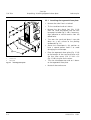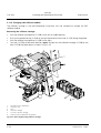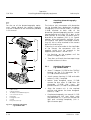
1
Operating Manual Axiostar plus Transmitted-Light Microscope Carl Zeiss Copyright Axiostar plus Knowledge of this manual is required for the operation of the instrument. Therefore please make yourself familiar with the contents of this manual and pay special attention to hints concerning the safe operation of the instrument. The specifications are subject to change; the manual is not covered by an update service. Unless expressly authorized, forwarding and duplication of this document, and the utilization and communication of its contents are not permitted. Violations will entail an obligation to pay compensation. All rights reserved in the event of granting of patents or registration of a utility model. Issued by: Carl Zeiss Light Microscopy P.O.B. 4041 D-37030 Göttingen Phone: ++49 551 5060 660 Fax: ++49 551 5060 464 Internet: www.zeiss.de/micro E-mail: [email protected] Number of this manual: B 40-815 e Date of issue: 12/01 II B 40-815 e 12/01 Axiostar plus Contents Carl Zeiss CONTENTS Page INTRODUCTION Title Page............................................................................................................. I Copyright ........................................................................................................... II Contents ........................................................................................................... III General Notes on Safety, Instrument Safety and EMC .........................................VI Notes on Safety: Unpacking, Transport, Storage, Disposal and operation............ VII Notes on Safety: Care and Maintenance, Warranty Notes ................................. VIII 1 1.1 1.2 1.3 1.4 1.5 1.6 1.7 INSTRUMENT DESCRIPTION ..........................................................................1-3 Name and intended application ........................................................................1-3 Description and main features ..........................................................................1-3 System overview ..............................................................................................1-4 Objectives ........................................................................................................1-7 Eyepieces .........................................................................................................1-8 Stage micrometers and eyepiece reticles ...........................................................1-9 Technical data................................................................................................1-11 2 2.1 2.2 START-UP .......................................................................................................2-3 Unpacking the instrument ................................................................................2-3 Attaching and setting ergonomic base plate .....................................................2-3 2.2.1 2.2.2 Attaching the ergonomic base plate ................................................................................2-4 Adjusting the ergonomic base plate.................................................................................2-6 2.3 Attaching tubes ...............................................................................................2-8 2.3.1 2.3.2 2.3.3 Attaching binocular tube 45°/20 ICS................................................................................2-8 Attaching binocular phototube 45°/20 ICS ......................................................................2-8 Attaching binocular tube 30°/20 ICS................................................................................2-9 2.4 2.5 Screwing in objectives ....................................................................................2-10 Inserting eyepieces .........................................................................................2-10 2.5.1 2.5.2 2.5.3 Inserting eyepiece reticles ..............................................................................................2-11 Compensation of ametropia when eyepiece reticles are used .......................................2-11 Attaching folding eyecups .............................................................................................2-12 2.6 2.7 Setting interpupillary distance and viewing height...........................................2-12 Setting mechanical stage 75 x 30 R/L..............................................................2-13 2.7.1 2.7.2 Setting drive length on the stage drive...........................................................................2-13 Setting drive knob torque for x/y adjustment on the mechanical stage...........................2-13 B 40-815 e 12/01 III Carl Zeiss IV Contents Axiostar plus 2.8 Attaching condensers.....................................................................................2-14 2.8.1 2.8.2 2.8.3 2.8.4 2.8.5 Attaching Abbe condenser 0.9/1.25.............................................................................. 2-14 Inserting slider for phase contrast or darkfield.............................................................. 2-14 Attaching Abbe condenser with 5-position turret.......................................................... 2-15 Attaching LED illuminator ............................................................................................. 2-16 Attaching darkfield condenser ...................................................................................... 2-18 2.9 2.10 2.11 Attaching colored disk carrier, polarizer or wide-angle unit .............................2-20 Inserting white-balance filter ..........................................................................2-21 Attaching epi-fluorescence illuminator ............................................................2-22 2.11.1 2.11.2 2.11.3 2.11.4 2.11.5 2.11.6 2.11.7 Filter stop carrier for epi-fluorescence illuminator ......................................................... 2-23 Attaching HBO 50 fluorescent lamp .............................................................................. 2-24 Replacing burner in fluorescent lamp ............................................................................ 2-25 Adjusting burner in fluorescent lamp ............................................................................ 2-27 Changing P&C reflector module.................................................................................... 2-28 Changing filter set in P&C reflector module .................................................................. 2-30 Changing color splitter in P&C reflector module............................................................ 2-31 2.12 Connecting the instrument to the line ...........................................................2-33 3 3.1 3.2 OPERATION....................................................................................................3-3 Switching on the instrument ............................................................................3-3 Using the transmitted-light brightfield technique ..............................................3-4 3.2.1 3.2.2 3.2.3 General principle............................................................................................................. 3-4 Transmitted-light brightfield configuration ...................................................................... 3-4 Setting transmitted-light brightfield for KÖHLER illumination .......................................... 3-4 3.3 Using transmitted-light phase contrast..............................................................3-6 3.3.1 3.3.2 3.3.3 General principle............................................................................................................. 3-6 Transmitted-light phase contrast configuration ............................................................... 3-6 Setting transmitted-light phase contrast.......................................................................... 3-7 3.4 Using the transmitted-light darkfield technique.................................................3-8 3.4.1 3.4.2 3.4.3 General principle............................................................................................................. 3-8 Transmitted-light darkfield configuration ........................................................................ 3-8 Setting transmitted-light darkfield................................................................................... 3-9 3.5 Using transmitted-light polarization contrast...................................................3-12 3.5.1 3.5.2 3.5.3 3.5.4 General principle........................................................................................................... 3-12 Transmitted-light polarization contrast configuration .................................................... 3-12 Setting transmitted-light polarization contrast............................................................... 3-12 Sample differentiation between gout and pseudogout.................................................. 3-13 3.6 Setting epi-fluorescence .................................................................................3-14 3.6.1 3.6.2 3.6.3 General principle........................................................................................................... 3-14 Epi-fluorescence configuration ...................................................................................... 3-14 Setting epi-fluorescence................................................................................................ 3-14 3.7 3.8 Measurement of lengths ................................................................................3-16 Attaching ICS double tube .............................................................................3-17 B 40-815 e 12/01 Axiostar plus Contents Carl Zeiss 3.9 Attaching photomicrography equipment ........................................................3-18 3.9.1 3.9.2 Attaching SLR camera, e.g. CONTAX ARIA ...................................................................3-18 Attaching MC 80 DX microscope camera (film cassette 35 mm) ....................................3-20 3.10 3.11 Attaching adapters for video cameras.............................................................3-21 Attaching adapters for digital compact cameras..............................................3-23 3.11.1 3.11.2 3.11.3 3.11.4 Digital cameras with 37 mm filter thread, e.g. SONY DCR-PC100 / SONY DSC – S50.....3-24 Digital cameras with 52 mm filter thread, e.g. SONY DSC–S70, DSC-S75, DSC-S85......3-25 Attaching cameras with other filter thread sizes ............................................................3-26 Sony DSC-S70, DC-S75, DSC-S85 on digital camera adapter 44 M52x0.75, 000000-1108-984 .......................................................................................3-26 3.12 Axiostar plus - Packing the mount ..................................................................3-27 4 4.1 4.2 CARE, MAINTENANCE AND TROUBLE-SHOOTING .......................................4-2 Care and maintenance of the instrument..........................................................4-2 Troubleshooting...............................................................................................4-3 4.2.1 4.2.2 4.2.3 4.2.4 Changing the fuses..........................................................................................................4-3 Changing the 6 V, 20 W halogen lamp..........................................................................4-4 Troubleshooting ..............................................................................................................4-5 Spares, wearing parts and tools .......................................................................................4-8 4.3 Requesting service............................................................................................4-9 APPENDIX Index .............................................................................................................................. A-2 List of Abbreviations ....................................................................................................... A-4 Physical and Technical Units............................................................................................ A-5 Certification in Accordance with DIN EN ISO 9001 / DIN EN 46001 EC Conformity Declaration B 40-815 e 12/01 V Carl Zeiss Safety General Notes, Instrument Safety and EMC Axiostar plus General Notes on Safety Before starting up the microscope, please ensure that you have familiarized yourself thoroughly with the contents of this manual. Further information can be obtained from our service department or from authorized representatives. The Axiostar plus microscope is an optical precision instrument which may be impaired in its performance or damaged if handled improperly. The microscope must be operated by trained personnel only who must be aware of the possible problems involved with microscopy and who are trained in the relevant application. To ensure that the working procedure is safe and that the microscope functions without problems, the notes and warnings included in the operating instructions must be observed. These are highlighted in the text by the following symbols: CAUTION! If the safety notes are not observed there is a risk for the user. CAUTION! Disconnect the instrument from the line. VI CAUTION! Hot surface; there is a risk of burns. CAUTION! If the safety notes are not observed, there is a risk that the instrument will be destroyed. ☞ NOTE! Notes that must be observed when working with the microscope. Instrument Safety and EMC The Axiostar plus microscope was designed, produced and tested in compliance with DIN EN 61010-1 (IEC 1010-1) “Safety requirements for electrical measuring, control and laboratory instruments“. It left the factory in a perfectly safe condition. The Axiostar plus microscope meets the requirements of the EC directives 73/23/ECappendix 1 and 89/336/EC and the EMC legislation of 09/18/1998: Radio interference suppression in accordance with EN 55011 class B Resistance to jamming in accordance with EN 50082-2 Conformity with the above EC directives is documented by the CE symbol. B 40-815 e 12/01 Axiostar plus Safety Unpacking Transport, Storage, Operation Carl Zeiss Unpacking, Transport, Storage Operation Please observe the following safety notes when unpacking, transporting and storing the Axiostar plus microscope: Please observe the following safety notes when using the Axiostar plus microscope: – In accordance with standard practice, the microscope is supplied in a plastic container with cardboard packaging; only use the original packaging for transport. – Retain the packaging for longer storage periods or to return the instrument to the manufacturer. – When unpacking, use the delivery note to ensure that all configurations and modules are present. – Observe transport and storage temperatures in accordance with the technical data. – Place the microscope on a stable work surface. – Keep optical surfaces free of fingerprints. Disposal When disposing of the Axiostar plus microscope, please observe the following safety notes: – The microscope can be operated with a line voltage of 100 − 240 V. – The microscope must be connected only to a properly installed outlet featuring a grounding contact; the grounding effect should not be made ineffective with an extension cable that does not have a protective ground wire. – If it is established that the protection measures are no longer effective: switch off the microscope ⇒ ⇒ safeguard against inadvertent operation ⇒ contact an authorized workshop or the manufacturer for repairs. – Always disconnect the instrument from the line before opening the instrument, changing the lamps or changing the fuses! – Only use fuses for the required rated power! The use of makeshift fuses and the shortcircuiting of the fuse holders are not permitted! – – Defective microscopes should not be placed in ordinary domestic waste; they should be disposed of in accordance with the relevant statutory provisions. B 40-815 e 12/01 Be careful of light-emitting diode radiation when using the LED illuminator. Do not look directly into the illuminator with your bare eyes from a short distance. When adjustments are required, the intensity must be reduced accordingly via the potentiometer, or a suitable attenuator must be used. VII Carl Zeiss Safety Care and Maintenance, Warranty Notes Axiostar plus Care and Maintenance Notes on Warranty Please observe the following safety notes when caring for and maintaining the Axiostar plus microscope: The Axiostar plus microscope, including its original accessories, may only be used for the microscope techniques described in this manual. The manufacturer cannot assume any liability for any other applications. With the exception of the work specified in Chapter 4 of this manual “Care, Maintenance and Troubleshooting“, no maintenance or repair may be performed by the user. We expressly indicate that any other repairs must only be carried out by our authorized personnel. Please observe the following warranty notes for the Axiostar plus microscope: – – – – Damaged instruments or components must only be repaired and maintained by our service department. Protect the microscope against dirt, dust and moisture; these influences can impair the performance of the instrument. If the microscope is not in use, protect it with the dust cover and make sure that it has been switched off. – Lamps are to be changed in accordance with the steps in Chapter 4 “Care, Maintenance and Troubleshooting“. – Disconnect the instrument from the line before changing a lamp. – Let the lamps cool down before changing them. – Keep the lamps free of fingerprints. VIII – The manufacturer guarantees that the instrument has no material and production defects when delivered. – You must inform us of any defects immediately and do everything possible to minimize the damage. – If the manufacturer is informed of such a defect, he is obligated to remove it; it is his decision whether he does this by repairing the instrument or by delivering an instrument free of any defect. – No guarantee is provided for defects caused by natural wear (wearing parts in particular) or improper use. – The manufacturer is not liable for damage caused by faulty operation, negligence or any other tampering with the instrument, in particular as a result of removing and exchanging microscope components or the use of accessories from other manufacturers. – Unauthorized intervention invalidates all claims against the warranty. B 40-815 e 12/01 Axiostar plus Instrument description Contents, List of Illustrations Carl Zeiss INSTRUMENT DESCRIPTION Contents 1 INSTRUMENT DESCRIPTION..................................................................................... 1-3 1.1 Name and intended application .................................................................................. 1-3 1.2 Description and main features .................................................................................... 1-3 1.3 System overview......................................................................................................... 1-4 1.4 Objectives .................................................................................................................. 1-7 1.5 Eyepieces ................................................................................................................... 1-9 1.6 Stage micrometers and eyepiece reticles ..................................................................... 1-9 1.7 Technical data .......................................................................................................... 1-11 List of illustrations Fig. 1-1 Overall view ............................................................................................................... 1-2 40-815 e 12/01 1-1 Carl Zeiss Fig. 1-1 1-2 Instrument description Overall view Axiostar plus Overall view B 40-815 e 12/01 Axiostar plus 1 1.1 Instrument description Intended application, Main features INSTRUMENT DESCRIPTION Name and intended application Manufacturer‘s name: Axiostar plus microscope The Axiostar plus is a transmitted light microscope for the visualization of fine structures and forms in biology and medicine. Typical applications of the Axiostar plus include: 1.2 Description and main features Thanks to its pyramid design, the Axiostar plus is a compact transmitted-light microscope. In addition to high-resolution ICS objectives and the major brightfield, darkfield, phase contrast, polarization contrast and epi-fluorescence techniques, an optional camera port for photo and video documentation is also available to the user. – Histology, pathology, Major instrument features: – Doctors’ offices, – – Training (schools and universities), – Routine laboratory microscopy, – Immunofluorescence and FISH methods, – – Field use with LED illumination. – – – Key to Fig. 1-1 1 2 3 4 5 6 7 8 9 10 11 12 13 14 15 16 17 18 Eyepieces Binocular tube Microscope stand Knurled screw for tube locking Brightness control On/off switch with integrated signal lamp Fine focusing drive (two-way) Coarse focusing drive (two-way) Drive for adjusting mechanical stage in X direction Drive for adjusting mechanical stage in Y direction Luminous field diaphragm Condenser carrier Lever for adjusting aperture diaphragm Condenser Centering screw for condenser (two-way) Mechanical stage with specimen holder Objective 4- or 5-position nosepiece – – – – – – – 40-815 e 12/01 Carl Zeiss Sturdy and convenient stand in the pyramid design Ergonomic base plate with hand rests and adjustable viewing height and angle User-friendly coaxial coarse and fine drive Mechanical stage 75 × 30 R/L with ceramiccoated stage surface and specimen holder; selectable stage drive right or left Space-saving and continuously adjustable integrated illuminator with long-life 6 V, 20 W halogen lamp 0.9/1.25 condenser for brightfield, darkfield and phase contrast Ph 1, 2, 3 Ball-bearing, 4- or 5-position objective revolver with W 0.8 thread ICS objectives in the price/performance categories CP-Achromat, A-Plan and Achroplan in finely graduated rows Binocular tube 45°/20 ICS with a viewing height of 425 − 470 mm and binocular phototube 45°/20 ICS with folding prism 100% vis / 100% doc Binocular tube 30°/20 ICS with a viewing height of 430 – 475 mm and binocular phototube 30°/20 ICS with folding prism 100 % vis / 100 % doc 10× eyepieces for the field numbers 20 or 18, suitable for eyeglass wearers, fixed and adjustable Visual white-balancing with the whitebalance filter 1-3 Carl Zeiss 1.3 1-4 Instrument description System overview Axiostar plus System overview B 40-815 e 12/01 Axiostar plus 40-815 e 12/01 Instrument description System overview Carl Zeiss 1-5 Carl Zeiss 1-6 Instrument description System overview Axiostar plus B 40-815 e 12/01 Axiostar plus 1.4 Instrument description Objectives Objectives The objectives are the optical centerpiece of the microscope. The following is an example of how objectives can be labeled: CP-ACHROMAT 10×/0.25 ∞/-. where: 10× = objective magnification, with a defined color ring on the objective allocated to each magnification step (Carl Zeiss color code) 0.25 = numerical aperture ∞ = infinite tube length − = can be used with cover slip thickness D = 0 or 0.17 mm or 0.17 = can be used with cover slip thickness D = 0.17 mm and Oil = oil immersion objective Ph 2 = phase contrast objective with green objective labeling and annular diaphragm Ph 2 The objective magnification multiplied by the eyepiece magnification (usually 10×) results in the visual overall magnification: e. g. 10 × 10 = 100×. Carl Zeiss ☞ The objective labeling "∞" indicates that these objectives may only be used with microscopes featuring infinite optics (ICS) and not with instruments whose objectives are marked with "160" as their mechanical tube length (in mm). The exact observance of the cover slip thickness of 0.17 mm is all the more necessary the higher the numerical aperture of the objective. Therefore, so-called "Corr" objectives can be set for different cover slip thickness via a correction ring. For this, a specimen area is searched, and the position of the correction ring where optimum focus and image contrast are obtained is determined (refocusing is always required). Immersion objectives are always insensitive to differences in cover slip thickness. When immersion objectives are used, the air between the cover slip and the objective is replaced with a liquid, which is immersion oil in most cases. The plastic oiler containing 20 ml of 581 N Immersol is particularly suitable for this purpose. Due to their short working distance, objectives 25× and higher feature resilient mounts (specimen protection). The numerical aperture × 1000, e. g. 0.25 × 1000 = 250×, is the highest useful magnification, i.e. no further details are resolved above that limit. 40-815 e 12/01 1-7 Instrument Description Objectives Carl Zeiss Axiostar plus The following objectives are available for the Axiostar plus microscope: Magnification/ Numerical aperture Microscopy Technique Objective Transm.-light brightfield CP-Achromat 5×/0.12 CP-Achromat Phase contrast Transm.-light brightfield Phase contrast Transm.-light brightfield Phase contrast Transm.-light brightfield 1-8 Free working distance FAA [mm] Cover slip thickness D [mm] Cat.No. 11.2 - 440920-0000-000 10×/0.25 5.1 - 440930-0000-000 CP-Achromat 40×/0.65 0.3 0.17 440950-0000-000 CP-Achromat 100×/1.25 Oil 0.07 0.17 440980-0000-000 CP-Achromat 10×/0.25 Ph 1 5.1 - 440931-0000-000 CP-Achromat 40×/065 Ph 2 0.3 0.17 440951-0000-000 CP-Achromat 100×/1.25 Oil Ph 2 0.07 0.17 000000-1007-159 CP-Achromat 100×/1.25 Oil Ph 3 0.07 0.17 440981-0000-000 A-Plan 5×/0.12 9.9 - 441020-0000-000 A-Plan 10×/0.25 4.4 - 441030-0000-000 A-Plan 20×/0.45 0.53 0.17 441040-0000-000 A-Plan 40×/0.65 0.43 0.17 441050-0000-000 A-Plan 100×/1.25 Oil 0.22 0.17 441080-0000-000 A-Plan 10×/0.25 Ph 1 4.4 - 441031-0000-000 A-Plan 20×/0.45 Ph 2 0.53 0.17 441041-0000-000 A-Plan 40×/0.65 Ph 2 0.43 0.17 441051-0000-000 A-Plan 100×/1.25 Oil Ph 3 0.22 0.17 441081-0000-000 Achroplan 4×/0.10 11.1 - 440020-0000-000 Achroplan 10×/0.25 4.8 - 440030-0000-000 Achroplan 20×/0.45 2.07 0.17 440040-0000-000 Achroplan 40×/0.65 0.59 0.17 440050-0000-000 Achroplan 50×/0.90 Oil 0.29 0.17 440057-0000-000 Achroplan 63×/0.80 0.29 0.17 440060-0000-000 0 (no cover slip) Achroplan 63×/0.95 0.15 Achroplan 100×/1.25 Oil 0.19 0.17 440080-0000-000 Achroplan 100×/1.25 Oil Iris 0.19 0.17 440086-0000-000 Achroplan 10×/0.25 Ph 1 4.8 - 440031-0000-000 Achroplan 20×/0.45 Ph 2 2.07 0.17 440041-0000-000 Achroplan 40×/0.65 Ph 2 0.59 0.17 440051-0000-000 Achroplan 100×/1.25 Oil Ph 3 0.19 0.17 440081-0000-000 2.5×/0.075 9.3 - 440310-0000-000 Plan-Neofluar 440068-0000-000 B 40-815 e 12/01 Instrument description Eyepieces, Stage micrometers, Eyepiece reticles Axiostar plus 1.5 Carl Zeiss Eyepieces The following eyepieces are offered for the Axiostar plus: Eyepiece Image angle Cat. No. Eyepiece PL 10×/18 Br. 39° 444131-9901-000 Eyepiece PL 10×/18 Br. foc. 39° 444132-9901-000 Eyepiece E-PL 10×/20 Br. 43° 444231-9901-000 Eyepiece E-PL 10×/20 Br. foc. 43° 444232-9902-000 Eyepiece PL 16×/16 Br. 54° 444053-0000-000 Eyepiece PL 16×/16 Br. foc. 54° 444054-0000-000 We recommend using eyecups for the eyepieces; they can be ordered in the Zeiss catalog under Cat. No. 444801-0000-000. 1.6 Stage micrometers and eyepiece reticles Measuring and counting using the microscope requires stage micrometers and eyepiece reticles, a small selection of which is listed below: Illustration 5 4 3 2 1 mm 0 0 1 2 3 4 5 6 7 8 9 10 11 12 13 14 0 1 2 3 4 5 6 7 8 9 10 11 12 13 14 40-815 e 12/01 Description, Technical Data Cat. No. Stage micrometer, positive 5 + 100/100 y D = 0.17 mm gradation on the +y-axis: 5 mm in 5 intervals; gradation on the −y-axis: 1 mm in 100 intervals with two opposing scales = 10 µm Accuracy ±1 µm 474026-0000-000 Crossline micrometer disk 14:140 / d = 26 mm gradation length = 14 mm increments = 0.1 mm gradation tolerance ≤ 0.001 mm 454060-0000-000 1-9 Instrument description Eyepieces, Stage micrometers, Eyepiece reticles Carl Zeiss 0 1 2 3 4 5 6 7 8 Axiostar plus Crossline disk / d = 26 mm 474064-0000-000 Crossline micrometer disk 10:100 / d = 26 mm gradation length = 10 mm increments = 0.1 mm gradation tolerance ≤ 0.001 mm 474066-9901-000 9 10 Net micrometer 474068-0000-000 12.5× ×12.5/5;10 / d = 26 mm area 12.5 × 12.5 mm, divided in fields of 5 × 5 or 10 × 10 Photo reticle MC 2.5× × / d = 26 mm for 35mm photography with an additional magnification of 2.5× × or for large-format photography with a 10× × additional magnification 454075-0000-000 Fixed pointer for focusing eyepiece / d = 26 mm To point toward objects in the specimen 000000-1095-418 ☞ If an eyepiece reticle is used, the binocular tube or the phototube must be equipped with two 1-10 B 40-815 e 12/01 foc. eyepieces containing an adjustable eye lens, into one of which the eyepiece reticle is mounted. Axiostar plus Instrument description Technical Data 1.7 Technical data (1) Dimensions (width × depth × height) Carl Zeiss Stand with binocular tube......................................................................approx. 200 × 350 × 450 mm Stand with phototube............................................................................approx. 200 × 350 × 450 mm Overall height including T2 adapter and CONTAX ARIA SLR camera housing.............. approx. 570 mm (2) Weight Axiostar plus with binocular tube ..................................................................................approx. 6.7 kg (3) Ambient conditions Storage and transport (in packaging): Permissible ambient temperature ...................................................................................-40 to +70 °C Permissible relative humidity (without condensation) ........................................................ max. 100 % Operation: Permissible ambient temperature ..................................................................................+10 to +35 °C Permissible relative humidity (without condensation) .......................................................... max. 85 % Air pressure........................................................................................................800 hPa to 1060 hPa (4) Operating data Category of use..............................................................................................................closed rooms Protection class.................................................................................................................................. I Protection type ........................................................................................................................... IP 20 Electrical safety.......................................... in compliance with DIN EN 61010-1 (IEC 1010-1) including .........................................................................................................................CSA and UL directives Pollution degree ...............................................................................................................................2 Excess voltage category .................................................................................................................... II Radio interference suppression....................................................in accordance with EN 55011 Class B Line voltage...................................................................................................... 100 to 240 V (±10 %) Conversion of the voltage is not necessary due to the wide range power supply! Line frequency..................................................................................................................50 to 60 Hz Power consumption.......................................................................................................... max. 65 VA Output voltage ........................................................................... stabilized, adjustable from 1.5 to 6 V (5) Fuses in accordance with IEC 127 for 100 − 240 V......................................................................................... T 0.8 A; 250 V; 5 × 20 mm 40-815 e 12/01 1-11 Carl Zeiss (6) Instrument description Technical Data Axiostar plus Light sources Halogen lamp.............................................................................................................. HAL 6 V, 20 W Adjustment of the light source ........................................................ continuous, 1.5 to 6 V DC 1 Color temperature at 6 V ...........................................................................................2800 K Light flux ..................................................................................................................... 280 lm Average life .................................................................................................................1000 h Luminous area ................................................................................................... 2.0 × 2.0 mm LED Illumination Constant, brightness independent color temperature of ................................................7480 K Homogenous image field illumination ................................................... up to 20 mm diameter Suitable for objectives with magnification of .......................................................2.5 x to 100 x Analog brightness control of ...................................................................approx. 15 to 100 % Operation with 9 V alkaline battery / 9 V NiCd Accublock: Operating voltage.......................................................................... approx. 4.5 to 10 V Current at maximum brightness depending on operating voltage of: Operating voltage 4.5 V.................................................................................... 90 mA Operating voltage 9 V....................................................................................... 51 mA Operating voltage 10 V..................................................................................... 45 mA Average life with 9 V alkaline battery (depending on battery capacity): Maximum intensity ................................................................................. approx. 6.5 h 65 % of maximum intensity..................................................................... approx. 16 h 50 % of maximum intensity..................................................................... approx. 21 h Operation with 12 V battery / Accu (voltage between 9-13.5 V): Operating voltage 9 V....................................................................................... 88 mA Operating voltage 12 V..................................................................................... 60 mA Operating voltage 13.5 V.................................................................................. 50 mA Fluorescent Lamp HBO 50 Lamp voltage .............................................................................. L1: 39 45 V / L2: 34 to 39 V Lamp current ....................................................................................... L1: 1.30 A / L2: 1.45 A Power ........................................................................................................................... 50 W Average light current ................................................................................................. 2000 lm Average life ...................................................................................................................100 h 2 Illumination surface................................................................................................ 0.3x1 mm Power supply mbq52ac-z for HBO 50 Line voltage ................... 100,110,120,127,230, 240 V, ±10 % AC, can be switched externally Line frequency ............................................................ 50 to 60 Hz, can be switched externally Power consumption. ........................................................................................... max. 350 VA 1 For photography using artificial light color reversal film for 3200 K, the conversion filter CB 3 (467852) produces the correct color temperature in the light path. 1-12 B 40-815 e 12/01 Axiostar plus (7) Instrument description Technical Data Carl Zeiss Opto-mechanical data 2 Stand with stage focusing........................................................................ with coarse drive (4 mm/U) and fine drive (0.4 mm/U) overall lift 15 mm Objective change...............................................................manually via 4/5-position objective revolver Objectives .............................................................................. ICS line of objectives with W 0.8 thread Eyepieces ...................................................................................................... 30 mm plug-in diameter with field number 18.................................................. PL 10×/18 Br. and PL 10×/18 Br. foc. or with field number 20................................................E- PL 10×/20 Br. and E- PL 10×/20 Br. foc. Specimen stage ..................................................... mechanical stage 75 × 30 R/L with ceramic surface Dimensions (width × depth).............................................................................. 170 × 140 mm Travel range (width × depth) ................................................................................ 75 × 30 mm Coaxial drive ..........................................................................................right or left selectable Vernier............................................................................................... readable from the right Specimen holder ..............................................................................with spring clip to the left Abbe-condenser 0.9/1.25 .............................for VObj. < 4× with wide angle function 445312-0000-000 Binocular tube 45°/20 maximum field number .......................................................................................................20 interpupillary distance ........................................................can be set between 55 and 75 mm viewing angle.................................................................................................................... 45° viewing height ................................................................................................. 425 − 470 mm visual port ......................................................................................................... tube factor 1× Binocular phototube 45°/20 maximum field number .......................................................................................................20 interpupillary distance ........................................................can be set between 55 and 75 mm viewing angle.................................................................................................................... 45° viewing height ................................................................................................. 425 − 470 mm visual port ......................................................................................................... tube factor 1× camera/video port ............................................................................................ tube factor 1× camera/video port ..........................................................................................interface 60 mm switched via folding prism ......................................................................100% vis / 100% doc 2 The scale on the coarse drive (0 to 400) permits the orienting measurement of the object thickness: 1 increment corresponds to approx. 5 µm 40-815 e 12/01 1-13 Carl Zeiss Instrument description Technical Data Axiostar plus Binocular tube 30°/20 maximum field number .......................................................................................................20 interpupillary distance.........................................................can be set between 55 and 75 mm viewing angle.................................................................................................................... 30° viewing angle with ergonomic base plate............30°, can be adjusted by ±6.5° in steps of 2.2° viewing height ................................................................................................430 to 475 mm viewing height with ergonomic base plate (4 settings each): binocular component turned down...................................... 480 / 490 / 500 / 510 mm binocular component turned up........................................... 525 / 535 / 545 / 555 mm visual port ......................................................................................................... tube factor 1x Binocular phototube 30°/20 maximum field number .......................................................................................................20 interpupillary distance.........................................................can be set between 55 and 75 mm viewing angle.................................................................................................................... 30° viewing angle with ergonomic base plate.................... 30°, adjustable by ±6.5° in steps of 2.2° viewing height ...........................................................................................................430 mm viewing height with ergonomic base plate (4 settings each): binocular component turned down...................................... 480 / 490 / 500 / 510 mm binocular component turned up........................................... 525 / 535 / 545 / 555 mm visual port ......................................................................................................... tube factor 1× camera/video port ............................................................................................ tube factor 1× camera/video port ..........................................................................................interface 60 mm switched via folding prism ......................................................................100% vis / 100% doc 1-14 B 40-815 e 12/01 Axiostar plus Start-Up Contents Carl Zeiss START-UP Contents 2 2.1 2.2 2.2.1 2.2.2 2.3 2.3.1 2.3.2 2.3.3 2.4 2.5 2.5.1 2.5.2 2.5.3 2.6 2.7 2.7.1 2.7.2 2.8 2.8.1 2.8.2 2.8.3 2.8.4 2.8.5 2.9 2.10 2.11 2.11.1 2.11.2 2.11.3 2.11.4 2.11.5 2.11.6 2.11.7 2.12 START-UP ..................................................................................................................2-3 Unpacking the instrument ...........................................................................................2-3 Attaching and setting ergonomic base plate ................................................................2-3 Attaching the ergonomic base plate ............................................................................2-4 Adjusting the ergonomic base plate .............................................................................2-6 Attaching tubes...........................................................................................................2-8 Attaching binocular tube 45°/20 ICS ............................................................................2-8 Attaching binocular phototube 45°/20 ICS...................................................................2-8 Attaching binocular tube 30°/20 ICS ............................................................................2-9 Screwing in objectives ...............................................................................................2-10 Inserting eyepieces ....................................................................................................2-10 Inserting eyepiece reticles ..........................................................................................2-11 Compensation of ametropia when eyepiece reticles are used ....................................2-11 Attaching folding eyecups .........................................................................................2-12 Setting interpupillary distance and viewing height ......................................................2-12 Setting mechanical stage 75 x 30 R/L .........................................................................2-13 Setting drive length on the stage drive .......................................................................2-13 Setting drive knob torque for x/y adjustment on the mechanical stage........................2-13 Attaching condensers ................................................................................................2-14 Attaching Abbe condenser 0.9/1.25 ..........................................................................2-14 Inserting slider for phase contrast or darkfield ...........................................................2-14 Attaching Abbe condenser with 5-position turret .......................................................2-15 Attaching LED illuminator ..........................................................................................2-16 Attaching darkfield condenser ...................................................................................2-18 Attaching colored disk carrier, polarizer or wide-angle unit.........................................2-20 Inserting white-balance filter......................................................................................2-21 Attaching epi-fluorescence illuminator .......................................................................2-22 Filter stop carrier for epi-fluorescence illuminator ......................................................2-23 Attaching HBO 50 fluorescent lamp ...........................................................................2-24 Replacing burner in fluorescent lamp .........................................................................2-25 Adjusting burner in fluorescent lamp .........................................................................2-27 Changing P&C reflector module ................................................................................2-28 Changing filter set in P&C reflector module ...............................................................2-30 Changing color splitter in P&C reflector module .........................................................2-31 Connecting the instrument to the line.......................................................................2-33 B 40-815 e 12/01 2-1 Start-Up List of illustrations Carl Zeiss Axiostar plus List of illustrations Fig. 2-1 Fig. 2-2 Fig. 2-3 Fig. 2-4 Fig. 2-5 Fig. 2-6 Fig. 2-7 Fig. 2-8 Fig. 2-9 Fig. 2-10 Fig. 2-11 Fig. 2-12 Fig. 2-13 Fig. 2-14 Fig. 2-15 Fig. 2-16 Fig. 2-17 Fig. 2-18 Fig. 2-19 Fig. 2-20 Fig. 2-21 Fig. 2-22 Fig. 2-23 Fig. 2-24 Fig. 2-25 Fig. 2-26 Fig. 2-27 Fig. 2-28 Fig. 2-29 Fig. 2-30 Fig. 2-31 Fig. 2-32 2-2 Inserting base pods......................................................................................................2-4 Attaching ergonomic base plate ..................................................................................2-5 Adjusting ergonomic base plate ...................................................................................2-7 Attaching binocular tube / phototube 45° ....................................................................2-8 Attaching binocular tube / phototube 30° ....................................................................2-9 Screwing in objectives ...............................................................................................2-10 Inserting eyepieces ....................................................................................................2-10 Inserting eyepiece reticle............................................................................................2-11 Setting interpupillary distance ....................................................................................2-12 Setting viewing height ...............................................................................................2-12 Setting torque ...........................................................................................................2-13 Attaching Abbe condenser 0.9/1.25...........................................................................2-14 Inserting slider for phase contrast or darkfield ...........................................................2-14 Attaching Abbe condenser with 5-position turret .......................................................2-15 Attaching LED illuminator ..........................................................................................2-16 Connecting LED illuminator to power source..............................................................2-17 Attaching darkfield condenser ...................................................................................2-18 Attaching colored disk carrier, polarizer or wide angle unit ........................................2-20 Inserting white-balance filter......................................................................................2-21 Attaching epi-fluorescence illuminator .......................................................................2-22 Filter stop carrier .......................................................................................................2-23 Attaching HBO 50 .....................................................................................................2-24 Replacing burner .......................................................................................................2-25 Adjusting burner .......................................................................................................2-27 Removing/inserting reflector carriage .........................................................................2-28 Removing/inserting P&C reflector module ..................................................................2-29 Changing filter set in P&C reflector module................................................................2-30 Inserting filter and color splitter .................................................................................2-31 Changing color splitter ..............................................................................................2-31 Changing color splitter ..............................................................................................2-32 Color splitter designation...........................................................................................2-32 Connecting the instrument to the line........................................................................2-33 B 40-815 e 12/01 Axiostar plus 2 Start-Up Attaching and adjusting ergonomic base plate START-UP ☞ Before installing and starting-up the Axiostar plus, carefully read through and observe the notes on safety (page VI ff.). To prevent fingerprints, do not touch optical surfaces when unpacking! 2.1 Unpacking the instrument The Axiostar plus microscope, including accessories, is delivered in standard packaging. • Remove the microscope from the transport case and place it on the work table. • Keep the packaging so that the instrument can be stored for a longer period of time or returned to the manufacturer. B 40-815 e 12/01 2.2 Carl Zeiss Attaching and setting ergonomic base plate Using the Axiostar plus with ergonomic base plate allows you to set the microscope to your individual ergonomic requirements. An experienced technician can attach the Axiostar plus (000000-1169-149, -150, -151) or Axiostar (000000-1122-098, -099, -100) to the ergonomic base plate on his own. However, we recommend asking another person to help. Before attaching the ergonomic base plate, unplug the microscope. 2-3 Carl Zeiss Start-Up Unpacking, Attaching ergonomic base plate 2.2.1 1 2 Plastic feet Base pod Fig. 2-1 2-4 Inserting base pods Axiostar plus Attaching the ergonomic base plate • Remove the tube if one is attached. • Tilt the stand back and set it down. • Remove the four plastic feet (Fig. 2-1/1) from the stand and replace with the four base pods included (Fig. 2-1/2). If necessary, clean adhesive or varnish remains from the drilled holes. • Turn over the stand and place it top side down on a soft surface or the packing cardboard (Fig. 2-2). • Secure the microscope in this position or have a second person hold it to avoid damaging the microscope. • Place the ergonomic base plate (Fig. 2-2/1) on the bottom of the stand and fasten with the four Allen screws (SW 2.5 M4 x 30 (Fig. 2-2/2) using the four washers. • Turn the microscope over and set it down on the ergonomic base plate. • Reattach tube and secure. B 40-815 e 12/01 Axiostar plus 1 2 Start-Up Attaching and adjusting ergonomic base plate Carl Zeiss Ergonomic base plate Allen screws M4 x 30 Fig. 2-2 Attaching ergonomic base plate B 40-815 e 12/01 2-5 Carl Zeiss 2.2.2 Start-Up Attaching and adjusting ergonomic base plate Adjusting the ergonomic base plate Adjusting viewing height The viewing height of the microscope can be adjusted by turning the feet (2-3/3) on the base plate in four levels. The height difference is 10 mm per level (total lift 30 mm). When the viewing height is changed, the slope of the hand rest on the ergonomic base plate is changed automatically. The adjustment range is approximately 6.5°. When setting the viewing height, there is a risk of getting your hands caught between the table and ergonomic base plate or adjustment feet; therefore, do not place your hands under the ergonomic base plate or adjustment feet when setting it down. • To set the height, lift the entire unit with the handle on the back of the ergonomic base plate (2-3/2). The adjustment feet (2-3/3) will disengage and can be adjusted. • Select desired height by turning the adjustment feet, then lower the entire unit. The adjustment feet will engage in position automatically. 2-6 Axiostar plus Setting viewing angle (microscope slope) The slope of the microscope in relation to the table surface or hand rest can be set according to your needs and application. There are four settings (adjustment range 6.5° in steps of 2.2°). In one setting, the microscope is basically parallel to the table surface. • Grasp the microscope at the top of the stand (2-3/1)and lift it by tilting it back slightly; while doing so, press the locking button (2-3/4) until it unlocks. • Tilt the microscope backward or forward until you reach the desired angle. • Let go the the locking button when you reach the desired position. When the button engages, let go of the stand. B 40-815 e 12/01 Axiostar plus 1 2 3 4 Start-Up Attaching and adjusting ergonomic base plate Carl Zeiss Top of stand Handle on back of ergonomic base plate Adjustment feet Locking button Fig. 2-3 Adjusting ergonomic base plate B 40-815 e 12/01 2-7 Carl Zeiss Start-up Attaching tube Axiostar plus 2.3 Attaching tubes One knurled screw and one hexagonal screw each are supplied with the mount. Any of these screws can be used to clamp the tube (phototube) to the stand. • Screw the required screw into the threaded hole (2-4/6) in the stand. 2.3.1 1 2 3 4 5 6 Dust cap Binocular tube Binocular phototube Hexagonal screw Knurled screw Threaded hole for knurled or hexagonal screw Fig. 2-4 2-8 Attaching binocular tube / phototube 45° Attaching binocular tube 45°/20 ICS • Remove dust cover (2-4/1) from tube underside. Unscrew the knurled screw (2-4/5) or the hexagonal screw (2-4/4) from the stand. • Press the binocular tube (2-4/2) in a slightly inclined position to the right into the spring of the stand. • Then set the tube in the dovetail of the stand by lowering the left side. The tube is now secured from falling out. • Turn the tube to the desired observation position; both the illustrated rotation and the 180° backward rotation are possible. • Tighten the knurled screw (2-4/5) or the hexagonal screw (2-4/4). 2.3.2 Attaching binocular phototube 45°/20 ICS • Remove dust caps (2-4/1); loosen knurled screw (2-4/5) or hexagonal screw (2-4/4). • Attach the binocular phototube (2-4/3) to the stand mount in the same way as the binocular tube. • Tighten knurled screw (2-4/5) or hexagonal screw (2-4/4). B 40-815 e 12/01 Axiostar plus 2.3.3 Start-up Attaching tube Carl Zeiss Attaching binocular tube 30°/20 ICS Using the binocular tube/phototube 30° ICS with the ergonomic base plate offers the best ergonomic conditions for microscope use. The adapter for 30° tubes is required to attach the binocular tube 30° ICS to the stand. If necessary, first remove the 45° ICS tube from the stand. • Remove dust cap from tube underside. Loosen knurled screw (2-5/4) or hexagonal screw on the stand. • Press the adapter for 30° tubes (2-5/2) slightly to the right into the spring of the stand. • Then set the adapter in the dovetail of the stand by lowering the left side. This secures the adapter from falling out. • Turn the adapter to align the outer edges parallel to the stand; the clamping screw for the 30° tube should point to the right. • Tighten knurled screw (2-5/4) or hexagonal screw. • Place the 30° ICS tube (2-5/1) in the adapter (2-5/2), align the outer edges and tighten clamping screws (2-5/3) on the adapter. Proceed the same way when attaching the phototube 30° ICS. 1 2 3 4 Fig. 2-5 B 40-815 e 12/01 Binocular tube 30° ICS Adapter for 30° tubes Clamping screw Knurled screw Attaching binocular tube / phototube 30° 2-9 Carl Zeiss Start-up Screwing in objectives, Inserting eyepieces 2.4 • Axiostar plus Screwing in objectives Remove dust caps (2-6/2) according to the number of objectives and screw objectives (2-6/3) into nosepiece (2-6/1) clockwise one by one, starting with the lowest magnification. ☞ Place dust caps on the unused objective openings in the nosepiece. 1 2 3 4- or 5-position nosepiece Dust cap Objective Fig. 2-6 Screwing in objectives 2.5 Inserting eyepieces • Remove both protection caps (2-7/4) from the binocular tube (2-7/3). • Insert the fixed eyepiece, e. g. PL 10×/18 Br. (2-7/2), into the right tube and the focusing eyepiece PL 10×/18 Br. foc. (2-7/1) into the left tube. ☞ The focusing eyepiece is used to compensate for ametropia of the eyes. 1 2 3 4 Focusing eyepiece Fixed eyepiece Binocular tube Dust caps Fig. 2-7 2-10 Inserting eyepieces B 40-815 e 12/01 Axiostar plus 2.5.1 Start-Up Inserting eyepieces Inserting eyepiece reticles The eyepieces PL 10×/18 Br. foc. and E-PL 10×/20 Br. foc. are intended for use with eyepiece reticles (see chapter 1.6). The slight image shift caused by the additional path through the glass is taken into account on the diopter scale by the fact that the zero point position is indicated not by the white dot (2-8/W) but by the red dot (2-8/R). The eyepiece reticles (2-8/3) have been attached to screw-in mounts (2-8/4) to allow easy replacement. Complete mounts with eyepiece reticles can be obtained directly from Zeiss. When reordering reticles for already available eyepieces 444232-9902-000, you must also order the mount 444232-0206-000. For eyepieces 444132-9901-000, order the mount 444132-0206-000. The eyepiece reticle will be delivered already in the mount. • Unscrew the mount containing eyepiece reticle from the eyepiece. the Screw in the new mount containing the required eyepiece reticle. 2.5.2 Compensation of ametropia when eyepiece reticles are used The correct use of an eyepiece reticle requires two focusing eyepieces, e. g. PL 10×/18 Br. foc., to make it possible to compensate for possible ametropia of the observer's eyes. • Use the eyelens of the focusing eyepiece to focus on the line figure of the eyepiece reticle. B 40-815 e 12/01 R W To replace the mount: • Carl Zeiss 1 2 3 4 Zero point position of diopter scale with eyepiece reticle Zero point position of diopter scale without eyepiece reticle Eyecup Spectacle protection ring Eyepiece reticle Mount Fig. 2-8 Inserting eyepiece reticle • Focus on the microscope image of a specimen via the focusing drive by looking through the eyepiece with reticle. • When the image and the eyepiece reticle are in focus in this eyepiece, focus the image for the second eye via the focusing eyelens of the second eyepiece. Both microscope images and plates should be focused. Now focusing should occur only with the focusing drive. 2-11 Carl Zeiss Start-Up Setting interpupillary distance and viewing height 2.5.3 Axiostar plus Attaching folding eyecups The eyepieces are equipped with rubber rings to protect eyeglasses from scratches. These rings can be replaced with folding eyecups, if desired. • Remove the rubber rings (2-8/2) from the eyepieces and attach the eyecups (2-8/1). Sometimes, the spectacle protection rings fit very tightly in the eyepiece groove so that a dull object (e.g. tooth pick) is required to press them out. 2.6 • Fig. 2-9 Setting interpupillary distance Setting interpupillary distance and viewing height The eyepiece distance is matched to the individual interpupillary distance of the observer by swinging the eyepiece tubes symmetrically towards one another (Fig. 2-9). The interpupillary distance is correct if you see only one round image when you look in both eyepieces! • The viewing height can be matched to the requirements of the individual user by swinging the eyepiece tubes up (2-10/A) or down (2-10/B). Fig. 2-10 Setting viewing height 2-12 B 40-815 e 12/01 Axiostar plus 2.7 Start-Up Setting mechanical stage 75 x 30 R/L Carl Zeiss Setting mechanical stage 75 x 30 R/L The Axiostar plus stand is delivered from the factory with right or left (R/L) drive operation. A specimen holder with spring lever is attached on the left of the mechanical stage. The verniers for x- and y- adjustment can be found on the right side of the mechanical stage. 2.7.1 Setting drive length on the stage drive The drive length of the x- and y-drive can be changed within a range of approximately 15 mm by moving the drive knob (2-11/4 or 1). 2.7.2 Setting drive knob torque for x/y adjustment on the mechanical stage 1 2 3 4 5 y-adjustment drive knob Nut for y torque setting Nut for x torque setting x-adjustment drive knob Adjustment pin Fig. 2-11 Setting torque The drive knob torque is set to an average value from the factory. To change the preset setting, proceed as follows: X-drive Y-drive • Move the drive knob for x-adjustment (2-11/4) all the way down. • Push drive knob for y-adjustment (2-11/1) all the way up. • Remove the adjustment pin (2-11/5) from the y-adjustment drive knob (2-11/1) and place into the bottom nut (2-11/3). • Place adjustment pin (2-11/5) in the top nut (2-11/2). • • Hold drive knob for x-adjustment (2-11/4) and turn nut with adjustment pin clockwise (lesser torque: -) or counterclockwise (greater torque: +). See Figure 2-11. Hold drive knob for y-adjustment (2-11/4) and turn nut with adjustment pin clockwise (lesser torque: -) or counterclockwise (greater torque: +). • Do not adjust by more than one rotation. • Do not adjust by more than one rotation. • Place adjustment pin back in y-adjustment drive knob (2-11/1). B 40-815 e 12/01 2-13 Carl Zeiss Start-Up Attaching condensers Axiostar plus 2.8 Attaching condensers 2.8.1 Attaching Abbe condenser 0.9/1.25 • Unscrew both centering screws (2-12/3) on the condenser carrier (2-12/1) until the Abbe condenser 0.9/1.25 (2-12/7) can be easily inserted into the condenser carrier. • Insert the condenser into the condenser carrier in such a way that 1. the dovetail of the condenser is pressed against the spring pin (2-12/2) in the condenser carrier and 1 2 3 4 5 6 7 Condenser carrier Spring pin Centering screws Groove Orientation screw Aperture diaphragm lever Condenser 2. the orientation screw (2-12/5) on the underside of the condenser enters the groove (2-12/4) on the condenser carrier. • Tighten both centering screws (2-12/3) on the condenser carrier (2-12/1) until they engage in the dovetail and keep the condenser (2-12/7) in position. Fig. 2-12 Attaching Abbe condenser 0.9/1.25 2.8.2 • 1 2 3 4 Inserting slider for phase contrast or darkfield Insert the slider (2-13/3) until it engages in the opening in the condenser (2-13/1); the orientation screw (2-13/4) must face upwards and engage in the guiding slot (2-13/2). Condenser Guiding slot Slider for phase contrast or darkfield Orientation screw Fig. 2-13 Inserting slider for phase contrast or darkfield 2-14 B 40-815 e 12/01 Axiostar plus 2.8.3 Start-Up Attaching condensers Carl Zeiss Attaching Abbe condenser with 5-position turret Attachment is made in the same way as the Abbe condenser 0.9/1.25. • Loosen both centering screws (2-14/3) on the condenser carrier (2-14/1). • Press condenser with dovetail against the spring pin (2-14/2) in the condenser carrier. Make sure that the orientation screw (2-14/5) on the condenser engages in the groove (2-14/4) of the condenser carrier. • Tighten both centering screws (2-14/3) on the condenser carrier (2-14/1) until they engage in the dovetail and hold the condenser (2-14/7) in position. 1 2 3 4 5 6 7 Condenser carrier Spring pin Centering screws Groove Orientation screw Aperture diaphragm lever Condenser Fig. 2-14 Attaching Abbe condenser with 5-position turret B 40-815 e 12/01 2-15 Carl Zeiss Start-Up Attaching condensers 2.8.4 1 2 3 4 Condenser carrier Centering screw LED illuminator Condenser drive Fig. 2-15 Attaching LED illuminator Axiostar plus Attaching LED illuminator • Insert LED illuminator (2-15/3) in the condenser carrier (2-15/1) in the same way as the Abbe condensers. • Tighten both centering screws (2-15/2) until the LED illuminator lies approximately in the center of the beam path. • Use condenser drive (2-15/4) to move the condenser carrier into the topmost position. Power to the LED illuminator can be supplied either via the line power unit or, independent of the line, via a commercially available 9 V Alkaline battery (9 V / Ni Cd battery block) in the integrated battery compartment, or via an external voltage source ranging from 11 to 14 V DC (e.g. 12 V car battery or battery pack C). The external voltage source must permit a constant current of up to approx. 120 mA to be loaded. The LED illuminator provides pleasant, colorneutral white light with a constant color temperature (approx. 7500 K) independent of the set brightness. It is suitable for objectives ranging from 2.5x to 100x. The light-emitting diodes have a long life and cause only minor operating costs. Operation with power unit Be careful of light-emitting diode radiation when using the LED illuminator. Do not look directly into the illuminator with your bare eyes from a short distance. When adjustments are required, the intensity must be reduced accordingly via the potentiometer, or a suitable attenuator must be used. ☞ The operation life with battery operation strongly depends on the brightness set and the loading capacity of the battery. 2-16 • Insert country-specific adapter (2-16/10) into the power unit (2-16/9). • Insert angled connector of the power unit (2-16/9) into the +12 V socket (2-16/2) of the LED illuminator (2-16/1). • Connect power unit to the line. • Attach the cable to the back of the stand with the self-sticking cable ties included. B 40-815 e 12/01 Axiostar plus Operation with 9 V Alkaline (9 V / Ni Cd battery block) Start-Up Attaching condensers Carl Zeiss battery • Turn holding screw (2-16/8) on the cover (2-16/7) of the battery compartment to the left and remove the cover. • Insert 9 V Alkaline battery (2-16/6) into the battery compartment (2-16/5) with the terminals in front (plus pole points upwards). • Attach cover, press it on and fix it in position by turning the holding screw to the right. Operation with external voltage supply • Insert angled connector of the 12 V batteryadapter cable (2-16/11) into the +12 V socket (2-16/2) of the LED illuminator. • Connect cable clamps of the 12 V batteryadapter cable (2-16/11) to the used 12 V battery: red clamp to plus pole (+), black clamp to minus pole (-). Depending on the brightness set via the control (2-16/3), the integrated current control guarantees constant brightness as long as there is sufficient loading capacity from the battery. If the loading capacity is exhausted, the LED illuminator will go out or drop to minimum brightness. • After connection to the voltage source, switch the LED illuminator on or off via the toggle switch (2-16/4). • Match the illumination intensity via the control (2-16/3). 1 2 3 4 5 6 7 8 9 10 11 12 LED illuminator +12 V socket Control Toggle switch Battery compartment 9 V Alkaline battery Cover Holding screw Power unit Country-specific adapter 12 V battery-adapter cable (4 m long) Cable holder Fig. 2-16 Connecting LED illuminator to power source Operation with battery pack C • Use the included connection cables to connect a 12 V connection box on the battery pack C (cigarette lighter) with the 12V connection box on the LED illuminator. For further details regarding the battery pack C, please see the applicable operating manual. B 40-815 e 12/01 Make absolutely sure to avoid moistening of the 12 V battery-adapter cable with liquids. 2-17 Carl Zeiss Start-Up Attaching condensers 2.8.5 Axiostar plus Attaching darkfield condenser The darkfield condenser frame (for Axiostar plus) is required to place darkfield condensers in the condenser carrier. • Place darkfield condenser (2-17/6) in the darkfield condenser frame (2-17/5) and screw on fastening ring (2-17/7) ☞ The dry darkfield condenser (0.7/0.85, 465506000-000) is screwed directly into the darkfield condenser frame without the fastening ring. 1 2 3 4 5 6 7 Condensor carrier Spring pin Centering screws Groove Darkfield condenser frame Darkfield condenser Fastening ring • Press the darkfield condenser frame (2-17/5) against the spring pin (2-17/2) into the condenser carrier (2-17/1). • Tighten both centering screws (2-17/3) on the condenser carrier (2-17/1) until they grip the ring mount and hold the darkfield condenser frame (2-17/5) in place. Fig. 2-17 Attaching darkfield condenser 2-18 B 40-815 e 12/01 Start-Up Darkfield condenser Axiostar plus Carl Zeiss Selection of recommended combinations: darkfield condenser – objective for best contrast when using transmitted-light darkfield technique Recommended darkfield condensers: A Ultracondenser 1.2 / 1.4 465500-0000-000 (applicable from 0.75 - 1) B Dry darkfield condenser 0.8 / 0.95 465505-0000-000 (applicable from 0.6 – 0.75) C Dry darkfield condenser 0.7 / 0.85 465506-0000-000 (applicable from 0.4 – 0.6) Objective Cat. No. Magnification Aperture Darkfield condenser A - Plan 441050-0000-000 40 X 0.65 B A - Plan 441040-0000-000 20 X 0.45 C Achroplan 440086-0000-000 100 X 1.25 A Achroplan 440057-0000-000 50 X 0.90 A Achroplan 440050-0000-000 40 X 0.65 B Achroplan 440040-0000-000 20 X 0.45 B, C Achroplan 440068-0000-000 63 X 0.95 A Achroplan 440060-0000-000 63 X 0.80 A Achrostigmat 440286-0000-000 100 X 1.25 A CP - Achromat 440953-9901-000 50 X 0.80 A CP - Achromat 440950-0000-000 40 X 0.65 B CP - Achromat 440943-0000-000 20 X 0.40 C Fluar 440257-0000-000 40 X 1.30 A Plan - Apochromat 440786-9902-000 100 X 1.30 A Plan - Apochromat 440786-0000-000 100 X 1.30 A Plan - Apochromat 440756-0000-000 40 X 1.00 A Plan - Neofluar 000000-1031-173 100 X 1.30 A Plan - Neofluar 440364-0000-000 63 X 0.95 A Plan - Neofluar 440456-0000-000 40 X 1.30 A Plan - Neofluar 440486-0000-000 100 X 1.30 A Plan - Neofluar 440544-0000-000 25 X 0.80 A Plan - Neofluar 440350-9902-000 40 X 0.75 A, B Plan - Neofluar 440466-0000-000 63 X 1.25 A Plan - Neofluar 440542-0000-000 25 X 0.80 A B 40-815 e 12/01 2-19 Carl Zeiss Start-Up Attaching colored disk carrier, polarizer or wide-angle unit 2.9 Axiostar plus Attaching colored disk carrier, polarizer or wide-angle unit The polarizer must be installed if you want to use polarization contrast. The wide-angle unit must be used with objectives having a magnification < 4x so that the entire visible field is illuminated. 1 2 3 4 5 6 7 8 Condenser carrier Colored disk carrier Stop bolt Adjusting lever Hexagonal key SW 2 Holding bolt Slotted screwdriver Wide angle unit or swiveling polarizer • Hold colored disk carrier (2-18/2) parallel to the underside of the condenser carrier (2-18/1) and screw the holding bolts of the colored disk carrier (2-18/6) into the front thread on the bottom left of the condenser carrier (2-18/1, position a) using the 90°offset hexagonal key SW 2 (2-18/5) until it engages. • Screw stop bolt (2-18/3) with adjusting lever (2-18/4) into the rear thread on the condenser carrier (2-18/1, position b) until it engages. • Screw wide-angle unit or swiveling polarizer (2-18/8) with exceeding threaded bolt (2-18/8, position a) into the front threaded hole of the condenser carrier (2-18/1, position a) by turning the entire wide-angle unit (or the polarizer). Screw in the unit until the threaded hole of the holding angle (2-18/8 position b) lies below the rear threaded hole (2-18/1, psition b) of the condenser carrier and contacts the underside of the condenser. • Then use the slotted screwdriver to screw the holding angle to the underside of the condenser carrier (2-18/1, position b) with the supplied slotted screw. Fig. 2-18 Attaching colored disk carrier, polarizer or wide angle unit 2-20 B 40-815 e 12/01 Axiostar plus 2.10 Start-Up Inserting white-balance filter Carl Zeiss Inserting white-balance filter The white-balance filter can be used to balance differing, subjective color perceptions when observing a specimen and to increase contrast. ☞ The white-balance filter is only for subjective balancing for the user. For microphotography, remove the white-balance filter and use an appropriate conversion filter (e.g. conversion filter 3200 ... 5500 K). • Carefully hold the white-balance filter (2-19/2) on the edge without smudging it and place it in the luminous field diaphragm opening (2-19/4). • To make sure the filter does not fall out, push the filter clamp (2-19/1) in the groove (2-19/3) until it engages. A maximum of two filters can be secured with the filter clamp. • There are depressions (2-19/5) on the top of the luminous field diaphragm (front and back) to make it easier to remove the filter clamp and white-balance filter. This allows you to pry out the filter clamp or filter with your fingers. 1 2 3 4 5 Filter clamp White-balance filter Groove Luminous field diaphragm Depression in the luminous field diaphragm Fig. 2-19 Inserting white-balance filter ☞ If a yellow tone appears when using objectives 5x and 2.5x even though a white-balance filter is inserted, use an additional neutral filter 25 % (467849-0000-000) on the luminous field diaphragm and increase the lamp intensity. B 40-815 e 12/01 2-21 Carl Zeiss Start-Up Attaching epi-fluorescence illuminator 2.11 Axiostar plus Attaching epi-fluorescence illuminator The epi-fluorescence illuminator can be used only in conjunction with a binocular tube/phototube 30°. 1 2 3 4 5 6 7 • If necessary, remove the 45° tube or 30° tupe and adapter for 30° tubes. • Replace knurled screws on the stage mount with hexagonal screws (2-20/5). • Press epi-fluorescence illuminator (2-20/2) at a slight angle to the right into the spring of the stand mount. • Then press the epi-fluorescence illuminator into the dovetail mount of the stand by lowering the left side. • Align epi-fluorescence illuminator to the outer contours of the stand. • Loosen the two back clamping screws (2-20/7) on the epi-fluorescence illuminator. • First tighten the hexagonal screws (2-20/5) and then the two back clamping screws (2-20/7) to hold the illuminator in position. • Place 30° ICS tube (2-20/1) with ring mount in the epi-fluorescence illuminator opening (2-20/2), align to the outer edges and tighten the clamping screws (2-20/6) on the epi-fluorescence. 30° ICS Tube Epi-fluorescence illuminator Filter stop carrier 2 Filter stop carrier 1 Hexagonal screw (on stand) Clamping screw Clamping screws (back) Fig. 2-20 Attaching epi-fluorescence illuminator 2-22 B 40-815 e 12/01 Start-Up Attaching epi-fluorescence illuminator Axiostar plus 2.11.1 Carl Zeiss Filter stop carrier for epi-fluorescence illuminator The epi-fluorescence illuminator has two slots for filter stop carriers. The carriers can be inserted and accessed from the right or left. Filter stop carrier 1 (designated with one dot) has three positions (a, b, c) for filters with a 25 mm diameter. O-rings are used to hold the filters in the stop carrier. Filter stop carrier 2 (designated with two dots) also has three positions. Position 1 (adjust) is used to adjust the fluorescent lamp (see section 2.11.4). Position 2 (open) is a free passage, but a 25 mm diameter filter can be used. Position 3 (closed) is the locking position for the fluorescent lamp. • When inserting the filter stop carrier, make sure that the markings on the top and right side of the carrier match the markings and the epi-fluorescence illuminator. 1 2 3 Filter stop carrier 1 Filter stop carrier 2 Epi-fluorescence illuminator Fig. 2-21 Filter stop carrier B 40-815 e 12/01 2-23 Carl Zeiss Start-Up Attaching epi-fluorescence illuminator 2.11.2 1 2 3 Axiostar plus Attaching HBO 50 fluorescent lamp • Place HBO 50 fluorescent lamp (2-22/1) on the connection supports on the epifluorescence illuminator (2-22/3), align and tighten with clamping screw (2-22/2) . • Plug the HBO 50 connection cable into the power supply and plug the power supply into an outlet. • Use switch on the front of the power supply to turn the HBO 50 lamp on and off. The lamp lights automatically when you turn on the switch. HBO 50 lamp Clamping screw Epi-fluorescence illuminator Fig. 2-22 Attaching HBO 50 2-24 B 40-815 e 12/01 Axiostar plus 2.11.3 • Start-Up Attaching epi-fluorescence illuminator Carl Zeiss Replacing burner in fluorescent lamp Turn off power supply for HBO 50. Remove HBO 50 plug from the socket on the power supply. Danger of getting burned! Allow lamp housing to cool off at least 15 minutes. • Remove HBO 50 lamp from the epifluorescence illuminator and place it on a work surface. • Loosen clamping screw (2-23/1) with Allen key (SW 3) and lift up lamp housing. • Press spring handle down (2-23/4) and lift up heat sink (2-23/2) with the burner (2-23/3) out of the lamp frame (2-23/5). Set it down so that clamping screw on the heat sink is accessible. The cable on the heat sink cannot be removed. • Loosen clamping screw on the heat sink with the Allen key and remove used burner. Dispose of used burner according to applicable ordinances. Pay attention to manufacturer‘s suggestions. B 40-815 e 12/01 1 2 3 4 5 Clamping screw Heat sink Brenner Spring handle Lamp frame Fig. 2-23 Replacing burner 2-25 Carl Zeiss • • Start-Up Attaching epi-fluorescence illuminator Hold the new burner on the labeled metal base and place it in the heat sink so that the reflective portion of the burner points down when placed in the lamp frame. If both sides are reflective, make sure that the electrode labeled “UP“ is at the top. If there is a sideways reflective melting position on the burner, it must point to the side in relation to the beam path. Axiostar plus The heat sink must be parallel to the lamp base. To aling, press spring handle and turn heat sink with burner in the lamp frame. • Replace lamp housing and tighten clamping screws (2-23/1). Make a note of the number on the operational hours counter. Carefully tighten clamping screw on the heat sink. Change the burner after reaching the expected life of 100 hours. Avoid getting fingerprints on the glass components of the burner. If fingerprints do occur, remove immediately. • Press down spring handle (2-23/4) and place burner with heat sink into the lamp frame (2-23/5). Make sure to touch only the heat sink. • Slowly let go of spring handle and release heat sink. 2-26 B 40-815 e 12/01 Axiostar plus 2.11.4 Start-Up Attaching epi-fluorescence illuminator Carl Zeiss Adjusting burner in fluorescent lamp Since the Axiostar plus epi-fluorescence illuminator has an adjustment guide (filter stop carrier 2, 2-24/5), the HBO 50 can remain on the illuminator while adjusting the burner. • Turn on completely connected fluorescent lamp via the power supply unit. The burner will ignite automatically. If the image of the light arc and the mirror image are superimposed, the burner will have a greater thermal load. This will reduce burner life. • Move filter stop carrier 2 (2-24/5) to the adjust position. The light arc of the burner (lighter) and its mirror image (somewhat darker) can be seen the filter stop carrier 2 window (2-24/6). • Use the knob (2-24/4) to focus the collector so both light arcs are sharply focused. • Use adjustment screw (2-24/1) to adjust burner in an axial direction to the mirror so that both light arcs appear the same size in the adjustment guide window (see Fig. 2-24/6). • Use the adjustment screws for height (2-24/2) and side adjustment (2-24/3) to position the light arc and mirror image centered and parallel to each other in the adjustment circle (2-24/6). The light arc and mirror image should not be superimposed. • When you are finished making the adjustments, use the knob to refocus again and move the filter stop carrier 2 (2-24/5) to the open or closed position. B 40-815 e 12/01 1 2 3 4 5 6 Axial burner adjustment Height adjustment for burner Side adjustment for burner Knob for collector adjustment Filter stop carrier 2 with adjustment guide Image from light arc and mirror image of burner in the adjustment guide Fig. 2-24 Adjusting burner 2-27 Carl Zeiss Start-Up Attaching epi-fluorescence illuminator Axiostar plus 2.11.5 Changing P&C reflector module The reflector carriage in the epi-fluorescence illuminator must be removed to change the P&C reflector module. Removing the reflector carriage: • Push the reflector carriage lever (2-25/5) to the left or middle position. • Push up the protective cap (2-25/2) on the epi-fluorescence illuminator (2-25/1) along the groove. • Push the reflector carriage lever (2-25/5) to the right. • Turn lock (2-25/3) to horizontal position (open) and pull out the reflector carriage (2-25/4) by the lock (2-25/3) and place down as shown in Fig. 2-26. 1 2 3 4 5 6 Epi-fluorescence illuminator Protective cap Lock Reflector carriage Lever for reflector carriage Label for filter combination or pigment Fig. 2-25 Removing/inserting reflector carriage 2-28 B 40-815 e 12/01 Start-Up Attaching epi-fluorescence illuminator Axiostar plus Carl Zeiss Inserting a P&C reflector module: • Hold the module (2-26/4) with thumb and index finger as shown in Fig. 2-26 and insert at a slant from the top with the right and left holding supports (2-26/5) into the bottom spring clamps (2-26/2) in the reflector carriage (2-26/3). • Press on the top of the module until it engages in the top spring clamps (2-26/1). Removing a P&C reflector module: • Slightly tip the module and disengage first from the top spring clamps (2-26/1) and then from the bottom spring clamps (2-26/2) to remove completely. Inserting the reflector carriage: • Turn the reflector carriage (2-25/4) to the correct insertion position and place in the epi-fluorescence illuminator. Hold the lever (2-25/5) to the right and push in the reflector carriage until it engages. • Turn lock position. • Push the reflector carriage lever to the left or middle position an insert the protective cap into the groove from above. (2-25/3) B 40-815 e 12/01 clockwise to lock 1 2 3 4 5 Top spring clamps Bottom spring clamps Reflector carriage P&C reflector module Holding supports Fig. 2-26 Removing/inserting P&C reflector module ☞ If the reflector carriage is not locked correctly, the imaging quality will be affected negatively. • The last step is to label the three reflector carriage positions (2-25/6) on the epifluorescence illuminator with the included labels (000000-1100-994) for filter combination and pigment. 2-29 Carl Zeiss Start-Up Attaching epi-fluorescence illuminator 2.11.6 Axiostar plus Changing filter set in P&C reflector module You can combine and attach the filter sets for the P&C reflector module as needed. Filter sets or completely equipped P&C reflector modules can be ordered from Carl Zeiss. 1 2 3 4 5 6 Fastening ring Locking filter P&C reflector module Orientation groove Excitation filter Mounting sheet Fig. 2-27 Changing filter set in P&C reflector module • Remove P&C reflector module (2-27/3) from the reflector revolver and place in a secure position. • Unscrew fastening ring (2-27/1) with mounting sheet (2-27/6) from the tool set. • Turn over reflector module so that the filter (2-27/2 or 5) falls onto a soft surface. • Insert the locking filter (emission filter) (2-27/2) and excitation filter (2-27/5) and secure with the fastening (2-27/1). The emission filter and excitation filter will be labeled with an inscription or arrow on the perimeter. The arrow indicates the installation direction for the filter into the reflector module and should point in (see arrow in Fig. 2-27). To minimize image shift during multiple fluorescence procedures, the emission filter can have an additional marking showing the position of the wedge angle. The marking should be aligned to the orientation groove (2-27/4) when inserting an emission filter in the reflector module. This guarantees that the wedge angle of the emission filter has the same defined position in the reflector modules used. This compensates for or minimizes the already slight image shift when Zeiss filter sets are used between modules. 2-30 B 40-815 e 12/01 Axiostar plus Start-Up Attaching epi-fluorescence illuminator Carl Zeiss We recommend the following procedure if you need to install filters without direction markings (arrow): Filters with reflective, dielectric layers should be installed so that the reflective layer – Points out for an excitation filter (in relation to the reflector module) and – Points in for an emission filter (Fig. 2-28). 2.11.7 Changing color splitter in P&C reflector module Extreme care is required when attaching the filter and color splitter to avoid damage to the optical elements and to keep them clean. We recommend ordering completely equipped P&C reflector modules since changing the color splitter has higher requirements. 1 2 3 4 5 6 7 Locking filter (emission filter) Reflective layer on emission filter Color splitter Reflective layer on the color splitter Excitation filter Reflective layer on the excitation filter Illumination/Imaging beam path Fig. 2-28 Inserting filter and color splitter To replace a color splitter: • Remove P&C reflector reflector revolver. • Loosen both slotted (2-29/1) with a screwdriver. • Hold together both halves of the reflector module, turn against the installation position and set aside. module from screws 1 2 3 Slotted screws Module half: emission Module half: excitation Fig. 2-29 Changing color splitter B 40-815 e 12/01 2-31 Carl Zeiss Start-Up Attaching epi-fluorescence illuminator Axiostar plus • Tip the top module half up (Excitation) (2-30/1) and remove the bottom module half (Emission) from the holding supports (2-30/5b). • Remove color splitter (2-30/2) and spring frame (2-30/3) from bottom module half. • Remove old color splitter and carefully place the new one on the spring frame (2-30/3) with the reflective side pointing up. Place both parts together in the bottom module half. Make sure that the side catch of the spring frame engages in the correct portion of the bottom module half. ☞ The reflective (layered) side of the color splitter has an angled edge or corner (Fig. 2-31). 1 2 3 4 5a 5b Module half: Excitation Color splitter Spring frame Module half: Emission Eyelets Holding supports • Place Excitation module half (2-30/1) onto Emission module half (2-30/4) (holding support 2-30/5b and eyelets 2-30/5a should connect). Hold both halves together and turn in the installation position. • Replace slotted screws and tighten. • Attach label with filter combination on the side of the module. Fig. 2-30 Changing color splitter 1 2 3 Angled edge Angled corner Layered side Fig. 2-31 Color splitter designation 2-32 B 40-815 e 12/01 Axiostar plus 2.12 Start-Up Connecting the instrument to the line Carl Zeiss Connecting the instrument to the line The microscope can be operated using line voltages of 100 - 240 V without conversion. • Connect the line cable with connector (2-32/2) to the instrument socket (2-32/1) and connect the earth-contact plug (2-32/3) to the line. • Switch on the instrument via the on/off switch (2-32/4) on the left-hand side of the instrument. • The green LED integrated into the on/off switch lights up to indicate that the instrument is ready for operation (switch in "I" position). The integrated halogen lamp 6 V, 20 W must also be on. 1 2 3 4 Instrument socket Line connector Line plug On/off switch Fig. 2-32 Connecting the instrument to the line B 40-815 e 12/01 2-33 Start-Up Carl Zeiss 2-34 Axiostar plus B 40-815 e 12/01 Axiostar plus Operation Contents Carl Zeiss OPERATION Contents 3 3.1 3.2 3.2.1 3.2.2 3.2.3 3.3 3.3.1 3.3.2 3.3.3 3.4 3.4.1 3.4.2 3.4.3 3.5 3.5.1 3.5.2 3.5.3 3.5.4 3.6 3.6.1 3.6.2 3.6.3 3.7 3.8 3.9 3.9.1 3.9.2 3.10 3.11 3.11.1 3.11.2 3.11.3 3.11.4 3.12 OPERATION ...............................................................................................................3-3 Switching on the instrument........................................................................................3-3 Using the transmitted-light brightfield technique..........................................................3-4 General principle .........................................................................................................3-4 Transmitted-light brightfield configuration ...................................................................3-4 Setting transmitted-light brightfield for KÖHLER illumination ........................................3-4 Using transmitted-light phase contrast .........................................................................3-6 General principle .........................................................................................................3-6 Transmitted-light phase contrast configuration.............................................................3-6 Setting transmitted-light phase contrast.......................................................................3-7 Using the transmitted-light darkfield technique ............................................................3-8 General principle .........................................................................................................3-8 Transmitted-light darkfield configuration .....................................................................3-8 Setting transmitted-light darkfield................................................................................3-9 Using transmitted-light polarization contrast ..............................................................3-12 General principle .......................................................................................................3-12 Transmitted-light polarization contrast configuration..................................................3-12 Setting transmitted-light polarization contrast ............................................................3-12 Sample differentiation between gout and pseudogout ...............................................3-13 Setting epi-fluorescence ............................................................................................3-14 General principle .......................................................................................................3-14 Epi-fluorescence configuration...................................................................................3-14 Setting epi-fluorescence ............................................................................................3-14 Measurement of lengths............................................................................................3-16 Attaching ICS double tube.........................................................................................3-17 Attaching photomicrography equipment....................................................................3-18 Attaching SLR camera, e.g. CONTAX ARIA................................................................3-18 Attaching MC 80 DX microscope camera (film cassette 35 mm) .................................3-20 Attaching adapters for video cameras ........................................................................3-21 Attaching adapters for digital compact cameras .........................................................3-23 Digital cameras with 37 mm filter thread, e.g. SONY DCR-PC100 / SONY DSC – S50 ..3-24 Digital cameras with 52 mm filter thread, e.g. SONY DSC–S70, DSC-S75, DSC-S85 ...3-25 Attaching cameras with other filter thread sizes .........................................................3-26 Sony DSC-S70, DC-S75, DSC-S85 on digital camera adapter 44 M52x0.75, 000000-1108-984.....................................................................................................3-26 Axiostar plus - Packing the mount..............................................................................3-27 B 40-815 e 12/01 3-1 Operation List of illustrations Carl Zeiss Axiostar plus List of illustrations Fig. 3-1 Fig. 3-2 Fig. 3-3 Fig. 3-4 Fig. 3-5 Fig. 3-6 Fig. 3-7 Fig. 3-8 Fig. 3-9 Fig. 3-10 Fig. 3-11 Fig. 3-12 Fig. 3-13 Fig. 3-14 Fig. 3-15 Fig. 3-16 Fig. 3-17 3-2 Switching on the instrument........................................................................................3-3 Inserting filters ............................................................................................................3-3 Transmitted-light brightfield for KÖHLER illumination, preparation................................3-4 Setting transmitted-light brighfield for KÖHLER illumination .........................................3-5 Setting transmitted-light phase contrast.......................................................................3-6 Setting Abbe condenser with 5-position turret disk .....................................................3-7 Centering phase stop ..................................................................................................3-7 Setting transmitted-light darkfield................................................................................3-9 Setting transmitted-light polarization contrast ...........................................................3-12 Setting epi-fluorescence ............................................................................................3-15 Length measurement.................................................................................................3-16 ICS double tube.........................................................................................................3-17 Attaching SLR camera, e.g. CONTAX ARIA ...............................................................3-18 Attaching MC 80 DX microscope camera..................................................................3-20 Attaching Sony "Digital Handycam DCR-PC100" camera ...........................................3-24 Attaching Sony "Digital Still Camera DSC-S70"..........................................................3-25 Inserting the Axiostar plus in the case ........................................................................3-28 B 40-81 e 12/01 Axiostar plus Operation Switching on the instrument 3 OPERATION 3.1 Switching on the instrument • Switch on the instrument via the On/Off switch (3-1/1). • The green LED integrated in the on/off switch lights up to indicate that the instrument is ready for operation (switch in "I" position). The integrated 6 V 20 W longlife halogen lamp must also be on. Carl Zeiss ☞ The Axiostar plus microscope is supplied with factory-aligned illumination. The illumination need not be adjusted even when the lamp is exchanged by the customer. • Set the required brightness brightness control (3-1/2). • Depending on the application, place one or several of the following dia. 32 filters (3-2/2) on the dust-protection glass of the luminous-field diaphragm (3-2/1) , e.g. – Interference wide-band filter, green, 32 x 4, for contrast enhancement in b/w photography of stained sections and for phase contrast. – CB 3 conversion filter, d = 32 x 2, to generate the correct color temperature of 3200 K when artificial light color reversal film is used. – CB 3, d = 32 x 2, and CB 12, d = 32 x 2, conversion filters for use with daylight color film. • If necessary, use the filter clamp (3-2/3) to secure the filter. via the When the LED illuminator is used instead of a condenser, it must be switched on via the toggle switch, and the illumination intensity must be set via the control. Setting Köhler illumination is not required on account of the homogeneous field illumination up to diameter 20 mm. Only when objective 2.5x is used, it may be necessary to center the LED illuminator and to slightly lower the condenser carrier. B 40-815 e 12/01 1 On/off switch with integrated control lamp 2 Brightness control Fig. 3-1 Switching on the instrument 1 Luminous-field diaphragm with dust protection glass 2 Filter 3 Filter clamp Fig. 3-2 Inserting filters 3-3 Operation Transmitted-light brightfield Carl Zeiss 3.2 Using the transmitted-light brightfield technique 3.2.1 General principle Transmitted-light brightfield microscopy is the most usual of all the optical microscopy techniques, since it allows the easy and fast viewing of high-contrast or stained specimens (e.g. blood smears). In order to obtain optimum resolution with full illumination of the field, the condenser, the luminous-field diaphragm and the aperture diaphragm must be set in accordance with the rules of the KÖHLER illumination principle. Here, the illumination cone is adapted to the objective's opening cone. In this way, the numerical aperture of the optical system is used, and "superfluous" light, which can cause interference in the form of scattered light, is prevented. Axiostar plus 3.2.2 – Transmitted-light brightfield configuration Each Axiostar plus microscope can be configured to permit the transmitted-light brightfield technique. – When using the Abbe condenser with 5position turret, set the brightfield position (H) via the turret disk. – If a slider for phase contrast is available with the 0.9/1.25 Abbe condenser, it must be removed when using the transmitted-light brightfield technique. 3.2.3 Setting transmitted-light brightfield for KÖHLER illumination Requirement: – As described in section 2, the microscope is ready for operation and switched on according to section 3.1. Settings: • First, place a high-contrast specimen with 0.17 mm cover slip on top of the mechanical stage 75x30 R/L. • Set image brightness using the brightness control (3-3/1) on the microscope stand. • Move the Abbe condenser 0.9/1.25 to the upper stop position via the condenser drive (3-3/3) and move aperture diaphragm lever (3-3/2) to the center position. ☞ 1 2 3 Brightness control Aperture diaphragm lever Condenser drive Fig. 3-3 3-4 Transmitted-light brightfield for KÖHLER illumination, preparation If the microscope is equipped with a mechanical stage 75 x 30 R (stage drive on the right side), then the condenser drive will be attached on the left in the factory. The condenser drive will be located on the right side of the microscope if the microscope is equipped with a mechanical stage 75 x 30 L. B 40-81 e 12/01 Axiostar plus Operation Transmitted light brightfield • Swivel 10x objective into beam path via knurled ring (3-4/5) of the nosepiece. • Look through the fixed eyepiece of the binocular tube (3-4/6) first and focus on the object via the focusing drive (3-4/1). • Then set the focus for the other eye by turning the eyelens of the focusing eyepiece if necessary. • Close luminous-field diaphragm (3-4/2) until it is visible in the field of view, even if not in focus (3-4/A). • Use the condenser (3-3/3) to adjust the condenser until the luminous-field diaphragm is sufficiently in focus (3-4/B) • Center luminous-field diaphragm (3-4/C) using both centering screws (3-4/4) and then open it until the edge of the diaphragm just disappears from the field of (3-4/D). • For aperture diaphragm setting (contrast), remove one eyepiece from the tube and look into the tube with your naked eye. Use lever (3-4/3) to set the aperture diaphragm to approx. 2/3 ... 4/5 of the diameter of the objective exit pupil (3-4/E). In most applications, this setting of the aperture diaphragm provides optimum contrast at almost full resolution and therefore the best compromise for the human eye. • Replace eyepiece into the tube. 1 2 3 4 5 6 Carl Zeiss Focusing drive Luminous field diaphragm Lever for aperture diaphragm setting Centering screws for condenser Knurled ring - nosepiece Binocular tube Fig. 3-4 Setting transmitted-light brighfield for KÖHLER illumination ☞ The size of the field of view and objective aperture changes every time an objective is switched; therefore, you must reset field of view and aperture settings for optimal results. For objectives < 4x, the swivel wide-angle unit must be moved into the light path (see section 2.9). B 40-815 e 12/01 3-5 Carl Zeiss Operation Transmitted-light phase contrast 3.3 Axiostar plus Using transmitted-light phase contrast 3.3.1 General principle The phase contrast technique is ideal for examinations of thin, unstained specimens, e.g. culture cells. The phase contrast technique uses the optical modulators "phase stop and phase ring" and the interference procedures during the formation of the intermediate image to change the small phase differences in intensity and color differences which are visible to the human eye. ☞ Optimum phase contrast requires a high level of cleanliness! Therefore, clean the front lens of the objective used, the visible condenser surfaces, the upper cover slip surface and the lower carrier plate surface of the specimen with particular care and carefully remove grease. 3.3.2 – 1 2 3 4 5 6 7 8 9 10 11 Eyelens – auxiliary microscope Knurled ring – auxiliary microscope Auxiliary microscope Binocular tube/phototube Hexagonal key SW 2 Centering screws for annular diaphragm centering Luminous field diaphragm Slider for phase contrast Lever for aperture diaphragm Phase contrast objective Diopter Fig. 3-5 3-6 Transmitted-light phase contrast configuration Abbe condenser 0.9/1.25 and slider for phase contrast, e.g. Ph 2. – When using the Abbe condenser with 5position turret, set the relevant phase stop position (Ph1, Ph2 or Ph3) via the turret disk. – Phase contrast objectives with phase rings Ph 1, Ph 2 or Ph 3 for different average numeric apertures which can also be used in brightfield without any restriction. – The name of the phase stop on the slider for phase contrast must correspond to the relevant name on the objective, e.g. Ph 1. Setting transmitted-light phase contrast B 40-81 e 12/01 Axiostar plus 3.3.3 Operation Transmitted-light phase contrast Carl Zeiss Setting transmitted-light phase contrast Requirements: – – As described in chapter 2, the microscope is ready for operation and switched on according to section 3.1. The microscope must be set for transmitted-light brightfield, as described in section 3.2. Settings: • Swivel phase contrast objective, e.g. 40×/0.65 Ph 2 (3-5/10), into the beam path. • Open luminous-field diaphragm (3-5/7) and aperture diaphragm (3-5/9) or (3-6/3) on the Abbe condenser with turret. If a phase stop position (or darkfield position) is set on the turret condenser, a green dot appears in the window of the aperture diaphragm indicating that the stop must be opened completely (green dot on the aperture diaphragm and in the window (3-6/4) are in the same row. • On the condenser, insert the slider for phase contrast (3-5/8) with the same name as on the phase contrast objective, e.g. Ph 2, or set the relevant position on the turret disk (3-6/2) of the Abbe condenser with 5-position turret. • Adjust the brightness. • Check whether phase stop centering complies with the figure (3-7). For this, remove one eyepiece and replace it with the diopter (3-5/11) or a auxiliary telescope (3-5/3). Focus on the phase ring by pulling out or pushing in the 3-5/1) of the auxiliary telescope via the knurled ring (3-5/2). • • If required, center the phase stop (3-7/A) via the two adjusting screws (3-5/6 or (3-6/5) using a 90° hexagonal key SW 1.5. Then replace the diopter or the auxiliary telescope with the eyepiece. B 40-815 e 12/01 1 2 3 4 5 Turret disk Window for turret disk position Aperture diaphragm lever Window for aperture diaphragm marking Adjusting screws for phase stops Fig. 3-6 Setting Abbe condenser with 5-position turret disk Fig. 3-7 Centering phase stop ☞ Complete phase contrast is only achieved if the bright phase stop (in the condenser) and the dark phase ring (in the objective) are exactly congruent in the illumination beam path (3-7/B). 3-7 Operation Transmitted-light darkfield Carl Zeiss 3.4 Using the transmitted-light darkfield technique 3.4.1 General principle Darkfield is mainly used for small or minute objects such as bacteria, but also for emulsions or unstained objects in watery solutions. In transmitted-light brightfield, unstained biological specimens, such as bacteria or living cell cultures, are often barely identifiable – if at all – on account of their light transmission. The situation changes significantly if such specimens are viewed in transmitted-light darkfield. In principle, the specimen is illuminated with an illumination aperture which is larger than that of the objective used. Only the diffracted and scattered light components which are so important for image production reach the objective, while the directly reflecting light bundles are guided past the objective. Condenser with slider for contrasting techniques Axiostar plus This is one of the reasons why even fine structures can be resolved and appear bright on a dark background although they partially lie below the resolving power of the light microscope. 3.4.2 Transmitted-light darkfield configuration – Condenser with slider for darkfield. – When using the Abbe condenser with 5-position turret, set the darkfield position (DF, 0.65 / 0.9) via the turret disk. ICS objectives with a numerical aperture smaller than that of the used darkfield stop can be used. – – For further details, please see the following table: Suitable objectives Slider for darkfield 0.25/0.9 (DF 10) CP-Achromat 5x/0.12 DF setting on condenser revolver (0.65/0.9) CP-Achromat 10x/0.25 A-Plan 5x/0.12 A-Plan 10x/0.25 Achroplan 4x/0.10 Plan-Neofluar 2.5x/0.075 Slider for darkfield 0 .65/0.9 (DF 40) CP-Achromat 10x/0.25 DF setting on condenser revolver (0.65/0.9) CP-Achromat 40x/0.65 A-Plan 10x/0.25 A-Plan 20x/0.45 Achroplan 10x/0.25 Achroplan 20x/0.45 Achroplan 40x/0.65 3-8 Slider for phase contrast Ph 1 All engraved Ph 1 Slider for phase contrast Ph 2 All engraved Ph 2 Slider for phase contrast Ph 3 All engraved Ph 3 B 40-81 e 12/01 Axiostar plus 3.4.3 Operation Transmitted-light darkfield Setting transmitted-light darkfield Before starting: – As described in section 2, the microscope is ready for operation and switched on according to section 3.1. Carl Zeiss ☞ Darkfield specimens require a considerably higher level of cleanliness than specimens for other methods; fingerprints, dirt and dust in particular brighten the background and reduce the contrast of the object image. Settings: • Set KÖHLER illumination in the same way as for transmitted-light brightfield, e.g. with objective 10x. • Insert the relevant slider (3-8/3) for darkfield − see table on page 3-8 − into the opening on the condenser until stop. • Open the luminous-field diaphragm (3-8/2). • Increase the brightness setting on the potentiometer. • If the image center is too bright or too dark, sensitively correct the condenser height until the field of view appears homogeneously dark or features homogeneous brightness distribution. For darkfield setting, it is also possible to use the slider for phase contrast 3 and the following objectives instead of the slider for darkfield 0.25/0.9: CP-Achromat, A-Plan and Plan-Neofluar 2.5x – 10x and Achroplan 4x. Darkfield condensers can be placed directly in the Axiostar plus when you use the darkfield condenser carrier. Chapter 2 offers an overview of applicable types and recommended objectives. 1 2 3 4 5 6 7 Focusing drive Luminous field diaphragm Dark field slider Aperture diaphragm lever Centering screws for condenser Nosepiece Binocular tube Fig. 3-8 B 40-815 e 12/01 Setting transmitted-light darkfield 3-9 Carl Zeiss Operation Transmitted-light darkfield Setting darkfield contrast with dry darkfield condensers • If necessary, swivel open the wide-angle unit, colored disk carrier, polarizer or λ plate. • Move condenser carrier down until it makes contact. • Place dry darkfield condenser in darkfield condenser carrier (see section 2). • Place darkfield condenser carrier in condenser carrier and center approximately so that when the condenser carrier moves the condenser moves into the gap of the mechanical stage without making contact. • Move condenser up until it makes contact. • Place specimen. • Set lamp brightness to maximum. • Swivel in objective with a small magnification (e.g. 5x or 10x) and focus the specimen with the focusing drive. • Since you can only see the specimen where small components illuminate when using darkfield illumination, set the specimen so that an even detail distribution can be seen. This will make the imaging of the luminous field diaphragm easier to identify. • Close luminous field diaphragm by turning luminous field diaphragm ring counterclockwise until it makes contact. • Lower the condenser until the edge of the luminous field diaphragm is displayed clearly (luminous field diaphragm focus level). There will be an increasing or decreasing light ring above or below the focus level of the luminous field diaphragm (circular “breathing” of the luminous field diaphragm depiction). • Recenter the luminous field diaphragm with the adjustment screws on the condenser carrier. • Swivel in desired objective. 3-10 Axiostar plus • Reset focus level with Z drive. Focus the luminous field diaphragm with the condenser drive. Open the luminous field diaphragm just above the viewing field edge. • Optimize the contrast with the condenser drive, if necessary. Additional information: Darkfield specimens require a considerably higher level of cleanliness than specimens for other methods; fingerprints, dirt and dust in particular brighten the background. B 40-81 e 12/01 Axiostar plus Operation Transmitted-light darkfield Setting darkfield contrast with immersion oil darkfield condensers • • If necessary, swivel open the wide-angle unit, colored disk carrier, polarizer or λ plate. Move condenser carrier down until it makes contact. • Place immersion oil darkfield condenser in darkfield condenser carrier (see section 2). • Place darkfield condenser carrier in condenser carrier and center approximately so that when the condenser carrier moves the condenser moves into the gap of the mechanical stage without making contact. • Move condenser up until it makes contact. • Place a drop of immersion oil (without bubbles, if possible) on the center of the condenser. • Place specimen. The immersion oil will disperse due to the capillary effect between the top of the condenser and the bottom of the specimen holder. • Slightly move the mechanical stage back and forth to dissipate any air bubbles in the immersion oil. • Set lamp brightness to maximum and open luminous field diaphragm completely. • Swivel in objective with a small magnification (e.g. 10x) and focus the specimen with the focusing drive. Then step by step swivel in the next largest dry objectives and focus the specimens with the focusing drive. • • Center the luminous field diaphragm on the condenser carrier with the adjustment screws and focus with the condenser drive. Place one drop of immersion oil on the specimen location, swivel in immersion oil objective and focus specimen. B 40-815 e 12/01 Carl Zeiss • Close luminous field diaphragm by turning luminous field diaphragm ring counterclockwise until it makes contact. • Lower the condenser until the edge of the luminous field diaphragm is displayed clearly. • Recenter the luminous field diaphragm with the adjustment screws on the condenser carrier. The luminous field diaphragm appears only as a circle segment on the edge of the viewing field due to the high magnification of the immersion oil objective. As a result, focusing and centering of the luminous field diaphragm must be repeated. If necessary, the luminous field objective should be opened slightly if the light intensity is too small. The luminous field diaphragm is centered when the edge of the luminous field diaphragm is centered or equidistant from the viewing field edge. • For a sharply focused specimen, open the sharply set luminous field diaphragm until just above the viewing field edge. • You can improve the contrast of the microscope image by slightly adjusting the focus level of the condenser with the condenser drive. • The last criteria is an equally background of the eyepiece image. • For immersion oil objectives with an iris diaphragm, the contrast can be further optimized by turning the adjustment of the iris diaphragm. dark Additional information: Darkfield specimens require a considerably higher level of cleanliness than specimens for other methods; fingerprints, dirt and dust in particular brighten the background. 3-11 Operation Transmitted-light polarization Carl Zeiss 3.5 Using transmitted-light polarization contrast 3.5.1 General principle The transmitted-light polarization technique is used with specimens that change the polarization state of light. Such specimens, e.g. crystals, minerals or polymers, are termed as birefringent. If birefringent substances are viewed between crossed polarizers (polarizer ⊥ analyzer), they appear bright while their surrounding remains dark. Axiostar plus Birefringent substances are recognized by the fact that they display 4 bright and 4 dark positions between crossed polarizers after rotation of the specimen around 360°. Depending on the level of birefringence, thickness and orientation of the object, interference colors will occur from gray (usually in biological objects) to white, yellow, red and st blue. These interference colors can be of 1 or higher order. 3.5.2 – – Transmitted-light polarization contrast configuration Polarizer, oriented in EAST-WEST direction, mounted on swivel-out carrier (3-9/1). Analyzer, oriented direction (3-9/2). 3.5.3 in NORTH-SOUTH Setting transmitted-light polarization contrast Before starting: – – As described in section 2, the microscope is ready for operation and switched on according to section 3.1. The microscope must be set for transmitted-light brightfield, as described in section 3.2. Settings: 1 2 Screw NORTH-SOUTH-oriented analyzer (3-9/2) into the tube (removed from the stand) from below. NORTH-SOUTH orientation is available when the marking on the analyzer is aligned along the longitudinal axis of the stand. • Swivel polarizer (3-9/1) into the beam path; the field of view appears dark because of the crossed polarizers. Polarizer on swivel-out carrier Analyzer Fig. 3-9 3-12 • Setting transmitted-light polarization contrast B 40-81 e 12/01 Axiostar plus • • Operation Transmitted-light polarization If necessary, correct the orientation of the analyzer to achieve an optimal, dark viewing field. Move the object to be examined into the field of view. Birefringent (anisotropic) objects now usually exhibit the optical effects described above. ☞ The orientation of the analyzer to the polarizer changes when you turn the tube, which in turn influences the contrast. 3.5.4 Carl Zeiss Sample differentiation between gout and pseudogout • Move two polarizers to the dark position (analyzer NORTH-SOUTH-oriented, polarizer EAST-WEST-oriented). • Swivel in lambda plate and, if a rotary lambda plate is available (e.g. 4452260000-000), set the oscillation direction to 45° (γ, stop position). • Select crystal needles that are oriented in the gamma direction (see marking on the lambda plate). Analysis: • If the crystal needles oriented parallel to the gamma direction of the lambda plate are yellow, and the crystal needles lying at a right angle to the gamma direction are blue, the crystals are monosodium urate crystals (gout). • If the crystal needles oriented parallel to the gamma direction of the lambda plate are blue, and the crystal needles lying at a right angle to the gamma direction are yellow, the crystals are calcium pyrophosphate crystals (pseudogout). This analysis is also possible using a polarizer with cemented lambda plate which can be placed on the luminous-field diaphragm. In that case, the lambda plate needs not be rotated. B 40-815 e 12/01 3-13 Operation Setting epi-fluorescence Carl Zeiss 3.6 Setting epi-fluorescence 3.6.1 General principle The epi-fluorescence method displays fluorescent substances with strong contrast in typical fluorescent colors. In an epi-fluorescence microscope, this occurs through a light generated via a heat protection filter on the excitation filter (bandpass). The filtered shortwave excitation beam is reflected by a dichromatic beam splitter and focused on the specimen via the objective. The specimen absorbs the short-wave beam and emits a longwave fluorescence beam (Stoke’s Law), which is registered by the objective and let through by the dichromatic beam splitter. Then the beams pass through a blocking filter (lowpass/bandpass) that only allows long-wave beams emitted from the specimen to pass. The excitation and blocking filters must be calibrated spectrally; they are both in the reflector module FL P&C, along with the dichromatic beam splitter. You can get an overview of filter sets and fluorochromes available from Zeiss at: www.zeiss.de/micro “Fluorescent microscopy” under “Techniques of Microscopy” 3-14 Axiostar plus 3.6.2 Epi-fluorescence configuration • Recommended objectives Plan-Neofluar or Fluar (UV excitation). • Epi-fluorescence illumination with reflector module FL P&C. • Mercury vapor short-arc lamp HBO 50 for incident illumination. ☞ The mercury vapor short-arc lamp must be adjusted with the adjustment guide before using it for the epi-fluorescence procedure. If necessary, you must readjust the setting depending on the amount of use. 3.6.3 Setting epi-fluorescence Before starting: – The microscope is ready for use as outlined in section 2. Settings: • Turn on the halogen lamp with the line switch on the microscope. • Swivel the desired objective into place. • Search for the specimen location to be observed in the transmitted light. If the 5position Abbe condenser with turret is to be used, set the turret to position H transmitted-light brightfield (or phase contrast). • Set KÖHLER illumination in the same way as transmitted-light brightfield. • Focus on the specimen with the focusing drive. • Turn off halogen lamp. B 40-81 e 12/01 Axiostar plus Operation Setting epi-fluorescence Carl Zeiss The first epi-fluorescence setting is significantly easier if you use the Plan-Neofluar 20x/0.50 objective and a strong fluorescent specimen. Demonstration specimens can be used first. • First leave the illumination beam closed; move the filter stop carrier 2 (3-10/2) to the closed position. • Turn on the HBO 50 with the power supply and let it warm up to operating temperature for about 15 minutes. • Use the adjustment handle (3-10/3) to move the desired fluorescence filter combination on the reflector carrier into the beam path. • Turn off the halogen lamp (on microscope) and open illumination beam path. Move the filter stop carrier 2 to the open position. • Refocus on the specimen and, if necessary, optimize the collector setting with the knob (3-10/1) on the HBO 50. Set the collector so that the illumination from the field of view appears as equal as possible to the shortwave excitation from the reflector module. Correction of the collector position is no longer necessary for modules with longwave excitation. B 40-815 e 12/01 1 2 3 Knob for collector adjustment Filter stop carrier 2 Adjustment handle Fig. 3-10 Setting epi-fluorescence 3-15 Operation Measurement of lengths Carl Zeiss 3.7 Measurement of lengths Before starting: The measurement of lengths using the Axiostar plus requires the following, for example: – stage micrometer, positive 5 + 100/100 y D = 0.17 mm – eyepiece crossline micrometer 10 : 100, d = 26 mm – An overview of available stage micrometers and eyepiece reticles is provided in chapter 1.6. ☞ The distance to be measured should be ≥ 5 mm in the intermediate eyepiece image in order to keep the influence of random measuring deviations as low as possible. Other measuring errors can occur if the eyepiece has not been inserted into the tube until stop. Axiostar plus Settings: Before the length measurement using the microscope can be performed, the micrometer or scale value of the used objective / eyepiece reticle combination must be determined. This scale value is exactly that distance in the specimen which complies to one interval of the used eyepiece crossline micrometer. For calibration, align the scales of the stage micrometer and the crossline micrometer parallel to each other by turning the eyepiece, and make the zero lines of both scales exactly congruent. If, for example, 99 increments (of 10 µm each) of the stage micrometer correspond to exactly 100 increments of the crossline micrometer, as in Fig. (3-11), the resulting scale value k’ for the used objective / eyepiece reticle combination (A-Plan 10x/0.25 and crossline micrometer 10:100) is k‘ = 99 100 x × 10 µm = 9.9 µm After exchange of the stage micrometer for the specimen to be measured, the measuring distance of interest L results from the number of increments of the eyepiece crossline micrometer (tenth estimated) multiplied with the scale value k’: L = 35.5 × 9.9 µm = 351.5 µm. Fig. 3-11 Length measurement 3-16 Particularly large object structures can also be determined by using the vernier scale gradations (0.1 mm) on the mechanical stage. Here, it might be necessary to determine the distance to be measured through calculation from a combined x and y measurement (Pythagoras). B 40-81 e 12/01 Axiostar plus 3.8 Operation Attaching ICS double tube Carl Zeiss Attaching ICS double tube • Loosen knurled screw (3-12/4) on the stand and remove binocular tube, if available. • Remove dust cover from the underside of the tube. • Press the ICS double tube (3-12/1) to the right in a slightly inclined position into the spring of the stand mount. • Then set the double tube ICS by lowering the left side into the dovetail of the stand mound. This will secure the double tube ICS from falling out. • Turn the ICS double tube into the required viewing direction and use the knurled screw (3-12/4 or hexagonal screw) to clamp it into the threaded opening (3-12/5). Both the illustrated rotation and the 90° backward rotation are possible. Accordingly, the controls of the ICS double tube are accessible to the user from the side or from the front. • Remove dust caps from the eyepiece tubes and insert two eyepieces each (3-12/2) with the same field number 18 or 20. • Insert country-specific adapter (3-12/9) into the power unit (3-12/8). • Insert angled connector (3-12/10) of the power unit into the 12 V 3-12/3) of the ICS double tube. • Use the enclosed self-adhesive cable holders (3-12/11) to suitably attach the cable to the stand and guide it to the back. • Connect power unit to the line. The ICS double tube is operated via: • Adjusting lever (3-12/7) for the positioning of the light pointer in the microscope image • Control (3-12/6) to set the brightness of the light pointer. 1 2 3 4 5 6 7 8 9 10 11 double tube Eyepieces 12 V socket Knurled screw (or hexagonal screw) Threaded opening for knurled screw or hexagonal screw Control for intensity of light pointer Adjusting lever for light pointer Power unit Country-specific adapter Angled connector Cable holder ICS Fig. 3-12 ICS double tube B 40-815 e 12/01 3-17 Carl Zeiss Operation Attaching photomicrography equipment 3.9 ☞ For the use of the photomicrography equipment, please observe the relevant separate manuals in addition to the information provided in this manual. Attaching photomicrography equipment The Axiostar plus microscope with phototube can be changed from observation to photomicrography via the control knob (3-13/7 and 3-14/8) attached to both sides of the phototube (photomicrography position: turned counterclockwise until stop: 100 % doc). In the other position of the knob, the beam path is directed to the eyepieces (100 % vis). Special adapters allow commercially available 35 mm SLR cameras and special microscope cameras (e.g. MC 80 DX) to be attached to the camera port of the Axiostar plus. If focusing is not to be made via the viewfinder of the camera, the component with the eyepiece reticle must be screwed in the eyepieces (see sections 1.6 and 2.5.1). • For focusing, set the eyepiece until the reticle is imaged in focus. • Then focus until both the microscope image and the reticle are in focus. 3.9.1 1 2 3 4 5 6 7 8 Cable release Camera housing T2-Adapter for CONTAX bayonet 2.5× connector for T2 Phototube Hexagonal screw Control Dust protection cap Axiostar plus Attaching SLR camera, e.g. CONTAX ARIA • Screw T-2 adapter (3-13/3) for the CONTAX bayonet on the 2.5x connector for T2 (3-13/4) (456005-0000-000). • Attach camera housing (3-13/2) and cable release (3-13/1), if required. • Loosen three hexagonal screws (3-13/6) remove the dust cover (3-13/8) from the phototube (3-13/5) and insert the premounted unit (3-13/A) in the phototube. • Align the camera unit in the required position and tighten the three hexagonal screws (3-13/6). • For photomicrography, turn control (3-13/7) counterclockwise until stop. When using the phototube 30° ICS, pull out the lever for light path switching completely (100 % light to camera port). Fig. 3-13 Attaching SLR camera, e.g. CONTAX ARIA 3-18 B 40-81 e 12/01 Axiostar plus Operation Attaching photomicrography equipment Carl Zeiss When artificial light color reversal film is used, the CB 3 conversion filter provides the correct color temperature of 3200 K. The filter must be placed on the dust cover of the luminous-field diaphragm (3-2/2), as mentioned in chapter 3.1. For daylight color reversal film, the CB 12 conversion filter must be used in addition to the CB 3 conversion filter. Various T2 adapters for SLR cameras are listed below: T2 adapters for SLR camera housings Cat. No. T2 adapter for CONTAX (CONTAX bayonet) 416010-0000-000 T2 adapter for OLYMPUS OM (OM bayonet) 416002-0000-000 T2 adapter for MINOLTA (SR bayonet) 416003-0000-000 T2 adapter for CANON (FD bayonet) 416004-0000-000 T2 adapter for NIKON (F bayonet) 416009-0000-000 T2 adapter for PENTAX (KA bayonet) 416011-0000-000 ☞ For detailed information on SLR cameras, please see the new operating manual B 40-046 e, “Photomicrography using 35 mm SLR cameras”, from Carl Zeiss. B 40-815 e 12/01 3-19 Carl Zeiss Operation Photomicrography equipment 3.9.2 Axiostar plus Attaching MC 80 DX microscope camera (film cassette 35 mm) • Loosen hexagonal screws (3-14/7) and remove dust protection cap (3-14/9) from the phototube. • Insert adapter 60 for microscope camera (3-14/5) (456006-0000-000) into phototube (3-14/6) and fix it using three hexagonal screws (3-14/7). • Insert P 2.5× projection lens (3-14/4) into adapter 60 for microscope cameras (3-14/5). • Attach MC 80 DX basic body (3-14/2) onto adapter 60 for microscope cameras until stop, align it and fix it by turning clamping ring (3-14/3) anti-clockwise. • Attach 35 mm Mot DX film cassette (3-14/1) to the basic body in such a way that the contact pins firmly engage in the relevant sockets. • For microphotography, turn control knob (3-14/8) counterclockwise until stop. When using the phototube 30° ICS, pull out the lever for light path switching completely (100 % light to camera port). When artificial light color reversal film is used, the CB 3 conversion filter provides the correct color temperature of 3200 K. The filter must be placed on the dust cover of the luminous-field (3-2/2), as mentioned in chapter 3.1. 1 2 3 4 5 6 7 8 9 35 mm Mot DX film cassette MC 80 DX basic body Clamping ring P 2.5× projection lens Adapter 60 for microscope cameras Phototube Hexagonal screws Control Dust protection cap For daylight color reversal film, the CB 12 conversion filter must also be used. The filter must be placed on the dust cover of the luminous-field diaphragm (3-2/2), as described in chapter 3.1. ☞ For detailed information on the MC 80 DX, please see manual B 40-036-d, “Microscope Camera MC 80 DX.” Fig. 3-14 Attaching MC 80 DX microscope camera 3-20 B 40-81 e 12/01 Operation Attaching adapters for video cameras Axiostar plus 3.10 Carl Zeiss Attaching adapters for video cameras The following video adapters and video zoom adapters with interface 60 permit the attachment of 1-chip b/w and color cameras Tube and 3-chip CCD cameras to the phototube of the Axiostar plus. Adapter 456105-0000-000 456107 000000-1069-414 Cameras 456106 456106-0000-000 456105 456108 60 C 2/3“ 1.0× 60 C 2/3“ 0.63× 456108-0000-000 452989-0000-000 60 C 1/3“ 0 .4× Zoom 44 C 1/3“ (3 CCD) 0 .35x ... 1 .6x 60 C 1/2“ 0.5× CAMERAS WITH C-MOUNT To be used with: 456140-0000-000 AXIOSTAR PLUS PHOTOTUBE WITH INTERFACE 60 MM 456115-0000-000 456115 456124 60 ENG 2/3“ 1 .0× CAMERAS WITH 2/3“ BAYONET 452984-0000-000 452992-0000-000 Zoom 44 ENG 1/2“ 0 .5× ... 2 .4× 44 ENG 1/2“ 0 .63× 3-CHIP CAMERAS WITH 1/2“ BAYONET To be used with: 456140-0000-000 B 40-815 e 12/01 3-CHIP 456118 To be used with: 456140-0000-000 3-21 Operation Attaching adapters for video cameras Carl Zeiss The connecting piece 60 - 44 also allows video adapters with 44 mm interface to be used with Video adapter (Cat. No.) 456140 Axiostar plus the phototube of the Axiostar plus with 60 mm interface. Suitable for Comments Microscopes with interface 60 mm and all video adapters for 44 mm interface. Connects video adapters for 44 mm interface to microscopes with 60 mm interface. 456140-0000-000 Connecting piece 60 - 44 ☞ The instructions of the camera manufacturer must also be observed when operating the video camera. Attachment and settings: • Loosen three hexagonal screws and remove dust cover from the camera tube of the Axiostar plus. • Screw video adapter or video zoom adapter with C-mount thread into the video camera. • Insert video adapter or video zoom adapter in ENG 2/3" or ENG 1/2“ bayonet of the video camera and clamp it tight. • Insert premounted unit (video camera with video adapter or video zoom adapter) in camera tube of the Axiostar plus, align it and fix it using the three hexagonal screws. 3-22 • Insert eyepiece with photo reticle in the binocular tube and align photo reticle parallel to the camera. • Swivel out folding prism on the binocular phototube 45° ICS to direct 100 % of the light to the port. When using the phototube 30° ICS, pull out the lever for light path switching completely (100 % light to camera port). • Set the required zoom magnification factor via the wheel of the video zoom adapter. • If required, adjust image brightness on the monitor by changing the lamp brightness on the microscope stand. B 40-81 e 12/01 Axiostar plus 3.11 Operation Attaching adapters for digital compact cameras Carl Zeiss Attaching adapters for digital compact cameras Tube Adapter AXIOSTAR 000000-1096-522 000000-1108-984 PLUS PHOTOTUBE WITH INTERFACE 60 MM D40 M52x0.75 44 M52x0.75 To be used with: To be used with: 456006-0000-000 456140-0000-000 B 40-815 e 12/01 Cameras COMPACT DIGITAL CAMERAS / CAMCORDERS WITH FILTER THREAD 37/52 MM 3-23 Carl Zeiss Operation Attaching adapters for digital compact cameras Axiostar plus 3.11.1 Digital cameras with 37 mm filter thread, e.g. SONY DCRPC100 / SONY DSC – S50 When delivered, the sliding mount with thread M37 (3-15/2) and the lens mount (3-15/4) are premounted. The drawing on the right shows the disassembled components. • Remove threaded adapter ring M37/M52 (3-16/3). • Plug connector 60 (3-15/6) onto the binocular phototube (3-15/7) and tighten the 3 set screws (3-15/8). • Screw the unit, consisting of sliding mount with thread M37 (3-15/2) and lens mount (3-15/4), into the M37 filter thread of the camera (3-15/1). • With the lens mount (3-15/4) pointing forward, plug the unit onto connector 60 (3-15/6) until stop and tighten the set screw (3-15/5). Depending on the microscope configuration and the camera used, the distance between the camera lens and the lens mount (3-15/4) must perhaps be optimized (see double arrow). This is required in particular if an untrimmed image cannot be achieved in any zoom position of the camera lens. Make the following settings on the camera: 1 2 3 4 5 6 7 8 SONY DCR-PC100 camera Sliding mount with thread M37 Set screw Lens mount Set screw Connector 60 for microscope camera Binocular phototube Set screw Fig. 3-15 Attaching Sony "Digital Handycam DCRPC100" camera 3-24 • Switch off the autofocus. • Set the distance to ∞. • Set the automatic exposure control to time priority. • Set the aperture as wide as possible (i.e. select a small f-stop number!). Not all cameras feature these possibilities. Please see the operating instructions of the camera used. • Loosen the set screw (3-15/3). • Vary the distance between the camera lens and the lens mount in steps, i.e. move the sliding mount with camera on the lens mount by defined steps. B 40-81 e 12/01 Axiostar plus Operation Attaching adapters for digital compact cameras • Zoom camera lens from wide-angle position (W) through to tele position (T). • Perform test until image is format-filling without trimming or vignetting. • Tighten the set screw (3-15/3) again. Carl Zeiss No untrimmed image may be obtained when a camera adapter combination is used which is not recommended by Carl Zeiss. 3.11.2 Digital cameras with 52 mm filter thread, e.g. SONY DSC–S70, DSC-S75, DSC-S85 When delivered, the sliding (3-16/4), the threaded adapter ring M37/M52 (3-16/3) and the lens mount (3-16/6) are premounted. The drawing on the right shows the disassembled status. Furthermore, the threaded ring adapter M37/52 (3-16/3) can be unscrewed from the sliding mount (3-16/4) (not shown), i.e. cameras with M37 can also be attached as described in the previous chapter. • Screw SONY VAD-S70 adapter ring (3-16/2) into the objective/filter thread of the DSC-S70 camera (3-16/1) until stop. • Plug connector 60 (3-16/8) onto the binocular phototube (3-16/9) and tighten the 3 set screws (3-16/10). • Screw the unit, consisting of sliding mount (3-16/4), the threaded adapter ring M37/M52 (3-16/3) and the lens mount (3-16/6) into the SONY VAD-S70 adapter ring (3-16/2). • With the lens mount (3-16/6) pointing forward, plug the unit onto connector 60 (3-16/8) until stop and tighten the set screw (3-16/7). 1 2 3 4 5 6 7 8 9 10 SONY DSC-S70 camera SONY VAD-S70 adapter ring M37/M52 threaded adapter ring Sliding mount Set screw Lens mount Set screw Connector 60 for microscope camera Binocular phototube Set screw Fig. 3-16 Attaching Sony "Digital Still Camera DSCS70" B 40-815 e 12/01 3-25 Carl Zeiss Operation Attaching adapters for digital compact cameras Depending on the microscope configuration or camera used, the distance between the camera lens and the lens mount (3-16/6) must perhaps be optimized (see double arrow). This is required in particular if an untrimmed image cannot be achieved in any zoom position of the camera lens. Make the following settings on the camera: • Switch off the autofocus. • Set the distance to ∞. • Set the automatic exposure control to time priority. • Set the aperture as wide as possible (i.e. select a small f-stop number!). Not all cameras feature these possibilities. Please see the operating instructions of the camera used. Axiostar plus 3.11.3 Attaching cameras with other filter thread sizes In principle, it is also possible to connect cameras with a filter thread other than M37 or M52. Suitable filter adapters or reduction rings are available in photo shops. As mentioned above, only a test can clarify whether such cameras are compatible with the digital adapters. 3.11.4 Sony DSC-S70, DC-S75, DSC-S85 on digital camera adapter 44 M52x0.75, 000000-1108-984 This adapter has been particularly designed for the Sony DSC-S70. Combined with “Plan” objectives, e.g. “Plan-Neofluar” objectives, increased edge definition and lower distortion can be achieved. Should future cameras prove to be suitable, too, our sales staff will be informed accordingly. • Loosen the set screw (3-16/5). • Vary the distance between the camera lens and the lens mount in steps, i.e. move the sliding mount with camera on the lens mount by defined steps. • Zoom camera lens from wide-angle position (W) through to tele position (T). • Perform test until image is format-filling without trimming or vignetting. Make the following settings on the camera: • Switch off the autofocus. Tighten the set screw (3-16/5) again. • Set the distance to ∞. • Set the automatic exposure control to time priority. • Set the aperture as wide as possible (i.e. select a small f-stop number!). • No untrimmed image may be obtained when a camera adapter combination is used which is not recommended by Carl Zeiss. 3-26 Attachment of the camera requires the SONY VAD S 70 adapter ring (Fig. 3-16/2). Furthermore, the connecting piece 60-44 (4561400000-000) must be inserted between the camera adapter and the binocular tube. B 40-81 e 12/01 Axiostar plus 3.12 Operation Packing the mount Axiostar plus - Packing the mount • Pull the binocular tube (3-17/6) completely apart and slide the rubber foam component (3-17/7) between the eyepiece tubes until stop. It might be necessary to apply some pressure on the rubber foam component until the holding flaps (3-17/8) are flush with the upper stand part. • Line cable, dust cover and any small accessories can be placed on the case floor beside the stand and fixed in position using the Velcro fasteners (3-17/3). • Attach the case cover, close the locks with a snap and, if required, lock them with the enclosed key. The portable case for the Axiostar plus can be used to transport the basic Axiostar plus equipment (stand with binocular tube 45° ICS, Abbe condenser, mechanical stage, line cable, dust cover). • Before inserting the Axiostar plus into the case, loosen the knurled screw on the stand (3-17/9) by turning it by ¼, turn the binocular tube 45° ICS (3-17/6) around 180° (eyepieces pointing backwards) and tighten the knurled screw again. • Open the four locks of the case and pull the case cover (3-17/1) upwards to remove it from the case floor (3-17/2). • Insert microscope into the recess in the case floor. • Push rubber foam component (3-17/4) over the condenser (3-17/5) and move the condenser upwards until it contacts the rubber foam component. B 40-815 e 12/01 Carl Zeiss 3-27 Carl Zeiss 1 2 3 4 5 6 7 8 9 Operation Packing the mount Axiostar plus Case cover Case floor Velcro fasteners Rubber foam component Condenser Binocular tube 45° ICS Rubber foam component Holding flaps Stand Fig. 3-17 Inserting the Axiostar plus in the case 3-28 B 40-81 e 12/01 Axiostar plus Care, maintenance and trouble-shooting Contents, List of illustrations Carl Zeiss CARE, MAINTENANCE AND TROUBLESHOOTING Contents 4 4.1 4.2 4.2.1 4.2.2 4.2.3 4.2.4 4.3 CARE, MAINTENANCE AND TROUBLE-SHOOTING.................................................. 4-2 Care and maintenance of the instrument .................................................................... 4-2 Troubleshooting ......................................................................................................... 4-3 Changing the fuses .................................................................................................... 4-3 Changing the 6 V, 20 W halogen lamp ..................................................................... 4-4 Troubleshooting ......................................................................................................... 4-5 Spares, wearing parts and tools .................................................................................. 4-8 Requesting service...................................................................................................... 4-9 List of illustrations Fig. 4-1 Changing the fuses .................................................................................................... 4-3 Fig. 4-2 Changing the 6 V, 20 W halogen lamp ....................................................................... 4-4 B 40-815 e 12/01 4-1 Carl Zeiss Care, maintenance and trouble-shooting Care and maintenance of the instrument 4 CARE, MAINTENANCE TROUBLE-SHOOTING 4.1 Care and maintenance of the instrument Axiostar plus AND Care of the Axiostar plus is limited to the following operations: When using the Axiostar plus in humid climatic zones, proceed as follows: • Cover the instrument with the dust cover after every use. • • Do not set up the instrument in a damp room, i.e. max. humidity < 85 %. • Cover open tubes with dust protection caps. • Remove dust and dirt from visible optical surfaces using a brush, airblower, Q-tip, optics cleaning paper or a cotton cloth. • • Remove water-soluble dirt (coffee, Coke, etc.) after breathing on it and wiping it off with a dust-free cotton cloth or a moistened cloth. A mild cleaning agent can also be added to the water. Remove stubborn, oily or greasy dirt (immersion oils, fingerprints) with a wad of cotton or a dust-free cotton cloth dipped in the optics cleaning mixture L. This cleaning mixture is produced of 90 Vol% benzoline and 10 Vol% isopropanol (IPA). The various components are also known under the following synonyms: benzoline: medical alcohol, petroleum ether Isopropanol 2-propanol, dimethyl carbinol, 2-hydroxypropane Store the Axiostar plus in bright, dry and well-ventilated rooms with a humidity of less than 85 %; store particularly sensitive components and accessories, such as objectives and eyepieces, in a dry closet. The risk of growth of fungus on optomechanical instruments always exists in the following conditions: − Relative humidity of more than 75% and temperatures between +15 °C and +35 °C for more than three days. − Installation in dark rooms without air ventilation. − Dust deposits and fingerprints on optical surfaces. Clean the optical surface by moving in circles starting in the middle. Slight pressure should be exerted on the optics during cleaning. 4-2 B 40-815 e 12/01 Axiostar plus 4.2 Care, maintenance and trouble-shooting Troubleshooting Carl Zeiss Troubleshooting Troubleshooting for the Axiostar plus is described in further detail using the following two examples: − changing the fuses and − changing the 6 V, 20 W halogen lamp Further measures are summarized in the table under4.2.2. 4.2.1 Changing the fuses • Switch off the instrument via the on/off switch (4-1/1), disconnect line plug from the line and disconnect line connector from instrument socket. • Check line cable and plugs and exchange them, if required. • Use a 2.0 mm clockmaker‘s screwdriver to press in the two spring shackles on the sides of the fuse holder over the slot and remove the fuse holder (4-1/3). • • Remove defective fuse inserts (4-1/2) from the fuse holder and replace them with new ones 1 2 3 On/off switch Fuse inserts Fuse holder Fig. 4-1 Changing the fuses Reinsert the fuse holder containing the new fuses; the spring shackles on the sides must audibly lock into place. T 0.8 A; 250 V; 5 × 20 mm fuse inserts must be used. For the catalog numbers of fuse inserts, please see Section 4.2.4. B 40-815 e 12/01 4-3 Carl Zeiss Care, maintenance and trouble-shooting Troubleshooting 4.2.2 Axiostar plus Changing the 6 V, 20 W halogen lamp Do not touch the lamp bulb with your bare hands; if required, clean the bulb with clean alcohol before switching it on for the first time to prevent dirt from burning in. The following procedure exchange the lamp: 1 2 Halogen lamp Cover When changing the lamp, it is not necessary to remove the microscope from the ergonomic base plate (in the event the base plate is being used). required to • Switch off lamp supply via on/off switch and allow lamp to cool down for approx. 15 minutes. • Disconnect the earth-contact plug from the line and remove the flat plug from the instrument connector. • Place the disconnected instrument on its back to make the underside of the pyramid stand accessible. • Fold down cover (4-2/2) and pull out defective halogen lamp (4-2/1). • Use the protective cover to hold the new 6 V, 20 W halogen lamp and insert both lamp pins carefully into the receptacles. Fig. 4-2 Changing the 6 V, 20 W halogen lamp ☞ is ☞ The Axiostar plus microscope is supplied with factory-aligned illumination. In general, the illumination need not be adjusted even when the lamp is exchanged by the customer. • Fold up cover again, return the stand to the upright position and reconnect the stand to the line. 4-4 B 40-815 e 12/01 Axiostar plus 4.2.3 Care, maintenance and trouble-shooting Troubleshooting Carl Zeiss Troubleshooting Problem Vignetting or inhomogeneous image brightness in the field of view; the field of view is not entirely visible Cause Remedy The vis/doc knob on the phototube is not in the correct position (intermediate position) Move the vis/doc knob on the phototube to the correct position (end position), see p. 3-18, 3-20 Nosepiece with objective not switched to stop position Switch nosepiece with objective to stop position Condenser not set correctly Set condenser correctly (adjustment, centering), see p. 3-4, 3-5 Aperture diaphragm not set correctly Set aperture diaphragm correctly (centering , opening), see p. 3-5 Luminous field diaphragm not set correctly Set luminous field diaphragm correctly (centering, opening), see p. 3-5 Filter not inserted correctly in filter Insert filter correctly in filter mount mount, see p. 3-3 Low resolving power and poor image contrast Aperture diaphragm opening not set correctly Set aperture diaphragm opening in accordance with the 2/3 rule or depending on the specimen features, see p. 3-5 Condenser not focused correctly Focus condenser, see p. 3-4, 3-5 Wrong cover slip thickness for 0.17 transmitted-light objectives Use the correct 0.17 mm cover slips, see p. 1-7 Use of no or unspecified immersion oil with CZ immersion objectives Use CZ immersion oil 518 N, see p. 4-8 Air bubbles in the immersion oil Remove air bubbles by applying new oil or by moving objective back and forth Immersion oil at the front lens of a Clean the front lens of the dry dry objective objective, see p. 4-2 Corr. ring is not set to the correct cover slip thickness Set the corr. ring to the correct thickness, see p. 1-7 Dirt or dust on the optical surfaces Clean the appropriate of objectives, eyepieces, components, see p. 4-2 condensers or filters B 40-815 e 12/01 4-5 Care, maintenance and trouble-shooting Troubleshooting Carl Zeiss Problem Image aberration Cause Axiostar plus Remedy Condenser not set correctly Set condenser correctly, see p. 3-3, 3-4, 3-5 Nosepiece not correctly switched to stop position Correctly click-stop nosepiece Specimen is not fixed on the mechanical stage Correctly set specimen in specimen holder and fix it Great focus differences after Focusing eyepieces are not set objective change correctly Set focusing eyepieces to the appropriate ametropia, see p. 2-11 and 2-12 Left and right fields of view Interpupillary distance of the cannot be combined into an binocular tube is not set correctly image Set interpupillary distance correctly, see p. 2-12 Eye-fatiguing microscopy Dirt or dust in the field of view Focusing eyepieces are not set correctly Set focusing eyepieces to the appropriate ametropia, see p. 2-11 and 2-12 Interpupillary distance of the binocular tube is not set correctly Set correct interpupillary distance, see p. 2-12 Focusing eyepieces are not set correctly Set focusing eyepieces to the appropriate ametropia, see p. 2-11 and 2-12 Image brightness not acceptable Reduce lamp voltage or insert conversion filter Condenser not focused correctly Focus condenser, see p. 3-3, 3-4, 3-5 Aperture diaphragm opening too small Set aperture diaphragm opening in accordance with the 2/3 rule or depending on the specimen features, see p. 3-5 Dirt or dust on the optical surfaces Clean the optical surfaces of the of objectives, eyepieces, appropriate components, condensers, filters or specimens see p. 4-2 4-6 B 40-815 e 12/01 Axiostar plus Problem The 6 V, 20 W halogen lamp does not function although the on/off switch is in “on“ position The 6 V, 20 W halogen lamp flickers, unstable brightness Care, maintenance and trouble-shooting Troubleshooting Cause Carl Zeiss Remedy Line cable not connected to the line Connect line cable to the line and make sure to check the instrument and line voltage, see p. 2-33 6 V, 20 W halogen lamp not installed Install 6 V, 20 W halogen lamp, see p. 4-4 6 V, 20 W halogen lamp defective Exchange 6 V, 20 W halogen lamp, see p. 4-4 The specified 6 V, 20 W halogen lamp is not used Use the specified 6 V, 20 W halogen lamp, see p. 4-8 Defective fuses Exchange fuses, see p. 4-3 Electronics module possibly defective Have electronics module checked by microscopy service and replaced, if required, see p. 4-9 End of average life of 6 V, 20 W halogen lamp Replace 6 V, 20 W halogen lamp, see p. 4-4 Incorrectly installed or broken line cable Correctly connect line cable or replace it, see p. 2-33 The pins of the 6 V, 20 W halogen Correctly insert pins of lamp are not correctly inserted 6 V, 20 halogen lamp into into the receptacle receptacle, see p. 4-4 B 40-815 e 12/01 4-7 Care, maintenance and trouble-shooting Troubleshooting Carl Zeiss 4.2.4 Axiostar plus Spares, wearing parts and tools Description Cat. No. Application 6 V, 20 W long-life halogen lamp 380079-9690-000 for the integrated illumination of the Axiostar plus SW 3 ball-headed screwdriver 000000-0069-551 for changing the phototube SW 2 90°-offset hexagonal key for attaching colored-disk condenser and swiveling polarizer Eyepiece eyecup 444801-0000-000 recommended for techniques with a low brightness level in order to suppress light reflection Dust cover for nosepiece Dust cover for eyepiece tube 462981-0000-000 000000-0168-373 for covering unused instrument openings Immersol 518 N ; 20 ml oiler 100 ml bottle 250 ml bottle 500 ml bottle 000000-1111-806 000000-1111-807 000000-1111-808 000000-1111-809 Cleaning paper, 300 sheets 462975-0000-000 for cleaning optical surfaces Fuse inserts (5 × 20 mm) for all line voltages; T 0.8 A; 250 V 000000-0127-019 electrical overload protection for the integrated power supply Light filters: Interference wide-band filter ,green, d = 32 × 4 Interference band filter, green 546, d = 32 × 3 for applications using immersion oil 467803-0000-000 for enhancing the contrast in b/w photography and phase contrast 467807-0000-000 CB 12 conversion filter, d = 32 × 2 CB 6 conversion filter, d = 32 × 2 CB 3 conversion filter, d = 32 × 2 3200-5500 K conversion filter, d = 32 × 2 467850-9901-000 467851-0000-000 467852-0000-000 467847-0000-000 for color photography using daylight color films and artificial light color reversal films N 0.25 neutral-density filter; d = 32 × 2 N 0.06 neutral-density filter; d = 32 × 2 467849-0000-000 467848-0000-000 White balance filter 000000-1155-789 0.50 gray filter, d = 32 × 4 0.12 gray filter, d = 32 × 4 0.03 gray filter, d = 32 × 4 467840-0000-000 467841-0000-000 467842-0000-000 for visual observation and b/w photography with transmission factor information for white light adjustment and contrast enhancement for visual observation for photography without color distortion, with transmission factor information KG 1 heat-protection filter, d = 32 × 2 Reflection heat protection filter, d = 32 × 2 467830-0000-000 467832-0000-000 for protecting sensitive specimens from heat Dust cover K Dust cover G (only in combination with binocular phototube) 459300-0000-000 459306-0000-000 for covering the instrument when it is not used 4-8 B 40-815 e 12/01 Axiostar plus 4.3 Care, maintenance and trouble-shooting Requesting service Carl Zeiss Requesting service All repairs of mechanical, optical or electronic components inside the instrument and of the electrical components of the Axiostar plus may only be performed by Carl Zeiss service staff or specially authorized personnel. To ensure the optimum setting and trouble-free function of your microscope over a longer period of time, we would recommend that you enter into a service/maintenance contract with Carl Zeiss. Within Germany, you can reach Carl Zeiss Microscopy Service as follows: Telephone: ++49 180 333 6 333 Fax: ++49 7364-20 4939 E-mail: [email protected] Further information is also available in the Internet at the following address: www.zeiss.de/micro In the case of subsequent orders or when service is required, please get in touch with your local Carl Zeiss agency. B 40-815 e 12/01 4-9 Care, maintenance and trouble-shooting Carl Zeiss 4-10 Axiostar plus B 40-815 e 12/01 Axiostar plus Appendix Carl Zeiss APPENDIX Index......... .................................................................................................................................. A-2 List of Abbreviations ..................................................................................................................... A-4 Physical and Technical Units .......................................................................................................... A-5 Certification in Accordance with DIN EN ISO 9001 / DIN EN 46001 EC Conformity Declaration B 40-815 e 12/01 A-1 Carl Zeiss Appendix Index Index A Abbe Condenser ....................2-14, 2-15, 2-16, .....................................................3-4, 3-6, 3-7 Adapter................ 3-17, 3-18, 3-19, 3-21, 3-23 Ambient conditions ...................................1-11 Ametropia .................................................2-11 Analyzer ....................................................3-12 Aperture diaphragm .............................3-4, 3-7 Aperture diaphragm lever ...................3-6, 3-11 Application..................................................1-3 Attaching photomicrography equipment ....3-18 Axiostar plus Disposal........................................................ VII Double tube ..............................................3-17 Dust cover ........................................3-19, 3-20 Dust-protection glass ...................................3-3 E EMC..............................................................VI Epi-fluorescence ........................................3-14 Epi-fluorescence illumination.............2-22, 3-14 Ergonomic base plate...... 2-3, 2-4, 2-6, 2-7, 2-9 Exchange the lamp ......................................4-4 Eyepiece .................................. 1-8, 2-10, 2-12, ......................................... 3-5, 3-7, 3-16, 3-17 Eyepiece reticle ................ 1-9, 2-11, 3-16, 3-18 B Binocular phototube 30°/20.........................2-9 Binocular phototube 45°/20.........................2-8 Binocular Tube 30°/20 .................................2-9 Binocular Tube 45°/20 .................................2-8 Birefringence .............................................3-12 Brightfield ...................................................3-4 Brightness control.................................3-3, 3-4 F Field number .............................................1-13 Filter...................................................3-3, 3-19 Filter clamp................................................2-21 Filter stop carrier......................2-23, 2-27, 3-15 Fluorescence.....................................2-22, 3-14 Fluorescent Lamp..............................1-12, 2-24 Fuses ........................................... VII, 1-11, 4-3 C Care..................................................... VIII, 4-2 Changing lamps .......................................... VIII Colored disk carrier....................................2-20 Condenser.......................1-3, 1-13, 2-14, 2-15, ...................................... 3-3, 3-4, 3-5, 3-6, 3-7 Condenser carrier .....................1-3, 2-17, 2-14, ......................................2-15, 2-16, 2-18, 2-20 Connecting the instrument to the line........2-33 Conversion filter ........................2-21, 3-3, 3-19 Cover slip thickness................................ 1-7, 1-8 H Halogen lamp ............. 1-3, 1-12, 2-33, 3-3, 4-3 I Immersion objectiv.......................................1-7 Instrument description .................................1-3 Instrument Safety ..........................................VI Interference wide-band filter ........................3-3 Interpupillary distance ................................1-13 K D Dark field slider ...........................................3-9 Darkfield condenser...................2-18, 2-19, 3-9 Digital compact cameras ............................3-23 Dimensions................................................1-11 Diopter.................................................3-6, 3-7 Disconnect .............................................VII, VIII A-2 Knurled ring ................................................3-5 KÖHLER illumination principle ...............3-4, 3-9 B 40-815 e 12/01 Axiostar plus Appendix Index Carl Zeiss L S LED illuminator ............................................3-3 Line voltage......................................1-11, 2-33 Luminous-field diaphragm ...................3-3, 3-4, .....................................................3-5, 3-7, 3-9 Service ........................................................4-9 Slider for darkfield .......................................3-8 Slider for phase contrast ..............2-14, 3-4, 3-6 Slider for phase darkfield ...........................2-14 SLR camera................................................3-18 Spares .........................................................4-8 Stage micrometers.......................................1-9 Start-Up ................................................ VII, 2-3 Storage .....................................................1-11 System overview ..........................................1-4 M Magnification ..............................................1-7 Magnification numerical aperture ................1-8 Maintenance ........................................ VIII, 4-2 Measurement of lengths............................ 3-16 Mechanical stage................................2-13, 3-4 Microscope camera ................................... 3-20 N Nosepiece ............................................1-3, 3-9 Numeric aperture ..........................1-7, 3-4, 3-6 O Objective........ 1-7, 1-8, 1-13, 2-10, 2-16, 2-19, ............................. 2-21, 3-4, 3-5, 3-6, 3-7, 3-8 Objective magnification ...............................1-7 On/off switch ..............................1-3, 2-33, 3-3 Operation.............................................. VII, 3-3 Opto-mechanical data ............................... 1-13 P P&C reflector module ................................ 2-30 Packing ..................................................... 3-27 Phase contrast .............................................3-7 Phase contrast objective...............................3-6 Polarizer .................................................... 3-12 Power unit ................................................ 3-17 B 40-815 e 12/01 T Tools ...........................................................4-8 Transmitted-light brightfield.........................3-4 Transmitted-light darkfield ....................3-8, 3-9 Transmitted-light polarization contrast .......3-12 Transport............................................. VII, 1-11 Troubleshooting ..........................................4-3 U Unpacking ............................................ VII, 2-3 V Video adapter............................................3-21 Viewing angle............................................1-13 Viewing height .................. 1-3, 1-13, 2-6, 2-12 W Warranty ..................................................... VIII Wearing parts..............................................4-8 Weight ......................................................1-11 White-balance filter ...................................2-21 Wide-angle unit.........................................2-20 A-3 Appendix Abbreviations Carl Zeiss Axiostar plus List of Abbreviations A-Plan Achromatic objectives featuring improved image flatness (ICS line) Br. suitable for eyeglass wearers CB CCD CE CP-Achromat CSA Correction Blue (conversion filter) Charge Coupled Device EC conformity declaration Achromatic objective (ICS line) Canadian Standards Association d D DIN doc DX diameter dark field or cover slip thickness Deutsches Institut für Normung (German standards association) documentation coding system for the storage of electronically legible information, e. g. film speed EG EMV EN ENG E-PL EWG European Community electromagnetic compatibility European standards Electronic News Gathering name of eyepiece type with aspheric lens and flat field of view European Economic Community FISH Fluorescence In Situ Hybridization foc. Focusing H HAL HBO bright field halogen lamp mercury vapor short-arc lamp ICS IEC IP ISO Infinity Color corrected System International Electrotechnical Commission International Protection (protection type) International Standard Organization MC Microscope Camera N N.A. neutral-density filter numerical aperture Ph PL phase contrast plan R right (control on the right of the mechanical stage) A-4 B 40-815 e 12/01 Appendix Physical and Technical Units Axiostar plus SLR SW s/w Single Lens Reflex wrench opening black-and-white T T2-Adapter slow-blow (fuse type) standard adapter for 35 mm cameras VObj VDE vis magnification of the objective association of German electrotechnicians visual W 0,8 Whitworth-type thread (inch thread) 0.8 Carl Zeiss Physical and Technical Units A ampere ° °C angular degree Centigrade h hPa Hz hour hectopascal Hertz K kg Kelvin kilogram lm Lumen (light flux) mm µm millimeter micrometer V Volt W Watt “ inch B 40-815 e 12/01 A-5 Appendix Carl Zeiss A-6 Axiostar plus B 40-815 e 12/01