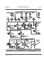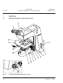
1
Operating Manual
Axioskop 40 / Axioskop 40 FL
Routine microscope
INTRODUCTION
Copyright
Carl Zeiss
Axioskop 40
Axioskop 40 FL
Knowledge of this manual is required for the operation of the instrument. Would you therefore please
make yourself familiar with the contents of this manual and pay special attention to hints concerning
safe operation of the instrument.
The specifications are subject to change; the manual is not covered by an update service.
©
Unless expressly authorized, forwarding and duplication of this document and the software
package, as well as utilization and communication of its contents are not permitted. Violations will
entail an obligation to pay compensation.
All rights reserved in the event of granting of patents or registration of a utility model.
Issued by:
Carl Zeiss
Light Microscopy
P.O.B. 4041
D-37030 Goettingen
GERMANY
Phone:
++49 551 5060 660
Telefax:
++49 551 5060 464
Internet:
www.zeiss.de/micro
E-Mail:
[email protected]
Number of this manual: B 40-810 e
Date of issue: 12/01/2001
0-2
B 40-810 e 12/01
Axioskop 40
Axioskop 40 FL
INTRODUCTION
Contents
Carl Zeiss
CONTENTS
Page
INTRODUCTION ........................................................................................................ 0-2
Copyright ................................................................................................................... 0-2
Contents .................................................................................................................... 0-3
Microscopy in a few steps ........................................................................................... 0-7
Notes on instrument safety ......................................................................................... 0-8
Notes on warranty .................................................................................................... 0-11
Overall view of Axioskop 40 ...................................................................................... 0-12
Overall view of Axioskop 40 FL.................................................................................. 0-13
CHAPTER 1
INSTRUMENT DESCRIPTION ..................................................................................... 1-2
1.1
Name and intended application................................................................................... 1-2
1.2
Instrument description and main features.................................................................... 1-2
1.3
System Overview Axioskop 40 / 40 FL.......................................................................... 1-4
1.4
Objectives................................................................................................................... 1-8
1.5
Eyepieces.................................................................................................................. 1-10
1.6
Stage micrometers and eyepiece reticules.................................................................. 1-11
1.7
Technical data .......................................................................................................... 1-13
B 40-810 e 12/01
0-3
Carl Zeiss
INTRODUCTION
Contents
Axioskop 40
Axioskop 40 FL
Page
CHAPTER 2
START-UP ..................................................................................................................2-4
2.1
2.1.1
2.1.2
2.1.3
2.1.4
2.1.5
2.1.6
2.1.7
2.1.8
2.1.9
2.1.10
2.1.11
2.1.12
2.1.13
Initial start-up............................................................................................................. 2-4
Unpacking and installing the microscope......................................................................2-4
Attaching binocular tube or phototube ........................................................................2-5
Inserting eyepieces and auxiliary microscope ................................................................2-5
Setting interpupillary distance of the binocular tube .....................................................2-7
Setting the viewing height ...........................................................................................2-7
Mounting objectives ....................................................................................................2-8
Inserting the refill set in the specimen marker...............................................................2-9
Attaching the universal condenser .............................................................................2-10
Connecting to power ................................................................................................2-11
Switching the microscope and HBO 100 W transformer on and off ............................2-11
Inserting and replacing the 12 V, 35 W halogen lamp ................................................2-12
HBO 103 illuminator (Axioskop 40 FL) ........................................................................2-13
HBO 50 fluorescence illuminator (Axioskop 40 FL) ......................................................2-17
2.2
2.2.1
2.2.2
2.2.3
2.2.4
2.2.5
2.2.6
2.2.7
2.2.8
2.2.9
2.2.10
2.2.11
2.2.12
2.2.13
2.2.14
2.2.15
2.2.16
2.2.17
Attachments and conversions ....................................................................................2-19
Changing the condenser............................................................................................2-19
Inserting stops (Ph, Var) and neutral filter in slider ......................................................2-20
Attaching filter mount ...............................................................................................2-21
Attaching polarizer D.................................................................................................2-21
Attaching overview adapter .......................................................................................2-22
Changing the HBO 103 W/2 mercury vapor short-arc lamp ........................................2-23
Changing the burner for the HBO 50 fluorescence illuminator ....................................2-26
Mechanical stage with torque setting.........................................................................2-28
Changing the mechanical stage .................................................................................2-29
Changing the binocular tube .....................................................................................2-33
Attaching Axioskop connector plate...........................................................................2-34
Installing and removing "Push&Click" modules on the Axioskop 40 FL........................2-35
Changing the filter set in the FL P&C reflector module................................................2-36
Changing the beam splitter in the FL P&C reflector module ........................................2-38
Attaching the active co-observation equipment for 2 users .........................................2-40
Attaching the multi-observation equipment ...............................................................2-42
Attaching the intermediate tube for image projection ................................................2-44
0-4
B 40-810 e 12/01
Axioskop 40
Axioskop 40 FL
INTRODUCTION
Contents
Carl Zeiss
Page
CHAPTER 3
OPERATION .............................................................................................................. 3-4
3.1
Axioskop 40 operation and function controls............................................................... 3-4
3.2
Axioskop 40 FL operation and function controls ........................................................ 3-10
3.3
Operation controls for accessory components............................................................ 3-16
3.4
Switching Axioskop 40 and Axioskop 40 FL on and off .............................................. 3-20
3.5
3.5.1
3.5.2
3.5.3
3.5.4
3.5.5
3.5.6
3.5.7
Illumination and contrast procedures......................................................................... 3-21
Setting transmitted-light bright field according to KÖHLER......................................... 3-21
Setting transmitted-light phase contrast .................................................................... 3-24
Setting transmitted-light VAREL contrast ................................................................... 3-26
Setting transmitted-light dark field ............................................................................ 3-27
Setting transmitted-light oblique illumination ............................................................ 3-29
Setting transmitted-light polarization......................................................................... 3-30
Setting epi-fluorescence............................................................................................ 3-34
3.6
3.6.1
3.6.2
3.6.3
3.6.4
3.6.5
3.6.6
3.6.7
Documentation......................................................................................................... 3-36
Attaching photomicrography equipment ................................................................... 3-36
Attaching adapters for video cameras........................................................................ 3-40
Attaching adapters for compact digital cameras ........................................................ 3-42
Digital cameras with 37 mm filter thread, e.g. SONY DCR-PC100 / SONY DSC-S50 .... 3-43
Digital cameras with 52 mm filter thread, e.g. SONY DSC-S70, DSC-S75, DSC-S85 .... 3-44
Adapting cameras with different filter thread sizes .................................................... 3-46
Sony DSC-S70, DC-S75, DSC-S85 on digital camera adapter 44 M52x0.75 ................ 3-46
3.7
3.7.1
3.7.2
Multiple observer equipment..................................................................................... 3-47
Using active co-observation equipment for 2 users .................................................... 3-47
Using multi-observation equipment ........................................................................... 3-49
3.8
3.8.1
Drawing device......................................................................................................... 3-51
Attaching switchable intermediate tube for image projection..................................... 3-51
3.9
Measuring lengths .................................................................................................... 3-52
B 40-810 e 12/01
0-5
INTRODUCTION
Contents
Carl Zeiss
Axioskop 40
Axioskop 40 FL
Page
CHAPTER 4
CARE, MAINTENANCE, TROUBLESHOOTING AND SERVICE ....................................4-2
4.1
Instrument care .......................................................................................................... 4-2
4.2
4.2.1
4.2.2
Instrument maintenance ............................................................................................. 4-3
Performing checks .......................................................................................................4-3
Changing fuses ...........................................................................................................4-3
4.3
Troubleshooting ......................................................................................................... 4-4
4.4
Spares, consumables and tools ................................................................................... 4-8
4.5
Requesting service ...................................................................................................... 4-9
APPENDIX
List of abbreviations.................................................................................................... A-2
Index.......................................................................................................................... A-4
Certification in accordance with DIN ISO 9001 / EN 46001
EC conformity declaration
0-6
B 40-810 e 12/01
Axioskop 40
Axioskop 40 FL
INTRODUCTION
Microscopy in a few steps ...
Carl Zeiss
Microscopy in a few steps using the example of "Setting of transmitted-light brightfield
according to KÖHLER"
☞
Please read the instructions regarding instrument safety and the Instrument Description
(Chapter 1) and Start-Up (Chapter 2) chapters before using the Axioskop 40 or Axioskop 40 FL
microscopes for the first time.
(1)
As described in Chapter 2 (Start-Up), the microscope is ready for operation and switched on
according to Section 3.4.
(2)
Place a high-contrast specimen on the mechanical stage (cover slip pointing up).
(3)
Swivel in 10x objective on the nosepiece and focus on the specimen; where possible, always
move the specimen away from the objective!
(4)
Move the condenser - e.g. condenser 0.9/1.25 H - to the upper stop position via the
condenser drive. The height adjustment must be preset in such a manner that the specimen is
not moved by the condenser. If the 0.9/1.25 H condenser is equipped with the slider for
PH/H/Var, place it in the middle position for bright field.
(5)
Close the luminous-field diaphragm until it is visible in the field of view, even if not in focus
(Fig. 0-1/A).
(6)
Lower condenser 0.9/1.25 H until the edge of the luminous-field diaphragm appears in focus
(Fig. 0-1/B).
(7)
Center the luminous-field diaphragm using centering screws on the condenser carrier (Fig. 01/C) and open it until its edge just disappears behind the field of view (Fig. 0-1/D).
(8)
To set the aperture diaphragm (contrast), remove one eyepiece from the binocular tube and
set the aperture diaphragm to approx. 2/3 of the diameter of the objective exit pupil
(Fig. 0-1/E). Optimum contrast setting is dependent on the specimen used.
(9)
Insert the eyepiece again and, where necessary, refocus on the specimen via the fine drive.
(10)
After each objective change, the field size and objective aperture change, which makes it
necessary to repeat steps (5) to (9).
Fig. 0-1
Diaphragm setting in transmittedlight bright field according to
KÖHLER
B 40-810 e 12/01
0-7
INTRODUCTION
Notes on instrument safety
Carl Zeiss
Axioskop 40
Axioskop 40 FL
Notes on instrument safety
The Axioskop 40 and Axioskop 40 FL microscopes have been designed, produced and tested in
compliance with DIN 61010-1 (IEC 1010-1) safety requirements for electrical measuring, control and
laboratory instruments, and meet the requirements of Appendix I of directive 73/23/EC.
The instruments meet the requirements of EC directive 89/336/EC and EMC legislation dated September
18, 1998. This operating manual includes information and warnings that must be observed by the user.
The following warning and information symbols are used in this manual:
☞
NOTE
This symbol is a warning which you must observe under all circumstances.
CAUTION
This symbol is a warning which indicates a hazard to the instrument or instrument system.
CAUTION
This symbol is a warning which indicates a hazard to the user of the instrument.
CAUTION
Hot surface!
CAUTION
UV radiation is emitted!
CAUTION
Disconnect the instrument from the line before opening it!
0-8
B 40-810 e 12/01
Axioskop 40
Axioskop 40 FL
INTRODUCTION
Notes on instrument safety
Carl Zeiss
The Axioskop 40 and Axioskop 40 FL microscopes, including original accessories, may only be used for
the microscope techniques described in this manual.
Particular attention must be paid to the following warnings:
The manufacturer cannot assume any liability for any other applications, including individual
modules or single parts. This also applies to all service or repair work that is not carried out by
authorized service personnel. All warranty claims shall be forfeited.
The power plug must be inserted in an outlet featuring a grounding (earth) contact. The
grounding effect must not be made ineffective by an extension cable that does not have a
protective ground wire.
If it is determined that protection measures are no longer effective, the instrument must be
switched off and safeguarded against inadvertent operation. Please contact a Zeiss service
agency or the Carl Zeiss Microscopy Service to repair the instrument.
☞
The power supply integrated in the stands of the microscopes enables line voltages to be used
in the range between 100 and 240 V ±10 %, 50 / 60 Hz, without the voltage on the
instrument having to be changed.
The transformers for the HBO 103 (ebq 100 dc) or XBO 75 (ebx 75 isolated) are designed for
the 100 to 240 V AC, 50 to 60 Hz range. They adapt automatically to the available line voltage.
There is no need to change the voltage.
However, the transformer for the HBO 50 must be set manually to the correct voltage (100,
110, 120, 127 or 230, 240 V), and the correct frequency must be preselected (50 or 60 Hz).
Before switching on the instrument, check as to whether it is suitable for the given line voltage.
Always disconnect the instrument from the power outlet before opening the instrument and
before changing the fuses.
Take sure to ensure you only use fuses of the rated power required. Use of makeshift fuses and
short-circuiting of the fuse holders are not permitted.
B 40-810 e 12/01
0-9
Carl Zeiss
INTRODUCTION
Notes on instrument safety
Axioskop 40
Axioskop 40 FL
The Axioskop 40 and Axioskop 40 FL microscopes are not equipped with any special devices for
protection from substances that are corrosive, toxic, radioactive or other substances that could
be hazardous to health. All the legal regulations, particularly the pertinent accident prevention
regulations of the respective country, must be observed when handling such substances.
Gas-discharge lamps, e.g. HBO 50 or HBO 103, emit ultraviolet radiation, which can cause
burns to the eyes and skin. Therefore, never look directly into the light of these lamps and
avoid direct, unprotected incidence of their light on your skin. When using the microscope,
always use the protective devices belonging to the instrument (e.g. special attenuation filters or
the fluorescence protection shield). When they are hot, gas-discharge lamps are under high
internal pressure and may therefore only be changed when they have cooled down, and
protective gloves and goggles must be used (for detailed information please see Operating
Manual B 40-065 e).
When fluorescence filters are used, the heat protection filter for heat emitted by the
microscope illuminator must not be removed, since fluorescence filters are sensitive to heat,
and their performance could be impaired.
Avoid touching the hot lamp housing. Always pull the power plug before changing the lamps
and allow the instrument to cool down for some 15 minutes.
Dust and dirt may impair the instrument’s performance. Therefore, the instrument must be
protected from these influences as far as possible and covered with the dust cover when not in
use. Always check whether the instrument is switched off before you cover it.
Placing objects against or covering ventilation slats may lead to heat build-up that will damage
the instrument and, in extreme cases, cause a fire. Always keep the ventilation slats clear and
ensure that no objects enter the instrument through the ventilation slats.
The instruments may only be operated by trained personnel who must be aware of the possible
dangers involved with microscopy and the particular application concerned. The Axioskop 40 /
Axioskop 40 FL is a high-precision instrument that can be impaired in its performance or
destroyed when handled improperly.
0-10
B 40-810 e 12/01
Axioskop 40
Axioskop 40 FL
INTRODUCTION
Notes on warranty
Carl Zeiss
Notes on warranty
The manufacturer guarantees that the instrument has no material or production defects when delivered.
You must inform us of any defects immediately and do everything to minimize any damage. If the
manufacturer is informed of such a defect, he is obligated to rectify it; it is his decision whether he does
this by repairing the instrument or by delivering an instrument free of any defect. No guarantee is
provided for defects caused by natural wear (wearing parts and consumables in particular) and improper
use.
The instrument manufacturer is not liable for damage caused by faulty operation, negligence or any
other tampering with the instrument, particularly the removal or replacement of instrument
components, or the use of accessories from other manufacturers. This forfeits all warranty claims.
With the exception of the work specified in this manual, no maintenance or repair of the Axioskop 40 /
Axioskop 40 FL may be undertaken. Repairs may only be performed by Zeiss service staff or specially
authorized personnel. Should any defect occur with the instrument, please get in touch with your local
Zeiss representative.
B 40-810 e 12/01
0-11
Carl Zeiss
INTRODUCTION
Overall view of Axioskop 40
Axioskop 40
Axioskop 40 FL
Overall view of Axioskop 40
1
2
3
4
5
6
7
0-12
Eyepieces
Binocular tube
Stand
Transmitted-light illuminator
Condenser 0.9/1.25 H
Mechanical stage
Objective
B 40-810 e 12/01
Axioskop 40
Axioskop 40 FL
INTRODUCTION
Overall view of Axioskop 40 FL
Carl Zeiss
Overall view of Axioskop 40 FL
1
2
3
4
5
6
7
8
Eyepieces
Ergo-tube 20°/23 with height adjustment
Stand
Incident-light illuminator
Transmitted-light illuminator
Universal condenser 0.9
Mechanical stage
Objective
B 40-810 e 12/01
0-13
INTRODUCTION
Carl Zeiss
0-14
Axioskop 40
Axioskop 40 FL
B 40-810 e 12/01
Axioskop 40
Axioskop 40 FL
INSTRUMENT DESCRIPTION
Table of contents / List of illustrations
Carl Zeiss
INSTRUMENT DESCRIPTION
Table of contents
1
1.1
1.2
1.3
1.4
1.5
1.6
1.7
INSTRUMENT DESCRIPTION.......................................................................................... 1-2
Name and intended application ....................................................................................... 1-2
Instrument description and main features ........................................................................ 1-2
System Overview Axioskop 40 / 40 FL .............................................................................. 1-4
Objectives ....................................................................................................................... 1-8
Eyepieces ...................................................................................................................... 1-10
Stage micrometers and eyepiece reticules ..................................................................... 1-11
Technical data ............................................................................................................... 1-13
List of illustrations
Fig. 1-1
Objective......................................................................................................................... 1-8
B 40-810 e 12/01
1-1
INSTRUMENT DESCRIPTION
Carl Zeiss
Name ... / Instrument description ...
1
INSTRUMENT DESCRIPTION
1.1
Name and intended application
Axioskop 40
Axioskop 40 FL
Manufacturer's designation: Axioskop 40 microscope
Axioskop 40 FL microscope
The Axioskop 40 and Axioskop 40 FL microscopes have been designed as universal microscopes for
biomedical applications. They can be used as pure transmitted-light microscopes or, with epifluorescence equipment, as combined transmitted-light / reflected-light microscopes
The Axioskop 40 and Axioskop 40 FL microscopes are mainly used in:
− hospitals
− doctor's offices
− universities and in
− industry
1.2
Instrument description and main features
With the advanced pyramid and modular design, the Axioskop 40/40 FL incorporates time-tested
principles in microscope construction, thus ideally combining modern requirements made on design,
ergonomics, operating convenience and function with technical performance. Due to the modular
design, existing microscope modules from the Axioskop 2 plus and Axioplan 2 imaging, such as tubes,
intermediate tubes and condensers, are easy to attach. A connector plate must be mounted for Axioplan
2 imaging modules.
Depending on the instrument configuration, the following microscopy and contrasting techniques are
possible:
Transmitted light:
Reflected light:
- bright field
- fluorescence contrast
- dark field
- phase contrast
- VAREL contrast
- polarization contrast
The Axioskop 40 FL stand includes epi-fluorescence equipment. The optional adjustment guide is
integrated into the microscope stand in the factory when ordered. For technical reasons, retrofitting
existing transmitted-light instruments with the epi-fluorescence equipment should be performed by Zeiss
service staff, since the nosepiece must also be retrofitted.
The binocular phototubes and suitable adapters enable one microscope camera, one reflex camera or
one digital / video camera to be attached for documentation purposes.
1-2
B 40-810 e 12/01
Axioskop 40
Axioskop 40 FL
INSTRUMENT DESCRIPTION
Instrument description and main features
Carl Zeiss
Major instrument features of the Axioskop 40:
− Stable and sturdy diecast stand with T-shaped base in pyramid design and integrated power supply
for the 12 V, 35 W halogen lamp
− Stage focusing via coaxial rough / fine drive
− Fixed and rotary mechanical stage with ceramic-coated surface and variable drive length
− Installed 12 V, 35 W lamp for transmitted light and HBO 50 or HBO 103 for reflected light
− Condenser 0.9/1.25 H for bright-field applications as well as for phase and variable contrast when
used in combination with a separate slider for Ph / Var
− Ball-bearing, 6-position nosepiece with W 0.8" thread
− ICS objectives in the cost-performance categories A-Plan, Achroplan, Plan-Neofluar, Plan-Apochromat
− Binocular tubes and phototubes with fixed and variable viewing angle as well as ergo-tubes and ergophototubes with infinitely variable height adjustment
− Adjustable 10x focusing eyepieces with field-of-view number 23, suitable for eyeglass wearers
− Wide variety of attachment possibilities for photography and video-microscopy
− Multi-observation equipment for 2 or 9 viewing stations
Additional features of the Axioskop 40 FL:
−
High-performance epi-fluorescence equipment
−
6-position nosepiece with W 0.8" thread and 5-position reflector turret
B 40-810 e 12/01
1-3
INSTRUMENT DESCRIPTION
Carl Zeiss
1.3
1-4
System overview Axioskop 40 / 40 FL
Axioskop 40
Axioskop 40 FL
System Overview Axioskop 40 / 40 FL
B 40-810 e 12/01
Axioskop 40
Axioskop 40 FL
B 40-810 e 12/01
INSTRUMENT DESCRIPTION
System overview Axioskop 40 / 40 FL
Carl Zeiss
1-5
INSTRUMENT DESCRIPTION
Carl Zeiss
1-6
System overview Axioskop 40 / 40 FL
Axioskop 40
Axioskop 40 FL
B 40-810 e 12/01
Axioskop 40
Axioskop 40 FL
B 40-810 e 12/01
INSTRUMENT DESCRIPTION
System overview Axioskop 40 / 40 FL
Carl Zeiss
1-7
INSTRUMENT DESCRIPTION
Carl Zeiss
Axioskop 40
Axioskop 40 FL
Objectives
1.4
Objectives
The objectives are the optical heart of the
microscope. The following is an example of how
objectives can be labelled:
Achroplan 10x/0.25 ∞/0.17
where
10x
: objective magnification, with a defined
color ring on the objective being allocated
to each magnification step (Zeiss color
code)
0.25 : numerical aperture
∞
Fig. 1-1
: infinite image distance; these objectives
can only be used with ICS microscopes
from Carl Zeiss
0.17 : can only be used with cover slip thickness
D = 0.17 mm
or
Objective
−
: can be used with cover slip thickness D = 0
or 0.17 mm
Other labels:
Oil : oil immersion objective
Ph 2 : phase contrast objective with a green color ring and phase stop Ph 2
Color ring code for objective magnification:
Color ring on
objective
black
brown
red
orange
yellow
green
light blue dark blue white
Magnification
factor
1.25x
2.5x
4x; 5x
6.3x
10x
16x;20x;
25x;32x
40x; 50x
63x
100x;
150x
Objective magnification multiplied by eyepiece magnification (usually 10x) results in overall visual
magnification: e.g. 10 x 10 = 100x.
When working with the microscopes, total magnification should not drop below or exceed the range of
useful magnification. The range of useful magnification was defined by Ernst ABBE as 500 to 1,000
times the numerical aperture of the objective used . There is no resolution for further details above that
limit. Accordingly, the range of useful magnification for an objective with a numerical aperture of 0.25 is
between 125x and 250x.
Exact observance of the cover slip thickness of 0.17 mm is all the more necessary the higher the
numerical aperture of the objective. For this reason, certain objectives can be set to different cover slip
thicknesses (due to correction mount). To this end, a specimen area is searched for, and the position of
the correction ring where optimum focus and image contrast are obtained is determined (refocusing is
invariably required).
1-8
B 40-810 e 12/01
INSTRUMENT DESCRIPTION
Axioskop 40
Axioskop 40 FL
Objectives
Carl Zeiss
When immersion objectives are used, the air between the cover slip and the objective is replaced by a
liquid, which in most cases is immersion oil . The plastic oiler containing 20 ml of 581 N immersion oil
(nD = 1.515) is particularly suitable for this purpose.
To prevent oil contamination of the specimen when the nosepiece is turned, the resilient mounts of the
immersion objectives can be locked in their lifted position by turning them clockwise (do not forget to
unlock them again!).
The following objectives are ideal for the Axioskop 40 and Axioskop 40 FL microscopes:
Microscopy technique
Magnification /
Num. Aperture
Free working
distance in mm
Cover slip
thickness D in mm
Cat. No
A-Plan
A-Plan
A-Plan
A-Plan
A-Plan
A-Plan
A-Plan
2.5x/0.06
5x/0.12
10x/0.25
20x/0.45
40x/0.65
100x/1.25 Oil
9.4
9.9
4.4
0.53
0.43
0.22
0.17
0.17
0.17
000000-1113-114
441020-0000-000
441030-0000-000
441040-0000-000
441050-0000-000
441080-0000-000
Phase contrast
A-Plan
A-Plan
A-Plan
A-Plan
10x/0.25 Ph 1
20x/0.45 Ph 2
40x/0.65 Ph 2
100x/1.25 Oil Ph 3
4.4
0.53
0.43
0.22
0.17
0.17
0.17
441031-0000-000
441041-0000-000
441051-0000-000
441081-0000-000
VAREL contrast
A-Plan
A-Plan
10x/0.25 Ph 1 Var 1
40x/0.65 Ph 2 Var 2
4.4
0.43
0.17
000000-1020-863
000000-1115-207
Achroplan
Achroplan
Achroplan
Achroplan
Achroplan
Achroplan
Achroplan
Achroplan
Achroplan
Achroplan
4x/0.10
10x/0.25
20x/0.45
40x/0.65
50x/0.90 Oil
63x/0.80
63x/0.95
100x/1.25 Oil
100x/1.25 Oil Iris
11.1
4.8
2.07
0.59
0.29
0.29
0.15
0.19
0.19
0.17
0.17
0.17
0.17
0 (no cover slip)
0.17
0.17
440020-0000-000
440030-0000-000
440040-0000-000
440050-0000-000
440057-0000-000
440060-0000-000
440068-0000-000
440080-0000-000
440086-0000-000
Achroplan
Achroplan
Achroplan
Achroplan
10x/0.25 Ph 1
20x/0.45 Ph 2
40x/0.65 Ph 2
100x/1.25 Oil Ph 3
4.8
2.07
0.59
0.19
0.17
0.17
0.17
440031-0000-000
440041-0000-000
440051-0000-000
440081-0000-000
Plan-Neofluar
Plan-Neofluar
Plan-Neofluar
Plan-Neofluar
Plan-Neofluar
Plan-Neofluar
Plan-Neofluar
2.5x/0.075
5x/0.15
10x/0.30
20x/0.50
40x/0.75
100x/1.30 Oil
9.3
13.6
5.6
1.3
0.47
0.06
0.17
0.17
0.17
0.17
440310-0000-000
440320-0000-000
440330-0000-000
000000-1004-072
440350-9902-000
000000-1018-595
Transmitted-light bright field
Transmitted-light bright field
Phase contrast
Transmitted-light bright field
B 40-810 e 12/01
Objective
1-9
INSTRUMENT DESCRIPTION
Carl Zeiss
Microscopy technique
Phase contrast
Transmitted-light bright field
1.5
Axioskop 40
Axioskop 40 FL
Objectives / Eyepieces
Objective
Magnification /
Num. Aperture
Free working
distance in mm
Cover slip
thickness D in mm
Cat. No
Plan-Neofluar
Plan-Neofluar
Plan-Neofluar
Plan-Neofluar
Plan-Neofluar
10x/0.30 Ph 1
20x/0.50 Ph 2
40x/0.75 Ph 2
100x/1.30 Oil Ph 3
5.6
1.3
0.47
0.06
0.17
0.17
0.17
0.17
440331-0000-000
000000-1004-989
440351-9902-000
000000-1031-171
Plan-Apochromat
Plan-Apochromat
Plan-Apochromat
Plan-Apochromat
40x/1.00 Oil Iris
63x/1.40 Oil
100x/1.40 Oil
0.31
0.09
0.09
0.17
0.17
0.17
440756-0000-000
440760-0000-000
440780-0000-000
Eyepieces
The following eyepieces are offered for the Axioskop 40 and Axioskop 40 FL:
Eyepiece
Image angle
Application
Cat. No.
Eyepiece W-PL 10x/23 Br. foc. (2x)
49.4°
with all tubes, see Pp. 1-4 ff.;
recommended for ergo-tubes
(000000-1104-293 / 296)
455043-0000-000,
aspheric
Eyepiece PL 10x/23 Br. foc. (2x)
49.4°
with all tubes, see Pp. 1-4 ff.;
recommended for ergonomic
binocular phototube
(452342-0000-000)
000000-1026-548
Eyepiece PL 16x/16 Br.
54°
with all tubes, see Pp. 1-4 ff.
444053-0000-000
Eyepiece PL 16x/16 Br. foc.
54°
with all tubes, see Pp. 1-4 ff.
444054-0000-000
W-PL and PL in the eyepiece designation refers to the excellent image flatness up to the edge of the field
of view. If required, eyecups for the eyepieces can be ordered under Cat. No. 444801-0000-000.
1-10
B 40-810 e 12/01
Axioskop 40
Axioskop 40 FL
1.6
INSTRUMENT DESCRIPTION
Stage micrometers and eyepiece reticules
Carl Zeiss
Stage micrometers and eyepiece reticules
Measuring and counting using a microscope requires stage micrometers and eyepiece reticules, a
selection of which is listed below:
Illustration
B 40-810 e 12/01
Designation, technical data
Cat. No.
Stage micrometer, positive 5 + 100/100 y
D = 0.17 mm
gradation on the +y-axis: 5 mm in 5 intervals
gradation on the -y-axis: 1 mm in 100 intervals
with 2 opposing scales = 10 µm, accuracy ±1µm
474026-0000-000
Crossline micrometer 14:140, d = 26 mm
gradation length = 14 mm
increments = 0.1mm
gradation tolerance ≤ 0.001 mm
454060-0000-000
Eyepiece reticule / d = 26 mm
474064-0000-000
Crossline micrometer 10:100, d = 26 mm
gradation length = 10 mm
increments = 0.1mm
gradation tolerance ≤ 0.001 mm
474066-9901-000
1-11
INSTRUMENT DESCRIPTION
Carl Zeiss
Stage micrometers and eyepiece reticules
Illustration
Axioskop 40
Axioskop 40 FL
Designation, technical data
Cat. No.
Net micrometer 12.5x12.5/5;10 / d = 26 mm
area 12.5x12.5 mm, divided into 10x10 fields
474068-0000-000
Photo reticule MC 2.5x / d = 26 mm
454075-0000-000
for 35 mm photography with 2.5x additional
magnification or for large-format photography
with 10x additional magnification.
☞
If an eyepiece reticule is used, the binocular tube or the phototube must be equipped with two
eyepieces with an adjustable lens, into one of which the eyepiece reticule is mounted.
1-12
B 40-810 e 12/01
Axioskop 40
Axioskop 40 FL
1.7
INSTRUMENT DESCRIPTION
Technical data
Carl Zeiss
Technical data
Dimensions (width x depth x height)
Axioskop 40 stand........................................................................ approx. 280 mm x 560 mm x 505 mm
Axioskop 40 FL stand with HBO 100
and adjustable ergo-tube 20°/23 in highest setting........................ approx. 280 mm x 730 mm x 555 mm
Weight
Axioskop 40 ...................................................................................................................... approx. 15 kg
Axioskop 40 FL with HBO 100......................................................................................... approx. 19.5 kg
Ambient conditions
Storage and transport (in packaging)
Permissible ambient temperature ...................................................................................... -10 to + 50 °C
Permissible relative humidity ............................................................................. 20 % to 75 % at +35 °C
Atmospheric pressure ............................................................................................. 800 hPa to 1060 hPa
Operation
Permissible ambient temperature ..................................................................................... +10 to + 35 °C
Permissible relative humidity ............................................................................. 20 % to 75 % at +35 °C
Atmospheric pressure ............................................................................................. 800 hPa to 1060 hPa
Altitude.............................................................................................................................. max. 2000 m
Pollution degree ................................................................................................................................... 2
Operating data
Operating environment.......................................................................................................... clean room
Protection class.......................................................................................................................................I
Protection type ................................................................................................................................IP 20
Electrical safety............................................................. in compliance with DIN EN 61010-1 (IEC 1010-1)
including CSA and UL directives
Overvoltage category.............................................................................................................................II
Radio interference suppression....................................................... in accordance with EN 55011, Class B
Insensitivity to interference ......................................................................in accordance with EN 50082-2
Line voltage.............................................................................................................100 to 240 V ±10 %
.......................................................................................................... Change of voltage is not required!
Line frequency...................................................................................................................... 50 to 60 Hz
Power consumption of Axioskop 40 / Axioskop 40 FL ................................................................... 100 VA
B 40-810 e 12/01
1-13
INSTRUMENT DESCRIPTION
Carl Zeiss
Technical data
Axioskop 40
Axioskop 40 FL
HBO 50 transformer mbq52ac-z
Operating environment ..........................................................................................................clean room
Protection class ...................................................................................................................................... I
Protection type................................................................................................................................ IP 20
Line voltage can be changed between ................................. 100, 110, 120, 127 VAC and 230, 240 VAC
Line frequency can be changed between ............................................................................ 50 and 60 Hz
Power consumption when HBO 50 is used........................................................................... max. 350 VA
HBO 100 W (HBO 100 W/2 and HBO 103 W/2) transformer
Operating environment ..........................................................................................................clean room
Protection class ...................................................................................................................................... I
Protection type................................................................................................................................ IP 20
Line voltage ...........................................................................................................100 VAC …240 VAC
Line frequency ..................................................................................................................... 50 … 60 Hz
Power consumption when HBO 103 is used ................................................................................. 260 VA
Fuses in accordance with IEC 127
Axioskop 40 microscope stand ....................................................................................T 1 A/H, 5x20 mm
Axioskop 40 FL microscope stand ................................................................................T 1 A/H, 5x20 mm
HBO 50AC transformer mbq52ac-z ...................................................................... 100 V, 127 V: 2x T 4 A
...................................................................................................................... 220 V – 240 V: 2x T 2.5 A
HBO 100 W transformer......................................................................................... T 3.15 A/H, 5x20 mm
Light sources
Halogen lamp ...................................................................................................................... 12 V / 35 W
Adjustment of the light source ......................................................................continuous, 3 to 12 V
Mercury vapor short-arc lamp...................................................................................................... HBO 50
Power consumption for HBO 50 ............................................................................................ 50 W
Mercury vapor short-arc lamp.............................................................................................HBO 103 W/2
Power consumption for HBO 103 W/2................................................................................. 100 W
1-14
B 40-810 e 12/01
Axioskop 40
Axioskop 40 FL
INSTRUMENT DESCRIPTION
Technical data
Carl Zeiss
Axioskop 40 / Axioskop 40 FL
Stand with manual stage focusing
rough drive .............................................................................................. approx. 5 mm / rotation
fine drive........................................................ approx. 0.5 mm / rotation; approx. 5 µm increments
lifting range ........................................................................................................... approx. 24 mm
height stop ................................................................................................can be set mechanically
Achromatic-aplanatic universal condenser 0.9 with
swivel front lens 0.9;
for objective magnifications Vobj. < 10 .....................................................swivel out front lens 0.9
for objective magnifications Vobj. ≥ 10x ..................................................... swivel in front lens 0.9
bright-field insert or turret disk with positions ............................................ for brightfield, darkfield
............................................................................................................... for phase contrast 1, 2, 3
Objective change................................................................. manually via 6-position nosepiece H, W 0.8"
Change of reflector module (only with Axioskop 40 FL) ................. manually via 5-position reflector turret
B 40-810 e 12/01
1-15
INSTRUMENT DESCRIPTION
Carl Zeiss
1-16
Axioskop 40
Axioskop 40 FL
B 40-810 e 12/01
Axioskop 40
Axioskop 40 FL
START-UP
Table of contents
Carl Zeiss
START-UP
Table of contents
2
START-UP ...................................................................................................................... 2-4
2.1
2.1.1
2.1.2
2.1.3
2.1.4
2.1.5
2.1.6
2.1.7
2.1.8
2.1.9
2.1.10
2.1.11
2.1.12
2.1.13
Initial start-up.................................................................................................................. 2-4
Unpacking and installing the microscope ......................................................................... 2-4
Attaching binocular tube or phototube ............................................................................ 2-5
Inserting eyepieces and auxiliary microscope .................................................................... 2-5
Setting interpupillary distance of the binocular tube ......................................................... 2-7
Setting the viewing height ............................................................................................... 2-7
Mounting objectives ........................................................................................................ 2-8
Inserting the refill set in the specimen marker .................................................................. 2-9
Attaching the universal condenser ................................................................................. 2-10
Connecting to power .................................................................................................... 2-11
Switching the microscope and HBO 100 W transformer on and off ................................ 2-11
Inserting and replacing the 12 V, 35 W halogen lamp .................................................... 2-12
HBO 103 illuminator (Axioskop 40 FL) ............................................................................ 2-13
HBO 50 fluorescence illuminator (Axioskop 40 FL) .......................................................... 2-17
2.2
2.2.1
2.2.2
2.2.3
2.2.4
2.2.5
2.2.6
2.2.7
2.2.8
2.2.9
2.2.10
2.2.11
2.2.12
2.2.13
2.2.14
2.2.15
2.2.16
2.2.17
Attachments and conversions ........................................................................................ 2-19
Changing the condenser ............................................................................................... 2-19
Inserting stops (Ph, Var) and neutral filter in slider .......................................................... 2-20
Attaching filter mount ................................................................................................... 2-21
Attaching polarizer D..................................................................................................... 2-21
Attaching overview adapter ........................................................................................... 2-22
Changing the HBO 103 W/2 mercury vapor short-arc lamp ............................................ 2-23
Changing the burner for the HBO 50 fluorescence illuminator ........................................ 2-26
Mechanical stage with torque setting ............................................................................ 2-28
Changing the mechanical stage ..................................................................................... 2-29
Changing the binocular tube ......................................................................................... 2-33
Attaching Axioskop connector plate............................................................................... 2-34
Installing and removing "Push&Click" modules on the Axioskop 40 FL ........................... 2-35
Changing the filter set in the FL P&C reflector module ................................................... 2-36
Changing the beam splitter in the FL P&C reflector module ............................................ 2-38
Attaching the active co-observation equipment for 2 users............................................. 2-40
Attaching the multi-observation equipment ................................................................... 2-42
Attaching the intermediate tube for image projection .................................................... 2-44
B 40-810 e 12/01
2-1
START-UP
List of illustrations
Carl Zeiss
Axioskop 40
Axioskop 40 FL
List of illustrations
Fig. 2-1
Fig. 2-2
Fig. 2-3
Fig. 2-4
Fig. 2-5
Fig. 2-6
Fig. 2-7
Fig. 2-8
Fig. 2-9
Fig. 2-10
Fig. 2-11
Fig. 2-12
Fig. 2-13
Fig. 2-14
Fig. 2-15
Fig. 2-16
Fig. 2-17
Fig. 2-18
Fig. 2-19
Fig. 2-20
Fig. 2-21
Fig. 2-22
Fig. 2-23
Fig. 2-24
Fig. 2-25
Fig. 2-26
Fig. 2-27
Fig. 2-28
Fig. 2-29
Fig. 2-30
Fig. 2-31
Fig. 2-32
Fig. 2-33
Fig. 2-34
Fig. 2-35
Fig. 2-36
Fig. 2-37
Fig. 2-38
Fig. 2-39
Fig. 2-40
Fig. 2-41
2-2
Setting up the microscope............................................................................................... 2-4
Attaching the binocular tube........................................................................................... 2-5
Inserting eyepieces .......................................................................................................... 2-5
Inserting the eyepiece reticule ......................................................................................... 2-6
Setting the interpupillary distance of the binocular tube................................................... 2-7
Setting the viewing height of the binocular tube.............................................................. 2-7
Mounting objectives........................................................................................................ 2-8
Inserting the refill set....................................................................................................... 2-9
Attaching the universal condenser ..................................................................................2-10
Axioskop 40...................................................................................................................2-11
HBO 100 W transformer (front and back) .......................................................................2-11
Removing the lamp housing ...........................................................................................2-12
Inserting the 12 V, 35 W halogen lamp ..........................................................................2-12
Attaching the HBO 103 illuminator.................................................................................2-13
HBO 100 W transformer ................................................................................................2-13
Adjustment guide ..........................................................................................................2-14
Adjusting the HBO 103 ..................................................................................................2-14
HBO 103 focal spot before rough adjustment .................................................................2-15
HBO 103 focal spot after rough adjustment....................................................................2-15
Focal spot image without adjustment guide on mechanical stage....................................2-16
Attaching HBO 50..........................................................................................................2-17
Adjusting HBO 50 ..........................................................................................................2-17
Changing the condenser ................................................................................................2-19
Inserting stops and neutral filter .....................................................................................2-20
Attaching the filter mount..............................................................................................2-21
Attaching polarizer D .....................................................................................................2-21
Attaching overview adapter............................................................................................2-22
HBO 100 W transformer ................................................................................................2-23
Removing HBO 103 illuminator.......................................................................................2-24
Removing HBO 103 housing...........................................................................................2-24
Changing the HBO 103 W/2 mercury vapor short-arc lamp .............................................2-25
Changing the burner......................................................................................................2-26
Setting the torque..........................................................................................................2-28
Changing the fixed mechanical stage .............................................................................2-29
Changing the rotary mechanical stage............................................................................2-29
Centering the rotary mechanical stage............................................................................2-30
Setting the ergo-drive ....................................................................................................2-31
Changing the specimen holder .......................................................................................2-32
Changing the binocular tube..........................................................................................2-33
Attaching Axioskop connector plate ...............................................................................2-34
Changing the FL P&C reflector module ...........................................................................2-35
B 40-810 e 12/01
Axioskop 40
Axioskop 40 FL
Fig. 2-42
Fig. 2-43
Fig. 2-44
Fig. 2-45
Fig. 2-46
Fig. 2-47
Fig. 2-48
Fig. 2-49
START-UP
List of illustrations
Carl Zeiss
Changing the filter set in the FL P&C reflector module ................................................... 2-36
Installing the filter and beam splitter .............................................................................. 2-37
Changing the beam splitter ........................................................................................... 2-38
Changing the beam splitter ........................................................................................... 2-38
Beam splitter designation .............................................................................................. 2-39
Attaching the active co-observation equipment for 2 users............................................. 2-41
Attaching the multi-observation equipment ................................................................... 2-43
Attaching intermediate tube for image projection .......................................................... 2-44
B 40-810 e 12/01
2-3
START-UP
Initial start-up
Carl Zeiss
2
Axioskop 40
Axioskop 40 FL
START-UP
The Axioskop 40 / Axioskop 40 FL can be installed, converted and started up by the customer.
However, it is also possible to have the microscope installed or converted by Zeiss service staff for an
extra charge.
☞
Before installing and starting-up the microscope, be sure to carefully read the notes on
instrument safety.
2.1
Initial start-up
2.1.1
Unpacking and installing the microscope
The basic instrument is supplied in a polyethylene case with cardboard packaging.
It contains the following components: Stand with 12 V, 35 W lamp. The binocular tube, objectives,
eyepieces, condenser, microscope mat and various small parts, such as spare lamp, dust cover and tool
bag and other optional accessories are delivered in a separate package.
The following components are factory-installed to the microscope stand: Mechanical stage, specimen
holder, filter mount or overview adapter and, for the Axioskop 40 FL, epi-fluorescence equipment,
adjustment guide (if ordered) and P&C reflector modules.
• Remove all components from the packaging
and use the delivery form to check for
completeness.
• Place instrument (2-1/1) onto the microscope
mat (2-1/2) on a vibration-free, flat worktable.
The area required by the Axioskop 40
microscope mat is some 500 mm x 400 mm
(width x depth).
• Dispose of original packaging properly, or keep
it for storage or for returning the instrument to
the manufacturer.
Fig. 2-1
2-4
Setting up the microscope
B 40-810 e 12/01
Axioskop 40
Axioskop 40 FL
2.1.2
Attaching
phototube
START-UP
Initial start-up
binocular
tube
Carl Zeiss
or
All the binocular tubes listed in the system
overview (refer to Section 1.3) can be attached to
the Axioskop 40 / Axioskop 40 FL stand as
described below.
• Undo hexagonal screw (2-2/3) using the SW3
ball-headed screwdriver. Remove dust caps
(2-2/2) from the bottom of the tube and the
dovetailed mount (2-2/5) on the stand.
• Hold the binocular tube (2-2/1) or the binocular
phototube in a slightly inclined position and
attach it to the stand mount (2-2/4). Turn the
binocular tube to the observation position
required and tighten the hexagonal screw using
the ball-headed screwdriver.
Fig. 2-2
Attaching the binocular tube
CAUTION
Do not hold the tube by the two eyepiece supports when moving it.
2.1.3
Inserting eyepieces and auxiliary
microscope
• Remove both dust caps (2-3/1 and 4) from the
binocular tube.
• Remove both eyepieces (2-3/2) from their cases
and insert them into the binocular tube as far
as they will go.
• The auxiliary microscope (2-3/3), which is used
to view aperture/phase/VAREL/dark-field stops
and to center phase/VAREL/dark-field stops, can
be inserted into one of the tubes instead of an
eyepiece. The adjustable lens enables you to
focus on these stops.
Fig. 2-3
☞
Inserting eyepieces
The eyepieces W-PL 10x/23 Br. foc. and PL 10x/23 Br. foc. can be used with the tubes offered
(refer to Section 1.5).
B 40-810 e 12/01
2-5
START-UP
Initial start-up
Carl Zeiss
Axioskop 40
Axioskop 40 FL
Inserting the eyepiece reticule
The PL 10x/23 Br. foc. eyepieces are intended for
use with eyepiece reticules.
The slight image shift caused by the additional
path through glass is taken into account on the
diopter scale by the fact that the zero point
position is indicated not by the white dot (2-4/W),
but by the red dot (2-4/R).
The eyepiece reticules (2-4/3) have been adhered
to screw-in mounts (2-4/4) by the manufacturer
for easy replacement. Complete mounts with
reticules adhered can be ordered directly from
Zeiss.
To change the mount, proceed as follows:
•
Fig. 2-4
☞
Inserting the eyepiece reticule
To replace an eyepiece reticule, unscrew the
mount (2-4/4) with eyepiece reticule (2-4/3)
from the eyepiece. Replace it with a new
mount containing the eyepiece reticule
required.
If you insert eyepiece reticules into the unscrewed mount, take care to ensure that the labeling
is visible the right way up after insertion.
Compensation of ametropia when eyepiece reticules are used
Correct use of an eyepiece reticule requires two focusing eyepieces, e.g. PL 10x/23 Br. foc., to enable the
user to compensate for differences in the visual acuity of his or her eyes.
• Use the focusing lens of the adjustable eyepiece to focus on the line figure of the eyepiece reticule.
• Use the focusing drive to focus on the microscope image of a specimen through the eyepiece with
reticule.
• As soon as the microscope image and eyepiece reticule are focused, use the second focusing eyepiece
to focus the microscope image for the second eye.
The two microscope images and reticules will now be focused.
From this point in time on, you should focus only with the focusing drive.
2-6
B 40-810 e 12/01
Axioskop 40
Axioskop 40 FL
START-UP
Initial start-up
Carl Zeiss
Inserting the fold-over eyecups
The eyepieces have a rubber eyeglass protection ring to prevent scratches on spectacles. The eyepieces
can be replaced with fold-over eyecups when required.
• Remove the eyeglass protection ring (2-4/2) from the eyepieces and insert the eyecups (2-4/1).
The eyeglass protection rings are set very tightly in the eyepiece groove, so you may need a blunt object
to prod them off.
2.1.4
Setting interpupillary distance of
the binocular tube
• The eyepiece distance is coordinated with your
individual interpupillary distance by swinging
the eyepiece tubes symmetrically toward one
another (Fig. 2-5).
The interpupillary distance is correct when you see
only one round image when you look through the
two eyepieces!
2.1.5
Fig. 2-5
Setting the interpupillary distance
of the binocular tube
Fig. 2-6
Setting the viewing height of the
binocular tube
Setting the viewing height
• The viewing height can be adjusted to
individual requirements by swiveling the
eyepiece tubes up (2-6/A) or down (2-6/B).
The viewing height also can be changed on the
ergonomic binocular phototube (452342-0000000), by adjusting the viewing angle infinitely
variably within a range from 6 to 25°.
The ergo-(photo)tubes (000000-1104-293 and
000000-1104-296) are equipped with continuous
height adjustment in the range of 50 mm.
B 40-810 e 12/01
2-7
START-UP
Initial start-up
Carl Zeiss
2.1.6
Axioskop 40
Axioskop 40 FL
Mounting objectives
• Move the mechanical stage with stage carrier
to the lower stop position.
• Remove dust caps (2-7/2) from the respective
openings on the nosepiece.
• Remove objectives (2-7/4) from the case and
screw them into the nosepiece (2-7/1) starting
with the lowest magnification (in a clockwise
direction).
• The specimen marker (2-7/5) can be inserted in
any nosepiece position instead of an objective.
Place the protection cap onto the specimen
marker during protracted periods when not in
use. See section 2.1.7 for further details.
Fig. 2-7
☞
Mounting objectives
Take care to ensure the dust protection caps are placed on the nosepiece positions not in use.
Adapter rings (2-7/3) can be placed on the objectives before their insertion into the nosepiece to allow
for visual brightness compensation when quickly switching between objectives Type A-Plan, Achroplan
and Plan-Neofluar of different magnification. This makes it unnecessary to constantly adjust the lamp
voltage when switching objectives.
For magnifications up to 20x, the 000000-1104-290 adapter ring is recommended; it contains a neutral
filter with 10% transmission. For magnifications from 20x to <63x, the 000000-1104-289 adapter ring
(with 25% transmission) is recommended. For magnifications from 63x to 100x, the 000000-1104-291
adapter ring (without neutral filter) is recommended. A steady brightness impression is possible only
when the indicated objective types are placed in the nosepiece; however, the impression is also
dependent on the coloring of the specimen.
Since the rings have a height of approximately 5 mm, an adapter ring must be placed on all objective
positions.
☞
The adapter rings are intended only for use with visual applications, since a slight red cast may
occur with CCD or photographic images. In addition, use of the ergo-phototube 20°/23 with
height adjustment (000000-1104-296) in the higher tube settings may slightly darken the
corners of the camera image.
• To use an adapter ring (2-7/3), simply screw it onto the objective and then insert both the objective
and ring into the nosepiece.
2-8
B 40-810 e 12/01
Axioskop 40
Axioskop 40 FL
START-UP
Initial start-up
Carl Zeiss
• Simply reverse the process to remove the adapter ring. If the objective comes out without the adapter
ring and the adapter ring is stuck in the nosepiece, then you must screw the objective back into the
adapter ring so that both are tightly interlocked. Then be sure to hold both the adapter ring and the
objective at the same time to unscrew them together.
• Dirt on the neutral filters of the adapter rings can be cleaned carefully with optical cotton dipped in
optical cleaning solution L (also refer to Chapter 4).
2.1.7
Inserting the refill set in the
specimen marker
• Unscrew the specimen marker from the
nosepiece holding it by the knurled ring on the
top section (2-8/1).
• Remove the refill set from the packaging. The
refill set consists of a marker insert (2-8/2) and
a cover (2-8/5).
• Unscrew top (2-8/1) of the specimen marker.
• If necessary, remove used marker insert from
the specimen marker receptacle (2-8/4).
• Insert the new marker insert (2-8/2) without the
cover along the 3 metal pins up to the end of
the coiled grooves without exerting any
pressure (2-8/3). It is best to hold the specimen
marker receptacle vertically and allow the
marker insert to slide in. Take care to ensure
that the top edge of the marker insert rests on
the 3 metal pins, otherwise the top cannot be
screwed on.
• Screw the top (2-8/1) onto the specimen
marker receptacle (2-8/4) and screw the
specimen marker back into the nosepiece.
• When necessary, attach the cover.
It is recommended to attach the cover if the
marker is not to be used for longer periods. Swing
the specimen marker out of the range of the beam
when not in use.
B 40-810 e 12/01
Fig. 2-8
Inserting the refill set
2-9
Carl Zeiss
START-UP
Initial start-up
Axioskop 40
Axioskop 40 FL
Do not allow the writing surfaces of the marker insert to get wet or dirty.
Before use, store a refill set (marker insert with cover) in its original packaging at 0 to 25 °C and with
approx. 50 % relative humidity. It is best stored in a refrigerator at approximately 8 °C.
2.1.8
Attaching the universal condenser
• Move the stage carrier to the upper stop
position via the focusing drive.
• Use lever (2-9/8) to fold out the front lens on
the condenser. Undo the two centering screws
(2-9/3) until their ends are no longer visible.
• Move condenser carrier (2-9/2) to the lowest
position via drive for height adjustment (2-9/5).
• Insert condenser (2-9/1) between condenser
carrier (2-9/2) and stage carrier (2-9/4). Orient
the locking screw on the bottom of the
condenser in the direction of the groove
(2-9/7).
Fig. 2-9
Attaching the universal condenser
• Press the dovetail of the condenser against the
spring mount (2-9/6) of the condenser carrier
until the condenser can be placed horizontally
onto the condenser carrier.
• Let the condenser go smoothly; the locking screw will lock in position in front of the groove (2-9/7).
• Screw in centering screws until they engage in the dovetail of the condenser.
☞
2-10
Proceed in the same manner when attaching other condensers (e.g. condenser 0.9/1.25 H
with slider for Ph, H, Var).
B 40-810 e 12/01
Axioskop 40
Axioskop 40 FL
2.1.9
START-UP
Initial start-up
Carl Zeiss
Connecting to power
• Connect microscope socket (2-10/2) to a power
outlet via a cable. The microscope can be
connected to a voltage between 100 and 240
VAC, 50…60 Hz. The power unit is set
automatically to the line voltage available
within this range.
• The HBO 100 illuminator (for epi-fluorescence)
receives power from a separate transformer.
The HBO 100 W transformer must be
connected to a power outlet via its power
socket (2-11/2) (only for the Axioskop 40 FL).
2.1.10
Fig. 2-10
Axioskop 40
Fig. 2-11
HBO 100 W transformer
(front and back)
Switching the microscope and
HBO 100 W transformer on and off
• Switch the microscope on and off using power
switch (2-10/1).
• For the Axioskop 40 FL, switch the HBO 100 W
transformer on and off using the power switch
(2-11/1).
B 40-810 e 12/01
2-11
START-UP
Initial start-up
Carl Zeiss
2.1.11
Axioskop 40
Axioskop 40 FL
Inserting and replacing the 12 V,
35 W halogen lamp
• Remove the power cable from the microscope.
• Turn off the microscope and allow it to cool
down for some 15 minutes.
• Undo the knurled screw (2-12/2) and pull up
the lamp housing (2-12/1).
• Hold the lamp carrier (2-13/3) by the knob
(2-12/3), pull it forward and move guide axis
(2-13/4) into the top click-stop position.
• Swivel lamp carrier (2-13/3) backwards, so you
have access to the lamp holder (2-13/2).
Fig. 2-12
Removing the lamp housing
• Remove the used lamp (when replacing it) by
pulling it up out of the lamp holder (2-13/2)
without tilting it.
• Push new lamp (2-13/1) with its two socket
pins carefully and without force into the lamp
holder (2-13/2). Do not touch the front
protective glass on the lamp. The lamp must
not be tilted, otherwise the socket pins may be
bent or the lamp holder damaged.
• Swivel the lamp carrier forward again (2-13/3)
until it makes contact.
• Then pull lamp carrier (2-13/3) from the top
click-stop position and move it to the bottom
position. Take care to ensure that the guide axis
is not tilted so that it does not become
jammed. The springs (2-13/5) will center the
lamp.
• Wiggle the knob slightly to ensure that the
lamp is lodged exactly in the compartment
funnel (2-13/6).
Fig. 2-13
2-12
Inserting the 12 V, 35 W halogen
lamp
• Replace lamp housing (2-12/1) and lock with
knurled screw (2-12/2).
• Reconnect power cable.
B 40-810 e 12/01
Axioskop 40
Axioskop 40 FL
2.1.12
(1)
START-UP
Initial start-up
Carl Zeiss
HBO 103 illuminator
(Axioskop 40 FL)
Inserting the HBO 103 W/2 mercury
vapor short-arc lamp
The HBO 103 illuminator and the HBO 103 W/2
mercury vapor short-arc lamp are delivered
separately packaged for safety reasons.
Therefore, inserting the HBO 103 W/2 lamp into
the lamp housing is the first step to start up this
illuminator.
The process to insert a new HBO 103 W/2 lamp is
described in detail in Section 2.2.6, "Changing the
HBO 103 W/2 mercury vapor short-arc lamp".
(2)
Attaching the HBO 103 illuminator
•
Remove cover from the incident-light tube
(2-14/1).
•
Insert the dovetail of the lamp housing
(2-14/3) into the incident-light tube (2-14/1)
on the back of the instrument and use a SW 3
ball-headed screwdriver to tighten the
clamping screw (2-14/2).
•
Insert multi-pin plug of the HBO 103
illuminator (2-14/3) into device connector
(2-15/1) on the HBO 100 W transformer and
secure it with a coupling ring.
•
Firstly, connect the power cable to the power
socket (2-15/2) on the HBO 100 W
transformer, then connect it to a power outlet.
B 40-810 e 12/01
Fig. 2-14
Attaching the HBO 103 illuminator
Fig. 2-15
HBO 100 W transformer
2-13
START-UP
Initial start-up
Carl Zeiss
(3)
Axioskop 40
Axioskop 40 FL
Adjusting the HBO 103 illuminator
Adjustment of the HBO 103 illuminator described
below is performed in two steps: Rough
adjustment and subsequently fine adjustment. The
latter can be performed with or without the
adjustment guide (2-16/2) on the incident-light
tube (2-16/1).
☞
Fig. 2-16
Adjustment guide
The adjustment guide must be ordered
separately.
Rough adjustment
• Remove the HBO 103 illuminator from the
microscope stand by undoing clamping screw
(2-14/2).
• Switch on the HBO 103 illuminator via the HBO
100 W transformer (2-11/1) and allow it to
warm up to the operating temperature. Take
care to ensure you do not look into the
illuminator’s light-output opening.
• Direct light beam against a projection surface
(wall) at least 3 m away. The focal spot should
correspond roughly to the image in Fig. 2-18.
• Use the knurled knob for collector adjustment
(2-17/6) to focus on the brighter focal spot.
Fig. 2-17
Adjusting the HBO 103
• Use adjusting screw (2-17/4) and the SW 3 ballheaded screwdriver to locate and focus on the
slightly darker, reflected focal spot.
• Use adjusting screws (2-17/1 and 3) and the
SW 3 ball-headed screwdriver to set the height
and side position of the brighter focal spot in
the same manner as the focal spot image (refer
to Fig. 2-19).
2-14
B 40-810 e 12/01
Axioskop 40
Axioskop 40 FL
Fig. 2-18
☞
START-UP
Initial start-up
HBO 103 focal spot before rough
adjustment
Fig. 2-19
Carl Zeiss
HBO 103 focal spot after rough
adjustment
The adjusting screws (2-17/2 and 5) should not be changed during rough adjustment of the
focal spots, because they influence the reflector setting in the HBO 103 illuminator. If
required, the above settings for rough adjustment of the HBO 103 illuminator can be
performed repeatedly.
Fine adjustment using the adjustment guide
• Attach the HBO 103 illuminator to the microscope stand as described in Section 2.1.12 (2).
• Pull adjustment guide (2-16/2) out of incident-light tube (2-16/1). The brighter focal spot of the HBO
103 W/2 lamp and its slightly darker reflector image become visible in the black glass window of the
adjustment guide.
• Use the knurled knob for collector adjustment (2-17/6) to focus on the brighter focal spot.
• Use the SW 3 ball-headed screwdriver on adjusting screw (2-17/4) to focus on the slightly darker,
reflected focal spot of the lamp.
• If required, set knurled knob (2-17/6) and adjusting screw (2-17/4) alternately until both focal spots
have the same dimension or size (refer to Fig. 2-17/8).
• Use adjusting screws (2-17/1 and 3) to adjust the brighter focal spot in the inscribed adjustment circle,
e.g. on the left side, in the same manner as the focal spot image (2-17/8).
• Use adjusting screws (2-17/2 and 5) to adjust the dark focal spot in the inscribed adjustment circle,
e.g. on the right side, in the same manner as the focal spot image (2-17/8).
• Push back the adjustment guide.
☞
The two focal spots of the HBO 103 W/2 lamp must not be superimposed on each other in
the adjustment circle of the adjustment guide! When required, the above settings for fine
adjustment of the HBO 103 illuminator can be performed repeatedly.
B 40-810 e 12/01
2-15
START-UP
Initial start-up
Carl Zeiss
Axioskop 40
Axioskop 40 FL
Fine adjustment without adjustment guide
• Attach the HBO 103 illuminator to the
microscope stand as described in Section 2.1.12
(2).
• Remove one objective from the nosepiece and
switch the empty opening into the function
position.
• Place a white sheet of paper onto the
mechanical stage.
• Completely open the pre-centered luminousfield and aperture diaphragms in the incidentlight illumination beam path.
Fig. 2-20
Focal spot image without
adjustment guide on mechanical
stage
• The pupil image with the two focal spot images
is now visible on the white sheet of paper.
When required, reduce the light intensity using
attenuation filters in the 3-position or
6-position filter slider.
• Use knurled knob (2-17/6) to focus on the brighter focal spot via the collector adjustment.
• Use the SW 3 ball-headed screwdriver on adjusting screw (2-17/4) to focus on the slightly darker,
reflected focal spot of the lamp.
• If required, set knurled knob (2-17/6) and adjusting screw (2-17/4) repeatedly until both focal spots
have the same dimension or size.
• Use adjusting screws (2-17/1 and 3) to set the brighter focal spot in accordance with Fig. 2-20.
• Use adjusting screws (2-17/2 and 5) to set the darker focal spot in accordance with Fig. 2-20.
2-16
B 40-810 e 12/01
Axioskop 40
Axioskop 40 FL
2.1.13
START-UP
Initial start-up
Carl Zeiss
HBO 50 fluorescence illuminator
(Axioskop 40 FL)
Attaching
illuminator
the
HBO
50
fluorescence
• Place the HBO 50 fluorescence illuminator
(2-21/3) on the connection tube (2-21/1) on the
Axioskop 40 FL stand, align and secure it with
clamping screw (2-21/2).
• Connect the HBO 50 connection cable to the
transformer and the power supply cable to a
power outlet.
• Turn the HBO 50 fluorescence illuminator on
and off with the power switch on the front of
the transformer. The lamp will ignite
automatically when switched on.
Adjusting the fluorescence illuminator
Providing the Axioskop 40 FL is fitted with an
adjustment guide (to be ordered separately), the
HBO 50 can remain attached to the epifluorescence illuminator while the burner is
adjusted.
Fig. 2-21
Attaching HBO 50
Fig. 2-22
Adjusting HBO 50
• Switch
on
the
completely
connected
fluorescence illuminator via the transformer.
The burner ignites automatically.
If the arc image and reflection are
superimposed, the burner will have
a higher thermal load, resulting in a
shorter service life.
• Pull out adjustment guide (2-22/5). The arc of
the burner (lighter) and its reflection (slightly
darker) are displayed in the adjustment guide
viewing window (2-22/6).
B 40-810 e 12/01
2-17
Carl Zeiss
START-UP
Initial start-up
Axioskop 40
Axioskop 40 FL
• Focus the collector with knurled knob (2-22/4), so that the two arcs are focused clearly.
• Use the adjusting screw (2-22/1) to set the burner in an axial direction to the reflection, so that both
arcs appear the same size in the viewing window of the adjustment guide (refer to Fig. 2-22/6).
• Use the adjusting screws for height (2-22/2) and side adjustment (2-22/3) to center the arc and
reflection parallel to each other in the adjustment circle of the viewing window (2-22/6). The arc and
reflection should not be superimposed on each other.
• After you finish adjusting, focus again with the knurled knob and push the adjustment guide back.
2-18
B 40-810 e 12/01
Axioskop 40
Axioskop 40 FL
2.2
☞
2.2.1
START-UP
Attachments and conversions
Carl Zeiss
Attachments and conversions
This chapter only describes the steps required for attachments to and conversions of the
microscope. The corresponding components must be reset to a functioning status as described
in Chapters 2 and 3.
Changing the condenser
• Move the stage carrier to the upper stop
position via the focusing drive.
• Use lever (2-23/8) to fold out the front lens of
condensers with a swiveling front lens.
• Move condenser carrier (2-23/2) to the lowest
position
via
height
adjustment
drive
(2-23/5).
• Undo both centering screws (2-23/3) until the
condenser can be easily taken out of its mount.
• Lift the condenser from the orientation groove
by slightly tilting it upwards and pull it out to
the front.
Fig. 2-23
Changing the condenser
• Use lever (2-23/8) to fold out the front lens (if
available) on the condenser to be newly
inserted.
• Insert condenser (2-23/1) between condenser carrier (2-23/2) and stage carrier (2-23/4). Orient the
locking screw on the bottom of the condenser in the direction of the groove (2-23/7).
• Press the dovetail of condenser against the spring mount (2-23/6) of the condenser carrier until the
condenser can be placed on the condenser carrier horizontally.
• Let the condenser go smoothly; the locking screw will lock in position in front of the groove (2-23/7)
at the front.
• Screw in centering screws until they engage in the dovetail.
B 40-810 e 12/01
2-19
Carl Zeiss
START-UP
Attachments and conversions
2.2.2
Axioskop 40
Axioskop 40 FL
Inserting stops (Ph,
neutral filter in slider
Var)
and
• Undo the four slot screws (2-24/1) and remove
cover (2-24/2).
• Insert stops (2-24/3 or 10) into the left or right
position with the metal facing down in the
slider (2-24/5). If two VAREL segment stops are
concerned, orientate these to the left uniform
with the opening. Place one insert each (2-24/9)
into the ring gaps.
• Hold the neutral filter (2-24/6) by the sides and
insert into the middle of the slider.
• Carefully place spring mount (2-24/7) over the
neutral filter and press down until it engages.
• Replace cover (2-24/2) and fasten with four slot
screws (2-24/1).
• Place pertinent labels into the gaps on the cover
(2-24/2) with the notches at the front.
• Insert slider (2-24/5) into the condenser
(notches at the front, cover on top) and allow it
to engage.
Fig. 2-24
2-20
Inserting stops and neutral filter
• Center stops (2-24/3 and 10) with two knurled
screws each (2-24/4 and 8), so that the image
matches that in Fig. 3-21.
B 40-810 e 12/01
Axioskop 40
Axioskop 40 FL
2.2.3
START-UP
Attachments and conversions
Carl Zeiss
Attaching filter mount
• If necessary, remove the polarizer or overview
adapter from the condenser carrier.
• Hold filter mount (2-25/5) parallel to the
bottom of the condenser carrier (2-25/3) and
screw holding pin (2-25/4) of filter mount
(2-25/5) into the front threaded hole to the left
below the condenser carrier (2-25/3) as far as it
will go using the SW 2 angled Allen key
(2-25/6).
• Use adjusting lever (2-25/1) to screw stop bolt
(2-25/2) into the rear threaded hole of the
condenser carrier as far as it will go.
• Different filters (32 mm) can be inserted in the
filter mount, e.g. the white-balance filter. If
only one filter is to be used, this can be placed
directly on the luminous field diaphragm.
2.2.4
Fig. 2-25
Attaching the filter mount
Fig. 2-26
Attaching polarizer D
Attaching polarizer D
• Where necessary, remove the filter mount or
overview adapter from the condenser carrier.
• Hold polarizer (2-26/6) parallel to the bottom of
the condenser carrier (2-26/3) and screw in
holding pin (2-26/4) of the polarizer (2-26/6)
with angled adjusting lever (2-26/5) into the
front threaded hole to the left below the
condenser carrier (2-26/3) as far as it will go.
• Screw in stop bolt (2-26/2) with adjusting lever
(2-26/1) as far as it will go into the rear
threaded hole of the condenser carrier (2-26/3).
B 40-810 e 12/01
2-21
Carl Zeiss
START-UP
Attachments and conversions
2.2.5
Axioskop 40
Axioskop 40 FL
Attaching overview adapter
• Where necessary, remove the filter mount or
polarizer from the condenser carrier.
• Hold overview adapter (2-27/4) parallel to the
bottom of the condenser carrier (2-27/3) and
screw in holding pin (2-27/6) of the overview
adapter with angled adjusting lever (2-27/7)
into the front threaded hole to the left below
the condenser carrier (2-27/3) as far as it will
go.
• Screw in stop bolt (2-27/2) with adjusting lever
(2-27/1) as far as it will go into the rear
threaded hole of condenser carrier (2-27/3).
Fig. 2-27
Attaching overview adapter
Centering the overview adapter
• Swivel overview adapter into the beam path.
Take care to ensure that it is properly engaged.
• Open the aperture
diaphragm completely.
and
luminous-field
• Reset the two adjusting screws (2-27/5) with
two Allen screwdrivers (SW 1.5) until the field
of view is optimally illuminated.
2-22
B 40-810 e 12/01
Axioskop 40
Axioskop 40 FL
2.2.6
START-UP
Attachments and conversions
Carl Zeiss
Changing the HBO 103 W/2 mercury vapor short-arc lamp
Before opening the lamp housing, ensure that there is no connection to the electrical mains.
HBO 103 W/2 lamps may only be changed after they have cooled down. Allow the HBO 103
microscope illuminator to cool down for some 15 minutes to avoid the risk of burns.
The HBO 103 W/2 lamp may only be removed from the packaging and inserted into the
HBO 103 illuminator if a protective mask and safety gloves are worn.
All electrical terminal connections must be made carefully. Pronounced heat during operation
may result in contacts loosening.
After the average operating time of 300 h has
expired, the HBO 103 W/2 mercury vapor short-arc
lamp must be replaced.
The lamp’s operating time can be checked on the
counter (2-28/1) of the HBO 100 W transformer.
Follow the operating steps below to change the
HBO 103 W/2 lamp:
• Disconnect the power plug and the HBO 103
plug from the HBO 100 W transformer.
• Wear a protective mask and safety gloves.
Fig. 2-28
B 40-810 e 12/01
HBO 100 W transformer
2-23
Carl Zeiss
START-UP
Attachments and conversions
Axioskop 40
Axioskop 40 FL
• Use the SW 3 ball-headed screwdriver to undo
clamping screw (2-29/2) and remove HBO 103
illuminator (2-29/3) from the incident-light tube
(2-29/1) on the microscope stand.
• Use focusing knob (2-30/3) to bring the
collector to the frontmost position (in the
direction of the light).
• Use a ball-headed screwdriver to undo the
clamping screw for lamp housing (2-30/2) and
carefully remove lamp housing (2-30/1)
upwards from the lamp holder (2-31/3).
• Press down the spring lever (2-31/4) and
remove the spent lamp (and transport locking
pin) with the heat sink from holder (2-31/2) in
the lamp holder (2-31/2). There is no need to
remove the wire connection.
Fig. 2-29
Removing HBO 103 illuminator
• Undo fixing screw (2-31/5) on heat sink
(2-31/6) and pull off the heat sink. If the heat
sink does not come off (or cannot be reset),
undo the fixing screw completely, screw it into
the next hole and press it down on the heat
sink.
• Dispose of the spent lamp properly.
• Insert the new lamp with the smaller diameter
first into the heat sink as far as it will go.
Pay attention to the correct hole
diameter when inserting the lamp
and do not apply any force.
The holder in the heat sink is marked
with H (for HBO) or X (for XBO).
Fig. 2-30
2-24
Removing HBO 103 housing
B 40-810 e 12/01
Axioskop 40
Axioskop 40 FL
START-UP
Attachments and conversions
Carl Zeiss
• Clamp fixing screw (2-31/5) onto the heat sink.
• Insert the new lamp (2-31/1) inside the heat
sink with the spring lever (2-31/4) pressed
down in the lamp holder (2-31/2) provided in
the illuminator mount (2-31/3). Take care to
ensure that the cable to the heat sink is free at
the side. Release the spring lever. Do not apply
force to the lamp.
• Fit the illuminator housing (2-30/1) onto the
illuminator mount (2-31/3) and clamp it in place
with clamping screw (2-30/2).
• Connect the HBO 103 illuminator plug and
power plug to the HBO 100 W transformer.
• Adjust the HBO 103 illuminator as described in
Section 2.1.12 (3).
Fig. 2-31
B 40-810 e 12/01
Changing the HBO 103 W/2 mercury
vapor short-arc lamp
2-25
START-UP
Attachments and conversions
Carl Zeiss
2.2.7
Axioskop 40
Axioskop 40 FL
Changing the burner for the
HBO 50 fluorescence illuminator
• Turn off the HBO 50 transformer. Remove the
HBO 50 plug from the transformer socket.
Danger of getting burned!
Allow lamp housing to cool down for
at least 15 minutes.
• Remove the HBO 50 fluorescence illuminator
from the epi-fluorescence component and place
it on a working surface.
• Undo clamping screw (2-32/1) with the SW 3
ball-headed screwdriver and remove the lamp
housing upwards.
• Press spring lever (2-32/4) down and pull the
heat sink (2-32/2) with the burner (2-32/3) up
out of the lamp holder (2-32/5). Put the heat
sink down, with the clamping screw accessible.
The cable on the heat sink should
not be removed.
Fig. 2-32
Changing the burner
• Undo the clamping screw on the heat sink with
the Allen key and remove the spent burner.
Dispose of the spent burner according to regulations. Observe the manufacturer’s
instructions!
• Hold the new burner by the labeled metal base and place it into the heat sink, so that the reflective
side of the burner faces down when placed into the lamp holder, or, if both sides are reflective, so
that the electrode labeled "UP" is on top. If there is a side reflective portion of the burner, it must
point to the side in relation to the beam path.
• Tighten the clamping screw on the heat sink carefully.
2-26
B 40-810 e 12/01
Axioskop 40
Axioskop 40 FL
START-UP
Attachments and conversions
Carl Zeiss
Avoid fingerprints on the burner’s glass parts. Where necessary, remove same immediately.
• Press down spring lever (2-32/4) and insert the burner with the heat sink into lamp holder (2-32/5).
Take care to ensure you only hold the heat sink.
• Slowly let go of the spring lever and release the heat sink.
The heat sink must be aligned parallel to the lamp base. To align it, press the spring lever and
turn the heat sink with the burner in the lamp holder.
• Replace the lamp housing and tighten clamping screw (2-32/1). Log the number on the operational
hour counter.
The burner must be replaced after it attains its service life of 100 hours.
B 40-810 e 12/01
2-27
START-UP
Attachments and conversions
Carl Zeiss
2.2.8
Mechanical
setting
Axioskop 40
Axioskop 40 FL
stage
with
torque
The Axioskop 40 / 40 FL stand is delivered with a
factory-installed mechanical stage with torque
setting. The specimen holder with single-handed
operation is attached to the standard mechanical
stage (000000-1104-300). Travel range and torque
for the x and y setting can be individually varied.
(1)
Setting travel range on the stage drive
The travel range of the x and y drives can be
changed within a range of approx. 15 mm by
moving the designated drive knob (2-33/4 or 1).
(2)
Fig. 2-33
Setting the torque
Setting the torque for the x/y drive
knobs on the mechanical stage
The torque for the drive knobs is set to an average
value at the factory. The setting can be changed as
follows:
a)
X drive
• Push drive knob for x adjustment (2-33/4) all the way down.
• Remove the enclosed adjustment pin (2-33/5) from the drive knob for y adjustment (2-33/1) and place
it into one of the holes in the bottom nut (2-33/3).
• Hold the drive knob for x adjustment (2-33/4) and turn the nut with the adjustment pin clockwise
(lesser torque: -) or counterclockwise (greater torque: +) until the desired adjustment is reached (refer
to Fig. 2-33).
• Do not adjust by more than one complete rotation.
b)
Y drive
• Push the drive knob for y adjustment (2-33/1) all the way up.
• Place adjustment pin (2-33/5) into one of the openings in the top nut (2-33/2).
• Hold the drive knob for y adjustment (2-33/1) and turn the nut with the adjustment pin clockwise
(lesser torque: -) or counterclockwise (greater torque: +) until the desired adjustment is reached.
• Do not adjust by more than one complete rotation.
• Place the adjustment pin back into the drive knob for y adjustment (2-33/1).
2-28
B 40-810 e 12/01
Axioskop 40
Axioskop 40 FL
2.2.9
START-UP
Attachments and conversions
Carl Zeiss
Changing the mechanical stage
Mechanical stages can be bolted on or turned and
centered depending on their type. The range of
movement in the x direction is 75 mm and in the y
direction 50 mm. Fixed stages with drive to right
or left are available. The drive is to the right on the
rotary mechanical stage.
(1)
Fixed mechanical stage 75x50 R
a)
Removing the stage
• Undo 4 fastening screws (2-34/5) on stage
carrier (2-34/6) using SW 3 Allen key (2-34/4).
• Remove stage (2-34/1) upwards from the stage
carrier.
b)
Fig. 2-34
Changing the fixed mechanical
stage
Attaching the stage
• Place stage (2-34/1) onto stage carrier (2-34/6) in such a manner that threaded holes (2-34/2) on the
bottom of the stage are positioned above the stage carrier openings (2-34/3).
• Insert four fastening screws (2-34/5) through the stage carrier from below and screw them into the
bottom of the stage; use the shorter screws for the front.
• Orient stage in the x-y direction and tighten the fastening screws.
(2)
Rotary mechanical stage 75x50/240° R
a)
Removing the stage
• Loosen screw cap (2-35/2) from the spring
housing with some 3 rotations.
• Press the stage to the front against spring pin
(2-35/1), lift it off the stage carrier (2-35/3)
from the back and remove it upwards.
• Retighten screw cap (2-35/2).
b)
Attaching the stage
• Loosen screw cap (2-35/2) from the spring
housing with some 3 rotations.
• Place the stage with the dovetail groove on the
spring pin (2-35/1).
B 40-810 e 12/01
Fig. 2-35
Changing the rotary mechanical
stage
2-29
START-UP
Attachments and conversions
Carl Zeiss
Axioskop 40
Axioskop 40 FL
• Press the stage to the front against the spring pin and lower towards the back of stage carrier
(2-35/3), then release it.
• Tighten screw cap (2-35/2).
c)
Centering the stage
When objectives with high magnification are used,
centering can be exact for one selected objective
only.
All stages are factory-precentered, i.e. a set
specimen detail remains in the image center when
the stage is rotated. If the image detail moves
from the image center (2-36/5) with the stage
rotating, recentering should be performed as
follows:
• Undo stage clamping screw (2-36/4) and the
screw cap on the stage carrier (2-36/1).
• Rotate the stage to determine the maximum
specimen deflection (2-36/5, tip of the arrow)
in the direction of the eyepiece reticule.
Fig. 2-36
Centering the rotary mechanical
stage
• Reset the two centering screws on the stage
carrier (2-36/2) using one SW 1.5 Allen
screwdriver (2-36/3) each to move the
specimen detail by half the arrow length in the
direction of the crossline center. Check whether
the specimen detail moves when the stage is
rotated again; repeat the procedure, when
required.
• When centering is finished, tighten screw cap
(2-36/1) again.
The stage can be turned through 240° up to a
range of y ≤ 27 mm. No rotation is possible above
this range of movement.
2-30
B 40-810 e 12/01
Axioskop 40
Axioskop 40 FL
(3)
START-UP
Attachments and conversions
Carl Zeiss
Adjusting the travel range on the ergodrive
On mechanical stages with ergo-drive, the travel
range of x and y adjustment can be extended by a
max. 15 mm by axial movement of the drive
knobs.
(4)
Removing and attaching additional
sleeves
The two drive knobs for mechanical stages with
catalog numbers 000000-1067-325, 0000001063-835 and 000000-1067-836 are fitted with
additional sleeves, providing an even finer setting
of the specimen position. They can be removed if
faster specimen movement is more important.
• Firstly, undo the two clamping screws (2-37/4)
on the bottom additional sleeve (2-37/3) and
remove the latter downwards, then undo the
two clamping screws (2-37/2) on the top
additional sleeve (2-37/1) and pull it
downwards as well.
Fig. 2-37
Setting the ergo-drive
• Mount the additional sleeves back on the drive knobs in reverse order and tighten both clamping
screws.
(5)
Setting the torque of the two drive knobs on the ergo-drive
The ergo-drive is set to an average torque value at the factory. This setting can be changed as follows:
a)
Adjusting the x direction
• Remove the additional sleeves (2-37/1 and 3) from the drive knobs when necessary. Undo the
clamping screws to do so.
•
Push the x drive knob (2-37/5) down and the y drive knob (2-37/7) up.
•
Hold the x drive knob (2-37/5) and turn the light knurled ring (2-37/6) above it clockwise (easy
action) or counterclockwise (sluggish action) until the desired torque is achieved.
B 40-810 e 12/01
2-31
START-UP
Attachments and conversions
Carl Zeiss
b)
Axioskop 40
Axioskop 40 FL
Adjusting the y direction
• Hold the y drive knob (2-37/7) and turn the light knurled sleeve (2-37/8) above it clockwise right
(sluggish action) or counterclockwise (easy action) until the desired torque is achieved.
• Replace the additional sleeves when necessary and tighten the clamping screws.
☞
To ensure the stage’s long service life, remove the build-up from the carrier plate at regular
intervals. Make sure that the build-up does not get into the guiding components for x
adjustment.
(6)
Changing the specimen holder on the
mechanical stages 000000-1046-520,
000000-1067-325,
000000-1063-835,
000000-1063-836
• Undo the two slotted screws (2-38/1) using a
screwdriver (2-38/3).
• Remove specimen holder (2-38/2) from the
mechanical stage (2-38/4) upwards.
• Attach desired specimen holder (2-38/5) to the
mechanical stage and tighten the two slotted
screws.
Fig. 2-38
2-32
Changing the specimen holder
☞
For technical reasons no other
specimen holder than the specimen
holder for one-handed operation
000000-1132-489 can be attached
to the mechanical stage 0000001104-300.
B 40-810 e 12/01
Axioskop 40
Axioskop 40 FL
2.2.10
START-UP
Attachments and conversions
Carl Zeiss
Changing the binocular tube
• Use an SW 3 ball-headed screwdriver to undo
clamping screw (2-39/3) and remove tube
(2-39/1) upwards.
• Place dust cap (2-39/2) for tube lens protection
on the dovetail of the binocular tube.
• Remove dust cap (2-39/5) from the desired
tube.
• Insert tube (2-39/1) with the dovetail into the
stand opening (2-39/4) and align the tube.
• Tighten clamping screw (2-39/3).
CAUTION
When moving the tube, do not hold it
by the two eyepiece supports.
Fig. 2-39
B 40-810 e 12/01
Changing the binocular tube
2-33
Carl Zeiss
START-UP
Attachments and conversions
2.2.11
Attaching
plate
Axioskop 40
Axioskop 40 FL
Axioskop
connector
The Axioskop connector plate is used to attach
intermediate tubes and/or tubes from Axioplan 2
to the Axioskop 40 / Axioskop 40 FL.
• Use a SW 3 ball-headed screwdriver to undo
clamping screw (2-40/5) and remove the
Axioskop 40 / 40 FL tube upwards.
• Insert the Axioskop connector plate (2-40/3)
with the dovetail on the bottom into the stand
opening (2-40/6) and slightly tighten clamping
screw (2-40/5).
Fig. 2-40
Attaching Axioskop connector plate
• Insert fastening screw (2-40/2) into the
countersunk hole of the connector plate
(2-40/3) and screw it into the drilled hole
(2-40/4) on the stand.
• Tighten clamping screw (2-40/5) and fastening
screw (2-40/2).
• Attach desired intermediate tube and/or tube
(2-40/1) of the Axioplan 2 to the connector
plate and clamp it in place using a screw
(2-40/7).
• Insert eyepieces with field number 23 into the
Axioplan 2 tube (2-40/1).
2-34
B 40-810 e 12/01
Axioskop 40
Axioskop 40 FL
2.2.12
START-UP
Attachments and conversions
Carl Zeiss
Installing and removing "Push&Click" modules on the Axioskop 40 FL
The FL P&C reflector modules and optovar and analyzer modules can only be installed on the Axioskop
40 FL, since its stand is equipped with a reflector turret. The following reflector modules can be installed:
FL P&C reflector module
# 000000-1046-281
Optovar module 1.25x (for transmitted light only)
# 000000-1046-284
Optovar module 1.6x (for transmitted light only)
# 000000-1046-283
Optovar module 2.5x (for transmitted light only)
# 000000-1046-282
Analyzer module D
# 000000-1050-958
Installing a module:
• Pull the cover cap (2-41/1) forwards off the
reflector turret.
• Insert the module (2-41/3) with the retaining
pins (2-41/4) from above to the left and right of
the module at an angle into the spring clamps
(2-41/2) on the reflector turret.
• Then press the module at the top until it snaps
securely into the top spring clamps of the
reflector turret.
Aligning the module when installing it in the
reflector turret:
FL P&C reflector module
the excitation filter faces forward; the emission
filter faces up
Optovar module
the lens faces up; the empty module opening
faces forward
Fig. 2-41
Changing the FL P&C reflector
module
Analyzer module
the analyzer faces up; the empty module
opening faces forward
B 40-810 e 12/01
2-35
START-UP
Attachments and conversions
Carl Zeiss
Axioskop 40
Axioskop 40 FL
Removing a module:
• Tip the module slightly. Pull it firstly out of the top springs and then out of the bottom springs.
Remove it completely.
• After removing or installing the reflector modules, insert the cover cap with the two top pins into the
stand housing and press until the bottom retaining pins snap in.
• Switch the reflector turret three positions clockwise to swivel the mounted module into the beam
path.
2.2.13
Changing the filter set in the
FL P&C reflector module
The filter sets for the FL P&C reflector module can
be combined and assembled individually by the
customer. Filter sets or fully assembled FL P&C
reflector modules can be ordered from Carl Zeiss.
• Remove FL P&C reflector module (2-42/3) from
the reflector turret and set it down (also refer
to Section 2.2.12).
• Use mounting device (2-43/6) from the tool set
to unscrew retainer ring (2-42/1).
Fig. 2-42
Changing the filter set in the FL P&C
reflector module
• Turn the reflector module around and allow the
filter (2-42/2 or 5) to drop out on a soft surface.
• The emission filter is inserted at (2-42/2), the
excitation filter at (2-42/5), and both are
secured using the retainer ring (2-42/1).
An arrow and designation can be provided on the circumference of the emission filter and excitation
filter. The arrow indicates the direction the particular filter is installed in the reflector module and must
always point inwards (refer to arrows in Fig. 2-42).
An additional label can be provided on the emission filter to show the position of the wedge angle in
order to minimize image movement during multiple fluorescence procedures.
The label should be aligned on the orientation groove (2-42/4) when the particular emission filter
concerned is installed in the reflector modules used. This ensures that the wedge angle of the emission
filter is in the same, defined position of the reflector modules, which minimizes the already minimal
image shift when Zeiss filter sets are used.
2-36
B 40-810 e 12/01
Axioskop 40
Axioskop 40 FL
START-UP
Attachments and conversions
Carl Zeiss
If it is necessary to install filters without direction
arrows, we recommend the following procedure:
Filters with reflective, dielectric layers need to be
installed, so that the reflective layer (2-43/6) on
the excitation filter (2-43/5) points outwards (in
relation to the reflector module). With the
emission filter (2-43/1), the reflective layer (2-43/2)
points inwards (Fig. 2-43).
The reflective layer (2-43/4) of the beam splitter
(2-43/3) should point downwards in its installation
position.
The arrows (2-43/7) mark the illumination or
imaging beam path.
Fig. 2-43
B 40-810 e 12/01
Installing the filter and beam
splitter
2-37
Carl Zeiss
2.2.14
START-UP
Attachments and conversions
Axioskop 40
Axioskop 40 FL
Changing the beam splitter in the FL P&C reflector module
Attachment of filters and the beam splitter requires utmost care to prevent damage to and
contamination of the optical components.
We recommend ordering completely equipped FL
P&C reflector modules, since changing the beam
splitter is quite demanding.
However, should you choose to change the beam
splitter, proceed as follows:
• Remove the FL P&C reflector module from the
reflector turret (also refer to Section 2.2.12).
• Undo the two slotted screws (2-44/1) with a
screwdriver.
Fig. 2-44
Changing the beam splitter
• Hold both halves of the reflector module
together (emission half (2-44/2) and
excitation half (2-44/3)), turn in the opposite
direction to the installation position and put it
down.
• Tip up the excitation half of the module
(2-45/1), which now is on top, and remove it
from the retaining pins (2-45/5b) on the
bottom half of the module (emission) (2-45/4).
• Remove the beam splitter (2-45/2) and spring
frame (2-45/3) from the bottom half of the
module.
• Remove the old beam splitter and carefully
place the new one onto the spring frame
(2-45/3) with the reflective side facing up and
place both parts together into the bottom half
of the module. Take care to ensure that the
side latch of the spring frame is in the
appropriate recess in the bottom half of the
module.
Fig. 2-45
2-38
Changing the beam splitter
B 40-810 e 12/01
Axioskop 40
Axioskop 40 FL
☞
START-UP
Attachments and conversions
Carl Zeiss
The reflective (layered) side (2-46/3) of
the beam splitter has a slanted edge
(2-46/1) or corner (2-46/2).
• Place the excitation half of the module
(2-45/1) onto the emission half of the module
(2-45/4) (the retaining pins 2-45/5b will grip the
eyelets 2-45/5a). Hold both halves together and
turn them back into the installation position.
• Re-insert the slotted screws and tighten them
up.
• Finally, attach the adhesive label with the name
of the filter combination to the side of the
module.
B 40-810 e 12/01
Fig. 2-46
Beam splitter designation
2-39
Carl Zeiss
2.2.15
START-UP
Attachments and conversions
Axioskop 40
Axioskop 40 FL
Attaching the active co-observation equipment for 2 users
The active co-observation equipment for 2 users can be attached to the Axioskop 40 stand without
restrictions. For the Axioskop 40 FL stand, the incident-light illuminator (e.g. HBO 103) must first be
removed.
• Use a SW 3 ball-headed screwdriver to undo clamping screw (2-47/12) and remove available tube
upwards.
• Remove dust cap (2-47/14) from the dovetail at (2-47/15) of the co-observation equipment (2-47/16).
• Insert the dovetail of the co-observation equipment in the stand opening (2-47/13) and tighten
clamping screw (2-47/12) only slightly at first.
• Align the co-observation equipment with the outer edges of the stand and ensure that the support
points on the co-observation equipment (bottom) and the stand (2-47/9) match exactly.
• Then tighten clamping screw (2-47/12) to prevent the co-observation equipment from shifting.
• Unscrew the tube lenses from the two binocular tubes (using the cover of one of the storage cases)
and put them into the cases.
• Remove upper dust caps (2-47/3 and 18) from the co-observation equipment.
• Place the ergonomic binocular phototube 6-25°/23 (100/100) (2-47/1) onto the front dovetail
(2-47/17) of the co-observation equipment. In addition to the ergonomic binocular phototube, the
binocular tube 30°/23 (2-47/2) can be placed onto the rear dovetail (2-47/4). Then align the tubes and
tighten with clamping screws (2-47/8 and 19).
The ergonomic binocular phototube 6-25°/23 provide an upright and unreversed image of the specimen
in the eyepiece.
• Screw heat protection shield (2-47/6) to co-observation equipment from below using the two
fastening screws (2-47/7).
• Insert power unit (2-47/10, 115 V or 230 V) plug (2-47/11) into the socket (2-47/5) on the coobservation equipment and tighten the ring to prevent shifting.
• Connect power unit to a power outlet (115 V or 230 V).
2-40
B 40-810 e 12/01
Axioskop 40
Axioskop 40 FL
START-UP
Attachments and conversions
Carl Zeiss
1
2
3
4
5
6
7
8
9
10
11
12
13
14
15
16
17
18
19
Fig. 2-47
Ergonomic binocular
phototube
Binocular tube
Dust cap
Rear dovetail
Socket for power unit
Heat protection shield
Fastening screw
Clamping screw
Support point (stand)
Power unit
Plug
Clamping screw
Dovetail (stand)
Dust cap
Dovetail
Co-observation equipment
Front dovetail
Dust cap
Clamping screw
Attaching the active co-observation equipment for 2 users
B 40-810 e 12/01
2-41
Carl Zeiss
2.2.16
START-UP
Attachments and conversions
Axioskop 40
Axioskop 40 FL
Attaching the multi-observation equipment
We recommend that you request Zeiss Microscopy Service to assemble the multi-observation
equipment (refer to Chapter 4), since careful optical adjustment and centering of the fields of
view of the various observation tubes is required.
Should you choose to assemble the equipment yourself, please proceed as follows:
• Remove binocular tube and intermediate tube (if provided) from the stand (also refer to Section
2.2.10).
• Insert the base unit of the multi-observation equipment into the stand opening and tighten with
clamping screw (2-48/1).
☞
Prior to attachment, the tube lens must be removed from the binocular tube (or phototube) for
the main user! The D-filter provided must be screwed in instead of the tube lens. This brings
the brightness of the image / pointer to the same level as the co-observation tubes.
• Insert the binocular tube or phototube 30°/23 (2-48/4) into the dovetail of the multi-observation
equipment (2-48/3), align it and tighten with clamping screw (2-48/2).
• Attach connecting piece L (2-48/5) or end connector L (2-48/7) to the left side of the multiobservation equipment, and connecting piece R (2-48/13) or end connector R (2-48/11) to the right
side. Do not forget to remove the protection caps from the tube ends. Each pin must engage in the
corresponding opening, then tighten the coupling nuts.
• Attach and clamp two binocular tubes for the multi-observation equipment (2-48/6) on each of the
connecting pieces L (2-48/5) and R (2-48/13).
• When mechanically aligning the connecting pieces and end connectors, take care to ensure that field
centering, based on the main observer tube (2-48/4), is retained. For this purpose, select a
conspicuous specimen area and place it at the point of intersection of the eyepiece reticule (also refer
to Section 1.6). After assembly of the first connecting piece, insert the eyepiece with reticule in the
respective co-observation tube and, in particular, adjust the height of the connecting tube using the
set screw (2-48/12) until the selected specimen area is again positioned at the point of intersection of
the eyepiece reticule.
• Attach and tighten a further connecting piece L (2-48/5) or end connector L (2-48/7) to the
connecting piece L, and a further connecting piece R (2-48/13) or end connector R (2-48/11) to
connecting piece R.
• Attach and clamp two binocular tubes for the multi-observation equipment on each of the end
connectors R and L. Perform height adjustment using the eyepiece reticule.
• Insert plug (2-48/9) of power unit (2-48/10, 115 V or 230 V) into the socket (2-48/8) on the multiobservation equipment and tighten the ring to prevent shifting.
• Connect power unit for light pointer (2-48/10) to a power outlet (115 V or 230 V).
2-42
B 40-810 e 12/01
Axioskop 40
Axioskop 40 FL
START-UP
Attachments and conversions
Carl Zeiss
The binocular tubes of the multi-observation equipment can be turned and fixed in position as required
by the user. The multi-observation equipment with light pointer enables up to 9 observation stations to
be set up with sufficient brightness for transmitted-light, bright-field. The weaker light phase and VAREL
contrast procedures are also possible with slight restrictions.
1
2
3
4
5
Clamping screw
Clamping screw
Dovetail
Binocular tube
Connecting piece L
Fig. 2-48
6
7
8
9
10
Co-observation tube
End connector L
Socket for power unit
Plug
Power unit
11
12
13
End connector R
Set screw
Connecting piece R
Attaching the multi-observation equipment
B 40-810 e 12/01
2-43
Carl Zeiss
START-UP
Attachments and conversions
2.2.17
Axioskop 40
Axioskop 40 FL
Attaching the intermediate tube
for image projection
The intermediate tube for image projection can
only be used with the binocular tube from the
Axioplan 2 range.
• Use an SW 3 ball-headed screwdriver to undo
clamping screw (2-49/8) and remove the tube
on the Axioskop 40 / 40 FL upwards.
• Insert the Axioskop connector plate (2-49/6)
with the dovetail on the bottom into the stand
opening (2-49/9) and slightly tighten clamping
screw (2-49/8).
• Insert fastening screw 2-49/5) into the
countersunk hole of the connector plate
(2-49/6) and screw it into the drilled hole
(2-49/7) on the stand.
• Tighten clamping screw (2-49/8) and fastening
screw (2-49/5).
• Attach the intermediate tube for image
projection (2-49/2) to the stand and tighten
clamping screw (2-49/10) only slightly at first.
The dovetail is used to help centering.
Fig. 2-49
Attaching intermediate tube for
image projection
• Insert the second clamping screw with washer
(2-49/3) into the drilled hole provided at the
back of the intermediate tube at (2-49/4),
screw it into the rear threaded hole of the
connector plate and tighten it slightly.
• Align the intermediate tube with the edges of the upper stand and alternately tighten clamping
screws (2-49/3) and (2-49/10).
• Use the cover of the tube lens storage case to unscrew the tube lens from the Axioplan 2 tube
(2-49/1) and store it in the case.
• Attach Axioplan 2 tube (2-49/1) without tube lens to the intermediate tube, align it with the edges of
the intermediate tube and tighten with clamping screw (2-49/11).
• Equip the Axioplan 2 tube (2-49/1) with eyepieces with field number 23.
☞
2-44
The zoom system invariably points to the right. The specimen is positioned on this side below
the input opening of the zoom tube.
B 40-810 e 12/01
Axioskop 40
Axioskop 40 FL
OPERATION
Table of contents
Carl Zeiss
OPERATION
Table of contents
3
3.1
3.2
3.3
3.4
3.5
3.5.1
3.5.2
3.5.3
3.5.4
3.5.5
3.5.6
3.5.7
3.6
3.6.1
3.6.2
3.6.3
3.6.4
3.6.5
3.6.6
3.6.7
3.7
3.7.1
3.7.2
3.8
3.8.1
3.9
OPERATION ................................................................................................................... 3-4
Axioskop 40 operation and function controls ................................................................... 3-4
Axioskop 40 FL operation and function controls ............................................................. 3-10
Operation controls for accessory components ................................................................ 3-16
Switching Axioskop 40 and Axioskop 40 FL on and off................................................... 3-20
Illumination and contrast procedures ............................................................................. 3-21
Setting transmitted-light bright field according to KÖHLER ............................................. 3-21
Setting transmitted-light phase contrast......................................................................... 3-24
Setting transmitted-light VAREL contrast........................................................................ 3-26
Setting transmitted-light dark field................................................................................. 3-27
Setting transmitted-light oblique illumination ................................................................. 3-29
Setting transmitted-light polarization ............................................................................. 3-30
Setting epi-fluorescence ................................................................................................ 3-34
Documentation ............................................................................................................. 3-36
Attaching photomicrography equipment........................................................................ 3-36
Attaching adapters for video cameras ............................................................................ 3-40
Attaching adapters for compact digital cameras ............................................................. 3-42
Digital cameras with 37 mm filter thread, e.g. SONY DCR-PC100 / SONY DSC-S50......... 3-43
Digital cameras with 52 mm filter thread, e.g. SONY DSC-S70, DSC-S75, DSC-S85......... 3-44
Adapting cameras with different filter thread sizes ......................................................... 3-46
Sony DSC-S70, DC-S75, DSC-S85 on digital camera adapter 44 M52x0.75..................... 3-46
Multiple observer equipment ......................................................................................... 3-47
Using active co-observation equipment for 2 users ......................................................... 3-47
Using multi-observation equipment................................................................................ 3-49
Drawing device ............................................................................................................. 3-51
Attaching switchable intermediate tube for image projection ......................................... 3-51
Measuring lengths......................................................................................................... 3-52
B 40-810 e 12/01
3-1
OPERATION
List of illustrations
Carl Zeiss
Axioskop 40
Axioskop 40 FL
List of illustrations
Fig. 3-1
Fig. 3-2
Fig. 3-3
Fig. 3-4
Fig. 3-5
Fig. 3-6
Fig. 3-7
Fig. 3-8
Fig. 3-9
Fig. 3-10
Fig. 3-11
Fig. 3-12
Fig. 3-13
Fig. 3-14
Fig. 3-15
Fig. 3-16
Fig. 3-17
Fig. 3-18
Fig. 3-19
Fig. 3-20
Fig. 3-21
Fig. 3-22
Fig. 3-23
Fig. 3-24
Fig. 3-25
Fig. 3-26
Fig. 3-27
Fig. 3-28
Fig. 3-29
Fig. 3-30
Fig. 3-31
Fig. 3-32
Fig. 3-33
Fig. 3-34
Fig. 3-35
Fig. 3-36
3-2
Axioskop 40 operation and function controls................................................................... 3-4
Luminous-field diaphragm............................................................................................... 3-7
Condenser 0.9/1.25 H..................................................................................................... 3-7
Mechanical stage with specimen holder........................................................................... 3-8
Nosepiece with openings for compensators ..................................................................... 3-8
Specimen marker ............................................................................................................ 3-9
Axioskop 40 FL operation and function controls..............................................................3-10
Universal condenser .......................................................................................................3-13
Mechanical stage with specimen holder..........................................................................3-14
Nosepiece with openings for compensators ....................................................................3-14
Luminous-field diaphragm..............................................................................................3-15
Binocular ergo-phototube ..............................................................................................3-16
Ergonomic binocular phototube .....................................................................................3-17
Setting viewing height on the binocular tube..................................................................3-17
Overview adapter...........................................................................................................3-18
Filter mount ...................................................................................................................3-18
Turning on the microscope.............................................................................................3-20
Axioskop 40 microscope settings for transmitted-light bright field...................................3-22
Setting the height stop on the condenser carrier.............................................................3-23
Centering phase stop on the universal condenser ...........................................................3-25
Centering phase stop (light, in the condenser) to the phase ring (dark, in the objective)...3-25
Condenser 0.9/1.25 H and slider with VAREL segment stop ............................................3-26
Centering dark field stop on the universal condenser ......................................................3-28
Condenser 0.9/1.25 H and slider with phase stops..........................................................3-29
Transmitted-light polarization components on the Axioskop 40 / 40 FL............................3-30
Determining the vibration direction nγ ' using the example of an artificial fiber ................3-31
Diagram of the Michel-Lévy color charts .........................................................................3-32
Epi-fluorescence components of the Axioskop 40 FL .......................................................3-35
Attaching an SLR camera (e.g. Contax 167 ARIA)............................................................3-37
Attaching a microscope camera (e.g. MC 80 DX) ............................................................3-38
Attaching the Sony-Kamera "Digital Handycam DCR-PC100" .........................................3-43
Attaching the Sony "Digital Still Camera DSC-S70" ........................................................3-44
Implementing active co-observation equipment for two users .........................................3-48
Using multi-observation equipment ................................................................................3-50
Attaching intermediate tube for image projection from Axioplan 2 .................................3-51
Length measurement using scale 1 on the stage micrometer (object) and scale 2 on the
crossline micrometer (eyepiece) ......................................................................................3-53
B 40-810 e 12/01
Axioskop 40
Axioskop 40 FL
B 40-810 e 12/01
OPERATION
Carl Zeiss
3-3
OPERATION
Axioskop 40 operation and function controls
Carl Zeiss
3
OPERATION
3.1
Axioskop 40 operation and function controls
Fig. 3-1
Axioskop 40 operation and function controls
3-4
Axioskop 40
Axioskop 40 FL
B 40-810 e 12/01
Axioskop 40
Axioskop 40 FL
OPERATION
Axioskop 40 operation and function controls
Carl Zeiss
Key to Fig. 3-1:
1
2
3
4
5
6
7
8
9
10
11
12
13
14
15
16
17
Eyepieces
Binocular tube
Light intensity regulator
On / Off switch with integrated control display
Focusing drive - rough adjustment (on both sides)
Focusing drive - fine adjustment (on both sides)
Color temperature key 3200 K at 12V
Drive knob for movement of the mechanical stage in y direction
Drive knob for movement of the mechanical stage in x direction
3 levers to swivel filters in / out in the transmitted-light filter magazine
Centering screw for universal condenser (on both sides)
Luminous-field diaphragm
Condenser 0.9/1.25 H
Mechanical stage with torque setting and specimen holder
Nosepiece with objectives
Drive knob for condenser height adjustment
Clamping lever for focusing drive height stop
Eyepieces (3-1/1)
− The two eyepiece versions, PL 10x/23 Br. foc. and W-PL 10x/23 Br. foc., enable ametropia of the
user's eyes to be compensated (see Section 2.1.3) and also eyepiece reticules to be accepted (see
Section 1.6).
Binocular tubes (3-1/2)
− The binocular tubes offered permit individual setting of the interpupillary distance and the viewing
height by swivelling the eyepiece supports (see also section 2.1.5) within set limits. Furthermore, the
two ergonomic binocular tubes enable the viewing angle to be set individually within a range from 6
to 25° or the viewing height in a range of 50 mm, depending on the model concerned.
Light intensity regulator (3-1/3)
− Regulates the voltage supply for the halogen illuminator in the range from approx. 3 to 12 V.
On / Off switch with control display (3-1/4)
− Position 0 = Instrument turned off.
− Position I = Instrument turned on, display glows green when the instrument is turned on.
Focusing drive - rough adjustment (rough drive) (3-1/5)
− Focusing drive for rough adjustment available on both sides of the instrument:
− 1 rough rotation = approx. 5 mm
− Overall lift: approx. 24 mm
Focusing drive - fine adjustment (fine drive) (3-1/6)
− Focusing drive for fine adjustment available on either side of the instrument:
− 1 fine rotation = 0.1 rough rotation
B 40-810 e 12/01
3-5
Carl Zeiss
OPERATION
Axioskop 40 operation and function controls
Axioskop 40
Axioskop 40 FL
Color temperature key 3200 K (3-1/7) (3-1/7)
− With the conversion filter switched on and full lamp voltage (12 V), the color temperature comes to
3200 K. This is required for photomicrography using color films (artificial light).
− Lever in front position:
conversion filter swiveled out
− Lever in back position:
conversion filter swiveled in
Drive knob for movement of the mechanical stage in the y direction (3-1/8)
− Movement of the mechanical stage in the y-direction, maximum travel range: 50 mm
− Adjustable torque (smoothness)
Drive knob for movement of the mechanical stage in the x direction (3-1/9)
− Movement of the mechanical stage in the x direction, maximum travel range: 75 mm
− Adjustable torque (smoothness)
Travel range adjustment for the mechanical stage with torque
− Distance adjustment of the mechanical stage, maximum adjustment range 15 mm
3 levers to swivel filter in / out in the transmitted-light filter magazine (3-1/10)
− 3 switchable neutral filters to adjust image brightness in the field of view,
maximum attenuation factor: 1:4400;
Lever corresponds to (on the stand from front to back): 1.5 %, 6 % and 25 % transmission
− lever in the front position:
filter swiveled out
− lever in the back position:
filter swiveled in
Centering screw for condenser (3-1/11)
− Centering screws for condenser centering attached on either side of the condenser carrier.
3-6
B 40-810 e 12/01
Axioskop 40
Axioskop 40 FL
OPERATION
Axioskop 40 operation and function controls
Carl Zeiss
Luminous field diaphragm (3-1/12)
− Wheel (3-2/2) for infinitely variable setting of
the luminous-field diaphragm (transmitted
light).
− Additional mount (3-2/1) for filters with
diameter d = 32 mm.
Fig. 3-2
Luminous-field diaphragm
Fig. 3-3
Condenser 0.9/1.25 H
Condenser 0,9/1,25 H (3-1/13)
The Abbe condenser (3-3/1) with aperture
diaphragm (3-3/4) can be equipped optionally
with:
Slider (3-3/2) for:
bright field (middle stop position)
phase contrast Ph 1, Ph 2, Ph 3 or VAREL contrast
Var1, Var2 (2 installation positions in right and left
stop position) as well as
oblique illumination (2 weaker stop positions).
The bright field position is also equipped with the
enclosed neutral filter (25 % transmission) for
compensating brightness differences between
contrast procedures.
The phase and VAREL segment stops can be centered with 2 knurled screws (3-3/3) each.
The Ph 3 stop can also be used as a dark field stop in combination with objectives with a numerical
aperture of < 0.5.
The VAREL segment stops with the corresponding VAREL objectives are appropriate for displaying
specimens in relief contrast. They are to be used for thicker, transparent specimens, which are unsuited
for phase contrast (with phase stops and objectives).
− Push slider from the right or left into the condenser 0.9/1.25 H opening and set the stop position
desired. Open the aperture diaphragm fully in the left and right stop position.
B 40-810 e 12/01
3-7
Carl Zeiss
OPERATION
Axioskop 40 operation and function controls
Axioskop 40
Axioskop 40 FL
Mechanical stage with torque setting and
specimen holder (3-1/14)
− Mechanical stage (3-4/2) for placement,
positioning and fastening of specimens with the
specimen holder (3-4/1).
Fig. 3-4
Mechanical stage with specimen
holder
Nosepiece with objectives (3-1/15)
− Contains
compartment
(3-5/1)
for
compensators.
− W 0.8” thread for six objectives.
− Quick change of objectives by turning the
nosepiece with the knurled wheel (3-5/2).
Fig. 3-5
3-8
Nosepiece with openings for
compensators
B 40-810 e 12/01
Axioskop 40
Axioskop 40 FL
OPERATION
Axioskop 40 operation and function controls
Carl Zeiss
Specimen marker
The specimen marker is screwed into the
nosepiece instead of an objective and can be used
to mark details of special interest in color on the
carrier plate. This enables you to find the marked
position more easily.
Three dot or arc markings with a diameter of
5 mm each can be placed on the cover slip.
The red ink adheres well to glass and quickly air
dries. However, the ink dissolves when it comes
into contact with immersion oil and can be wiped
off.
Do not allow the writing surface to become wet or
dirty.
• Move specimen detail to the center of the
viewing field.
• Remove cover (3-6/4) by pulling it down and
swivel specimen marker (3-6/1) into the beam
path.
Fig. 3-6
Specimen marker
• Press specimen marker ring (3-6/2) down. Exert slight pressure until contact with the cover slip
generates 3 dots. Additional pressure on the ring triggers rotation of the ink pins (3-6/3), which
generates 3 arcs.
− The spring position of the ink pins prevents damage to the cover slip and writing surface.
− To find the marked specimen detail later, it is best to use an overview objective. This aligns the
marking symmetrically to the viewing field edge. Then swivel in an objective with greater
magnification (up to approx. 40x). The specimen detail will now be in the viewing field range.
☞
It is recommended to swing the specimen marker out of the beam path after use and to attach
the cover if it is not to be used for longer periods.
Drive knob for condenser height adjustment (3-1/16)
− Condenser height adjustment to set KÖHLER illumination.
Clamping lever for focusing drive height stop (3-1/17)
− Adjustable height stop for stage focusing..
• Turn the clamping lever up against the pin stop. Move stage to the highest position with the focusing
drive to prevent collisions with the stage or carrier plate. Clamp in position by pressing on the
clamping lever.
B 40-810 e 12/01
3-9
Carl Zeiss
OPERATION
Axioskop 40 FL operation and function controls
3.2
Axioskop 40 FL operation and function controls
Fig. 3-7
Axioskop 40 FL operation and function controls
3-10
Axioskop 40
Axioskop 40 FL
B 40-810 e 12/01
Axioskop 40
Axioskop 40 FL
OPERATION
Axioskop 40 FL operation and function controls
Carl Zeiss
Key to Fig. 3-7:
1
2
3
4
5
6
7
8
9
10
11
12
13
14
15
16
17
18
19
20
21
22
23
24
Eyepieces
Ergo-tube 20°/23 with height adjustment
Centering screws for luminous-field diaphragm
Push rod for luminous-field diaphragm (incident light)
Compartment for filter slider
Centering screws for aperture diaphragm
Push rod for aperture diaphragm (incident light)
Adjustment guide
Light intensity regulator
On / Off switch with integrated control display
Focusing drive – rough adjustment (either side)
Focusing drive – fine adjustment (either side)
Color temperature key 3200 K
Drive knob for mechanical stage adjustment in the y direction
Drive knob for mechanical stage adjustment in the x direction
3 levers to swivel filters in / out in the filter magazine
Condenser centering screws (either side)
Universal condenser)
Mechanical stage with torque setting and specimen holder
Nosepiece with objectives
Reflector turret
Luminous-field diaphragm
Drive knob for condenser height adjustment
Clamping lever for focusing drive height stop
Eyepieces (3-7/1)
− See Section 1.6 and 2.1.3.
Binocular tubes (3-7/2)
− See Section 2.1.4 and 2.1.5.
Centering screws for luminous-field diaphragm (incident light) (3-7/3)
− Center the luminous-field diaphragm with an SW 3 ball-headed screwdriver (see also Section 3.5.7).
Push rod for luminous-field diaphragm (incident light) (3-7/4)
− Infinitely variable setting of the luminous-field diaphragm diameter (see also Section 3.5.7).
− Push rod pushed in:
fully open
− Push rod pulled out:
locked
B 40-810 e 12/01
3-11
Carl Zeiss
OPERATION
Axioskop 40 FL operation and function controls
Axioskop 40
Axioskop 40 FL
Compartment for filter slider (3-7/5)
− For 3-position or 6-position filter slider with 18 mm filter diameter.
Centering screws for aperture diaphragm (incident light) (3-7/6)
− Center the aperture diaphragm with an SW 3 ball-headed screwdriver (also see Section 3.5.7).
Push rod for aperture diaphragm (incident light) (3-7/7)
− Infinitely variabe setting of the aperture diaphragm diameter (see Section 3.5.7).
− Push rod pushed in:
fully open
− Push rod pulled out:
locked
Adjustment guide for lamp adjustment (3-7/8)
− The adjustment guide is used to adjust and center the HBO 100 and HBO 50 fluorescence illuminators
Light intensity regulator (3-7/9)
− Regulates the direct-current voltage supply for the halogen illuminator in the range from approx. 3 to
12 V.
On / Off switch with control display (3-7/10)
− Position 0 = Instrument turned off.
− Position 1 = Instrument turned on, display glows green when the instrument is turned on.
Focusing drive - rough adjustment (rough drive) (3-7/11)
− Focusing drive for rough adjustment available on either side of the instrument:
− 1 rough rotation = approx. 5 mm
− Overall lift: approx. 24 mm
Focusing drive - fine adjustment (fine drive) (3-7/12)
− Focusing drive for fine adjustment available on either side of the instrument:
− 1 fine rotation = 0.1 rough rotation
Color temperature key 3200 K (3-7/13)
− With the conversion filter switched on and full lamp voltage (12 V), the color temperature comes to
3200 K. This is required for photomicrography using color films (artificial light).
− Lever in the front position: conversion filter swiveled out
− Lever in the back position: conversion filter swiveled in
Drive knob for movement of the mechanical stage in the y direction (3-7/14)
− Movement of the mechanical stage in the y direction, maximum travel range: 50 mm
− Adjustable torque (smoothness)
3-12
B 40-810 e 12/01
Axioskop 40
Axioskop 40 FL
OPERATION
Axioskop 40 FL operation and function controls
Carl Zeiss
Drive knob for movement of the mechanical stage in x the direction (3-7/15)
− Movement of the mechanical stage in the x direction, maximum travel range: 75 mm
− Adjustable torque (smoothness)
Travel range adjustment
− Distance adjustment of the mechanical stage, maximum adjustment range 15 mm
3 levers to swivel filter in / out in the transmitted-light filter magazine (3-7/16)
− 3 switchable neutral filters to adjust image brightness in the field of view,
maximum attenuation factor: 1:4400;
lever corresponds to (on the stand from front to back): 1.5 %, 6 % and 25 % transmission
− lever in the front position:
filter swiveled out
− lever in the back position:
filter swiveled in
Centering screw for condenser (3-7/17)
− Centering screws for condenser centering attached to either side of the condenser carrier.
Universal condenser (3-7/18
Depending on the version, the achromaticaplanatic universal condenser (3-8/1) is equipped
as follows:
− fold-out front lens.
− turret disk for:
bright field
dark field - D
phase contrast - Ph 1, Ph 2, Ph 3.
− aperture diaphragm (iris stop).
The front lens is folded in / out using lever (3-8/2).
Turning the turret wheel (3-8/4) swivels the bright
field insert and the contrast stops in the beam
path. The abbreviation of the set turret position
(e.g. D) is displayed in one of the windows (3-8/5).
Slider (3-8/3) opens and closes the aperture
diaphragm.
B 40-810 e 12/01
Fig. 3-8
Universal condenser
3-13
Carl Zeiss
OPERATION
Axioskop 40 FL operation and function controls
Axioskop 40
Axioskop 40 FL
Mechanical stage with torque setting and
specimen holder (3-7/19))
− Mechanical stage (3-9/2) for placement,
positioning and fastening of specimens with the
specimen holder (3-9/1).
Fig. 3-9
Mechanical stage with specimen
holder
Nosepiece with objectives (3-7/20)
See page 3-8.
Reflector turret (3-7/21)
− Accepts a maximum of five reflector modules,
such as the FL P&C reflector module and
shutter plate for incident light as well as the
optovar module and analyzer module for
transmitted light.
− Quick change of reflector modules by turning
the knurled ring (3-10/3) of the reflector turret.
Specimen marker
See page 3-9.
Fig. 3-10
3-14
Nosepiece with openings for
compensators
B 40-810 e 12/01
Axioskop 40
Axioskop 40 FL
OPERATION
Axioskop 40 FL operation and function controls
Carl Zeiss
Luminous-field diaphragm (3-7/22)
See page 3-7.
Drive knob for condenser height adjustment
(3-7/23)
− Height adjustment of the condenser to set
KÖHLER illumination.
Clamping lever for focusing drive height stop
(3-7/24)
− Adjustable height stop for stage focusing..
• Turn clamping lever up against the pin stop.
Move stage to the highest position with the
focusing drive to prevent collisions with the
stage or carrier plate. Clamp in position by
pressing on the clamping lever.
B 40-810 e 12/01
Fig. 3-11
Luminous-field diaphragm
3-15
Carl Zeiss
OPERATION
Operation controls for accessory components
3.3
Axioskop 40
Axioskop 40 FL
Operation controls for accessory
components
Binocular ergo-tube / ergo-phototube 20°/23
The ergo-tubes are designed for viewing field 23.
The viewing angle is 20°.
The two ergo-tubes can be infinitely variably
adjusted in a range of 50 mm.
In addition, there is an even greater adjustment
range by the binocular component being swiveled
from the bottom to the top observation position
(depending on interpupillary distance).
In the binocular, you will see a reversed, upside
down image of the specimen.
Fig. 3-12
Binocular ergo-phototube
− Use the knobs (3-12/2) to adjust the height.
− Adjustment can be determined by the
centimeter interval scale on the side (marked
every ½ cm) (3-12/3).
The ergo-phototube has two adjustment positions (Distribution: 100:0 / 0:100).
− Push rod (3-12/1) pushed in:
− Push rod (3-12/1) pulled out:
100% to the eyepieces.
100% to the photo port.
The maximum weight allowed on the camera port for camera and cable is 2.5 kg. The Carl Zeiss Service
can set a higher maximum load capacity (at a charge) on request.
We recommend placing the ergo-phototube in the lowest position for optimal illumination of the camera
image. Light shadows on camera image corners may occur with objectives with large and/or low exit
pupils in conjunction with certain camera adapters. The shadows cannot be detected visually. In addition,
objective adapter rings should not be used with a camera.
3-16
B 40-810 e 12/01
Axioskop 40
Axioskop 40 FL
OPERATION
Operation controls for accessory components
Ergonomic binocular phototube 6-25°/23
(100/100)
Carl Zeiss
1
The camera port (3-13/1) of the ergonomic
binocular
phototube
enables
SLR-cameras,
microscope cameras and video cameras to be
attached via the corresponding adapters. The light
can be directed either to the eyepieces or to the
attached camera via a push rod.
An upright, unreversed image of the specimen
appears in the binocular.
− Push rod (3-13/2) pushed in:
100 % of the light to the eyepieces
− Push rod (3-13/2) pulled out:
100 % of the light to the camera
− To set the best ergonomic viewing angle, swivel
the binocular component (see Fig. 3-13) up or
down.
− The interpupillary distance can be changed by
separate adjustment of the two eyepiece tubes
− The viewing angle can be changed by swiveling
the eyepiece tubes up (3-14/A) or down
(3-14/B).
B 40-810 e 12/01
2
Fig. 3-13
Ergonomic binocular phototube
Fig. 3-14
Setting viewing height on the
binocular tube
3-17
OPERATION
Operation controls for accessory components
Carl Zeiss
Axioskop 40
Axioskop 40 FL
Overview adapter 2.5x-4x
The overview adapter makes for complete image
field illumination when a low magnification
objective is used (2.5x-4x) combined with the
0.9/1.25 H condenser (000000-1104-285).
The adapter can be centered, remaining in the
beam path while the objective is being inserted.
− Swivel overview adapter (3-15/2) into or out of
the beam path with the handle (3-15/1). Take
sure that the overview adapter engages
correctly.
Fig. 3-15
Overview adapter
Filter mount
The filter mount enables three 32mm diameter
color filters to be swiveled into the beam path
jointly or separately.
• Insert filter into one of the three filter plates
(3-16/1).
•
Fig. 3-16
3-18
Swivel the filter plate into the beam path using
handle (3-16/2) until it engages in locking pin
(3-16/3).
Filter mount
B 40-810 e 12/01
Axioskop 40
Axioskop 40 FL
OPERATION
Operation controls for accessory components
Carl Zeiss
Transmitted-light filter magazine
The transmitted-light filter magazine is controlled by the 3 levers on the stand (3-1/10 and 3-7/16) and
can accommodate 3 swivel-in neutral-density filters for attenuation of image brightness in the field of
view. Each of these 3 neutral-density filters features graded transmission values of 1.5 %, 6 % and 25 %
(viewed on the stand from front to back), thus permitting the maximum attenuation factor of 1:4400
when used individually or combined.
When several filters are combined, the transmission value is calculated as a percentage of the product of
the individual transmission values.
Filter slider (Axioskop 40 FL only)
The 3-position or 6-position FL filter slider permits 18 mm diameter filters (e.g. PINKEL-type filters) to be
directed into the incident-light beam path. The position of the various filters in the beam path is locked
via corresponding notches in the filter slider.
The filter sliders can be equipped individually. For this purpose, only the rubber retaining ring must be
removed.
Since the 6-position FL filter slider juts to the other side of the stand when pushed through and can be
inserted from either side, the cover attached to the left of the stand must be exchanged for a special
guiding piece, which is supplied together with the 6-position FL filter slider. The cover and guiding piece
just need to be inserted into the stand opening, which makes them easy to mount.
B 40-810 e 12/01
3-19
Carl Zeiss
OPERATION
Switching Axioskop 40 and Axioskop 40 FL on and off
3.4
Axioskop 40
Axioskop 40 FL
Switching
Axioskop
40
Axioskop 40 FL on and off
and
Switching on
• Switch the instrument on with the on / off
switch on the side of the instrument (3-17/1).
− The control display lights up.
• Set the brightness desired using the light
intensity regulator.
Switching off
Fig. 3-17
Turning on the microscope
• After you have finished using the instrument,
switch it off with the on / off switch on the side
of the instrument (3-17/1).
− The control display is turned off.
• Place the dust cover on the instrument.
3-20
B 40-810 e 12/01
Axioskop 40
Axioskop 40 FL
OPERATION
Illumination and contrast procedures
3.5
Illumination and contrast procedures
3.5.1
Setting transmitted-light bright field according to KÖHLER
(1)
Carl Zeiss
General principle
Transmitted-light bright field microscopy is the most commonly used of all the optical techniques, since
it makes for fast and easy viewing of high-contrast or stained specimens (e.g. blood smears).
For as true-to-object imaging as possible, indirect ray bundles, i.e. ray bundles diffracted and scattered
on the specimen details, are of major importance in addition to so-called direct ray bundles. The greater
the portion of indirect rays (aperture), the more realistic the microscope image will be, according to
ABBE.
To make full use of the entire optical performance of the microscope - especially of the objective, the
condenser, the luminous-field diaphragm and the aperture diaphragm should be set in accordance with
the rules for KÖHLER illumination. These basic rules of microscope setting are described in detail in
Section 3.5.1 (3) "Transmitted-light bright field settings according to KÖHLER on the Axioskop 40 and
Axioskop 40 FL".
(2)
Transmitted-light bright field equipment on the Axioskop 40 and Axioskop 40 FL
Each Axioskop 40 microscope permits performance of the transmitted-light bright field technique.
Either the 0.9/1.25 H condenser or the 0.9 universal condenser can be used.
(3)
Setting transmitted-light bright field according to KÖHLER on the Axioskop 40 and
Axioskop 40 FL
− The Axioskop 40 has been set up as described in Chapter 2.
− The Axioskop 40 has been switched on.
• Set image brightness via voltage control (3-18/1) on the microscope stand.
• Place a high-contrast specimen on the specimen holder of the mechanical stage.
• Swivel in front lens of universal condenser (for objectives ≥ 10x) and use drive for height adjustment
(3-19/2) to move the condenser to the upper stop. The stop must be set in such a manner that the
specimen is not touched by the condenser (for setting of condenser stop, see Section 3.5.1 (4)).
• Depending on the condenser used:
Switch slider (3-19/3) of the 0.9/1.25 H condenser (3-19/4) to the middle position for bright field or
the turret disk of the 0.9 universal condenser (if equipped with a turret disk) to position H (bright
field).
☞
The 0.9/1.25 H condenser (000000-1104-285) can be used for transmitted-light bright field
without the slider.
B 40-810 e 12/01
3-21
Carl Zeiss
OPERATION
Illumination and contrast procedures
Axioskop 40
Axioskop 40 FL
• Swivel in 10x objective (yellow ring, also refer to
Chapter 1) into the nosepiece (3-18/5) and
focus on the specimen using the drive knob
(3-18/2).
• Close luminous-field diaphragm (3-18/4) until it
becomes visible (even if not in focus) in the field
of view (3-18/A).
• Use drive for height adjustment (3-19/2) to
lower the universal condenser until the edge of
the luminous-field diaphragm appears in focus
(3-18/B).
• Use both centering screws (3-18/3) to center
the luminous-field diaphragm image (3-18/C)
and then open the diaphragm until its edge just
disappears from the field of view (3-18/D).
Fig. 3-18
Axioskop 40 microscope settings for
transmitted-light bright field
• For aperture diaphragm setting (contrast),
remove one eyepiece from the tube and look
through the tube with your naked eye. Use
adjusting lever (3-19/5) to set the aperture
diaphragm
to approx. 2/3 ... 4/5 of the
diameter of the objective exit pupils (3-18/E). In
most applications, this aperture diaphragm
setting provides optimum contrast at almost
ideal resolution, and is therefore the best
compromise for the human eye.
• Insert eyepiece back in the tube.
☞
Since specimen field size and objective aperture change after every objective change as does
the centering slightly under some circumstances, the settings of the luminous-field diaphragm
and aperture diaphragm must be repeated to obtain optimum results.
For < 10x objectives, the front lens of the universal condenser (if used) must be folded out and
the aperture diaphragm fully opened. In the case of such large fields, the luminous-field
diaphragm can also be used for better contrasting by its opening being reduced by a certain
range. Closing it too much should be avoided in order not to worsen the evenness of the field
of view illumination.
3-22
B 40-810 e 12/01
Axioskop 40
Axioskop 40 FL
OPERATION
Illumination and contrast procedures
(4)
stop
Setting the height
condenser carrier
on
Carl Zeiss
the
• Undo the fastening screw of height stop
(3-19/1) using an SW 3 ball-headed screwdriver.
• Use focusing drive to focus on the specimen.
• Close luminous-field diaphragm and focus via
the height adjustment control (3-19/2) of the
condenser.
• Carefully raise the condenser slightly without
lifting the specimen.
• Tighten fastening screw (3-19/1) on the height
adjustment.
Fig. 3-19
B 40-810 e 12/01
Setting the height stop on the
condenser carrier
3-23
OPERATION
Illumination and contrast procedures
Carl Zeiss
3.5.2
(1)
Axioskop 40
Axioskop 40 FL
Setting transmitted-light phase contrast
General principle
The phase contrast technique is ideal for examining thin, unstained specimens, e.g. culture cells. The
human eye is unable to see phase differences (differences in refractive index and thickness) between the
different cell components.
The phase contrast technique uses the optical modulators "phase stop and phase ring" as well as the
interference procedures during formation of the intermediate image in order to change the small phase
differences into intensity and color differences that are visible to the eye.
The high-intensity, direct light components are attenuated with the optically defined ring channel "phase
stop and phase ring" and given a constant phase shift. The indirect light components diffracted at
different cell components, however, bypass this optical channel and are influenced by the in-phase
refractive index and the thickness differences in the specimen.
In the intermediate image plane, the partial beams subject to different influence interfere and are
enhanced or attenuated - depending on the phase position. This interference results in image contents
displaying distinctions in intensity and color that can be perceived by the human eye.
(2)
Axioskop 40 and Axioskop 40 FL configurations
− Phase-contrast objectives with phase rings Ph 1, Ph 2 or Ph 3 for different average numerical
apertures that can also be used in bright field without restriction.
− Universal condenser with turret disk containing centering phase stops Ph 1, Ph 2 and Ph 3 for
different average numerical apertures or
0.9/1.25 H condenser with slider with any combination of a max. 2 phase stops (Ph 1, Ph 2, Ph 3).
− The inserted phase stop on the condenser must match the corresponding label on the objective, e.g.
Ph 1.
3-24
B 40-810 e 12/01
Axioskop 40
Axioskop 40 FL
(3)
OPERATION
Illumination and contrast procedures
Carl Zeiss
Setting
transmitted-light
phase
contrast on the Axioskop 40 and
Axioskop 40 FL
• Swivel phase-contrast objective, e.g. designated
with Ph 1, into the beam path.
• Insert phase stop labeled the same as the
phase-contrast objective, e.g. Ph 1, on the
turret disk of the universal condenser. When
using the 0.9/1.25 H condenser, place slider
into the right or left stop position
• To check centering and congruence of the
bright phase stop (in the condenser) with the
dark phase ring (in the objective), remove one
eyepiece from the tube and replace it with the
auxiliary microscope. Use the correction device
on the auxiliary microscope to focus on the
phase stop and the phase ring in the objective
exit pupil.
Fig. 3-20
Centering phase stop on the
universal condenser
Fig. 3-21
Centering phase stop (light, in the
condenser) to the phase ring (dark,
in the objective)
• If congruence is not perfect (3-21/A), use the
two SW 1.5 screwdrivers (3-20/1 and 4) on the
two centering screws (3-20/2 and 3) to recenter
the bright phase stop until complete
congruence with the dark phase ring is
achieved (3-21/B). With the 0.9/1.25 H
condenser, use the knurled screws on the slider
for your centering procedure (see Fig. 3-22/1).
• Remove the auxiliary microscope from the tube
and re-insert eyepiece in the tube.
Normally, however, centering is not required, since
the phase stops are factory-centered and centering
is retained even if the universal condenser is
removed from the condenser carrier and attached
again.
To enhance the image contrast, an interference wide-band filter, green 32 x 4, can be placed on the
luminous-field diaphragm or inserted into the filter mount (where available).
Complete phase contrast is only achieved if the bright phase stop (in the condenser) and the dark phase
ring (in the objective) are exactly congruent in the illumination beam path (3-21/B).
B 40-810 e 12/01
3-25
OPERATION
Illumination and contrast procedures
Carl Zeiss
3.5.3
(1)
Axioskop 40
Axioskop 40 FL
Setting transmitted-light VAREL contrast
General principle
VAREL contrast offers a relief-like and rich contrast display of specimen structures.
It can also be used for thick, transparent specimens that cannot be contrasted with phase contrast.
Using the segment-like, high aperture illuminated, absorbing VAREL ring in the objective the intensities
of direct and deflected light are balanced. This gives a richer contrast, relief-like image.
(2)
Transmitted-light dark field components for Axioskop 40 and Axioskop 40 FL
− 0.9/1.25 H condenser with slider, equipped with VAREL segment stops Var 1 / Var 2
− Objectives suitable for VAREL segment stops: A-Plan 10x/0.25 Ph 1 Var 1 and/or A-Plan 40x/0.65 Ph 2
Var 2.
(3)
Setting
transmitted-light
VAREL
contrast on the Axioskop 40 and
Axioskop 40 FL
• Completely open aperture diaphragm (3-22/4)
on the 0.9/1.25 H condenser (3-22/3).
• Move VAREL segment stop into the beam path
and guide slider (3-22/2) to the right or left
stop position.
• Swivel in appropriate VAREL objective on the
nosepiece.
Fig. 3-22
3-26
Condenser 0.9/1.25 H and slider
with VAREL segment stop
• Where necessary, center the VAREL segment
stops by adjusting the knurled screws (3-22/1)
in the same manner as for the phase stops
(cf. fig. 3-21).
B 40-810 e 12/01
Axioskop 40
Axioskop 40 FL
3.5.4
(1)
OPERATION
Illumination and contrast procedures
Carl Zeiss
Setting transmitted-light dark field
General principle
Due to their transparency, unstained biological specimens, such as bacteria or living cell cultures, are
often barely or not at all visible in transmitted-light bright field. This changes markedly if such specimens
are viewed in transmitted-light dark field, where the specimen is illuminated using an aperture that is
larger than that of the objective used.
In dark field, only the diffracted and scattered light components, which are important for imaging, reach
the objective, while the direct unchanged light bundles are directed past the objective. This is one of the
reasons why even fine structures can be resolved, although they are sometimes below the resolving
power of the light microscope and appear very bright on a dark background.
(2)
Transmitted-light dark field configuration for the Axioskop 40 and Axioskop 40 FL
− Universal condenser with dark field stop in position D and 0.76 to 0.90 aperture on the illumination
side
− Use of ICS objectives up to the maximum aperture of 0.65. Objectives with a higher aperture can only
be used in conjunction with the above universal condenser if they feature an integrated aperture iris
stop.
☞
(3)
The Ph 3 stop of the PH, H and Var slider can be used with the 0.9/1.25 H condenser and
objectives < 40x as a dark field stop.
Setting transmitted-light dark field on the Axioskop 40 and Axioskop 40 FL
• Setting KÖHLER illumination is identical to transmitted-light brightfield, except that the highest
aperture objective (not oil immersion) must be used instead of the 10x objective.
• Turn the turret disk of the universal condenser to position D and switch in condenser front lens.
• Remove the eyepiece from the tube (replace it with the auxiliary microscope) and check the centering
of the dark field stop in the objective exit pupil. If the central dark field stop D in the universal
condenser is outside or out of the center of the objective exit pupil, and if the exit pupil is not
homogeneously dark, the dark field stop must be recentered.
• To center the dark field stop, use the two SW 1.5 Allen screwdrivers (3-23/1 and 4) and adjust the
two centering screws (3-23/2 and 3) until the objective exit pupil is homogeneously dark. After
centering, remove both SW 1.5 screwdrivers from the condenser.
☞
Since the apertures of objectives with an integrated aperture iris stop are too high for
transmitted-light dark field, the aperture iris stop must be closed to the limit aperture of 0.65.
As dark a background of the field of view as possible is always the performance criterion for the dark
field technique.
B 40-810 e 12/01
3-27
Carl Zeiss
OPERATION
Illumination and contrast procedures
Axioskop 40
Axioskop 40 FL
• Replace eyepiece in the tube.
• If the height of the dark field condenser has
been set correctly enough, any visible
brightening in the field of view can be reduced,
and the luminous-field diaphragm image will be
almost perfectly in focus.
• Finally, match the diameter of the luminousfield diaphragm to the size of the field of view.
Fig. 3-23
☞
3-28
Centering dark field stop on the
universal condenser
Dark field microscopy requires specimens to be considerably cleaner than in other techniques.
Fingerprints, dirt or dust particles, in particular, have negative effects, since they brighten the
background of the field of view and decrease the contrast of the object image.
B 40-810 e 12/01
Axioskop 40
Axioskop 40 FL
3.5.5
(1)
OPERATION
Illumination and contrast procedures
Carl Zeiss
Setting transmitted-light oblique illumination
General principle
Oblique illumination offers an additional opportunity to display a relief-like structure of specimens.
Oblique illumination allows light into the objective from one side only, which results in a relief-like image.
(2)
Axioskop 40 and Axioskop 40 FL configuration for oblique illumination
− 0.9/1.25 H condenser with slider, equipped preferably with phase stops Ph 1 / Ph 2 or Ph 1 / Ph 3
with stop Ph 1 in adjacent slider position to the soft stop notches.
The procedure of oblique illumination would constitute a worsening in comparison with the VAREL
contrast therefore the application is not recommended in connection with sliders equipped for VAREL
contrast.
(3)
Setting oblique illumination on the
Axioskop 40 and Axioskop 40 FL
• Place slider (3-24/1) for phase stops on the
0.9/1.25 H condenser (3-24/2) into one of the
weaker stop positions (to the side of the middle
stop position).
• Open luminous-field diaphragm completely.
• Open aperture diaphragm (3-24/3) completely.
With a different slider equipment to the one
recommended incident light through an
adjacent stop with a larger opening is possible
in the weaker stops. In such cases close the
aperture diaphragm until you obtain an
optimum image.
Fig. 3-24
B 40-810 e 12/01
Condenser 0.9/1.25 H and slider
with phase stops
3-29
OPERATION
Illumination and contrast procedures
Carl Zeiss
3.5.6
Setting transmitted-light polarization
3.5.6.1
Detecting birefringence
(1)
Axioskop 40
Axioskop 40 FL
Application
The transmitted-light polarization technique is used for specimens that change the polarization condition
of light. These are called birefringent, e.g. crystals, minerals or polymers. If these birefringent substances
are viewed between crossed polarizers (polarizer ⊥ analyzer), they are brightened while their
surroundings remain dark.
Birefringent substances are recognized by the fact that they feature 4 bright and 4 dark positions
between crossed polarizers when turned through 360°. Interference colors ranging from gray (mostly
with biological specimens) to white, yellow, red and blue occur as a function of birefringence, thickness
st
and orientation of the specimen. These interference colors can be of the 1 or a higher order.
(2)
Axioskop 40 and Axioskop 40 FL
configuration
− Objectives suitable for polarization (tensionfree)
− Rotary mechanical stage 75x50/240° R
− Polarizer D (rotary or fixed), unless a polarizer is
integrated in the condenser
− Analyzer carrier or analyzer module D in the
reflector turret (only available with Axioskop 40 L
(3)
Fig. 3-25
Transmitted-light polarization
components on the Axioskop 40 /
40 FL
Setting Axioskop 40 and
Axioskop 40 FL
• Set the microscope as for transmitted-light
bright field according to KÖHLER (refer to
Section 3.5.1 (3)).
• Center rotary mechanical stage (3-25/3) (refer
to Chapter 2).
• Swivel polarizer (3-25/2) into the beam path
and position it to 0°, provided that a rotary
polarizer is used.
• Insert analyzer carrier (3-25/1) above the nosepiece or swivel in analyzer module on reflector turret
(Axioskop 40 FL). The field of view now appears dark on account of the crossed polarizers.
3-30
B 40-810 e 12/01
Axioskop 40
Axioskop 40 FL
OPERATION
Illumination and contrast procedures
Carl Zeiss
• Bring examination object into the field of view and turn it through 240° with the mechanical stage.
Birefringent (anisotropic) specimens now normally display the aforesaid color and intensity changes
occurring during rotation between crossed polarizers. However, optically anisotropic substances can
also remain dark if an isotropic direction, e.g. of optically uniaxial or biaxial crystals, is oriented parallel
to the direction of observation.
3.5.6.2
(1)
Determining the vibration direction nγ '
Application
Determination of vibration directions nγ, nγ' (direction with the absolutely or relatively highest refractive
index), nα and nα' (direction with the absolutely or relatively lowest refractive index) in relation to
morphological directions, e.g. of crystal surfaces, crystal needles or fibers, is an important criterion for
recognition. It is also used for the diagnosis of biocrystals (e.g. gout, pseudo-gout).
Fig. 3-26
(2)
−
−
−
−
−
−
Determining the vibration direction nγ ' using the example of an artificial fiber
Axioskop 40 and Axioskop 40 FL configuration
Eyepiece with reticule
Objectives suitable for polarization (tension-free)
Rotary mechanical stage 75x50/240° R (3-25/3)
Polarizer D (rotary or fixed), unless a condenser with integrated polarizer is used.
Analyzer carrier or analyzer module D in the reflector turret (only available with Axioskop 40 FL)
Adjusting specimen for polarization microscopy (453679-0000-000)
B 40-810 e 12/01
3-31
Carl Zeiss
(3)
OPERATION
Illumination and contrast procedures
Axioskop 40
Axioskop 40 FL
Setting the Axioskop 40 and Axioskop 40 FL microscopes
• Set the microscope as for transmitted-light bright field (refer to Section 3.5.1 (3)), taking care to
ensure that the interpupillary distance of the binocular tube has been set correctly (refer to Section
2.1.3).
• Center the rotary mechanical stage (3-25/3) (refer to Section 2.2.9 (2)).
• Swivel polarizer (3-25/2) into the beam path and position it to 0°, provided that a rotary polarizer is
used.
• Swivel in the analyzer module on reflector turret (3-25/1). The field of view now appears dark on
account of the crossed polarizers.
• Place Pol adjusting specimen on the microscope stage and turn the stage until the adjusting specimen
is in the dark position.
• Switch off analyzer and align crosslines with the fissures of the specimen.
• Then switch on the analyzer again and remove the adjusting specimen. The transmission directions of
the polarizer and analyzer are now parallel to the crosslines (polarizer EW, analyzer NS).
• Turn the mechanical stage with specimen, e.g. an artificial fiber, until the specimen displays maximum
darkness. The fiber is now parallel to one of the two directions of the crosslines. If pronounced
deviations (5° and more) occur, a polarization microscope must be used.
☞
Do not change the interpupillary distance of the binocular tube now, since otherwise the angle
position of the crosslines with reference to the fiber will be changed.
• Turn the stage by approximately 45° until the
longitudinal axis of the fiber is oriented in a NESW direction (3-26). The specimen now
features
optimum
brightness
(diagonal
position). In this position, the specimen may
display any color.
• Push in compensator λ (only in Axioskop 40 FL
because the opening is otherwise occupied by
the analyzer slider).
Fig. 3-27
3-32
Diagram of the Michel-Lévy color
charts
Like the specimen, compensator λ is a birefringent
object, though with a defined path difference of
550 nm and the greatest vibration direction nγ
defined to be oriented in a NE-SW direction.
The specimen changes its color when
compensator λ is pushed in. The type of color
change depends on the specimen’s orientation
(NE-SW or NW-SE).
The changes in color are caused by optical
interference. Here, the interference colors (path
differences) in both diagonal positions (NE-SW and
NW-SE) of the specimen must be compared.
B 40-810 e 12/01
Axioskop 40
Axioskop 40 FL
OPERATION
Illumination and contrast procedures
Carl Zeiss
The path difference results from the superimposition (interference) of the vibration direction of the
specimen and that of compensator λ.
The greater path difference occurs if the vibration direction of the specimen with the absolutely or
relatively highest refractive index (nγ or nγ') is parallel to the greatest vibration direction of
compensator λ. The specimen will then appear greenish-blue, for example.
The smallest path difference occurs if the vibration direction of the specimen with the absolutely or
relatively lowest refractive index (nα or nα') is perpendicular to the vibration direction of compensator λ.
The specimen will then appear yellow, for example.
(4)
Conclusions
The grayish white color first occurring in the bright position in the above example corresponds to a path
difference of 150 nm according to the Michel Lévy color chart (3-27).
The "surroundings" of the artificial fiber, which are not birefringent, appear dark red when
st
compensator λ is inserted, which corresponds to the compensator’s path difference of 550 nm (1 order
interference color for the path difference 550 nm, corresponds to 1 λ).
If the vibration direction of the birefringent specimen to be examined is parallel to the greatest vibration
direction of the compensator λ, i.e. in NE-SW direction, the specimen’s path difference (e.g. grayish
white: 150 nm) and the path difference of compensator λ (red: 550 nm) add up. This results in a color
change to the specimen from grayish white to greenish blue (resulting path difference = 700 nm).
If the vibration direction of the specimen to be examined is perpendicular to the greatest vibration
direction of compensator λ, i.e. in NW-SE direction, the specimen’s path difference (e.g. grayish white:
150 nm) is subtracted from the path difference of compensator λ (red: 550 nm). A change in the
interference color of the specimen from grayish white to orange (resulting path difference = 400 nm)
then becomes visible.
☞
Michel Lévy color charts are available under Cat. No. 42-312.
B 40-810 e 12/01
3-33
OPERATION
Illumination and contrast procedures
Carl Zeiss
3.5.7
(1)
Axioskop 40
Axioskop 40 FL
Setting epi-fluorescence
General principle
The epi-fluorescence technique enables high contrast images of fluorescent substances to be displayed in
typical fluorescence colors. In the epi-fluorescence microscope, light generated by a high-performance
illuminator reaches the excitation filter (band pass) via a heat protection filter. The filtered, short-wave
excitation emission is reflected by a dichroic beam splitter and focused on the specimen via the objective.
The specimen absorbs the short-wave emission and then emits the long-wave fluorescence (Stoke’s law),
which is now gathered by the objective and transmitted by the dichroic beam splitter. Finally, the rays
pass a barrier filter (long pass/band pass), which only allows the long-wave emission from the specimen
to be transmitted.
Excitation and barrier filters, which are both positioned in the FL P&C reflector module together with the
corresponding dichroic beam splitter, must be perfectly matched.
(2)
−
−
−
−
Axioskop 40 FL configuration
Recommended objectives: Plan-Neofluar or Fluar (UV-excitation)
FL P&C reflector module and shutter plate in the reflector turret
HBO 103 or HBO 50 mercury vapor short-arc lamp for incident-light illumination
Fluorescence protection shield
☞
(3)
Before the epi-fluorescence technique is used, it is absolutely essential to adjust the mercury
vapor short-arc lamp. If required, re-adjustment must be performed depending on the
operation time.
Setting epi-fluorescence on the Axioskop 40 FL
The first epi-fluorescence setting is considerably simplified if the Plan-Neofluar objective 20x/0.50 and a
specimen of pronounced fluorescence is used. It is also possible to use demonstration specimens first.
☞
If compensator λ has not been removed from the compartment above the nosepiece after
transmitted-light polarization microscopy, it must be taken out before setting epi-fluorescence.
• Insert the fluorescence protection shield into the compensator compartment above the nosepiece.
• Swivel in Plan-Neofluar 20x/0.50 objective.
• Move the condenser turret to position H, transmitted-light bright field (or phase contrast also), and
then move to the specimen area to be examined.
• Keep the light path in the incident-light illuminator blocked at first using the shutter plate on reflector
turret (3-28/1) or the barrier position of the incident-light filter slider (3-28/4).
3-34
B 40-810 e 12/01
Axioskop 40
Axioskop 40 FL
OPERATION
Illumination and contrast procedures
Carl Zeiss
• Switch on the HBO 103 or HBO 50 mercury
vapor short-arc lamp (3-28/2) and allow it to
warm up to operation temperature for some 15
minutes.
• Select the FL P&C reflector module in the
reflector turret (3-28/1) containing the required
fluorescence filter combination (depending on
excitation) and switch it on.
• When using the incident-light filter slider
(3-28/4), unblock the light path in the incidentlight illuminator.
• Remove one eyepiece from the tube and set
the aperture diaphragm (3-28/3) by opening it
until the entire objective exit pupil is unblocked.
• Insert the eyepiece in the tube again and close
luminous-field diaphragm (3-28/5) until it is
visible in the field of view.
• Use the two centering screws (3-28/6 to center
the luminous-field diaphragm in relation to the
edge of the field of view.
Fig. 3-28
Epi-fluorescence components of the
Axioskop 40 FL
• Either open the luminous-field diaphragm until
it just disappears behind the edge of the field of
view or, if there is a risk of specimen bleaching,
reduce it until it is visible in the field of view.
• Finally, refocus on the specimen and optimize
the HBO 103 collector position as described in
Section 2.1.12. Set the collector in such a
manner that homogeneous illumination is
obtained with the short-wave excitation
reflector module. When modules with longwave excitation are used, correction of the
collector position is not required.
B 40-810 e 12/01
3-35
OPERATION
Documentation
Carl Zeiss
3.6
Documentation
3.6.1
Attaching photomicrography equipment
Axioskop 40
Axioskop 40 FL
The Axioskop 40 / Axioskop 40 FL microscope equipped with a binocular phototube (3-29/5) can be
switched from observation to photomicrography using the push rod (3-29/7) (push rod pulled out).
The special T2 adapter 2.5x and other T2 adapters enable commercially available 35 mm SLR cameras
(SLR - single lens reflex) and special microscope cameras (e.g. MC 80 DX or MC 200 CHIP from Carl Zeiss)
to be attached to the 60 mm interface camera port of the binocular phototube.
For working with photomicrography equipment, please see the corresponding operating manuals in
addition to the information provided here.
Objective adapter rings with neutral filters should only be used for visual brightness compensation,
however, not in conjunction with phototubes for photographic purposes.
Depending on film sensitivity, using the neutral filter NG 0.10 may lead to a red cast.
When using the ergonomic binocular phototube 20°/23 with height adjustment (000000-1104-296), the
corners of the camera image may darken slightly at higher tube settings.
☞
Zeiss-Service can accommodate a camera weight greater than 2.5 kg when required.
When using artificial color reversal film, the CB 3 conversion filter generates the correct color
temperature of 3200 K. The filter is switched on with the 3200 K color temperature key (3-1/7 and
3-7/13). Lamp voltage must be set to the maximum setting (12 V).
For daylight color reversal film, you must use the CB 12 conversion filter in addition to the CB 3
conversion filter. This filter is placed on the dust protection glass of the luminous-field diaphragm.
3-36
B 40-810 e 12/01
Axioskop 40
Axioskop 40 FL
(1)
OPERATION
Documentation
Carl Zeiss
Attaching an SLR camera,
e.g. CONTAX 167 ARIA
•
Screw T-2 adapter (416010-0000-000, 3-29/3)
for the camera used onto the 2.5x connector
for T2 (456005-0000-000, 3-29/4).
•
Attach camera (416181-0000-000, 3-29/2)
and, if necessary, the shutter release cable
(416167-0000-000, 3-29/1).
•
Remove the dust cap (3-29/8) from the
phototube (3-29/5) and insert the peassembled unit A into the phototube.
•
Align the camera unit in the required position
and tighten with the three hexagonal screws
(3-29/6).
• When artificial light color reversal film is used,
the color temperature key 3200 K (3-1/7
3-7/13) must be pressed, and the lamp voltage
must be set to maximum.
☞
If focusing is not to be performed
through the viewfinder of the
camera, a focusing eyepiece with
photo
reticule
MC 2.5x
/
d = 26 mm (454075-0000-000, see
p. 1-12) must be used.
For detailed information on SLR
cameras
please
see
manual
G 42-406 II, "35 mm SLR cameras
for microscopes and stereomicroscopes".
B 40-810 e 12/01
Fig. 3-29
Attaching an SLR camera
(e.g. Contax 167 ARIA)
3-37
OPERATION
Documentation
Carl Zeiss
(2)
Axioskop 40
Axioskop 40 FL
Attaching a microscope camera, e.g. MC 80 DX or MC 200 CHIP, from Carl Zeiss
R
The Axioskop 40 / Axioskop 40 FL microscope allows not only attachment of SLR cameras, but also of
microscope cameras, e.g. MC 80 DX or MC 200 CHIP from Carl Zeiss. You can order the MC 80 DX and
MC 200 CHIP microscope cameras directly from Carl Zeiss. You will also need all the other components,
for which catalog numbers are given.
• Insert 60 mm interface adapter (456006-0000000, 3-30/5) into phototube (3-30/6) and
tighten three hexagonal screws (3-30/7).
• Insert P 2.5x projection lens (456021-0000-000,
3-30/4) into the microscope camera adapter
(3-30/5).
• Attach MC 80 DX basic unit (3-30/2) to the
microscope camera adapter as far as it will go,
align it and tighten clamping ring (3-30/3)
counterclockwise.
• Attach 35 mm Mot DX film cassette (3-30/1) to
the basic body in such a manner that the
contact pins tightly lodge in the appropriate
sockets.
• Use the focusing eyepiece with MC 2.5x /
d = 26 mm photo reticule (454075-0000-000,
see page 1-12).
• If artificial light color reversal film is used, the
color temperature key 3200 K must be set with
the lever (3-1/7 and 3-7/13) at maximum lamp
voltage.
The microscope camera is operated directly from
the console (MC 80 DX) or via a PC / Notebook
separately connected to the microscope camera
irrespective of the microscope used.
Fig. 3-30
3-38
Attaching a microscope camera
(e.g. MC 80 DX)
B 40-810 e 12/01
Axioskop 40
Axioskop 40 FL
OPERATION
Documentation
Carl Zeiss
Various T2 adapters for SLR camera housings are listed below:
T2 adapters for SLR camera housings
Cat. No.
T2 adapter for CONTAX (CONTAX bayonet)
416010-0000-000
T2 adapter for OLYMPUS OM (OM bayonet)
416002-0000-000
T2 adapter for MINOLTA (SR bayonet)
416003-0000-000
T2 adapter for CANON (FD bayonet)
416004-0000-000
T2 adapter for NIKON (F bayonet)
416009-0000-000
T2 adapter for PENTAX (KA bayonet)
416011-0000-000
B 40-810 e 12/01
3-39
OPERATION
Documentation
Carl Zeiss
3.6.2
Axioskop 40
Axioskop 40 FL
Attaching adapters for video cameras
Thanks to ICS optics from Carl Zeiss, the completely corrected intermediate image falls directly onto the
target of the video camera. This makes for optimum quality of the image transfer, even if high-resolution
video cameras are used.
The following video adapters with 60 mm interface enable 1-chip b/w and color CCD cameras and 3chip color/high-resolution CCD cameras to be connected to the phototube of the Axioskop 40 / 40 FL.
Tube
Adapters
456105-0000-000
456107
000000-1069-414
Cameras
456106
456106-0000-000
456105
456108
60
C 2/3"
1.0×
60
C 2/3"
0.63×
456108-0000-000
452989-0000-000
60
C 1/3"
0.4×
Zoom 44 C 1/3"
(3 CCD)
0.35x ... 1.6x
60
C 1/2"
0.5×
CAMERAS
WITH
C-MOUNT
To be used with:
456140-0000-000
PHOTO456115-0000-000
TUBE
AXIOSKOP 40
456115
WITH
60 MM
3-CHIPCAMERAS
WITH 2/3"
BAYONET
60
ENG 2/3"
1.0×
INTERFACE
456124
452984-0000-000
452992-0000-000
Zoom 44
ENG 1/2"
0.5× ... 2.4×
44
ENG 1/2"
0.63×
To be used with:
456140-0000-000
3-40
456118
3-CHIPCAMERAS
WITH 1/2"
BAYONET
To be used with:
456140-0000-000
B 40-810 e 12/01
Axioskop 40
Axioskop 40 FL
OPERATION
Documentation
Carl Zeiss
The 60 - 44 adapter also allows video adapters with 44 mm interface to be used with the phototube of
the Axioskop 40 / 40 FL with 60 mm interface.
Video adapter
(Cat. No.)
456140
Suitable for
Comments
Microscopes with 60 mm interface and
all video adapters for 44 mm interface.
Connects video adapters for 44 mm
interface to microscopes with 60 mm
interface.
456140-0000-000
Adapter 60 - 44
Attachment steps and settings:
•
Undo the three hexagonal screws and remove the dust cap from the photo tube.
•
Screw the video adapter or video zoom adapter with C-mount thread into the video camera.
•
Insert the video adapter or video zoom adapter into ENG 2/3" or ENG 1/2" bayonet of the video
camera and clamp it tightly.
•
Insert the pe-assembled unit (video camera with video adapter or video zoom adapter) into the
photo tube, align it and fasten it using the three hexagonal screws.
•
Insert the eyepiece with photo reticule into the binocular tube and align the photo reticule parallel to
the camera.
•
Pull out the push rod for light path switching completely (100 % light to the camera port).
•
Set the required zoom magnification factor via the wheel of the video zoom adapter.
•
Where necessary, adjust image brightness on the monitor by changing the lamp brightness on the
microscope stand.
B 40-810 e 12/01
3-41
OPERATION
Documentation
Carl Zeiss
3.6.3
Attaching adapters for compact digital cameras
Tube
PHOTOTUBE
AXIOSKOP 40
WITH
60 MM
INTERFACE
3-42
Axioskop 40
Axioskop 40 FL
Adapters
000000-1096-522
000000-1108-984
D40
M52x0.75
44
M52x0.75
To be used with:
To be used with:
456006-0000-000
456140-0000-000
Cameras
COMPACT
DIGITALCAMERAS /
CAMCORDER
WITH
37/52 MM
FILTER
THREAD
B 40-810 e 12/01
Axioskop 40
Axioskop 40 FL
3.6.4
OPERATION
Documentation
Carl Zeiss
Digital cameras with 37 mm filter
thread, e.g. SONY DCR-PC100 /
SONY DSC-S50
When delivered, the sliding mount with M37
thread (3-31/2) and the lens mount (3-31/4) are
pe-assembled. The drawing alongside shows the
disassembled components.
•
Remove M37/M52 threaded adapter ring
(3-32/3).
•
Plug connector 60 (3-31/6) onto the binocular
phototube (3-31/7) and tighten with the 3
grub screws (3-31/8).
•
Screw the unit, consisting of sliding mount
with M37 thread (3-31/2) and lens mount
(3-31/4), into the M37 filter thread of the
camera (3-31/1).
•
With the lens mount (3-31/4) pointing
forward, plug the unit onto connector 60
(3-31/6) as far as it will go and tighten with
the grub screw (3-31/5).
Depending on the microscope configuration and
the camera used, the distance between the
camera lens and the lens mount (3-31/4) might
need to be optimized (see double arrow). This is
required, in particular, if an untrimmed image
cannot be achieved in any zoom position of the
camera lens.
Make the following settings on the camera:
•
Switch off the autofocus.
•
Set the distance to ∞.
•
Set the exposure control to timed automatic.
•
Set the aperture as wide as possible (i.e. select
a small f-stop number!).
Fig. 3-31
Attaching the Sony-Kamera "Digital
Handycam DCR-PC100"
Not all cameras feature these options. Please refer to the operating instructions of the camera used.
•
Undo grub screw (3-31/3).
•
Vary the distance between the camera lens and lens mount in stages, i.e. move the sliding mount
with the camera on the lens mount by defined steps.
B 40-810 e 12/01
3-43
OPERATION
Documentation
Carl Zeiss
Axioskop 40
Axioskop 40 FL
•
Zoom the camera lens from the wide-angle
position (W) to the telephoto position (T).
•
Perform the test until the image fills the format
without trimming or vignetting.
•
Tighten grub screw (3-31/3) again.
If you use a camera adapter combination not
expressly recommended by Carl Zeiss, it is possible
that you will be unable to generate an untrimmed
image.
3.6.5
Digital cameras with 52 mm filter
thread, e.g. SONY DSC-S70, DSCS75, DSC-S85
When delivered, the sliding mount (3-32/4), the
M37/M52 threaded adapter ring (3-32/3) and lens
mount (3-32/6) are pre-assembled. The drawing
alongside shows the disassembled components.
Furthermore, the M37/52 threaded ring adapter
(3-32/3) can be unscrewed from the sliding mount
(3-32/4) (not shown), so that cameras with M37
can also be attached as described in the previous
section.
Fig. 3-32
Attaching the Sony "Digital Still
Camera DSC-S70"
•
Screw SONY VAD-S70 adapter ring (3-32/2)
into the objective/filter thread of the DSC-S70
camera (3-32/1) as far as it will go.
•
Plug connector 60 (3-32/8) onto the binocular
phototube (3-32/9) and tighten with the 3
grub screws (3-32/10).
•
Screw the unit, consisting of the sliding mount
(3-32/4), the M37/M52 threaded adapter ring
(3-32/3) and lens mount (3-32/6) into the
SONY VAD-S70 adapter ring (3-32/2).
• With the lens mount (3-32/6) pointing forward,
plug the unit onto connector 60 (3-32/8) as far
as it will go and tighten grub screw (3-32/7).
3-44
B 40-810 e 12/01
Axioskop 40
Axioskop 40 FL
OPERATION
Documentation
Carl Zeiss
Depending on the microscope configuration or the camera used, the distance between the camera lens
and the lens mount (3-32/6) might need to be optimized (see double arrow). This is required, in
particular, if an untrimmed image cannot be achieved in any zoom position of the camera lens.
Make the following settings on the camera:
•
Switch off the autofocus.
•
Set the distance to ∞.
•
Set the exposure control to timed automatic.
•
Set the aperture as wide as possible (i.e. select a small f-stop number!).
Not all cameras feature these options. Please refer to the operating instructions of the camera used.
•
Undo grub screw (3-32/5).
•
Vary the distance between the camera lens and lens mount in steps, i.e. move the sliding mount with
the camera on the lens mount by defined steps.
•
Zoom the camera lens from the wide-angle position (W) to the telephoto position (T).
•
Perform the test until the image fills the format without trimming or vignetting.
•
Tighten grub screw (3-32/5) again.
If you use a camera adapter combination not expressly recommended by Carl Zeiss, it is possible that you
will be unable to generate an untrimmed image.
B 40-810 e 12/01
3-45
OPERATION
Documentation
Carl Zeiss
3.6.6
Axioskop 40
Axioskop 40 FL
Adapting cameras with different filter thread sizes
Cameras with a filter thread other than M37 or M52 can also be connected with filter adapters or
reduction rings available in specialty photo stores. As mentioned above, the compatibility of the cameras
and digital adapters can be determined only by testing them.
3.6.7
Sony DSC-S70, DC-S75, DSC-S85 on digital camera adapter 44 M52x0.75
This adapter (000000-1108-984) is designed especially for the Sony DSC-S70 / 75. In conjunction with
the Plan objectives, e.g. "Plan-Neofluar", photographs can be generated with increased edge clarity and
decreased distortion. Our marketing department will provide information as to whether future cameras
will also be suitable for these requirements.
The SONY VAD-S70 adapter ring (3-32/2) must be used to attach the camera. In addition, the 60-44
adapter (456140-0000-000) must be attached between the camera adapter and the binocular tube.
Make the following settings on the camera:
• Turn off the autofocus.
• Set the distance to ∞.
• Set the exposure control to timed automatic.
• Set the aperture as wide as possible (i.e. select a small f-stop number!).
3-46
B 40-810 e 12/01
Axioskop 40
Axioskop 40 FL
OPERATION
Multiple observer equipment
3.7
Multiple observer equipment
3.7.1
Using active co-observation equipment for 2 users
Carl Zeiss
The active co-observation equipment is designed for two users and consists of the following:
−
−
−
−
Basic unit (3-33/3) with two mounts for the binocular tubes
Power unit (3-33/4) 115 V or 230 V for voltage supply to the light pointer
Ergonomic binocular phototube 6-25°/23 (3-33/1) for the front tube mount of the microscope
Binocular tube 30°/23 (3-33/2) or ergonomic binocular phototube 6-25°/23 for the back tube mount
of the co-observation equipment.
The ergonomic binocular phototube 6-25°/23 provides an upright, unreversed image in the eyepiece.
☞
For technical reasons, the epi-fluorescence technique cannot be used combined with the active
co-observation equipment.
Operation of the active co-observation equipment is performed via:
− levers (3-33/6) to position the light pointer on the microscope image, which can be operated by both
users in the same manner
− adjusting knob (3-33/5) to control the brightness of the light pointer.
B 40-810 e 12/01
3-47
OPERATION
Multiple observer equipment
Carl Zeiss
1
2
3
4
5
6
Ergonomic binocular phototube
Binocular tube
Basic unit
Power unit
Adjusting knob for brightness control
Lever for light pointer
Fig. 3-33
3-48
Axioskop 40
Axioskop 40 FL
Implementing active co-observation equipment for two users
B 40-810 e 12/01
Axioskop 40
Axioskop 40 FL
3.7.2
OPERATION
Multiple observer equipment
Carl Zeiss
Using multi-observation equipment
The multi-observation equipment is designed in such a manner that there is no noticeable loss in optical
image quality for a maximum of 9 viewing stations.
The multi-observation equipment consists of the following:
−
−
−
−
−
−
Basic unit (3-34/2) and two connection tubes for additional viewing stations
Binocular tube 30°/23 / ergonomic binocular phototube 6-25°/23 (3-34/4, 452340 / 452342)
Power unit (3-34/9) (100 V to 240 V) for voltage supply to the light pointer
Connecting piece R (3-34/11, right) and connecting piece L (3-34/5, left)
End connector R (3-34/10, right) and end connector L (3-34/8, left)
Binocular tubes 30°/23 for additional viewing stations (3-34/6 and 7, 452927)
☞
We recommend that you have the multi-observation equipment assembled by Carl Zeiss service
staff, since careful optical adjustment and centering of the fields of view of the various
observation tubes is required. Should you want to perform assembly yourself, refer to Section
2.2.16, "Attaching the multi-observation equipment".
The multi-observation equipment is operated via:
− lever (3-34/3) to position the light pointer in the microscope image
− adjusting knob (3-34/1) to control the brightness of the light pointer
The co-observation tubes can be rotated toward one another and clamped in position in the dovetail
mount to achieve as favorable a viewing position as possible. The 90° configuration is not required.
Focus can be corrected via the diopter setting of the foc. eyepieces both on the tube of the
Axioskop 40 / 40 FL as well as on all the other co-observation tubes.
☞
Positioning of the light pointer and its brightness control can be performed on the basic unit of
the multi-observation equipment.
B 40-810 e 12/01
3-49
OPERATION
Multiple observer equipment
Carl Zeiss
1
2
3
4
5
6
Adjusting knob for brightness control
Basic unit
Lever for light pointer
Binocular tube
Connecting piece L
Co-observation tube
Fig. 3-34
3-50
7
8
9
10
11
Axioskop 40
Axioskop 40 FL
Co-observation tube
End connector L
Power unit
End connector R
Connecting piece R
Using multi-observation equipment
B 40-810 e 12/01
Axioskop 40
Axioskop 40 FL
OPERATION
Drawing device
Carl Zeiss
3.8
Drawing device
3.8.1
Attaching switchable intermediate tube for image projection
The intermediate tube for image projection
(3-35/1) can also be attached to the new
Axioskop 40 and Axioskop 40 FL routine microscopes via the connector plate (3-35/2). See
section 2.2.17 for startup.
The intermediate tube for image projection
projects reference material positioned beside the
microscope onto the intermediate image of the
microscope. This enables the user to copy
microscope specimens by drawing.
Furthermore, transparent references, e.g. grainsize plates, reference scales for photomicrography
or other self-made patterns with bright lines on a
dark background, can be placed on a light box and
projected onto the microscope image.
Due to its good imaging performance, the
intermediate tube can also be combined with a
microscope camera, e.g. MC 200 CHIP from Carl
Zeiss, and used for macrophotography. This
requires the transmitted light to be switched off.
Fig. 3-35
B 40-810 e 12/01
Attaching intermediate tube for
image projection from Axioplan 2
3-51
OPERATION
Measuring lengths
Carl Zeiss
3.9
Axioskop 40
Axioskop 40 FL
Measuring lengths
Measurement of lengths using the Axioskop 40 / 40 FL requires the following:
− stage micrometer, positive 5 + 100/100 y, D = 0.17 mm as the specimen
− eyepiece crossline micrometer 10:100, d = 26 mm in the eyepiece
An overview of available stage micrometers and eyepiece reticules is available in Chapter 1.
Before length measurement can be performed with the microscope, the micrometer or scale value of the
objective / eyepiece reticule combination must be determined. This scale value is exactly that distance in
the specimen that corresponds to one interval of the crossline micrometer used.
For calibration, align the scales of the stage micrometer and crossline micrometer parallel to each other
by turning the eyepiece, and make the zero line of both scales exactly congruent. If, for example, 99
increments (of 10 µm each) of the stage micrometer correspond to exactly 100 increments of the
crossline micrometer, as in Fig. 3-36, the resulting scale value k’ for the objective / eyepiece reticule
combination used (A-Plan 10x/0.25 and crossline micrometer 10:100) is
k' =
☞
3-52
99
x 10 µm = 9.9 µm
100
The distance to be measured should be ≥ 5 mm in the eyepiece field to keep the influence of
random measuring deviations as low as possible. Other measuring errors may occur if the
eyepiece has not been inserted into the tube as far as it will go.
B 40-810 e 12/01
Axioskop 40
Axioskop 40 FL
OPERATION
Measuring lengths
Carl Zeiss
After the stage micrometer has been exchanged
for the specimen to be measured, the measuring
distance of interest results from the number of
increments of the crossline micrometer (tenth
estimated), multiplied by the scale value k’.
Example:
L = 35.5 x 9.9 µm = 351.5 µm
Particularly large sample structures can also be
determined by using the vernier scale gradations
(0.1 mm) on the mechanical stage. Here, it might
be necessary to determine the distance to be
measured by calculating a combined x and y
measurement (Pythagoras).
1
Fig. 3-36
B 40-810 e 12/01
2
Length measurement using scale 1
on the stage micrometer (object)
and scale 2 on the crossline
micrometer (eyepiece)
3-53
OPERATION
Carl Zeiss
3-54
Axioskop 40
Axioskop 40 FL
B 40-810 e 12/01
Axioskop 40
Axioskop 40 FL
CARE, MAINTENANCE, TROUBLESHOOTING AND SERVICE
Table of contents / List of illustrations
Carl Zeiss
CARE, MAINTENANCE, TROUBLESHOOTING AND
SERVICE
Table of contents
4
4.1
4.2
4.2.1
4.2.2
4.3
4.4
4.5
CARE, MAINTENANCE, TROUBLESHOOTING AND SERVICE ........................................ 4-2
Instrument care ............................................................................................................... 4-2
Instrument maintenance.................................................................................................. 4-3
Performing checks........................................................................................................... 4-3
Changing fuses ............................................................................................................... 4-3
Troubleshooting .............................................................................................................. 4-4
Spares, consumables and tools ........................................................................................ 4-8
Requesting service........................................................................................................... 4-9
List of illustrations
Fig. 4-1
Changing fuses ............................................................................................................... 4-3
B 40-810 e 12/01
4-1
Carl Zeiss
CARE, MAINTENANCE, TROUBLESHOOTING AND SERVICE
Instrument care
4
CARE, MAINTENANCE, TROUBLESHOOTING AND SERVICE
4.1
Instrument care
Axioskop 40
Axioskop 40 FL
Care of the Axioskop 40 / Axioskop 40 FL is limited to the following operations:
• Switch off the instrument each time after use and place the instrument cover on it to protect it from
dust and humidity.
• Do not set up the instrument in a humid room; maximum humidity < 85%.
• Cover the open tubes with dust caps.
• Dust and loose dirt on visible optical surfaces should be removed with a brush, bulb, cotton swab,
optical paper or cotton cloth.
• Remove water-soluble dirt (coffee, cola, etc.) by blowing on it and wiping it off with a lint-free cotton
cloth or a damp cloth. You can use a mild cleaner in the water.
• To remove stubborn, oily dirt, e.g. fingerprints or traces of grease and immersion oil, use a cotton
swab or lint-free cotton cloth and the optics cleaning solution L. The cleaning solution comprises 90
Vol% gasoline and 10 Vol% isopropanol (IPA). The individual components are also known as:
Gasoline:
Medical alcohol, Petrolether
Isopropanol:
2-Propanol,
Dimethylcarbinol,
2-Hydroxypropan
Clean the optical surface by polishing in circles starting in the middle and moving to the edges (only use
slight pressure).
When using the microscope in humid climatic zones, proceed as follows:
• Store the instrument in bright, dry and well-ventilated rooms with humidity not in excess of 85 %;
optical components and accessories that are particularly susceptible to fungus growth, e.g. objectives
and eyepieces, should be stored in a dry closet.
The risk of fungus growth on opto-mechanical instruments invariably exists in the following conditions:
− Relative humidity in excess of 75 % and temperatures between +15 °C and +35 °C for more than
three days.
− Installation in dark rooms without air ventilation.
− Dust deposits and fingerprints on optical surfaces.
4-2
B 40-810 e 12/01
Axioskop 40
Axioskop 40 FL
CARE, MAINTENANCE, TROUBLESHOOTING AND SERVICE
Instrument maintenance
4.2
Instrument maintenance
4.2.1
Performing checks
Carl Zeiss
• Take sure to ensure that the instrument complies with the required line voltage (e.g. on the HBO 100
transformer).
• Check the power cable and plug for defects.
• If any damage is visible, switch off the instrument. Have the instrument repaired only by a qualified
technician.
• Ensure that the maximum operation time of the halogen and mercury vapor short-arc lamps is not
exceeded.
4.2.2
Changing fuses
Always pull the power plug before
changing the fuses.
The fuse compartment is on the back of the
microscope. It is combined with the instrument
socket and contains two T 1 A/H 250 V fuses.
• Pull the power plug.
• Pull out fuse holder (4-1/1) until it can be folded
down.
• Remove the fuses from the holder upwards and
insert new fuses.
• Fold up the fuse holder and push it back into
the compartment as far as it will go.
• Insert the power plug in the outlet.
B 40-810 e 12/01
Fig. 4-1
Changing fuses
4-3
Carl Zeiss
4.3
Problem
CARE, MAINTENANCE, TROUBLESHOOTING AND SERVICE
Troubleshooting
Axioskop 40
Axioskop 40 FL
Troubleshooting
Cause
Shadows or inhomogeneous
The vis/fot push rod on the camera
image brightness in the field of tube is not in the correct position
view; the field is not entirely
(intermediate position).
visible.
Remedy
Move the vis/fot push rod on the
phototube to the correct position
(end position), see Pages 3-16 /
3-17.
The nosepiece with objective is not Switch the nosepiece with
in click-stop position.
objective to click-stop position.
4-4
The condenser is not set correctly.
Set the condenser correctly
(adjustment, centering),
see P. 3-21 ff.
The aperture diaphragm is not set
correctly.
Set the aperture diaphragm
correctly (centering, opening),
see P. 3-21 ff.
The luminous-field diaphragm is
not set correctly.
Set the luminous-field diaphragm
correctly (centering, opening),
see P. 3-21 ff.
The filter is not inserted correctly
in the filter mount.
Insert the filter correctly in the
filter mount.
B 40-810 e 12/01
Axioskop 40
Axioskop 40 FL
CARE, MAINTENANCE, TROUBLESHOOTING AND SERVICE
Troubleshooting
Carl Zeiss
Problem
Cause
Remedy
Low resolving power and poor
image contrast.
The aperture diaphragm opening
is not set correctly.
Set the aperture diaphragm
opening as per the 2/3 rule or the
specimen features, see P. 3-22 ff.
The condenser is not focused
correctly and front lens 0.9 not
switched correctly.
Focus the condenser and switch
front lens 0.9 on or off correctly,
see P. 3-22 ff.
Wrong cover slip thickness for
transmitted-light objectives with
cover slip thickness 0.17 mm.
Use standardized cover slips with
thickness 0.17 mm.
The carrier plate is incorrectly
placed.
Turn over the carrier plate; the
specimen side should point up.
Use of no or non-specified
immersion oil with immersion
objectives.
Use immersion oil 518 N from Carl
Zeiss, see P. 4-8.
Air bubbles in the immersion oil.
Apply new oil to remove the
bubbles.
Immersion oil on the front lens of
a dry objective.
Clean the front lens of the dry
objective, see P. 4-2.
The corr. ring is not set to the
correct cover slip thickness.
Set the corr. ring to the correct
thickness, see P. 1-8.
Dirt or dust on the optical surfaces Clean the appropriate optical
of objectives, eyepieces,
components, see P. 4-2.
condensers or filters.
Asymmetrically blurred images, The condenser is not set correctly.
e.g. one side in focus, one side
not in focus.
The nosepiece is not in click-stop
position.
The specimen is not clamped in
position on the mechanical stage.
B 40-810 e 12/01
Set the condenser correctly,
see P. 3-22 ff.
Correctly click-stop the nosepiece.
Correctly insert the specimen in
the specimen holder and clamp it.
4-5
Carl Zeiss
CARE, MAINTENANCE, TROUBLESHOOTING AND SERVICE
Troubleshooting
Axioskop 40
Axioskop 40 FL
Problem
Cause
Greater focus differences after
objective change.
The focusing eyepieces are not set Set the focusing eyepieces to the
correctly.
appropriate ametropia, s. P. 2-6.
The objective is not screwed in to
the stop position.
Remedy
Screw in the objective to the stop
position.
The tube lens is either not inserted Insert the tube lens or remove the
or inserted superfluously.
superfluous tube lens.
Left and right fields of view
cannot be combined to one
image.
Eye-fatiguing microscopy.
Interpupillary distance of the
binocular tube is not set correctly.
Set the interpupillary distance
correctly, see P. 2-7.
The focusing eyepieces are not set Set the focusing eyepieces to the
correctly.
appropriate ametropia, s. P. 2-6.
Interpupillary distance of the
binocular tube is not set correctly.
Set the interpupillary distance
correctly, see P. 2-7.
The focusing eyepieces are not set Set focusing eyepieces to the
correctly.
appropriate ametropia, s. P. 2-6.
Image brightness is not
acceptable.
Adjust the lamp voltage or insert
the conversion filter.
Binocular tube optically /
mechanically out of alignment.
Check / repair by microscopy
service.
Dirt or dust in the field of view. The condenser is not focused
correctly and front lens 0.9 not
switched correctly.
The aperture diaphragm opening
is too small.
Focus the condenser and switch
front lens 0.9 on or off correctly,
see P. 3-22 ff.
Set the aperture diaphragm
opening as per the 2/3 rule or the
specimen features, see P. 3-22 ff.
Dirt or dust on the optical surfaces Clean the optical surfaces of the
of the objectives, eyepieces,
affected components,
condensers, filters or specimens.
see P. 4-2.
4-6
B 40-810 e 12/01
Axioskop 40
Axioskop 40 FL
CARE, MAINTENANCE, TROUBLESHOOTING AND SERVICE
Troubleshooting
Problem
Cause
The 12 V 35 W halogen lamp
The power plug is not connected
does not function although the to the power outlet.
on / off switch is in the "on"
position.
Carl Zeiss
Remedy
Connect the power plug to the
power outlet and be sure to check
the instrument and line voltage.
12 V 35 W halogen lamp is not
installed.
Attach 12 V 35 W halogen lamp,
see P. 2-12.
12 V 35 W halogen lamp is
defective.
Replace 12 V 35 W halogen lamp,
see P. 2-12.
The specified 12 V 35 W halogen
lamp is not used.
Use the specified 12 V 35 W
halogen lamp, see P. 4-8.
The fuses are defective.
Replace fuses, see P. 4-3.
The electronic module is possibly
defective.
Have electronic module checked
by service and replaced, if required
(see P. 4-9).
The power outlet does not provide Use a different power outlet.
voltage.
The 12 V 35 W halogen lamp
flickers; the illumination
intensity is unstable.
B 40-810 e 12/01
End of average service life of 12 V
35 W halogen lamp.
Replace the 12 V 35 W halogen
lamp, see P. 2-12.
Incorrectly installed or broken
power cable.
Connect the power cable correctly
or replace it.
The pins of the 12 V 35 W
halogen lamp are not correctly
inserted into the receptacle.
Correctly insert the pins of 12 V
35 W halogen lamp in the
receptacle, see P. 2-12.
4-7
CARE, MAINTENANCE, TROUBLESHOOTING AND SERVICE
Spares, consumables and tools
Carl Zeiss
4.4
Axioskop 40
Axioskop 40 FL
Spares, consumables and tools
Description
Cat. No.
Application
12 V 35 W halogen lamp
000000-0425-360
for transmitted-light illumination
HBO 50AC mercury vapor short-arc lamp
381619-0000-000
for HBO 50 lamp
HBO 103 W/2 mercury vapor short-arc
lamp
380301-9350-000
for HBO 103 lamp
SW 3 ball-headed screwdriver
000000-0069-551
to change tubes and lamps
SW 2.5 ball-headed screwdriver
to change FL P&C reflector modules
Eyepiece eyecup
444801-0000-00
recommended for low-brightness
techniques to suppress reflected light
(not so suitable for eyeglass wearers,
where necessary, fold over eyecup)
Dust cover for nosepiece
Dust cover for eyepiece tube
462981-0000-000
000000-0168-373
to close unused instrument openings
518 N immersion oil;
20 ml oiler
100 ml bottle
250 ml bottle
500 ml bottle
000000-1111-800
000000-1111-807
000000-1111-808
000000-1111-809
Cleaning paper, 300 sheets
462975-0000-000
to clean optical surfaces
000000-0149-685
protects the integrated power unit
from excessive load
459311-0000-000
459312-0000-000
to cover the instrument after use
G-fuse inserts (5x20 mm);
T 1 A/H 250 V
2x
Dust cover M
Dust cover G (only in combination with
binocular phototube)
4-8
for oil immersion applications
nD = 1.515
B 40-810 e 12/01
Axioskop 40
Axioskop 40 FL
4.5
CARE, MAINTENANCE, TROUBLESHOOTING AND SERVICE
Requesting service
Carl Zeiss
Requesting service
All repairs of mechanical, optical or electronic components inside the instrument and of the electrical
components of the Axioskop 40 / Axioskop 40 FL may only be performed by Carl Zeiss service staff or
specially authorized personnel.
To ensure optimum setting and trouble-free function of your microscope over a longer period of time,
we recommend that you enter into a service/maintenance agreement with Carl Zeiss.
For subsequent orders or when service is required, please get in touch with your local Carl Zeiss
representative.
Additional information is available on the Internet:
[email protected]
http://www.zeiss.de.
B 40-810 e 12/01
4-9
CARE, MAINTENANCE, TROUBLESHOOTING AND SERVICE
Carl Zeiss
4-10
Axioskop 40
Axioskop 40 FL
B 40-810 e 12/01
Axioskop 40
Axioskop 40 FL
APPENDIX
Overview
Carl Zeiss
APPENDIX
List of abbreviations...........................................................................................................................A-2
Index.................................................................................................................................................A-4
Certification in accordance with DIN ISO 9001 / EN 46001
EC conformity declaration
B 40-810 e 12/01
A-1
APPENDIX
List of abbreviations
Carl Zeiss
Axioskop 40
Axioskop 40 FL
List of abbreviations
AC
Alternating Current
A-Plan
Achromatic objectives featuring improved image flatness (ICS optics line)
Br.
Suitable for eyeglass wearers
CCD
Charge Coupled Device
cod.
Coded
CSA
Canadian Standards Association
D
Cover slip thickness
D
Dark field
d
Diameter (e.g. of filters)
d
Height value (height measurement)
d'
Difference of stage height setting (focus difference in height measurement)
DC
Direct Current
DIC
Differential Interference Contrast
DIN
Deutsches Institut für Normung (German Standards Institute)
DL
Transmitted light
DX
Coding system for the storage of electronically legible information
(e.g. film speed)
EG
European Community
EN
European standards
EMV
Electromagnetic compatibility
ENG
Electronic News Gathering
ERGO
Ergonomic
EWG
European Economic Community
FAA
Free working distance
FL
Fluorescence
foc.
Focusing
fot
Photographic
H
Bright feld
HAL
Halogen lamp
HBO
Mercury vapor short-arc lamp for fluorescence
A-2
B 40-810 e 12/01
Axioskop 40
Axioskop 40 FL
APPENDIX
List of abbreviations
ICS
Infinity Color-Corrected System
IEC
International Electrical Engineering Commission
IP
International Protection (protection class)
ISO
International Organization for Standardization
L
Measuring distance (length measurement)
LED
Light Emitting Diode
MC
Microscope Camera
MOT / mot.
Motorized
nM
Refractive index of the medium
nD
Refractive index of D-line (sodium)
nP
Refractive index of the specimen
Ph
Phase contrast
PL
Flat field
R
Right (drive knob to the right of the mechanical stage)
SLR
Single Lens Reflex
SW
Wrench opening
T
Slow-blow (fuse type)
TV
Television
T2-Adapter
Standardized connector for 35 mm cameras
UL
Underwriter Laboratories
UV
Ultraviolet
VDE
Verband Deutscher Elektrotechniker (Association of German Electricians)
vis
Visual
Vobj
Magnification of the objective
W 0,8"
Whitworth-type thread 0.8"
W-PL
Wide-angle eyepiece
XBO
Xenon short-arc lamp
B 40-810 e 12/01
Carl Zeiss
A-3
Carl Zeiss
APPENDIX
Index
Axioskop 40
Axioskop 40 FL
Index
Page
A
Adapter ..........................................................................................................................................3-40
Adjustment guide ......................................................................................................... 2-15, 3-11, 3-12
Ambient conditions .........................................................................................................................1-13
Ametropia .........................................................................................................................................2-6
Analyzer .........................................................................................................................................3-30
Analyzer module ................................................................................................................... 3-14, 3-30
Analyzer slider .................................................................................................................................3-30
Aperture diaphragm ...................................................................................... 3-7, 3-12, 3-13,3-22, 3-35
Application .......................................................................................................................................1-2
B
Beam splitter ...................................................................................................................................2-38
Binocular tubes ................................................................................................................ 2-5, 3-5, 3-11
Birefringence ...................................................................................................................................3-30
Brightfield .................................................................................................................... 3-13, 3-21, 3-27
Burner ............................................................................................................................................2-26
C
Centering screws for condenser ............................................................................................... 3-5, 3-11
Changing fuses .................................................................................................................................4-3
Color temperature 3200 K ...................................................................................... 3-5, 3-6, 3-11, 3-12
Condenser ...................................................................2-10, 2-19, 2-21, 3-5, 3-6, 3-7, 3-11, 3-13, 3-26
Condenser carrier ........................................................................................................... 2-7, 2-18, 3-23
Condenser height adjustment .................................................................................. 3-5, 3-9, 3-11, 3-15
Connector plate ..............................................................................................................................2-34
Consumables ....................................................................................................................................4-8
Control display ........................................................................................................................ 3-5, 3-12
Conversions ....................................................................................................................................2-19
Conversion filter ..............................................................................................................................3-38
Co-observation equipment .................................................................................................... 2-40, 3-47
D
Dark field ........................................................................................................................................3-27
Diaphragm ......................................................................................................................................2-20
Digital compact cameras .................................................................................................................3-42
Dimensions .....................................................................................................................................1-13
Documentation ...............................................................................................................................3-36
Drawing device ...............................................................................................................................3-51
A-4
B 40-810 e 12/01
Axioskop 40
Axioskop 40 FL
APPENDIX
Index
Carl Zeiss
E
Epi-fluorescence ...................................................................................................................... 1-2, 3-36
Ergo-drive ...................................................................................................................................... 2-31
Ergonomic binocular phototube ...................................................................................................... 3-17
Ergo-tube .............................................................................................................................. 3-11, 3-16
Eyecups ............................................................................................................................................ 2-7
Eyepieces .........................................................................................................1-10, 2-5, 2-6, 3-5, 3-11
Eyepiece reticle ................................................................................................................................. 2-6
F
Filter ...............................................................................................3-5, 3-6, 3-7, 3-11, 3-13, 3-18, 3-36
Filter mount ................................................................................................................. 2-21, 3-18, 3-25
Filter set ................................................................................................................................ 2-36, 2-37
Filter slider ............................................................................................................................. 3-12, 3-34
Fine adjustment ....................................................................................................................... 3-5, 3-11
Fine drive ................................................................................................................................ 3-5, 3-12
Fluorescence ................................................................................................................................... 3-34
FL P&C reflector module ........................................................................................................ 2-35, 2-36
Focusing drive .......................................................................................3-5, 3-9, 3-11, 3-12, 3-15, 3-23
Focusing drive stop .................................................................................................. 3-5, 3-9, 3-11, 3-15
Front lens .......................................................................................1-3, 2-7, 2-15, 3-8, 3-12, 3-24, 3-26
Front optics ........................................................................................................................... 2-10, 2-19
H
Halogen lamp ................................................................................................................................. 2-12
HBO 50 fluorescence illuminator ..................................................................................................... 2-17
HBO 103 illuminator ....................................................................................................................... 2-13
Height stop on condenser carrier .................................................................................................... 3-23
I
Illumination and contrasting procedures .......................................................................................... 3-21
Incident light .............................................................................................. 3-11, 3-12, 3-14, 3-19, 3-34
Incident-light aperture diaphragm ................................................................................................... 3-11
Incident-light filter stop carrier ........................................................................................................ 3-19
Incident-light fluorescence ....................................................................................................... 1-2, 3-34
Incident-light illuminator ................................................................................................................. 3-35
Incident-light luminous-field diaphragm .......................................................................................... 3-11
Intermediate tube .................................................................................................................. 2-42, 2-44
Intermediate tube for image projection, adjustable .......................................................................... 3-51
Installation ........................................................................................................................................ 2-4
Instrument care ................................................................................................................................ 4-2
Instrument description ...................................................................................................................... 1-2
Instrument maintenance ................................................................................................................... 4-3
Interpupillary distance ....................................................................................................................... 2-7
K
KÖHLER ......................................................................................................................... 3-9, 3-15, 3-21
B 40-810 e 12/01
A-5
Carl Zeiss
APPENDIX
Index
Axioskop 40
Axioskop 40 FL
L
Light intensity ................................................................................................................ 3-5, 3-11, 3-12
Light sources ...................................................................................................................................1-14
Line voltage .......................................................................................................................... 1-13, 1-14
Line connection ...............................................................................................................................2-11
Luminous-field diaphragm ............................................................ 3-5, 3-7, 3-11, 3-15, 3-22, 3-25, 3-35
M
Maintenance .....................................................................................................................................4-3
Measurement of lengths .................................................................................................................3-52
Mechanical stage .........................................................2-28, 2-29, 2-32, 3-5, 3-6, 3-8, 3-11, 3-12, 3-14
Mercury vapor short-arc illuminator .................................................................................................3-35
Mercury vapor short-arc lamp ..........................................................................................................2-23
Microscope camera .........................................................................................................................3-38
Multi-observation equipment ........................................................................................ 2-42, 3-47, 3-49
N
Nosepiece ............................................................................................... 2-8, 3-5, 3-8, 3-11, 3-14, 3-22
O
Objectives ........................................................................................................................ 1-8, 2-8, 2-30
Oblique illumination ........................................................................................................................3-29
On / off switch ............................................................................................................... 3-5, 3-11, 3-12
Operating the instrument ................................................................................................................1-13
Operation and function controls .............................................................................................. 3-4, 3-10
Overview equipment ............................................................................................................. 2-22, 3-18
P
Performing checks .............................................................................................................................4-3
Phase contrast .................................................................................................................................3-24
Phase stop ......................................................................................................................................2-20
Photomicrography ...........................................................................................................................3-36
Phototube .................................................................................................................... 3-36, 3-37, 3-47
Polarization .....................................................................................................................................3-30
Polarizer ................................................................................................................................ 2-21, 3-30
Push&Click module .........................................................................................................................2-34
R
Refill set for specimen marker ............................................................................................................2-9
Reflector module .............................................................................................................................3-14
Reflector turret ...................................................................................................................... 3-11, 3-14
Requesting service .............................................................................................................................4-9
Reticules .........................................................................................................................................1-11
Rough drive ............................................................................................................................ 3-5, 3-12
Rough adjustment ................................................................................................................... 3-5, 3-11
A-6
B 40-810 e 12/01
Axioskop 40
Axioskop 40 FL
APPENDIX
Index
Carl Zeiss
S
Spares .............................................................................................................................................. 4-8
Specimen holder .............................................................................................2-32, 3-5, 3-8, 3-11, 3-14
Specimen marker .............................................................................................................. 2-9, 3-9, 3-14
Stage carrier ........................................................................................................ 2-10, 2-19, 2-29, 2-30
Stage focusing ......................................................................................................................... 3-9, 3-15
Stage micrometer ........................................................................................................................... 1-11
Start-up ..................................................................................................................................... 2-1, 2-4
Stop carrier .................................................................................................................................... 2-20
Switching off .................................................................................................................................. 3-20
Switching on .................................................................................................................................. 3-20
Switch-on / switch-off .................................................................................................................... 2-11
System overview ............................................................................................................................... 1-4
T
Technical data ................................................................................................................................ 1-13
Tools ................................................................................................................................................ 4-8
Torque setting ....................................................................................................................... 2-28, 2-31
Transformer ................................................................................................................................... 2-11
Transmitted light ......................................................................3-14, 3-21, 3-24, 3-26, 3-27, 3-29, 3-30
Transmitted-light filter magazine ..................................................................................................... 3-19
Travel distance ....................................................................................................................... 2-28, 2-31
Travel distance adjustment ....................................................................................................... 3-6, 3-13
Troubleshooting ............................................................................................................................... 4-4
Tube ............................................................................................2-5, 2-33, 2-34, 2-40, 2-42, 2-44, 3-5
U
Universal condenser ...................................................................................................... 3-13, 3-21, 3-27
Unpacking ........................................................................................................................................ 2-4
V
VAREL contrast ............................................................................................................................... 3-26
Vibration direction .......................................................................................................................... 3-31
Video microscopy ........................................................................................................................... 3-40
Viewing height ................................................................................................................................. 2-7
W
Weight ........................................................................................................................................... 1-13
White-balance filter ........................................................................................................................ 2-21
B 40-810 e 12/01
A-7
APPENDIX
Carl Zeiss
A-8
Axioskop 40
Axioskop 40 FL
B 40-810 e 12/01