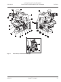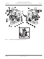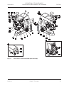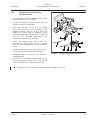
1
Operating Manual
Axio Lab.A1
www.ryfag.ch
Bettlachstrasse 2
2540 Grenchen
tel 032 654 21 00
fax 032 654 21 09
Ryf AG
Carl Zeiss
Copyright / Trade Marks
Axio Lab.A1
Knowledge of this manual is essential for the operation of the instrument. Please familiarize yourself with
the contents of this manual and pay special attention to instructions concerning safe operation of the
instrument.
The specifications are subject to change; the manual is not covered by an update service.
©
Unless expressly authorized, dissemination or duplication of this document and commercial
exploitation or communication of its contents is not permitted. Persons in contravention of this
copyright are liable to pay compensation for damages.
All rights reserved, including rights created by patent grant or registration of a utility model.
All names of companies and products mentioned in this manual may be trademarks or registered
trademarks. Third party products are cited for information purposes only and this does not represent
approval or recommendation of these products.
Carl Zeiss Microscopy GmbH accepts no liability for the performance or use of such products.
Publisher:
Carl Zeiss Microscopy GmbH
Carl-Zeiss-Promenade 10
07745 Jena, Germany
[email protected]
www.zeiss.com/microscopy
Carl Zeiss Microscopy GmbH
Königsallee 9-21
37081 Göttingen, Germany
Order No. 430037-7144-001
Date of issue: Version 1 - 19 April 2013
2
430037-7144-001
04/2013
Axio Lab.A1
Contents / List of Illustrations
Carl Zeiss
CONTENTS
Page
1
INTRODUCTION ................................................................................................... 7
1.1
1.2
1.3
Notes on instrument safety .......................................................................................... 7
Notes on ergonomics of the microscope .................................................................... 12
Notes on warranty ...................................................................................................... 13
2
DESCRIPTION OF THE INSTRUMENT ................................................................. 14
2.1
2.2
2.3
2.3.1
2.3.2
2.4
2.4.1
2.4.2
2.4.3
2.4.4
2.4.5
2.4.6
2.4.7
2.5
2.5.1
2.5.2
2.5.3
2.5.4
Intended use ................................................................................................................ 14
System overview ......................................................................................................... 16
Technical data.............................................................................................................. 21
Viewing height and tube angle ....................................................................................... 23
Assignment of dust covers, intermediate plate and base plate ......................................... 24
Control and functional elements on microscope ....................................................... 26
Stand models ................................................................................................................. 26
Stand for transmitted light.............................................................................................. 26
Stand for transmitted light polarization ........................................................................... 28
Stand for transmitted light and reflected light fluorescence ............................................. 30
Stand for reflected light .................................................................................................. 32
Stand for transmitted light conoscopy ............................................................................. 34
Ergo stands with TÜV certificate "Ergonomically tested" ................................................. 36
Control and functional elements on optional components ...................................... 37
Tubes/photo tubes ......................................................................................................... 37
Microscope stages .......................................................................................................... 41
Condensers .................................................................................................................... 43
Reflector turret 4x .......................................................................................................... 45
3
START-UP ........................................................................................................... 46
3.1
3.1.1
3.1.2
3.1.3
3.1.4
3.1.5
3.1.6
3.1.7
3.1.8
3.1.9
3.1.10
3.1.11
3.1.12
Installing standard components ................................................................................. 46
Unpacking and setting up microscope stand ................................................................... 46
Mounting base plate for use of larger tubes .................................................................... 47
Attaching the binocular tube/photo tube ........................................................................ 48
Installing eyepieces or auxiliary microscope or diopter...................................................... 49
Screwing in objectives .................................................................................................... 50
Fitting and removing push&click modules in the reflector turret ....................................... 51
Mounting a mechanical stage ......................................................................................... 52
Mounting rotary stage Pol .............................................................................................. 54
Attaching condenser ...................................................................................................... 57
Installing or replacing 35 W halogen bulb or 3 W white light LED lamp ............................ 58
Installing or changing the 12 V 50 W halogen lamp ........................................................ 60
Installing or replacing LED modules ................................................................................. 61
04/2013
430037-7144-001
3
Carl Zeiss
Contents / List of Illustrations
Axio Lab.A1
3.2
3.2.1
3.2.2
3.2.3
3.2.4
3.3
3.4
3.5
3.5.1
3.5.2
3.5.3
3.5.4
3.5.5
3.5.6
3.5.7
Mounting optional components ................................................................................ 62
Mounting the light intensive co-observer unit ................................................................. 62
Mounting polarizer D or filter holder .............................................................................. 62
Mounting and centering the overview fixture.................................................................. 63
Inserting modulator disk in condenser 0.9 H Pol.............................................................. 64
Connecting to the power supply................................................................................ 65
Switching the microscope on/off ............................................................................... 65
Basic setting of the microscope from an ergonomic point of view.......................... 66
Configuration of an ergonomic microscope workplace .................................................... 66
TÜV certificate ID:0000025994 "Ergonomically tested" .................................................. 67
Ergonomic configuration of the microscope workstation ................................................. 67
Ergonomic adjustment of the microscope ....................................................................... 68
Setting interpupillary distance on the binocular tube ....................................................... 70
Setting the viewing height ............................................................................................. 70
Adjusting for ametropia when using eyepiece reticles ..................................................... 71
4
OPERATION ....................................................................................................... 72
4.1
4.1.1
4.1.2
4.1.3
4.1.4
4.1.5
4.1.6
4.1.7
4.2
4.2.1
4.2.2
4.2.3
4.2.4
Lighting and contrasting method in transmitted light ............................................. 72
Adjusting the transmitted light brightfield according to KÖHLER ..................................... 72
Adjusting the transmitted light darkfield according to KÖHLER ........................................ 75
Setting the transmitted light phase contrast .................................................................... 76
Setting transmitted light polarization .............................................................................. 78
Setting transmitted light polarization with the conoscopy stand ...................................... 87
Determining the optical character of crystals ................................................................... 87
Setting transmitted light polarization for conoscopic observation – determining the optical
character of crystals ....................................................................................................... 98
Lighting and contrasting method in reflected light ................................................ 101
Adjusting the reflected light brightfield according to KÖHLER........................................ 101
Adjusting the reflected light darkfield ........................................................................... 103
Adjusting reflected light polarization – Proof of bireflexion and reflexion-pleochroism .... 104
Adjusting reflected light fluorescence ........................................................................... 105
5
CARE, FUSE REPLACEMENT AND SERVICE ..................................................... 107
5.1
5.2
5.2.1
5.2.2
5.3
5.4
Instrument care ......................................................................................................... 107
Instrument maintenance .......................................................................................... 108
Checking the instrument .............................................................................................. 108
Replacing the fuses in the stand ................................................................................... 108
Troubleshooting........................................................................................................ 109
Service ....................................................................................................................... 112
6
ANNEX ............................................................................................................. 113
6.1
6.2
6.3
List of abbreviations ................................................................................................. 113
Index .......................................................................................................................... 114
Industrial property rights ......................................................................................... 118
4
430037-7144-001
04/2013
Axio Lab.A1
Contents / List of Illustrations
Carl Zeiss
LIST OF ILLUSTRATIONS
Fig. 1-1
Fig. 1-2
Fig. 2-1
Fig. 2-2
Fig. 2-3
Fig. 2-4
Fig. 2-5
Fig. 2-6
Fig. 2-7
Fig. 2-8
Fig. 2-9
Fig. 2-10
Fig. 2-11
Fig. 2-12
Fig. 2-13
Fig. 2-14
Fig. 2-15
Fig. 2-16
Fig. 2-17
Fig. 2-18
Fig. 2-19
Fig. 2-20
Fig. 2-21
Fig. 2-22
Fig. 2-23
Fig. 2-24
Fig. 3-1
Fig. 3-2
Fig. 3-3
Fig. 3-4
Fig. 3-5
Fig. 3-6
Fig. 3-7
Fig. 3-8
Fig. 3-9
Fig. 3-10
Fig. 3-11
Fig. 3-12
Fig. 3-13
Fig. 3-14
Fig. 3-15
Fig. 3-16
Fig. 3-17
04/2013
"RADIATION" and "LED APERTURE" warning labels on Axio Lab.A1 for transmitted
and reflected light fluorescence ........................................................................................ 10
"Hot surface below" warning label on Axio Lab.A1 for reflected light ................................ 11
Axio Lab.A1, stand transmitted light ................................................................................. 27
Axio Lab.A1, stand transmitted light polarization ............................................................... 29
Axio Lab.A1, stand transmitted and reflected light fluorescence ......................................... 31
Axio Lab.A1, stand reflected light...................................................................................... 33
Axio Lab.A1, stand transmitted light conoscopy................................................................. 35
Axio Lab.A1 ergonomic stand with TÜV certificate "Ergonomically tested"......................... 36
Binocular photo tube 30°/20 with fixed graduation 50:50.................................................. 37
Binocular photo tube 30°/23 with toggle graduation 100:0/0:100 ..................................... 37
Setting the viewing height on the binocular tube............................................................... 38
Binocular ergo photo tube 8-38°/20 with fixed graduation 50:50....................................... 38
Binocular ergo tube 8-33°/20 with vertical adjustment 50 mm ........................................... 39
Binocular ergo photo tube 20°/23 with vertical adjustment ................................................ 40
Binocular ergo photo tube 15°/23, telescopic with vertical adjustment ............................... 40
Mechanical stage 75x30 R with specimen holder ............................................................... 41
Mechanical stage 75x30 R ergonomic with stationary drive ................................................ 41
Mechanical stage reflected light 75x30 R with specimen holding plate ............................... 41
Rotary stage Pol................................................................................................................ 42
Filter mount on luminous-field diaphragm operating ring for filter d=32x4 mm .................. 42
Condenser 0.9/1.25 H, D, Ph1, Ph2, Ph3 with modulator disk ............................................ 43
Condenser 0.9/1.25 H ...................................................................................................... 43
Overview fixture ............................................................................................................... 44
Polarizers.......................................................................................................................... 44
4-position reflector turret.................................................................................................. 45
Nosepiece of the transmitted light polarization stand with mount for compensators ........... 45
Setting up the microscope ................................................................................................ 46
Placing tools in the storage compartment .......................................................................... 46
Stowing power cord in cover flap for shipping ................................................................... 47
Mounting the base plate................................................................................................... 47
Attaching the binocular tube ............................................................................................ 48
Installing the eyepieces ..................................................................................................... 49
Installing the eyepiece reticle............................................................................................. 49
Screwing in objectives....................................................................................................... 50
Replacing the reflector module ......................................................................................... 51
Replacing a mechanical stage............................................................................................ 52
Setting friction torque....................................................................................................... 53
Replacing snap-in rotary stage Pol, detachable specimen guide Pol and stage clips ............. 54
Centring rotary stage Pol .................................................................................................. 55
Centering objectives ......................................................................................................... 56
Attaching condenser ........................................................................................................ 57
Removing the cover .......................................................................................................... 58
Removing the LED lamp .................................................................................................... 58
430037-7144-001
5
Carl Zeiss
Fig. 3-18
Fig. 3-19
Fig. 3-20
Fig. 3-21
Fig. 3-22
Fig. 3-23
Fig. 3-24
Fig. 3-25
Fig. 3-26
Fig. 3-27
Fig. 3-28
Fig. 3-29
Fig. 3-30
Fig. 3-31
Fig. 3-32
Fig. 3-33
Fig. 3-34
Fig. 3-35
Fig. 4-1
Fig. 4-2
Fig. 4-3
Fig. 4-4
Contents / List of Illustrations
Axio Lab.A1
Fig. 4-5
Fig. 4-6
Fig. 4-7
Inserting the LED lamp ......................................................................................................58
Attaching condenser .........................................................................................................59
Removing the cover...........................................................................................................60
Removing the 12V 50W halogen bulb ...............................................................................60
Installing the 12V 50W halogen bulb .................................................................................60
Removing the cover...........................................................................................................61
Removing the LED module.................................................................................................61
Installing the LED module ..................................................................................................61
Mounting the polarizer D ..................................................................................................62
Mounting the overview fixture...........................................................................................63
Modulator disk in condenser 0.9 H Pol...............................................................................64
Mains power supply on rear side of stand ..........................................................................65
Mains switch on left side of microscope .............................................................................65
Light intensity control and FL/TL toggle switch....................................................................65
TÜV certificate "Ergonomically tested" ..............................................................................67
Ergonomic adjustment of the microscope ..........................................................................68
Setting the interpupillary distance on the binocular tube .................................................... 70
Setting the viewing height on the binocular tube ............................................................... 70
Microscope settings in transmitted light brightfield ............................................................73
Setting the height stop on the condenser carrier ................................................................74
Center darkfield stop on condenser, achromatic-aplanatic 0.9 H D Ph DIC .......................... 76
Centering the phase stop (bright ring in condenser) to the phase ring
(dark ring in objective) .......................................................................................................77
Components for transmitted light polarization ...................................................................79
Gamma direction ..............................................................................................................80
Determining the direction of oscillation nJ’ using a synthetic fiber as an example ................ 81
Fig. 4-8
Fig. 4-9
Fig. 4-10
Fig. 4-11
Fig. 4-12
Fig. 4-13
Fig. 4-14
Schematic diagram of the color charts according to Michel-Lévy ......................................... 83
Components for circular polarization contrast ....................................................................86
Axio Lab.A1 for transmitted light conoscopy ......................................................................88
Determining optical character ............................................................................................89
Components for transmitted light polarization on conoscopy stand .................................... 91
Gamma direction ..............................................................................................................91
Determining the direction of oscillation nJ’ using a synthetic fiber as an example................. 92
Fig. 4-15
Fig. 4-16
Fig. 4-17
Fig. 4-18
Fig. 4-19
Fig. 4-20
Fig. 5-1
Schematic diagram of the color charts according to Michel-Lévy ......................................... 94
Components for circular polarization contrast on conoscopy stand ..................................... 97
Axio Lab.A1 for transmitted light conoscopy ......................................................................99
Determining the optical character .................................................................................... 100
Microscope settings in reflected light brightfield............................................................... 102
Components for reflected light fluorescence .................................................................... 106
Replacing the fuses in the stand ...................................................................................... 108
6
430037-7144-001
04/2013
Axio Lab.A1
INTRODUCTION
Notes on instrument safety
1
INTRODUCTION
1.1
Notes on instrument safety
Carl Zeiss
Axio Lab.A1 microscopes have been designed, produced and tested in compliance with DIN EN 61010-1
(IEC 61010-1) and IEC 61010-2-101 safety requirements for electrical measuring, control and laboratory
instruments.
These instruments meet the requirements of EC Directive IVDD 98/79/EC (In Vitro Diagnostic); they carry
the
mark.
This operating manual includes information and warnings which must be observed by the user.
The following warning and instruction symbols are used in this manual:
CAUTION
This symbol indicates a potential hazard to the user.
CAUTION
Hot surface!
CAUTION
UV radiation emitted!
CAUTION
Disconnect the instrument from the power supply before opening!
CAUTION
This symbol indicates a potential hazard to the instrument or system.
NOTE
This symbol denotes a warning to which special attention must be paid.
Axio Lab.A1 microscopes, including original accessories, may only be used for the microscopy methods
described in this manual.
The following instructions must be carefully observed:
The manufacturer is unable to assume any liability for any other use, including individual
modules and individual components. This also applies to all service and repair work not
performed by authorized service personnel. Furthermore, all warranty rights will be forfeited.
The socket into which the power cable is plugged must be earthed. The grounding effect must
not be made ineffective by an extension cable that does not have a protective ground wire.
04/2013
430037-7144-001
7
Carl Zeiss
INTRODUCTION
Notes on instrument safety
Axio Lab.A1
Should it be determined that protective measures are no longer effective, the instrument must
be taken out of service and secured against unintended use. Please contact a Zeiss service
agency or the Carl Zeiss Microscopy Service to repair the instrument.
Axio Lab.A1 microscopes are equipped with a power supply unit incorporated in the stand,
which allows line voltages ranging from 100 V to 240 V ±10 % (50/60 Hz) to be used without
requiring any change of voltage on the unit.
Always pull the mains plug before opening the instrument or replacing a fuse!
Only fuses for the rated power may be used. The use of provisional fuses or short-circuiting the
fuse-holder is prohibited.
Axio Lab.A1 microscopes are not equipped with any special devices for protection from
substances that are corrosive, potentially infectious, toxic, radioactive, or other substances that
could be hazardous to health. Make sure to observe all legal regulations, particularly the
relevant national accident prevention regulations when handling such substances.
Dirt and dust may impair the performance of the instrument. Therefore, the instrument must be
protected from these influences as far as possible and covered with the dust cap when not in
use. Always check whether the instrument is switched off before you cover it.
Obstruction or covering of ventilation slits may result in heat build-up which may damage the
instrument and in extreme cases cause fire. Always keep the ventilation slits clear and ensure
that no objects enter the instrument through the ventilation slits.
Instruments may only be operated by trained personnel. The latter must be aware of the
possible hazards involved with microscopy and the respective application. Axio Lab.A1
microscopes are high-precision instruments whose functional capabilities may be impaired or
destroyed when handled improperly.
Instruments should not be operated in an explosive environment.
Instruments should only be operated on a solid, non-combustible surface.
8
430037-7144-001
04/2013
Axio Lab.A1
INTRODUCTION
Notes on instrument safety
Carl Zeiss
LED Risk Group 2 to IEC 62471, LED radiation will be emitted.
Never look into the LED beam of the illuminating device – either with or without optical
instruments. Failure to observe this precaution may results in eye injuries!
Combustible and easily inflammable materials should not be held close to the light beam.
Be sure to read the safety data sheets for the Immersol 518 N£, Immersol 518 F£ and
Immersol W£.
Immersol 518 N£ immersion oil irritates the skin. Avoid any contact with skin, eyes and
clothing.
In the case of inadvertent skin contact, wash the oil off with plenty of water and soap.
In the case of inadvertent eye contact, immediately rinse the eye with plenty of water for at
least five minutes. Seek medical attention if irritation persists.
Proper disposal of immersion oil 518 N£: do not discharge into the surface water or sewerage
system.
Do not put defective microscopes into the household waste; they must be disposed of in
accordance with WEEE Directive 2002/96/EC.
Samples must likewise be properly disposed of in accordance with statutory provisions and
internal work instructions.
04/2013
430037-7144-001
9
Carl Zeiss
INTRODUCTION
Notes on instrument safety
Axio Lab.A1
Warning labels on Axio Lab.A1 stands
Warning label: Hot surface!
Affixed to all stands with transmitted light illumination.
Fig. 1-1
10
"RADIATION" and "LED APERTURE" warning labels on Axio Lab.A1 for transmitted and
reflected light fluorescence
430037-7144-001
04/2013
Axio Lab.A1
Fig. 1-2
04/2013
INTRODUCTION
Notes on instrument safety
Carl Zeiss
"Hot surface below" warning label on Axio Lab.A1 for reflected light
430037-7144-001
11
INTRODUCTION
Notes on ergonomics of the microscope
Carl Zeiss
1.2
Axio Lab.A1
Notes on ergonomics of the microscope
The Axio Lab.A1 light microscope was developed and designed in cooperation with occupational health
physicians and the TÜV Rhineland to meet the most exacting demands with regard to ergonomics at the
microscope workstation. This first light microscope worldwide is available with a special ergonomic
configuration and bears TÜV certificate ID:0000025994 "Ergonomically tested".
Particularly laboratory microscopes in the Axio Lab.A1 device class are used continuously for many routine
applications (e.g. haematological, histological and cytological examinations) over a period of several
hours. With light microscopes which have not been ergonomically designed, such regular, extended
periods of use could cause health problems in terms of posture control of the user. This health risk can be
significantly reduced by careful ergonomic design and arrangement of the control elements, wellconceived and individual eyepiece adjustment options, together with correct set-up of the entire
microscope workstation.
This results in improved working conditions, greater comfort of employees and higher work productivity.
More and more countries are also introducing stringent workplace regulations for microscope
workstations, particularly in the medical field. In addition, the regulations of employers' liability insurance
associations increasingly place obligations on employers to provide ergonomic workplaces and
microscopes.
TÜV certificate ID:0000025994 "Ergonomically tested" prescribes the distance of control elements from
the table, user and between instruments. Furthermore, it defines a broad adjustment range for the
eyepiece to cater for the different body heights of male and female microscope users worldwide. For this
purpose the ergonomic tube or ergo tube must be vertically and angle-adjustable. This enables it to be
adjusted to suit different body heights (static ergonomics) and occasionally varied by the user during
extended periods of use (dynamic ergonomics). The following workplace standards apply as a basis for
this TÜV certificate of ergonomics:
DIN 58959-4:
DIN EN 1335-1:
DIN EN 12464-1:
DIN EN 12665:
DIN EN 13150:
DIN EN ISO 15189:
DIN EN ISO 9241-11:
DIN EN ISO 60601-1-6:
Quality management in medical microbiology – Part 4:
Requirements for investigations using light microscopes
Office work chairs – Part 1: Dimensions – Determination of dimensions
Lighting of work places – Part 1: Indoor work places
Basic terms and criteria for specifying lighting requirements
Workbenches for laboratories – Safety requirements and test methods
Medical laboratories – Particular requirements for quality and competence
Ergonomic requirements for office work with visual display terminals –
Guidance on usability
Medical-electrical equipment – General requirements for basic safety and
essential performance
and the following ergonomic standards:
12
DIN 33402-2:
DIN 33406:
DIN 33408:
DIN 33411:
DIN 68877:
DIN EN 614-1:
Ergonomics – Human body dimensions – Part 2: Values
Workplace dimensions in production
Body templates for seats
Human physical strength
Swiveling work chair - Safety requirements, testing
Ergonomic design principles – Part 1: Terms and general principles
430037-7144-001
04/2013
Axio Lab.A1
INTRODUCTION
Notes on warranty
Carl Zeiss
DIN EN 894-1:
Ergonomic requirements for the design of displays and control actuators – Part 1:
General principles for human interaction with displays and control actuators
DIN EN 894-3:
Ergonomic requirements for the design of displays and control actuators – Part 3:
Control actuators
DIN EN 62079:
Preparation of instructions – Structuring, content and presentation
DIN EN ISO 7250:
Basic human body measurements for technological design
DIN EN ISO 14738: Safety of machinery – Anthropometric requirements for the design of
workstations at machinery
ISO 11226:
Ergonomics – Evaluation of static working postures
SEMI S8-0307:
Safety guidelines for ergonomics engineering of semiconductor manufacturing
equipment
For a further explanation of TÜV certificate of ergonomics and basic ergonomic setting and operation of
the Axio Lab.A1 microscope please refer to Section 3.5.
1.3
Notes on warranty
The manufacturer guarantees that the instrument has no material or production defects when delivered.
Any defects occurring must be reported without delay; every effort must be made to minimize damage.
Once a defect has been notified, the manufacturer shall have the right to choose whether to repair the
instrument or deliver a replacement in perfect condition. No warranty is provided for normal attrition (in
particular of wearing parts) or improper use.
The instrument manufacturer shall not be liable for damage caused by faulty operation, negligence or
any other tampering with the instrument, particularly the removal or replacement of instrument
components, or the use of accessories from other manufacturers. Such actions will render any warranty
claims invalid.
With the exception of the work specified in this manual, no maintenance or repair of microscopes may be
carried out. Repairs are only to be performed by Carl Zeiss service or personnel specifically authorized by
Carl Zeiss. Should a malfunction of the instrument occur, please contact the Carl Zeiss Microscopy Service
in Germany (see page 112) or the responsible Carl Zeiss representative abroad.
04/2013
430037-7144-001
13
DESCRIPTION OF THE INSTRUMENT
Intended use
Carl Zeiss
2
DESCRIPTION OF THE INSTRUMENT
2.1
Intended use
Axio Lab.A1
Axio Lab.A1 microscopes were designed as all-purpose microscopes for biological and medical
applications, as well as material analyses.
Depending on the microscope stand selected, they may also be used as true transmitted or reflected light
microscopes or as combined transmitted/reflected light fluorescence microscopes.
Typical biomedical applications of Axio Lab.A1 microscopes include:
Medical examinations in laboratories, clinics and medical practices
Science and research (colleges, universities) in the fields of medicine and biology
Industrial applications (pharmacology, food technology)
Analysis of human blood and tissue samples.
Typical material analysis applications of Axio Lab.A1 microscopes include:
Metallographic laboratories
Automotive industry
Microsystems engineering
Geoscientific institutes
Exploration industry
Depending on the instrument configuration, the following microscopy and contrasting techniques are
possible:
Transmitted light
Reflected light
Brightfield (H)
Brightfield (H)
Darkfield (D)
Darkfield (D)
Phase contrast (Ph)
Polarization (Pol)
Polarization (Pol)
Fluorescence (FL)
Polarization (conoscopy)
Differential Interference Contrast (DIC)
Polarization (C-Pol)
The binocular photo tubes and suitable adapters permit one microscope camera, one reflex camera or
one digital/video camera to be attached for documentation purposes.
The Axio Lab.A1 was specially developed and designed for ergonomic use in lengthy routine applications,
e.g. haematological, histological and cytological laboratory examinations.
14
430037-7144-001
04/2013
Axio Lab.A1
DESCRIPTION OF THE INSTRUMENT
Intended use
Carl Zeiss
The ergonomic design elements of the Axio Lab.1 are:
Vertically adjustable, swivel-type and swivel/vertically adjustable ergo tubes
Skin-friendly surfaces on binocular section of the tubes, control elements and stand
Ergo table with fixed stage drive
Vertical and friction adjustable stage drives
Optional use of fine focusing knobs in standard form or as a jog/shuttle knob
Special, ergonomic arrangement of the three main control elements: focusing drive, stage drive and
brightness control
A TÜV certificate of ergonomics was prepared for the light microscope in cooperation with occupational
health physicians and the TÜV Rhineland and granted for the following basic configuration:
Transmitted light brightfield stand with ergo table and comfortable ergo tube
04/2013
430037-7144-001
15
DESCRIPTION OF THE INSTRUMENT
System overview
Carl Zeiss
2.2
16
Axio Lab.A1
System overview
430037-7144-001
04/2013
Axio Lab.A1
04/2013
DESCRIPTION OF THE INSTRUMENT
System overview
430037-7144-001
Carl Zeiss
17
Carl Zeiss
18
DESCRIPTION OF THE INSTRUMENT
System overview
430037-7144-001
Axio Lab.A1
04/2013
Axio Lab.A1
04/2013
DESCRIPTION OF THE INSTRUMENT
System overview
430037-7144-001
Carl Zeiss
19
Carl Zeiss
20
DESCRIPTION OF THE INSTRUMENT
System overview
430037-7144-001
Axio Lab.A1
04/2013
Axio Lab.A1
2.3
DESCRIPTION OF THE INSTRUMENT
Technical data
Carl Zeiss
Technical data
Dimensions (width x depth x height)
Axio Lab.A1 basic microscope stand
without tube (430037-9000-000) ................................................ approx. 219 mm x 410 mm x 3395 mm
The other stand types differ slightly in depth and significantly in height, depending on the tube used. An
overview of viewing heights (eyepoint heights) of the various tubes can be found in Section 2.3.1.
An estimate of the height of the stand with the respective tube can be obtained by adding:
10 mm to the viewing height in the lower position of binocular section
for tubes with a fixed viewing angle
10 mm to the viewing height of the upper limit for ergo tubes
Weight
Axio Lab.A1 microscope stand (depending on version and accessories) ......................... approx. 8 to 20 kg
Ambient conditions
Shipping (in packaging):
Permissible ambient temperature ................................................................................... -40 to +70 °C
Storage:
Permissible ambient temperature .................................................................................. +10 to +40 °C
Permissible humidity (without condensation) ......................................................... max. 75 % at 35 °C
Operation:
Permissible ambient temperature .................................................................................. +10 to +40 °C
Permissible relative humidity (without condensation) ............................................. max. 75 % at 35 °C
Highest permitted altitude of use .................................................................................... max. 2000 m
Air pressure ........................................................................................................800 hPa to 1060 hPa
Degree of pollution .......................................................................................................................... 2
Operational specifications
Operational area .................................................................................................................Closed rooms
Protective class .......................................................................................................................................I
Protection type ................................................................................................................................ IP 20
Electrical safety .............................................................................. acc. to DIN EN 61010-1 (IEC 61010-1)
in conformance with CSA and UL regulations
Overvoltage category .............................................................................................................................II
RFI suppression ...................................................................................... conforming to EN 55011 Class B
Noise immunity ..................................................................................... conforming to DIN EN 61326/A1
Mains voltage Axio Lab.A1 ....................................................................................... 100 to 240 V ±10 %
.......................................................................................... Conversion of mains voltage unnecessary!
Mains frequency ........................................................................................................................50/60 Hz
Power consumption Axio Lab.A1.................................................................................................. 110 VA
Fuses to IEC 127
Microscope stand Axio Lab.A1 ......................................................................... 2 x T 3.15 A/H, 5x20 mm
04/2013
430037-7144-001
21
Carl Zeiss
DESCRIPTION OF THE INSTRUMENT
Technical data
Axio Lab.A1
Light sources
LED transmitted light
Power consumption ............................................................................................................. max. 3 W
Adjustment of light source ...................................................................continuous approx. 0.5 to 12 V
Halogen lighting transmitted light
Power consumption ........................................................................................................... max. 35 W
Halogen lighting reflected light
Power consumption ................................................................................................................... 50 W
Adjustment of light source ...................................................................continuous approx. 0.5 to 12 V
LED lighting reflected light fluorescence with replaceable LED modules
Wavelengths optional .............................................. 365, 380, 455, 470, 505, 530, 590, 615, 625 nm
.......................................................................................................... or neutral white (540 - 580 nm)
LED classification ...................................................................................... LED Risk Group 2 to IEC 62471
Axio Lab.A1:
Stand with manual stage focusing
Coarse focusing ............................................................................................ approx. 4 mm/revolution
Fine focusing ................................................................approx. 0.4 mm/revolution; 4 μm scale interval
Lifting range ............................................................................................................................ 30 mm
Height stop ............................................................................................................... pre-set at works
Optional condenser 0.9/1.25 H with or without modulator disk for
...................................................................................Brightfield, darkfield and phase contrast 1, 2, 3
Manual objective change ...............................................................by nosepiece, 4x H Pol or 5x H D, M27
Manual reflector module change ..............................................................................by reflector turret 4x
22
430037-7144-001
04/2013
Axio Lab.A1
2.3.1
DESCRIPTION OF THE INSTRUMENT
Technical data
Carl Zeiss
Viewing height and tube angle
Order No.
Binocular tube
Viewing
angle
Adjustment
Viewing
height* in mm
425522-9000-000
Binocular tube 30°/20
30°
- none -
434 / 470
425522-9010-000
Binocular photo tube 30°/20 (50:50)
30°
- none -
434 / 470
425522-9020-000
Binocular ergo tube 8-38°/20
8-38°
Angle
407 - 534
425522-9030-000
Binocular ergo photo tube 838°/20
(50:50)
8-38°
Angle
407 - 534
425522-9040-000
Binocular ergonomic tube 8-38°/22
8-33°
Angle
Height
412 - 603
425520-9000-000
Binocular tube 30°/23
30°
- none -
449 / 485
425520-9010-000
Bin. photo tube 30°/23 (50:50)
30°
- none -
449 / 485
425520-9020-000
Bin. photo tube 30°/23 (100:100) Bio
30°
- none -
449 / 485
425520-9030-000
Binocular photo tube 20°/23 (100:100)
20°
- none -
442 / 481
425520-9040-000
Bin. ergo tube (100/100), angleadjustable, upright image
5-30°
Angle
395 - 537
425520-9050-000
Bin. ergo tube 15°/23 (50/50),
telescopic, height, upright image
15°
Height
telescopic
410 - 509
425520-9090-000
Binocular tube 20°/23
20°
442 / 481
425520-9100-000
Bin. photo tube 20°/23 Pol (100:100)
20°
442 / 481
425511-0000-000
Binocular ergo tube 20°/23,
44 mm height
20°
Height
457 - 574
425512-0000-000
Bin. ergo tube 20°/23 (100/100), reverse
image, 44 mm height
20°
Height
457 - 574
425514-0000-000
Bin. ergo tube 20°/23 (100/100),
upright image, 44 mm height
20°
Height
457 - 574
*
Viewing heights:
Tubes with fixed viewing angle without ergo function:
Binocular part lower/upper e.g. 442 / 481 o 442 to 481 mm
Angle- and vertically adjustable ergo tubes:
Binocular part lower/upper e.g. 457 / 574 o 457 to 574 mm
All specifications are for an interpupillary distance of 65 mm.
04/2013
430037-7144-001
23
DESCRIPTION OF THE INSTRUMENT
Technical data
Carl Zeiss
2.3.2
Axio Lab.A1
Assignment of dust covers, intermediate plate and base plate
Cat. No.
Binocular tube
Transmitted
light
430037-9000-000
430037-9010-000
430037-9030-000
Conoscopy
430037-9040-000
425522-9000-000
Binocular tube
30°/20 Bio
Small
Small
---
---
---
---
Small
Small
---
---
---
---
425522-9010-000
425522-9020-000
Binocular photo tube
30°/20 (50:50)
Binocular ergonomic tube
8-38°/20
Reflected light
430037-9020-000
430037-9050-000
Small
--M*
425522-9030-000
Binocular photo tube
8-38°/20 (50:50)
Medium
--M*
425522-9040-000
425520-9000-000
425520-9010-000
425520-9020-000
425520-9030-000
425520-9040-000
24
Binocular ergonomic tube
8-33°/22
Binocular tube
30°/23 Bio
Binocular photo tube
30°/23 (50:50) Bio
Binocular photo tube 30°/23
(100:100) Bio
Binocular photo tube
20°/23 (100:100) upright
image
Binocular ergo photo tube
(100/100), angle-adjustable,
upright image
Medium
Medium
Medium
Spacer
Spacer
---
M*
M*
M*
Small
Small
Small
Spacer
Spacer
---
---
---
---
Medium
Medium
Medium
Spacer
Spacer
---
M
M
M
Medium
Medium
Medium
Spacer
Spacer
---
M
M
M
Medium
Medium
Medium
Spacer
Spacer
---
M
M
M
Medium
Medium
Medium
Spacer
Spacer
---
M
M
M
430037-7144-001
04/2013
DESCRIPTION OF THE INSTRUMENT
Technical data
Axio Lab.A1
Carl Zeiss
Cat. No.
Binocular tube
Transmitted
light
430037-9000-000
430037-9010-000
430037-9030-000
Conoscopy
430037-9040-000
Reflected light
430037-9020-000
430037-9050-000
425520-9050-000
Binocular ergo tube 15°/23
(50/50), telescopic, height,
upright image
Medium
Medium
Medium
Spacer
Spacer
---
M
M
M
Small
Small
Small
Spacer
Spacer
---
---
---
---
Medium
Medium
Medium
Spacer
Spacer
---
M
M
M
Medium
Medium
Medium
Spacer
Spacer
---
M
M
M
Medium
Medium
Medium
Spacer
Spacer
---
M
M
M
Medium
Medium
Medium
Spacer
Spacer
---
M
M
M
425520-9090-000
425520-9100-000
425511-0000-000
425512-0000-000
425514-0000-000
Binocular tube
20°/23 Mat
(analog 9030 without
camera output)
Binocular photo tube
20°/23 Pol (100/100)
Binocular ergo tube 20°/23,
reverse image, 44 mm
height
Binocular ergo tube 20°/23
(100/100),
44 mm height
Binocular ergonomoic tube
20°/23 (100/100), upright
image,
44 mm height
* Included in shipping contents
Explanation of table:
Dust cover
Small: 415510-1901
Medium: 459306-0000-000
Intermediate plate for tubes
(423732-9040-000)
Intermediate plate: necessary
--- unnecessary
Base plate
(430037-910-000)
M: Mandatory
M: recommended
04/2013
430037-7144-001
---- unnecessary
25
DESCRIPTION OF THE INSTRUMENT
Control and functional elements on microscope
Carl Zeiss
2.4
Control and functional elements on microscope
2.4.1
Stand models
Axio Lab.A1
Five stand models are available in the delivery program:
1. Transmitted light stand for biomedical applications in brightfield, darkfield and phase contrast
2. Transmitted light stand for biomedical applications in brightfield, darkfield, phase contrast and
polarization
3. Transmitted and reflected light stand for biomedical applications in brightfield, darkfield, phase
contrast, polarization (transmitted light) and fluorescence (reflected light)
4. Reflected light stand for material applications in brightfield, darkfield, phase contrast, polarization
and C-DIC
5. Transmitted light stand for material applications in brightfield, darkfield, phase contrast, polarization,
conoscopy
In addition, the delivery program also includes two TÜV certificates, "Ergonomically tested" stand
models.
A TÜV certificate of ergonomics was prepared for the light microscope in cooperation with occupational
health physicians and the TÜV Rheinland and granted for the following two basic configurations:
Transmitted light stand with ergo stage and comfortable ergonomic tube
Reflected light fluorescence stand with ergo stage and comfortable ergo tube
2.4.2
Stand for transmitted light
Key to Fig. 2-1:
1
2
3
4
5
6
7
8
9
10
11
12
13
14
15
16
17
18
19
20
21
22
23
24
25
26
Basic stand
Stage carrier for mechanical stages
Light intensity control
Focusing drive – fine adjustment (right side, finger wheel)
Focusing drive – coarse adjustment (right side)
Drive knob for mechanical stage adjustment in X direction
Drive knob for mechanical stage adjustment in Y direction
Drive knob for vertical adjustment of condenser (right side)
Centering screw for condenser (right side)
Luminous-field diaphragm
Condenser with aperture diaphragm (optional with modulator disk)
Mechanical stage 75x30 (optionally for right- or left-hand operation or with ergo-drive right) with specimen holder
Nosepiece 5x H
Slot for 6x20 slider
Eyepieces
Binocular section of tube
Binocular tube/photo tube
Carrying handle
Centering screw for condenser (left side)
Drive knob for vertical adjustment of condenser (left side)
Focusing drive – coarse adjustment (left side)
Focusing drive – fine adjustment (left side)
ON/OFF switch
Transmitted light lamp in stand base
Tool flap/cable holder
430037-7144-001
04/2013
Axio Lab.A1
Fig. 2-1
04/2013
DESCRIPTION OF THE INSTRUMENT
Control and functional elements on microscope
Carl Zeiss
Axio Lab.A1, stand transmitted light
430037-7144-001
27
DESCRIPTION OF THE INSTRUMENT
Control and functional elements on microscope
Carl Zeiss
2.4.3
Axio Lab.A1
Stand for transmitted light polarization
Key to Fig. 2-2:
1
2
3
4
5
6
7
8
9
10
11
12
13
14
15
16
17
18
19
20
21
22
23
24
25
28
Basic stand
Stage carrier for rotary stages (also suitable for mechanical stages)
Light intensity control
Focusing drive – fine adjustment (right side, finger wheel)
Focusing drive – coarse adjustment (right side)
Drive knob for vertical adjustment of condenser (right side)
Centering screw for condenser (right side)
Luminous-field diaphragm
Locking screw for rotary table (arrests rotation)
Condenser with aperture diaphragm (optional with modulator disk)
Rotary stage lock in stage carrier
Rotary stage Pol with specimen guide
Nosepiece 4x H Pol (3 eyes centering, 1 eye fixed)
Slot for 6x20 slider
Eyepieces
Binocular section of tube
Binocular tube/photo tube
Carrying handle
Centering screw for condenser (left side)
Drive knob for vertical adjustment of condenser (left side)
Focusing drive – coarse adjustment (left side)
Focusing drive – fine adjustment (left side)
ON/OFF switch
Tool flap/cable holder
Transmitted light lamp in stand base
430037-7144-001
04/2013
Axio Lab.A1
Fig. 2-2
04/2013
DESCRIPTION OF THE INSTRUMENT
Control and functional elements on microscope
Carl Zeiss
Axio Lab.A1, stand transmitted light polarization
430037-7144-001
29
DESCRIPTION OF THE INSTRUMENT
Control and functional elements on microscope
Carl Zeiss
2.4.4
Axio Lab.A1
Stand for transmitted light and reflected light fluorescence
Key to Fig. 2-3:
1
2
3
4
5
6
7
8
9
10
11
12
13
14
15
16
17
18
19
20
21
22
23
24
25
26
27
28
29
30
30
Reflected light intensity control
FL/TL switch (FL - reflected light fluorescence; TL - transmitted light)
Push-pull rod for switching between LED 1 and LED 2
Cover flap for LED reflected light illumination in top part of stand
Basic stand
Nosepiece 5x H FL-LED
Stage carrier for mechanical stages
Transmitted light intensity control
Base plate for microscope stand
Focusing drive – fine adjustment (right side, finger wheel)
Focusing drive – coarse adjustment (right side)
Drive knob for mechanical stage adjustment in X direction
Drive knob for mechanical stage adjustment in Y direction
Drive knob for vertical adjustment of condenser (right side)
Centering screw for condenser (right side)
Luminous-field diaphragm
Condenser with aperture diaphragm (optional with modulator disk)
Slot for 6x20 slider
Mechanical stage 75x30 (optionally for right- or left-hand operation or with ergo-drive right) with specimen holder
Reflector turret 4x
Eyepieces
Binocular section of tube
Binocular comfortable ergo tube
Centering screw for condenser (left side)
Drive knob for vertical adjustment of condenser (left side)
Focusing drive – coarse adjustment (left side)
Focusing drive – fine adjustment (left side)
ON/OFF switch
Tool flap/cable holder
Transmitted light illumination in stand base
430037-7144-001
04/2013
Axio Lab.A1
Fig. 2-3
04/2013
DESCRIPTION OF THE INSTRUMENT
Control and functional elements on microscope
Carl Zeiss
Axio Lab.A1, stand transmitted and reflected light fluorescence
430037-7144-001
31
DESCRIPTION OF THE INSTRUMENT
Control and functional elements on microscope
Carl Zeiss
2.4.5
Axio Lab.A1
Stand for reflected light
Key to Fig. 2-4:
1
2
3
4
5
6
7
8
9
10
11
12
13
14
15
16
17
18
19
20
21
22
Reflected light illumination
Luminous-field diaphragm (centered)
Aperture diaphragm (centered)
Basic stand
Nosepiece 5x H HD
Stage carrier for mechanical stages
Light intensity control
Focusing drive – fine adjustment (right side, finger wheel)
Focusing drive – coarse adjustment (right side)
Drive knob for mechanical stage adjustment in X direction
Drive knob for mechanical stage adjustment in Y direction
Mechanical stage 75x30 A with specimen holder A for reflected light
Slot for 6x20 slider
Reflector turret 4x
Eyepieces
Binocular section of tube
Binocular tube/photo tube
Focusing drive – coarse adjustment (left side)
Focusing drive – fine adjustment (left side)
ON/OFF switch
Filter slide reflected light
Tool flap/cable holder
32
430037-7144-001
04/2013
Axio Lab.A1
Fig. 2-4
04/2013
DESCRIPTION OF THE INSTRUMENT
Control and functional elements on microscope
Carl Zeiss
Axio Lab.A1, stand reflected light
430037-7144-001
33
DESCRIPTION OF THE INSTRUMENT
Control and functional elements on microscope
Carl Zeiss
2.4.6
Axio Lab.A1
Stand for transmitted light conoscopy
Key to Fig. 2-5:
1
2
3
4
5
6
7
8
9
10
11
12
13
14
15
16
17
18
19
20
21
22
23
24
25
26
27
28
29
Rotary knob A: Swiveling analyzer in/out
Rotary knob BL: Swiveling Bertrand lens in/out
Basic stand
Nosepiece 4x H Pol (3 eyes centering, 1 eye fixed)
Stage carrier for rotary stages (also suitable for mechanical stages)
Light intensity control
Focusing drive – fine adjustment (right side)
Focusing drive – coarse adjustment (right side)
Drive knob for vertical adjustment of condenser (right side)
Centering screw for condenser (right side)
Luminous-field diaphragm
Locking screw for rotary stage (arrests rotation)
Condenser with aperture diaphragm (optional with modulator disk)
Rotary stage Pol with specimen holder
Slot for 6x20 slider
Setting wheel for direction of oscillation of analyzer
Setting wheel for focusing of Bertrand lens
Eyepieces
Binocular section of tube
Binocular tube/photo tube
Centering screw for condenser (left side)
Drive knob for vertical adjustment of condenser (left side)
Focusing drive – coarse adjustment (left side)
Focusing drive – fine adjustment (left side)
ON/OFF switch
Carrying handle
Storage compartments for two 6x20 slides
Transmitted light illumination
Tool flap/cable holder
CAUTION
The movements of rotary knobs A and BL (Fig. 2-5/1 and 2) and the respective setting wheels
(Fig. 2-5/16 and 17) are coupled with one another. Only one control element should therefore
be operated at a time and the movement of the other should not be inhibited or blocked.
Mechanical damage may otherwise occur.
34
430037-7144-001
04/2013
Axio Lab.A1
Fig. 2-5
04/2013
DESCRIPTION OF THE INSTRUMENT
Control and functional elements on microscope
Carl Zeiss
Axio Lab.A1, stand transmitted light conoscopy
430037-7144-001
35
DESCRIPTION OF THE INSTRUMENT
Control and functional elements on microscope
Carl Zeiss
2.4.7
Axio Lab.A1
Ergo stands with TÜV certificate "Ergonomically tested"
Key to Fig. 2-6:
1
2
3
Binocular ergo tube 8-33°, 50 mm vertically adjustable
Mechanical stage 75x30 ergonomic with stationary drive
Stand for transmitted light and reflected light fluorescence
Fig. 2-6
Axio Lab.A1 ergonomic stand with TÜV certificate "Ergonomically tested"
Further information on the ergonomically correct setting of the microscope and its ergonomic
operation can be found in Section 3.5.
36
430037-7144-001
04/2013
Axio Lab.A1
DESCRIPTION OF THE INSTRUMENT
Control and functional elements on optional components
2.5
Control and functional elements
on optional components
2.5.1
Tubes/photo tubes
Carl Zeiss
The appropriate adapters for reflex cameras,
microscope cameras and video cameras may be
plugged into the camera port (Fig. 2-7/1 or
Fig. 2-8/1) of the binocular photo tubes.
Binocular photo tube 30°/20 with fixed
graduation 50:50
Fig. 2-7
Binocular photo tube 30°/20 with
fixed graduation 50:50
Fig. 2-8
Binocular photo tube 30°/23 with
toggle graduation 100:0/0:100
50% of the light is directed to the eyepieces and
camera port respectively (Fig. 2-7).
Binocular photo tube 30°/23 (100:0/0:100)
The light can be directed using a shift knob to
either the eyepieces or the mounted camera.
Shift knob (Fig. 2-8/2) to front (eye symbol):
100 % light to eyepieces.
Shift knob (Fig. 2-8/2) to the rear (camera
symbol):
100 % light to camera
Push-pull rod (Fig. 2-8/3) pushed in:
Eyepiece shutter closed.
Pull-push rod (Fig. 2-8/3) pulled out:
Eyepiece shutter opened.
Particularly for camera shots with extended
exposure times it is recommended that
possible residual light incidence through the
eyepiece is prevented either by means of a
tube shutter or eyepiece cover (included in
dustguard set). If neither is available, remove
the eyepiece and insert the supplied dust cap
onto the eyepiece socket!
04/2013
430037-7144-001
37
Carl Zeiss
DESCRIPTION OF THE INSTRUMENT
Control and functional elements on optional components
Axio Lab.A1
Interpupillary distance and viewing height
In all tubes:
The interpupillary distance can be varied by
adjusting the eyepiece sockets relative to one
another.
The viewing height can be varied by
swiveling
the
eyepiece
sockets
up
(Fig. 2-9/A) or down (Fig. 2-9/B).
For
polarization
microscopy
we
recommend the photo tube Pol with
upright crosshairs.
Fig. 2-9
Setting the viewing height on the
binocular tube
Binocular ergo tube/ergo photo tube 8-38°/20
These tubes are designed for a field of view of 20.
The viewing angle can be continuously adjusted
between 8° and 38° by swiveling the binocular
section (Fig. 2-10/1).
The splitting ratio of the ergo photo tube is 50:50,
i.e.: 50 % of the light is directed to the eyepieces
and 50 % to the camera port.
Fig. 2-10
Binocular ergo photo tube 8-38°/20
with fixed graduation 50:50
CAUTION
The binocular ergo tube/ergo photo tube 8-38°/20 may only be used on the Axio Lab.A1 with
the base plate (430037-9100-000) installed, as the microscope is otherwise liable to tip over,
resulting in damage to the instrument or injury to the user.
38
430037-7144-001
04/2013
Axio Lab.A1
DESCRIPTION OF THE INSTRUMENT
Control and functional elements on optional components
Carl Zeiss
Binocular comfortable ergo tube 8-33°/22
with vertical adjustment 50 mm
This comfortable ergo tube is designed for a field
of view of 22.
The viewing angle can be continuously adjusted
between 8° and 33° by swiveling the binocular
section (Fig. 2-11/3) with the aid of the angle scale
(Fig. 2-11/2).
The viewing height can be adjusted independently
of the viewing angle. The latter can be
continuously varied within a range of 0 mm to
50 mm with the aid of the rotary knobs
(Fig. 2-11/1). The setting can be read off a vertical
scale (Fig. 2-11/4).
In addition an even greater adjustment range can
be used (depending on interpupillary distance) by
swiveling the binocular section from the lower to
the upper observation position.
CAUTION
The binocular comfortable ergo tube
8-33°/22 may only be used on the
Axio Lab.A1 with the base plate
(430037-9100-000) installed, as the
microscope is otherwise liable to tip
over, resulting in damage to the
instrument or injury to the user.
04/2013
Fig. 2-11
430037-7144-001
Binocular ergo tube 8-33°/20 with
vertical adjustment 50 mm
39
Carl Zeiss
DESCRIPTION OF THE INSTRUMENT
Control and functional elements on optional components
Axio Lab.A1
Binocular ergo tube/ergo photo tube 20°/23
and ergo photo tube 15°/23, each with
continuous vertical adjustment
These ergo photo tubes are designed for field of
view of 23. For use on the Axio Lab.A1 they are
recommended for max. field of view of 22. The
viewing angle is 20° or 15°.
Ergo tubes are continuously vertically adjustable in
the range 0 mm to 44 mm.
Fig. 2-12
Binocular ergo photo tube 20°/23
with vertical adjustment
In addition an even greater adjustment range can
be used (depending on interpupillary distance) by
swiveling the binocular section from the lower to
the upper observation position.
Continuous height adjustment is achieved by
means of the rotary knobs (Fig. 2-12/1 and
Fig. 2-13/1).
The adjustment range of the ergo photo
tube 20°/23 can be read off the lateral scale
(Fig. 2-12/3).
The ergo photo tube 20°/23 has two switch
positions (Graduation: 100:0/0:100).
Push-pull rod (Fig. 2-12/2) pushed in: 100 %
to the eyepieces.
Push-pull rod(Fig. 2-12/2) pulled out: 100 %
to camera port.
Fig. 2-13
Binocular ergo photo tube 15°/23,
telescopic with vertical adjustment
The ergo photo tube 15°/23 (Fig. 2-13) is only
available with an upright image and fixed
graduation (50:50).
The binocular part of the ergo photo tube 15°/23
is also continuously horizontally retractable up to
50 mm.
40
430037-7144-001
04/2013
Axio Lab.A1
2.5.2
DESCRIPTION OF THE INSTRUMENT
Control and functional elements on optional components
Carl Zeiss
Microscope stages
Mechanical stage 75x30 R or L or mechanical
stage 75x30 R ergonomic with stationary drive
Mechanical stage (Fig. 2-14/7) for seating,
positioning and securing specimens with
specimen holder.
Specimen holder (Fig. 2-14/2) for singlehanded operation or specimen holder for
counting chambers (replaceable after
loosening the two knurled screws,
Fig. 2-14/1).
Drive knobs for X (Fig. 2-14/6) and Y
adjustment (Fig. 2-14/5).
The drive knobs for X and Y can be adjusted
to suit the personal needs of the user, both
in height and friction. The required tools
(Fig. 2-14/8) are located in the upper drive
knob.
Fig. 2-14
Mechanical stage 75x30 R with
specimen holder
Fig. 2-15
Mechanical stage 75x30 R
ergonomic with stationary drive
Fig. 2-16
Mechanical stage reflected light
75x30 R with specimen holding
plate
Vernier scale for display of adjustment
range in X (Fig. 2-14/3) and Y direction
(Fig. 2-14/4).
Drive knobs on right (R) or left (L) side
depending on model.
Furthermore,
a
mechanical
stage
(Fig. 2-15/1) with ergonomic, stationary X-Y
drive (Fig. 2-15/2) is located on the righthand side.
Mechanical stage reflected light 75x30 R
Mechanical stage (Fig. 2-16/2) for seating,
positioning and securing specimens with
specimen holding plate (Fig. 2-16/1) with
spring clips.
Drive knobs for X and Y adjustment on righthand side.
Vernier scale for display of adjustment range
in X and Y direction.
Removable specimen holding plate (after
loosening the two knurled screws).
04/2013
430037-7144-001
41
Carl Zeiss
DESCRIPTION OF THE INSTRUMENT
Control and functional elements on optional components
Axio Lab.A1
Rotary stage Pol 360° with lock
Rotary stage Pol (Fig. 2-17) for seating,
positioning and securing specimens with
specimen guide (Fig. 2-17/5) and specimen
holder (Fig. 2-17/7).
360° rotation with lock using knurled screw
(Fig. 2-17/6).
Angle of rotation with vernier
(Fig. 2-17/1) readable on angle scale.
scale
Specimen guide (Fig. 2-17/5) removable
(after loosening clamp screw, Fig. 2-17/4;
two cylindrical pins on the underside serve as
orientation for mounting the specimen guide
on the rotary stage).
Fig. 2-17
Specimen guide equipped with specimen
holder which can be shifted in X and Y
direction using drive knobs (Fig. 2-17/3 and
2). The shift in X and Y direction can be read
off via the two vernier scales on the
respective scale.
Rotary stage Pol
Filter mount on luminous-field diaphragm
operating ring for filter 32x4 mm
Place the filter (Fig. 2-18/2) on the luminousfield diaphragm operating ring (Fig. 2-18/3).
To secure the filter insert the filter clamp
(Fig. 2-18/1)
on
the
luminous-field
diaphragm operating ring.
To replace the filter, grip the recesses of the
filter clamp and pull it off the luminous-field
diaphragm operating ring.
Fig. 2-18
42
Filter mount on luminous-field
diaphragm operating ring for filter
d=32x4 mm
430037-7144-001
04/2013
Axio Lab.A1
2.5.3
DESCRIPTION OF THE INSTRUMENT
Control and functional elements on optional components
Carl Zeiss
Condensers
Condenser 0.9/1.25 H, D, Ph1, Ph2, Ph3
Condenser 0.9/1.25 H (Fig. 2-19/1) with aperture
diaphragm (Fig. 2-19/4) with modulator disk
(Fig. 2-19/3) for:
Brightfield (H)
Darkfield (D)
Phase contrast Ph 1, Ph 2, Ph 3
Position adjustment of modulator disk by
turning knurled ring (Fig. 2-19/2).
This condenser is also available without modulator
disk, i.e. only brightfield.
Fig. 2-19
Condenser 0.9/1.25 H, D, Ph1, Ph2,
Ph3 with modulator disk
Fig. 2-20
Condenser 0.9/1.25 H
Condenser 0.9/1.25 H
Condenser 0.9 H (Fig. 2-20/1) with
diaphragm (Fig. 2-20/2) for brightfield
aperture
This condenser is also available with a modulator
disk.
04/2013
430037-7144-001
43
DESCRIPTION OF THE INSTRUMENT
Control and functional elements on optional components
Carl Zeiss
Axio Lab.A1
Overview fixture 2.5x–4x
The overview fixture is for full display field
illumination when using an objective with a weak
magnification factor (2.5x–4x) in combination with
the Abbe condenser 0.0/1.25 H (424227-9000000).
It can be centered and remains swiveled into the
beam path for as long as the respective objective is
in use.
Swivel overview fixture (Fig. 2-21/2) into/out
of the beam path with the aid of a handle
(Fig. 2-21/1). Ensure that the overview fixture
snaps in securely when swiveled in.
Fig. 2-21
Overview fixture
The illumination of weak objective magnifications
can be centered with the centering screws. For this
purpose, the condenser should be centered on the
other objectives without the overview fixture.
With the overview fixture attached it can be mounted on the luminous-field diaphragm
operating unit, provided the condenser carrier is shifted as far down as possible!
Polarizer D, 90° rotatable, switchable
(Fig. 2-22/3)
Polarizer can be swiveled in/out using handle
(Fig. 2-22/1)
Polarizer with
rotatable
lever
(Fig. 2-22/2),
90°
Polarizer, fixed with lambda plate, rotatable,
(Fig. 2-22/4)
Polarizer can be swiveled in/out using handle
(Fig. 2-22/6)
Lambda plate can be swiveled in/out using
handle (Fig. 2-22/7)
Lambda plate with lever (Fig. 2-22/5), rotatable
Fig. 2-22
Polarizers
With the polarizer attached it can be mounted on the luminous-field diaphragm operating
unit, provided the polarizer carrier is shifted as far down as possible!
44
430037-7144-001
04/2013
Axio Lab.A1
2.5.4
DESCRIPTION OF THE INSTRUMENT
Control and functional elements on optional components
Carl Zeiss
Reflector turret 4x
The 4x reflector turret is equipped with reflector
positions P&C.
The reflector position is adjusted by turning the
knurled
ring
(Fig. 2-23/1).
The
marking
(Fig. 2-23/3) on the knurled ring shows the
reflector position in the beam path.
The stickers supplied can be used to identify the
reflector modules employed. The stickers can be
applied to the areas provided (Fig. 2-23/2).
Fig. 2-23
4-position reflector turret
Fig. 2-24
Nosepiece of the transmitted light
polarization stand with mount for
compensators
Nosepiece with objectives
Nosepiece 4x or 5x depending on stand type
with M27 threaded insert for four or five
objectives.
Quick change of objectives by turning
nosepiece on knurled ring (Fig. 2-24/2).
Incorporates slot (Fig. 2-24/3) for 6x20-slide
(compensators, analyzers, quarter plates).
Stand for transmitted light polarization and
stand for transmitted light conoscopy with
4x nosepiece, of which 3 positions can be
centered with the aid of two screws each
(Fig. 2-24/1).
CAUTION
Do not overtighten the
(Fig. 2-24/1) on the stop.
screws
Filter slide for reflected light stand
Filter slide for reflected light with two
positions for filter d=25 mm (neutral and
color filter, white balance filter)
Insert the filter slide from the left and
operate (Fig. 2-4/22)
04/2013
430037-7144-001
45
START-UP
Installing standard components
Carl Zeiss
3
Axio Lab.A1
START-UP
The Axio Lab.A1 microscope can be independently installed, converted and started up by the customer.
On request, the microscope can also be installed or converted by Zeiss Service at an extra charge.
Before installing and starting-up the microscope, be sure to carefully read the Notes on
instrument safety (see Section 1.1).
The activities described in the following are
illustrated for the most part by way of examples for
one microscope stand type. However, they apply
analogously to other microscope stand models.
Special features are described separately.
3.1
Installing standard components
3.1.1
Unpacking
and
microscope stand
setting
up
x Remove all components from the packaging
and check that all components described on the
delivery note are present.
Fig. 3-1
Setting up the microscope
x Set up microscope stand (Fig. 3-1/1) on a
vibration-free, level, hard and non-combustible
surface.
x Keep the original packaging for storage or for
returning the instrument to the manufacturer,
or dispose of it properly.
x Tools (Fig. 3-2/1) required for set-up and
adjustment of the microscope are located in the
storage compartment (Fig. 3-2/2) on the rear
side of the stand. Press the underside of the
cover flap at PRESS to open.
The following tools are included in the shipping
contents:
Angled socket wrench SW 3
Two socket wrenches SW 1.5 for adjusting
the phase contrast diaphragms in the
respective condenser positions.
Fig. 3-2
46
Placing tools in the storage
compartment
430037-7144-001
04/2013
Axio Lab.A1
START-UP
Installing standard components
Carl Zeiss
x For shipping the power cord (Fig. 3-3/1) can be
rolled up and stowed in the open cover flap
(Fig. 3-3/2).
Fig. 3-3
3.1.2
Stowing power cord in cover flap
for shipping
Mounting base plate for use of larger tubes
The base plate must be mounted on Axio Lab.A1 stands in order to increase stability during
operation for the majority of tubes/photo tubes/ergo tubes, or this is at least recommended.
Please refer to the corresponding notes in the system overview, Section2.2.
The base plate is not required for binocular tube or photo tube 30°/20 (425522-9000-000 and
425522-9010-000) or binocular tubes 20°/23 and 30°/23 (425520-9090-000 and 4255209000-000).
x Lay the stand on its rear side.
x Unscrew or pull the rear rubber feet (Fig. 3-4/3)
out of the holes in the stand.
x Set the base plate (Fig. 3-4/2) on the underside
of the stand and secure with two screws.
(Fig. 3-4/1).
x Set the stand in an upright position.
Save the rubber feet of the microscope
stand for use at a later date.
Fig. 3-4
04/2013
430037-7144-001
Mounting the base plate
47
Carl Zeiss
3.1.3
START-UP
Installing standard components
Axio Lab.A1
Attaching the binocular tube/photo tube
All binocular tubes listed in the system overview (refer to Section 2.2) can be mounted on the microscope
stand as described below. Regardless of the stand type and tube used, in some cases an intermediate
plate must also be mounted (see Section 2.2).
For tubes mounted without an intermediate plate, proceed as follows:
x Loosen the hexagonal socket head screw (Fig. 3-5/3) with a SW 3 socket wrench. Remove dust caps
(Fig. 3-5/2, 5) from the underside of the tube and the dovetail ring mount on the stand side.
x Hold the binocular tube/photo tube (Fig. 3-5/1) at an angle, insert it with the dovetail ring into the
stand mount (Fig. 3-5/4) and turn into a horizontal position. Rotate the binocular tube into the desired
observation position and re-tighten the hexagonal socket head screw with the ball-headed
screwdriver.
Fig. 3-5
Attaching the binocular tube
For tubes mounted with an intermediate plate, proceed as follows:
x Loosen the hexagonal socket head screw (Fig. 3-5/6) with a SW 3 socket wrench. Remove dust caps
(Fig. 3-5/8, 11) from the underside of the tube and the dovetail ring mount on the stand side.
x Insert the intermediate plate (Fig. 3-5/10) with its dovetail ring into the stand mount (Fig. 3-5/7) and
tighten hexagonal socket head screw (Fig. 3-5/6).
x Insert the binocular tube/photo tube (Fig. 3-5/12) into the intermediate plate, align and tighten the
hexagonal socket head screw (Fig. 3-5/9) with the ball-headed screwdriver.
48
430037-7144-001
04/2013
Axio Lab.A1
3.1.4
START-UP
Installing standard components
Carl Zeiss
Installing eyepieces or auxiliary
microscope or diopter
x Remove both dust caps (Fig. 3-6/1 and 5) from
the binocular tube.
x Remove the two eyepieces (Fig. 3-6/2) from
their cases and insert them into the binocular
tube as far as they will go.
Before inserting Pol eyepieces with
tubes without upright reticle, the
orientation screw on the reverse side
of the eyepieces must be unscrewed.
The eyepieces cannot otherwise be
fully inserted.
x The auxiliary microscope (Fig. 3-6/3) can be
Fig. 3-6
Installing the eyepieces
used in place of the eyepiece in one of the
binocular sockets for observing aperture, phase
and darkfield stops or for centering phase
stops. Focusing on these apertures is possible using the adjustable eye lens.
x The auxiliary microscope (Fig. 3-6/3) or diopter (Fig. 3-6/4) can be used for observing conoscopic
images.
Installing the eyepiece reticle
Eyepiece reticles (Fig. 3-7/3) can be used with
eyepieces with a red dot.
The slight image shift caused by the additional
path through glass is taken into account on the
diopter scale by the fact that the zero point
position is indicated not by the white dot, but the
red dot.
Make sure the line figure always faces the field
stop.
The eyepiece reticles must be inserted
under dust-free conditions. This should
only be carried out by Carl Zeiss
Service.
Fig. 3-7
04/2013
430037-7144-001
Installing the eyepiece reticle
49
START-UP
Installing standard components
Carl Zeiss
Axio Lab.A1
Inserting reversible eyecups
The eyepieces have rubber protection rings to avoid scratches on the eyeglasses. These may be replaced
by reversible eyecups as desired.
x For this purpose remove the eyeglass protection rings (Fig. 3-7/2) from the eyepieces and mount the
eyecups (Fig. 3-7/1).
Sometimes the eyeglass protection rings are seated very tightly in the eyepiece groove, so you may need
a blunt object (wooden stick) to prod them off.
3.1.5
Screwing in objectives
x Move mechanical stage with stage carrier to
lower stop.
x Remove the dust caps (Fig. 3-8/5) from the
openings in the nosepiece.
x Remove objectives (Fig. 3-8/4) from the case
and screw them into the nosepiece (Fig. 3-8/1)
starting with the lowest magnification
(clockwise rotation).
x Instead of an objective, the specimen marker
(Fig. 3-8/3) with a spacer ring W0.8/M27
(Fig. 3-8/2) can be screwed on in any desired
nosepiece position. If the specimen marker is
not to be used for an extended period, apply
the protective cap to prevent it from drying out.
Fig. 3-8
Screwing in objectives
Always replace the dust caps on any empty positions on the nosepiece.
Spacer ring W0.8/M27 is required when using W0.8 objectives.
50
430037-7144-001
04/2013
Axio Lab.A1
3.1.6
START-UP
Installing standard components
Carl Zeiss
Fitting and removing push&click modules in the reflector turret
The reflector turret 4x stand transmitted and reflected light (BioMed) and the reflected light stand
(Material) is firmly installed.
The modules must be inserted and removed from the front after removing the cover cap.
Inserting a module:
x Remove cover cap (Fig. 3-9/4) from the stand
towards the front.
x Insert the module (Fig. 3-9/2) as illustrated
together with the retaining brackets on the
right and left (Fig. 3-9/3) diagonally from below
into the upper spring clips (Fig. 3-9/1) of the
reflector turret.
x Then apply pressure to the module from below
until it also securely engages with the lower
spring clips of the reflector turret.
The position number of the P&C module is
shown on the right hand side of the reflector
turret adjacent to the position of the respective
P&C module.
x Apply the supplied stickers with the filter
combination data of the respective module to
the corresponding field of the cover cap
(Fig. 3-9/5, position 1 to 4).
Fig. 3-9
Replacing the reflector module
Removing a module:
x Slightly tilt the module in order to detach it
from the lower spring clips, then from the
upper spring clips of the reflector turret.
x Once the reflector modules have been installed /
removed, refit the cover cap. The cover cap
should be fitted onto the stand as straight as
possible to avoid the knurled ring of the
reflector turret becoming jammed and
damaged.
x Apply pressure to the cover cap until the
retaining brackets have engaged.
04/2013
430037-7144-001
51
START-UP
Installing standard components
Carl Zeiss
3.1.7
Axio Lab.A1
Mounting a mechanical stage
Axio Lab.A1 stands are fitted with the respective
mechanical stage at the factory according to
customer requirements.
The friction torque of the drive knobs is set at an
average value at the factory.
Should the stage need to be replaced or the stage
settings changed, proceed as follows:
3.1.7.1
Dismantling a stage
x Remove the four fastening screws (Fig. 3-10/4)
on the stage carrier (Fig. 3-10/2) using an SW 3
(Fig. 3-10/3) Allen wrench.
x Remove stage (Fig. 3-10/1) upwards from the
stage carrier.
Fig. 3-10
3.1.7.2
Replacing a mechanical stage
Installing the stage
x Place stage (Fig. 3-10/1) onto stage carrier (Fig. 3-10/2) in such a manner that the threaded holes on
the bottom of the stage (Fig. 3-10/6) are positioned above the stage carrier openings (Fig. 3-10/5).
x Insert four fastening screws (Fig. 3-10/4) through the stage carrier from below and screw them into
the bottom of the stage.
x Turn the stage to orient it in an XY direction and tighten the fastening screws.
3.1.7.3
Setting drive length on stage drive
The length of the X and Y drives can be changed by shifting the respective drive knob (Fig. 3-11/4 or 1)
axially within a range of approx. 15 mm.
52
430037-7144-001
04/2013
Axio Lab.A1
3.1.7.4
START-UP
Installing standard components
Carl Zeiss
Setting friction torque of drive
knobs for X/Y adjustment of the
mechanical stage
(1) X drive
x Push drive knob for X adjustment (Fig. 3-11/4)
right to the bottom.
x Remove supplied adjusting pin (Fig. 3-11/5)
from the drive knob for Y adjustment
(Fig. 3-11/1) and insert into one of the holes of
the lower hole nut(Fig. 3-11/3).
x Hold the drive knob for X adjustment
(Fig. 3-11/4) and turn the hole nut with the
adjusting pin in a clockwise direction (small
friction torque: –) or counter-clockwise (large
friction torque: +) until the desired freedom of
movement has been achieved (see Fig. 3-11).
Fig. 3-11
Setting friction torque
x It should not be shifted more than one
revolution.
(2) Y drive
x Push drive knob for Y adjustment (Fig. 3-11/1) right to the top.
x Insert the supplied adjusting pin (Fig. 3-11/5) into the hole of the upper hole nut (Fig. 3-11/2).
x Hold the drive knob for Y adjustment (Fig. 3-11/1) and turn the hole nut with the adjusting pin in a
clockwise direction (small friction torque: –) or counter-clockwise (large friction torque: +) until the
desired freedom of movement has been achieved.
x It should not be shifted more than one revolution.
x Re-insert the adjusting pin into the drive knob for Y adjustment (Fig. 3-11/1).
Set the friction torque on the mechanical stage with ergonomic, stationary X-Y drive
analogously. No tool is required for the purpose. The lock-nut (silver) of the respective drive
can be adjusted manually; hold the drive knob tight.
04/2013
430037-7144-001
53
Carl Zeiss
START-UP
Installing standard components
3.1.8
3.1.8.1
Axio Lab.A1
Mounting rotary stage Pol
Removing rotary stage pole
x Loosen screw cap (Fig. 3-12/6) from the spring
housing (about three rotations).
x Press rotary stage Pol (Fig. 3-12/4) to the front
against spring-loaded pin (Fig. 3-12/7), lift it off
the stage carrier (Fig. 3-12/5) from the back and
remove it upwards.
x Re-tighten screw cap (Fig. 3-12/6).
3.1.8.2
Attaching rotary stage Pol
x Where necessary, loosen screw cap (Fig. 3-12/6)
of spring housing with approx. three rotations.
x Place rotary stage Pol with the groove of the
dovetail (stage bottom) on spring-loaded pin
(Fig. 3-12/7).
Fig. 3-12
Replacing snap-in rotary stage Pol,
detachable specimen guide Pol and
stage clips
x Attach the rotary stage with the clamp screw
(Fig. 3-12/8) pointing to the front right.
x Press the rotary stage Pol to the front against
the spring-loaded pin and lower it towards the
back into the stage carrier (Fig. 3-12/5), then
release it.
x Re-tighten screw cap (Fig. 3-12/6).
The rotary stage must be mounted so that the vernier scale is on the left side and clamp is on
the right side.
3.1.8.3
Dismantling the detachable specimen guide and mounting stage clips
x Loosen the knurled screw (Fig. 3-12/1) on the specimen guide Pol. Withdraw the specimen guide Pol
(Fig. 3-12/2) upwards.
x Insert the stage clips (Fig. 3-12/9) into the holes provided.
54
430037-7144-001
04/2013
Axio Lab.A1
3.1.8.4
START-UP
Installing standard components
Carl Zeiss
Removing the stage clips and mounting the detachable specimen guide Pol
x Remove the stage clips (Fig. 3-12/9) from the rotary stage Pol.
x Insert the specimen guide Pol (Fig. 3-12/2) with the two cylindrical pins on the underside into the holes
provided (Fig. 3-12/3) and tighten the clamp screw (Fig. 3-12/1).
3.1.8.5
Centering rotary stage Pol
With high-power objectives centering can be exact
only for one selected objective.
All stages are factory-precentered, i.e. while
rotating the stage the specimen feature set to the
center will remain in the center. If the specimen
feature moves off the center of the field of view
(Fig. 3-13/5), while rotating the stage, the stage
should be re-centered by following this procedure:
x The KÖHLER illumination on the microscope
must be adjusted before centering the stage
(see Section 4.1.1).
x Turn the nosepiece to swing the non-centring
objective mount.
x For centering the stage, use a contrasting
specimen and an eyepiece with crossline reticle.
x Loosen the stage clamping screw (Fig. 3-13/1)
and screw cap on the stage carrier (Fig. 3-13/3).
Fig. 3-13
Centring rotary stage Pol
x Rotate the stage to determine the position of
maximum offset of the specimen feature
(Fig. 3-13/5, origin of arrow) from the center of
the eyepiece reticle.
x Reset the two centering screws on the stage carrier (Fig. 3-13/2) using a SW 1.5 socket wrench
(Fig. 3-13/4) to move the specimen detail by half an arrow length in the direction of the crossline
center. Check whether the specimen detail moves when the stage is rotated again; repeat the
procedure, when required.
The SW 1.5 socket wrenches are located in the storage compartment on the rear side of the
microscope stand.
x When centering is finished, re-tighten screw cap (Fig. 3-13/3).
04/2013
430037-7144-001
55
START-UP
Installing standard components
Carl Zeiss
3.1.8.6
Centring
objectives
polarization stand
Axio Lab.A1
of
the
The nosepiece 4x Pol is equipped with one fixed
and three centerable objective positions.
Stage centering of the non-centering objective
mount is necessary to ensure that a specimen
feature located in the center of the field of view
does not drift out while rotating the stage. By
centering of the remaining objectives, the
specimen feature remains in the center of the field
of view even after changing the objective.
x The KÖHLER illumination on the microscope
must be adjusted before centering the stage
(see Section 4.1.1).
x For centering the stage, use a contrasting
specimen and an eyepiece with crossline reticle.
Fig. 3-14
Centering objectives
x First turn the nosepiece to swing the noncentering objective mount. Center the rotary
stage for the non-centering objective mount as
described under 3.1.8.5.
x Turn the nosepiece to move a centering objective mount into the light path.
x Rotate the stage to determine the position of maximum offset of the specimen feature (Fig. 3-14/3,
origin of arrow) from the center of the eyepiece reticle.
x Reset the two centering screws on the stage carrier (Fig. 3-14/2) using one SW 1.5 Allen screw-driver
(Fig. 3-14/1) each to move the specimen detail by half the arrow length in the direction of the
crossline center. Check whether the specimen detail moves when the stage is rotated again; repeat
the procedure, when required.
x Center the other two objectives in the same manner.
To maintain this centering accuracy, when replacing it is important not to hold the objective
itself, but the knurled ring of the nosepiece to rotate the latter.
56
430037-7144-001
04/2013
Axio Lab.A1
3.1.9
START-UP
Installing standard components
Carl Zeiss
Attaching condenser
x Move the stage carrier with focusing drive to
the higher stop position.
CAUTION
The objectives should not collide with
other parts.
x Swivel out front lens (if shiftable) on condenser
using lever (Fig. 3-15/7).
x Remove both centering screws (Fig. 3-15/5) on
the condenser carrier until their ends are no
longer visible.
x Push the condenser carrier (Fig. 3-15/3) with
drive knob for vertical adjustment (Fig. 3-15/2)
down as far as it will go.
If using an overview fixture, make sure that this
does not come to rest on the luminous-field
diaphragm.
Fig. 3-15
Attaching condenser
x Insert the condenser (Fig. 3-15/8 or 9) between the condenser carrier (Fig. 3-15/3) and the stage
carrier (Fig. 3-15/1). In doing so, align the screwed stud bolt on the underside of the condenser with
the groove (Fig. 3-15/6).
x Press the condenser with the dovetail ring against the mainspring (Fig. 3-15/4) of the condenser carrier
until the condenser sits horizontally on the condenser carrier.
x Position the condenser on the carrier so that the screwed stud bolt sits against the groove (Fig. 3-15/6)
at the front.
x Insert the centering screws until they engage with the dovetail ring of the condenser.
For mounting other types of condensers, proceed analogously.
04/2013
430037-7144-001
57
START-UP
Installing standard components
Carl Zeiss
3.1.10
Axio Lab.A1
Installing or replacing 35 W
halogen bulb or 3 W white
light LED lamp
If desired, Axio Lab.A1 transmitted light stands can
be equipped with a 3 W white light LED lamp with
spectrum daylight or warm light.
To insert or replace the halogen/LED lamp, proceed
as follows:
x Switch off the microscope, remove the power
cord on the microscope and allow it to cool
down at least 15 min.
Fig. 3-16
Removing the cover
x Press the clamping jaw (Fig. 3-16/1) on the
cover (Fig. 3-16/2) downwards. Swing the cover
down, remove from the retaining channel
(Fig. 3-17/4) on the stand and set aside.
x Remove the lamp plug (Fig. 3-17/2) from the
lighting unit (Fig. 3-17/3).
x Press the loops (Fig. 3-18/1) on the securing clip
(Fig. 3-17/1) of the lamp holder together and
swing it out to the front.
x When changing the lamp remove the lighting unit
(Fig. 3-17/3).
Fig. 3-17
Removing the LED lamp
x Position the new lighting unit/LED lamp
(Fig. 3-18/1) with the lower front edge between
the contact surface and securing clip. Set the
light fitting on the securing clip.
x Lift the securing clips (Fig. 3-18/2) of the lamp
holder with the lighting unit until the latter is
completely enclosed in the lamp holder. In
doing so, slightly press the ends of the securing
clips together and guide past the two upper
retaining elements. Release the pressure until
the clamping bracket opens and engages on
both sides in the retaining elements.
Fig. 3-18
58
Inserting the LED lamp
430037-7144-001
04/2013
Axio Lab.A1
START-UP
Installing standard components
Carl Zeiss
x Check that the lighting unit is seated correctly
and insert the plug (Fig. 3-19/2) onto the pins
of the lighting unit (Fig. 3-19/1). Ensure that it
engages properly to avoid bending the pins.
x Insert the cable of the lamp plug into the stand
so that it is not damaged when the cover is
mounted.
x Insert the lower edge of the cover (Fig. 3-16/2)
into the retaining channels (Fig. 3-17/4) of the
stand and swing upwards until the clamping
jaw (Fig. 3-16/1) locks in.
x Reconnect the power cord.
Fig. 3-19
04/2013
430037-7144-001
Attaching condenser
59
START-UP
Installing standard components
Carl Zeiss
3.1.11
Axio Lab.A1
Installing or changing the
12 V 50 W halogen lamp
Each Axio Lab.A1 reflected light stand is equipped
with a 12 V 50 W halogen lamp. To insert or
replace a halogen lamp, proceed as follows:
x Switch off the microscope, remove the power
cord on the microscope and allow it to cool
down at least 15 min.
x Loosen the screw (Fig. 3-20/2) in the cover.
Fig. 3-20
x Swing the cover (Fig. 3-20/1) slightly upwards
and apply pressure from below to remove it
from the stand.
Removing the cover
x Remove lamp plug (Fig. 3-21/1) from the
halogen lamp (Fig. 3-21/3).
x Press the securing clips (Fig. 3-21/2) on both
sides of the lamp holder and swing the latter
downwards.
x When changing the lamp remove the lighting
unit (Fig. 3-21/3).
x Position the halogen lamp (Fig. 3-22/2) on the
contact surface of the lamp holder (lamp will be
held secure by the groove).
Fig. 3-21
Removing the 12V 50W halogen
bulb
x Press the securing clip (Fig. 3-22/1) on both
sides of the lamp holder and swing it upwards
until the securing clip is seated against the
halogen lamp. Slowly release the securing clip
so that it opens and engages in the retaining
elements to the right and left.
x Check that the halogen lamp is correctly seated
and insert the plug (Fig. 3-22/3) on the pins of
the latter. Ensure that it engages properly to
avoid bending the pins.
x Insert the cable of the lamp plug into the stand
so that it is not damaged when the cover is
mounted.
x Insert the cover (Fig. 3-20/1) at a slant from
below into the upper holding elements of the
stand, swivel down and press into place.
Tighten screw (Fig. 3-20/2).
x Reconnect the power cord.
Fig. 3-22
60
Installing the 12V 50W halogen
bulb
430037-7144-001
04/2013
Axio Lab.A1
3.1.12
START-UP
Installing standard components
Carl Zeiss
Installing or replacing LED modules
Axio Lab.A1 transmitted/reflected light stands may
be equipped for transmitted light applications with
a 35 W halogen lamp or an 3 W LED lamp (see
Section 3.1.10) and for reflected light fluoresce
applications with up to two LED modules from the
delivery program (see also Section 2.2). To insert or
replace LED modules, proceed as follows:
x Switch off the microscope, disconnect the
power cord.
x Loosen the screw (Fig. 3-23/2) in the cover.
x Swing the cover (Fig. 3-23/1) slightly upwards
and apply pressure from below to remove it
from the stand.
Fig. 3-23
Removing the cover
Fig. 3-24
Removing the LED module
Fig. 3-25
Installing the LED module
x Use the push-pull rod (Fig. 3-24/4) to swivel the
placement position in the center (beam path).
x Remove the plug (Fig. 3-24/1) of the LED
module to be replaced (Fig. 3-24/2) from the
connecting socket while pressing the notched
lever on the plug. Remove the securing clip
(Fig. 3-24/3) of the module holder upwards and
pull out the LED module (Fig. 3-24/2).
x Press the securing clip (Fig. 3-25/5) upwards
and push in the replacement LED module
(Fig. 3-25/3) until it locks into the LED holder.
When the securing clip is release, it will secure
the LED module in its slot.
x Insert the plug (Fig. 3-25/2) of the LED module
into the respective connecting socket until it
engages.
x Use the pull-push rod (Fig. 3-24/4) to swivel the
other placement position into the beam path
and insert or replace the second LED module
(Fig. 3-25/1) in the same way.
x Insert the cover (Fig. 3-23/1) at a slant from
below into the upper holding elements of the
stand, swivel down and press into place.
Tighten screw (Fig. 3-23/2).
x Record the data of the LED modules on the
supplied stickers and apply at the positions
intended on the upper part of the stand.
x Reconnect the power cord.
04/2013
430037-7144-001
61
Carl Zeiss
3.2
START-UP
Mounting optional components
Axio Lab.A1
Mounting optional components
The microscope must be switched off and unplugged before starting work.
Upon completion of work the respective modules must be restored to function (see Sections
3.1 to 3.4).
3.2.1
Mounting the light intensive co-observer unit
The light-intensive co-observer unit is mounted on the Axio Lab.A1 with a main observer and one or two
co-observers in accordance with the separate instructions for use for multi-discussion facilities (Order No.
425145-7144-001).
3.2.2
Mounting polarizer D or filter
holder
x Lift the condenser carrier together with its drive
knob upwards as far as it will go.
x If necessary, unscrew locking and holding pins
together with the overview fixture from the
condenser carrier.
x Hold the polarizer or filter holder (Fig. 3-26/4)
parallel to the underside of the condenser
carrier (Fig. 3-26/1) and screw the holding pin
(Fig. 3-26/2) of polarizer (Fig. 3-26/4) with the
angled adjusting lever (Fig. 3-26/3) into the
front left threaded hole below the condenser
carrier as far as it will go.
Fig. 3-26
Mounting the polarizer D
x Screw the locking pin (Fig. 3-26/6) with
adjusting lever (Fig. 3-26/5) as far as it will go
into the rear threaded hole of the condenser
carrier.
The other components shown in the system
overview at this point must be mounted
analogously.
62
430037-7144-001
04/2013
Axio Lab.A1
3.2.3
START-UP
Mounting optional components
Carl Zeiss
Mounting and centering the
overview fixture
x Lift the condenser carrier together with its drive
knob upwards as far as it will go.
x Remove the polarizer or filter holder from the
condenser carrier as applicable.
x Hold the overview fixture or filter holder
(Fig. 3-27/2) parallel to the underside of the
condenser carrier (Fig. 3-27/1) and screw the
holding pin (Fig. 3-27/4) of overview fixture
with the angled adjusting lever (Fig. 3-27/6) into
the front left threaded hole below the
condenser carrier as far as it will go.
x Screw the locking pin (Fig. 3-27/8) with
adjusting lever (Fig. 3-27/7) as far as it will go
into the rear threaded hole of the condenser
carrier.
x Swivel the overview fixture into the beam path.
Ensure that it is securely locked into place.
x Completely open the aperture and luminousfield diaphragm.
Fig. 3-27
Mounting the overview fixture
x Use the SW 1.5 hexagonal socket wrench to set
the two adjusting screws (Fig. 3-27/3 and 5)
until the field of view is optimally illuminated.
The overview fixture should only be mounted on condenser 0.9/1.25.
04/2013
430037-7144-001
63
Carl Zeiss
START-UP
Mounting optional components
3.2.4
Inserting
modulator
condenser 0.9 H Pol
Axio Lab.A1
disk
in
x Remove the condenser (Fig. 3-28/1) from the
condenser carrier (see Section 3.1.9).
If the condenser carrier cannot be lowered
sufficiently, e.g. when the overview fixture is
fitted, the latter must be detached to enable the
condenser to be pushed down as far as it will
go and removed.
x Loosen the clamp screw (Fig. 3-28/5) of the
condenser scale segment (Fig. 3-28/3) with a
SW 3 screwdriver and pull the scale segment
out to the front.
Fig. 3-28
Modulator disk in condenser
0.9 H Pol
x Insert the modulator disk (Fig. 3-28/4) with the
two-pronged fork opening at the front into the
condenser so that the internal guide is enclosed
on both sides and serves as a stop for the
modulator disk. At the same time it must be
ensured that the pin of the clamp screw of the
modulator disk is located in the orientation
groove of the condenser.
x Tighten the clamp screw of the modulator disk
with an SW 3 screwdriver.
x Re-insert the condenser into the condenser
carrier (see Section 3.1.9).
64
430037-7144-001
04/2013
Axio Lab.A1
3.3
START-UP
Connecting to the power supply
Carl Zeiss
Connecting to the power supply
In all stand models the power supply of the
Axio Lab.A1 is located in the rear side of the
instrument.
x Connect microscope (Fig. 3-29/1) to the mains
power supply via a power cord and mains
socket.
x The Axio Lab.A1 can be connected to a line
voltage of 100 to 240 VAC, 50/60 Hz. The
power unit is set automatically to the line
voltage available.
3.4
Fig. 3-29
Mains power supply on rear side of
stand
Fig. 3-30
Mains switch on left side of
microscope
Fig. 3-31
Light intensity control and
FL/TL toggle switch
Switching the microscope on/off
x Switch the microscope on/off using (Fig. 3-30/1)
the mains switch.
x Adjust image brightness (Fig. 3-31/3) using the
brightness control.
To do this, take hold of the recessed grips of
the rotary knob with the fingertips and turn it
to the desired position.
Only transmitted light and reflected light
fluorescence stand:
x Turn the FL/TL toggle switch (Fig. 3-31/1) to the
desired position (FL = fluorescence lighting for
reflected light; TL = lighting for transmitted
light).
x Depending on the position of the FL/TL toggle
switch, the image brightness is adjusted using
the controller for transmitted light TL
(Fig. 3-31/3) or for reflected light fluorescence
FL (Fig. 3-31/2).
04/2013
430037-7144-001
65
Carl Zeiss
START-UP
Basic setting of the microscope from an ergonomic point of view
3.5
Basic setting of the microscope from an ergonomic point of view
3.5.1
Configuration of an ergonomic microscope workplace
Axio Lab.A1
The Axio Lab.A1 light microscope was developed and designed in cooperation with occupational health
physicians and the TÜV Rhineland to meet the most exacting demands with regard to ergonomics at the
microscope workstation. This first light microscope worldwide is available with a special ergonomic
configuration and bears TÜV certificate ID:0000025994 "Ergonomically tested". Besides this special TÜVcertified ergonomic configuration, the Axio Lab.A1 offers many other product characteristics and
ergonomic components for configuring the microscope workstation according to ergonomic aspects.
Particularly laboratory microscopes in the Axio Lab.A1 device class are used continuously for many routine
biomedical applications (e.g. haematological, histological and cytological examinations) over a period of
several hours. With this regular use of the light microscope for extended periods it is particularly
important to keep the burden on the user's musculoskeletal system to a minimum. The ergonomic design
and arrangement of the control elements on the microscope, individual eyepiece adjustment and correct
configuration of the entire microscope workstation, including lighting, chair and workbench permits a
significant reduction in this burden for the user.
This results in improved working conditions, greater comfort of microscope users and higher work
productivity. More and more countries are also introducing stringent workplace regulations for
microscope workstations, particularly in the medical field. In addition, the regulations of employers'
liability insurance associations increasingly place obligations on employers to provide ergonomic
workplaces and microscopes.
The following sections of this user manual contain advice for the correct basic setting of the Axio Lab.A1,
particularly from an ergonomic point of view. They also include notes on ergonomic configuration of the
overall microscope workstation.
66
430037-7144-001
04/2013
Axio Lab.A1
3.5.2
START-UP
Basic setting of the microscope from an ergonomic point of view
Carl Zeiss
TÜV certificate ID:0000025994 "Ergonomically tested"
TÜV certificate ID:0000025994 "Ergonomically tested" prescribes the distance of control elements from
the table, the user and each other. Furthermore, it defines a broad adjustment range for the eyepiece to
cater for the different body heights of male and female microscope users worldwide. For this purpose the
ergonomic tube or ergo tube must be vertically and angle-adjustable. This enables it to be adjusted to
suit different body heights (static ergonomics) and occasionally varied by the user during extended
periods of use (dynamic ergonomics). Furthermore, the TÜV ergonomic certificate is only fully valid when
the remaining microscope workstation is ergonomically designed in terms of lighting, height-adjustable
chair and table.
A series of fundamental workplace standards, as
listed in Section 1.2 Notes on ergonomics of the
microscope, serve as a basis for this TÜV
ergonomic certificate. The TÜV certificate
(Fig. 3-32) is located on the comfort ergo tube of
the special ergonomically certified configuration.
Details of this certificate can be found online at:
www.tuv.com upon entering ID:0000025994.
Fig. 3-32
Besides this Axio Lab.A1 configuration explicitly
distinguished with the TÜV ergonomic certificate it
is also possible to enhance the ergonomic
friendliness of the microscope workstation in
stages by selecting an adjustable ergo tube or
special ergonomically designed table with fixed
stage drive.
3.5.3
TÜV certificate "Ergonomically
tested"
Ergonomic configuration of the microscope workstation
In addition to the use of an ergonomically designed microscope, e.g. the Axio Lab.A1, the microscope
workstation must exhibit further ergonomically relevant aspects in terms of lighting conditions, air
temperature, humidity, general workplace configuration, height-adjustable chair and table surface. These
aspects are described in more detail in the following; references to standards with additional information
in square brackets. It is recommended that the Axio Lab.A1 be used in a sitting position.
Lighting requirements are determined by the visual comfort and acuity of the user. Visual comfort gives
the user a feeling of well-being, thus contributing to a high level of productivity. Visual acuity enables the
user to perform visual tasks even under difficult conditions over extended periods of time. Specifically,
this means that the workstation should not be in direct sunlight or reflected light from other sources. For
fluorescence microscopy the workstation must be capable of darkening [EN 58959, 1997] with an
illumination level of less than 50 lux [EN 12464-1].
Air temperature, humidity, air speed and ambient radiation temperature are variables which influence the
heat exchange of the human body with the surroundings. Well-being, health and efficiency of the user
can only be guaranteed if the above variables remain within a certain range. Target values are, e.g. a
room temperature of 20 °C and relative humidity of approx. 60 % [DIN 33403-2].
04/2013
430037-7144-001
67
Carl Zeiss
START-UP
Basic setting of the microscope from an ergonomic point of view
Axio Lab.A1
The microscope workstation must be separate from the general laboratory area to enable uninterrupted
work, in particular for medium and long periods of work. The workstation must be free of dust and acidic
vapors which could impact the proper performance of the microscope, Each microscope workstation
must be configured to permit adequate storage of the materials required for the examination and work
regulations. Laboratory workbenches must be installed so as to enable vibration-free work on the
microscope [EN 58959, 1997; EN 12464-1]. Minimum leg space must be provided beneath the
workbench [see Section 6 in EN 527-1:2000; DIN EN 13150].
Individual adjustments must be possible at sitting workplaces with a fixed working height at least by
means of height-adjustable chairs with foot supports [DIN 33406: Workplace dimensions in production].
A height-adjustable workbench is even better.
The sitting position should adjusted so that the underarms rest horizontally on the work surface and the
upper arms are relaxed. It should not be necessary to raise the shoulders or bend over. The upper part of
the body should be as upright as possible [DIN EN 1335-1: Office work chairs]. The seat and backrest of
the office swivel chair should satisfy ergonomic requirements and support dynamic sitting postures
[TÜV 2PfG974: Ergonomic requirements for office swivel chairs]. These requirements must be satisfied for
users of different sizes by means of a height-adjustable chair.
3.5.4
Ergonomic
microscope
adjustment
of
the
Individual adjustment of the microscope, in
particular the eyepiece, to different body sizes
must be possible. This is referred to as “static
ergonomics”. In particular, the correct eyepiece
height must be achieved with a height-adjustable
tube which supports an ergonomic sitting position.
The fine adjustment of the viewing height may also
be achieved in part by swiveling the eyepiece
sockets. Ideally, however, it should be a
combination of continuous height adjustment and
swiveling range.
To relieve the neck and shoulder muscles the
forward inclination of the head should not exceed
approx. 30° from the horizontal. On the other
hand, the angle should not be less than 8°, as this
corresponds to the natural head posture and
viewing direction of the eyes in a relaxed state.
Individual adjustment of the viewing angle and/or
height also permits dynamic variation of the
working posture. This is referred to as "dynamic
ergonomics".
Fig. 3-33
68
Ergonomic adjustment of the
microscope
430037-7144-001
04/2013
Axio Lab.A1
START-UP
Basic setting of the microscope from an ergonomic point of view
Carl Zeiss
For example, in the TÜV-certified ergonomic configuration, including the comfortable ergo tube (4255229040-000), the eyepiece viewing height and angle are continuously variable to cover the range from the
5th percentile female to the 95th percentile male. When using other ergo tubes from the Axio Lab.A1
program the coverage of the setting range is somewhat smaller.
In all cases it is important that the user can adjust his/her sitting position and eyepiece viewing height
according to individual needs. The sitting position should adjusted so that the underarms rest horizontally
on the work surface and the upper arms are relaxed. It should not be necessary to raise the shoulders or
bend over. The upper part of the body should be as upright as possible [DIN EN 1335-1: Office work
chairs]. The seat and backrest of the office swivel chair should satisfy ergonomic requirements and
support dynamic sitting postures [TÜV 2PfG974: Ergonomic requirements for office swivel chairs].
Accordingly, eyepiece viewing should be achieved by swinging the binocular part up or down for fixed
observation tubes or by continuous adjustment of an inclinable eyepiece and/or viewing angle for ergo
tubes. The viewing angle should in this case be between 8° and approx. 30°. The eyepiece viewing height
should be selected to enable the user to assume a relaxed, upright posture. The static muscle work of the
user's musculoskeletal system should be reduced as far as possible, to minimize the risk of muscle strain
in the back and neck. In addition, insofar as he/she is working on the variable ergo tube, the user should
slightly vary the settings from time to time in order to reduce muscle strain caused by extended periods of
work.
The TÜV ergonomically certified configuration of the Axio Lab.A1 provides optimum conditions. Other
Axio Lab.A1 configurations, in particular those equipped with the ergo stage and/or ergo tube, likewise
enable a wide range of adjustments to maximize ergonomics at the microscope workstation. These
ergonomic design considerations should always be taken into account when setting up a microscope
workstation in the laboratory. The longer the user works continuously at a microscope workstation, the
more important these considerations are.
04/2013
430037-7144-001
69
Carl Zeiss
START-UP
Basic setting of the microscope from an ergonomic point of view
3.5.5
Axio Lab.A1
Setting interpupillary distance on
the binocular tube
x Adjusting individual interpupillary distance by
symmetrical swiveling of the two eyepiece
sockets against each other (Fig. 3-34).
The correct interpupillary distance has been set
when the observer sees only one round image
when looking into the eyepiece!
Fig. 3-34
Setting the interpupillary distance
on the binocular tube
3.5.6
Setting the viewing height
x The viewing height can be adjusted to
individual requirements by swiveling the
eyepiece sockets up (Fig. 3-35/A) or down
(Fig. 3-35/B).
Fig. 3-35
Setting the viewing height on the
binocular tube
This individual height adjustment in two stages
(upper and lower position) is basically possible with
all tubes of the Axio Lab.A1 program. The viewing
height difference thus achieved depends on the
selected interpupillary distance and the tube
viewing angle, which may be stationary or variable,
depending on the model. With an interpupillary
distance of 65 mm and a tube viewing angle of
30°, this adjustable height difference is approx.
40 mm.
Binocular ergo (photo) tubes (425511-0000-000,
425512-0000-000, 425514-0000-000, 4255209050-000) are provided with continuous height
adjustment in the range 44 mm or 50 mm.
The binocular part of the ergo photo tube 425520-9050-000 is also continuously horizontally retractable
up to 50 mm.
The eyepoint angle of binocular ergo (photo) tubes (425522-9020-000 and 425522-9030-000) is
continuously adjustable in the range 8° to 38°.
The comfortable binocular ergo tube (425522-9040-000) is continuously height-adjustable by up to
50 mm and angle-adjustable from 8° to 33°. This is the ergo tube which received the highest distinction
and recommendation in terms of ergonomics at the microscope workstation within the scope of the TÜV
certificate of economics.
70
430037-7144-001
04/2013
Axio Lab.A1
3.5.7
START-UP
Basic setting of the microscope from an ergonomic point of view
Carl Zeiss
Adjusting for ametropia when using eyepiece reticles
The prerequisite for correct use of an eyepiece reticle is two adjustable eyepieces to compensate for
different degrees of ametropia of the user.
x Focus on the line figure of the eyepiece reticle with the focusable eye lens of the adjustable eyepiece.
x Focus on the microscopic image of a loaded specimen with the focusing drive while observing with
the eyepiece containing the eyepiece reticle.
x When both the microscopic image and the eyepiece reticle are in focus, the image for the second eye
is brought into focus with the focusable eye lens of the second eyepiece.
Both microscopic images including the eyepiece reticle are thus in focus. Focusing work should now be
carried out only by means of the focusing drive.
04/2013
430037-7144-001
71
OPERATION
Lighting and contrasting method in transmitted light
Carl Zeiss
4
OPERATION
4.1
Lighting and contrasting method in transmitted light
4.1.1
Adjusting the transmitted light brightfield according to KÖHLER
Axio Lab.A1
(1) General principle
Transmitted light brightfield microscopy is the most common optical microscopy method, as highcontrast or colored specimens (e.g. blood smears) can be viewed quickly and easily.
For a true-to-object imaging, indirect ray bundles, i.e. ray bundles diffracted and scattered on the
specimen details, are of major importance in addition to the so-called direct ray bundles. The higher the
portion of indirect bundles of rays (aperture), the more realistic the microscopic image according to ABBE
will be.
To make use of the microscope's full optical performance, and in particular that of the objective, the
condenser, luminous-field diaphragm and aperture diaphragm should be adjusted in line with the
requirements for KÖHLER illumination. These fundamental rules of microscope adjustment are described
in detail below in Section 4.1.1 (3) "Setting transmitted light brightfield according to KÖHLER".
(2) Instrumentation for transmitted light brightfield
The equipment of every Axio Lab.A1 microscope, except the stand for reflected light, allows the
transmitted light brightfield method to be used.
All available condensers, except special condensers such as darkfield condensers, can be used for the
transmitted light brightfield.
(3) Adjusting the transmitted light brightfield according to KÖHLER
The Axio Lab.A1 has been started up correctly (see Section3).
The Axio Lab.A1 is switched on.
x Adjust image brightness with controller for light intensity (Fig. 4-1/2) on microscope stand.
x Insert a high-contrast specimen into the specimen holder of the mechanical stage.
x If condensers with a swiveling front lens are used, swivel these in with t 10x objectives and set the
condenser with drive head for height adjustment (Fig. 4-1/3 or Fig. 4-2/2) to the upper stop. The stop
must be adjusted so that the specimen is not lifted by the condenser (for setting the condenser stop
see Section 4.1.1 (4)).
x When using condensers with a turret/modulator disk and knurled ring (Fig. 4-2/3) set position H
(brightfield).
x Swivel in 10x objective on nosepiece (Fig. 4-1/6) and focus with drive knob (Fig. 4-1/2) on the
specimen.
72
430037-7144-001
04/2013
Axio Lab.A1
OPERATION
Lighting and contrasting method in transmitted light
Carl Zeiss
x Close luminous-field diaphragm (Fig. 4-1/5)
until it is visible (even if not in focus) in the field
of view (Fig. 4-1/A).
x Turn the vertical control of the condenser drive
to lower the condenser until the edge of the
luminous-field diaphragm appears in focus
(Fig. 4-1/B).
x Center the luminous-field diaphragm using the
two centering screws (Fig. 4-1/4) on the
condenser carrier (Fig. 4-1/C) and then open the
luminous-field diaphragm until the edge of the
diaphragm just disappears from the field of
vision (Fig. 4-1/D).
x For aperture diaphragm adjustment (contrast)
remove an eyepiece from the tube socket and
look into the socket with the naked eye. Set the
aperture diaphragm with the adjusting lever
(Fig. 4-2/4) to approx. 2/3 ... 4/5 of the
diameter of the exit pupil of the objective
(Fig. 4-1/E). In most applications, this aperture
diaphragm setting provides optimum contrast at
almost ideal resolution, and is therefore the best
compromise for the human eye.
x Reinsert the eyepiece into the tube socket.
Fig. 4-1
Microscope settings in transmitted
light brightfield
Every change of objective will result in a change in specimen field size and objective aperture,
together with a possible slight change in centering, so that for optimum results the luminousfield and aperture diaphragm adjustments must be repeated.
With objectives < 10x the front lens of the condenser (if swivelable) must be folded out and the
aperture diaphragm completely opened. For better contrast with such large object fields, the
luminous-field aperture can be used by reducing its aperture by a certain amount. Overclosing
should be avoided so as not to impair the uniformity of the illumination of the field of view.
04/2013
430037-7144-001
73
Carl Zeiss
OPERATION
Lighting and contrasting method in transmitted light
Axio Lab.A1
(4) Setting the height stop on the condenser
carrier
x Loosen the fastening screw (Fig. 4-2/1) of the
height stop using a SW 3 ball-headed
screwdriver.
x Use focusing drive to focus on the specimen.
x Close luminous-field diaphragm and focus it by
turning the vertical control (Fig. 4-2/2) of the
condenser.
x Carefully raise the condenser slightly without
lifting the specimen.
Fig. 4-2
74
Setting the height stop on the
condenser carrier
x Tighten fastening screw (Fig. 4-2/1) of height
stop.
430037-7144-001
04/2013
Axio Lab.A1
4.1.2
OPERATION
Lighting and contrasting method in transmitted light
Carl Zeiss
Adjusting the transmitted light darkfield according to KÖHLER
(1) General principle
Due to their transparency, unstained biological specimens, such as bacteria or living cell cultures, are
often barely or not at all visible in transmitted light bright field. This is radically changed when such
specimens are observed in the transmitted light darkfield. In principle, the specimen is exposed to light
from an illumination aperture which is larger than that of the objective used.
In dark field, only the diffracted and scattered light components, which are important for imaging, reach
the objective, while the direct unchanged light bundles are directed past the objective. This is one of the
reasons why even fine structures can be resolved, which are sometimes below the resolving power of the
light microscope and which appear very bright on a dark background.
(2) Instrumentation
All Axio Lab.A1 microscopes, except stands for reflected light, are suitable for darkfield applications.
Condenser with darkfield stop in position D e.g.:
Condenser 0.9/1,25 H with modulator disk H, D, Ph 1, Ph 2, Ph 3,
Condenser, achrom.-aplan. 0.9 H D Ph DIC,
Darkfield condenser with dry darkfield,
Ultra condenser.
(3) Adjusting the transmitted light darkfield
x Adjust the brightness according to KÖHLER analogously to the method for the transmitted light
brightfield. Instead of the 10x objective, however, swivel in the objective with the highest aperture
which does not exceed the limit aperture for the darkfield with the condenser used.
x Position the turret/modulator disk of the condenser at D and swivel in the condenser front lens (if
existing).
x Remove the eyepiece from the tube (or replace with auxiliary microscope) and check the centering of
the darkfield diaphragm in the exit pupil of the objective. If the central darkfield stop D in the universal
condenser is partly outside of or de-centered to the exit pupil of the objective, and if the exit pupil is
not homogeneously dark, the darkfield stop must be re-centered.
x To center the darkfield stop (not possible with all condensers), use the two SW 1.5 Allen screwdrivers
(Fig. 4-3/1and 4) to turn the two centering screws (Fig. 4-3/2 and 3) until the exit pupil of the objective
appears homogeneously dark. After centering, remove both SW 1.5 screwdrivers from the condenser.
Since the apertures of objectives with an integrated aperture iris stop are too high for
transmitted light dark field, the aperture iris stop must at least be closed to the limit aperture
of 0.65.
The performance criterion for the darkfield method is always the darkest possible background of the field
of view.
04/2013
430037-7144-001
75
OPERATION
Lighting and contrasting method in transmitted light
Carl Zeiss
Axio Lab.A1
x Reinsert the eyepiece into the tube.
x If the height of the darkfield condenser is set
correctly and sensitively, it is possible to reduce
any brightening in the field of view left, and the
luminous-field diaphragm image appears almost
perfectly in focus.
x Finally, match the size of the luminous-field
diaphragm to the size of the field of view.
Dark field microscopy requires specimens to be
considerably cleaner than in other techniques. In
particular fingerprints, dirt or dust particles have a
negative effect, as they brighten the background
of the field of view and decrease the contrast of
the object image.
Fig. 4-3
Center darkfield stop on condenser,
achromatic-aplanatic 0.9 H D Ph DIC
4.1.3
Setting the transmitted light phase contrast
(1) General principle
The phase contrast technique is ideal for examining thin, unstained specimens such as cultured cells.
Generally, the human eye is unable to perceive phase differences (index and thickness differences)
between the different cell components.
The phase contrast technique uses "phase stop and phase ring" optical modulators and interference
procedures in forming the intermediate image in order to transform small phase differences into
differences in intensity and color which are visible to the human eye.
High-intensity, direct light components are attenuated with the optically defined ring channel "phase
stop and phase ring" and given a constant phase shift. The indirect light components diffracted at
different cell components, however, bypass this optical channel and are influenced in phase by the
refractive index and the thickness differences in the specimen.
Interference in the intermediate image level occurs due to the differently influenced partial beams, and
these strengthen or weaken according to phase position. This interference results mainly in image
contents displaying differences in intensity which can be perceived by the human eye.
(2) Instrumentation
All Axio Lab.A1 microscopes, except stands for reflected light, are suitable for phase contrast
applications.
Phase contrast objectives with phase rings Ph 1, Ph 2 or Ph 3 for different average numerical
apertures which can also be used in the brightfield.
Condenser with turret/modulator disk containing centering phase stops Ph 1, Ph 2 and Ph 3 for
different average numerical apertures.
The phase stop used on the condenser must correspond to the label on the objective used,
e.g. Ph 1.
76
430037-7144-001
04/2013
Axio Lab.A1
OPERATION
Lighting and contrasting method in transmitted light
Carl Zeiss
(3) Setting transmitted light phase contrast
x Swivel the phase contrast objective, e.g. labeled Ph 1, into the beam path.
x On the turret disk of the condenser, swivel in the phase stop to match the phase contrast objective,
e.g. 1.
x To check the centering and the congruence of the bright phase stop (in the condenser) with the dark
phase ring (in the objective), remove one eyepiece from the tube and replace it with the centering
telescope. Using the centering telescope's correction facility, focus on the phase stop and the phase
ring in the exit pupil of the objective.
x If congruence is not perfect (Fig. 4-4/A), use the
two SW 1.5 hexagonal screwdrivers (Fig. 4-3/1
and 4) to turn the two centering screws
(Fig. 4-3/2 and 3) to recenter the bright phase
stop until complete congruence with the dark
phase ring is achieved (Fig. 4-4/B).
x Finally, remove the auxiliary microscope from
the tube and reinsert the eyepiece.
To enhance the image contrast, an interference
wide-band filter, green 32 x 4, can be inserted on
the luminous-field diaphragm or in the color glass
holder (if existing).
Perfect phase contrast is only achieved if the bright
phase stop (in the condenser) and the dark phase
plate (in the objective) are precisely congruent in
the illumination beam path (Fig. 4-4/B).
04/2013
Fig. 4-4
430037-7144-001
Centering the phase stop (bright
ring in condenser) to the phase ring
(dark ring in objective)
77
OPERATION
Lighting and contrasting method in transmitted light
Carl Zeiss
4.1.4
4.1.4.1
Axio Lab.A1
Setting transmitted light polarization
Detecting birefringence
(1) Application
The transmitted light polarization method is used for specimens which change the state of polarization of
light. These specimens, such as crystals, minerals or polymers, are referred to as birefringent. When these
birefringent substances are viewed between crossed polarizers (polarizer A analyzer), they appear bright
while their surroundings remain dark.
Birefringent substances are identified in that they show four bright and four dark positions when rotated
through 360° between crossed polarizers. Depending on birefringence, thickness and orientation of the
specimen, interference colors ranging from gray (mostly with biological specimens) to white, yellow, red
and blue appear in this process. These interference colors can be of the first or any higher order.
(2) Instrumentation
Polarization methods can be used in the transmitted light on Axio Lab.A1 microscopes for transmitted
light polarization and conoscopy.
Tension-free objectives
Rotary stage Pol
Polarizer D (rotatable or fixed)
Analyzer slide D, fixed, or lambda or lambda/4 compensator
Depolarizer (for screwing into Axio Lab.A1 tubes) to avoid undesirable polarization effects
The depolarizer is already incorporated in the Axio Lb.A1 stand for conoscopy.
A depolarizer (quartz depolarizer) should be installed in all microscopes used for examining
mineral/geological specimens.
A depolarizer suppresses undesirable polarization effects which may occur after the analyzer (e.g. on
prism surfaces in the tube), or shifts these to higher orders.
78
430037-7144-001
04/2013
Axio Lab.A1
OPERATION
Lighting and contrasting method in transmitted light
Carl Zeiss
(3) Setting the microscope
x Set the microscope as in the transmitted light
brightfield according to KÖHLER (see Section
4.1.1 (3)).
x Center rotary stage Pol (Fig. 4-5/1) (see Section
3.1.8.5) and objectives (see Section 3.1.8.6).
x Swivel polarizer (Fig. 4-5/3) into the light path
and, if it is rotatable, position it at 0°.
x Insert the analyzer slider (Fig. 4-5/2) into the slit
for compensators (if tube does not already have
an analyzer). The field of view will appear dark
due to the crossed polarizers. With screw-on
analyzers, care must be taken to ensure that
they are aligned with polarizer D (i.e. crossed
position).
x Move the specimen into the field of view and
turn it with the rotary stage. As a rule, while
being rotated between crossed polarizers,
birefringent (anisotropic) specimens will now
display the variations in color and intensity
described above. However, optically anisotropic
substances may also remain dark, if an isotropic
direction, e.g. of optically uniaxial or biaxial
crystals, is oriented parallel to the direction of
observation.
04/2013
Fig. 4-5
430037-7144-001
Components for transmitted light
polarization
79
Carl Zeiss
4.1.4.2
OPERATION
Lighting and contrasting method in transmitted light
Axio Lab.A1
Determination of gout and pseudogout
x Set the microscope as in the transmitted light brightfield according to KÖHLER (see Section 4.1.1 (3)).
x Then swivel the polarizer, rigidly fixed to the lambda plate (445226-0000-000) (Fig. 4-5/3) into the
beam path.
x Insert the analyzer slider (Fig. 4-5/2) into the slit for compensators.
x The field of view will appear dark due to the crossed polarizers.
x Swivel the rotatable lambda plate into the beam path and set the metal adjusting lever of the lambda
plate to 45°.
The gamma direction is orthogonal to the position of the level and is indicated by a white line on the
top of the lambda plate.
The 45° position is at the third white marking on the scale. The scale graduation from one
marking to the next is 15°. The 45° position is at the third marking, calculated from the 0°
marking. As a further reference point for the correct position of the lever, the Greek letter O has
been engraved on the upper side of the lambda plate, likewise at the 45° position.
x Select crystals which are oriented in the gamma
direction (Fig. 4-6).
Evaluation:
If the crystal needles parallel to the gamma
direction are yellow and those perpendicular to the
gamma direction are blue, they are monosodium
urate crystals (gout).
If the crystal needles parallel to the gamma
direction are blue and those perpendicular to the
gamma direction are yellow, they are calcium
pyrophosphate crystals (pseudogout).
Fig. 4-6
Gamma direction
Alternatively, a combination of fixed polarizer (427701-0000-000) and analyzer with fixed
lambda plate, 45° (453681-0000-000) can also be used. This offers the advantage that the
lambda plate is pre-set to 45°, precluding the risk of incorrect setting. The evaluation is
performed in the same way as described above.
80
430037-7144-001
04/2013
OPERATION
Lighting and contrasting method in transmitted light
Axio Lab.A1
4.1.4.3
Carl Zeiss
Determining the direction of oscillation nJ
(1) Application
The determination of the direction of oscillation of nJ and nJ' (direction of oscillation with the absolutely
or relatively highest refractive index) and nD and nD' (direction of oscillation with the absolutely or
relatively lowest refractive index) in relation to the morphological directions, e.g. of crystal surfaces,
crystal needles or fibers, provides an important criterion for recognition. It is also employed for the
diagnosis of biocrystals (e.g. gout, pseudogout).
Fig. 4-7
Determining the direction of oscillation nJ’ using a synthetic fiber as an example
(2) Instrumentation
Eyepiece with crossline reticle
Tension-free objectives
Rotary stage Pol (Fig. 4-5/1)
Polarizer D (rotatable or fixed)
Screw-in fixed analyzer slide D or lambda/lambda4 compensator combined with analyzer (in
Axio Lab.A1 tubes)
Alignment specimen for polarization microscope (453679-0000-000)
04/2013
430037-7144-001
81
Carl Zeiss
OPERATION
Lighting and contrasting method in transmitted light
Axio Lab.A1
(3) Setting the microscope
x Set the microscope as for the transmitted light brightfield (see Section 4.1.1), taking care to ensure the
correct interpupillary distance in the binocular tube (see Section 3.5.5).
x Center rotary stage Pol (Fig. 4-5/1) and objectives (see Sections 3.1.8.5 and 3.1.8.6).
x Swivel polarizer (Fig. 4-5/3) into the light path and, if it is rotatable, position it at 0°.
x Insert the analyzer slider (Fig. 4-5/2) into the slit for compensators (if tube does not already have an
analyzer). The field of view will appear dark due to the crossed polarizers. If not, align the analyzer in
the tube or the intermediate plate.
x Set the alignment specimen Pol on the microscope stage and turn to the dark position of the
alignment specimen.
x Remove the analyzer and align the crossline reticle with the cracks in the specimen.
x Subsequently reinsert the analyzer and remove the alignment specimen. The pass directions of the
polarizer and analyzer will now be parallel to the crossline reticle (polarizer EW, analyzer NS).
An adjustment of the crossline reticle is not necessary when working with the intermediate
plate and the binocular photo tube Pol (425520-9100-000).
x Rotate the rotary stage Pol with the specimen, e.g. a synthetic fiber, until the specimen appears as
dark as possible. In this position, the fiber extends parallel to one of the two directions of the crossline
reticle.
Do not change the interpupillary distance on the binocular tube, as the the angle of the
crossline reticle to the fiber will be changed.
x Now turn the stage on by 45° so that the longitudinal axis of the fiber is oriented NE-SW (Fig. 4-8).
The specimen will display the greatest brightness here (diagonal position). In this position the
specimen may have any color.
x Insert the compensator O (only possible if used with screw-in analyzer in tube or intermediate plate).
Like the specimen, the compensator O is a birefringent object, albeit with a defined path difference of
550 nm and the principal direction of oscillation nJ definitely oriented in a NE-SW direction.
By moving compensator O into the light path, the specimen changes its color. The type of color change
depends on the orientation of the specimen (NE-SW or NW-SE).
The changes in color are attributable to optical interference. The interference colors (path differences) in
both diagonal positions (NE-SW and NW-SE) of the specimen must be compared in this connection.
The path difference results from the superposition (interference) of the direction of oscillation of the
specimen with the direction of oscillation of the compensator O.
The greater path difference occurs, if the direction of oscillation of the specimen with the absolutely or
relatively highest refractive index (nJ or nJ’) is parallel to the principal direction of oscillation of the
compensator O. The specimen will then appear, for instance, in greenish-blue (Fig. 4-7/2).
The smallest path difference occurs, if the direction of oscillation of the specimen with the absolutely or
relatively lowest refractive index (nD or nD’) is perpendicular to the direction of oscillation of the
compensator O. The specimen will then appear, for instance, in yellow (Fig. 4-7/3).
82
430037-7144-001
04/2013
Axio Lab.A1
OPERATION
Lighting and contrasting method in transmitted light
Carl Zeiss
(4) Conclusions
The grayish-white color appearing first in the
bright position in the above example (Fig. 4-7/1)
corresponds to a path difference of 150 nm
according to the Michel-Lévy color chart (Fig. 4-8).
When the compensator O is brought into the light
path, the non-birefringent "surroundings" of the
synthetic fiber appear in a dark red color, which
corresponds to the path difference of the
compensator of 550 nm (1st order interference
color for the path difference of 550 nm
corresponds to 1 O).
If the direction of oscillation (nJ or nJ') of the
birefringent specimen to be examined is parallel to
the principal direction of oscillation (nJ) of the
compensator O, i.e. in NE-SW direction, the path
difference of the specimen (e.g. grayish-white:
150 nm) and the path difference of the
compensator O (red: 550 nm). This results in a
color change of the specimen from grayish white
to greenish-blue (resulting path difference =
700 nm).
Fig. 4-8
Schematic diagram of the color
charts according to Michel-Lévy
If the direction of oscillation of the specimen to be examined is perpendicular to the principal direction of
oscillation of the compensator O, i.e. in NW-SE direction, the path difference of the compensator O (e.g.
grayish-white: 150 nm) is subtracted from the path difference of the compensator (red: 550 nm). In this
case, the interference color of the specimen visibly changes from grayish-white to orange (resulting path
difference = 400 nm).
Color charts according to Michel-Lévy are available under Cat. No. 42-312.
4.1.4.4
Measuring path differences
The measurement compensators are required for exact measurement of path differences. These return,
i.e. compensate, the path difference created by the specimen to zero (black of the first order).
Whereas in the above-described methods the addition or subtraction position was of interest, solely the
subtraction position is of interest in the measurement.
Path differences in the specimen can assume very small values (1/50 O or 10 nm) and very great values
(more than 10 O or approx. 5500 nm and more) and with that determine the compensator appropriate
for the measurement.
The suitable compensator is determined as follows:
x Set the microscope as for the transmitted light brightfield (see Section 4.1.1), taking care to ensure the
correct interpupillary distance in the binocular tube (see Section 3.5.5).
x Accurately position the specimen to be examined on the center of the crosshairs.
04/2013
430037-7144-001
83
Carl Zeiss
OPERATION
Lighting and contrasting method in transmitted light
Axio Lab.A1
x Limit to aperture to a value of about 0.2.
x Turn the rotary stage Pol until the specimen is almost obliterated, i.e. completely dark, and set the
45° locking position.
x Rotate the stage once (by 45°) so that the specimen is in a diagonal position (bright).
The interference intensity or color leads to the following conclusion:
If more or less strong interference colors appear on the specimen, the path difference ranges
approximately between 1/2 O and 5 O.
The suitable compensator is:
Tilting compensator B 0-5 O.
If the specimen-side color changes from light gray/white to a strong interference color, when a
compensator O (473704-0000-000) is inserted in the compensator slot, the path difference is
(1/4 ...1/2) O.
A prerequisite for occurrence of the color change effect may be the evaluation in two specimen
positions rotated at an angle of 90° from one another, plus a centered stage.
The suitable compensator is:
Tilting compensator B 0-5 O or the compensation method according to DE SENARMONT up to 1 O
using the Senarmont compensator 546/4 nm.
The compensation method according to DE SENARMONT requires the use of the rotatable
analyzer.
After insertion of the compensator O and rotation of the object by 90°, the interference color remains
white; in this case, however, it is a "higher-order white" and thus the path difference is > 5 O.
The suitable compensator is:
Tilting compensator K 0-30 O (Accessory 000000-1115-698)
A dark gray as appearing interference color suggests very small path differences (O/10 or 54.6 nm).
The suitable compensator is:
Brace–Köhler rotary compensator O/10 (Accessory 000000-1115-703).
x Insert the compensator into the slit as far as it will go.
The accompanying instructions must be observed for measurement preparation and procedure.
84
430037-7144-001
04/2013
OPERATION
Lighting and contrasting method in transmitted light
Axio Lab.A1
4.1.4.5
Carl Zeiss
Circular polarization contrast
(1) Application
Unlike standard polarization contrast, circular polarization contrast does not show any dark (extinction)
positions that depend on the angle of rotation (azimuth) of the specimen relative to polarizer or analyzer.
This means that when the stage is rotated the same image impression remains, as the light/dark positions
are omitted. With optical anisotropy, all transparent specimens display the characteristic interference
colors.
(2) Instrumentation
Tension-free objectives
Rotary stage Pol
Circular polarizer D (no polarizers may be adapted on the condenser) including corresponding
O/4 plate.
Stationery analyzer slide D or screw-in analyzer (in Axio Lab.A1 tubes).
(3) Setting the microscope
x Set the microscope as in the transmitted light brightfield according to KÖHLER (see Section 4.1.1).
x Center rotary stage Pol or objective (if this has not already been done – see Section 3.1.8.5 or 3.1.8.6).
x Initially do not use a specimen for the further settings.
x Swivel in the lower part of circular polarizer D (Fig. 4-9/2) into the beam path until it engages and
evaluate the extinction (darkening) of the field of view without the specimen at full light intensity.
If this is not optimum, align the analyzer in the tube or intermediate plate as necessary.
x Insert the respective slide 6x20 with O/4 plate (Fig. 4-9/1) as far as it will go into the slot for
compensators above the nosepiece.
x Then swivel the upper part of circular polarizer D (Fig. 4-9/4) into the beam path.
x Rotate the lever of the O/4 plate of the circular polarizer D (Fig. 4-9/3) until the extinction is maximum
(dark-gray field of view) (lever points 45° to the right).
04/2013
430037-7144-001
85
Carl Zeiss
1
2
3
4
5
OPERATION
Lighting and contrasting method in transmitted light
Axio Lab.A1
Slide 6x20 with O/4 plate
Lower section of circular polarizer
Lever for rotating O/4 plate
O/4 plate in upper part of the circular polarizer
Adjustment slits
Fig. 4-9
Components for circular polarization contrast
x An (anisotropic) specimen should not be observed until after the above adjustment.
x Reinsert the specimen to be examined.
In their interference color – dependent on material, specimen thickness and orientation – the specimens
appear constant and independent of stage rotation.
For a high-contrast image with higher objective enlargements (from approx. 20x) the
illumination aperture must be reduced to a value between 0.15 and 0.20, i.e. the aperture
diaphragm must be closed accordingly.
The effect of the O/4 plate (Fig. 4-9/4) can be undone by either swiveling it out of the light path
or turning it with lever (Fig. 4-9/3) into one of its two click-stop positions.
86
430037-7144-001
04/2013
Axio Lab.A1
OPERATION
Lighting and contrasting method in transmitted light
4.1.5
Setting transmitted light polarization with the conoscopy stand
4.1.6
Determining the optical character of crystals
Carl Zeiss
For the classification (and thus identification) of crystalline material – instead of the observation of the
specimen itself – the analysis of an interference image in the objective pupil provides the more valuable
information. This image is visible in the eyepiece when an additional lens (so-called Bertrand lens) is
switched on. Alternatively, the auxiliary microscope or a diopter may be used to view the interference
image.
In contrast to orthoscopy, this is referred to as conoscopy, because the illumination is ideally provided by
a wide open cone. In practice this means that the aperture diaphragm is fully open and the objective
should likewise have a high aperture.
(1) Application
The crystal diagnosis is for determining the optical character of transparent and weakly absorbent
crystals. This method is also referred to as conoscopy.
Its main application is the classical mineral microscopy. However, synthetic crystals, industrial minerals
and plastics (e.g. films) can also be identified and characterized.
(2) Instrumentation
Conoscopic viewing is preferably carried out on the Axio Lab.A1 microscope for transmitted light
conoscopy.
Tension-free objectives; recommended:
N-Achroplan 50x/0.8 Pol objective or
EC Plan-Neofluar 40x/0.9 Pol objective
Rotary stage Pol
Polarizer D (rotatable or fixed)
0.9 Pol condenser
(3) Setting the microscope for conoscopy
In the case of uniaxial crystals, the most favorable orientation for conoscopic viewing is obtained with
those specimen features (e.g. of a thin section) that in orthoscopic viewing change the brightness as little
as possible. In this case, the direction of viewing and the optical axis are parallel. The same applies to
biaxial crystals if viewed in or approximately in the direction of one of the two optical axes.
04/2013
430037-7144-001
87
Carl Zeiss
OPERATION
Lighting and contrasting method in transmitted light
Axio Lab.A1
x Set the microscope as in the transmitted light
brightfield according to KÖHLER (see Section
4.1.1).
x Place the specimen on the stage and focus on
it.
x Swivel the analyzer into the beam path (on
position) with rotary knob A (Fig. 4-10/2). The
direction of oscillation can be changed using
the setting wheel (Fig. 4-10/4) of the analyzer.
Fig. 4-10
Axio Lab.A1 for transmitted light
conoscopy
CAUTION
The movements of rotary knobs A and BL and the respective setting wheels are coupled with
one another. Only one control element should therefore be operated at a time and the
movement of the other should not be inhibited or blocked. Mechanical damage may otherwise
occur.
If rotary knob BL is set to the on position, if it is not already at the on position rotary knob A is
automatically carried.
If, on the other hand, rotary knob A is set to the off position, if it is not already at the off
position rotary knob BL is automatically carried.
x Place a selected crystal in the center of the crossline reticle.
x Swivel in objective N-Achroplan 50x/0.8 Pol or EC Plan-Neofluar 40x/0.9 Pol and focus with the
focusing drive.
x If necessary, close the luminous-field aperture to avoid superimposition of the axial figure by axial
figures of neighboring crystals. The smallest crystal range that can be faded out is 170 μm.
x Switch on Bertrand lens BL (Fig. 4-10/1) (Position on). The axial figure will appear in the field of view.
x Bring the axial figure into focus with setting wheel (Fig. 4-10/5).
(4)
Evaluation
Crystalline anisotropic specimens can be separated into optical uni- and biaxial, in each case with
"optically positive" or "negative" character.
Uniaxial crystals display a black cross when the optical axis is parallel to the direction of view.
Depending on the size of the birefringence and specimen thickness, concentrically arranged
colored interference rings (so-called isochromes) may appear (see also Fig. 4-11 second row).
This cross remains closed when the stage is rotated. Depending on the section it may lie within or outside
the displayed objective pupil.
With optically biaxial crystals, the cross resolves into two dark hyperbola branches (the so-called
isogyres) depending on stage rotation, which are surrounded by colored interference patterns
depending on the amount of birefringence and specimen thickness (suggestive of the figure "8").
88
430037-7144-001
04/2013
Axio Lab.A1
OPERATION
Lighting and contrasting method in transmitted light
Carl Zeiss
Inserting a compensator O (473704-0000-000) or O/4 (473714-0000-000) or a wedge compensator 0-4 O
(000000-1140-663) in the compensator slot with the initial state of the axial figure being as illustrated in
Fig. 4-11 results in the following changes in color shown schematically (blue and yellow areas) to the axial
figure, thus allowing differentiation in "optically positive" and "optically negative".
Optically uniaxial
Positive
Negative
Optically biaxial
Positive
Negative
O-Plate
(white o blue
o yellow)
+ = blue
Quartz wedge
(Direction of motion
at insertion)
Ò Direction of
Ó movement
– = yellow
O/4 plate
(position of black
spots)
Fig. 4-11
Determining optical character
In the case of less favourable sections in which the cross-hair center is optically uniaxial or the isogyres are
optically biaxial specimens outside the objective pupil, an assessment is possible as follows:
x If the black isogyres are straight and they run parallel to the pupil (in relation to the cross-hairs), the
specimen is optically uniaxial.
x If the black isogyres are curved lines which wander on a circular path through the pupil, the
specimen is optically biaxial.
Paying appropriate attention, also such axial figures not illustrated in Fig. 4-11 can be interpreted.
Axial figures can often be better displayed with circular polarization. Particularly the axis angle
of optically biaxial specimens (quasi distance between the isogyres) can be determined more
clearly. The optical character can also be determined. For this purpose the compensator O
(6 x 20), arranged in the compensator slot, is used.
Two storage compartments for 6x20 slides are located on the reverse side of the conoscopy
stand.
04/2013
430037-7144-001
89
OPERATION
Lighting and contrasting method in transmitted light
Carl Zeiss
4.1.6.1
Axio Lab.A1
Demonstrating birefringence with the Axio Lab for conoscopy
(1) Application
The transmitted light polarization method is used for specimens which change the state of polarization of
light. These specimens, such as crystals, minerals or polymers, are referred to as birefringent. When these
birefringent substances are viewed between crossed polarizers (polarizer A analyzer), they appear bright
while their surroundings remain dark.
Birefringent substances are identified in that they show four bright and four dark positions when rotated
through 360° between crossed polarizers. Depending on birefringence, thickness and orientation of the
specimen, interference colors ranging from gray (mostly with biological specimens) to white, yellow, red
and blue appear in this process. These interference colors can be of the first or any higher order.
(2) Instrumentation
On the Axio Lab.A1 microscope for transmitted light conoscopy:
Tension-free objectives
Rotary stage Pol
Polarizer D (rotatable or fixed)
Compensator lambda or lambda/4
The depolarizer is already incorporated in the Axio Lb.A1 stand for conoscopy.
A depolarizer (quartz depolarizer) should be installed in all microscopes used for examining
mineral/geological specimens.
A depolarizer suppresses undesirable polarization effects which may occur after the analyzer (e.g. on
prism surfaces in the tube), or shifts these to higher orders.
90
430037-7144-001
04/2013
Axio Lab.A1
OPERATION
Lighting and contrasting method in transmitted light
Carl Zeiss
(3) Setting the microscope
x Set the microscope as in the transmitted light
brightfield according to KÖHLER (see Section
4.1.1 (3)).
x Center rotary stage Pol (Fig. 4-12/1) (see Section
3.1.8.5) and objectives (see Section 3.1.8.6).
x Swivel polarizer (Fig. 4-12/3) into the light path
and, if it is rotatable, position it at 0°.
x Swivel the analyzer into the beam path and
adjust on the setting wheel until the field of
view is dark.(Fig. 4-12/2)
x Move the specimen into the field of view and
turn it with the rotary stage. As a rule, while
being rotated between crossed polarizers,
birefringent (anisotropic) specimens will now
display the variations in color and intensity
described above. However, optically anisotropic
substances may also remain dark, if an isotropic
direction, e.g. of optically uniaxial or biaxial
crystals, is oriented parallel to the direction of
observation.
4.1.6.2
Fig. 4-12
Components for transmitted light
polarization on conoscopy stand
Fig. 4-13
Gamma direction
Determination of gout and
pseudogout
x Set the microscope as in the transmitted light
brightfield according to KÖHLER (see Section
4.1.1 (3)).
x Swivel the polarizer, rigidly fixed or rotatable
(Fig. 4-12/3) into the beam path. Set the
rotatable polarizer to the 0° position.
x With the Axio Lab for polarization push the
analyzer (453681-0000-000) into the 6x20
slider mount. (Fig. 4-12/2)
x With the AxioLab for conoscopy swivel the
analyzer into the beam path and bring it into a
crossed position using the setting wheel.
Additionally, insert compensator 6x20 (4737040000-000) into the slider mount 6x20.
x The field of view will appear dark due to the
crossed polarizers.
x Select crystals which are oriented in the gamma
direction (Fig. 4-13).
04/2013
430037-7144-001
91
OPERATION
Lighting and contrasting method in transmitted light
Carl Zeiss
Axio Lab.A1
Evaluation:
If the crystal needles parallel to the gamma direction are yellow and those perpendicular to the gamma
direction are blue, they are monosodium urate crystals (gout).
If the crystal needles parallel to the gamma direction are blue and those perpendicular to the gamma
direction are yellow, they are calcium pyrophosphate crystals (pseudogout).
4.1.6.3
Determining the direction of oscillation nJ
(1) Application
The determination of the direction of oscillation of nJ and nJ' (direction of oscillation with the absolutely
or relatively highest refractive index) and nD and nD' (direction of oscillation with the absolutely or
relatively lowest refractive index) in relation to the morphological directions, e.g. of crystal surfaces,
crystal needles or fibers, provides an important criterion for recognition. It is also employed for the
diagnosis of biocrystals (e.g. gout, pseudogout).
Fig. 4-14
Determining the direction of oscillation nJ’ using a synthetic fiber as an example
(2) Equipment configuration for Axio Lab for conoscopy
Eyepiece with crossline reticle
Tension-free objectives
Rotary stage Pol (Fig. 4-12/1)
Polarizer D (rotatable or fixed)
Compensator lambda or lambda/4 as required
Alignment specimen for polarization microscope (453679-0000-000)
92
430037-7144-001
04/2013
Axio Lab.A1
OPERATION
Lighting and contrasting method in transmitted light
Carl Zeiss
(3) Setting the microscope
x Set the microscope as for the transmitted light brightfield (see Section 4.1.1), taking care to ensure the
correct interpupillary distance in the binocular tube (see Section 3.5.5).
x Center rotary stage Pol (Fig. 4-5/1) and objectives (see Sections 3.1.8.5 and 3.1.8.6).
x Swivel polarizer (Fig. 4-5/3) into the light path and, if it is rotatable, position it at 0°.
x Swivel the analyzer into the beam path and bring it into a crossed position using the setting wheel
(Fig. 4-5/2). The field of view will appear dark due to the crossed polarizers.
x Set the alignment specimen Pol on the microscope stage and turn to the dark position of the
alignment specimen.
x Swivel out the analyzer and align the crossline reticle with the cracks in the specimen.
x Subsequently swivel the analyzer back in and remove the alignment specimen. The pass directions of the
polarizer and analyzer will now be parallel to the crossline reticle (polarizer EW, analyzer NS).
An adjustment of the crossline reticle is not necessary when working with the intermediate
plate and the binocular photo tube Pol (425520-9100-000).
x Rotate the rotary stage Pol with the specimen, e.g. a synthetic fiber, until the specimen appears as
dark as possible. In this position, the fiber extends parallel to one of the two directions of the crossline
reticle.
Do not change the interpupillary distance on the binocular tube, as the the angle of the
crossline reticle to the fiber will be changed.
x Now turn the stage on by 45° so that the longitudinal axis of the fiber is oriented NE-SW (Fig. 4-15).
The specimen will display the greatest brightness here (diagonal position). In this position the
specimen may have any color.
x Inserting compensator O (473704-0000-000).
Like the specimen, the compensator O is a birefringent object, albeit with a defined path difference of
550 nm and the principal direction of oscillation nJ definitely oriented in a NE-SW direction.
By moving compensator O into the light path, the specimen changes its color. The type of color change
depends on the orientation of the specimen (NE-SW or NW-SE).
The changes in color are attributable to optical interference. The interference colors (path differences) in
both diagonal positions (NE-SW and NW-SE) of the specimen must be compared in this connection.
The path difference results from the superposition (interference) of the direction of oscillation of the
specimen with the direction of oscillation of the compensator O.
The greater path difference occurs, if the direction of oscillation of the specimen with the absolutely or
relatively highest refractive index (nJ or nJ’) is parallel to the principal direction of oscillation of the
compensator O. The specimen will then appear, for instance, in greenish-blue (Fig. 4-14/2).
The smallest path difference occurs, if the direction of oscillation of the specimen with the absolutely or
relatively lowest refractive index (nD or nD’) is perpendicular to the direction of oscillation of the
compensator O. The specimen will then appear, for instance, in yellow (Fig. 4-14/3).
04/2013
430037-7144-001
93
Carl Zeiss
OPERATION
Lighting and contrasting method in transmitted light
Axio Lab.A1
(4) Conclusions
The grayish-white color appearing first in the
bright position in the above example (Fig. 4-14/1)
corresponds to a path difference of 150 nm
according to the Michel-Lévy color chart
(Fig. 4-15).
When the compensator O is brought into the light
path, the non-birefringent "surroundings" of the
synthetic fiber appear in a dark red color, which
corresponds to the path difference of the
compensator of 550 nm (1st order interference
color for the path difference of 550 nm
corresponds to 1 O).
Fig. 4-15
Schematic diagram of the color
charts according to Michel-Lévy
If the direction of oscillation (nJ or nJ') of the
birefringent specimen to be examined is parallel to
the principal direction of oscillation (nJ) of the
compensator O, i.e. in NE-SW direction, the path
difference of the specimen (e.g. grayish-white:
150 nm) and the path difference of the
compensator O (red: 550 nm). This results in a
color change of the specimen from grayish white
to greenish-blue (resulting path difference =
700 nm).
If the direction of oscillation of the specimen to be examined is perpendicular to the principal direction of
oscillation of the compensator O, i.e. in NW-SE direction, the path difference of the compensator O (e.g.
grayish-white: 150 nm) is subtracted from the path difference of the compensator (red: 550 nm). In this
case, the interference color of the specimen visibly changes from grayish-white to orange (resulting path
difference = 400 nm).
Color charts according to Michel-Lévy are available under Cat. No. 42-312.
4.1.6.4
Measuring path differences with the Axio Lab for conoscopy
The measurement compensators are required for exact measurement. These return, i.e. compensate, the
path difference created by the specimen to zero (black of the first order).
Whereas in the above-described methods the addition or subtraction position was of interest, solely the
subtraction position is of interest in the measurement.
Path differences in the specimen can assume very small values (1/50 O or 10 nm) and very great values
(more than 10 O or approx. 5500 nm and more) and with that determine the compensator appropriate
for the measurement.
The suitable compensator is determined as follows:
x Set the microscope as for the transmitted light brightfield (see Section 4.1.1), taking care to ensure the
correct interpupillary distance in the binocular tube (see Section 3.5.5).
x Accurately position the specimen to be examined on the center of the crosshairs.
94
430037-7144-001
04/2013
Axio Lab.A1
OPERATION
Lighting and contrasting method in transmitted light
Carl Zeiss
x Limit to aperture to a value of about 0.2.
x Turn the rotary stage Pol until the specimen is almost obliterated, i.e. completely dark.
x Rotate the stage once (by 45°) so that the specimen is in a diagonal position (bright).
The interference intensity or color leads to the following conclusion:
If more or less strong interference colors appear on the specimen, the path difference ranges
approximately between 1/2 O and 5 O.
The suitable compensator is:
tilting compensator B 0-5 O.
If the specimen-side color changes from light gray/white to a strong interference color, when a
compensator O (473704-0000-000) is inserted in the compensator slot, the path difference is
(1/4 ...1/2) O.
A prerequisite for occurrence of the color change effect may be the evaluation in two specimen
positions rotated at an angle of 90° from one another, plus a centered stage.
The suitable compensator is:
tilting compensator B 0-5 O or the compensation method according to DE SENARMONT up to 1 O
using the Senarmont compensator 546/4 nm.
The compensation method according to DE SENARMONT requires the use of the rotatable
analyzer.
After insertion of the compensator O and rotation of the specimen by 90°, the interference color
remains white; in this case, however, it is a "higher-order white" and thus the path difference is > 5 O.
The suitable compensator is:
tilting compensator K 0-30 O (Accessory 000000-1115-698)
A dark gray as appearing interference color suggests very small path differences (O/10 or 54.6 nm).
The suitable compensator is:
Brace-Köhler rotary compensator O/10 (Accessory 000000-1115-703).
x Insert the compensator into the slit as far as it will go.
The accompanying instructions must be observed for measurement preparation and procedure.
04/2013
430037-7144-001
95
OPERATION
Lighting and contrasting method in transmitted light
Carl Zeiss
4.1.6.5
Axio Lab.A1
Circular polarization contrast with Axio Lab for conoscopy
(1) Application
Unlike standard polarization contrast, circular polarization contrast does not show any dark (extinction)
positions that depend on the angle of rotation (azimuth) of the specimen relative to polarizer or analyzer.
This means that when the stage is rotated the same image impression remains, as the light/dark positions
are omitted. With optical anisotropy, all transparent specimens display the characteristic interference
colors.
(2) Instrumentation
Tension-free objectives
Rotary stage Pol
Circular polarizer D (no polarizers may be adapted on the condenser) including corresponding O/4
plate.
(3) Setting the microscope
x Set the microscope as in the transmitted light brightfield according to KÖHLER (see Section 4.1.1).
x Center rotary stage Pol or objective (if this has not already been done – see Section 3.1.8.5 or 3.1.8.6).
x Initially do not use a specimen for the further settings.
x Swivel the analyzer into the beam path.
x Swivel in the lower part of circular polarizer D (Fig. 4-16/2) into the beam path until it engages and
evaluate the extinction (darkening) of the field of view without the specimen at full light intensity.
If this is not optimum, align the analyzer as necessary.
x Insert the respective slide 6x20 with O/4 plate (Fig. 4-16/1) as far as it will go into the slot for
compensators above the nosepiece.
x Then swivel the upper part of circular polarizer D (Fig. 4-16/4) into the beam path.
x Rotate the lever of the O/4 plate of the circular polarizer D (Fig. 4-16/3) until the extinction is maximum
(dark-gray field of view) (lever points 45° to the right).
96
430037-7144-001
04/2013
Axio Lab.A1
1
2
3
4
5
OPERATION
Lighting and contrasting method in transmitted light
Carl Zeiss
Slide 6x20 with O/4 plate
Lower section of circular polarizer
Lever for rotating O/4 plate
O/4 plate in upper part of the circular polarizer
Adjustment slits
Fig. 4-16
Components for circular polarization contrast on conoscopy stand
x An (anisotropic) specimen should not be observed until after the above adjustment.
x Reinsert the specimen to be examined.
In their interference color – dependent on material, specimen thickness and orientation – the specimens
appear constant and independent of stage rotation.
For a high-contrast image with higher objective enlargements (from approx. 20x) the
illumination aperture must be reduced to a value between 0.15 and 0.20, i.e. the aperture
diaphragm must be closed accordingly.
The effect of the O/4 plate (Fig. 4-16/4) can be undone by either swiveling it out of the light
path or turning it with lever (Fig. 4-16/3) into one of its two click-stop positions.
04/2013
430037-7144-001
97
OPERATION
Lighting and contrasting method in transmitted light
Carl Zeiss
4.1.7
Axio Lab.A1
Setting transmitted light polarization for conoscopic observation – determining the
optical character of crystals
For the classification (and thus identification) of crystalline material – instead of the observation of the
specimen itself – the analysis of an interference image in the objective pupil provides the more valuable
information. This image is visible in the eyepiece when an additional lens (so-called Bertrand lens) is
switched on. Alternatively, the auxiliary microscope or a diopter may be used to view the interference
image.
In contrast to orthoscopy, this is referred to as conoscopy, because the illumination is ideally provided by
a wide open cone. In practice this means that the aperture diaphragm is fully open and the objective
should likewise have a high aperture.
4.1.7.1
Application
The crystal diagnosis is for determining the optical character of transparent and weakly absorbent
crystals. This method is also referred to as conoscopy.
Its main application is the classical mineral microscopy. However, synthetic crystals, industrial minerals
and plastics (e.g. films) can also be identified and characterized.
(1) Instrumentation
Conoscopic viewing is preferably carried out on the Axio Lab.A1 microscope for transmitted light
conoscopy.
Tension-free objectives; recommended:
N-Achroplan 50x/0.8 Pol objective or
EC Plan-Neofluar 40x/0.9 Pol objective
Rotary stage Pol
Polarizer D (rotatable or fixed)
0.9 Pol condenser
(2) Setting the microscope for conoscopy
In the case of uniaxial crystals, the most favorable orientation for conoscopic viewing is obtained with
those specimen features (e.g. of a thin section) that in orthoscopic viewing change the brightness as little
as possible. In this case, the direction of viewing and the optical axis are parallel. The same applies to
biaxial crystals if viewed in or approximately in the direction of one of the two optical axes.
98
430037-7144-001
04/2013
OPERATION
Lighting and contrasting method in transmitted light
Axio Lab.A1
Carl Zeiss
x Set the microscope as in the transmitted light
brightfield according to KÖHLER (see Section
4.1.1).
x Swivel polarizer (Fig. 4-12/3) into the light path
and, if it is rotatable, position it at 0°.
x Swivel the analyzer into the beam path and
bring into a crossed position with the setting
wheel. (The field of view will now appear dark)
x Place the specimen on the stage and focus on
it.
x Swivel the analyzer into the beam path (on
position) with rotary knob A (Fig. 4-17/2). The
direction of oscillation can be changed using
the setting wheel (Fig. 4-17/4) of the analyzer.
Fig. 4-17
Axio Lab.A1 for transmitted light
conoscopy
CAUTION
The movements of rotary knobs A and BL and the respective setting wheels are coupled with
one another. Only one control element should therefore be operated at a time and the
movement of the other should not be inhibited or blocked. Mechanical damage may otherwise
occur.
If rotary knob BL is set at the on position, rotary knob A is automatically carried if it is not
already in the on position.
If, on the other hand, rotary knob A is set to the off position, if it is not already at the off
position rotary knob BL is automatically carried.
x Place a selected crystal in the center of the crossline reticle.
x Swivel in objective N-Achroplan 50x/0.8 Pol or EC Plan-Neofluar 40x/0.9 Pol and focus with the
focusing drive.
x If necessary, close the luminous-field aperture to avoid superimposition of the axial figure by axial
figures of neighboring crystals. The smallest crystal range that can be faded out is approx. 170 μm.
x Switch on Bertrand lens BL (Fig. 4-17/1) (Position on). The axial figure will appear in the field of view.
x Bring the axial figure into focus with setting wheel (Fig. 4-17/5).
4.1.7.2
Evaluation
Crystalline anisotropic specimens can be separated into optical uni- and biaxial, in each case with
"optically positive" or "negative" character.
Uniaxial crystals display a black cross when the optical axis is parallel to the direction of view.
Depending on the size of the birefringence and specimen thickness, concentrically arranged
colored interference rings (so-called isochromes) may appear (see also Fig. 4-11 second row).
This cross remains closed when the stage is rotated. Depending on the section it may lie within or outside
the displayed objective pupil.
04/2013
430037-7144-001
99
OPERATION
Lighting and contrasting method in transmitted light
Carl Zeiss
Axio Lab.A1
With optically biaxial crystals, the cross resolves into two dark hyperbola branches (the so-called
isogyres) depending on stage rotation, which are surrounded by colored interference patterns
depending on the amount of birefringence and specimen thickness (suggestive of the figure "8").
Inserting a compensator O (473704-0000-000) or O/4 (473714-0000-000) or a wedge compensator 0-4 O
(000000-1140-663) in the compensator slot with the initial state of the axial figure being as illustrated in
Fig. 4-18 results in the following changes in color shown schematically (blue and yellow areas) to the axial
figure, thus allowing differentiation in "optically positive" and "optically negative".
Optically uniaxial
Positive
Negative
Optically biaxial
Positive
Negative
O-Plate
(white o blue
o yellow)
+ = blue
Quartz wedge
(Direction of motion
at insertion)
Ò Direction of
Ó movement
– = yellow
O/4 plate
(position of black
spots)
Fig. 4-18
Determining the optical character
In the case of less favorable sections in which the cross-hair center is optically uniaxial or the isogyres are
optically biaxial specimens outside the objective pupil, an assessment is possible as follows:
x If the black isogyres are straight and they run parallel to the pupil (in relation to the cross-hairs), the
specimen is optically uniaxial.
x If the black isogyres are curved lines which wander on a circular path through the pupil, the
specimen is optically biaxial.
Paying appropriate attention, also such axial figures not illustrated in Fig. 4-18 can be interpreted.
Axial figures can often be better displayed with circular polarization. Particularly the axis angle
of optically biaxial specimens (quasi distance between the isogyres) can be determined more
clearly. The optical character can also be determined. For this purpose the compensator O
(6 x 20), arranged in the compensator slot, is used.
Two storage compartments for 6x20 slides are located on the reverse side of the conoscopy
stand.
100
430037-7144-001
04/2013
Axio Lab.A1
OPERATION
Lighting and contrasting method in reflected light
4.2
Lighting and contrasting method in reflected light
4.2.1
Adjusting the reflected light brightfield according to KÖHLER
Carl Zeiss
(1) Application
Reflected light brightfield microscopy is the simplest and most common optical microscopy method for
examining opaque samples or specimens, e.g. material sections or wafers.
For a true-to-object imaging, indirect ray bundles, i.e. ray bundles diffracted and scattered on the
specimen details, are of major importance in addition to the so-called direct ray bundles. The higher the
portion of indirect bundles of rays (aperture), the more realistic the microscopic image according to ABBE
will be.
The bundled light emitted by the reflected light unit is reflected on a color-neutral beam splitter and then
passes through the objective which focuses the beam on the surface of the sample (so-called condenser
function). The objective collects the light reflected by the specimen and together with the tube lens it
generates the microscopic intermediate image which can then be visually observed or objectively
documented.
(2) Instrumentation
Reflected light brightfield viewing is only possible with the stand for reflected light.
Reflector module brightfield ACR P&C for reflected light in the reflector turret
(3) Setting the reflected light brightfield
The microscope has been started up correctly as described in Section 3.
The microscope is switched on.
x Adjust the light intensity by turning the regulator (Fig. 4-19/4).
x Position a high-contrast reflected light specimen on the microscope stage.
x Swivel in the 10x objective on the nosepiece (Fig. 4-19/3).
x On the nosepiece (Fig. 4-19/6) swivel in the position with the reflector module brightfield.
x Bring the specimen into focus with the focusing drive (Fig. 4-19/5). If possible, always focus away from
the specimen in order to avoid a collision between the objective and the specimen.
x Set the knurled wheel of aperture diaphragm A (Fig. 4-19/1) in the middle position (about half
open/closed).
x Adjust (reduce) the knurled wheel of luminous-field aperture F (Fig. 4-19/2) until the luminous-field
aperture is visible in the field of vision.
x Use the focusing drive to adjust the focus on the edge of the luminous-field aperture.
x Now open the luminous-field aperture until the latter just disappears behind the edge of the field of
view.
x For aperture diaphragm adjustment (image contrast) remove an eyepiece from the tube socket and
look into the socket with the naked eye, or use the auxiliary microscope instead of the eyepiece.
This functions only with sufficiently reflecting specimens.
04/2013
430037-7144-001
101
Carl Zeiss
OPERATION
Lighting and contrasting method in reflected light
Axio Lab.A1
x For specimens with medium contrast characteristics, set the aperture diaphragm with the knurled
wheel (Fig. 4-19/1) to about 2/3 to 4/5 of the exit pupil diameter of the objective.
In most applications, this aperture diaphragm setting provides optimum contrast at almost ideal
resolution, and is therefore the best compromise for the human eye.
x Then reinsert the eyepiece, adjust the focus with the coaxial coarse and fine focusing knob and adjust
the brightness to the reflected light specimen.
Never use the aperture diaphragm for controlling image brightness. Use the adjusting knob
(Fig. 4-19/4) for illumination intensity!
1
2
3
4
5
6
Knurled wheel of aperture diaphragm A
Knurled wheel of luminous-field diaphragm F
Nosepiece
Light intensity control
Focusing drive
Reflector turret
Fig. 4-19
102
Microscope settings in reflected light brightfield
430037-7144-001
04/2013
Axio Lab.A1
4.2.2
OPERATION
Lighting and contrasting method in reflected light
Carl Zeiss
Adjusting the reflected light darkfield
(1) Application
The reflected light darkfield method is used for examining incompletely reflecting surfaces with different
degrees of reflectivity (ideal reflected light brightfield specimens), i.e. with scratches, ruptures, pores or
other disruptions to the even surface. All these light-scattering details light up brightly in the darkfield,
whereas the even surface remains dark.
(2) Instrumentation
Observations in the reflected light darkfield can only be made on Axio Lab.A1 microscopes for reflected
light.
Epiplan-Neofluar, EC Epiplan-Neofluar, Epiplan objectives with the additional designation “HD”
Reflector module darkfield ACR P&C for reflected light
The stand for reflected light is equipped with a built-in darkfield stop.
(3) Adjusting the reflected light darkfield
x Adjust the microscope as described in Section 4.2 for the reflected light brightfield. In order to avoid
reflexes, the displayed luminous-field aperture should be located slightly beyond the edge of the field
of view.
x If used, remove the 6x20 compensator slide.
x Swivel in the objective position with darkfield objective (HD) on the nosepiece.
x If necessary, swivel in the reflector module darkfield on the reflector turret.
x Completely open the aperture diaphragm and switch off or remove the neutral filter as necessary.
x Place the specimen on the stage and sharpen the image.
04/2013
430037-7144-001
103
OPERATION
Lighting and contrasting method in reflected light
Carl Zeiss
4.2.3
Axio Lab.A1
Adjusting reflected light polarization – Proof of bireflexion and reflexionpleochroism
(1) Application
Reflected light polarization is a further contrasting method for cut surfaces of mineral ore, coal, ceramic
products, certain metals and alloys, as depending on the orientation of the crystals and object details, the
cut surfaces often react differently when reflected in linear polarized light
The illumination light is linear polarized by the polarizer before passing through the objective onto the
specimen surface, where it is reflected. Here the beam parts experience phase differences depending on
the structure and polarization optical rotations which, when passing through the analyzer, are displayed
in different shades of grey. The grey can be converted into a color contrast with the aid of a compensator
with O-plate.
With objectives of very low magnification, a rotatable O/4 plate arranged in front of the objective
(Antiflex cap) permits the reflections to be eliminated even with "dark" specimen surfaces, which
otherwise would be unavoidable.
(2) Instrumentation
Observations in the reflected light darkfield can only be made on Axio Lab.A1 microscopes for reflected
light.
Rotary stage Pol
Epiplan-Neofluar Pol, EC Epiplan-Neofluar Pol, Epiplan Pol objectives
C DIC/DIC/TIC ACR P&C or DIC/Pol ACR P&C or DIC Red I ACR P&C reflector module
or Pol ACR P&C reflector module in reflector turret
Analyzer slide D, fixed or compensator Lambda, 6x20 or Lambda/4, 6x20
(3) Adjusting reflected light polarization
x Adjust the microscope as described in Section 4.2 for the reflected light brightfield.
x Swivel reflector module P&C (for DIC or Pol) on the reflector turret into the beam path and insert the
analyzer slide (or lambda, lambda/4 compensator) into the 6x20 compartment.
x Insert a specimen, adjust the desired magnification level, focus and observe the specimen in the
polarization contrast now present while turning the rotary stage Pol.
A specimen is bireflective when the details display differences in brightness and color which change when
the stage is rotated.
For samples with low bireflexion it is advisable to use the analyzer with lambda plate, rotatable.
Pleochroism can be detected when the color of the specimen changes when the stage is rotated
(reflected light polarizer turned on, analyzer turned off).
104
430037-7144-001
04/2013
Axio Lab.A1
4.2.4
OPERATION
Lighting and contrasting method in reflected light
Carl Zeiss
Adjusting reflected light fluorescence
(1) General principle
The reflected light fluorescence method is used to show fluorescent substances in typical fluorescence
colors in high contrast. The light originating from a high-performance illuminator in a reflected light
fluorescence microscope passes through a heat protection filter to an excitation filter (bandpass). The
filtered, short-wavelength excitation beam is reflected by a dichroic beam splitter and is focused on the
specimen above the objective. The specimen absorbs the short-wave radiation before emitting longerwave fluorescence radiation (Stokes’ Law). This radiation is then captured from the image side by the
objective and passes through the dichroic beam separator. Finally, the beams pass through a band
elimination filter (longpass/bandpass) which only permits the long-wave radiation emitted by the
specimen to pass through.
The spectra of the excitation and the band-elimination filters must match very closely. They must be
inserted in a reflector module FL P&C together with the respective dichroic beam splitter.
Only powerful LED are supplied as FL excitation light sources in the Axio Lab.A1 program.
(2) Instrumentation
Observations in reflected light fluorescence can only be made on Axio Lab.A1 microscopes for reflected
light and reflected light fluorescence.
Recommended objectives: EC Plan-Neofluar or Fluar (UV excitation)
LED modules for FL excitation (maximum two configurable)
Reflector modules FL P&C equipped with respective filter sets
Fluorescence protection shield
(3) Adjusting reflected light fluorescence
The adjustment of reflected light fluorescence is facilitated by starting with an objective of average
magnification, e.g. EC Plan-Neofluar 20x/0.50, and a specimen of high fluorescence. Demonstration
samples can also be used for the start-up.
If the compensator O used for the transmitted light polarization method is still in its
compartment above the nosepiece, it must be removed before adjusting reflected light
fluorescence.
x Slide the fluorescence protection shield (Fig. 4-20/8) into the compensator compartment above the
nosepiece.
x Swivel in the 20x/0.50 EC Plan-Neofluar objective on the nosepiece (Fig. 4-20/4).
x Initially set the FL/TL toggle switch (Fig. 4-20/2) to position TL (transmitted light).
x If necessary, turn the condenser turret (Fig. 4-20/7) to position H transmitted light-brightfield (or phase
contrast if using a Ph objective) and seek the specimen detail to be examined.
x Adjust the light intensity by turning the regulator (Fig. 4-20/5) and focus (Fig. 4-20/6).
04/2013
430037-7144-001
105
Carl Zeiss
OPERATION
Lighting and contrasting method in reflected light
Axio Lab.A1
x On the reflector turret (Fig. 4-20/9), select reflector module FL P&C with the desired fluorescence filter
combination (depending on excitation mode) and switch on.
x Use the push-pull rod (Fig. 4-20/1) to swivel the desired LED (1 or 2) into the beam path.
When switching between the two LEDs the current brightness setting is adopted.
To avoid dazzling when switching between the LEDs, the brightness should be reduced slightly
beforehand.
x Set the FL/TL toggle switch to position FL (reflected light fluorescence).
x Adjust the light intensity for reflected light by turning the regulator (Fig. 4-20/3).
x Finally, sharpen the image of the specimen.
1
2
3
4
5
6
7
8
9
Push-pull rod for swiveling in LED 1/LED 2.
FL/TL toggle switch (reflected light fluorescence/transmitted light)
Reflected light intensity control
Nosepiece
Transmitted light intensity control
Focusing drive
Modulator disk
Fluorescence protection shield
Reflector turret
Fig. 4-20
106
Components for reflected light fluorescence
430037-7144-001
04/2013
Axio Lab.A1
CARE, FUSE REPLACEMENT AND SERVICE
Instrument care
5
CARE, FUSE REPLACEMENT AND SERVICE
5.1
Instrument care
Carl Zeiss
The only care required for the Axio Lab.A1 is as follows:
x Switch the device off each time after use and apply the protective cover (protects against dust and
moisture).
x Do not set the instrument up in a moist environment (max. humidity < 75 %).
x Cover all open tubes with dust caps.
x Remove dust and loose dirt on visible, optical surfaces by means of a fine brush, blower brush, cotton
wool bud, optical paper or cotton cloth.
x Remove water-soluble dirt (coffee, cola, etc.) by breathing on it and wiping with a dust-free cotton
cloth or a moistened cloth. A mild cleaning agent may be added to the water.
x Remove stubborn, oily or greasy dirt (immersion oils, finger prints) using cotton wool buds or a dustfree cotton cloth and optical cleanser L.
This cleaning agent is manufactured from 85 vol% gasoline and 15 vol% isopropyl alcohol (IPA). The
components are also known by the following names:
Gasoline:
rubbing alcohol, light petroleum,
Isopropanol:
2-propyl alcohol,
dimethylcarbinol,
2-hydroxypropane
Clean the optical surfaces with circular movements from the middle to the edge. Exert only slight
pressure on optical surfaces.
The front lens of the condenser Pol should not be cleaned with acetone.
Please observe the following guidelines if the microscope is to be used in a hot and humid climate:
x Keep the instrument in bright, dry and well-aired rooms; humidity < 75 %; especially sensitive parts
such as objectives and eyepieces should be kept in special dry closets.
Precision optical instruments are always susceptible to mould if they are kept and used under following
conditions:
Relative humidity > 75 % over periods exceeding three days at temperatures between +15 °C and
+35 °C.
Set up in dark rooms without sufficient ventilation.
Dust and finger prints on optical surfaces.
04/2013
430037-7144-001
107
Carl Zeiss
CARE, FUSE REPLACEMENT AND SERVICE
Instrument maintenance
5.2
Instrument maintenance
5.2.1
Checking the instrument
Axio Lab.A1
x Ensure that the prescribed line voltages are observed.
x Check the power cable and the plug for possible damage.
x If any damage is observed, turn the instrument off and secure immediately. Call in a qualified
professional to remedy the problem.
5.2.2
Replacing the fuses in the stand
Always disconnect the instrument from
the power supply before replacing
fuses.
If the fuses fail, the reason must first of all be
ascertained and technical problems properly
remedied.
The fuse box is located on the rear side of the
microscope. It is combined with the supply plug
and contains two type T 3.15 A/H /250 V fuses.
Fig. 5-1
Replacing the fuses in the stand
x Disconnect from the power supply
x Remove the fuse holder (Fig. 5-1/2) by pulling it
to the front. Use a small screwdriver for the
purpose if necessary.
x Remove the fuses from the fuse holder and
replace with new fuses.
x Push the fuse holder back into the fuse box
(Fig. 5-1/1) until it engages.
x Connect the power plug.
108
430037-7144-001
04/2013
Axio Lab.A1
5.3
CARE, FUSE REPLACEMENT AND SERVICE
Troubleshooting
Carl Zeiss
Troubleshooting
Problem
Cause
Shadows or inhomogeneous
image brightness in the field of
view; the field is not entirely
visible.
The vis/fot push-pull rod/shift knob on Move the vis/fot push-pull rod/shift
the photo tube is not in the correct
knob to the correct position (end
position (intermediate position).
position).
Low resolving power and poor
image contrast.
04/2013
Troubleshooting
Nosepiece with objective not engaged Push in nosepiece with objective until
in click-stop.
it engages.
Condenser not correctly adjusted.
Set the condenser correctly
(adjustment, centering);
see p. 72 ff.
Aperture diaphragm not correctly
adjusted.
Set the aperture diaphragm correctly
(opening);
see p. 72 ff.
Luminous-field aperture not correctly
adjusted.
Set the luminous-field diaphragm
correctly (aperture);
see p. 72 ff.
The filter has not been inserted
correctly in the filter mount.
Insert filter correctly in the filter
mount.
Aperture diaphragm not correctly
adjusted.
Set the aperture diaphragm as per
2/3 rule or the specimen features;
see p. 72 ff.
The condenser has not been correctly
focused and front lens 0.9 not
swiveled in/out correctly.
Focus the condenser and swivel front
lens 0.9 in or out correctly;
see p. 72ff.
Wrong frame glass thickness for
transmitted light objectives corrected
for 0.17 mm frame glass thickness.
Use standard frame glass with a
thickness of 0.17 mm.
Specimen slide placed upside down.
Turn the specimen slide over; the
specimen side should be on top.
Use of no or non-specified immersion
oil with immersion objectives.
Use immersion oil 518 N or 518 F
from Carl Zeiss
Air bubbles in immersion oil.
Repeat oiling procedure with fresh oil.
Immersion oil on the front lens of a
dry objective.
Clean the front lens of the dry
objective
Correction setting is not set to the
proper thickness of the frame glass.
Adjust the correction setting ring to
the correct frame glass thickness.
Dirt or dust on the optical surfaces of
objectives, eyepieces, condensers or
filters.
Clean the respective optical
components.
430037-7144-001
109
Carl Zeiss
CARE, FUSE REPLACEMENT AND SERVICE
Troubleshooting
Axio Lab.A1
Problem
Cause
Troubleshooting
Asymmetric image sharpness, e.g.
one side is sharp, one is side
blurred.
Condenser is not correctly adjusted.
Adjust the condenser correctly;
see p. 72 ff.
Nosepiece is not engaged in its
locking position.
Engage the nosepiece in its locking
position (click-stop).
The specimen has not been fixated on Insert and fixate correctly in the
the mechanical stage.
specimen holder.
Distinct focus differences when
changing the objective.
Focusable eyepieces are not adjusted Adjust the focusable eyepieces
correctly or a Pol eyepiece was used in according to the vision defect,
a binocular tube without upright
see p. 70.
reticle.
Objective is not screwed in all the
way.
Screw the objective in to the stop.
Tube lens is not mounted, or mounted Mount the tube lens or remove it, as
unnecessarily.
appropriate.
Left and the right field of view
Eyepiece distance (interpupillary
cannot be brought together in one distance) is not adjusted correctly.
image.
Eye fatigue when using the
microscope.
Dirt or dust in the field of view.
110
Adjust the interpupillary distance
correctly;
see p. 70.
Focusable eyepieces are not adjusted
correctly.
Adjust the focusable eyepieces
according to the vision defect,
see p. 70.
Eyepiece distance (interpupillary
distance) is not adjusted correctly.
Adjust the interpupillary distance
correctly;
see p. 70.
Focusable eyepieces are not adjusted
correctly.
Adjust the focusable eyepieces
according to the vision defect,
see p. 70.
Image brightness is unacceptable.
Adjust the lamp voltage or insert a
conversion filter.
Binocular tube is
optically/mechanically misaligned.
Call in service personnel for
check/repair.
The condenser has not been correctly
focused and front lens 0.9 not
swiveled in/out correctly.
Focus the condenser and swivel front
lens correctly in or out;
see p. 72ff.
Opening of the aperture diaphragm is
too small.
Set the aperture diaphragm as per
2/3 rule or the specimen features;
see p. 72 ff.
Dirt or dust on optical surfaces of
objectives, eyepieces, condensers,
filters or specimens.
Clean the optical surfaces of the
soiled components,
see p. 107.
430037-7144-001
04/2013
Axio Lab.A1
CARE, FUSE REPLACEMENT AND SERVICE
Troubleshooting
Carl Zeiss
Problem
Cause
Troubleshooting
LED/halogen lamp does not light
up although the switch in the on
position.
Power plug is not plugged into the
mains outlet.
Insert the plug into the mains outlet.
Ensure that the outlet and instrument
have the same voltage.
Lamp is not installed.
Install lamp, see p. 58.
Lamp is defective.
Replace lamp, see p. 58.
Fuses are defective.
Replace fuses, see p. 108.
Installed electrical equipment may be
defective.
Call in the service personnel to check
components and replace if necessary,
see p. 112.
No voltage from the power socket.
Use a different mains power socket.
Halogen lamp is reaching the end of
its life span.
Replace the halogen lamp, see p. 58.
Power cable is not installed properly
or is damaged.
Install the power cable properly or
replace it.
Pins of the LED/halogen lamp are not
properly inserted in the socket.
Insert the pins of the lamp correctly,
see p. 58.
LED/halogen lamp flickers,
illumination intensity is not stable.
04/2013
430037-7144-001
111
CARE, FUSE REPLACEMENT AND SERVICE
Service
Carl Zeiss
5.4
Axio Lab.A1
Service
Repairs of mechanical, optical or electronic components inside the instrument and electrical components
of Axio Lab.A1 microscopes may only be performed by Carl Zeiss service staff or specially authorized
personnel.
To ensure optimum setting and trouble-free function of your microscope over a longer period of time, we
recommend that you enter into a service/maintenance agreement with Carl Zeiss.
For subsequent orders or when service is required, please get in touch with your local Carl Zeiss
representative.
If servicing is required, please contact your local representative or
Carl Zeiss Microscopy GmbH
Carl-Zeiss-Promenade 10
07745 Jena, Germany
[email protected]
www.zeiss.com/microscopy
Carl Zeiss Microscopy GmbH
Königsallee 9-21
37081 Göttingen, Germany
112
430037-7144-001
04/2013
Axio Lab.A1
6
ANNEX
6.1
List of abbreviations
AC
BL
Br.
CSA
D
D
d
DIC
DIN
EC
EN
Ergo
FL
foc.
fot
H
IEC
IP
ISO
L
LED
Ph
PL
Pol
P&C
R
SLR
SW
T
TIC
TL
UL
UV
VAC
vis
ANNEX
List of abbreviations
Carl Zeiss
Alternating current
Bertrand lens
Suitability for spectacle wearers
Canadian Standards Association
Frame glass thickness
Darkfield
Diameter (e.g. filter)
Differential Interference Contrast
Deutsches Institut für Normung (German Standards Institute)
European Community
Euronorm (European standard)
Ergonomic
Fluorescence
focusable
photographic
brightfield
International Electrotechnical Commission
Internal protection (through housing)
International Standardization Organization
Left (Drive knob left on mechanical stage)
Light emitting diode
Phase contrast
Plan
Polarization
Push&click
Right (drive knob to right of mechanical stage)
Single lens reflex
Width across flats
Delayed action (fuse type)
Total interference contrast in circular polarized light
Transmitted light
Underwriter Laboratories
Ultraviolet
Volt AC
visual
04/2013
430037-7144-001
113
ANNEX
Index
Carl Zeiss
6.2
Axio Lab.A1
Index
Page
A
Ametropia...........................................................................................................................................71
Analyzer.......................................................................................................... 34, 78, 85, 90, 91, 93, 96
Analyzer slider .........................................................................................................................79, 80, 82
Aperture diaphragm ................................................................................................................32, 43, 73
Auxiliary microscope ............................................................................................................................49
B
Base plate .....................................................................................................................................30, 47
Basic setting ........................................................................................................................................66
Bertrand lens ...........................................................................................................................34, 87, 98
Binocular photo tube ...........................................................................................................................37
Binocular tube .....................................................................................................................................48
Bireflexion .........................................................................................................................................104
Birefringence .................................................................................................................................78, 90
Brightfield ...................................................................................................................................72, 101
C
Cable holder ............................................................................................................... 26, 28, 30, 32, 34
Care .................................................................................................................................................107
Carrying handle.......................................................................................................................26, 28, 34
Centering screw for condenser .......................................................................................... 26, 28, 30, 34
Checking the instrument ...................................................................................................................108
Circular polarization contrast .........................................................................................................85, 96
Coarse adjustment ...................................................................................................... 26, 28, 30, 32, 34
Color charts ..................................................................................................................................83, 94
Color glass holder ...............................................................................................................................77
Comfortable ergo tube ........................................................................................................................30
Components .......................................................................................................................................37
Optional ..........................................................................................................................................62
Standard .........................................................................................................................................46
Condenser .......................................................................................... 26, 28, 30, 34, 43, 57, 64, 72, 75
Condenser carrier ................................................................................................................................57
Conoscopy ..............................................................................................................................34, 87, 98
Contrasting techniques........................................................................................................................14
Control and functional elements ...................................................................... 26, 28, 30, 32, 34, 36, 37
Co-observer unit .................................................................................................................................62
D
Darkfield .....................................................................................................................................75, 103
Darkfield stop .....................................................................................................................................75
Depolarizer .........................................................................................................................................78
Description of the instrument ..............................................................................................................14
Determining crystal character.........................................................................................................87, 98
Dimensions .........................................................................................................................................21
Diopter ...............................................................................................................................................49
Direction of oscillation ...................................................................................................................81, 92
Drive length ........................................................................................................................................52
114
430037-7144-001
04/2013
Axio Lab.A1
ANNEX
Index
Carl Zeiss
E
Ergo photo tube ................................................................................................................................. 38
Ergo tube ..........................................................................................................................36, 38, 39, 40
Ergonomics .................................................................................................................................. 12, 36
Eyecups .............................................................................................................................................. 50
Eyeglass protection ring ...................................................................................................................... 50
Eyepiece reticle ............................................................................................................................. 49, 71
Eyepieces .............................................................................................................. 26, 28, 30, 32, 34, 49
F
Filter holder ........................................................................................................................................ 62
Filter mount........................................................................................................................................ 42
Filter slide ........................................................................................................................................... 45
Filter slide reflected light ..................................................................................................................... 32
Fine adjustment ...........................................................................................................26, 28, 30, 32, 34
FL/TL toggle switch ............................................................................................................................. 65
Fluorescence............................................................................................................................... 30, 105
Focusing drive ....................................................................................................... 26, 28, 30, 32, 34, 74
Friction torque .................................................................................................................................... 53
Front lens ..................................................................................................................................... 57, 72
G
Gout ............................................................................................................................................ 80, 91
H
Halogen lamp 12 V 50 W.................................................................................................................... 60
Height stop on condenser carrier......................................................................................................... 74
I
Industrial property rights ................................................................................................................... 118
Instrument care ................................................................................................................................ 107
Instrument maintenance ................................................................................................................... 108
Instrument safety .................................................................................................................................. 7
Intended use ...................................................................................................................................... 14
Interpupillary distance ................................................................................................................... 38, 70
K
KÖHLER ....................................................................................................................................... 72, 75
L
LED lamp............................................................................................................................................ 58
LED module ........................................................................................................................................ 61
Light intensity ....................................................................................................................26, 28, 30, 32
Light intensity control ......................................................................................................................... 34
Light sources ...................................................................................................................................... 22
Lighting and contrasting method ................................................................................................ 72, 101
Luminous-field diaphragm ............................................................................... 26, 28, 30, 32, 34, 73, 77
M
Mains switch ...................................................................................................................................... 65
Maintenance .................................................................................................................................... 108
Mechanical stage ................................................................................................... 26, 30, 32, 36, 41, 52
04/2013
430037-7144-001
115
Carl Zeiss
ANNEX
Index
Axio Lab.A1
Microscope adjustment .......................................................................................................................68
Microscope stages ...............................................................................................................................41
Modulator disk ....................................................................................................................................64
N
Nosepiece ........................................................................................... 26, 28, 30, 32, 34, 45, 50, 56, 72
O
Objective centering .............................................................................................................................56
Objectives .....................................................................................................................................50, 55
ON/OFF switch ............................................................................................................ 26, 28, 30, 32, 34
Operation ...........................................................................................................................................72
Overview fixture ............................................................................................................................44, 63
P
Phase contrast .....................................................................................................................................76
Phase stop ..........................................................................................................................................77
Photo tube ............................................................................................................ 26, 28, 32, 34, 37, 48
Polarization ............................................................................................................... 28, 78, 87, 98, 104
Polarizer .............................................................................................. 44, 62, 78, 79, 80, 82, 90, 91, 93
Power cord .........................................................................................................................................47
Power supply ......................................................................................................................................65
Pseudogout...................................................................................................................................80, 91
Push&click module ..............................................................................................................................51
R
Reflected light ............................................................................................... 30, 32, 101, 103, 104, 105
Reflected light brightfield ..................................................................................................................101
Reflected light darkfield .....................................................................................................................103
Reflected light fluorescence ...............................................................................................................105
Reflected light illumination ............................................................................................................30, 32
Reflected light polarization ................................................................................................................104
Reflector turret .................................................................................................................. 30, 32, 45, 51
Reflexion-pleochroism .......................................................................................................................104
Replacing the fuses ...........................................................................................................................108
Reticle .................................................................................................................................................49
Rotary stage Pol ....................................................................... 28, 34, 42, 54, 55, 78, 81, 85, 90, 92, 96
S
Service ..............................................................................................................................................112
Setting up ...........................................................................................................................................46
Slot ............................................................................................................................. 26, 28, 30, 32, 34
Specimen guide.............................................................................................................................42, 54
Specimen holder ...........................................................................................................................41, 42
Specimen holding plate .......................................................................................................................41
Stage carrier........................................................................................ 26, 28, 30, 32, 34, 52, 54, 55, 57
Stage clip ............................................................................................................................................54
Stand .................................................................................................................... 26, 28, 30, 32, 34, 36
Stand models ......................................................................................................................................26
Stand, ergonomic ................................................................................................................................36
START-UP............................................................................................................................................46
Switching on/off..................................................................................................................................65
System overview..................................................................................................................................16
116
430037-7144-001
04/2013
Axio Lab.A1
ANNEX
Index
Carl Zeiss
T
Technical data .................................................................................................................................... 21
Toggle switch FL/TL............................................................................................................................. 30
Tool ................................................................................................................................................... 46
Tool flap ......................................................................................................................26, 28, 30, 32, 34
Transmitted light ........................................................................... 26, 28, 30, 34, 72, 75, 76, 78, 87, 98
Transmitted light brightfield ................................................................................................................ 72
Transmitted light illumination ........................................................................................................ 30, 34
Transmitted light lamp .................................................................................................................. 26, 28
Transmitted light phase contrast ......................................................................................................... 76
Transmitted light polarization ........................................................................................................ 78, 90
Troubleshooting ............................................................................................................................... 109
Tube ............................................................................................................... 26, 28, 30, 32, 34, 37, 49
TÜV certificate .................................................................................................................................... 67
U
Unpacking .......................................................................................................................................... 46
V
Vertical adjustment ....................................................................................................................... 30, 40
Vertical adjustment of condenser .......................................................................................26, 28, 30, 34
Vertical adjustment of tube ................................................................................................................. 39
Viewing height ....................................................................................................................... 23, 38, 70
W
Warning label ..................................................................................................................................... 10
Warranty ............................................................................................................................................ 13
Weight ............................................................................................................................................... 21
White light LED lamp .......................................................................................................................... 58
Workspace configuration .................................................................................................................... 66
04/2013
430037-7144-001
117
ANNEX
Industrial property rights
Carl Zeiss
6.3
Axio Lab.A1
Industrial property rights
Instruments, instrument components or methods described in this manual are protected by the following
patents:
see label on microscope stand
118
430037-7144-001
04/2013