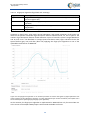
1
AQUAculture USEr driven operational Remote Sensing information services Deliverable 5.1 IS data quality control NIVA, WI, VU/VUmc, FFCUL, SGM 2014-11 AQUA-USERS is funded under the European Community’s 7th Framework Program (Theme SPA.2013.1.1-06: Stimulating development of downstream services and service evolution, Grant Agreement No 607325) D5.1: IS data quality control 30/11/2014 Task 5.1: Methods of quality control of IS data Deliverable 5.1: IS data quality control Lead beneficiary NIVA (5) Contributors NIVA(5), WI (1), VU/VUmc (2), FFCUL (4), SGM(8) Due date 31/09/2014 Actual submission date 1/12/2014 Dissemination level PU Change record Issue Date Change record Authors 0.1 09/09/14 Initial outline NIVA (KAS) 0.2 1.0 Initial draft 1/12/14 Final version NIVA, WI, VU/VUmc, FFCUL, SGM Consortium No Name Short Name 1 Water Insight BV WI 2 Stichting VU-VUMC VU/VUmc 3 Plymouth Marine Laboratory PML 4 Fundação da Faculdade de Ciências da Universidade de Lisboa FFCUL 5 Norsk institutt for vannforskning NIVA 6 DHI-GRAS GRAS 7 DHI DHI 8 Sagremarisco-Viveiros de Marisco Lda SGM To be cited as Sørensen, K., Johnsen, T., Ghebrehiwot, S., Poser, K., Eleveld, M.A., Sá, C., Fragoso, B.D.D., Icely, J.D. (2014) “IS data quality control”, AQUA-USERS deliverable D5.1, EC FP7 grant agreement no: 607325, 56p. © Copyright 2014, the member of the AQUA-USERS consortium. All rights reserved. 2 D5.1: IS data quality control 30/11/2014 Task objective (from DoW) The objective of this task T5.1 is to ensure high quality of in situ data entered into the AQUA-USERS database as a fundament for developing high-quality services for the aquaculture users. This subtask T5.1 is a part of WP5, which has the overall objective to ensure good routines for collection and storage of high quality in situ (IS) data from all of the involved users. IS-data from the users will be supplemented with IS-data gathered by the partners through field campaigns and Ferry box operations. Scope of this document The aim with this report is to give the best practice for in situ measurements, water sampling and analysis of parameters used by the partners in their field campaigns, and the users at the aquaculture sites. The parameters in focus are those needed for the developments of the products in AQUAUSERS like improved satellite products, algorithm development, development of indicator and input for the decision support tool. The partners are using different protocols depending of the local adjustments, different equipment and instrumentation. It is not the plan that the partner should adopt specific protocols, but to document the protocols in use and give a best practice guideline. AQUA-USERS has NOT the primary aim to do satellite validation even if many of the partners are using methods and protocols which also are used in the e.g. ESA validations programs. This means we have a more practical approach in this report and focus on what are the minimum requirements to fulfill the aim of the project. The following chapter is describing the best practices to perform an in situ measurement or analysis. Some of the methods are also used by end users and need to be practical in their form, but keeping the minimum requirements for a good method. One will in the following describe the most essential elements on the methods and give reference to more scientific material and eventually include important attachments. Abstract This deliverable gives an overview of the AQUA-USERS partner methods and summarizes some main best practices for the methods that will be used in the project. It is not the aim to include all details of the methods, but to point to the literature and other official protocols. The methods included cover i) use of some core water quality sensors where some are proxies for geophysical quantities, ii) analytical methods used on water samples and finally iii) optical methods to determine water reflectance to be used to validate the remote sensing algorithms. 3 D5.1: IS data quality control 30/11/2014 List of related documents Short Description Date D2.1 Initial user requirements document 31/01/2014 D2.3 GIS site selection application blueprint 28/02/2014 D2.5 WISP embedded software technote 30/04/2014 D2.6 Data policy guidelines report 30/04/2014 List of abbreviations Abbreviation Description AOP Apparent optical properties apig Pigment absorption BGC Biogeochemical BPA Bleached particle absorption CDOM Coloured dissolved organic matter Chl-a Chlorophyll-a CTD Conductivity, temperature and depth DO Dissolved oxygen FOV Field of view HAB Harmful algal bloom HPLC High-performance liquid chromatography IOP Inherent optical properties IS In situ L Luminance MERIS MEdium Resolution Imaging Spectrometer Rrs Remotes sensing reflectance TSM Total Suspended Matter UTC Coordinated universal time 4 D5.1: IS data quality control 30/11/2014 Notation and nomenclature for optical parameters and implementation in the optical model. For brevity spectral ( ) and angular factors ( , ) are usually left out (Adapted from Eleveld, 2012) Symbol b , bk Name, units total absorption, absorption for one of the individual optical components CHL, TSM, CDOM, m-1. specific absorption coefficients for any of the single optical components, for CHL m2 mg-1, for TSM m2 g-1, for CDOM m-1. total scattering, scattering for one of the individual the optical components, m-1. bk* specific scattering coefficients for any of the single optical components, for TSM m2 g-1. bb total backscattering, m-1 Ck concentration of a single optical substance, for CHL mg m-3, for TSM g m-3, for CDOM absorption normalized by CDOM absorption at 440 nm -. downwelling incident irradiance on a horizontal plane above the water surface, W m-2 nm1 . a reflectance model factor, -. a , ak ak* Ed0 f' k Kd, Ku KE K0 K ( , ) L0u v , a single optical substance such as chlorophyll (CHL), total suspended matter (TSM) or colored dissolved organic matter (CDOM), -. Vertical attenuation for downward irradiance, upward irradiance, m-1 Vertical attenuation for net downward irradiance, m-1 Vertical attenuation for scalar irradiance, m-1 Vertical attenuation for radiance, m-1 0 water-leaving radiance (the upwelling radiance measured above the water surface in the sensor viewing direction), W m-2 nm-1 sr-1. a factor that relates radiance below the water surface to irradiance below the water surface, -. Secchi disk depth (m) solar zenith angle, deg. v sensor view zenith angle, deg. rs wavelength of light, nm. remote sensing reflectance, sr-1. w0 water-leaving reflectance from a plane above the water surface, MERIS irradiance reflectance (-) relative sensor-sun azimuth angle, deg. Q zSD 5 D5.1: IS data quality control 30/11/2014 Table of contents 1 Introduction ................................................................................................................ 9 2 In situ measurements and inherent optical properties (IOP) .......................................10 2.1 Secchi Disc depth ......................................................................................................................... 10 2.1.1 Purpose of parameter ........................................................................................................... 10 2.1.2 Measurement principle and measurement challenges ......................................................... 10 2.1.3 Protocol ................................................................................................................................. 10 2.1.4 Quality control ...................................................................................................................... 10 2.2 Weather, colour of the sea and general field (metadata) observation. ...................................... 11 2.2.1 Purpose of parameter(s) ....................................................................................................... 11 2.2.2 Measurement principle and measurement challenges ......................................................... 11 2.2.3 Protocol ................................................................................................................................. 11 2.2.4 Quality control ...................................................................................................................... 11 2.3 Temperature and salinity measurements.................................................................................... 11 2.3.1 Purpose of parameter (s) ...................................................................................................... 11 2.3.2 Measurement principle and measurement challenges ......................................................... 12 2.3.3 Protocol ................................................................................................................................. 12 2.3.4 Quality control ...................................................................................................................... 13 2.4 Oxygen measurements ................................................................................................................ 13 2.4.1 Purpose of parameter ........................................................................................................... 13 2.4.2 Measurement principle and measurement challenges ......................................................... 13 2.4.3 Protocol ................................................................................................................................. 14 2.4.4 Quality control ...................................................................................................................... 14 2.5 pH measurements ........................................................................................................................ 14 2.5.1 Purpose of parameter ........................................................................................................... 14 2.5.2 Measurement principle and measurement challenges ......................................................... 14 2.5.3 Protocol ................................................................................................................................. 15 2.5.4 Quality control ...................................................................................................................... 15 2.6 Turbidity measurements .............................................................................................................. 15 2.6.1 Purpose of parameter ........................................................................................................... 15 2.6.2 Measurement principle and measurement challenges ......................................................... 15 2.6.3 Protocol ................................................................................................................................. 16 2.6.4 Quality control ...................................................................................................................... 16 2.7 Chl-a fluorescence measurements .............................................................................................. 16 6 D5.1: IS data quality control 30/11/2014 2.7.1 Purpose of parameter ........................................................................................................... 16 2.7.2 Measurement principle and measurement challenges ......................................................... 16 2.7.3 Protocol ................................................................................................................................. 18 2.7.4 Quality control ...................................................................................................................... 19 3 Analysis on water samples .........................................................................................20 3.1 Phytoplankton pigments .............................................................................................................. 20 3.1.1 Purpose of parameter(s) ....................................................................................................... 20 3.1.2 Measurement principle and measurement challenges ......................................................... 22 3.1.3 Protocol(s) ............................................................................................................................. 22 3.1.4 Quality control ...................................................................................................................... 23 3.2 Phytoplankton absorption ........................................................................................................... 23 3.2.1 Purpose of parameter(s) ....................................................................................................... 23 3.2.2 Measurement principle and measurement challenges ......................................................... 23 3.2.3 Protocol(s) ............................................................................................................................. 23 3.2.4 Quality control ...................................................................................................................... 24 3.3 Suspended material ..................................................................................................................... 24 3.3.1 Purpose of parameter(s) ....................................................................................................... 24 3.3.2 Measurement principle and measurement challenges ......................................................... 24 3.3.3 Protocol(s) ............................................................................................................................. 24 3.3.4 Quality control ...................................................................................................................... 25 3.4 Turbidity ....................................................................................................................................... 25 3.4.1 Purpose of parameter(s) ....................................................................................................... 25 3.4.2 Measurement principle and measurement challenges ......................................................... 26 3.4.3 Protocol(s) ............................................................................................................................. 26 3.4.4 Quality control ...................................................................................................................... 26 3.5 Coloured dissolved organic material ........................................................................................... 26 3.5.1 Purpose of parameter ........................................................................................................... 26 3.5.2 Measurement principle and measurement challenges ......................................................... 26 3.5.3 Protocol ................................................................................................................................. 26 3.5.4 Quality control ...................................................................................................................... 27 3.6 Phytoplankton abundance and composition ............................................................................... 27 3.6.1 Purpose of parameter ........................................................................................................... 27 3.6.2 Measurement principle and measurement challenges ......................................................... 27 3.6.3 Protocol ................................................................................................................................. 27 3.6.4 Quality control ...................................................................................................................... 28 7 D5.1: IS data quality control 30/11/2014 3.7 Nutrients ...................................................................................................................................... 28 3.7.1 Purpose of parameter(s) ....................................................................................................... 28 3.7.2 Measurement principle and measurement challenges ......................................................... 28 3.7.3 Protocol ................................................................................................................................. 29 3.7.4 Quality control ...................................................................................................................... 38 4 Apparent optical properties (AOP) measurements ......................................................39 4.1 WISP-3: hyperspectral radiances and reflectance, Chl-a, TSM, CDOM, Kd ................................. 39 4.1.1 Purpose of parameter ........................................................................................................... 39 4.1.2 Measurement principle and measurement challenges ......................................................... 39 4.1.3 Protocol ................................................................................................................................. 41 4.1.4 Quality control ...................................................................................................................... 42 4.2 TriOS hyperspectral radiometers ................................................................................................. 47 4.2.1 Purpose of parameter ........................................................................................................... 47 4.2.2 Measurement principle and measurement challenges ......................................................... 47 4.2.3 Protocol ................................................................................................................................. 49 4.2.4 Quality control ...................................................................................................................... 49 5 References .................................................................................................................50 6 Appendices ................................................................................................................54 8 D5.1: IS data quality control 30/11/2014 1 Introduction AQUA-USERS is strongly user driven to ensure sustainable and user-relevant services after the project. A pivotal part of the AQUA-USERS project is the collection and integration of in-situ data into the database and application. In close collaboration with the users, in-situ data will be collected at the users' production sites during the project period. These data include WISP-3 measurements, Secchi disc depth, cell counts, concentrations of pigments, solids and coloured dissolved organic matter, data on phytoplankton composition, data on environmental physical conditions (temperature, oxygen levels etc.) as well as the actual response of the aquaculture species (e.g. mortality, growth, yield, and fish behaviour) produced. Furthermore there will be additional in-situ data collected by some of the partners during the project period. Finally, whenever available, historical data from the users’ sites will be submitted along with data previously collected from relevant sites by the partners. Quality control of data is a crucial part of data management, and hence the data policy of the project (cf D2.6.). In the following, for each parameter that will be measured by the consortium partners and/or users, a description is given of The purpose of measuring the parameter, i.e. its relevance to aquaculture The principle behind the measurement and the challenges it provides The measurement protocols that are followed within AQUA-USERS The quality control procedures that are followed In some instances, data from national monitoring programs will also be used by the AQUA-USERS partners, these, however are not the subject of this report. 9 D5.1: IS data quality control 30/11/2014 2 2.1 In situ measurements and inherent optical properties (IOP) Secchi Disc depth 2.1.1 Purpose of parameter The threshold depth of observation for the Secchi disc is a direct measure of the vertical visibility in water, and it is one of several parameters used by environmental authorities to describe water quality. In some branches of aquatic science it is termed transparency. 2.1.2 Measurement principle and measurement challenges See Aas et al. (2014) for a historic summary of the Secchi Disc depth describing that Alessandro Cialdi, Commander of the Papal Navy, in 1866, published a report containing a section by Frater Pietro Angelo Secchi, where the factors influencing the visibility in the sea of submerged disks of different sizes and colourings were discussed (Secchi, 1866). In the years to come the white version of this device became a standard instrument in marine investigations. An important factor that enters the theory of the Secchi depth is the properties of the human eye as a contrast sensor. In Aas et al. (2014) the theory of the Secchi Disc Depth and its relationships to other quantities are described. Studies in air (Blackwell, 1946) have demonstrated that the human eye is able to distinguish a target from its background down to a lower limit or threshold value of the contrast between the target and its background. In our case the target is the Secchi disk, and the definition of the contrast C becomes C = (LD −L)/L, where LD is the luminance from the disk and L the luminance from the background. The depth is determined by the optical properties of the water and can therefore be related to these properties. Observations of the Secchi Disc depth can never be satisfactory substituted for direct recordings of the other optical properties, but they can serve as independent checks of these properties. In Aas et al. (2014) the different papers and experiment done by several scientists are discussed. Preisendorfer (1986) discussed the assumptions and limitations of the Secchi depth theory and procedure, using attenuation coefficients of photopic quantities. Originally the Secchi Disc depth was measured on the sun side of the ship, but there are some protocols and groups that measure on the shadow side. Aas et al. (2014) discuss the gains and losses related to the absence or presence of direct sunlight on the Secchi Disc. In average the Secchi Disc depth are reduced with 7% if one measure in shadow. 2.1.3 Protocol Today the marine standard method of measurement is to lower a white disk, with a diameter of approximately 30 cm, on the sunny side of the ship, supported on a cord and with its plane horizontal, from the ship rail and into the sea to a depth where the disk cannot be seen any longer. The disk is then hauled upwards to a depth where it can be recognized once again. The mean value of the two threshold depths is termed “the Secchi Disc depth”. As described the observation should be performed on the sunny side of the ship, but if this by some reason is not possible one should make a note on the condition of the measurements. Registration of time of the day, sun/shadow, wind speed, foam and discolouring of the sea. 2.1.4 Quality control If there are difficult measuring conditions make two recordings of the Secchi Disc Depth and if they differ more than 10% make a third recording. Take the average value of the two best observations and make note in the forms. It is also a good practice to make several recordings of the Secchi Disc Depth during the time at the station since this will help to understand any variability during any 10 D5.1: IS data quality control 30/11/2014 measuring campaign. The colour of the water can be registered using the Secchi Disc Depth. See also next Chapter 2.2. 2.2 Weather, colour of the sea and general field (metadata) observation. 2.2.1 Purpose of parameter(s) The colour of water is a complex optical feature, influenced by the composition of the natural water body and the illumination conditions. Recordings of foams and discolouring of the sea can be important for later interpretation of the data. Also the general observation metadata during a field campaign is important to register. 2.2.2 Measurement principle and measurement challenges Visual description of the colour is best made by describing the colour of the water column above a white disk, at half the Secchi disk depth, under shaded conditions. This requires that one also observe the Secchi Disc at the shadow side or make a note that the colour observation is recorded on the sunny side (cf section 2.1). The angle of observation should be kept close to nadir ≤ 42° (but not capture your own reflection), and somewhat with your back to the sun (preferably at an azimuth angle of either ca. 120° or 235°). The solar zenith angle should be < 70° (Van der Woerd et al., 2013). 2.2.3 Protocol A systematic recording of these quantities should follow a standard procedure using a standard form where all important factors that can influence the in situ measurements are recorded. Specific importance is also the time stamp of all observation on a ship, instrument deployment and automatic data recordings follows the same time zone (UTC is recommended). Depending of what ISobservation one includes in the campaign one should prepare a form that fits the purpose. An example of a field form is presented in Appendix A. In the appendix, some overview tables are shown of codes to be used for discolouring of the sea, sky code, sea state, surface code, visibility. For cloud coverage one uses the oktas scale where clear sky is 0 oktas and fully overcast is 8. 2.2.4 Quality control Before ending measurements on a station check that all recordings and important notes are performed. It is important to synchronize the timestamps of instruments and observation time as well as agree on the time zone (UTC). When ending the measurement, check the form and confirm that all measurements have been taken. Back up the data as soon as possible, preferably daily. 2.3 Temperature and salinity measurements 2.3.1 Purpose of parameter (s) For aquaculture, temperature is a key input for both site selection (cf D2.3) and for the management of an existing farm. For instance, food intake, growth and survival rates are significantly related to water temperature. Temperature is also one of the environmental factors that regulates phytoplankton growth rate (Cloern et al., 2014). Temperature (and salinity) also impacts the optics, notably the reflectance at the air-sea interface. It is also one of the oceanographic parameters defining water types, which are traditionally measured by a CTD sensor. Water temperature is therefore a parameter that is regularly measured by almost all of the users in the AQUA-USERS project, often on a daily basis (cf. D2.1). 11 D5.1: IS data quality control 30/11/2014 Figure 1. Primary productivity is the product of phytoplankton biomass (regulated by import, export, sinking, mortality, nutrient supply, and growth rate) times phytoplankton growth rate (regulated by light, temperature, and nutrient concentrations) (from Cloern et al., 2014) Salinity is important parameter that can affect stock species in aquaculture and therefore is a routine parameter to be registered by aquaculture farmers (cf. D2.1). As described by Hamer et al. (2008) seawater salinity variation (i.e., hypoosmotic stress) in the marine environment can affect various biological parameters of mussels, for example an increased oxygen consumption up to 58%. A tendency towards reduced growth with decreasing salinity, reflected as reduced shell growth rate and decreasing weight specific growth rate with falling salinity. (Riisgård et al., 2012). Salinity is an important parameter to be measured especially in mussel farming areas where big salinity fluctuations occur (e.g. estuaries, river runoff). 2.3.2 Measurement principle and measurement challenges Water temperature should be measured directly at the site, whenever possible, if not it should be measured as soon as possible, because sample temperature changes quickly after collection, especially in warm/cold atmospheric conditions. Salinity is most commonly reported using the Practical Salinity Scale 1978 (Lewis, 1980). Before development of the Practical Salinity Scale (PSS), salinity was reported in parts per thousand. Salinity expressed in the PSS is a dimensionless value, although by convention, it is reported as practical salinity units (PSU). Salinity in practical salinity units is nearly equivalent to salinity in parts per thousand (Wagner et al., 2006). More often, salinity is not measured directly, but is instead derived from the conductivity measurement (Wagner et al., 2006). Electrical conductance is a measure of the capacity of water (or other media) to conduct an electrical current. Electrical conductance of water is a function of the types and quantities of dissolved substances in water, but there is no universal linear relation between total dissolved substances and conductivity. Conductivity is defined as a measure of the electrical conductance of a substance normalized to unit length and unit cross section at a specified temperature (Radtke et al., 2005). 2.3.3 Protocol Measurements will be carried out according to instructions given by the manufacturer. There are many sensors on the marked like SeaBird CTD, SAIV STD, YSI multiprobes and WTW-instruments. As mentioned earlier in the document, salinity values are derived from the conductivity of water, using instruments like the CTD Seabird SBE SeaCat 19plus which include temperature, depth, PAR 12 D5.1: IS data quality control 30/11/2014 sensors as well a SBE 4 conductivity cell which can provide accurate readings up to large depth. For adequate maintenance and calibration, see the manufactures user’s manual (Sea-Bird, 2013). Using the WTW Profiline Condi 197i the calibration can be done by immersing the conductivity measuring cell in the 0.01 mol/l KCl (1413µS/cm at 25°C) control standard solution, in order to determine the cell constant. After the calibration, the measuring instrument automatically evaluates the current status of the calibration. A fixed cell constant 0.475 1/cm can be used or it can also be manually adjusted (WTW, 2009). After the calibration the instrument is ready to use and the measurement is done by introducing the conductivity measuring cell in the sample and wait enough time to allow for temperature adjustment in order to obtain a stable reading. The YSI multiparametric probe includes a number of useful sensors assembled in one cable, such as temperature, Chl-a fluorescence, O2, and pH. The probe can retrieve salinity, specific conductance or conductivity, calibration can be done for each one of these parameters. For adequate use details see manufacture’s Users Manual (YSI, 2009). 2.3.4 Quality control Instruments like CTD Seabird SBE SeaCat 19plus need to be shipped back to manufacturer for calibration checks from time to time. Recommendations for quality measurements (Radiometer Analytical, 2004): Conductivity is temperature dependent, for example the conductivity measured in a 0.01 mol/l KCl solution at 20°C is 1.273 mS/cm whereas, at 25°C, it is 1.409 mS/cm. To perform correct conductivity measurements, it is recommended to use a temperature sensor or a conductivity cell with built-in temperature sensor (Radiometer Analytical, 2004). For reliable conductivity measurements it is recommended to perform frequent calibrations, the cell constant value is an important factor of conductivity measurements, therefore the cell constant value must be checked before starting measurements. The temperature and stirring conditions during calibration should be as close as possible to the sample measurement conditions. Also is important to make sure that the measuring cell is totally covered by the sample (Radiometer Analytical, 2004). Probe maintenance and storage should be done according to the manufacturer’s manuals; but it is recommended that the cell is clean and rinsed with de-ionised water between samples measurement and before storage. After long term storage, condition the cell for 8 hours in de-ionised water before use. For salinity measurement the calibration should be carried out using a standard seawater solution K15 (STD) (salinity = 35, conductivity equals 42.896 mS/cm at 15°C) (Radiometer Analytical, 2004). A good practice to control the salinity is to take a water sample for control in the laboratory using a salinometer (e.g. Portasal type). A control diagram on the sensor should be established. 2.4 Oxygen measurements 2.4.1 Purpose of parameter Dissolved Oxygen (DO) is an important factor in chemical reactions in water and essential for the survival of aquatic organisms (Wagner et al., 2006). 2.4.2 Measurement principle and measurement challenges Sources of DO in surface waters are primarily atmospheric aeration and photosynthetic activity of aquatic plants (Lewis, 2005). Dissolved Oxygen is an important factor in chemical reactions in water and in the survival of aquatic organisms. In surface waters, DO concentrations typically range from 2 13 D5.1: IS data quality control 30/11/2014 to 10 milligrams per liter (mg/L). DO saturation decreases as water temperature increases, and DO saturation increases with increased atmospheric pressure. Occasions of super saturation (greater than 100-percent DO saturation) often are related to excess photosynthetic production of oxygen by aquatic plants as a result of nutrient (nitrogen and phosphorus) enrichment, sunlight, and warm water temperatures (Wagner et al., 2006). DO may be depleted by inorganic oxidation reactions or by biological and chemical processes that consume dissolved, suspended, or precipitated organic matter (Hem, 1989). The measuring process consumes DO; therefore, water flow past the sensor is critical. If the water velocity at the point of measurement is less than 1 foot per second (ft/s), an automatic or manual stirring mechanism is required (Wagner et al, 2006). Details on dissolved oxygen calibration, measurement, and limitations can be found in Lewis (2005). 2.4.3 Protocol Measurements will be carried out according to instructions given by manufacturer. Before use, a calibration needs to be carried out using the calibration vessel, OxiCal®-SL, following the manufacturer’s Operating Manual (WTW, 2004). For samples with salt content higher than 1g/L salinity correction is necessary (WTW, 2004). 2.4.4 Quality control After the calibration, the measuring instrument evaluates the current status of the probe against the relative slope. The evaluation appears on the display (WTW, 2004). As a quality control one could preserve a water sample and determine DO according to the Winkler titration methods. 2.5 pH measurements 2.5.1 Purpose of parameter Intensive aquaculture is known to cause impacts on the sea bottom; accumulation of organic matter under farming structures can induce a reduction on the pH on the sediment and surrounding water. This effect can cause impacts on the benthic community below the aquaculture sites. Measuring pH at one meter above the sea bottom and one meter below the surface can be a minimum measurement strategy. This is e.g. currently done at Sagres site to fulfill with the monitoring requirements demanded by the Portuguese authorities for the aquaculture site, to evaluate the changes on the sediment. At a global scale, in a world where there is increasing discussion on ocean acidification and its effects, pH may be an important parameter to measure at aquaculture sites with particular interest in bivalve aquaculture. Bivalve’s shells are made of calcium carbonate and decreasing pH may induce an additional stress factor as bivalves might spend additional energy for shell deposition, or to avoid shell dissolution. 2.5.2 Measurement principle and measurement challenges The pH of a solution is a measure of the effective hydrogen-ion concentration (Radtke et al., 2003). More specifically, pH is a measure that represents the negative base-10 logarithm of hydrogen-ion activity of a solution, in moles per litre. Solutions having a pH below 7 are described as acidic, and solutions with a pH greater than 7 are described as basic or alkaline. Dissolved gases, such as carbon dioxide, hydrogen sulfide, and ammonia, appreciably affect pH (Wagner et al, 2006). Measurements using pH electrodes are normally called the NBS scale (pHNBS), but one should be aware that in marine acidification and carbon system studies the pH definition are somewhat different and called the total scale (pHtot). 14 D5.1: IS data quality control 30/11/2014 2.5.3 Protocol With probes such as the WTW Multi 340i, pH measurements can be done with or without a temperature sensor as well as with the temperature sensor of an oxygen sensor or a conductivity measuring cell. The measuring instrument recognizes which sensors are connected and switches automatically to the correct mode for the temperature measurement (WTW, 2004). The pH sensors age with time, which causes changes in the asymmetry (zero point) and slope of the pH electrode. As a result, an inexact measured value is displayed. Calibration determines the current values of the asymmetry and slope of the electrode and stores them in the measuring instrument; for this reason is essential to calibrate at regular intervals (WTW, 2004). Normally a two-point calibration is done considering the range of the samples to be analyzed, using standard buffer solutions (pH values at 25 °C: 2.00 / 4.01 / 7.00 / 10.01) (WTW, 2004), the pH-7 buffer is used to establish the null point, and a pH-4 or pH-10 buffer is used to establish the slope of the calibration line at the temperature of the solution (Wagner et al., 2006). Expiration dates for the pH-4, 7, and 10 buffer solutions must be checked (Wagner et al., 2006). After calibration the probe is ready to use, to measure the samples pH is important to make sure that the probe is fully immersed and wait enough time for temperature to adjust and retrieve a stable measurement. After measurements the probe needs to be rinsed with distilled water; store the clean electrode in the watering cap that is filled with reference electrolyte (KCl 3 mol/L, Ag+ free) (WTW, 2010). 2.5.4 Quality control Regular checking of the instrument and sensor performance are done using pH standard buffer 4, 7 and 10. 2.6 Turbidity measurements 2.6.1 Purpose of parameter The turbidity gives an indication of the amount of particles in the water column and is a good proxy for the total suspended material (TSM). It is used in water quality studies and is common in many multiprobe sensors (e.g. YSI 6600) and in Ferrybox systems. Turbidity in open water may be caused by phytoplankton, runoff from land and re-suspension of bottom sediments. 2.6.2 Measurement principle and measurement challenges The most widely used measurement unit for turbidity is the Formazin Turbidity Unit (FTU). ISO standard 7027:1999 refers to its units as FNU (Formazin Nephelometric Units). Historically there have been several practical ways of checking water transparency (cf section 2.1 about Secchi Disc Depth). The most direct are to measure of attenuation of light as it passes through a sample column of water. The alternatively used Jackson Candle method (Jackson Turbidity Unit or JTU) is essentially the inverse measure of the length of a column of water needed to completely obscure a candle flame viewed through it. Modern instruments do not use candles, and the Jackson method was replaced by scattering methods. Particles’ optical property to scatter a light beam focused on them is now considered a more meaningful measure of turbidity in water. Turbidity measured this way uses an instrument called a nephelometer with the detector set up to the side of the light beam. More light reaches the detector if there are lots of small particles scattering the source beam than if there are few. The units of turbidity from a calibrated nephelometer are called Nephelometric Turbidity Units (NTU). Some older instruments used the unit Formazin Turbidity units (FTU), but now the ISO standard use Formazin Nephelometric unit (FNU). FTU, NTU and FNU are for practical use equivalent while JTU are not. The 15 D5.1: IS data quality control 30/11/2014 principle should follow the ISO standard EN-ISO 7027:1999, even if this standard refers to an laboratory instrument method for use for water samples. 2.6.3 Protocol There are many sensors on the marked (YSI, Seapoint), but one should secure that the specification of the sensors follows the principle of the ISO standard 7027:1999 which describes that the wavelength where the scattering are performed should be greater than 800 nm. Measurement at lower wavelength can be affected by water containing high concentration blue absorbing optical quantities like cDOM and phytoplankton. Otherwise one should follow the operation recommendation from the manufacturer. 2.6.4 Quality control One can use the small compact portable HACH 2100Q IS turbidimeter and measure the turbidity in parallel on a water sample from the same site as the sensor. The IS refers to the use the wavelength at 860 nm. Same type of instrument can be used for calibration of the instruments using Formazin standards. Standard formazin solution can be purchased, but if one needs large volumes this Formazin solution can be produced in your own laboratories. 2.7 Chl-a fluorescence measurements 2.7.1 Purpose of parameter Sensors for measuring Chlorophyll-a fluorescence are used to give a proxy for Chlorophyll-a. This is one of the most used biogeochemical sensors in marine research. 2.7.2 Measurement principle and measurement challenges Biogeochemical sensors (Jaccard, et al., 2014) often measure a proxy of the physical parameter, like Chl-a fluorescence as proxy for Chl-a or CDOM fluorescence for CDOM. In order to use or compare the measured data, the relationship between both has to be defined. In this section we will use measurements of Chl-a fluorescence from Ferrybox systems as case studies to illustrate the discussed scientific background. Measurements of in situ Chl-a fluorescence are also used on profiling fluorometers placed on CTD or in multiprobe sensors. The relationship between in situ Chl-a fluorescence and Chl-a concentration may vary between night and day time, between different growth stages of the algae population, and with the algae species composition. Therefore, the Chl-a fluorescence values cannot be directly used to determine the Chl-a concentration. However, water samples taken by e.g. Ferrybox system along the ship’s transect are used to determine the Chl-a concentration (by the HPLC method) for different conditions throughout the year. These data can then be used to study variations in the Chl-fluorescence to concentration relationship. This relationship was studied and reported in the EC-Ferrybox project (Sørensen et al., 2006). In that project it was found that an overall relationship for each year (encompassing all the abovementioned sources of variations) could be applied to make the Chl-a fluorescence values a proxy for the concentration. An overall relationship between Chl-a fluorescence and concentration is calculated for each year by linear regression between corresponding HPLC and fluorescence measurements. The Chl-a fluorescence can thereby be used as a proxy for the Chl-a concentration. The Chl-a fluorescence (CHLAFL) values can thereby be converted into Chl-a concentration (CHLACONC) by: CHLACONC = aCHL * CHLAFL + bCHL, where (aCHL) and (bCHL) are respectively a slope and offset of calibration. The seasonal and diurnal variation in the Chl-a_fluorescence/Chl-a ratio has been studied to improve the Chl-a_fl as proxy for 16 D5.1: IS data quality control 30/11/2014 the Chl-a concentration and to derive a better delayed mode calibration of the real-time chlorophylla fluorescence data. The comparisons are done by using water samples from fixed stations and compare with the Chl-a_fl after biofouling corrections. Figure 2 shows one example of water samples collected for such a “calibration”. In Figure 3 the scatterplot of all data from the period from 2003 to 2008 are shown. Figure 2. Chlorophyll-a fluorescence (red dots) and chlorophyll-a from water samples for one year (2011) at on station along a ship transect in the Skagerrak/Kattegat area. Some data from the monitoring programs are using spectrophotometric Chl-a analysis (SP) shown with black upward triangles, and points used for satellite validation are based on HPLC method, shown with grey downward looking triangles. Figure 3. Scatterplott (log-log) of all calibration points in the Skagerrak/Kattegat area 2003-2008. The data are 2 based on one common calibration per year. The coefficient of correlation are R = 0.653. One knows that Chlorophyll-a fluorescence is directly linked to the photochemistry of the alga as well as the species, so the seasonal and diurnal variations are large. This has led to an assumption that we can introduce a more seasonal calibration of the data. In Figure 4 a plot of the data based on the yearly calibration are plotted seasonally giving Chl-a_fl/Chl-a ratio variations of 3-4. This has led to the hypothesis that the Chl-a fluorescence could be calibrated on a seasonal basis rather than on a yearly basis and that also species (which contribute to the seasonal changes), night and day differences need to be considered. As seen in Figure 4 the ratio was >3 in winter and low during more productive periods. This is an effect of the high activity in the photosystem during productive periods giving a low Chl-a fluorescence relative to Chl-a. 17 D5.1: IS data quality control 30/11/2014 Figure 4. Seasonal plot of Chl-a_fl/Chl-a_HPLC ratio based on a yearly calibration of the data. Based on the seasonal calibration factor derived from the analysis of the data plotted in Figure 4, using a monthly calibration factor has been studied. The conclusions from this preliminary study showed that more advanced “calibration” procedures must be applied to the delayed mode data for using Chl-a fluorescence sensor data as proxy for Chl-a concentration. The study is based on Ferrybox data, but will also apply for in situ sensor data that measure directly under natural light condition (the Ferrybox data are somewhat dark adapted). For in situ sensor data like from profiling instruments the variation can be higher than shown for the Ferrybox sensor data. Examples for such profiles are shown in figure 5 illustrating that the Chl-a_F/Chl-a ratio in the surface (PAR >400) at one the same station during 24 hours varied with a factor 5-6. Figure 5. Vertical profiles of Chl-a, Chl-a_fl/Chl-a_HPLC ratio and PAR from a 24 hours measurement every 3 hours from same station in the Oslofjord area (Norway). 2.7.3 Protocol The commercial Chl-a fluorescence sensors on the marked operate with different calibration procedures both for the factory calibration and recommended procedures to be used by the operator. Each operator needs to pay attention to the procedures for their own sensor. The recommendation is to do a calibration against the algae that are expected to be present in the area of consideration or at least the most dominating species. The Chl-a fluorescence signal will vary 18 D5.1: IS data quality control 30/11/2014 depending on algae species, season, nutrient situation and light. In theory when one or more of the factors varies a new calibration should be performed. This is obviously not possible for an operational real-time system so one needs to agree on one system for their regional calibration and eventually introduce delayed mode calibration if one wants to correct for some of these factors. Moreover, the ratio between in vivo Chl-a fluorescence measurements and the Chl-a concentration based on in vitro HPLC or spectrophotometric Chl-a determination may vary with a factor 3-6 depending on various conditions described above. Hence, the method used to calibrate BGC-sensors will influence the measurements and lead to an additional factor that needs to be taken into account in the quality control routines. As an example, the Chl-a fluorescence sensor from Ferrrybox system is calibrated annually using a “standard” algae from NIVA’s algal culture collection. This is done by bringing a sample (in exponential growth phase) onboard the ship, and diluting the concentrated sample to a series of samples with Chl-a concentration within the range ~0.1 to 100 mg m-3. The Chl-a fluorescence sensor is removed from its cuvette and lowered into the water samples. The Chl-a concentration in the water samples are thereafter determined by the HPLC method, and compared by linear regression to the corresponding fluorescence sensor values. This calibration is applied to set or update the conversion factor (gain and offset) from the raw sensor values to the Chl-a fluorescence values (in mg m-3) stored in the log file. 2.7.4 Quality control To control the sensor some of the manufacturer has produced solid standards that routinely can be used to check the sensor drift. This is only for a long term quality control and cannot be used for calibration. Also the solid standards are different so one should use the same standard for the same sensor. It is also possible to use algal cultures for control, but this procedure is more laboriously. 19 D5.1: IS data quality control 30/11/2014 3 3.1 Analysis on water samples Phytoplankton pigments 3.1.1 Purpose of parameter(s) Chlorophyll a and other pigments (This introduction has been extracted from Sá, 2013) Phytoplankton contains three types of pigments involved in light harvesting and photoprotection: chlorophylls, carotenoids and biliproteins (Wright and Jeffrey, 2006). All photosynthetic phytoplankton contain one or more types of chlorophylls as part of the light-harvesting complexes in their chloroplasts, with chlorophyll a (Chl-a) being ubiquitous and commonly used as a biomass proxy. Chlorophyll a consists of magnesium coordination complexes of conjugated cyclic tetrapyrroles with a fifth isocyclic ring and often esterified long-chain alcohol (Figure 6). Other chlorophylls differ according to the oxidation state of the macrocycle, the type of side-chains, and the type of esterifying alcohol, if present. For instance, the Divinyl form of Chl, which can be found in Prochlorophytes, results from a substitution of an ethyl group into a second vinyl one. Figure 6. Chlorophyll a structure Many Chl a derivatives can be found both naturally and as artefacts of sample extraction or degradation. They may lose only the magnesium atom (pheophytins) or the phytol chain (chlorophyllides), or lose both the magnesium atom and phytol (pheophorbides). They may also spontaneously rearrange (epimers) or oxidize (allomers). Significant peaks of chlorophyllide a (Chlide a) are often seen in chromatograms because chlorophyllase enzymes can be activated when a cell is damaged (e.g., during filtration, storage or extraction). Significant degradation of Chl a may occur if the cells are left too long on the filter, frozen too slowly or not cold enough, or extracted in a solvent that does not inactivate the chlorophyllase. Chlide a concentration is generally included in the total Chl a fraction for biomass estimation. Carotenoids are a diverse family of yellow, orange or red isoprenoid, polyene pigments, which are involved in light-harvesting or in photoprotection. These pigments can absorb light in the blue and green parts of the spectrum (420-550 nm) and, although variable in amount as response to irradiance, are very useful taxonomically as some carotenoids can be exclusive of specific taxa. Pigment information can therefore be used to assess phytoplankton community structure at some level (e.g. Class). This method has been widely used in oceanographic studies (e.g., Barlow et al., 2008; Kyewalyanga et al., 2007; Leal et al., 2009; Mendes et al., 2007; Sá et al., 2013; Silva et al., 2008). A summary table of major pigments and its taxonomical occurrence are presented in Table 1 (Jeffrey et al., 1997). Phycobiliproteins, which can be of three subtypes: phycoerythrobilins, phycocyanobilins and phycourobilins, are generally the third type of light harvesting pigment, mostly found in cyanobacteria, rhodophytes and cryptophytes. However, biliproteins are water soluble and not extractable by organic solvents used in the analysis of chlorophylls and carotenoids. 20 D5.1: IS data quality control 30/11/2014 Table 1 List of most relevant pigments and their correspondent occurrence in phytoplankton communities (Jeffrey et al., 1997) Pigment Abbreviation Occurrence Chlorophyll a Chl a A proxy of algae biomass Divinyl chlorophyll a DvChl a Prochlorococcus sp Total chlorophyll a TChl a A proxy of total algae biomass Chlorophyll b Chl b Chlorophytes, prasinophytes Chlorophyll c3 Chl c3 Crysophytes and prymnesiophytes Chlorophyll c1+c2 Chl c1,c2 Diatoms, crysophytes, prymnesiophytes and dinoflagellates Fucoxanthin Fuco Diatoms, crysophytes and prymnesiophytes 19’Hexafucoxanthin Hexa Prymnesiophytes 19’Butafucoxanthin Buta Crysophytes and Prymnesiophytes Alloxanthin Allo Cryptophytes Zeaxanthin Zea Cyanobacteria and chlorophytes B-B-carotene B-car (Chl a + DvChl a) euglenophytes, and HAB specific pigments AQUA-USERS is mostly focused on phytoplankton species that can form blooms and be harmful to aquaculture production. An extensive list of toxins and pigments associated with harmful algae bloom species (HABs) is presented in the Appendix 14A of Roy et al. (2011). We here present a selected list (Table 2) of the species being considered in the framework of the AQUA-USERS project, which takes into consideration the blooms occurring in the location of the users aquacultures. Table 2. Pigments of species. Algal species Algal class Harmful effect Chlorophylls Carotenoids Chattonella antiqua Raphidophyceae Fish mortality, neurotoxic Chl a Fuco, viola Gymnodinium catenatum Dinophyceae PSP Chl a, Chl c2 BB-car, diadino, dino, Peri Karenia mikimotoi Dinophyceae Fish and invertebrate mortality Chla, Chla c1/2, Chl c3 But-fuco, BBcar, BE-car, diadino, Fuco, Gyro-e, Hexfuco Lingulodinium polyedrum Dinophyceae Toxic to shellfish Chla, Chlc Peri Phaeocystis globosa Prymnesiophyceae haemolysis Chla, Chl c1, Chl c2, Chl c3 But-fuco, BBcar, BE-car, Diadino, Diato, Fuco, Hex-fuco, Hex-kfuco Pseudonitzschia australis Bacillariophyceae ASP Chla, Chl c1, Chl c2 Fuco, Diadino 21 Other pigments MAAs MAAs B-car, D5.1: IS data quality control 30/11/2014 3.1.2 Measurement principle and measurement challenges Whatever the method chosen to determine algal pigments, the measure itself relies on spectroscopic characteristics: light absorption or fluorescence (Aminot and Rey, 2000). Chlorophylls exhibit two major light absorption bands, one in the blue part of the visible spectrum (460 nm) and one in the red (630-670nm). In discrete samples, photosynthetic pigments can be measured either by the traditional non-separative spectroscopic methods or after chromatographic separation, using HPLC. Spectroscopy allows a low-cost easier method to determine pigment samples, however, HPLC is often recommended for pigment studies since it provides, qualitatively and quantitatively, complete information on major phytoplankton pigments. Both methods though require filtering the water to obtain a concentrated sample of phytoplankton cells, filter storage and extraction of cells with an appropriate solvent prior to analysis. Storage temperature and time are critical points, as degradation of pigments can be generated at inadequate storage temperatures and the lower the temperature, the longer the storage time can be. The SCOR Working Group 78 concluded that storage at -18°C to -20°C would be acceptable only up to one week of storage. For periods up to one year, samples should be stored at temperature of liquid nitrogen (-196°C). Extraction of phytoplankton cells should be adequate in order to extract all pigments present in a sample as some algae are more difficult to extract than others. Planktonic diatoms and naked flagellates are easier to disrupt as opposed to armoured dinoflagellates, heavily silicated benthic diatoms, cyanobacteria or thick-walled green algae. Knowledge of the phytoplankton community of the area is helpful in making a decision. Acetone 90% is commonly used, however other solvents like ethanol or methanol are also used (Aminot and Rey, 2000). 3.1.3 Protocol(s) Spectrophotometric method For Chlorophyll determination, spectrophotometric measurements are limited to the red absorption bands as carotenoids have also strong absorption maxima in the blue. Problems also occur due to the degradation products. For instance, it is not possible to differentiate chlorophyllides. Pheopigments also show similar spectra but have a slight red shift and a decrease of the molar extinction coefficents that can be taken into account. There are two types of spectrophotometric methods suitable for routine use: trichromatic and monochromatic. The former have been developed to determine three types of chlorophyll (a, b and c) in the absence of degradation products. Absorbances must be measured at three wavelengths of the three Chls, plus a blank wavelength, then a set of three equations is used to calculate the concentrations. The equations of Jeffrey and Humphrey (1975) are the only ones recommended for the three chlorophylls (Aminot and Rey, 2000). The monochromatic methods are recommended for Chl a in coastal and estuarine waters. These methods have been developed to correct Chl a for pheopigments a. Absorbances are measured at the red maximum (plus a blank wavelength) before and after acidification. It is assumed that acidification degrades all chlorophyll-like pigments into pheopigments by eliminating the magnesium ion from the tetrapyrrole complex. The drop in absorbance allows both chl a and pheopigments a to be calculated. The correction equations for pheopigments have been published by Lorenzen (1967). Marker et al., (1980) discuss the monochromatic, both with and without correction for pheaopigments versus trichromatic methods and recommend to use the monochromatic methods . The monochromatic methods without pheaopigment corrections are also used in several national standards and some of the partners use those methods in their monitoring programs. 22 D5.1: IS data quality control 30/11/2014 HPLC High Performance Liquid Chromatography (HPLC) enables chemical separation (i.e. based on molecular polarity) and quantification of the pigments individually (i.e. even degradation products can be determined), allowing therefore a more accurate measurement. An accuracy for Chl in the order of <5% can be achieved with HPLC (Hooker et al., 2012). There is no unique HPLC method and several protocols have been developed by different authors depending on the number of pigments of interest and presence of different phytoplankton communities. In this project HPLC data from FCUL is analyzed following the method of Zapata et al. (2000). 3.1.4 Quality control It is strongly recommended to participate in intercomparison studies that are arranged among partners like in the satellite validation teams (Sørensen et al., 2007a) or in national or international laboratory performance studies for pigment analysis e.g. arranged by Quasimeme (www.quasimeme.org). Even if a set of few laboratories can achieve high accuracy (< 5%) normally a lower accuracy (< 20%) are common when many laboratories (> 15) with different methods are involved (Sørensen et al., 2007a). 3.2 Phytoplankton absorption 3.2.1 Purpose of parameter(s) The pigment absorption (APIG) and the bleached particle absorption (BPA) (using the MERIS acronyms) are determined to be used in the algorithm developments and to verify the satellite apig/Chl-a-ratios as well as contribute to the calculations of the non-pigment absorption (BPA+CDOM). 3.2.2 Measurement principle and measurement challenges After filtration of the water samples on a glassfiber filter the absorption coefficients for the unbleached and bleached filters are determined with an integrating sphere and calculated as described by Tassan and Ferrari (1995). To convert the result into the absorption of particles in a suspension a divisor of 2 (the so-called β factor (Doerffer, 2002) is applied. Pigment absorption apig is calculated as the difference between the absorption spectra of the unbleached and bleached filters, adjusting the whole spectrum of apig so that it becomes zero at 750nm. Bleached particles absorption at 442 nm, abp(442), is determined directly from the absorption spectrum of the bleached filter. This value is again added to ay(442), and this sum is defined as the yellow substance (YSBPA) in the MERIS protocol (Doerffer, 2002). The spectral shape of the bleached particle absorption is supposed to follow an exponential function (Montagner, 2001). Sørensen et al. (2007b) describe the methods used for NIVAs satellite products validation and the findings of the bio-optical relations for Skagerrak area. 3.2.3 Protocol(s) The protocol being used by both NIVA and FCUL is the one described in Tassan and Ferrari 1995, 2002. Shortly summarized the water samples should be filtered through 25 mm glass fibre filters (GF/F) from Whatman Inc. (0.7 μm retention efficiency). The diameter of the particulate material should be fitted to the actual integrating sphere used. Example for a 20 mm sphere (Labsphere model RSA-PE-20) a diameter of the particles retained on the filter is 15 mm. The transmission and 23 D5.1: IS data quality control 30/11/2014 reflection spectra of the filters were determined using a spectrophotometer with an integrating sphere. For the bleaching of the filters 3-4 drops of a solution of sodiumhypochlorite (0.1% active chlorine according to Ferrari and Tassan (1999)) are applied for approximately 5 minutes, then the filters is flushed with 5 ml of distilled water, and then measured. 3.2.4 Quality control Notes on the quality assurance of this protocol can be found in the REVAMP report (Tilstone et al., 2002). The spectra should be inspected and if one detects a significant peak around 665 this will indicate incomplete bleaching of the sample. Also the ratio of absorption at 443/665 should be less <1. 3.3 Suspended material 3.3.1 Purpose of parameter(s) The total suspended material (TSM) gives estimate of the total amount of particles in the water masses and comprises the organic particulate material (POM) and the inorganic fraction (PIM). 3.3.2 Measurement principle and measurement challenges Suspended solids in water are determined by gravimetric techniques, after filtering a certain volume of water sample throw a burned and pre-weighed filter; the filter is dried and weighed, and later is burned for 4 hours at 450°C and weighed again after cooling in a desiccator. The Total Suspended Matter is given by the weight of the dry filter subtracting the initial filter weight. The Particulate Inorganic Matter is given by subtracting the weight of the burned filter to the initial filter weight. Particulate Organic Matter is calculated as the difference between the Total Suspended Matter and the Particulate Inorganic Matter. The obtained weight values for each parameter, are divided by the correspondent filtered volume, results are expressed in mg/L. Total Suspended Matter = Particulated Organic Matter + Particulate Inorganic Matter Using a vacuum or pressure filtration apparatus, the sample is filtered through a glass-fibre filter. The filter is then dried at 105 °C and the mass of the residue retained on the filter is determined by weighing (ISO 11923, 1997). Some protocols operate with drying temperature down to 60 - 70 °C. 3.3.3 Protocol(s) The protocol that is used for Ria Formosa and Sagres samples is adapted from the ECASA Toolbox protocol for Particulate matter in seawater. (http://www.ecasatoolbox.org.uk/the-toolbox/eia-country/book-of-protocols/particulate-matter-inseawater). Pre-washed, ashed and weighed GF/F 47mm filters, prepared as below, stored in individual aluminium foil. Clean membrane forceps Freshly distilled water in wash bottle MilliQ water Filtration manifold with filter holders for 47mm filters Dessicator 24 D5.1: IS data quality control 30/11/2014 Drying oven Muffle furnace (450°C) Filter-preparation: a) To remove fine loose particles of filter, separate and soak in distilled water for > 1h; agitate and rinse 3-4 times in distilled water. b) Partially dry each filter on suction head to remove excess water (this prevents sticking to foil in the next step). c) Place filters individually into foil envelope/fan and oven dry overnight. d) Carefully number each filter on the exposed margin (soft lead pencil or pre-tested pen) and lay out (slightly overlapping) on foil tray, fit a lid and ash in muffle furnace at 450°C for >4h. e) Cool in desiccator; all handling of filters, from this point on, using clean (acetone) forceps only to avoid contamination. f) Remove individually and weigh to 5 places, standardizing the time it takes to weigh (filters increase in weight as they take up atmospheric moisture), and store place in numbered petrislides. Particulate Organic Matter and Total Particulate Matter determination: a) Filter the required volume of homogenized water sample (2L for Sagres, 1L for Ria Formosa) b) After the sample volume is filtered, add 50 ml of MilliQ water (3x) into the filtration cup with the pump running, to guarantee that salt is removed from the filter. Remove the filtration funnel and rinse carefully the rim of the filter under the funnel. c) Oven dry filters (60°C for 2 days, 40°C for 1 week) and store in desiccator. d) Weigh (from desiccator, to 5 places, as above, preferably with the same balance) for total suspended matter (TSM). e) Ash at 450°C in muffle furnace for > 4h f) Weigh (from desiccator to 5 places, as above, preferably with the same balance) for inorganic particulates (PIM). g) Do all of the above using at least 10 blank filters (prepared and processed as above, but without sample) for each experimental day (changes in weight before and after experimentation is used to correct for changes in balance calibration and/or filter water content). Absolute care in the preparation and processing of these filters as described is essential, for small errors in weight at these stages will significantly bias ratios and other results calculated later. 3.3.4 Quality control For quality control of Suspended Solids in waters the protocol is defined by the International Standard ISO Standard 11923:1997(E) Water Quality - Determination of Suspended Solids by filtration through glass-fiber filters; this protocol uses a reference suspension of microcrystalline cellulose ρ=500mg/L. 3.4 Turbidity 3.4.1 Purpose of parameter(s) The turbidity gives an indication of the amount of particles in the water column and is a good proxy for the total suspended material (TSM). Turbidity in open water may be caused by phytoplankton, runoff from land and re-suspension of bottom sediments. 25 D5.1: IS data quality control 30/11/2014 3.4.2 Measurement principle and measurement challenges The most widely used measurement unit for turbidity is the Formazin Turbidity Unit (FTU). ISO standard 7027:1999 refers to its units as FNU (Formazin Nephelometric Units). See section 2.6 for a historical overview and the use of turbidity sensors The principle should follow the ISO standard EN-ISO 7027:1999. There are several laboratory instruments on the marked, but one should secure that the ISO standard are used and that the principle of using 860 nm is fulfilled. 3.4.3 Protocol(s) One should follow the operation recommendation from the manufacturer. Be careful with the measuring cuvettes and clean for humidity on the outside of the glass. Be aware of error on large floating particles (zooplankton) that give errors in the readings. 3.4.4 Quality control Use standard Formazin solution purchased from the manufactory and calibrate as described in the instrument protocols. 3.5 Coloured dissolved organic material 3.5.1 Purpose of parameter The coloured dissolved organic material gives estimate of the optically measurable component of the dissolved organic matter in water. Also known as the chromophoric dissolved organic matter,[ yellow substance, gelbstoff or CDOM. 3.5.2 Measurement principle and measurement challenges Spectrophotometric determination of yellow substances The measurement of yellow substances (YS) in the samples and blanks, done at Sagres site, follows the Ocean Optics Protocols for Satellite Ocean Colour Sensor Validation (Revision 2). See the REVAMP Protocols (Tilstone et al., 2002). 3.5.3 Protocol The CINTRA dual beam spectrophotometer is used to record spectra for YS. Before measurements are taken, both field samples and the MilliQ water are left to adjust to room temperature. The 10 cm quartz path length cuvette is inspected for cleanliness before any measurements, and, if needed, soaked in 10% HCl and rinsed thoroughly with MilliQ water. The cuvettes, as well as the optical windows of the spectrophotometer, are cleaned with MilliQ water and dried thoroughly with lint free laboratory tissues. The instrument scan speed was programmed to 120 and to slit width 2, and a baseline was recorded between 350-800 nm. The blank spectrum is observed by filling the cuvette carefully with filtered MilliQ water to avoid bubbles and compared to the scan with that of air in the reference cell. After recording the spectrum, the MilliQ is discarded and the cuvette is rinsed three times with 5 to 10 ml of a field sample. The spectrum is recorded for this field sample under the same conditions used for the blank. To check the stability of the instrument, a MilliQ scan is run after completing the scans for the field samples. The data processing consists first in subtracting the MilliQ spectrum from the sample spectrum. The absorption coefficient, aYS, of dissolved organic matter is calculated from the measured absorbance, aYS, using the following equation (Icely et al, 2013). 26 D5.1: IS data quality control 30/11/2014 Where l is the cuvette path length. 3.5.4 Quality control The laboratory spectrophotometer should follow normal quality control routines for calibration. Carefully inspection of drift of blank spectra and rinsing of cuvettes are important. Several blank measurements during a measuring day should be performed (e.g. 1 blank every 10 sample). 3.6 Phytoplankton abundance and composition 3.6.1 Purpose of parameter The abundance and composition of phytoplankton is changing during the growing season. High concentrations of algae are normally advantageous for mussel producers, while for fish-farmers they may cause problems. Sometimes the occurrence of toxic or noxious algae can result in huge losses for the aquaculture industry and just low concentrations of some algal species can be disastrous. In all these cases it is vital to receive information of what algal species is causing the problem and in which concentrations they occur to actuate action if possible and different species needs different actions. To identify and quantify the algae, microscopic analyses is necessary, and to get comparable results within the whole region the phytoplankton should be analyzed in a uniform way. 3.6.2 Measurement principle and measurement challenges Sampling, preservation, and counting of algae can be done in several different ways. For that reason it is important to follow fixed routines to obtain comparable results. EN 15972:2011 Water quality – Guidance on quantitative and qualitative investigations of marine phytoplankton is a standard describing among other factors sampling procedures, needed equipment for sampling, species identification, and sample processing. For finding the phytoplankton abundance and composition in the AQUA-USERS project it seems most easy to follow a simple, but fixed procedure based on EN 15972:2011 that is giving both the users and the scientists the needed information. 3.6.3 Protocol For monitoring of phytoplankton, water samples can be collected either individually from fixed depths, as combined samples or as integrated samples adjusted to the hydrographical situation at each site and the aim of the investigation. The sampling frequency and duration has to be decided in each case according to the aim of the investigation. Water samples have to be stored in bottles made of material that does not affect the phytoplankton or the preservative before analysis and for longterm storage of samples the bottles has to be impermeable. The samples should be fixed immediately after sampling with neutral Lugol's solution (0.2 ml/100 ml sample), and stored in a cold, dark place for not more than 6 months. For quantification the sedimentation technique (Utermöhl method (fully described in EN 15204:2006, short version given in EN 15972, Annex F)) shall be used with subsequent analysis under inverse microscopy. After adaptation of the preserved phytoplankton samples to room temperature and gently homogenization of the bottles for 1-3 min subsamples of normally 10-50 ml is extracted into a tube placed over a horizontally orientated chamber with a transparent bottom plate. The sedimentation time for the samples is 8-24 hours depending on the volume of the subsamples. As a general rule, all phytoplankton species shall be identified to the lowest certain taxonomic level, and algae that cannot be identified to taxon/taxa level by using a regular microscope shall be grouped 27 D5.1: IS data quality control 30/11/2014 into size ranges within each class. Flagellates and other algae that cannot be identified to taxon, genus or class level shall be separated and grouped into agreed size classes. For each water sample a table comprising a taxa list should be recorded to the lowest certain taxonomic level and concentration per unit volume of the various taxa and taxon groups. To get information about the algal biomass in the water sample calculations of cellular carbon content has to be done according to Menden-Deuer and Lessard (2000). 3.6.4 Quality control Since there are not possible to have any reference material or similar like for chemical analysis it is therefore recommended to participate in inter calibration exercises and do parallel analysis to establish some error budgets for such analysis 3.7 Nutrients 3.7.1 Purpose of parameter(s) Natural enrichment with nutrients of coastal waters due to the occurrence of upwelling, important to explain the primary production at Sagres, where it is suggested that nitrogen is the most important nutrient regulating the microalgal growth, well as altering the relative microplanktonic composition in favour of diatoms (Loureiro et al, 2008). For the Ria formosa lagoon diatoms are also referred as the most sensitive group to nutrient enrichment (Loureiro et al, 2005). 3.7.2 Measurement principle and measurement challenges A review of the methods for nutrient analysis was done by Marta Zacarias, Priscila Goela and Alice Newton and assembled in a document (Zacarias et al, 2014); for the Sagres and Ria Formosa sites, based on the ISO for each method determination and on the book “Methods of seawater analysis” (Grasshoff et al, 1999). The methods described in the following are based on nutrients methods measured at Sagres and Ria Formosa: ammonium, nitrates, nitrites, phosphates, silicates. Different laboratories and partners could have small differences in the adopted methods according to their national monitoring programs. Ammonium The ammonium dissolved in seawater reacts with hypochlorite, donated by dichlorocyanuric acid, to form monochloramine which, in the presence of phenol, makes indophenol blue. The tri-sodium citrate solution acts as a buffer. The reaction is catalyzed by sodium nitroprusside. Nitrite The water nitrite determination ISO method is 6777:1984 - Water quality - Determination of nitrite Molecular absorption spectrophotometric method. This method is based on the reaction of an aromatic amine, leading to the formation of a diazonium compound which reacts with a second aromatic amine giving an azo compound. The method used is adapted for smaller volumes and is given in Grasshoff et al. (1999). Nitrate The water nitrate determination ISO method is ISO 7890-3:1988 – Water quality – Determination of nitrate – Spectrophotometric method using sulfosalicylic acid. The method used for nitrates determination is based on the reduction of nitrate by passing through a cadmium reductor column. Nitrate ions are reduced to nitrite ions. The nitrite concentration is determined. The yield of the reduction of nitrate depends upon the metal used in the reductor, on the pH of the solution and on the activity of the metal surface. The reaction is buffered with ammonium chloride to ensure a complete reduction and that reaction will not continue after the first 28 D5.1: IS data quality control 30/11/2014 product has been formed. The initial concentration of nitrite in samples has to be known and subtracted to this result after reduction of nitrate to nitrite. The method used is adapted for smaller volumes and is given in Grasshoff et al. (1999). Phosphate The ISO method for determination of phosphates in water is ISO 6878:2004 – Determination of Phosphorus – Ammonium Molybdate spectrophotometric method. All the methods for determination of inorganic phosphate in seawater are based on the reaction of the ions with a mixture of acidified molybdate and antimony tartrate, giving a phosphomolybdate complex. This product is reduced, by ascorbic acid, giving a bluish complex containing antimon. There may be some interference with dissolved silicate if the final reaction pH is greater than 1 or if measurements are made after 30 minutes. Therefore absorbance should be read after the addition of reagents. The method used was adapted for smaller volumes and is given in Grasshoff et al. (1999). Silicate The used method is based on the reaction of inorganic silicate with an acidic reagent of molybdate, giving a silicomolybdate complex. This complex is reduced, by ascorbic acid action, giving a blue silicomolybdic complex. This reaction is dependent of pH (pH 3-4) and there may be some interferences from some phosphate dissolved if the final pH is less than 3. This interference is removed by the addition of oxalic acid. The method used was adapted for smaller volumes and is given in Grasshoff et al. (1999). 3.7.3 Protocol Ammonium Equipment: Analytical Balance; Spectrophotometer ( UV-VIS), with 630 nm filter; Chemicals: Ammonium chloride (NH4Cl); Sodium Hydroxide (NaOH); Phenol; Disodium nitroprusside dehydrate (Na2Fe(CN)5NO.2H2O); Tri-sodium citrate dihydrate (C6H5Na3O7.2H2O); Dichloroisocyanuric acid. Reagents: Sodium hydroxide solution, 0,5 M: Dissolve 2 g of sodium hydroxide (NaOH) in bidistilled water, making up the volume to 100 ml. Store in a polyethylene bottle. Phenol Reagent: Dissolve 3,8 g of phenol and 40 mg of disodium nitroprusside dehydrate (Na2Fe(CN)5NO.2H2O) in bidistilled water, making up the volume to 100 ml. The solution should be stored in a refrigerator in a tightly closed amber glass bottle. Buffer Solution: Dissolve 24 g of tri-sodium citrate dihydrate (C6H5Na3O7.2H2O) in about 50 ml bidistilled water. Add 2 ml sodium hydroxide solution 0,5 M, making up the volume to 100 ml. The solution should be stored, in a refrigerator, in a polyethylene bottle. 29 D5.1: IS data quality control 30/11/2014 Oxidant Solution (Trione): Dissolve 20 mg of dichloroisocyanuric acid in 10 ml of sodium hydroxide solution 0,5 M. The solution should be used only during the next 24 hours. Procedure: Preparation of Standard Solutions Stock solution NH4 100 mmol.dm-3 Dissolve 535 mg of anhydrous ammonium chloride (NH4Cl), dried at 100ºC for 1 h, in bidistilled water. The solution is made up to a 100 cm3 volume with bi-distilled water. The solution should be stored cool (a refrigerator is not required but is preferable). Working solution NH4 500 mol.dm-3 3 Dilute 0.5 cm of the stock solution in bi-distilled water. The solution is made up to a 100 cm3 volume with bi-distilled water. The solution should be stored cool and dark (a refrigerator is not required but is preferable). Standard solutions Prepare adequate dilutions in order to get ammonium standard solutions of 0.5,1.0, 2.0 and 5 mol.dm-3. These ammonium standard solutions should be used to build the calibration curve through the least square method. The calibration curve will allow determining the concentration of ammonium in seawater samples Calibration In each test tube, put 5 ml of each standard solution, add 150 l of buffer solution (citrate solution), next add 150 l phenol reagent and 150 l oxidant solution. Mix well by swirling between additions. Close the test tubes and keep them in dark at least 6 hours. After, measure the absorbance, using glass cuvettes of 1 cm, at 630 nm. Each standard is analyzed in triplicate, with exception of the blank for which there are 10 replicates. Analysis of Samples The procedure described in calibration, with respect to used volumes, addition of reagents, waiting time of reaction and reading of absorbances should be used for analysis of samples. Example Table 3. Ammonium concentration in the standard solutions and respective absorbance. [NH4+] (M) 0.00 0.5 1.00 2.00 5.00 Abs 630 nm 0.003 0.049 0.086 0.172 0.416 30 Note: Absorbance values are the average of absorbances obtained in each of triplicates, with exception of blank, which was made in 10 replicates. D5.1: IS data quality control 30/11/2014 Figure 7. Graph of ammonium concentrations in the standard solutions and respective absorbance. Nitrites Equipment: Analytical Balance; Spectrophotometer (UV-VIS), with 540 nm filter. Chemicals: Sulphanilamide; Hydrochloric acid (HCl 37%); N-(1-naphtyl)-etylenediamine dihydrochloride solution (NEED) Reagents: Sulphanilamide solution: Dissolve 1g of sulphanilamide in a mixture of 60 cm3 bi-distilled water and 10 mL of hydrochloric acid (HCl 37%). After cooling, the solution is made up to a 100 cm3 volume with bi-distilled water. Store this reagent in the dark at < 8 ºC. The reagent is stable for at least one month. N-(1-naphtyl)-etylenediamine dihydrochloride solution (NEED): Dissolve 100 mg of NEED in bi-distilled water. The solution is made up to a 100 cm3volume with bi-distilled water. The solution should be stored in a brown bottle at < 8 ºC. The reagent is stable for more than a month and can be used until a brown discolouration occurs. Procedure: Preparation of Standard Solutions Stock solution 100 mmol.dm-3 Dissolve 690 mg of anhydrous sodium nitrite (NaNO2), dried at 100ºC for 1 h, in bi-distilled water. The solution is made up to a 100 cm3 volume with bi-distilled water. The solution should be stored cool and dark (a refrigerator is not required but is preferable). 500 mol.dm-3 Dilute 0.5 cm3 of the stock solution in bi-distilled water. The solution is made up to a 100 cm3 volume with bi-distilled water. The solution should be stored cool and dark (a refrigerator is not required but is preferable). Working solution 31 D5.1: IS data quality control 30/11/2014 Standard solutions Prepare adequate dilutions in order to get nitrite standard solutions of 0.1, 0.25, 0.5, 1 and 5 mol.dm-3. These nitrite standard solutions should be used to build the calibration curve through the least square method. The calibration curve will allow determining the concentration of nitrites in seawater samples. Calibration Put 5 cm3 of standard solution, in each test tube. Add 100 l Sulphanilamide solution and mix well by swirling. After 3 minutes, add 100 l NEED solution in the test tubes and mix again. The absorbance is read at 540 nm, using glass cuvettes of 1 cm. Each standard is analyzed in triplicate, with the exception of the blank for which there are 10 replicates. Analysis of samples The procedure described in calibration, with respect to used volumes, addition of reagents, waiting time of reaction and reading of absorbances should be used for analysis of samples. Example Table 4. Nitrite concentration in the standard solutions and respective absorbance. [NO2-] (M) 0.00 0.10 0.25 0.50 1.00 Abs 540 nm 0.000 0.017 0.038 0.076 0.146 Note: Absorbance values are the average of absorbances obtained in each of triplicates, with exception of blank, which was made in 10 replicates. Figure 8. Graph of nitrite concentrations in the standard solutions and respective absorbance. 32 D5.1: IS data quality control 30/11/2014 Nitrates Equipment: Analytical Balance; Spectrophotometer (UV-VIS), with 540 nm filter; pH meter; Activated Cadmium column (reductor). Chemicals: Sulphanilamide; Hydrochloric acid (HCl 37%); N-(1-naphtyl)-etylenediamine dihydrochloride solution (NEED); Ammonium chloride (NH4Cl); Concentrated ammonia (NH3); Potassium nitrate (KNO3). Procedure: Preparation of Standard Solutions Stock solution 100 mmol.dm-3 Dissolve 1.1011 g of anhydrous potassium nitrate (KNO3), dried at 100ºC for 1 h, in bi-distilled water. The solution is made up to a 100 cm3 volume with bi-distilled water. The solution should be stored cool and dark (a refrigerator is not required but is preferable). Working solution 1 mmol.dm-3 Dilute 1 cm3 of the stock solution in bi-distilled water. The solution is made up to a 100 cm3 volume with bi-distilled water. The solution should be stored cool and dark (a refrigerator is not required but is preferable). Standard solutions Prepare adequate dilutions in order to get nitrate standard solutions of 0.5, 1, 2, 5 and 10 mol.dm-3. These nitrate standard solutions should be used to build the calibration curve through the least square method. The calibration curve will allow determining the concentration of nitrites in seawater samples. Determination of the Efficiency of the redactor The practical efficiency of the reductor is usually somewhat less than 100 % but should not be less than 90 %. To determine the efficiency of the reductor, it should be compared the absorbance of diluted nitrate standard solution (10 mol.dm-3), passed through a cadmium column, with a standard solution of nitrite (5 mol.dm-3). The reductor efficiency should be calculated, using the following formula: The reductor efficiency should be checked after a set of 10 samples, passing the buffer through the cadmium column. If the efficiency reductor is below 90% it should be reactivated with concentrated nitrate solution. 33 D5.1: IS data quality control 30/11/2014 Calibration Put 5 cm3 of each standard solution in test tubes. Add 5 cm3 of ammonium chloride buffer solution. The content of each test tube should be passed through a cadmium reductor column, discarding first 5 cm3 and collecting the remaining 5 cm3 to the test tube again. Add 200 l sulphanilamide solution, and shake it. After 3 minutes, add to each test tube 200 l NEED solution, and mix well by swirling. The absorbance is read at 540 nm, using glass cuvettes of 1 cm. Each standard is analysed in triplicate, with the exception of the blank for which there are 10 replicates. Analysis of samples The procedure described in calibration, with respect to used volumes, addition of reagents, waiting time of reaction and reading of absorbances should be used for analysis of samples. Example Table 5. Nitrate concentration in the standard solutions and respective absorbance. [NO3-] (M) 0 0.5 1 2 5 Abs 540 nm 0.002 0.052 0.095 0.207 0.520 Note: Absorbance values are the average of absorbances obtained in each of triplicates, with exception of blank, which was made in 10 replicates. Figure 9. Graph of nitrate concentrations in the standard solutions and respective absorbance. 34 D5.1: IS data quality control 30/11/2014 Phosphates Equipment: Analytical Balance; Spectrophotometer (UV-VIS), with 880 nm filter. Chemicals: Potassium dihydrogen phosphate (KH2PO4); Concentrated sulphuric acid (H2SO4); Ammonium heptamolybdate tetrahydrate (NH4)6 Mo7 O24 . 4 H2O); Potassium antimony tartrate (K(SbO) C6H4O6); Ascorbic acid (C6H8O6). Reagents: Sulphuric acid, 9 mol.dm-3: Carefully add 25 cm3 concentrated sulphuric acid to 75 cm3 bi distilled water. After cooling, the solution is made up to a 100 cm 3 volume with bi-distilled water. Store in a polyethylene bottle. Molybdate solution: Dissolve 95 mg ammonium heptamolybdate tetrahydrate in bi-distilled water, the solution is made up to 10 cm3 volume with bi-distilled water. Store in a laboratory glass bottle. Tartrate solution: Dissolve 325 mg potassium antimony tartrate (K(SbO) C6H4O6) in bi-distilled water, the solution is made up to 10 cm3 volume with bi-distilled water. Store in a laboratory glass bottle. Mixed reagent: Add, carefully, 4,5 cm3 molybdate solution to 20 cm3 sulphuric acid 9 mol.dm3 , stirring continuously. Add, quickly, 0.5 cm3 tartrate solution and mix well. Store the solution in tightly closed amber glass bottle. This mixed reagent is stable for several months. Ascorbic acid solution: Dissolve 700 mg ascorbic acid in bi-distilled water, the solution is made up to 10 cm3 volume with bi-distilled water. The solution should be stored dark in a brown bottle at < 8ºC and it is stable for several weeks as long as it remains colourless. Procedure: Preparation of Standard Solutions Stock solution 100 mmol.dm-3 Dissolve 1.361 g of anhydrous potassium dihydrogen phosphate (KH2PO4), dried at 100ºC for 1 h, in 50 cm3 of bi-distilled water. Add 1 cm3 of sulphuric acid (H2SO4). The solution is made up to a 100 cm3 volume with bi-distilled water. The solution should be stored cold in a glass bottle and it is stable for several months. 500 mol.dm-3 Dilute 0.5 cm3 of the stock solution in bi-distilled water. The solution is made up to a 100 cm3 volume with bi-distilled water. Working solution Standard solutions Prepare adequate dilutions in order to get phosphate standard solutions of 0.1, 0.25, 0.5, 1 and 2 mol.dm-3. These phosphate standard solutions should be used to build the calibration curve through the least square method. The calibration curve will allow determining the concentration of phosphates in seawater samples. 35 D5.1: IS data quality control 30/11/2014 Calibration Put 5 cm3 of each standard solution in test tubes. Add 150 l mixed reagent and mix well. Add 150 l ascorbic acid solution and mix well again. The absorbance is read at 880 nm, using glass cuvettes of 1 cm. Each standard is analyzed in triplicate, with exception of the blank for which there are 10 replicates. Analysis of samples The procedure described in calibration, with respect to used volumes, addition of reagents, waiting time of reaction and reading of absorbances should be used for analysis of samples. Example Table 6 – Phosphate concentration in the standard solutions and respective absorbance. [PO43-] (M) 0 0.25 0.5 1 2 Abs 880 nm 0.000 0.023 0.047 0.101 0.206 Note: Absorbance values are the average of absorbances obtained in each of triplicates, with exception of blank, which was made in 10 replicates. Figure 10. Graph of phosphate concentrations in the standard solutions and respective absorbance. 36 D5.1: IS data quality control 30/11/2014 Silicates Equipment: Analytical Balance; Spectrophotometer (UV-VIS), with 810 nm filter. Chemicals: Concentrated sulphuric acid (H2SO4); Disodium hexafluoro silicate (Na2SiF6); Ammonium heptamolybdate tetrahydrate ((NH4)6Mo7O24.4H2O); Ascorbic acid (C6H8O6); Oxalic acid dehydrate (COOH)2 . 2 H2O. Reagents: Sulphuric acid, 7 mol.dm-3: Pipette 20 cm3 of concentrated sulphuric acid to 70 cm3 bi-distilled water. After cooling, the solution is made up to a 100 cm3 volume with bi-distilled water. Molybdate solution: Dissolve 20 g of ammonium heptamolybdate tetrahydrate ((NH4)6Mo7O24.4H2O) in bi-distilled water, the solution is made up to a 100 cm3 volume with bidistilled water. The solution should be stored in a polyethylene bottle protected from direct sunlight. Mixed reagent: Add 25 cm3 molybdate solution to 25 cm3 sulphuric acid, 7 mol.dm-3. The solution should be stored in a polyethylene bottle protected from direct sunlight. Ascorbic acid solution: Dissolve 175 mg of ascorbic acid in bi-distilled water. The solution is made up to a 10 cm3 volume with bi-distilled water. The solution should be stored in a polyethylene bottle, in refrigerator. Discard when a yellow tinge appears. Oxalic acid solution: Dissolve 1 g of oxalic acid in bi-distilled water. The solution is made up to a 10 cm3 volume with bi-distilled water. Store this solution in a polyethylene bottle at room temperature. Procedure: Preparation of Standard Solutions Stock solution 10 mmol.dm-3 Dissolve 188 mg of anhydrous disodium hexafluoro silicate (Na2SiF6), dried at 100ºC for 1 hour, in bi-distilled water. The solution is made up to a 100 cm3 volume with bi-distilled water. Transfer the solution immediately into a polycarbonate bottle (or high pressure polyethylene or polypropylene). The solution should be stored cool (a refrigerator is not required but is preferable). Working solution 1 mmol.dm-3 Dilute 10 cm3 of the stock solution in bi-distilled water. The solution is made up to a 100 cm3 volume with bi-distilled water. The solution should be stored cool (a refrigerator is not required but is preferable). Standard solutions Prepare adequate dilutions in order to get phosphate standard solutions of 1, 2, 5, and 10 mol.dm-3. These silicate standard solutions should be used to build the calibration curve through the least square method. The calibration curve will allow determining the concentration of silicates in seawater samples. 37 D5.1: IS data quality control 30/11/2014 Calibration Put 5 cm3 of each standard solution in test tubes. Add 150 l mixed reagent and mix well. Wait 15 minutes, add 100 l oxalic acid solution, and finally, add 100 l ascorbic acid solution. Test tubes would be shaken between additions. Wait about 3 hours before read the absorbances. The absorbance is read at 810 nm, using glass cuvettes of 1 cm. Each standard is analyzed in triplicate, with exception of the blank for which there are 10 replicates. Analysis of samples The procedure described in calibration, with respect to used volumes, addition of reagents, waiting time of reaction and reading of absorbances should be used for analysis of samples. Example Table 7. Silicate concentration in the standard solutions and respective absorbance. [(SiO4)4+] (M) 0 1 2 5 10 Abs 810 nm 0.000 0.023 0.042 0.110 0.218 Note: Absorbance values are the average of absorbances obtained in each of triplicates, with exception of blank, which was made in 10 replicates. Figure 11. Graph of silicate concentrations in the standard solutions and respective absorbance. 3.7.4 Quality control A normal quality control procedure are to use control samples (Control reference material, CRM) with relevant concentration ranges and establish a control chart system with alarm thresholds and action threshold. If the control results from running the CRM show one value outside the action threshold or 2 values out of 3 on the same side of the alarm threshold the analysis should be stopped until the error are found. 38 D5.1: IS data quality control 30/11/2014 4 4.1 Apparent optical properties (AOP) measurements WISP-3: hyperspectral radiances and reflectance, Chl-a, TSM, CDOM, Kd 4.1.1 Purpose of parameter The WISP-3 is a handheld hyperspectral radiometer for assessing surface water quality (see also D2.5). The three hyperspectral radiometers within the WISP-3 are used to measure upwelling and downwelling radiance (Lu and Ld) and irradiance(Ed), these measurements are combined to yield subsurface irradiance reflectance (R(0-)). This marine reflectance is a very important measurement to validate satellite observations of water quality. The WISP-3 also instantaneously derives biogeochemical water quality parameters from the spectral measurements including Chl-a and TSM. CDOM and Kd can be calculated using the accompanying web system WISPweb. The relevance of these parameters has been explained in the relevant sections in chapter 3. Figure 12: Performing a WISP-3 measurement 4.1.2 Measurement principle and measurement challenges The collector on top measures the down-welling irradiance (Ed) that is incident on the water surface. The two channels with gershun tubes at the front are used to determine the fraction of light that interacted with substances in the water. One of these collectors points downward at a 42 degree angle to capture upwelling radiance (Lu) that includes all light leaving the water as well as sky light reflected at the water surface. The collector that looks up at a 42 degree angle collects the downwelling radiance (Ld) or the sky light separately so that its influence on observed water color can be determined. The water colour or subsurface irradiance reflectance (R(0-)) is immediately calculated after each measurement by combining the information from the three measurements. The WISP-3 applies built-in water quality algorithms on the reflectance spectrum, resulting in concentrations of phytoplankton biomass (as chlorophyll-a), cyanobacteria biomass (as phycocyanin) and suspended sediments concentrations as well as the water transparency on its display. Under standard settings, the WISP-3 takes five measurements for each radiometer in a total of 3090s depending on the light condition. It calculates the average Ld, Lu and Ed and derives the average reflectance. It automatically corrects for dynamic dark readings, which are measured on a number of separate pixels that are not irradiated by external light. The radiance and irradiance are calculated from raw instrument counts according to the following equations: Lu (W m-2 nm-1 sr-1) = 0.01 × (counts × cal/t)/(A× dλ× Ω) -2 -1 -1 Equation 1 Ld (W m nm sr ) = 0.01 × (counts × cal/t)/(A× dλ× Ω) Equation 2 Ed (W m-2 nm-1) = 0.01 × (counts × cal/t)/(A× dλ) Equation 3 R(0-) = Q × f (Lu – r × Ld)/Ed Equation 4 39 D5.1: IS data quality control 30/11/2014 where cal is the calibration factor, t is the integration time of the measurement, A is the collection area (surface area of the optical fiber for radiance channels, and the surface of the cosine collector for the irradiance channel), dλ is pixel width, and Ω corrects for the solid angle of the radiance measurement (Ω = 2π[1cos(FOV/2)], where FOV is the 3° fieldofview). Q denotes the conversion coefficient for Lwu (upwelling radiance below water) to Ewu (upwelling irradiance below water), f is the conversion constant of Lu (upwelling radiance above water) to Lwu (upwelling radiance below water), r is the radiance of skylight at zenith angle of 42°. The challenge in developing reflectance algorithms is to relate light absorption and scattering properties of the substances that are present in a water body (e.g. phytoplankton pigments, dissolved matter) to their influence on water colour, at varying concentrations of each substance. In addition, the bulk attenuation of light by the combined substances is of prime interest to define the penetration of sunlight into the water column, fuelling aquatic photosynthesis. Various approaches to the inverse problem of water colour have been researched in the last decades, and it is important that the algorithms used to derive the concentrations of these substances from the measured reflectance are appropriately chosen. The algorithms that are built into the WISP-3 by default are considered suitable for a range of moderately to highly turbid water types, which includes a large number of lakes and other inland waters. Additionally, the algorithms provided through WISPweb are more complex and can handle an even wider range of water types. If the WISP-3 measurement is carried out properly, it is likely that an algorithm exists that can derive the concentrations of dominant optical substances. If a suitable algorithm does not exist, some algorithms can be tuned or trained to handle rare optical conditions. Most of the built-in WISP-3 algorithms target specific areas in the reflectance spectrum which correspond to wavelength ranges where the substance of interest has a large influence on the amount of reflected light, while other substances do not have much influence on the reflectance spectrum. The optical signals are extracted from differences between these spectral bands, or from band ratios against a reference bands. These algorithms typically target one to four bands simultaneously to solve the inverse problem, and are computationally inexpensive so that they can be embedded on an instrument such as the WISP-3. Advanced algorithms may use substantially more information from the reflectance spectrum, and use bio-optical models to match the full spectral absorption and scattering profiles of individual substances to the observed reflectance. The main advantage of these bio-optical models is that spectral information of individual substances can be easily changed to match locally expected conditions, such as red sediments or specific phytoplankton groups. However, the complexity of these models require more computing power. In the WISP-3 data processing chain, results from such algorithms become available only after uploading the measured data to WISPweb. The preliminary data that appear on the WISP-3 display are calculated using the band algorithms adopted from the literatures listed in Table 8. Parameter Reference Chlorophyll-a Gons et al., 2005 Total Suspended Matter Rijkeboer , 2001 Light attenuation Gons et al., 1998 Phycocyanin Simis et al., 2005, Simis et al., 2007 Table 8: Default algorithms used with the WISP-3, which can be adapted to local algorithm on request. 40 D5.1: IS data quality control 30/11/2014 4.1.3 Protocol Preparing the WISP-3: To measure enough light reflected in the water column, it is recommended to measure when the sun elevation is at least 30° above the horizon. Also, weather conditions are important: Sunny days are best, full cloud cover is usually fine, but low light due to thick rain clouds, as well as fog and rain are best avoided since the WISP-3 may not take a measurement due to lack of light or produce very noisy reflectances. Partially clouded conditions require careful positioning; making sure the sky radiance sensor is pointed towards the same type of sky that illuminates the water. Positioning the WISP-3: It is very important to operate the WISP-3 at the intended horizontal and vertical angles. The bubble level helps to keep the instrument level during measurements. Equally important is the angle towards the sun and possible shadows. The WISP-3 should be positioned 135° away from the sun, or in other words at 45° away from where shadows cast by the sun reach. There are thus two suitable angles, measured either clockwise or counter-clockwise from the sun. Reflections of the sun and the sky on the water surface are kept to a minimum when measuring at these angles. According to Mobley. (1999), the angle from the sun should at least be 90°, although closer to 135° is optimal. Angles < 90° (towards the sun) and ~180° (opposite to the sun) should absolutely be avoided. The correct position should be kept during the measurement, until the display indicates the measurement is finished by flashing the screen. If the sky is fully overcast and shadows are not visible, the angle is less critical but measuring towards the position of the sun is still not advised. Figure 13. Handling and positioning of WISP-3 during measurements Measurement conditions: It is important to stand close enough to the water so that the sensor looking down will actually be pointed at the water surface. Clear and completely overcast skies provide the best measurement conditions. Scattered clouds may hamper accuracy, because the light collector may point at the sky might not represent the same light as reflected on the water surface and captured by the downward looking sensor. If clouds are moving in and out of view, it is advised to wait a while for homogeneously open or closed cloud cover. Taking additional measurements is also recommended under doubtful conditions. Areas with floating vegetation, leaves, garbage, bottom visibility and shadows cast from boats or jetties are to be avoided. Waves can also interfere with accuracy, although this is normally sufficiently reduced by measuring in the correct direction relative to the sun. The WISP-3 averages five measurements, which further reduces the effect of the darker and lighter wave slopes. Boats, jetties, rafts, and bridges without superstructures can provide ideal locations. Performing measurements: To record a measurement, the 'Measure (#)' button has to be pressed. The display will show “Adapting to light”, followed by the percentage completion of the current measurement. It is important to keep the WISP-3 steady until the screen blinks several times to 41 D5.1: IS data quality control 30/11/2014 indicate that the measurement is finished. The measurement is automatically saved. The message “Not enough light” warns of low light conditions. This can be an indication of unsuitable weather conditions or a blocked sensor. If the WISP-3 is exposed to direct sunlight for some time on a warm day, the message too much light' may show. Place the instrument in the shade for a while to cool down. It is good practice to return the instrument to its case in a shaded spot when it is not in use. The WISP-3 will record measurements even when the solar elevation is not correct, when it is not kept horizontal, or pointed towards the sun. These considerations are the responsibility of the user, as with any measurement device. Viewing measurements: The display screen of the WISP-3 will show estimates of chlorophyll-a (Chl-a), phycocyanin (PC), light attenuation (Kd) and total suspended matter (TSM). It is also possible to view the reflectance spectrum on the screen. The WISP-3 saves its measurements on an SD card. These measurements can be uploaded to the WISPweb system (see Figure 13), where they can be visualized, analysed further and exported. In particular, the more advanced WISP algorithm (Peters, in preparation) can be used to derive water quality parameters. Figure 13. A glimpse inside WISPweb. 4.1.4 Quality control A WISP-3 is calibrated (absolute radiometric and wavelength calibration) once a year. If the instrument is damaged or broken then recalibration is recommended. Water Insight has implemented basic quality control flags (Figure 14) for the measurements uploaded on the WISPweb. Below is the detailed description of the type of flags applied on WISPweb. 42 D5.1: IS data quality control 30/11/2014 Figure 14. Flow chart of the Quality control flag types and steps Saturated and under-saturated spectra (signals): Once the raw-data (count) are uploaded to WISPweb, the data will be scanned for saturated or under-saturated signals. For WISP-3, which is 16bit resolution sensor we set saturation flag for signals >62000 counts and under saturation flag for signals <32768 counts. Figure 15. Flags to identify saturated and under-saturated signals Cloud radiance distribution: High cloud cover is one factor that might impede or at least influence derived reflectance spectra. Furthermore, in the process of deriving a reflectance spectrum, the surface-reflected sky radiance is subtracted from the water-leaving radiance. Especially for lake measurements, trees and buildings can be close to the water body and thus their reflection would end up in the measurement - which is not per se an issue, but it would be good to flag such measurements accordingly. Below two approaches to identify cloud distribution are explained. In these two approaches the down-welling light measurements, Ed and Ld, are used to derive a parameter that represents the cloud cover situation. A byproduct of the procedure is a flag for Ld spectra that are not exclusively observing sky radiance. The underlying idea here is that two different scattering processes may occur in the atmosphere, of which only one introduces spectral effects. In a clear sky, photons scatter mostly on gas molecules in the atmosphere, which are much smaller than their wavelength/energy - Rayleigh scattering. The size of water droplets, e.g. in clouds/haze/fog, are of approximately the same order than the wavelengths in visible light. Both, Rayleigh- and Mie scattering, are approximations of the Maxwell equations for different energy/size ratios of the 43 D5.1: IS data quality control 30/11/2014 involved photons and particles in which only Rayleigh scattering is wavelength dependent (λ-4), while Mie scattering, at least for visible wavelength, is spectrally neutral. That is why the sky is blue and clouds are white. Down-welling irradiance and sky radiance as typically measured for water reflectance spectra (Ɵ=450 ɸ=1350) should be spectrally different only due to their different composition of direct and diffuse/scattered light. The ratio Ld/Ed should therefore only dependent on the ratio of Rayleigh to Mie scattering. Approach 1. This flag selects or categorizes the type of cloud coverage as scattered, complete overcast and clear sky based on the normalized ratio of down-welling radiance to down-welling irradiance. The thresholds for the rations were validated using the sky photos taken during measurements. As mentioned above the clear sky plot is wavelength dependent while the cloud overcast plot is spectrally neutral. Table 9. Cloud detecting thresholds Normalized Ratio Range Flag type Ld(426nm)/Ed(426nm) <0.67 Cloud overcast Ld(550nm)/Ed(550nm) >0.25 Scattered cloud <0.25 Clear sky Figure 16. Cloud coverage classification Approach 2. Fit a model a (λ-4) + (1-a) to the ratio Ld/Ed (normalized by mean) for a random spectrum in the WISP database. This works quite well for the whole WISP database of >17k spectra. 44 D5.1: IS data quality control 30/11/2014 Figure 17. The plot still has a and b in it and was not normalized. Still, same result for the one-parameter model. The model parameter varies roughly from 0 to 1. The 'perfect' fits, e.g. R>0.99, are mostly represented by Rayleigh-scattering dominated skies (cloud free), whereas the spectra where the fit didn't work out perfectly (0.95<R<0.99), tend to be Mie-dominated. Especially the 'bad' fits (R<0.95) might result from obstacles in the FOV of the radiance measurements, e.g. trees or buildings, which introduce spectral features. In any case, these measurements should be regarded with care and flagged accordingly. At this moment, both cloud radiance distribution methods are tested on the WISPweb database. In the near future, the best methods will be implemented and the results will be integrated with the reflected skylight correction discussed below. Position of sun angle: Based on the UTM time registration during the measurement the position of sun angle is calculated on WISPweb. A flag will raise if the sun angle is lower than 30 degree. Repeated measurement accuracy: In one measurement, the WISP-3 takes the mean of five readings minimising the noise and error due to instrument stablization. When the user uploads spectra with repeated measurement (usually >=3 Spectra) then spectra of Ed , Ld , Lu will be flagged if their values at 550 nm differ by more than 25% (Ruddick et al. 2006). Correcting for the reflected skylight: Because of surface reflectance, Rrs or water-leaving radiance is challenging to measure from above the surface. It usually is estimated by correcting for the reflected skylight in the measured above-water upwelling radiance (Lu) using a theoretical Fresnel reflectance value. WISPweb uses the “Fingerprint algorthim” developed by Simis and Olsson (2013) to estimate the correct Fresnel value. It is based on the assumption that features in Rrs of water are spectrally smooth, whereas downwelling and reflected upward (ir)radiance contain a multitude of narrow features caused by gas absorption in the outer layers of the sun and the Earth atmosphere. In field measurements, when an unsuitable value for r is applied, these features can be recognized in resulting erroneous Rrs spectra. Reciprocally, the value of r can be optimized to minimize the presence of these features in Rrs, which is adapted in this approch. Because the atmospheric absorption features are both numerous and spectrally narrow, the optimization of r can be based on a series of these features without risk that the underlying absorption, scattering, or fluorescence features in Rrs influence the estimate of r (Simis and Olsson 2013). The optimization technique converges on a value of r labeled high, low, suspect and empty (table 10). 45 D5.1: IS data quality control 30/11/2014 Table 10. Fingerprint algorithm flags labels and meanings. Flag High Low Suspect Empty Meaning Flag raised if the fingerprint optmization terminates at the upper limit of r (to prevent negative Rrs) Flag raised if the fingerprint optmization terminates at the lower limit of r(0.0246) Flag raised if any value of rho within upper and lower limit would result in negative Rrs Flag raised if the fingerprint returns no band features. (r will be NaN) Variations in cloud cover, solar angle, aerosol absorption, and optical properties of the water can influence the position and width of gas absorption features observed in Ed, Lw, and Ls spectra. The number of gas absorption features found dominant in pairs of Lw and Ls spectra ranged between 2 and 16, with 11.3 ± 2.9 identified on average (Simis and Olsson 2013). Figure 18 below shows the spectra before (grey line) and after (blue line) applying the finger print algorithm to one of the uploaded measurements to WISPweb. Figure 18. The fingerprint algorithm is an iterative procedure to remove the effects of light reflected at the water surface from the reflectance spectrum. For more information refer to Simis and Olsson, 2013. (Blue line is the corrected spectra after applying the finger print algorithm). At this moment, the finger print algorithm is implemented in WISPweb but only for internal WI use. In the course of the AQUA-USERS project it will be made available to all users. 46 D5.1: IS data quality control 30/11/2014 Additional flagging: The table below shows additional flagging conditions recorded in WISPweb. Table 11. Other additional flags based on band ratio based on R(0-) values on WISPweb Band ratio and thresholds Flag If R(620)-R(560)/R(620)+R(560)>0 Extreme SPM If R(753.75)-R(681.25)/R(753.75)+R(681.25)>0 Floating layers and R(708.5)-R(681.25)/R(708.5)+R(681.25)>0 If R(753.75)-R(681.25)/R(753.75)+R(681.25)>0 Bottom visibility and R(620)-R(560)/R(620)+R(560)>0 4.2 TriOS hyperspectral radiometers 4.2.1 Purpose of parameter TriOS RAMSES radiance and irradiance hyperspectral radiometers are used to measure upwelling and downwelling radiance and irradiance. The purpose of these parameters is to determine marine reflectance. It is also possible to deduce other biogeochemical parameters from such measurements, as with WISP-3. However, these are not calculated automatically. On the other side TriOS sensors offer measurements in 190 channels in the range 320-950nm with high accuracy. They are also more sensitive to light and can be lowered below surface enabling underwater measurements as well. In the following the above water measurements performed from ship are described in more detail. Figure 19. Typical TriOS Ramses installation: irradiance Ed pointing toward zenith, radiances Ld and Lu sensor pointing in the same plane upward and downward with the opposite angles. 4.2.2 Measurement principle and measurement challenges The measurement principle follows what is decribed in chap. 4.1 for the WISP-3 for above water reflectance measurements. In this chapter there is focus on autonomous measurement from ships of opportunity systems (Ferrybox). Such installation is used by a few partner working in the validation community like NIVA Ferrybox network in Norwegian waters. 47 D5.1: IS data quality control 30/11/2014 For such an installation a set of 3 hyperspectral measurements is required: 1. downwelling radiance, Ld, instrument looking upward 2. upwelling radiance, Lu, sensor looking downward 3. irradiance, Ed, sensor looking towards zenith If installed on a vessel, measurements can be taken on station or underway. The former is well described in Ruddick et.al. (2006). The case of fixed installations can be considered as an on station measurement. Figure 20 show an arrangement of sensor for an intercalibration on a ship before a measurement campaign, and in Figure 21 a typical installation on a ship of opportunity system n Norwegian waters. Figure 20. Installation for on station measurements on board a vessel during an intercomparison exercise. Irradiance on the left and radiance the right. 48 D5.1: IS data quality control 30/11/2014 Figure 21. Installation on a ship of opportunity in Skagerrak. Radiance sensors can be mounted at different place on the ship. Azimuth and zenith/nadir angles must be match as close as possible the installation angles of both sensors. Both radiance sensors (Ld and Lu) should look in the same vertical plane with opposite zenith and nadir angles of the same value. Irradiance sensor (Ed) is place as high as possible in order to avoid shadow or hidden sky parts from surrounding structures. Measurements are taken by all three sensors at the same time. Sensor direction should not point towards shadow on sea surface, or towards sun glint. A key factor for successful measurements is stable light conditions. If data are to be compared with satellite measurements, clear sky conditions at time of overpass and sampling are necessary. 4.2.3 Protocol A general protocol for marine reflectance measurements is described by Zibordi et.al (2012). Specific protocol for on station measurements from a vessel is well described in Ruddick et.al (2006). For underway measurements, the operator may not have control of the ship’s heading, hereby the relative azimuth angle between the direction of measurement and the sun. This case requires some additional processing in order to select good measurements. Protocols for such measurement are developed under the ESA VAMP II contract (Jaccard, in prep.) In all cases, sensors should be checked and cleaned as often as the situation allows. TriOS also provides a field control lamp which can be used to illuminate sensors with a known spectra. While this cannot be used to calibrate them, it provides a good way to check their functionality. 4.2.4 Quality control Field calibrator should be used to monitor the drift and cleanness of the sensor optics. For underway measurements, a special processor was developed for NIVA in order to comment out data of lower quality, such as cloudy days, measurements from shadow or sun glint. Please refer to the deliverables of the ESA VAMP project (Santer et.al., 2014, In Jaccard, (In prep.)). 49 D5.1: IS data quality control 30/11/2014 5 References Aas, E., Høkedal, J. & Sørensen, K. (2014). Secchi depth in the Oslofjord–Skagerrak area: theory, experiments and relationships to other quantities. Ocean Sci., 10, 177–199. Aminot, A., & Rey, F. (2000). Standard procedure for the determination of chlorophyll a by spectroscopic methods. ICES Techniques in Marine Environmental Sciences (TIMES) publication No.31. ISSN 0903-2606. Barlow, R., Kyewalyanga, M., Sessions, H., van den Berga, M. & Morris, T. (2008). Phytoplankton pigments, functional types, and absorption properties in the Delagoa and Natal Bights of the Agulhas ecosystem. Estuarine, Coastal and Shelf Science, 80, 201 - 211. Blackwell, H.R. (1946). Contrast threshold of the human eye, J.Opt.Soc. Am., 36, 624-632. Cloern, J.E., Foster, S.Q. & Kleckner, A.E. (2014). Phytoplankton primary production in the world’s estuarine-coastal ecosystems. Biogeosciences 11, 2477–2501. Doerffer, R., (2002). Protocols for the validation of MERIS water products. European Space Agency Document no. PO-TN-MEL.GS-0043. Eleveld, M.A. (2012). Wind-induced resuspension in a shallow lake from Medium Resolution Imaging Spectrometer (MERIS) full-resolution reflectances. Water Resources Research 48(4). Ferrari, G.M. and Tassan, S., (1999). A methods using chemical oxidation to remove light absorption by phytoplankton pigments. Journal of Phycology, 35, pp 1090-1098. Gons, H.J., Ebert, J. & Kromkamp, J. (1998). Optical teledetection of the vertical attenuation coefficient for downward quantum irradiance of photosynthetically available radiation in turbid inland waters. Aquatic Ecology, 31, 299–311. Gons, H.J., Rijkeboer, M. & Ruddick, K.G. (2005). Effect of a waveband shift on chlorophyll retrieval from MERIS imagery of inland and coastal waters. Journal of Plankton Research, 27 (1), 125-127. Grasshoff, K., Kremling, K. & Ehrhardt, M. (Eds). (1999). Methods of seawater analysis, 3rd edition, Weinheim, Germany: Verlag Chemie. Hamer, B., Jakšić, Ž., Pavičić-Hamer, D., Perić, L., Medaković, D., Ivanković, D., Pavičić, J., Zilberberg, C., Schröder, H.C., Müller, W.E.G., Smodlaka, N., & Batel, R. (2008). Effect of hypoosmotic stress by low salinity acclimation of Mediterranean mussels Mytilus galloprovincialis on biological parameters used for pollution assessment. Aquatic Toxicology, 89(3), 137–151. Hem, J.D. (1989). Study and interpretation of the chemical characteristics of natural water: U.S. Geological Survey Water-Supply Paper 2254, 264 p. Hooker, S.B., Clementson, L., Thomas, C.S., Schlüter, L., Allerup, M., Ras, J., Claustre, H., Normandeau, C., Cullen, J., Kienast, M., Kozlowski, W., Vernet, S., Chakraborty, S., Lohrenz, S., Tuel, M., Redalje, D., Cartaxana, P., Mendes, C.R., Brotas, V., Prabhu Matondkar, S.G., Neeley, A. & Skarstad, E. (2012). The Fifth SeaWiFS HPLC Analysis Round-Robin Experiment (SeaHARRE-5). NASA Technical Memoradum 2012-217503, NASA Goddard Space Flight Center, Greenbelt, Maryland. Icely, J., Cristina, S., Goela, P., Moore, G., Danchenko, S., Zacarias, M. & Newton, A. (2013). Technical Assistance for the Validation of MERIS Marine Products at Portuguese Oceanic and Coastal Sites” - A summary of the project outputs between 2008-2012 (NEW CCN-CONTRACT NUMBER 21464/08/I-OL) FINAL REPORT Jaccard, P., Norli, M., Ledang, A.B., Hjermann, D.Ø., Reggiani, E,R., Sørensen, K., Wehde, H., Kaitala, S., Folkestad, A., (2014). MyOcean2. Real Time Quality Control of biogeochemical measurements, V2.4. 39 p. 50 D5.1: IS data quality control 30/11/2014 Jeffrey, S.W., & Humphrey, G.F. (1975). New spectrophotometric equations for determining chlorophylls a, b, c and c2 in higher plants, algae and natural phytoplankton. Biochemie and Physiologie der Pflanzen, 167, 191-194. Jeffrey, S.W. & Vesk, M. (1997). Introduction to marine phytoplankton and their pigment signatures. In: Jeffrey, S.W., Mantoura, R.F.C., Wright, S.W. (Eds.), Phytoplankton Pigments in Oceanography: Guidelines to Modern Methods. UNESCO Monogr.Oceanogr. Methodol., Vol. 10. UNESCO Publishing, Paris, 37 – 84 pp. Kyewalyanga, M.S., Naik, R., Hegde, S., Raman, M., Barlow, R. & Roberts, M. (2007). Phytoplankton biomass and primary production in Delagoa Bight Mozambique: Application of Remote Sensing. Estuarine, Coastal and Shelf Science, 74, 429 - 436. Leal, M.C., Sá, C., Nordez, S., Brotas, V. & Paula, J. (2009). Distribution and vertical dynamics of planktonic communities at Sofala Bank, Mozambique. Estuarine, Coastal and Shelf Science, 84, 605 616. Lewis, E.L. (1980). The Practical Salinity Scale 1978 and its antecedents: IEEE Journal of Oceanic Engineering, v. OE–5, 1, 3−8. Lewis, M.E. (2005). Dissolved oxygen: U.S. Geological Survey Techniques of Water-Resources Investigations, book 9, chap. A6, section 6.2, 34 p. Lorenzen, C.J. (1967). Determination of chlorophyll and pheopigments: spectrophotometric equations. Limnology and Oceanography, 12, 343-346. Loureiro, S; Icely,J; Newton, A (2005). Effects of nutrient enrichments on primary production in the Ria Formosa coastal lagoon (Southern Portugal), Hydrobiologia, November 2005, Volume 550, Issue 1, pp 29-45 Loureiro, S; Icely,J; Newton, A (2008). Enrichment experiments and primary production at Sagres (SW Portugal) Journal of Experimental Marine Biology and Ecology 359 (2008) 118–125 Marker, A.F.H., Nusch, E.A.,, Rai, H., and Rieman,. B. (1980). The measurements of photosynthetic pigments in freshwater and standardization of methods: Conclusions and recommendations. Arch.Hydribiol.Beih. Ergebn. Limnol. 14, 91-106. Menden-Deuer, S. & Lessard, E.J. (2000). Carbon to volume relationships for dinoflagellates, diatoms, and other protist plankton. Limnol. Oceanogr, 45(3), 569-579. Mendes, C.R., Cartaxana P. & Brotas, V. (2007). HPLC determination of microalgae pigments: comparing resolution and sensitivity of a C18 and a C8 method. Limnology and Oceanography: Methods, 5, 363 - 370. Mobley, C.D. (1999). Estimation of the remote-sensing reflectance from above-surface measurements. Appl. Opt., 38(36), 7442–7455. Montagner, F., (2001). Reference model for MERIS level 1 processing, European Space Agency, Document no. PO-TN-MEL-GS-0026. Preisendorfer, R. W. (1986). Secchi Disk Science: Visual Optics of Natural Waters. Limnology and Oceanography, 31, 909-926. Radiometric Analytical SAS (2004). Conductivity Theory and Practice D61M002. Villeurbanne, France: Analytical SAS, 49p. 51 D5.1: IS data quality control 30/11/2014 Radtke, D.B., Busenberg, E., Wilde, F.D. & Kurklin, J.K (Eds). (2003). pH (version 1.2): U.S. Geological Survey Techniques of Water-Resources Investigations, book 9, chap. A6, section 6.4, 28 p. Radtke, D.B., Davis, J.V., & Wilde, F.D. (Eds). (2005). Specific electrical conductance (version 1.2): U.S. Geological Survey Techniques of Water-Resources Investigations, book 9, chap. A6, section 6.3, 22 p. Rijkeboer, M. (2001) Optische teledetectie: algoritmenvoor het bepalen van de concentratie chlorofyl-a en zwevend stof. STOWA report (2001-5), Utrecht. Riisgård, H.U., Bøttiger, L. & Pleissner, D. (2012). Effect of Salinity on Growth of Mussels, Mytilus edulis, with Special Reference to Great Belt (Denmark). Open Journal of Marine Science, 2, 167-176. Roy, S., Llewellyn, C., Egelend, E. S. & Johnsen, G. (2011). Phytoplankton pigments: Characterization and applications in oceanography. Cambridge University Press. 874pp. Ruddick, K.G, De Cauwer, V., Park, Y.-J. & Moore, G. (2006). Seaborne measurements of near infrared water-leaving reflectance: The similarity spectrum for turbid waters. Limnology and Oceanography, 51(2), 1167–1179. Available at: http://www.aslo.org/lo/toc/vol_51/issue_2/1167.html. Santer, F. Zagolski, P. Jaccard, and K. Sørensen (2014). RAMSES-TriOS/Ferrybox Measurements with Concurrent MERIS/in-situ Reflectance Matchups - A New Protocol for in-situ Data Processing – In Jaccard, (Ed.), (In prep.). VAMP – Sky Dome Correction of above Water Radiometric Measurements. NIVA. Sá, C. (2013). Ocean Colour off the Portuguese Coast: Chlorophyll products validation and applicability. Ph.D. Thesis, University of Lisbon, pp. 228. Sá, C., Leal, M.C., Silva, A., Nordez, S., André, E., Paula, J. & Brotas, V. (2013). Variation of phytoplankton assemblages along the Mozambique coast as revealed by HPLC and microscopy. Journal of Sea Research, 79, 1-11. Silva, A., Mendes, C.R., Palma, S. & Brotas, V. (2008). Short time variation of phytoplankton succession, during one year, in Lisbon Bay (Portugal) as revealed by microscopy cells counts and HPLC pigment analysis. Estuarine, Coastal and Shelf Science, 79, 230 238. Sea-Bird (2013). Sea-Bird SBE 19plus V2 SeaCAT Profiler - Conductivity, Temperature, and Pressure Recorder with RS-232 Interface, User’s Manual. Washington, DC, USA: Sea-Bird Electronics. Secchi, A. (1866). Esperimente per determinare la transparenza del mare. In A. Cialdi (Ed), Sul moto ondoso del mare e su le correnti di esso specialmente su quelle littorali (258–288). Rome. Simis, S.G.H., Ruiz-Verdú, A., Domínguez-Gómez, J.A., Peña-Martinez, R., Peters, S.W.M. & Gons, H.J. (2007). Influence of phytoplankton pigment composition on remote sensing of cyanobacterial biomass. Remote Sensing of Environment, 106(4), 414–427. Available at: http://linkinghub.elsevier.com/retrieve/pii/S0034425706003518. Simis, S.G.H. & Olsson, J. (2013). Unattended processing of shipborne hyperspectral reflectance measurements. Remote Sensing of Environment, 135, 202–212. Simis, S.G.H., Peters, S.W.M. & Gons, H.J. (2005). Remote sensing of the cyanobacterial pigment phycocyanin in turbid inland water. Limnology and Oceanography, 50(1), 237–245. Available at: http://www.aslo.org/lo/toc/vol_50/issue_1/0237.html. Sørensen, K. (Ed) (2006). FerryBox- From On-line Oceanographic Observation to Environmental Information. Report on the use of FerryBox data for validation purposes of satellite data. Deliverable D-5-4. EU-project FerryBox, Contract no. EVK2-2002-00144. Sørensen, K., Aas, E. Høkedal, J. (2007). Validation of MERIS water products and bio-optical relations in the Skagerrak. (IJRS, Vol. 28, No 3-4, 2007). 52 D5.1: IS data quality control 30/11/2014 Sørensen, K., Grung, M. Röttgers, R. (2007). An intercomparison of in vitro chlorophyll a determinations for MERIS level 2 data validation. (IJRS, Vol. 28, No 3-4. 2007). Tassan, S. and Ferrari, G.M., (1995). An Alternative approach to absorption measurements of aquatic particles reatined on filters. Limonology and Oceanography 40. pp 1358-1368. Tassan, S., and Ferrari, G. M. (2002). Sensitivity analysis of the “Transmittance- Reflectance” method for measuring light absorption by aquatic particles retained on filters , J. Plankton Res., 24: 757-774. Tilstone, G. H., Moore, G. F., Sorensen, K., Doerffer, R., Rottgers, R., Ruddick, K. G., Pasterkamp, R. & Jørgensen, P.V. (2002). Regional validation of MERIS chlorophyll products in North Sea coastal waters. https://earth.esa.int/workshops/mavt_2003/MAVT-2003_802_REVAMPprotocols3.pdf Van der Woerd, H., Eleveld, M., Zielinski, O., Busch, J., Friedrichs, A., Wernand, M., Novoa, S., Piera, D., Simon, C., Bardají, R. & Bernard, E. (2013). Crowdsourcing technologies for the monitoring of the colour, transparency and fluorescence of the sea: Key scientific aspects of Quality Control. CitClops Deliverable 2.4 http://www.citclops.eu/the-project/public-deliverables. Wagner, R.J., Boulger, R.W., Jr., Oblinger, C.J. & Smith, B.A. (2006). Guidelines and standard procedures for continu¬ous water-quality monitors—Station operation, record computation, and data reporting: U.S. Geological Survey Tech¬niques and Methods 1–D3, 51 p. WTW (2009), Operating Manual, Cond 197i. Weilheim, Germany: WTW GmbH. WTW (2004). Operating Manual, Multi 340i, pH/Dissolved Oxygen/Conductivity Measuring Instrument. Weilheim, Germany: WTW GmbH. WTW (2010). Operating manual, SenTix, pH electrode with gel electrolyte. Weilheim, Germany: WTW GmbH. Wright, S.W. & Jeffrey, S.W. (2006). Pigment markers for phytoplankton production. In: Volkmann, J.K. (Ed.), Marine Organic Matter: Biomarkers, Isotopes and DNA. Springer-Verlag, Berlin, 71–104 pp. YSI (2009). Professional Plus, User Manual, Item # 605596, Yellow Spring, OH, USA: YSI Incorporated. Zapata, M., Rodriguez, F. & Garrido, J.L. (2000). Separation of chlorophylls and carotenoids from marine phytoplankton: A new HPLC method using a reversed phase C8 column and pyridinecontaining mobile phases. Marine Ecology Progress Series, 195, 2945. Zacarias, M., Goela, P., Newton, A (2014). Laboratory Protocols – Nutrients analysis, 3rd October 2014, University of the Algarve, Faro, Portugal. 16p. Zibordi, G., K. Ruddick, I. Ansko, G. Moore, S. Kratzer, J. Icely, and A. Reinart. (2012). In situ determination of the remote sensing reflectance: an inter-comparison Ocean Sci., 8, 567–586, 2012. 53 D5.1: IS data quality control 30/11/2014 6 Appendices 54 D5.1: IS data quality control 30/11/2014 MERIS-validation Norwegian Institute for Water Research - NIVA Project: Participants: Station: Location name: Position: Vessel: N: E: Date: Arrival UTC: Air temp.: Barometer: Hygrometer: Wind speed: Wind direction: Current speed: Current direction: Wave height: Wave direction: Wave type: Visibility: Cloud-cover: Surface material: Secchi UTC So Sg Sr Sb Ss Colour at ½ So Sun Shadow CTD File: UTC: STD No: Data line no: UTC: Water sample Secchi depth, So: 1/2 Secchi depth, ½ So Surface Depth (m): UTC: AC9 UTC: File: Hydroscat UTC: File: Ed UTC: File: Eu UTC: File: Edekk UTC: File; Secchi UTC So (White) Comments UTC So (White) UTC So (White) Sun Shadow Position: N: E: Date: Departure UTC: Air temp.: Barometer: Hygrometer: Wind speed: Wind direction: Current speed: Current direction: Wave height: Wave direction: Wave type: Visibility: Cloud-cover: Surface material: Save copy CTD STD BB6 AC9 PRR 55 Analysis ready: D5.1: IS data quality control 30/11/2014 Discolour code of the sea 0 No change 1 Red tides (red-brown) 2 Coccolithophores (milky blueish water) 3 Surface cyanophycea (ochre) 4 Phaeocystes foam (beige foam) 5 Other Sky code 0 Clear sky - no clouds 1 Thin cirrus, sun clearly visible 2 Thin cirrus and or persistend contrails, sun visible 3 Scattered cluds, sky coverage in octas, but area of measurement under sun 4 Scattered cluds, sky coverage in octas, but area of measurement in cloud shade 5 Mostly overcast, but sun is partly visible through clouds 6 Total overcast sky with high stratus clouds, sun not visible 7 Total overcast with low stratus, sun not visible 8 Other Surface code (used to evaluate satellite match up situation) 0 1 2 3 Ok - no influence Xcarcely influence Little influence Significant influence Air visibility 0 1 2 3 4 5 6 7 8 9 <50m 50-200m 200-500m 500-1000m 1-2km 2-4km 4-10km 10-20km 20-50km >50km Sea state 0 1 2 3 4 5 6 7 8 9 Calm-glassy Calm-rippled Smoth-wavelet Slight Moderate Rought Very rought High Very high Phenomenal 0 0-1dm 1-5dm 0.5-1.25m 1.25-2.5m 2.5-4m 4-6m 6-9m 9-14m >14m 56