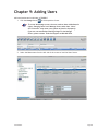
1
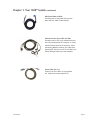
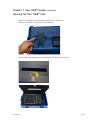
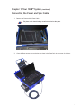
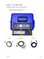
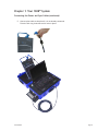
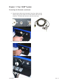
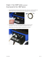
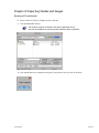
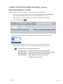
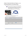
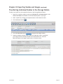
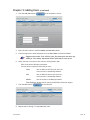
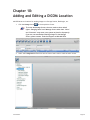
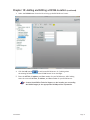
Tour 1008™
User Manual
YOUR IMAGE IS OUR BUSINESS.
Important Safety Information
Warning
There are no user serviceable parts inside.
Refer all servicing to qualified service personnel.
To reduce the risk of fire or electric shock, do not expose the unit to
moisture or water. Do not allow foreign objects to get into the enclosure.
If the unit is exposed to moisture, or a foreign object gets into the
enclosure, immediately disconnect the power cord from the wall. Take the
unit to a qualified service person for inspection and necessary repairs.
Caution
Read all the instructions before connecting or operating the component.
Keep this manual so you can refer to these safety instructions.
Heed all warnings and safety information in these instructions
and on the product itself. Follow all operating instructions.
Clean the enclosure only with a dry cloth or a vacuum cleaner.
Clean the DR plate with a damp cloth. Use no chemicals or alcohol.
You must allow 10 cm or 4 inches of unobstructed clearance around
the unit. Do not place the unit on a bed, sofa, rug, or similar surface
that could block the ventilation slots. If the component is placed
in a bookcase or cabinet, there must be ventilation of the cabinet
to allow proper cooling. Keep the component away from radiators,
heat registers, stoves, or any other appliance that produces
heat. Keep the component away from flammable materials.
The unit must be connected to a power supply only of the
type and voltage specified on the side panel of the unit.
Connect the component to the power outlet only with the supplied
power supply cable or an exact equivalent. Do not modify the
supplied cable in any way. Do not attempt to defeat grounding
and/or polarization provisions. Do not use extension cords.
Do not route the power cord where it will be crushed, pinched, bent at severe
angles, exposed to heat, or damaged in any way. Pay particular attention
to the power cord at the plug and where it exits the back of the unit.
Sound-Eklin®
09012012
Page i
Important Safety Information (continued)
The power cord should be unplugged from the wall outlet if the unit is to be
left unused for a long period of time. Immediately stop using the component
and have it inspected and/or serviced by a qualified service agency if:
•
•
•
•
•
The power supply cord or plug has been damaged.
Objects have fallen or liquid has been spilled into the unit.
The unit has been exposed to rain.
The unit shows signs of improper operation
The unit has been dropped or damaged in any way
Place the unit on a fixed, level surface strong enough to support its
weight. Do not place it on a moveable cart that could tip over.
System will become unresponsive when exposed to Electrostatic
Discharge (ESD) or Electrical Fast Transient (EFT) conditions.
This is the normal response to these environmental conditions.
Please restart the system to return to normal operation.
Specifications
Power Requirements
USA: 115 Volts, 60 Hz
Europe: 230 Volts, 50 Hz
Power Consumption
200 Watts
Dimensions (W x H x D)
20 x 14x 10½ inches, 508 x 355 x 267 mm
Weight (net)
37 lb., 16.8 kg
CAUTION
Risk of electric shock.
Do not open.
Caution: To reduce the risk of electric shock, do
not remove cover. No user-serviceable parts inside.
Refer servicing to qualified service personnel.
This symbol is to alert the user to
important operating and maintenance
(service) instructions in this manual and
literature accompanying the product.
Sound-Eklin®
09012012
Applicable for USA, Canada or
where approved for the usage.
Caution: To prevent electric shock, match wide
blade of plug to wide slot. Insert fully.
This symbol is to alert the user to the
presence of uninsulated dangerous voltages
inside the product’s enclosure that may
constitute a risk of electric shock.
Page ii
Table of Contents
Tour 1008™ User Manual
Important Safety Information ............................................................................... i
Warning...................................................................................................... i
Caution...................................................................................................... i
Specifications.............................................................................................. ii
Chapter 1: Tour 1008™ System Setup...................................................................... 1
Opening the Tour 1008™ Case......................................................................... 4
Connecting the Power and Sync Cables.............................................................. 5
Connecting the Generator.............................................................................. 9
Powering Up the Tour 1008™ System................................................................ 11
Chapter 2: Opening the Tour 1008™ Software.......................................................... 13
Chapter 3: Entering Patient Information................................................................ 14
Searching and Adding Patients ....................................................................... 14
Editing an Existing Patient’s Information .......................................................... 16
Chapter 4: Creating Studies................................................................................ 17
Creating a New Study for an Existing Patient ..................................................... 17
Adding New Images to an Existing Study............................................................ 18
Chapter 5: Acquiring Radiographs ........................................................................ 19
Choosing Anatomy and Positioning .................................................................. 19
Acquiring a Radiograph ................................................................................20
Cropping a Radiograph.................................................................................22
Chapter 6: Managing Protocols............................................................................24
Using Protocols...........................................................................................24
Adding New Protocols................................................................................... 27
Editing Existing Protocols ............................................................................. 31
Renaming Protocols..................................................................................... 32
Deleting Protocols.......................................................................................34
Chapter 7: Reviewing Images.............................................................................. 35
Reviewing a Study....................................................................................... 35
Chapter 8: Exporting Studies and Images...............................................................38
Burning a CD..............................................................................................38
Exporting Images to a Folder.......................................................................... 41
Transferring Studies to the Storage Station........................................................44
Transferring Individual Studies to the Storage Station...........................................46
Confirm Storage of a Study ........................................................................... 47
Chapter 9: Adding Users....................................................................................48
Chapter 10: Adding and Editing a DICOM Location.....................................................50
Chapter 11: Gain Calibration............................................................................... 53
Sound-Eklin®
09012012
Page iii
Chapter 1: Tour 1008™ System Setup
The Tour 1008™ works seamlessly with the X-ray generator to provide the best image quality in a
small, lightweight, self-contained system. The Tour 1008™ portable Acquisition Stations are intended
to be used by Veterinarians for intermittent mobile diagnostic applications. They will connect the
Acquisition laptop in the case to a Varian Amorphous Silicon Digital X‐Ray Detector (2520E+).
Sound-Eklin®
Page 1
Chapter 1: Tour 1008™ System
(continued)
The system comes with several components that integrate the X-ray machine, plate, and the
computer. If you need to order replacement parts, please refer to the part numbers in parenthesis.
Tour 1008™ Transit Case (70-309)
Wheel Kit for Tour 1008™ case (30-580):
Carries and protects the computer, plate, and
plate cable. Wheel kit allows for easy transport
in airports and on smooth surfaces.
2520E+ Plate (91-402):
Captures digital radiographs.
Handswitch (90-412):
Syncs the genrator’s prep and expose
cycle with the computer.
Ethernet Cable (90-403):
Connects the computer to the network to allow
for easy image transfers from the Acquisition
Station to the Storage Station.
Sound-Eklin®
Page 2
Chapter 1: Tour 1008™ System
(continued)
ODU Plate Cable (10-835):
Provides power to the plate and syncs the
plate and Tour 1008™ Transit System.
Generator Power Sync Cable (10-748):
Provides power to the X-ray machine and syncs
the X-ray machine with the computer. It comes
standard with a Min-X HF 80 connector. Other
connectors (Medison, Poskom, Vet X-Ray Ultra,
Ajex 90-15HF, Sternes Icon 90/20, and the Porta
1030 & 8020 generators) are also available.
Power Cable (10‐171):
Connects the Tour 1008™ to any standard
U.S. 110W power outlet (Nema 5-15).
Sound-Eklin®
Page 3
Chapter 1: Tour 1008™ System
(continued)
Opening the Tour 1008™ Case
1. Press in on the metal pin on each latch to open the Tour 1008™ Case.
With the pin pressed, lift the latch to unlock the case.
2. Open the plate door by pulling up on the silver tab and lifting the cover back.
Sound-Eklin®
Page 4
Chapter 1: Tour 1008™ System
(continued)
Connecting the Power and Sync Cables
1. Remove the Plate and the Plate Cable.
The plate cable should always remain attached to the plate.
2. Close the Plate storage door and place the plate in the Plate Park in the center of the door.
Sound-Eklin®
Page 5
Chapter 1: Tour 1008™ System
Connecting the Power and Sync Cables (continued)
3. Twist the knob counter clockwise on the Connector
Panel door to reveal the connection ports.
Power Cable
Sound-Eklin®
Generator Power Sync Cable
ODU Plate Cable
Page 6
Chapter 1: Tour 1008™ System
Connecting the Power and Sync Cables (continued)
4. Connect the ODU Plate Cable by aligning the red dot and notch on
the cable with the red dot and notch on the receptacle.
Sound-Eklin®
Page 7
Chapter 1: Tour 1008™ System
Connecting the Power and Sync Cables (continued)
5. Connect plate cable to the plate if it is not already connected.
Twist the lock ring clockwise until it locks in place.
Sound-Eklin®
Page 8
Chapter 1: Tour 1008™ System
(continued)
Connecting the Generator
The power sync cable should stay connected to the
generator and should be stored in the generator box.
1. Plug the Generator Power Sync Cable into the generator by connecting
the power adapter to the power port on the generator.
2. Plug the hand switch end of the Generator Power Sync
Cable into the generator’s hand switch port.
If using the system Internationally or on a 220 power, any device
plugged into the side power must be able to handle 220.
Sound-Eklin®
Page 9
Chapter 1: Tour 1008™ System
Connecting the Generator (continued)
3. Plug the Lemo cable on the Generator Power Sync Cable into the
case by lining up the red dot on the Lemo cable to the top of the
connector. To release the connector, pull out on the ring.
4. Plug the power cable into the case.
Sound-Eklin®
Page 10
Chapter 1: Tour 1008™ System
(continued)
Powering Up the Tour 1008™ System
1. Plug the female end of the power cable into the power cable slot on the Tour 1008™ box.
Plug the other end into a grounded power outlet (see page ii Power Requirements).
2. With the cables connected, turn on the Tour 1008™ by pressing the white
ON switch to “I” (O = off, I = on) located in the upper left corner. The
battery light on the laptop will light up, showing power to the unit.
Sound-Eklin®
Page 11
Chapter 1: Tour 1008™ System
Powering Up the Tour 1008™ System (continued)
3. Open the computer by sliding the gray clip on the front edge to the right and lifting the lid.
4. Turn on the laptop by pressing the Power button Sound-Eklin®
on the keyboard.
Page 12
Chapter 2:
Opening the Tour 1008™ Software
Open the Tour 1008™ software to begin the process of taking a radiograph.
1. Double-click the TruDRlx™ icon on the desktop. This will load
the TruDRlx™ software for acquiring digital radiographs.
2. Enter the user name and password when the login screen appears.
The default login is:
User Name: admin
Password: admin
The software will load directly into the Patient Screen
if the “Remember Me” box is checked.
Sound-Eklin®
Page 13
Chapter 3: Entering Patient Information
Searching and Adding Patients
Search for existing patients or add new patients easily by using the filtered search fields.
1. Search for a patient in the Patient Screen by entering the Patient ID, the Patient Name,
or the Owner Last Name. The patient list will automatically filter as you type.
Patient list is displayed based on the User Selected Default.
2. If you do not see the patient you are searching for, click the Add button or
press the Enter key on the keyboard to bring up the Add Patient Screen.
3. Enter the patient’s information. Patient Name and Last Name are required fields.
Sound-Eklin®
Page 14
Chapter 3: Entering Patient Information
Searching and Adding Patients (continued)
4. Click one of the Save buttons.
Save Saves the current patient and
returns you to the Patient Screen.
Save+ Add Saves the current patient and
returns to the Add Patient Screen.
Save+ Acquire
Saves the current patient then
opens the Anatomy/Protocol Screen.
Save+ Protocol Saves the patient and brings you to the Protocol
Screen to select the desired series.
Sound-Eklin®
Page 15
Chapter 3: Entering Patient Information
(continued)
Editing an Existing Patient’s Information
Edit an existing patient’s information for future DICOM tagging.
Previous exam information cannot be changed
1. Select a patient to edit from the Patient Screen.
2. Click the Edit button.
3. Edit the information as needed.
If the patient is from an owner previously entered, owner
can be selected from owner list on the left hand side.
4. Click the appropriate Save button (see page 15).
Sound-Eklin®
Page 16
Chapter 4: Creating Studies
Creating a New Study for an Existing Patient
Create a new study for a patient previously entered in TruDRlx™ .
1. Search for a patient in the Patient Screen by entering the Patient ID, the Patient Name,
or the Owner Last Name. The Patient List will automatically filter as you type.
2. Select a patient by clicking on the name in the Patient List.
3. Click the New button to create a new study.
4. Select the anatomy and the view from the Anatomy Screen (see page 19).
Sound-Eklin®
Page 17
Chapter 4: Creating Studies
(continued)
Adding New Images to an Existing Study
Add new images to an existing patient’s study.
1. Search for a patient in the Patient Screen by entering the Patient ID, the Patient Name,
or the Owner Last Name. The Patient List will automatically filter as you type.
2. Click the Acquire button.
This will take you to the Views Screen using the anatomy you selected
from the original study. This is shown in the description field.
Sound-Eklin®
Page 18
Chapter 5: Acquiring Radiographs
Choosing Anatomy and Positioning
Choose the anatomy and positioning to automatically enhance and position the radiograph.
1. Select the Tech and ordering Vet from the drop-down menus.
2. Move the mouse over the model to display anatomical region
of interest and select the appropriate anatomy.
3. Click on the appropriate button to select the view position.
This will bring you to the acquisition screen.
Sound-Eklin®
Page 19
Chapter 5: Acquiring Radiographs (continued)
Acquiring a Radiograph
1. Set the X-ray generator according to the Sound-Eklin® Technique Chart using
the appropriate KVP and mAs (depending on the X-ray generator).
Due to variations between X-ray generators, optimal techniques may vary
somewhat from the values printed on the Sound-Eklin® Technique Chart.
Users should be fully acquainted with State and local regulations
governing radiation protection and the use of diagnostic X-ray
equipment as well as the manual for the X-Ray Generator being used.
2. Align the generator 22 inches from the plate using the light beam
to fill the entire capture area defined on the plate.
If the generator is aligned perpendicular, the light will form
a rectangle with even sides filling the capture area.
The image capture area is highlighted by a white box
with the dotted cross-hairs in the middle.
3. From the Acquisition Screen press the hand switch halfway down to
prep and all the way down to expose the X-ray machine.
Proper Two-Stage Hand Switch Technique
1. To Prep, push the hand switch halfway down.
2. Wait until the Generator is ready to fire (i.e. prep
light on the X-ray machine is illuminated, etc.).
3. To expose, push the hand switch completely down.
Sound-Eklin®
Page 20
Chapter 5: Acquiring Radiographs
Acquiring a Radiograph (continued)
4. The image will appear in less than 3 seconds. Use the Left, Right, Flip, and Reverse
buttons to make orientation adjustments before shooting the next shot.
Crop the images as needed (see Pages 22).
If you need to reshoot your X-ray, press the hand switch and fire again.
5. Click the Anatomy button to change views.
6. Select the next view from the Views Screen. Click the Anatomy button
from the Views Screen if another anatomy is needed (see page 19).
7. Click the Patient button Sound-Eklin®
to return to the Patient Screen or click the Review button.
Page 21
Chapter 5: Acquiring Radiographs (continued)
Cropping a Radiograph
Remove unwanted portions of the radiograph.
1. Click the Crop button in the Acquisition Screen.
The new image will appear lighter after clicking the crop button.
The image will enhance once the cropping is complete.
2. Frame the desired image by holding the left mouse button in the upper left
corner of the image and dragging to the lower right hand corner.
Sound-Eklin®
Page 22
Chapter 5: Acquiring Radiographs
Cropping a Radiograph (continued)
3. The image will enhance once it is cropped.
4. Re-crop the image if necessary before taking the next
radiograph by pressing the crop button.
Images must be cropped before shooting the next image.
Sound-Eklin®
Page 23
Chapter 6: Managing Protocols
Using Protocols
Shoot a series of X-rays in a preset order which automatically
advances to the next shot after each shot taken.
1. From the Patient Screen, select the Patient and click Protocol.
Or, from the Patient Screen, click New and click Protocol.
OR, from the Add Patient or Edit Patient screen, click Save + Protocol.
(To add a Patient see page 14).
Sound-Eklin®
Page 24
Chapter 6: Managing Protocols
Using Protocols (continued)
2. Select a Protocol button to begin the protocol series.
3. The Acquisition Screen now has a Protocol Information bar showing the Protocol shot
number. The current Shot Information is located in the upper left portion of the screen.
Sound-Eklin®
Page 25
Chapter 6: Managing Protocols
Using Protocols (continued)
4. Shoot the first image. The software will automatically advance to the next shot in
the protocol series. The shot number in the Protocol Information Bar keeps track of
the shot sequence, while the current Shot Information appears in the upper left.
5. Use the Protocol Information bar to repeat a shot, skip a shot, restart the
protocol, move to the last shot in the protocol, or stop the protocol.
Move to the first shot in the protocol series
Repeat the previous shot
Skip to the next shot
Skip to the last shot in the protocol series
Stop the protocol
6. Click the Review button to open the Review screen (see page 35).
7. Click the Patient button to return to the Patient Screen.
Sound-Eklin®
Page 26
Chapter 6: Managing Protocols
(continued)
Adding New Protocols
Create new protocols for your individual studies.
1. Click the Manage button in the Patient Screen.
To access the Manage screen, the user needs to have Administrative
rights. Changing items in the Manage Screen other than “Users”
and “Protocols” may cause your system to function improperly.
If you are concerned about making changes in the Manage
Screen, please contact Technical Support at 800.268.5354.
2. Click on the Plus Sign next to the Protocols List to show the existing protocols.
3. Select the Protocols menu from the left side of the screen to load the Protocol Screen.
Sound-Eklin®
Page 27
Chapter 6: Managing Protocols
Adding New Protocols (continued)
4. Enter the name of your new protocol in the Protocol Name field.
5. Click the Save button.
6. Filter the shots as necessary by selecting the Filter by Laterality drop down menu.
Sound-Eklin®
Page 28
Chapter 6: Managing Protocols
Adding New Protocols (continued)
7. Select the first shot from the Shots for Species list.
8. Click the Link button to add the shot to the Shots Linked to Protocol box.
9. Repeat step 6 through 8 as necessary.
Sound-Eklin®
Page 29
Chapter 6: Managing Protocols
Adding New Protocols (continued)
10. Click the Unlink button to remove any shots
from the Shots Linked to Protocol box.
11. Select a shot in the Shots Linked to Protocol box and change the order
using the Move Up, Move Down, Move First, and Move Last buttons.
Moves your protocol shot up one shot in the series.
Moves your protocol shot down one shot in the series.
Moves your protocol shot from its location to the first shot in the series.
Moves your protocol shot from its location to the last shot in the series.
12. Click the Save button to save your protocols.
13. Click the Patient button to return to the Patient Screen.
If you have forgotten to save your protocols, a Save dialogue box will appear.
Click the Yes button to save your changes and return to the Patient Screen.
Sound-Eklin®
Page 30
Chapter 6: Managing Protocols
(continued)
Editing Existing Protocols
Edit current Protocols to customize them to your shot series.
1. Click the Manage button in the Patient Screen.
To access the Manage screen, the user needs to have Administrative
rights. Changing items in the Manage Screen other than “Users”
and “Protocols” may cause your system to function improperly.
If you are concerned about making changes in the Manage
Screen, please contact Technical Support at 800.268.5354.
2. Click on the Plus Sign next to the Protocols list to show the existing protocols.
3. Select a Protocol to edit from the left side menu, make changes and click Save.
4. Continue with Step 8 on page 29.
Closing the TruDRlx™ software and re-opening is
required to save protocol database changes.
Sound-Eklin®
Page 31
Chapter 6: Managing Protocols
(continued)
Renaming Protocols
Rename current protocols.
1. Click the Manage button in the Patient Screen.
To access the Manage screen, the user needs to have Administrative
rights. Changing items in the Manage Screen other than “Users”
and “Protocols” may cause your system to function improperly.
If you are concerned about making changes in the Manage
Screen, please contact Technical Support at 800.268.5354.
2. Click on the Plus Sign next to the Protocols List to show the existing protocols.
3. Select the Protocol to rename from the left side menu click the Rename button.
Sound-Eklin®
Page 32
Chapter 6: Managing Protocols
Renaming Protocols (continued)
4. Rename the protocol in the text box and click the OK button when finished.
5. Restart the TruDRlx™ software by clicking the close button in the upper right hand corner.
Sound-Eklin®
Page 33
Chapter 6: Managing Protocols
(continued)
Deleting Protocols
Delete current protocols.
1. Click the Manage button in the Patient Screen.
To access the Manage screen, the user needs to have Administrative
rights. Changing items in the Manage Screen other than “Users”
and “Protocols” may cause your system to function improperly.
If you are concerned about making changes in the Manage
Screen, please contact Technical Support at 800.268.5354.
2. Click on the Plus Sign next to the Protocols List to show the existing protocols.
3. Select the Protocol to delete from the left side menu click the Delete button.
4. Click the OK button to permanently remove the protocol.
5. Restart the TruDRlx™ software by clicking the close button in the upper right corner.
Sound-Eklin®
Page 34
Chapter 7: Reviewing Images
Reviewing a Study
Change the layout or delete any unwanted images before
the study is archived on the Storage Station.
1. Select a patient by clicking on the name in the Patient List.
2. Click on the Study from the list on the right.
3. Click the Review button.
4. Select a thumbnail in the left-hand pane to load the image into the review
pane on the right. The selected image is highlighted by a yellow box.
5. Review the images by using the following techniques:
Zoom: Use the LEFT and RIGHT arrow keys.
Hold the arrow keys down for continu-ous zoom.
Pan: Click and hold the left touch pad button and
move your finger across the touch pad.
Window Level: Click and hold the right touch-pad button
then move your finger diagonally across the touch pad.
Sound-Eklin®
Page 35
Chapter 7: Reviewing Images
Reviewing a Study (continued)
6. Use the toolbar for advanced review features.
Patient: Returns to the Patient Screen.
New: Opens the Anatomy/Protocol Screen for creating a new study or protocol.
Acquire: Opens the Views Screen using the anatomy you selected from the original study.
Anatomy: Opens the Anatomy/Protocol Screen to select
a new view to add into the same study.
Save: Saves the image with any changes that
have altered the original image.
Warning message will appear to choose “Yes” to overwrite
(replace) the image or “No” to create a new (additional) image.
Delete: Removes any unwanted images.
Deleting an image permanently removes it from the computer.
Revert: Reverts to the original image.
Left: Rotates the image 90˚ counterclockwise.
Right: Rotates the image 90° clockwise.
Flip: Flips the image vertically.
Reverse: Flips the image horizontally.
Magnify: Activates the magnifier which will zoom on the selected area.
Actions: Shows a drop-down menu with additional review features.
Win Level: Selects the window level feature
(also accessible by using the right mouse button).
Invert: Inverts blacks and whites, replicating a fluoroscopy.
Pan: Pans the image (also accessible by
using the left mouse button).
Crop: Trims an image without applying enhancements.
In the Review Screen, cropping does not re-enhance
the image based on the captured area. It only trims
the image without re-enhancing the image.
Sound-Eklin®
Page 36
Chapter 7: Reviewing Images
Reviewing a Study (continued)
Annotations: Shows a drop-down menu with annotations.
Display: Displays the annotations.
Hide: Hides the annotations.
Add: Adds annotations to the image using the
Annotation Toolbar (shown below).
Select: Selects an annotation button or previous annotation.
Line: Click and drag to draw a line.
Rectangle: Click and drag to create a square.
Ellipse: Click and drag to create a circle.
Text: Click and drag a box to open for text to be typed.
Ruler: Click and drag to draw a line measurement.
Poly Ruler: Create a measurement using multiple
lines by clicking at each point.
Protractor: To measure an angle, click the center of the angle,
then click the base line, then click the angle line.
Cross Product: Click and drag to create two
perpendicular lines of equal length.
Layout: Click a view box, then click the image to load.
1 Up: 1 image displays on the screen.
2 Up Horizontal: Image windows tile horizontally.
2 Up Vertical: Image windows tile vertically.
4 Up: 4 images display.
Overlay: Hide: Hides all DICOM information.
Summary: Displays patient and shot information.
Detail: Displays patient, shot, and additional detailed information.
7. Click the Patient button Sound-Eklin®
Opens menu for DICOM text display.
to return to the Patient Screen.
Page 37
Chapter 8: Exporting Studies and Images
Burning a CD
Burn a copy of a study to a CD.
1. Search for a patient in the Patient Screen by entering the Patient ID, the Patient Name,
or the Owner Last Name. The Patient List will automatically filter as you type.
2. Select a patient and a study. The patient and study are highlighted in a blue box.
3. Click the CD/DVD button.
4. Select either the DICOM or JPEG image format from the Export As: drop-down menu.
5. When selecting the DICOM option from Step 4, Click the Add DICOM viewer check
box only if the computer does NOT have a full DICOM viewer already installed.
The options below are unchecked by default.
Annotation Burnin
Anonymize Images
Repository Format Add DICOM Viewer
–
–
–
–
Imprints annotations made to saved images
Strips all client and clinic information from the images
Places images in a repository folder format
Adds a DICOM viewer on the CD to view the images on
a computer without a full DICOM viewer installed.
Efilm Lite and Reveal Lite are optional DICOM viewers to add.
Sound-Eklin®
Page 38
Chapter 8: Exporting Studies and Images
Burning a CD (continued)
6. When selecting the JPEG format from Step 4, select the
Overlay Burnin options from the drop-down menu:
The default option is set to None:
None:
Does not show overlays on the JPEG image
Detail:
Shows a detailed DICOM overlay on the JPEG image (shows Image
Number, Manufacturers Model Number, Study ID, Image Anatomy,
View Position, Image Laterality Extended, Columns X Rows,
Technique, Institution Name, Physician of Record, Patients Name,
Patient ID, Patient Breed Description, Patients Birth Date, Patients
Sex, Acquisition Number, Acquisition Date, Acquisition Time.)
Summary: Shows a summary DICOM overlay on the JPEG image (shows
Image Number, Image Anatomy, View Position, Image
Laterality Extended, Institution Name, Physician of Record,
Patients Name, Patient Breed Description, Patients Birth
Date, Patients Sex, Acquisition Date, Acquisition Time).
7. Click the Select button on the Export As dialog box.
Sound-Eklin®
Page 39
Chapter 8: Exporting Studies and Images
Burning a CD (continued)
8. Insert a blank CD (CD-R or CD-RW) into the computer.
9. Click the Write Disc button.
The CD burn progress is shown in the lower right hand corner.
You will be prompted to remove the disc when the burn is finished.
10. Click the OK button to complete writing the CD and remove the CD from the CD-Rom.
Sound-Eklin®
Page 40
Chapter 8: Exporting Studies and Images
(continued)
Exporting Images to a Folder
Export images from a study to a folder to save on a computer, thumb drive, etc.
1. Search for a patient in the Patient Screen by entering the Patient ID, the Patient Name,
or the Owner Last Name. The Patient List will automatically filter as you type.
2. Select a patient and a study. The patient and study are highlighted in a blue box.
3. Click the Folder button.
4. Select either the DICOM or JPEG image format from the Export As: drop-down menu.
5. When selecting the DICOM option from Step 4, Click the Add DICOM viewer check
box only if the computer does NOT have a full DICOM viewer already installed.
The options below are unchecked by default.
Annotation Burnin –
Anonymize Images –
Repository Format –
Create DICOM DIR –
Sound-Eklin®
Imprints annotations made to saved images
Strips all client and clinic information from the images
Places images in a repository folder format
Adds a DICOM directory to quickly import
all images into a PACS system.
Page 41
Chapter 8: Exporting Studies and Images
Exporting Images to a Folder (continued)
6. When selecting the JPEG format from Step 4, select the
Overlay Burnin options from the drop-down menu:
The default option is set to None:
None:
Does not show overlays on the JPEG image
Detail:Shows a detailed DICOM overlay on the JPEG image (shows Image
Number, Manufacturers Model Number, Study ID, Image Anatomy,
View Position, Image Laterality Extended, Columns X Rows,
Technique, Institution Name, Physician of Record, Patients Name,
Patient ID, Patient Breed Description, Patients Birth Date, Patients
Sex, Acquisition Number, Acquisition Date, Acquisition Time.)
Summary:Shows a summary DICOM overlay on the JPEG image (shows
Image Number, Image Anatomy, View Position, Image
Laterality Extended, Institution Name, Physician of Record,
Patients Name, Patient Breed Description, Patients Birth
Date, Patients Sex, Acquisition Date, Acquisition Time).
Sound-Eklin®
Page 42
Chapter 8: Exporting Studies and Images
Exporting Images to a Folder (continued)
7. Click the Select button
8. Select a location to save the images.
Images are saved to a selected location in a folder with the
animals name and the clients last name (e.g. Trigger-Smith).
Sound-Eklin®
Page 43
Chapter 8: Exporting Studies and Images
(continued)
Transferring Studies to the Storage Station
Transfer the studies from the Acquisition Station to the Storage/Review Station.
You must have your Tour 1008™ and your Tour 1008™ Review
Station networked. Please contact your local IT Person for
any networking needs and Sound-Eklin® technical support for
configuring DICOM Server for the TruDRlx™ Software.
1. Connect a Cat-5 Internet cable into the Internet port on the computer.
2. Press the F4 key on your keyboard to bring up the Store Files
to DICOM Server screen from the Patient Screen.
Sound-Eklin®
Page 44
Chapter 8: Exporting Studies and Images
Transferring Studies to the Storage Station (continued)
3. Click the Start Store button to begin the DICOM image transfer to the Storage Station.
This will store all the studies that were taken since your
last transfer and clear the queue. If any studies are
missing, you can transfer them individually.
4. Status box displays transfer results. When the status box states “completed,”
your images have successfully transferred to the DICOM server. Click the
Red close button in the upper right corner to close the screen.
Sound-Eklin®
Page 45
Chapter 8: Exporting Studies and Images (continued)
Transferring Individual Studies to the Storage Station
Transfer the studies from the Acquisition Station to the Storage/Review Station.
1. Search for a patient’s study by entering the Patient ID, the Patient Name, or the
Owner Last Name. The Patient List will automatically filter as you type.
2. Select a patient by clicking on the patient name in the Patient List field.
3. Click the Server button.
4. Click the Start Store button to begin DICOM image transfer.
5. Status box displays transfer results. When the status box states “completed”,
your images have successfully transferred to the DICOM server. Click the
Red close button in the upper right corner to close the screen.
Sound-Eklin®
Page 46
Chapter 8: Exporting Studies and Images
(continued)
Confirm Storage of a Study
Confirm the study successfully sent to the Storage/Review Station.
1. On the Patient Screen, the Archive field will show a server name (e.g. SOUND-SERV1).
2. Hold the mouse over the server name to show a pop-up window with the transfer data.
The pop-up box displays how many images have been transferred and the
date and time of the transfer. The information is color coded for easy
reference. If only one study appears, it will be highlighted in blue and the
color coding will not appear:
Green Box — Image transfer complete
Red Box — Image transfer failed
Sound-Eklin®
Page 47
Chapter 9: Adding Users
Add veterinarians and technicians to TruDRlx™.
1. Click the Manage button in the Acquisition Screen.
To access the Manage screen, the user needs to have Administrative
rights. Changing items in the Manage Screen other than “Users”
and “Protocols” may cause your system to function improperly.
If you are concerned about making changes in the Manage
Screen, please contact Technical Support at 800.268.5354.
2. Select the Users menu from the left side of the screen to load the Users Screen.
Sound-Eklin®
Page 48
Chapter 9: Adding Users
(continued)
3. Click the Add User button in the Acquisition Screen.
4. Enter the User’s name in the First Name and Last Name fields.
5. Create a unique user name and password in the User Name and Password fields.
Suggested user name: First initial of your first name plus last name (e.g.
jsmith for John Smith). Passwords can be left blank for ease of use.
6. Select a Group or Groups for each user by clicking Tech or Vet.
Each Group allows different permissions.
You may select multiple check boxes per user:
Tech: User is added to the Tech pull down list
has no access to the Manage features.
Vet: User is added to the Vet pull down list,
has no access to the Manage features.
Admin: Has full access to the Manage features.
Tech Support: Should only be used by Sound-Eklin® Technical Support
7. Click the Save button to add the User.
8. Repeat steps 3 through 7 to add additional Users.
Sound-Eklin®
Page 49
Chapter 10:
Adding and Editing a DICOM Location
Add DICOM server locations for sending images to a Storage Station, Radiologist, etc.
1. Click the Manage button in the Acquisition Screen.
To access the Manage Screen, the user needs to have Admin
rights. Changing items in the Manage Screen other than “Users”
and “Protocols” may cause your system to function improperly.
If you are concerned about making changes in the Manage
Screen, please contact Technical Support at 800.268.5354.
2. Select the Configuration menu from the left side of the screen to load the Users screen.
Sound-Eklin®
Page 50
Chapter 10: Adding and Editing a DICOM Location
(continued)
3. Select the DICOM button from the left to bring up the DICOM Server Screen.
4. Click the Add button to add a new DICOM server. Or if editing select
the existing DICOM server from the DICOM Servers list on the Right.
5. Enter the AE Title, IP Address, and Port number for your DICOM server. OR if editing
enter the corrected AE Title, IP Address, and Port number for your DICOM server.
Contact Sound-Eklin® Technical Support or the location you are sending
the DICOM images for the appropriate DICOM provider information.
Sound-Eklin®
Page 51
Chapter 10: Adding and Editing a DICOM Location
(continued)
6. Select additional information by choosing Yes from the drop-down menus.
Only select the following options if you are directed to
do so by a Technical Support representative:
Auto Send Server: Automatically sends images
to the selected DICOM location.
Allow Worklist: Designates the DICOM location as a worklist modality.
Telemedicine Server: Designates the DICOM
location as a telemedicine server.
Archive Server: 7. Click the Save button Designates the DICOM location as an archive server.
to save the current information.
8. Click the Verify button to verify that you can connect to the DICOM Server.
9. A smiley face will appear when you have verified the connection to the DICOM Server.
If a frowning face appears, please contact Sound-Eklin®
Technical Support at 800.268.5354.
10. Click the Patient button Sound-Eklin®
to return to the Patient Screen.
Page 52
Chapter 11: Gain Calibration
Calibrate the X-ray Generator and the plate.
Before beginning, make sure the TruDR software is closed
and that there is nothing on the digital panel. Remove
any X-ray grid that is directly on the panel.
1. Manually set the X-ray machine to the following settings
and open the collimator to it’s full size:
62 KVP
10 seconds
2. Set the collimator 40 inches (102 cm) from the plate and
ensure that the light field covers the entire plate.
3. Open the Viva program from the desktop using START > Varian > Paxscan > Viva.
4. Click the OK button on the status window when the program opens.
5. Click the Acquisition menu from the toolbar menus on top and select Gain Calibration.
6. Click the Continue button when the pop-up window appears.
The software will automatically begin an offset
calibration that will last about one minute.
Sound-Eklin®
Page 53
Chapter 11: Gain Calibration
(continued)
7. Shoot an X-ray when the screen prompts you to “Initiate X-ray exposure number: 1”.
8. Shoot again when prompted until you have reached a total of 8 shots.
9. Click the Finish button when the status box reads “Finish” OR “Initiate Next Exposure (#9)”.
10. Read the Gain Median in the pop-up window. The number should be between 1000 to 1500.
11. If the Gain Median is not between 1000 and 1500, repeat
steps 7-10 using a different technique.
Whether to increase or decrease your technique:
Gain Median is lower than 1000: RAISE the technique
Gain Median is higher than 1500: LOWER the technique
12. Click the Acquisition menu from the toolbar menus on top, and select Acquire Image.
13. When the text box pops up, shoot an X-ray using the same
technique you used to determine your gain median.
14. Examine the image to make sure it is a uniform milky-gray color.
15. Close the Viva software and open the TruDRlx™ software to begin shooting.
Sound-Eklin®
Page 54
Dr. Marc Laxineta, DVM
Sound-Eklin™ Technical Support
6359 Paseo Del Lago
Carlsbad . California 92011 USA
toll free: 800.268.5354 option 3
phone:760.918.9626
fax:76.918.9620
international:+1.760.918.9626
www.soundeklin.com
© 2012 All Rights Reserved
Sound-Eklin® — a VCA ANTECH company