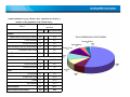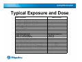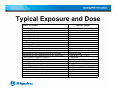
1
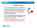
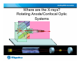
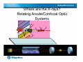
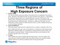
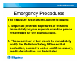
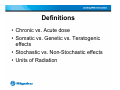
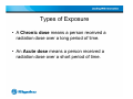
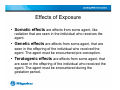
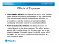
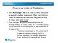
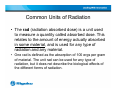
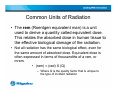
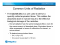
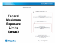
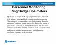
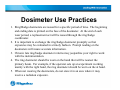
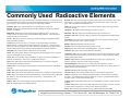
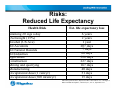
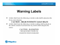
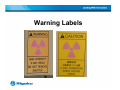
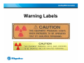
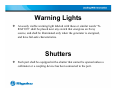
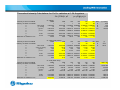
X-ray Radiation and Safety: What Everyone Should Know Kris F. Tesh, Ph.D. Director, Macromolecular Products Rigaku Americas Corporation 9009 New Trails Drive,The Woodlands, TX 77381-5209 (281)362-2300 http://www.Rigaku.com [email protected] Outline 1. Basics of X-ray Diffraction 2. Where are the X-rays 3. General X-ray Safety -definitions -procedures -videos -handouts 4. At the Instrument 5. Some Software Instruction Personnel Training All personnel involved in the installation, maintenance, repair or use of analytical X-ray units must be registered with the Radiation Safety Office. Prior to beginning work with an analytical unit, the user shall attend a radiation safety training session provided by the Radiation Safety Office. This session is intended to provide basic safety information and to introduce the administrative procedures of the Safety Office at Rigaku Americas Corporation. Detailed instructions on the operations, hazards and radiation warning devices of a specific analytical unit , must be provided by the owner of the equipment. Before starting to work on an analytical unit, make sure you receive specific instruction on the unit’s operation from the person responsible for the unit. General Radiation • Radiation is energy in transit in the form of high speed particles and electromagnetic waves. We encounter electromagnetic waves every day. They make up our visible light, radio and television waves, ultra violet (UV), and microwaves with a spectrum of energies. These examples of electromagnetic waves do not cause ionizations of atoms because they do not carry enough energy to separate molecules or remove electrons from atoms. General Radiation • Ionizing radiation is radiation with enough energy so that during an interaction with an atom, it can remove tightly bound electrons from their orbits, causing the atom to become charged or ionized. Ionizing radiation deposits energy at the molecular level, causing chemical changes which lead to biological changes. These include cell death, cell transformation, and damage which cells cannot repair. Effects are not due to heating. General Radiation • X-rays are a form of ionizing radiation. They are electromagnetic waves emitted by energy changes in electrons. These energy changes are either in electron orbital shells that surround an atom (Rigaku FRE+ or Micromax 007HF generators) or in the process of slowing down (synchrotron). General X-ray • X-rays are produced from the excitation of electrons followed by the cascading of these electrons back down to the ground state • The typical X-rays used in crystallography range from 0.6 to 2.5Å • Your instrument ideally emits X-rays of only one wavelength (1.54Å or 0.7107Å) out of the end of the collimator: But other wavelengths are produced while the primary wavelength is being produced X-rays Where are the X-rays? Rotating Anode/Confocal Optic Systems Where are the X-rays? Rotating Anode/Confocal Optic Systems Three Regions of High Exposure Concern 1. Primary Beam The critical radiation exposure problem with analytical X-ray equipment is the primary beam. Exposure to the primary beam can cause localized acute exposure. Consequently, the analytical operator must never intentionally place any part of their body in the primary beam. Typically, these beams are relatively “soft” X-rays resulting in maximal energy deposition in epithelial tissues. Erythema or reddening of the skin can occur when skin is acutely exposed to 300 R (much less than a second). Radiation burns may occur from longer exposures. 2. Scattered Radiation When the primary beam intersects a material such as a sample or elements of the X-ray unit including the beam stop, some of the radiation is scattered out of the primary beam. While these radiation fields are considerably less intense than the primary beam, they still represent a potential hazard. Scattered radiation fields can be measured by the analytical operators with a survey meter. 3. Leakage Some radiation may leak around the tube housing structure. State law requires that source housing construction shall be that when all the shutters are closed the leakage radiation must not exceed that of radiation limits for the general public. Rotating Anode Systems: What are the danger areas? 2. Scattered Radiation 3. Leakage 1. Primary Beam Rotating Anode Systems: What are the danger areas? Rotating Anode Systems: What are the danger areas? 3. Leakage 1. Primary Beam 2. Scattered Radiation Emergency Procedures If an exposure is suspected, do the following: 1. Report all potential exposures of this kind immediately to your supervisor and/or person responsible for the analytical unit. 2. The supervisor in turn needs to immediately notify the Radiation Safety Office so that evaluation, corrective action and if necessary, medical evaluation can be initiated. Definitions • Chronic vs. Acute dose • Somatic vs. Genetic vs. Teratogenic effects • Stochastic vs. Non-Stochastic effects • Units of Radiation Types of Exposure • A Chronic dose means a person received a radiation dose over a long period of time. • An Acute dose means a person received a radiation dose over a short period of time. Effects of Exposure • Somatic effects are effects from some agent, like radiation that are seen in the individual who receives the agent. • Genetic effects are effects from some agent, that are seen in the offspring of the individual who received the agent. The agent must be encountered pre-conception. • Teratogenic effects are effects from some agent, that are seen in the offspring of the individual who received the agent. The agent must be encountered during the gestation period. Effects of Exposure • Stochastic effects are effects that occur on a random basis with its effect being independent of the size of dose. The effect typically has no threshold and is based on probabilities, with the chances of seeing the effect increasing with dose. Cancer is a stochastic effect. • Non-stochastic effects are effects that can be related directly to the dose received. The effect is more severe with a higher dose, i.e., the burn gets worse as dose increases. It typically has a threshold, below which the effect will not occur. A skin burn from radiation is a non-stochastic effect. Common Units of Radiation • The Roentgen (R) is a unit used to measure a quantity called exposure. This can only be used to describe an amount of gamma and X-rays, and only in air. • One roentgen is equal to depositing in dry air enough energy to cause 2.58x 10⁻⁴ coulombs per kg. It is a measure of the ionizations of the molecules in a mass of air. - The main advantage of this unit is that it is easy to measure directly, but it is limited because it is only for deposition in air, and only for gamma and X-rays. Common Units of Radiation • The rad (radiation absorbed dose) is a unit used to measure a quantity called absorbed dose. This relates to the amount of energy actually absorbed in some material, and is used for any type of radiation and any material. • One rad is defined as the absorption of 100 ergs per gram of material. The unit rad can be used for any type of radiation, but it does not describe the biological effects of the different forms of radiation. Common Units of Radiation • The rem (Roentgen equivalent man) is a unit used to derive a quantity called equivalent dose. This relates the absorbed dose in human tissue to the effective biological damage of the radiation. • Not all radiation has the same biological effect, even for the same amount of absorbed dose. Equivalent dose is often expressed in terms of thousandths of a rem, or mrem. • (rem) = (rad) X (Q) – Where Q is the quality factor that is unique to the type of incident radiation Common Units of Radiation • The sievert (Sv) is a unit used to derive a quantity called equivalent dose. This relates the absorbed dose in human tissue to the effective biological damage of the radiation. – Not all radiation has the same biological effect, even for the same amount of absorbed dose. Equivalent dose is often expressed in terms of millionths of a sievert, or micro-sievert. – To determine equivalent dose • (Sv) = (Gy) x (Q) • One sievert is equivalent to 100 rem. Other Units of Radiation • • • The curie(Ci) is a unit used to measure a radioactivity. One curie is that quantity of a radioactive material that will have 37,000,000,000 transformations in one second. Often radioactivity is expressed in smaller units like: thousandths (mCi), millionths (uCi) or even billionths (nCi) of a curie. The relationship between becquerels and curies is: 3.7 x 10¹⁰ Bq in one curie. The gray (Gy) is a unit used to measure a quantity called absorbed dose. This relates to the amount of energy actually absorbed in some material, and is used for any type of radiation and any material. One gray is equal to one joule of energy deposited in one kg of a material. The unit gray can be used for any type of radiation, but it does not describe the biological effects of the different radiations. Absorbed dose is often expressed in terms of hundredths of a gray, or centi-grays. One gray is equivalent to 100 rads. The Becquerel (Bq) is a unit used to measure a radioactivity. One Becquerel is that quantity of a radioactive material that will have 1 transformations in one second. Often radioactivity is expressed in larger units like: thousands (kBq), one millions (MBq) or even billions (GBq) of a becquerels. As a result of having one Becquerel being equal to one transformation per second, there are 3.7 x 10¹⁰ Bq in one curie. Federal Maximum Exposure Limits Limits for Exposures Occupational Dose limit (US - NRC) Occupational Exposure Limits for Minors (10%) Occupational Exposure Limits for Fetus Public dose limits (ouside radiation area) Occupational Limits (eye) Occupational Limits (skin) Occupational Limits (extremities) Exposure 50 mSv/year (5 rem) 0.5 rem/year 0.5 rem/9 months 1 mSv/year (0.1 rem) 15 rem/year 50 rem/year 50 rem/year ALARA: The above limits are the Maximum Permissible Doses allowed by regulation. However, all doses should be maintained As Low As Reasonably Achievable (ALARA). ANSI/HPS N43.2-2001 and Federal CFR Federal Maximum Exposure Limits (areas) Personnel Monitoring Ring/Badge Dosimeters Operators of analytical X-ray equipment will be provided with a finger (ring) and body (badge) monitoring device. The dosimeter is designed to record information about the amount of radiation which you receive during the course of your work. However, it is important to note that the crosssectional area of the primary radiation beam is usually small and that the monitoring device may not indicate the maximum exposure to the operator. Dosimeter Use Practices 1. Ring/Badge dosimeters are issued for a specific period of time. The beginning and ending date is printed on the face of the dosimeter. At the end of each wear period, a replacement set will be issued through the ring/badge coordinator. 2. It is important to exchange the ring/badge dosimeter promptly so that exposures may be evaluated in a timely fashion. Prompt reading on the dosimeters will insure accurate information. 3. Chronic late ring/badge dosimeter returns may jeopardize your right to work with the instrumentation. 4. The ring dosimeter should be worn on the hand that will be nearest the primary beam. For example, if the operator sets up an experiment working mainly with the right hand, the ring dosimeter should be worn on the at hand. 5. When not wearing the dosimeters, do not store it in an area where it may receive a radiation exposure. Dosimeter Use Practices (cont.) 6. Hand carry your badge through Airport Security…Do not allow it to be Xrayed! 7. If you lose your ring or badge dosimeter, promptly inform your Radiation Safety Officer for a replacement. If the lost dosimeter is subsequently recovered, return it to the Radiation Safety Office for processing and continue to wear the replacement dosimeter. 8. If your dosimeter is damaged, return it to the Radiation Safety Office for replacement. 9. Do not lend your ring or badge dosimeter to another person; and do not wear another person’s dosimeter. 10. Do not wear your dosimeter during personal medical procedures involving nuclear medicine or X-ray radiation. The exposure recorded by the dosimeter must be restricted to your occupational exposure. If you inadvertently wear the dosimeter while being exposed to radiation for medical reasons, promptly report this to the Radiation Safety Office and obtain a replacement dosimeter. Annual estimated average effective dose equivalent received by a member of the population of the United States. Average annual effective dose Source equivalent (mrem) (µSv) (percent of total) Natural Inhaled (Radon and Decay Products) 200 2 55% Cosmic Radiation 27 0.27 8% Terrestrial Radiation 28 0.28 8% Other Internally Deposited Radionuclides 39 0.39 11% Cosmogenic Radioactivity 1 10 0% 300 3 82% Medical X ray 39 0.39 11% Nuclear medicine 14 0.14 4% Consumer products 10 0.1 3% 1 <0.3 Total Natural Artificial Other Exposure table and graph Occupational 0.9 Nuclear Fuel Cycle <1 1 <0.03 Fallout <1 1 <0.03 Miscellaneous <1 1 <0.03 Total Artificial 63 0.63 18% Total Artificial and Natural 360 3.6 100 Typical Exposure and Dose S ource of Exposure Average Dose to US public from All sources Average Dose to US Public From Natural Sources Average Dose to US Public From M edical Sources Average Dose to US Public from Weapons Fallout Average Dose to US Public From Nuclear Power Coal Burning Power Plant X-rays from TV set (1 inch) Airplane ride (39,000 ft.) Nuclear Power Plant (normal operation at plant boundary) Natural gas in home Average Natural Background Average US Cosmic Radiation Average US Terrestrial Radiation Terrestrial background (Atlantic coast) Terrestrial background (Rocky M ountains) Cosmic Radiation (Sea level) Cosmic Radiation (Denver) Background Radiation Total (East, West, Central US) Background Radiation Total (Colorado Plateau) Background Radiation Total (Atlantic and Gulf in US) Radionuclides in the body (i.e., potassium) Building materials (concrete) Drinking Water Pocket watch (radium dial) Exposure (Range) 360 mrem/year 300 mrem/year 53 mrem/year < 1 mrem/year < 0.1 mrem/year 0.165 mrem/year 0.500 mrem/hour 0.500 mrem/hour 0.600 mrem/year 9 mrem/year 0.008 mR/hour (0.006-0.015) 27 mrem/year 28 mrem/year 16 mrem/year 40 mrem/year 26 mrem/year 50 mrem/year 46 mrem/year (35-75) 90 mrem/year (75-140) 23 mrem/year (15-35) 39 mrem/year 3 mrem/year 5 mrem/year 6 mrem/year Typical Exposure and Dose S ource of Exposure Chest X-ray Extemities X-ray Dental X-ray Head/neck X-ray Cervical Spine X-ray Lumbar spinal X-rays Pelvis X-ray Hip X-ray Shoe Fitting Fluroscope (not in use now) Upper GI series Lower GI series CT (head and body) Therapeutic thyroid treatment (dose to the thyroid) Therapeutic thyroid treatment (dose to the whole body) Earliest Onset of Radiation Sickness Onset of hematopoietic syndrome Onset of gastrointestinal syndrome Onset of cerebrovacular syndrome Thershold for cataracts (dose to the eye) Expected 50% death without medical attention Doubling dose for genetic effects Doubling dose for cancer Dose for increase cancer risk of 1 in a 1,000 Consideration of theraputic abortion threshold (dose in utero) Exposure (Range) 8 mrem (5-20) 1 mrem 10 mrem 20 mrem 22 mrem 130 mrem 44 mrem 83 mrem 170 mrem 245 mrem 405 mrem 1,100 mrem 10,000,000 mrad 7,000 mrem (5,000-15,000) 75,000 mrad 300,000 mrad (100,000 - 800,000) 1,000,000 mrad (500,000 - 1,200,000) 10,000,000 mrad (>500,000) 200,000 mrad 400,000 mrad (300,000 - 500,000) 100,000 mrad 500,000 mrad 1,250 mrem 10,000 mrem Commonly Used Radioactive Elements Americium -241: Used in many smoke detectors for homes and business...to measure levels of toxic lead in dried paint samples...to ensure uniform thickness in rolling processes like steel and paper production...and to help determine where oil wells should be drilled. Krypton - 85: Used in indicator lights in appliances like clothes washer and dryers, stereos and coffee makers...to gauge the thickness of thin plastics and sheet metal, rubber, textiles and paper...and to measure dust and pollutant levels. Cadmium -109: Used to analyze metal alloys for checking stock, sorting scrap. Nickel - 63: Used to detect explosives...and as voltage regulators and current surge protectors in electronic devices. Calcium - 47: Important aid to biomedical researchers studying the cell function and bone formation of mammals. Phosphorus - 32: Used in molecular biology and genetics research. Californium - 252: Used to inspect airline luggage for hidden explosives...to gauge the moisture content of soil in the road construction and building industries...and to measure the moisture of materials stored in silos. Plutonium - 238: Has safely powered at least 20 NASA spacecraft since 1972. Carbon - 14: Helps in research to ensure that potential new drugs are metabolized without forming harmful by-products. Promethium - 147: Used in electric blanket thermostats...and to gauge the thickness of thin plastics, thin sheet metal, rubber, textiles, and paper. Cesium - 137: Used to treat cancers...to measure correct patient dosages of radioactive pharmaceuticals...to measure and control the liquid flow in oil pipelines...to tell researchers whether oil wells are plugged by sand...and to ensure the right fill level for packages of food, drugs and other products. (The products in these packages do not become radioactive.) Radium - 226: Makes lightning rods more effective. Chromium - 51: Used in research in red blood cell survival studies. Strontium - 85: Used to study bone formation and metabolism. Cobalt - 57: Used in nuclear medicine to help physicians interpret diagnosis scans of patients' organs, and to diagnose pernicious anemia. Technetium - 99m: The most widely used radioactive isotope for diagnostic studies in nuclear medicine. Different chemical forms are used for brain, bone, liver, spleen and kidney imaging and also for blood flow studies. Cobalt - 60 : Used to sterilize surgical instruments...to improve the safety and reliability of industrial fuel oil burners...and to preserve poultry fruits and spices. Copper - 67: When injected with monoclonal antibodies into a cancer patient, helps the antibodies bind to and destroy the tumor. Polonium - 210: Reduces the static charge in production of photographic film and phonograph records. Selenium - 75: Used in protein studies in life science research. Sodium - 24: Used to locate leaks in industrial pipelines...and in oil well studies. Thallium - 204: Measures the dust and pollutant levels on filter paper...and gauges the thickness of plastics, sheet metal, rubber, textiles and paper. Curium - 244: Used in mining to analyze material excavated from pits slurries from drilling operations. Thoriated tungsten: Used in electric arc welding rods in the construction, aircraft, petrochemical and food processing equipment industries. It produces easier starting, greater arc stability and less metal contamination. Iodine - 123: Widely used to diagnose thyroid disorders. Thorium - 229: Helps fluorescent lights to last longer. Iodine - 129: Used to check some radioactivity counters in vitro diagnostic testing laboratories. Thorium - 230: Provides coloring and fluorescence in colored glazes and glassware. Iodine - 131: Used to diagnose and treat thyroid disorders. (Former President George Bush and Mrs. Bush were both successfully treated for Grave's disease, a thyroid disease, with radioactive iodine.) Tritium: Used for life science and drug metabolism studies to ensure the safety of potential new drugs... for self-luminous aircraft and commercial exit signs... for luminous dials, gauges and wrist watches...and to produce luminous paint. Iridium - 192: Used to test the integrity of pipeline welds, boilers and aircraft parts. Uranium - 234: Used in dental fixtures like crowns and dentures to provide a natural color and brightness. Iron - 55: Used to analyze electroplating solutions. Uranium - 235: Fuel for nuclear power plants and naval nuclear propulsion systems...also used to produce fluorescent glassware, a variety of colored glazes and wall tiles. Xenon - 133: Used in nuclear medicine for lung ventilation and blood flow studies. Adapted from Nuclear Energy Institute, 17706 I Street, N.W., Suite 400Washington, DC 20006-3708 Risks: Reduced Life Expectancy Health Risk Smoking 20 cigs a day Overweight (15%) Alcohol (US Ave) All Accidents All Natural Hazards All Industries Agriculture Construction Mining and quarrying Manufacturing Occupational dose (1 rem/yr) Occupational dose (300 mrem/yr) Est. life expectancy loss 6 years 2 years 1 year 207 days 7 days 60 days 320 days 227 days 167 days 40 days 51 days 15 days NRC Draft guide DG-8012, adapted from B.L Cohen and I.S. Lee, "Catalogue of Risks Extended and Updates", Health Physics, Vol. 61, September 1991. Risks: 1 in a Million Another way of looking at risk, is to look at the Relative Risk of 1 in a million chances of dying of activities common to our society. •Smoking 1.4 cigarettes (lung cancer) •Eating 40 tablespoons of peanut butter •Spending 2 days in New York City (air pollution) •Driving 40 miles in a car (accident) •Flying 2500 miles in a jet (accident) •Canoeing for 6 minutes •Receiving 10 mrem of radiation (cancer) Adapted from DOE Radiation Worker Training, based on work by B.L Cohen, Sc.D. Ways to Reduce Risk There are 3 general ways to reduce exposure risk • Time: Reduce the amount of time you are near the source of radiation • Distance: Get as far away from the source as possible • Shielding: Place something between you and the source to absorb approaching X-rays Administrative Controls Equipment Registration All analytical X-ray equipment shall be registered with the Radiation Safety Office. The Radiation Safety Office must be notified prior to initial use, if the unit is moved, modified or serviced. Administrative Controls Operating Procedures Detailed written operating procedures shall be available to each registered unit. These procedures shall include all routine operating conditions for which the instrument will be used. At a minimum this shall include: sample insertion and manipulation, equipment alignment, routine maintenance, as well as emergency procedures. -User Manual is a good start for your procedures. Administrative Controls Safety Overrides Under some circumstances it may be necessary to override the analytical unit’s safety devices. All overrides must be approved in writing by the Radiation Safety Officer. Safety Devices Analytical units shall have the following safety devices as required by State Regulations. Unused ports shall be secure in a manner which will prevent accidental opening. Open beam units shall have a shutter over the port which cannot be opened unless a collimator or coupling has been connected. Safety interlocks shall not be used to de-activate the X-ray beam except in an emergency or during testing of the interlock system. Warning Devices All units with an open beam configuration shall have an easily identified device located near the radiation source housing and labeled what gives a clear, visible indication of the X-ray generation status (on-off) Safety interlocks shall not be used to de-activate the X-ray beam except in an emergency or during testing of the interlock system. Warning Labels A label which bears the following or similar words shall be placed on the X-ray source housing: CAUTION - HIGH INTENSITY X-RAY BEAM A label which bears the following or similar wording shall be placed on the control console of each unit near any switch which energizes the source: CAUTION - RADIATION THIS EQUIPMENT PRODUCES RADIATION WHEN ENERGIZED Warning Labels Warning Labels Warning Lights An easily visible warning light labeled with these or similar words “XRAY ON” shall be placed near any switch that energizes an X-ray source, and shall be illuminated only when the generator is energized, and have fail-safe characteristics. Shutters Each port shall be equipped with a shutter that cannot be opened unless a collimator or a coupling device has been connected to the port. Emergency Stop Buttons “Panic Buttons” All instruments are designed with a panic button which powers off the generator immediately upon activating. In an emergency, the X-ray On lamp can be jarred and the filament broken (if for example there is water on the floor). Radiation Surveys The Radiation Safety Office will perform a survey annually and following major repairs and/or system modifications. This survey will include inspection of all safety systems and a radiation exposure survey. The results of the survey will be kept on file in the Radiation Safety Office. Users of analytical equipment should also routinely perform radiation surveys. The surveys should include monitoring for stray radiation in the immediate vicinity of the X-ray apparatus. All labs should have a radiation survey meter readily available!!! Survey Meter Instrumentation Survey should be performed with a portable Geiger-Mueller survey instrument although the results are not necessarily quantitative. If accurate measurements are desired, the instrument should be calibrated with the source of low energy X-rays. Consideration should also be given to possible monitoring errors due to the cross-sectional area of the monitored radiation beam being smaller than the sensitive area of the survey meter. When the Operator Should Perform a Radiation Survey 1. Upon installation of your instrument. 2. After any major changes in equipment configuration or minor system maintenance to insure that no unanticipated exposure hazards exist. 3. Following any maintenance requiring the disassembly or removal of local components. 4. During the performance of maintenance and alignment procedures. 5. When visual inspection of the local components in the system reveals an abnormal condition. General Precautions Only Trained personnel shall be permitted to operate an analytical unit. Be familiar with the procedure to be carried out. Never expose any part of your body to the primary beam. Turn the X-ray beam OFF before attempting to make any changes to the experimental set-up (except for beam alignment) While the beam is on DO NOT attempt to handle, manipulate or adjust any object (sample, sample holder, collimator, etc.) which is in the direct beam path (except for beam alignment procedures). Examine the system carefully for any system modifications or irregularities. Follow the operating procedures carefully. DO NOT take short cuts! Never leave the energized system unattended in an area where access in not controlled. General Precautions Survey the area frequently to evaluate scatter and leakage radiation fields. Never remove auxiliary shielding without authorization from the owner of the analytical equipment or Radiation Safety Officer. Never bypass safety circuits, such as interlocks. Report all unusual occurrences to the owner of the analytical unit for possible corrective actions. Only authorized, trained individuals as specified by the unit’s owner and the Radiation Safety Office may repair, align or make modifications to the X-ray apparatus. Notice to Employees Theoretical Intensity Calculations for Cu Kα radiation at 1.54 Angstrom -ln ( I/Io)= μt Intensity at front of material ntensity out back of material V V V V V V V Thickness of material in mm ratio of I/Io μ/ρ cm2/g ρ g/ cm3 μ cm−1 ex: N2(air) 10000 1 0.000100 7.5 0.001210 0.009075 1000 1 0.001000 7.5 0.001210 0.009075 μ=ρΣgi(μ/ρ)i 100 100 100 100 atom 1 50 90 99 H 0.010000 0.500000 0.900000 0.990000 N 7.5 7.5 7.5 7.5 O 0.001210 0.001210 0.001210 0.001210 Pb 0.009075 0.009075 0.009075 0.009075 μ/ρ cm2/g 0.40 7.50 11.50 232.00 Absorption Copper Kα ln I/Io -ln I/Io Intensity at front of material ntensity out back of material V ratio of I/Io V Σgi(μ/ρ)i cm2/g V ρ g/ cm3 V μ cm−1 V V ln I/Io V -ln I/Io Thickness of material in mm Intensity at front of material ntensity out back of material V V V V V V V Thickness of material in mm ratio of I/Io μ/ρ cm2/g ρ g/ cm3 μ cm−1 ln I/Io -ln I/Io -9.210340 9.210340 10149 -6.907755 -4.605170 -0.693147 -0.105361 -0.010050 6.907755 4.605170 0.693147 0.105361 0.010050 7612 5075 764 116 11 ex: water (body fluids) 10000 1000 1 1 0.000100 0.001000 10.23 10.23 1.00 1.00 10.23 10.23 -9.210340 9.210340 9.00 gi 2/18 16/18 -6.907755 -4.605170 -0.693147 -0.105361 -0.010050 6.907755 4.605170 0.693147 0.105361 0.010050 6.75 4.50 0.68 0.10 0.01 ex: lead (beam stop) 10000 1000 1 1 0.000100 0.001000 232.00 232.00 11.30 11.30 2621.60 2621.60 -9.210340 9.210340 3.51E-02 100 100 100 100 atom 1 50 90 99 H 0.010000 0.500000 0.900000 0.990000 O 10.23 10.23 10.23 10.23 1.00 1.00 1.00 1.00 10.23 10.23 10.23 10.23 100 100 100 100 1 50 90 99 0.010000 0.500000 0.900000 0.990000 232.00 232.00 232.00 232.00 11.30 11.30 11.30 11.30 2621.60 2621.60 2621.60 2621.60 1.00E+300 1 1.00E-300 232.00 11.30 2621.60 -6.907755 -4.605170 -0.693147 -0.105361 -0.010050 6.907755 4.605170 0.693147 0.105361 0.010050 2.63E-02 1.76E-02 2.64E-03 4.02E-04 3.83E-05 -690.78 690.78 2.63E+00 Walk In Radiation Enclosure Landauer Service Guide 1 Landauer Service Guide 2 Landauer Service Guide 3 Landauer Service Guide 4 Landauer Service Guide 5 Sources of Information University of Pittsburgh Vanderbilt University International Energy Agency, Division of Public Information UCLA Radiation Safety Handout (8/92) http://www.tdh.state.tx.us/ech/rad/pages/brc.htm -Texas Department of Health, Bureau of Radiation Control http://www.physics.isu.edu/radinf/index.html http://www.physics.isu.edu/radinf/law.htm -Idaho State University http://liley.physics.swin.oz.au/~dtl/sp407/projrad/ -University of Swinburne Technology http://www.umich.edu/~radinfo/ -University of Michigan http://www.access.gpo.gov/nara/ -National Archives and Records Administration, Office of the Federal Register http://www.dhs.ca.gov/rhb/ -California Department of Health Services, Radiologic Health Branch http://www.hhmi.org/home/publication/3.html http://www.ntis.gov/nac/index.html THANK YOU FOR YOUR INTEREST Rigaku Americas Corporation 9009 New Trails Drive,The Woodlands, TX 77381-5209 (281)362-2300 http://www.Rigaku.com [email protected]