1
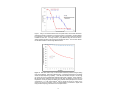
※本プロトコールは参考用の資料になり ます。商品ご購入の際は必ず商品に添付 されている資料をご参照ください。 Enabling Discovery in Life Science® Mito-ID® Membrane Potential Cytotoxicity Kit for microplates Instruction Manual Cat. No. ENZ-51019-KP002 For research use only. Rev. 2.1.1 September 2010 for 2 x 96-well plates Notice to Purchaser The Mito-ID® Membrane Potential Cytotoxicity Kit is a member of the CELLestial® product line, reagents and assay kits comprising fluorescent molecular probes that have been extensively benchmarked for live cell analysis applications. CELLestial® reagents and kits are optimal for use in demanding cell analysis applications involving confocal microscopy, flow cytometry, microplate readers and HCS/HTS, where consistency and reproducibility are required. This product is manufactured and sold by ENZO LIFE SCIENCES, INC. for research use only by the end-user in the research market and is not intended for diagnostic or therapeutic use. Purchase does not include any right or license to use, develop or otherwise exploit this product commercially. Any commercial use, development or exploitation of this product or development using this product without the express prior written authorization of ENZO LIFE SCIENCES, INC. is strictly prohibited. Limited Warranty These products are offered under a limited warranty. The products are guaranteed to meet appropriate specifications described in the package insert at the time of shipment. Enzo Life Sciences’ sole obligation is to replace the product to the extent of the purchase price. All claims must be made to Enzo Life Sciences, Inc. within five (5) days of receipt of order. Trademarks and Patents Enzo, CELLestial and Mito-ID are trademarks of Enzo Life Sciences, Inc. Several of Enzo’s products and product applications are covered by US and foreign patents and patents pending. Contents I. Introduction ............................................................... 1 II. Reagents Provided and Storage.............................. 2 III. Additional Materials Required ................................. 2 IV. Safety Warnings and Precautions........................... 3 V. Methods and Procedures ......................................... 3 A. CELL PREPARATION.......................................................... 3 B. REAGENT PREPARATION ................................................... 3 C. MEMBRANE POTENTIAL CYTOTOXICITY ASSAYS ................. 4 VI. Appendices ............................................................... 6 A. FILTER SET SELECTION ................................................... 6 B. RESULTS ........................................................................ 6 VII. References ................................................................ 9 VIII. Troubleshooting Guide ......................................... 10 I. Introduction Mitochondria play a central role in diverse biological phenomena including metabolism, bioenergetics, cancer and apoptosis. Recently, it has been found that compromised membrane potential induced by drug accumulation in the mitochondria contributes to the toxicity of various organs, especially the heart and liver. Adverse drug reactions often remain undetected until large numbers of patients have already been exposed. The ability to test a compound’s safety at the stage of “lead drug selection” (10-100 compounds) would provide a valuable tool for drug screening research. Enzo Life Sciences’ Mito-ID® Membrane Potential Cytotoxicity Kit measures mitochondrial membrane potential with a cationic dye that fluoresces either green or orange depending upon mitochondrial membrane potential status. In energized cells, the Mito-ID™ membrane potential dye exists as a green fluorescent monomer in the cytosol and also accumulates as orange fluorescent aggregates in the mitochondria. However, in cells with compromised mitochondrial membrane potential, the Mito-ID® membrane potential dye exists primarily as green fluorescent monomers throughout the cytosol and no longer exhibits orange fluorescence in the mitochondria. Compared with the commonly used cationic carbocyanine dye JC-1, the Mito-ID® membrane potential dye offers greater solubility, better photostability, and higher sensitivity to mitochondrial membrane potential changes. A control mitochondrial membrane potential perturbation agent, carbonyl cyanide 3-chlorophenylhydrazone (CCCP), is provided for monitoring changes in mitochondrial dynamics. The Mito-ID® Membrane Potential Cytotoxicity Kit enables monitoring of mitochondrial potential changes using a simple fluorescence microplate reader. Potential applications of the kits are in preclinical drug safety assessment using in vitro cell culture models to aid in the drug development process, and especially to differentiate among test compounds and rank their order of potency. 1 II. Reagents Provided and Storage All reagents are shipped on dry ice. Upon receipt, the kit should be stored upright and protected from light at ≤-20°C. When stored properly, these reagents are stable for at least twelve months. Avoid repeated freezing and thawing. The reagents provided in the kit are sufficient for 2 x 96well microplates. Reagent Quantity ® Mito-ID MP Detection Reagent 200 µL CCCP Control (2 mM) 100 µL 10X Assay Buffer 1 2.5 mL 50X Assay Buffer 2 0.5 mL III. Additional Materials Required Fluorescence microplate reader or imaging plate reader with a filter set or monochromator setting that provides Ex=490 nm/Em=570-590 nm 96-well tissue culture microplate with black wall and clear bottom Calibrated, adjustable precision pipetters, preferably with disposable plastic tips Glass microscope slides (optional) Glass cover slips (optional) Deionized water Anhydrous DMSO (optional) Serum (optional) Growth medium (e.g. Dulbecco’s modified Eagle medium, D-MEM) 2 IV. Safety Warnings and Precautions This product is for research use only and is not intended for diagnostic purposes. Some components of this kit may contain hazardous substances. Reagents can be harmful if ingested or absorbed through the skin and may cause irritation to the eyes. Reagents should be treated as possible mutagens and should be handled with care and disposed of properly. Observe good laboratory practices. Gloves, lab coat, and protective eyewear should always be worn. Never pipet by mouth. Do not eat, drink or smoke in the laboratory areas. All blood components and biological materials should be treated as potentially hazardous and handled as such. They should be disposed of in accordance with established safety procedures. To avoid photobleaching, perform all manipulations in subdued light environments, in amber microcentrifuge tubes or protected from light by other means. V. Methods and Procedures A. CELL PREPARATION Cells should be maintained in 96-well microplates via standard tissue culture practices. Seed cells in microplate(s) the day before test compound/probe addition. The protocols described herein are based upon Hela cells, for which it is highly recommended that cells in culture medium are seeded on plates at a density of 2 x 10⁴ to 3 x 10⁴ cells per well at a seeding volume of 100 L per well. Keep the microplate(s) in a humidified CO2 incubator overnight. NOTE: 96-well black wall and clear bottom plates are highly recommended for this assay. Any cell number and plate coating requirements should be optimized for the selected cell. B. REAGENT PREPARATION NOTE: Allow all reagents to thaw at room temperature before starting with the procedures. Upon thawing, gently hand-mix or vortex the reagents prior to use to ensure a homogenous solution. Briefly centrifuge the vials at the time of first use, as well as for all subsequent uses, to gather the contents at the bottom of the tube. 3 1. Positive Control CCCP (carbonyl cyanide 3-chlorophenylhydrazone) is a proton ionophore and uncoupler of oxidative phosphorylation in mitochondria. As such, it is useful for depolarizing mitochondria. Addition of CCCP should cause a dose-dependent reduction in mitochondrial orange fluorescence. Prepare this perturbation agent as a positive control for mitochondrial membrane potential loss. Before beginning the experiment, ensure that the vial of CCCP Control has equilibrated to room temperature. Dilute the CCCP Control (supplied as a 2 mM stock solution in DMSO) in medium or buffer of choice to a final concentration optimized for the cells being used. For HeLa or U2OS cells, it is recommended to perform treatment with the agent using a final assay concentration of 2~4 µM CCCP in medium for about 15~30 minutes in order to observe changes in mitochondria membrane potential changes. Unused stock should be stored in aliquots at -20°C for about 6 months. 2. Mito-ID™ MP Dye Loading Solution NOTE: The Mito-ID® MP Detection Reagent is light sensitive. Avoid direct exposure of the reagent to intense light. Aliquot and store unused reagent at -20°C, protected from light. Avoid repeated freeze/ thaw cycles. For each 96-well plate, add 100 µL of Mito-ID® MP Detection Reagent, 1 mL 10X Assay Buffer 1 and 0.2 mL 50X Assay Buffer 2 into 8.7 mL deionized water. Mix well. C. MEMBRANE POTENTIAL CYTOTOXICITY ASSAYS I. Rapid Kinetics Measurement (for compounds that change mitochondrial membrane potential within 30 mins) 1. Obtain prepared microplates containing cells (see section A). 2. Dispense 100 µL of the prepared Mito-ID® MP Dye Loading Solution (see section B-2, above) directly into the growth medium. NOTE: It is NOT necessary to remove medium or wash the cells. However, some media may result in high background levels, and some test compounds may be sensitive to the presence of serum. In these instances, growth media and serum should be removed. Then, add 100 µL of the buffer of choice to the wells before adding the dye loading solution. 3. Incubate the microplates for 30 mins at room temperature, or 37°C prior to addition of the test compound. 4 NOTE: The Mito-ID® MP dye is light sensitive. Be sure to protect samples from light. The appropriate incubation time depends upon the individual cell type and cell concentration used. Optimize the incubation time for each experiment. DO NOT wash the cells after dye loading. 4. Prepare the test compound by dissolving it in the buffer of choice. The Mito-ID® Membrane Potential Cytotoxicity Assay is optimized for compound addition at one-tenth of the final well volume. For positive control, dispense 20 µL of the provided CCCP Control to a final concentration of 2~4 µM in medium or buffer. NOTE: Titration of the CCCP Control may be required for optimal results with different cell types. 5. Run the Mito-ID® membrane potential cytotoxicity assay by monitoring the fluorescence at Ex=490 nm/Em=590 nm with a fluorometric imaging plate reader or a fluorescence microplate reader equipped with liquid dispensing capabilities. In live and healthy cells, the mitochondria will fluoresce orange following aggregation of the Mito-ID® MP dye. The orange J-aggregates emit at 590 nm. NOTE: Faster addition speeds can lead to better mixing of compounds and lower signal variance across the plate. Make sure to follow the recommended experimental setup parameters provided by the instrument manufacturer before reading the plate. It is also important to run the signal test before the experiment. Different instruments have their own intensity range. II. Slow Kinetics Measurement (for compounds that change mitochondrial membrane potential after more than 30 min treatment) 1. Obtain prepared microplates containing live cells (see section A). 2. Treat the cells with 10~20 µL of your compound under optimal culture or buffer conditions for a time period sufficient for assessing the effects of the compound. For positive control, dispense 10~20µL of provided CCCP Control to a final concentration of 2~4 µM in medium or buffer for 15~30 minutes. Negative control cells should be treated with the same vehicle (media or buffer) used to reconstitute or dilute the inducer or inhibitor for an equal length of time under similar conditions. NOTE: It is NOT necessary to remove medium or wash the cells. However, some media may result in high background levels, and some test compounds may be sensitive to the presence of serum. In these instances, growth media and serum should be removed. Then, add 100 µL of the buffer of choice to the wells before adding the dye loading solution. 5 NOTE: Titration of the CCCP Control may be required for optimal results with different cell types. 3. Dispense 100 µL of the prepared Mito-ID® MP Dye Loading Solution (see section B-2, page 4) into each well. Incubate for 30 minutes at room temperature or 37°C. NOTE: The Mito-ID® MP dye is light sensitive. Be sure to protect samples from light. The appropriate incubation time depends upon the individual cell type and cell concentration used. Optimize the incubation time for each experiment. 4. Observe immediately with a fluorescent microplate reader or imaging plate reader using an excitation setting of about 480 nm and an emission setting of about 590 nm. In live and healthy cells, the mitochondria will fluoresce orange following aggregation of the Mito-ID® MP dye. The orange J -aggregates emit at 590 nm. NOTE: It is imperative that samples be analyzed immediately following completion of staining. DO NOT wash the cells after Mito-ID® MP dye loading. The fluorescence value in blank wells added with the growth medium should be subtracted from the values for those wells with cells treated with the test compounds. VI. APPENDICES A. FILTER SET AND MONOCHROMATOR SETTING SELECTION The selection of optimal filter sets or monochromator settings for a fluorescence microplate reader application requires matching the instrument optical specifications to the spectral characteristics of the dyes employed in the analysis. Please consult your instrument or filter set manufacturer for assistance in selecting optimal filter or monochromator settings. Settings suitable for Texas Red or tetramethylrhodamine (TRITC) are recommended for imaging the orange-fluorescent signal of the Mito-ID® MP dye, indicating energized mitochondria. B. EXPECTED RESULTS In most eukaryotic cells the majority of ATP production is via oxidative phosphorylation by the mitochondrial respiratory chain. Therefore, the mitochondrial membrane potential makes up a large part of the bioenergetic state of the cell and it is altered directly, depending upon the cell’s energy needs. The ability to accurately measure mitochon6 drial membrane potential can give invaluable information about the general health and function of the mitochondria, in particular of the overall function of the respiratory chain and the potential of the mitochondria to generate ATP and provide energy for other cellular components. Measurement of the mitochondrial membrane potential is useful in a wide variety of research areas and mitochondrial dysfunction is implicated in diseases such as cancer, diabetes, Parkinson’s disease, and stroke. Typical results of fluorescence microplate-based analysis of cell populations using the Mito-ID® Membrane Potential Cytotoxicity Kit are shown in Figure 1. Using a conventional fluorescence microplate reader, mitochondrial membrane potential is shown to decrease as a function of CCCP concentration, as demonstrated by a decrease in orange signal. The high Z’-factor (>0.9) obtained using this kit demonstrates excellent signal-to-background ratios. Figure 2 shows real time kinetic measurement of mitochondrial membrane potential changes with CCCP treatment using a conventional fluorescence microplate reader with liquid dispensing capability for test agent addition. The Mito-ID® MP dye provides a more robust method to monitor mitochondrial membrane potential changes compared to JC-1, due to its higher photostability, better aqueous solubility and higher sensitivity to mitochondrial membrane potential changes. This kit offers true mixand-read capability and can be readily used for screening of large compound libraries. 7 Figure 1. Using a conventional fluorescence microplate reader, mitochondrial membrane potential was shown to decrease as a function of CCCP concentration, as demonstrated by a decrease in orange signal. Enzo’s Mito-ID® MP dye is at least 10-fold more sensitive to MMP loss than JC-1. The high Z’-factor (>0.9) obtained using the Enzo kit demonstrates excellent signal-to-noise and signal-to-background ratios. The error bars denote the standard deviation of at least eight determinations. Figure 2. Real time kinetic study of mitochondrial membrane potential changes arising from CCCP treatment. Data were collected using a conventional fluorescence microplate reader with liquid dispensing capability: Hela cells were seeded overnight in 25,000 cells per 100 µl per well in a 96-well black wall/clear bottom plate. Next day, 100 µl of Mito-ID® MP dye was directly added into each well and cells were incubated for 30 mins at room temperature. CCCP (20 µl/well) was added using a BioTek dual liquid dispensor to achieve concentrations of 1 µM. Orange Mito-ID® MP dye signal was shown to change in the presence of CCCP as a function of time, as demonstrated by a decrease in signal, read using the BioTek Synergy Mx fluorescence microplate reader. 8 VII. References 1. Smiley, Reers, Mottola-Hartshorn, Lin, Chen, Smith, Steele, and Chen (1991) “Intracellular heterogeneity in mitochondrial membrane potentials revealed by a J-aggregate forming lipophilic cation JC-1.” Proc. Natl. Acad. Sci. USA 88: 3671-3675. 2. Cossarizza, Baccarani-Contri, Kalashnikova, and Franceschi (1993) “A new method for the cytofluorimetric analysis of mitochondrial membrane potential using the J-aggregate forming lipophilic cation 5,5’,6,6’-tetrachloro-1,1’,3,3’ tetraethylbenzimidazolylcarbocyanine iodide (JC-1).” Biochem. Biophys. Res. Commun. 197 (1): 40-45. 3. Woollacott and Simpson. (2001) “High throughput fluorescence assays for the measurement of mitochondrial activity in intact human neuroblastoma cells.” J Biomol Screen 6(6): 413-420. 9 VIII. Troubleshooting Guide Problem Potential Cause Suggestion Fixed and/or permeabilized cells fail to stain with the mitochondrial membrane potential dye. This dye is only suitable for live-cell staining. Use the dye only for livecell analysis. Stained cells have been exposed to strong light. Protect samples from exposure to strong light and analyze them immediately after staining. Kit reagent has degraded. Verify that the reagents are not past their expiration dates before using them. Control cells without treatment show low orange signal. Control cells are not healthy. Extended storage of cells after staining may adversely affect their health. Use healthy cells. Acquire data as soon as possible after staining the cells. Cell staining is too strong. Mitochondrial membrane potential dye is too concentrated for the cell type being employed. Dilute the staining solution further with Assay Buffer. Orange fluorescent mitochondrial membrane potential signal increases upon treatment with the CCCP Control. The orange emission filter in your instrument may be too wide and green fluorescent signal is being read, too. Replace emission filter with a narrower band pass filter. Accuracy of data from experiment to experiment is poor. The concentration of the stained cell sample for data acquisition is too low or inconsistent between wells. Repeat experiments using a higher density of cells, and carefully seed the wells so that each well has the same number of cells. Poor staining observed. 10 www.enzolifesciences.com Enabling Discovery in Life Science® NORTH/SOUTH AMERICA GERMANY UK & IRELAND ENZO LIFE SCIENCES INTERNATIONAL, INC. 5120 Butler Pike Plymouth Meeting, PA 19462-1202 USA T 1-800-942-0430/(610) 941-0430 F (610) 941-9252 E [email protected] ENZO LIFE SCIENCES GMBH Marie-Curie-Strasse 8 DE-79539 Lörrach Germany T +49/0 7621 5500 526 Toll Free 0800 664 9518 F +49/0 7621 5500 527 E [email protected] www.enzolifesciences.com ENZO LIFE SCIENCES (UK) LTD. Palatine House Matford Court Exeter EX2 8NL UK T 0845 601 1488 (UK customers) T +44/0 1392 825900 (from overseas) F +44/0 1392 825910 E [email protected] www.enzolifesciences.com www.enzolifesciences.com SWITZERLAND & REST OF EUROPE BENELUX FRANCE ENZO LIFE SCIENCES AG Industriestrasse 17, Postfach CH-4415 Lausen Switzerland T +41/0 61 926 89 89 F +41/0 61 926 89 79 E [email protected] www.enzolifesciences.com ENZO LIFE SCIENCES BVBA Melkerijweg 3 BE-2240 Zandhoven Belgium T +32/0 3 466 04 20 F +32/0 3 466 04 29 E [email protected] www.enzolifesciences.com ENZO LIFE SCIENCES c/o Covalab s.a.s. 13, Avenue Albert Einstein FR -69100 Villeurbanne France T +33 472 440 655 F +33 437 484 239 E [email protected] www.enzolifesciences.com