1
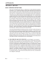
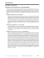
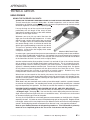
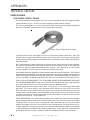
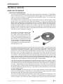
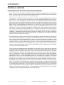
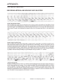
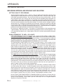
APPENDICES
TECHNICAL ARTICLES
BASIC OPERATING INSTRUCTIONS
USE OF THE PENCIL PROBE IN THE DIAGNOSIS OF ARTERIAL DISEASE IN THE LIMBS
1. THE PROBE
The probe consists of two crystals; one for transmitting the ultrasound waves and the other for
receiving the reflected waves. If either crystal is damaged, the probe will not work properly or
will not work at all. The crystals are covered by a material that is vulnerable to attack by ECG
paste or cream. Therefore, DO NOT use ECG paste as the contact medium between the skin
and the probe. Use AQUASONIC or any gel made for ultrasonic physical therapy equipment. In
an emergency use any surgical jelly or lubricant, even petroleum jelly or mineral oil. Remove
the gel after use with a soft tissue. If you should find the probe with dried gel on it, wash it off
under running water. Do NOT scrape off the gel because you may damage the coating over
the crystals. Do NOT autoclave the probe.
2. POSITION OF THE PROBE
Invariably, people not accustomed to our probe use it incorrectly. The probe we furnish is
different from that of the other manufacturers and is used differently. If you hand someone
the probe and say “Here, try it for yourself”, they will almost always put it over their radial
artery and place the probe perpendicular to the artery—and perhaps with no coupling gel.
Many people have tried to compare our Doppler with other makes by this method. Keep in
mind that you are not buying a Doppler for use on the radial artery, but for use on vessels
you cannot feel. The best testing ground is therefore in your particular area of interest.
We believe our instruments will permit you to find the vessels easier, let you hear the venous
sounds easier and follow the vessels better than any other device on the market, regardless
of price. But it takes some practice in order to be able to do this. We believe the arm is
a good and most convenient limb for you to learn on—to learn how to hold the probe
depending on the depth of the artery and vein. The area about 150 mm each side of the
elbow is a good place to start.
First, put some gel on the tip of the probe. The gel squeeze-bottle must be shaken downward
and then gently squeezed to get the gel to come out. Pile up about 7 mm of gel on the
probe, making certain there are no large air bubbles in the pile, because ultrasound does
not go readily through air. It needs a continuous conducting medium, and the gel is ideal.
Turn the VOLUME control fully down (counter clockwise) and turn the instrument on.
Gradually turn up the volume. You should hear a rumbling sound if you are holding the
probe. This is caused by the vibration of the gel due to tremor in your arm. Now place the
probe over an artery in the arm about half way between the elbow and the wrist. Tilt the
back of the probe toward the hand at an angle of about 45 degrees, making certain there is
gel in the pathway between the probe and the skin. Move the probe and the skin sideways
to try to find the center of the artery and the hissing noise at heart rate, which is the Doppler
sound for an artery. If the sounds you hear are more or less continuous, that is simply the
background noise of the instrument and it means that you are not over the artery. The
main energy of the beam is only about as wide as the crystals in the probe, so there isn’t
much room for error in aiming the probe. For this reason you must always search the area
of the artery and tilt the probe for best Doppler sounds.
2100-SX, US 05M Appendices, Basics, Tech Arts-1.0 10/25/06
PARKS Flo-Lab Operating Manual
V.1
APPENDICES
TECHNICAL ARTICLES
BASIC OPERATING INSTRUCTIONS
When you are looking for deep arteries, or for small or obstructed arteries, you will have to turn
the VOLUME control near maximum. This also means that every time you move the head of
the probe you are going to get some pretty big thumping noises in the earphones. Therefore
you want to avoid moving the head of the probe with respect to the skin as much as possible.
That is why you place the probe over the area where you think the artery is and then you search
for the exact point by moving the skin with the probe and changing the angle of the probe with
respect to the skin. You might wonder why these big transient noises can’t be filtered. We do
limit their intensity, but we do not filter. The reason is that in the search for low-velocity blood
flow, such as in occluded arteries and in the veins, the pitch of the Doppler sounds associated
with the blood flow are very low. Any filtering to eliminate or minimize the sounds accompanying
movement of the probe would also reduce the response to low-velocity blood flow sounds, and
of course this is undesirable.
3. DIAGNOSIS OF ARTERIAL DISEASE
The Doppler method of diagnosing arterial disease of the limbs is only one of several good
methods. It is probably the most convenient and least expensive of the better methods. It
is only qualitative but can be made semi-quantitative by permitting you to make systolic
blood pressure measurements along the leg with the aid of a proper cuff and manometer.
The great sensitivity of the transcutaneous Doppler can cause a doctor or technician to
conclude improperly that an arterial pathway is open when it isn’t. Collateral flow around
an obstruction can be well-developed, especially in the thigh, and cause pulsatile blood to
flow in the distal arteries. Or a major artery may be narrowed, causing pulsatile flow distally.
These mistakes in diagnosis can be avoided almost entirely by simple means and a little
bit of experience. An experienced user of the Doppler can recognize the characteristic
sounds of open and obstructed arteries. Remember that Doppler sounds vary in pitch
(frequency) with the velocity of blood flow. When you hear the Doppler sound on a normal
artery and compare it with a normal arterial pulse-pressure wave, you will recognize the
sound of the dicrotic notch, the very fast rise time of the wave and perhaps a third sound
just before the onset of a new pulse wave. While the origin of these second and third waves
in the descending branch of a pulse wave may be in dispute, their absence in vessels distal
to an obstruction is not disputed. So a diagnostic rule is that whenever you hear the second
and perhaps third sounds of a pulse wave of a major artery, you can be sure the artery is
open proximal to the probe. Plethysmographic studies also show a delayed crest to the
wave, associated with a slower rise time to the wave when there is an obstruction proximally.
Though the Doppler is permitting you to hear velocity changes rather than true volume
changes, the correlation is good enough to be quite valuable diagnostically.
Now the opposite is not necessarily true—that when you can’t hear second and perhaps
third sounds the artery is obstructed proximally to the probe. In the digits and smaller
vessels the pulse wave is smoothed out more, especially when there is some
vasoconstriction. Now of course there are cases that are in doubt. If you cannot clearly
hear the second and third sounds (the third sound is frequently missing), compare with the
same artery on the other limb. If you find a radical difference in the sound of the Doppler,
both in pitch and in amplitude, you are justified in being quite suspicious of the patency of
the artery of the first limb you studied provided you are now fairly skilled at optimizing the
sounds.
V.2
PARKS Medical Electronics, Inc.
Aloha, Oregon
U.S.A.
APPENDICES
TECHNICAL ARTICLES
BASIC OPERATING INSTRUCTIONS
Another thing you listen for is the relative clarity of the arterial wave. How well it stands
out from the background noise of the instrument and perhaps the venous flow adjacent to
the artery. Move the probe a little to each side of the artery to make this estimation. In a
normal person you will find that you can make the arterial pulse wave almost completely
separate from the venous sounds by positioning of the probe.
The way you really come to a final conclusion that the artery is obstructed proximal to the
probe is by measuring the systolic pressure at the ankle with an ordinary arm cuff. If you
want to measure pressure at other places on the leg you will need a special cuff, the bladder
of which encircles the limb. We sell such cuffs. The method is as follows:
Wrap the cuff around the ankle or slightly above it so you can get the probe on the
posterior tibial and hear the arterial sounds adequately. Inflate the cuff to a pressure
well above the patient’s arm pressure or at least 30 points above the pressure at which
the Doppler sounds disappear. Gradually reduce cuff pressure until you hear blood flow,
though the sound won’t be normal. At that point read the pressure to obtain systolic
pressure at the ankle. If you have doubts, center the probe on the artery and inflate
the cuff again. You can observe at what cuff pressure the blood flow stops and again
where it starts. Where it starts is normally used.
This procedure is very similar to taking pressure on the arm using a stethoscope. There
you are using sounds of turbulence or wall motion. Here we are sensing the flow of blood
under the cuff with a much more sensitive device. You can get a clear indication of systolic
pressures as low as 30 mm of Hg. The only problem is keeping the probe right on the
center of the artery while you are inflating and deflating the cuff. An aneroid manometer
mounted on the inflation bulb of the cuff is preferable. Tycos makes such a device and
perhaps others do too.
The possibility of misdiagnosing is greatly reduced by this method provided you make two
or more measurements and you are skilled at holding the probe in the right place and at
the right angle. A low pressure reading is quite reliable. On diabetics you may get readings
of 300 mm Hg or more, even though they have ulcers on their toes. These people with
end-artery disease studied plethysmographically with the mercury-in-silastic strain gage,
which we also make, will have quite large and normal looking pulsations in the toes. Their
arterial walls are sclerosed so badly sometimes that they will not compress with cuff
pressure.
The normal pressure in the ankles should be about the same as the systolic pressure in
the arm, or a little higher. If the ankle pressure is 30 mm Hg or more lower than the arm
pressure, an obstruction is almost certainly present. Normally one finds that people with
arterial obstructions have pressures of 100 mm Hg or less.
If you have a proper cuff you can take pressures in the same manner (with the probe at the
posterior tibial) just below the knee, just above it and at the top of the thigh. By measuring
systolic pressure (the pressure measurement is always where the cuff is, not where the
probe is) you will find radical differences between measuring sites if the obstruction is between
them or you will find that pressures at corresponding points on the two legs are quite different.
An exception is bilateral obstruction of the bifurcation of the abdominal aorta which may
give you fairly symmetrical pressures on both legs. Unfortunately you cannot use the
Doppler above the top of the thigh. The pressure measurements made on the thigh with a
narrow cuff will be clinically useful, though not accurate.
2100-SX, US 05M Appendices, Basics, Tech Arts-1.0 10/25/06
PARKS Flo-Lab Operating Manual
V.3
APPENDICES
TECHNICAL ARTICLES
BASIC OPERATING INSTRUCTIONS
Once you have determined that there is an obstruction it is often desirable to determine
just where it is. It is permissible to check at certain points provided you are quite familiar
with normal sounds—second and perhaps third sounds. Start at the top of the thigh and
listen for the normal arterial sounds. A little to one side you should hear venous flow varying
with respiration. The adjacent venous flow assures you that you are indeed listening to a
major artery. This is important because you can get beautiful sounds from a collateral that
is aimed toward your probe and giving a tremendous Doppler effect. But a collateral follows
a tortuous path and the venous sounds will not be found adjacent to it. If you have a little
problem hearing the vein (and you shouldn’t over big veins) give the leg a slight squeeze
distal to the probe to increase the velocity of the venous blood and make its pitch higher.
As you follow the superficial femoral artery down toward the knee you will lose the sound,
even on normals, in some parts of the path because of tendons or other anatomical
obstructions between the probe and the artery. You should be able to pick it up again
easily in the popliteal region. Your ear and concentration make a filter to extract wanted
information from background noise that exceeds anything that can be done electronically.
You can follow these small arteries distal to the knee and in some cases they can be followed
all the way to the ankle and beyond. Keep in mind that some people don’t have a dorsalis
pedis artery. If you are working on arteries in the foot, make sure they are dilated by immersing
the foot in a bucket of warm water for a few minutes. Some people are vasoconstricted most
of the time. They usually will dilate for a while after the immersion and in a few minutes be
constricted again. Also they usually do not have arterial disease.
If you want to quickly determine the efficiency of flow in the arterial system of the leg, pick
up the posterior tibial and listen for 2nd and perhaps 3rd sounds. If you hear them, and
you are sure you know the difference between normal and abnormal, go no further. If they
do not sound normal or there is doubt, make a blood pressure measurement and compare
it with systolic pressure on the other ankle and on the arm. To find the location of the
obstruction you can listen with the Doppler, or using a special cuff you can make blood
pressure readings farther up the leg. If the obstruction is in the iliacs you can note it by
the Doppler sound distal to the obstruction or by a much lower than normal blood pressure
at the top of the thigh as measured with the cuff and the Doppler.
4. PRE-OPERATIVE AND POST-OPERATIVE use of the Doppler is very important.
When the patient is on the table, measure systolic pressure at both ankles and record it.
After blood is again permitted to flow, measure both pressures again. The pressure on the
operated leg should be UP compared to the pressure in the other leg, the control. If it
isn’t, then it is pretty safe to assume something is wrong. On rare occasions a limb will
have such a high degree of reactive hyperemia that pressure will not be up and may even
be lower, but the leg will be hot. A large percentage of patients are blocked to some degree
before they get off the table. Blood-pressure measurements will give you an objective
evaluation of the surgery. Some surgeons use the pencil probe directly on the artery (using
sterile jelly for coupling) just distal to the repair. The characteristic of the flow sound is
important. If the runoff is inadequate an experienced ear can detect it and often correct
the cause on the table. You can also use Doppler and pressure measurements for follow
up, comparing pressures at both ankles with systolic pressure at the arm, measured either
with a Doppler or stethoscope.
V.4
PARKS Medical Electronics, Inc.
Aloha, Oregon
U.S.A.
APPENDICES
TECHNICAL ARTICLES
TECHNIQUE OF EVALUATING CALF VENOUS DISEASE
The assessment of calf venous disease by Doppler ultrasound may be achieved with an accuracy
of up to 85% compared to venography when one is experienced with the technique. The status of
the calf veins can be assessed by listening with the Doppler at the posterior tibial vein at the ankle,
the popliteal vein, the superficial femoral vein, and the common femoral vein. The status of the calf
veins is determined by a combination of augmentation maneuvers when listening at these various
points.
NORMAL RESPIRATION FLOW SOUNDS
The Doppler is initially placed over the posterior tibial vein at the ankle behind the medial
malleolus. Generous amounts of acoustic gel must be used, and one must be careful to avoid
undue pressure with the probe which might result in obstruction of venous flow. Initially the
posterior tibial artery signal is elicited. The probe is then moved slightly to either side of the
arterial signal until the windstorm like venous signal is heard. Normally this signal should wax
and wane with respiration. In the presence of calf vein thrombosis, the signal may be more
continuous or there may be no audible signal present. If the feet are vasoconstricted, a venous
flow signal may not be heard until the venous velocity is increased by gentle compression of
the foot.
CHECKING COMPETENCY OF THE VALVES
Once the optimal venous signal is elicited, the calf is then compressed with the hand which is
not holding the probe. The fingers should be spread so that much of the calf muscle is
compressed. During this procedure, no venous flow should be heard. If venous flow signals
are elicited, this is a sign of deep venous valvular incompetence, usually secondary to old deep
vein thrombosis.
AUGMENTING VENOUS VELOCITY BY COMPRESSION
Next the calf is released and one should normally hear an augmentation of venous flow as blood
enters the previously decompressed calf veins. The magnitude and duration of the augmented
signal can be influenced by several factors including the temperature of the foot, the general
vasomotor tone of the patient and the presence or absence of venous thrombosis in the calf. It
is important to compare the augmentation signals in each foot. In vasoconstricted individuals
with cold feet, the posterior tibial venous augmentation may be very minimal but it should be
symmetrical. If there is good augmentation in one leg and poor augmentation in the other, the
latter leg is usually the site of venous thrombosis. Next, the common femoral and then the
superficial femoral veins are examined and the signals assessed for augmentation upon calf
compression. Calf-vein thrombosis will result in a decreased augmentation of the venous signals
at these sites. Similarly the popliteal vein should be examined. In general, the most sensitive
indicator of calf-vein thrombosis is a relative decrease in augmentation upon release of calf
compression with the probe positioned over the posterior tibial vein at the ankle. There are
certain conditions which will imitate calf-vein thrombosis. Such problems as subfascial
hematoma, a ruptured Baker’s cyst, extensive edema, or other conditions which cause increased
pressure on the calf veins may result in a decreased augmentation of flow during the
aforementioned maneuvers. Such conditions can be best diagnosed by a venogram if the
diagnosis is in question.
2100-SX, US 05M Appendices, Basics, Tech Arts-1.0 10/25/06
PARKS Flo-Lab Operating Manual
V.5
APPENDICES
TECHNICAL ARTICLES
V.6
PARKS Medical Electronics, Inc.
Aloha, Oregon
U.S.A.
APPENDICES
TECHNICAL ARTICLES
USING PROBES
USING THE DOPPLER ON DIGITS
ALWAYS USE THE HIGHEST FREQUENCY PROBE YOU HAVE ON DUAL-FREQUENCY DOPPLERS.
That will normally be a nominal 8 MHz probe. At lower frequencies, such as nominal 4 MHz,
the efficiency of reflection of the ultrasound signal is poorer and performance on surface vessels
will not be as good as it is with a higher frequency probe.
Line up the long way of the crystals and the pencil
probe body with the length of the artery for best
separation of arteries and veins at the ankle and best
efficiency on the digital arteries.
Whenever you try to pick up arterial flow from the
digits you must consider digit temperature. It is often
difficult to get a digital pulse when feet are cold or
cool. The digits can be so vasoconstricted that blood
only oozes through, which is sufficient to nourish but
doesn’t give a good recording or sound on any device.
This occurs in perfectly healthy digits under normal
conditions. It also occurs after surgery when severe
vasospasm may occur.
Nominal 8 MHz Pencil Probe
In order to get a good sound, blood pressure measurement or recording under these conditions
you must cause vasodilatation to take place. One method is to warm the extremity by immersing
it in warm water-not hot water. Within a few minutes you will be able to evaluate the condition
of the flow when the limb is at heart level or slightly above.
Another method used to dilate peripheral vessels is to occlude all flow at the ankle or forearm
with an ordinary arm cuff inflated to well above systolic pressure. This is not normally painful,
especially when vascular disease is present in the arterial system. If too much discomfort is
apparent, a different procedure could be tried. Five minutes is usually enough time. On release
of cuff pressure a reactive hyperemia will take place and last for a short time at least. There
should be enough time to make an evaluation of arterial patency. Diabetics may have
incompressible arteries so this technique may not work with them.
When there are two sounds to the arterial pulse wave, the first caused by the filling of the
vessel with systole and the second being either forward or reverse flow in the diastolic phase,
vessels are usually patent. However, blockage of major proximal arteries may be present with
good collateral flow around them.
When there is only one sound with each cardiac cycle and the sound is not brisk, a proximal stenosis
or occlusion may be present. On recording such sounds, you will find the recording has a rounded
top and respectively slow upstroke. You must be sure the filter control is in the proper position.
PROPER PROBE PLACEMENT AND PROPER USE OF GEL ARE VERY IMPORTANT!
The pencil probe body should be in line with the artery, not crossways to it, and should be at about
a 45 degree angle. (See page V.4) You must be very careful about probe pressure, because a
slight amount of pressure against the skin can occlude the artery. You must use a small amount
of ultrasound coupling gel in front of the probe.
You may find it impossible to make blood pressure measurements on the digits with a Doppler,
especially the toes. It is better to use our Photoplethysmograph for that purpose. Doppler sounds
from digital arteries will be very helpful once you become familiar with normal and pathological
sounds. Making digital pressure measurements on the toes is not a very popular procedure.
Refer to your medical literature for diagnostic procedures. This information is primarily
meant to be a simplified guide to the use of the instruments and the probe.
2100-SX, US 05M Appendices, Basics, Tech Arts-1.0 10/25/06
PARKS Flo-Lab Operating Manual
V.7
APPENDICES
TECHNICAL ARTICLES
USING PROBES
THE SKINNY PENCIL PROBE
Our standard probes for the Doppler are a 3/8" (10 mm) diameter for the high frequency probes
(nominal 8 MHz) and ½" (12 mm) for the low frequency probe (nominal 4 MHz).
The “skinny” pencil probe is ¼" (6.5 mm) diameter and is made only for the higher frequencies
because at the lower frequencies the small crystals don’t work well.
Skinny Pencil Probe, Nominal 8 MHz
If you do vascular work, we suggest you have a skinny pencil probe to work with. What you
will find is that you get a better signal-to-noise ratio in many cases. That means the flow signal
stands out above the background noise better which automatically means better recordings on
weak flow sounds.
But a second benefit is better separation of artery and vein. Mostly the two lie close together,
and you’ll find supraorbitals much easier to do with the skinny pencil. Also on the digits with
care you can get pretty good separation of artery and vein. Don’t hesitate to try it on carotids
on skinny people. On thick necks the 4 MHz will probably work best.
You probably know that diagnosis of venous disease below the knee is pretty poor. You’ll find the
posterior tibial vein and others easier with the skinny pencil and the high frequency will let you hear
venous flow-velocity changes with respiration easier than a low-frequency probe will, especially if
you can use about a 45 degree angle to the vein, which you can do easily with surface vessels.
FOR LEG BLOOD PRESSURES, THE STANDARD PROBE IS RECOMMENDED BECAUSE
OF THE WIDER BEAM WIDTH. Align the crystals with the vessels for best artery-vein separation
and across the vessels for less critical positioning to take blood pressures.
Another place the skinny pencil works best, so we’re told, is for measuring penile blood
pressures. This can be difficult, since the deep penile arteries are very small in the flaccid
state and there is a lot of spongy tissue over them. The skinny pencil probe works best for two
reasons. One is that the power is concentrated into a much smaller crystal, so the beam intensity
is higher. The second is that the small probe can be pushed down into the tissue closer to the
artery much easier than a larger probe can.
PRICE DIFFERENCE. Our standard pencil probes are far lower in cost than any other Doppler
probes made by other manufacturers. And they work better, too. The skinny pencil costs about
10 percent more. It is wise to have a backup probe, because probes do fail. If you do vascular
studies, we suggest you have a skinny pencil in your armamentarium. You’ll find you use it a lot.
To order, specify the frequency engraved on your standard high frequency probe or indicated by
a label attached to the standard high frequency probe cable. If your probe has a label attached
to the cable, DO NOT remove, it contains important reordering and warranty information.
V.8
PARKS Medical Electronics, Inc.
Aloha, Oregon
U.S.A.
APPENDICES
TECHNICAL ARTICLES
USING THE PPG SENSOR
PHOTOPLETHYSMOGRAPH
This instrument will provide both arterial and venous operation of the photocell. The ARTERIAL
mode is used when you just want to get pulsations from the digit. The arterial mode is much
easier to use than the venous, not requiring the use of the RESET button. It is also five times
more sensitive than the venous mode.
The VENOUS mode is used when you want to record slow variations in blood changes, usually
on the calf for a skin perfusion study. After the transducer is in place, you must press the
RESET button momentarily so the electronic circuitry can balance itself for the amount of light
getting back into the photocell and hold that balance in memory. RESET then establishes a
baseline or beginning point from which gross variations in light intensity changes entering the
photocell caused by blood flow under the sensor will be compared. If the baseline on the chart
recording varies, it is due to variations in light entering the photocell and may be due to faulty
technique as well as blood flow variations.
The sensor is a black disc and has two
round “eyes” clearly visible. These “eyes”,
one a light source and the other a light
sensor, must be placed against the skin.
For most tests the patient should be lying
down so there is no pooling of the blood
as there would be if the limb were well
below heart level. If the light is very bright
it may be best to cover the sensor with a
dark cloth when in use.
PPG Sensor
For finger or toe pulses, the sensor is placed over the pad of the digit. It can be held in place
by the Velcro band supplied or by tape. You may use ordinary office tape but be sure you do
not bind the transducer to the digit so tightly that you restrict blood flow. A clear tape (not
frosted) that is sticky on both sides can be obtained at stationery stores and may be preferable.
No band is used. The tape is affixed to the “eyes” side of the sensor and then to the skin.
This method is more prone to interference by outside light, so you may need a dark cloth to
cover the toe. You may need to anchor the cable to the digit with ordinary tape if there is foot
tremor. Good anchoring is especially necessary when the photoplethysmograph is used in the
VENOUS mode, because slow variations in contact pressure will cause a baseline shift. Light
variations may cause problems so the dark cloth cover is recommended.
If the digit is quite cold, the recording may be minimal or absent, even on a “normal”. In such
cases you must release vasoconstriction by warming the foot or using a cuff to occlude flow at
the ankle for three to ten minutes, depending on how much reactive hyperemia is required for
your tests. With peripheral vascular disease this occlusion is not painful and causes release
of vasomotor tone. It allows a better assessment of the distal vascular bed and the inflow rate.
The dilation obtained may not last very long. The patient may well vasoconstrict in a few minutes
as tissue anoxia lessens.
PULSE CONTOURS A normal toe pulse will have a rapid upstroke, a dicrotic (descending)
notch and a fairly sharp peak. Digits filling via small collaterals fill slowly, so pulses are quite
rounded with slower filling time and usually much lower amplitude.
The fingers and toes of older people will often show a rapid upstroke with a sharp peak. This
means the main arterial pathway is open. The notch in the descending part of the wave will be
minimal or absent. This is because arterial compliance has been lost due to atherosclerosis.
2100-SX, US 05M Appendices, Basics, Tech Arts-1.0 10/25/06
PARKS Flo-Lab Operating Manual
V.9
APPENDICES
TECHNICAL ARTICLES
USING THE PPG SENSOR
The photocell can be applied to a toe and used in conjunction with an ordinary arm cuff placed
around the ankle to measure systolic pressures. After pressing the ARTERIAL button and
assuming a reasonably good toe pulse, you inflate the cuff well above systolic pressure, then
gradually deflate while watching the recording for the return of the pulse wave. When pulsatility
can first be detected, read systolic pressure from the manometer. Pressure readings made at
the ankles are normally equal to or somewhat above arm pressures. Diabetics often have highly
elevated systolic pressures measured in this way because of hardening of the arterial walls. In
such cases the measured value does not correspond to true systolic pressure.
In some cases of diminished pulsatility and bright light, it maybe advisable to cover the digit
with a dark cloth when the sensor is attached.
DO NOT IMMERSE THE SENSOR FOR CLEANING.
The sensor is not water tight and will not function with water inside! Should you inadvertently
immerse it, hang with the sensor above the cable so the liquid can drain out. If the sensor
was immersed in a sterilizing solution, a conductive residue may remain inside, ruining it. In
such a case it would be best to reimmerse it in plain water and drain, repeating this several
times, in an effort to salvage it.
V . 10
PARKS Medical Electronics, Inc.
Aloha, Oregon
U.S.A.
APPENDICES
TECHNICAL ARTICLES
TECHNIQUES FOR BETTER DOPPLER RECORDINGS
A few hints on use of the probe will probably help you to get better recordings than you otherwise
would. Basically, they involve the angle of the probe, how much gel to use, how much pressure to
use, and how to orient the crystals in the end of the probe.
The angle of the probe to the vessel is important and there are two opposing factors which you
must balance. If you are studying a vessel near the surface, holding the probe almost tangent to
the vessel, or at least 30 degrees off tangency, will give you a good recording with minimum filtering.
You want to use minimum filtering because it gives you a truer picture of velocity changes occurring
during the cardiac cycle which is an important part of diagnosis. So whenever you are studying
digital vessels, supraorbitals dorsalis pedis, etc., try to make your angle to the skin less than 45
degrees. Use no more filtering than is necessary to get an acceptable recording, usually 14 or
even 28 Hz. if you can tolerate the raggedness of the recording. The reason this works better is
that the shift in frequency by the Doppler effect is a function of the angle of the incident ultrasound
to the blood flow. The pitch of the sound is higher, and with higher pitched sounds you need less
smoothing. Smoothing introduces distortion into the true velocity picture, so minimal smoothing
gives you more realistic picture of flow velocity changes. Of course you may choose to use more
smoothing to have a better looking graph.
A problem with small vessels is that it is difficult or impossible to separate the artery from an adjacent
vein. Venous flow is low velocity, and when the beam of ultrasound hits the vein, low-pitched sounds
will be mixed in with the higher-pitched arterial sounds. They will contaminate the recording. About
the only thing you can do to minimize venous contamination is to orient the crystals in the end of
the probe so they are vertical with respect to the skin. The size of the ultrasound beam sent into
the skin, near the crystal at least, is approximately the size of the crystal. This means that if
artery and vein are lying side by side, vertical crystals will give you a narrow beam and a better
chance of isolating the desired vessel.
In the case of making blood pressure measurements at the ankle, you are really not that interested
in getting a pure arterial signal. You would prefer a wider beam so that slight movements of the
foot during inflation or deflation of the cuff don’t cause you to move the beam off the vessel. Therefore
you would want the crystals on the probe to be parallel to the skin, utilizing the full width of the
crystal. Furthermore, in order to get a good signal, you may have to tolerate a wider angle between
the probe and the skin. In fact, the deeper the vessel the more the probe has to be aligned toward
perpendicular, which is the worst possible position from a Doppler shift standpoint. The reason
you have to go more vertical is because of the power loss of the ultrasound energy as it passes
through the tissue. When vessels are deep and you want a good recording, align the crystals in
the probe along the length of the artery so that as much ultrasound as possible is hitting the target
artery. This means you will possibly have to use more filtering to get an acceptable recording, but
that is the trade off. If your sound is weak and there is considerable background noise, the recording
is not going to be good anyway. You must get the best signal-to-noise ratio you can, and you do
that by a combination of crystal alignment, angle, and how close you can get to the artery. Don’t
hesitate to use pressure on deep vessels. If you use pressure on shallow ones such as the digits
and dorsalis pedis, the edge of the probe will shut off the flow.
2100-SX, US 05M Appendices, Basics, Tech Arts-1.0 10/25/06
PARKS Flo-Lab Operating Manual
V . 11
APPENDICES
TECHNICAL ARTICLES
TECHNIQUES FOR BETTER DOPPLER RECORDINGS
For vessels near the surface, higher frequency probes work best. This is because, in theory at
least, the efficiency of reflection of ultrasound varies as the fourth power of the frequency. This
means, in theory, that for near-surface vessels 10 MHz. is 16 times better than 5 MHz. In practice
there is a noticeable difference but it is nowhere near that great. When it comes to depth, the
inverse applies. Many people got the impression that you need 4 or 5 MHz for veins. You need
lower frequencies for an advantage in depth. It has nothing to do with arteries or veins. In fact, the
Doppler shift is twice as much at 10 as it is at 5 so the sounds are easier to hear. Use whatever
works best for you and don’t try to stick to anybody’s rules. If lower frequencies were better, why
don’t we use 2 MHz. The reason is that the efficiency of reflection off red cells in the smaller
vessels is very poor. The radial artery does not give a good signal, but the highly vascular placenta
does and so does the abdominal aorta. 2 MHz. is used mostly for obstetrics and for the detection
of air emboli in neurosurgery done in the sitting position. In the first case there is much more
blood involved plus the beating fetal heart, and in the second case the air embolus is much larger
than a red cell. So in both cases the targets are bigger. 10 MHz. makes a very poor obstetrical
Doppler and 5 isn’t much better.
We make a smaller probe we call a skinny pencil. Its outside diameter is 6.5 mm as opposed to
the 9.5 mm diameter of our standard probe. It is a bit easier to manipulate around digits and its
smaller beam separates small arteries and veins better. Some people like it especially for the
supraorbital arteries.
We also make two sizes of flat probes which can be attached to the skin. They are mostly used
for blood pressure measurements in the O.R. and in nurseries, but sometimes they are affixed over
the dorsalis pedis or posterior tibial during vascular surgery. There can be a problem of the gel
running out and compression of the artery inadvertently, especially the dorsalis pedis. The posterior
tibial at the level of the external malleolus may be under quite a bit of tissue and not give a good
signal to noise ratio except on skinny people. A further complication is that after vascular surgery
you may get vasospasm and need a pencil probe to pick up the signal. If you have spasm or
suspect it, you have to make repeated ankle pressure measurements over a period of time and
see if pressure continues to rise.
Knees should be bent to pick up popliteals. The groin crease is an easy place to pick up deep
venous flow. With the patient lying down, you should hear considerable variation in venous velocity
with respiration.
With a special cuff you can measure finger and penile pressures. You can monitor the effects of
warming or drugs on the fingers of people with vasospastic disease with an infant probe attached
with Velcro or tape.
You can monitor or record supraorbital flow with a flat probe above the supraorbital notch. When
you try to get a supraorbital recording, which can be difficult, it is easy to get a nearby artery that
feeds the eyelid and flows in the wrong direction. This can be avoided to a large extent by laying
the probe flat against the cheek, pointed upward. With older people you may have to stretch the
skin above the notch to straighten out a twisted artery. You’ll probably get the best recordings by
using the probe from above the notch, but be aware of the hazard of getting the wrong artery.
Internal carotids can be picked up just below the angle of the jaw and on the anterior pillar of the
tonsillar fossa. Internal flow tends to be more continuous and is fairly easily distinguished from
external carotid or common carotid flow. Always know your own flow sounds well so you know
what is normal. It takes some time to get familiar with normal sounds, but after a while you will
know what the recording would look like just by listening.
V . 12
PARKS Medical Electronics, Inc.
Aloha, Oregon
U.S.A.
APPENDICES
TECHNICAL ARTICLES
RECORDING ARTERIAL AND VENOUS FLOW VELOCITIES
Although the following recordings were made with a Model 806-C Doppler, they all apply to all
directional Dopplers of our manufacture using the 8 MHz probe. It is usually better to use stereo
earphones when doing carotid and small-vessel work because the stereo effect lets you know when
the ultrasound beam is intercepting more than one vessel. To minimize this, keep the length of
the probe crystals in line with the vessel being studied. The probe body must NOT be
perpendicular to the vessel. An adjacent vein may unavoidably interfere with a small artery and
reduce the quality of the recording.
The zero-flow line is obtained by pressing down the ZERO/CAL button. Reposition the stylus if
necessary while holding this button down. If you change the OUTPUT FILTER-Hz control, check
the zero-flow line again.
If you are pointing the probe toward the direction of flow which may reverse during the cardiac cycle,
it is suggested you set the zero-flow line two major divisions from the bottom (right side) of the
paper. The DOPPLER RECORDING button should be in the NORMAL position so that flow toward
the probe causes an upstroke and flow away from the probe causes a downstroke (below the zeroflow line).
If you want the recording reversed in polarity, push the DOPPLER RECORDING selector to indicate
INVERT. Use the 28 Hz position of the OUTPUT FILTER-Hz selector for best representation of
velocity changes, but the recording will be ragged. Too much filtering will eliminate recording of a
momentary reversal of flow. If the flow is not going to reverse during the cardiac cycle, use the
NORMAL or INVERT position of the DOPPLER RECORDING selector, depending on whether flow
is toward or away from the probe. The quality of the recordings will be better.
Please keep in mind that these recordings are primarily meant to help you understand and therefore
effectively use our instruments. Diagnostic information MUST come from other doctors and the
scientific literature.
SUPRAORBITAL ARTERY
The most difficult vessel of interest to record from is the supraorbital artery. This recording is
of a normal right supraorbital artery using the 10 MHz pencil probe almost flat against the cheek.
With pathology, the recording would not be so clean. Three things are important in assessing
this recording. First, a rapid upstroke is indicative of blood moving very quickly, something
that doesn’t happen when small-vessel collaterals supply the flow. Second, the presence of a
dicrotic notch is important, for the same reasons just cited. Third, the flow is in the right directioncoming out of the eye socket. Fourth, there is considerable flow. The flow is the area under
the curve to the zero flow line, not just the pulse. No augmentation can be obtained with temporal
artery compression on this side. Lack of augmentation does not necessarily mean carotid
obstruction.
2100-SX, US 05M Appendices, Basics, Tech Arts-1.0 10/25/06
PARKS Flo-Lab Operating Manual
V . 13
APPENDICES
TECHNICAL ARTICLES
RECORDING ARTERIAL AND VENOUS FLOW VELOCITIES
V . 14
PARKS Medical Electronics, Inc.
Aloha, Oregon
U.S.A.
APPENDICES
TECHNICAL ARTICLES
RECORDING ARTERIAL AND VENOUS FLOW VELOCITIES
2100-SX, US 05M Appendices, Basics, Tech Arts-1.0 10/25/06
PARKS Flo-Lab Operating Manual
V . 15
APPENDICES
TECHNICAL ARTICLES
RECORDING ARTERIAL AND VENOUS FLOW VELOCITIES
GETTING QUALITY RECORDINGS
All the preceding recordings are on a “normal”. They are good quality recordings and they show
rapid changes in velocity which means that little filtering was used. There are some rules you
must follow to get good recordings: You must illuminate only one artery with the probe. The size
of the ultrasonic beam is the same as the size of one crystal of the probe close to the probe. As
it goes deeper, it gets larger in all directions, so if possible, you push the probe into the flesh. This
gets you closer to the vessel which gives you a better signal-to-noise ratio and also lets you push
tissue and vessels to positions where they won’t interfere. For shallow vessels, like digits, temporal,
dorsalis penis, etc., pushing on the probe will shut off the flow. Instead, you should use a big glob
of gel so you can get a 45 degree angle (tangent to the vessel is theoretically best) and line up the
crystals with the artery so you don’t illuminate the adjacent vein. In practice you use an angle that
produces the best higher-pitched sound above the background noise.
Be sure there is adequate circulation. If the subject is vasoconstricted it will be impossible to get
a good recording. Warm the limb or shut off flow long enough to induce a reactive hyperemia. You
can’t record flow that doesn’t exist. A high velocity of flow and a shallow vessel produces the better
recordings.
WHICH FREQUENCY TO USE, 8 OR 4 MHZ?
PARKS Dual Frequency Directional Dopplers have a choice of either 8 MHz or 4 MHz probes.
The most used frequency now by far is 8 MHz. Some people never use the lower frequency, but it
is useful in edema and in the popliteals and superficial femoral. For the low frequency, 4 MHz will
give better penetration and a better signal-to-noise ratio. You must be certain you have the
probe connected to the appropriate panel connectors (which are marked).
The high-frequency probe (8 MHz) is for use on all shallow vessels. Do all studies of facial
arteries, fingers, toes, skin flaps, etc. with it. The efficiency of reflection of ultrasound varies
approximately as the fourth power of the frequency. This means that where depth is not a
factor, 10 MHz is theoretically 16 times more sensitive than 5 MHz. In practice, the difference
doesn’t seem that great, but there is a pronounced difference which is obvious when you study
supraorbital and digital arteries. Furthermore, since the pitch of the sound is twice as high at
10 as at 5 (for a given flow velocity and angle) the sound is easier to hear and the recordings
are smoother with less filtering. Smaller crystals are used at 8 MHz so the size of the ultrasonic
beam is smaller, making separation of flow from adjacent vessels easier.
The 4 MHz frequency may be preferable for deeper vessels. The attenuation of ultrasound energy
in tissue varies linearly with depth, so theoretically 5 goes twice as deep as 10 PROVIDED
THAT acoustic power output of both transducers is the same. Practically speaking, acoustic
power output at the low levels used in instruments of this type is so low that it is extremely
difficult or impossible to measure accurately. Also, noise generated by internal circuitry and
probe efficiency varies among manufacturers and among instruments of the same manufacture.
The result is that you just have to see which frequency works best for your applications. In
general, deeper vessels must be large to be detected.
As a guide, you may find you can find popliteals (with knee bent), vertebras or deep arteries
and veins easier with 4 than 8. Usually the probe must be more toward (but not at) a 90degree angle to the vessel because of the depth. Because of the lower-pitched sound and the
more perpendicular angle, the recording will not be as smooth as you might like. If you use
more filtering to make it smooth, you may be eliminating useful information such as the rise
time of the wave, the depth of the dicrotic notch, momentary reversal of flow, in fact, any of the
fast changes in velocity that m ay occur in normal and pathological (stenotic) states.
DO NOT POINT THE PROBE DIRECTLY TOWARD A BONE just under the surface of the skin
because the reflection will be so great you may overload the sensitive circuitry and get a poor
quality or false recording. Use 8 MHz if you can, and always aim at an angle of about 45
degrees to the plane of the bone.
V . 16
PARKS Medical Electronics, Inc.
Aloha, Oregon
U.S.A.