1
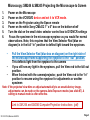
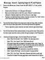
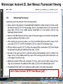
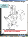
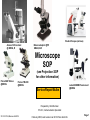
Student Scopes (various) Axiovert 25 Inverted @ BRDG, R124 Nikon Labophot @FV SM244, 245 Microscope SOP (see Projection SOP for other information) Parco XMZ Stereo @BRDG Parco LTM-800 @BRDG Service/Repair/Bulbs Leica 4000DMI Fluorescent @BRDG Prepared by: Bob Morrison STLCC, Instrumentation Specialist STLCC-CPLS;Morrison 9/4/2015 February 2008, Last revision Jan 2014 Parco bulb info Page 1 SM244 and SM245 : Nikon Labophot -2 Camera Control CMACamera 3CCD (details Slide #7) Turn “on” (green light) on bottom shelf of cart View Selector Control Rod Eyepiece (push in) Camera ( pull out) VCR/DVD Player, Turn On And set to VCR mode Turn dial on selector box To DVD/VCR setting. Selector box 1 2 Epson remote used To power on projector 3 4 Objectives 4,10,20,40x Focus & Fine focus Condenser Diaphragm Adjustment STLCC-CPLS;Morrison 9/4/2015 Off/On Light Source Slide Adjustment Page 2 Microscopy: SM244 & SM245 Projecting the Microscope to Screen 1. 2. 3. 4. 5. 6. Power on the Microscope Power on the VCR/DVD device and set it to VCR mode. Power on the Projector using the Epson remote Power on the white Sony CMA-D2 1” x 6” box on the bottom shelf Turn the dial on the small video selector control box to DVD/VCR settings Focus the specimen in the microscope eyepiece as you would for normal observations. Note, this requires that the View Selector Rod (blue on diagram) is in the full “in” position to deflect light toward the eyepieces. • • • Pull the View Selector Rod (blue box on diagram) on the right side of the microscope frame supporting the eyepiece to the “out” position. This deflects light from the eyepiece to the camera If you still see any light in the eyepieces, pull the View rod to the full out position. When finished with the camera/projector, push the View rod to the “in” position to resume using the eyepiece for adjustments or another specimen Note: If the projected view dims or adjust automatically to an unsatisfactory image, adjustments can be made on the camera Auto Exposure modes (see slide #7). A setting to manual mode is often effective. Link to SM 244 and SM245 Computer/Projection Instructions (pdf) STLCC-CPLS;Morrison 9/4/2015 Page 3 Microscope: Axiovert 25 @ BRDG R124 / SLCC # 0097770 Occular/Eyepiece Light Source Condenser Light Shunt To Projector Filter Guide Phase, Clear, Varel PC/Monitor Condenser Light Diaphragm (must be pulled fully forward) HBO Aux Light Source Svideo to USB box Aperture Center Screws Diaphragm Pushrod Off/On MTI-DAGE Camera Control Unit (CCU) Light Source Adjust Set Gain to “manual” Off/On W green indicator STLCC-CPLS;Morrison 9/4/2015 Reflector Modules Filters/Fluorescence Camera Focus & Fine focus View Selector Knob Eyepiece (clockwise) Projection (ccw) Page 4 Microscopy: Axiovert ; Capturing Images for PC and Projector Power on the Microscope, Camera Control Unit (MIT–DAGE 2” x 5” box), and the Projector 1. 2. Verify that the CCU box is “on” with green LED shown. Make sure the Gain switch on the CCU is set to “Manual” to avoid camera attempts to rebalance brightness and thus dim your scope image. Projector is controlled by Epson remote, red power-on button. The “Video” button at the 6-9pm position gives control to the camera. Turn the View Selector Knob on the scope (next to the main focus dials) clockwise until it hits stop at about 4pm position, this directs light toward the eyepieces • Focus on specimen, adjust condenser, and light controls to get desired image 3. Turn the View Selector Knob counterclockwise to stop position about 10am position, this directs light toward the camera and then through an adapter to the PC/projector 4. Viewing and Capturing Images ; Start the Adobe Photoshop Program • Select “Start from Scratch”, then “CANCEL” the next popup menu • Select “File”, then “Import”, then “USB Camera Device” • If the scope image does not appear in the popup window, select “Image” at the bottom and then select the “Image” tab. On this menu set the input mode to S-Video and NTSC (should be the default settings) • Adjust brightness and other parameters using slide bar controls • Select “Capture Still Image”, and verify capture in popup window • Select “File”, then “Save”, then enter a filename and format (usually .jpg) • Close/Exit the Adobe Photoshop application when finished with all captures STLCC-CPLS;Morrison 9/4/2015 Page 5 Microscope: Inverted, Image Capture, Set Video Source 1. If the scope image does not appear, select Image. 2. Select the Image tab and then make sure NTSC and S-Video boxes are set, 3. Scope image should now appear STLCC-CPLS;Morrison 9/4/2015 Page 6 Microscope: Axiovert, Camera and Control Unit MTI-DAGE DC-330 Camera and Control Unit (CCU) Camera cable input port Off/On W green indicator Camera output ports on rear(Svideo) 10-bit, digital processing Resolution up to 750 TV lines On-chip integration to 8 seconds (256 frames) Digital horizontal and verticle detail enhancement On screen programming for 41 internal camera functions Three user-defined memories retain camera settings RS-232c interface Automatic color shading correction RGB, Y/C, NTSC, or B/W video outputs Set Gain to “manual” to avoid auto adjustment changing scope view. HotLink to DC-330 Camera Specs on a website STLCC-CPLS;Morrison 9/4/2015 Page 7 Microscope: Axiovert 25, User Manual, Fluorescent Viewing Link to Zeiss Axiovert 25 CFI User Manual (pdf) STLCC-CPLS;Morrison 9/4/2015 Page 8 Microscope: Axiovert 25, Fluorescence, Ref. Figure Lamp Halogen w clipmount HLWS-A 6V30W, NARVA Part#:000000-0402-943 (have spares @BRDG) Link to Zeiss Axiovert 25 CFI User Manual (pdf) STLCC-CPLS;Morrison 9/4/2015 Page 9 Microscope: Fluorescence, Typical Path and Filters STLCC-CPLS;Morrison 9/4/2015 Page 10 Microscope: Axiovert 25 @BRDG, Countertop / SN #668654 Light Source Note: Make sure the Condenser is pulled fully forward and locked into position. Condenser Light Diaphragm Adjustment Filter Guide Phase, Clear, Varel Light Source Adjustment Link to Zeiss Axiovert 25 CFI User Manual (pdf) Off/On Eyepieces Focus & Fine focus STLCC-CPLS;Morrison 9/4/2015 Note: This bench device does not have a camera. Page 11 Microscope: Stereo/ Dissecting, @BRDG, Parco XMZ XMZ-833-10L Binocular 10x WF 7.5x to 35x Top Halogen Bottom LED Parco, XMZ series XMZ 0620131 1/25/10 RGM, BS recd, Binocular head inclined at at 45º angle Paired 10x wide-field eyepieces Working distance of 85mm Interpupillary adjustment of 55 to 75mm Dual diopter adjustments Coated optics for crisp image and superior resolution Illumination and stand for examining opaque or translucent specimens Heavy-duty rack and pinion focusing with slip clutch and tension adjustment Includes a 75mm frosted stage plate and 75mm reversible black/white stage Locked-on spring mounted stage clips 120V 20W STLCC-CPLS;Morrison 9/4/2015 OK for Use 7/12/12 RGM Power supply out, thus incident light does not work, seeking supplier of part Power Supply Internal, reqd for incident lighting 110VAC to 12V DC Page 12 Microscope: Parco XMZ Series Instruction Manual Focus: Holding problem; Correction STLCC-CPLS;Morrison 9/4/2015 Page 13 Microscope: Bulb, XMZ-833-10L Binocular 10x WF 7.5x to 35x Top Halogen Bottom LED STLCC-CPLS;Morrison 9/4/2015 Page 14 Microscope: Parco LTM-800, @BRDG, Binocular LTM-802-P Monocular 10x WF w/Pointer 4x, 10x, 40xR, 100xR Halogen Use choke ring collar around left side course focus knob to tighten tension on stage (prevent sagging stage) Choice of 45º monocular, 90º dual view video, binocular or trinocular head 10x wide-field eyepiece with calibrated pointer (on select models) DIN achromatic objectives are parfocaled and parcentered Quadruple nosepiece has precision ball bearing movement with precision stops Coaxial coarse and fine focusing with planetary reduction gear system Positive stops at both ends of the stage to prevent damage to specimens and optics 6V halogen illumination with two element condenser and advanced electronics Adjustable light intensity control know Built-in mechanical stage, 1.25 N.A. Abbe condenser, iris diaphragm and blue filter Spring loaded stage clamp for exact positioning of specimen Tension adjustment eliminates stage drift STLCC-CPLS;Morrison 9/4/2015 Page 15 Microscope: Parco, LTM 800 Series Instruction Manual Focus: Holding problem; Correction STLCC-CPLS;Morrison 9/4/2015 Page 16 Microscope: Parco, LTM-800 , Bulb Replacement STLCC-CPLS;Morrison 9/4/2015 Page 17 Microscope: Parco, Lamp replacement LTM-800 6V 20W, LBB3 STLCC-CPLS;Morrison 9/4/2015 Page 18 Microscope: Lamp 5W replacement STLCC-CPLS;Morrison 9/4/2015 Page 19 Microscope: Student, Leica DM750 New FV 11/11/09 M1-M27 Designated for Microlab Microscope for university advanced life science courses Key Features Focusable or fixed eyepieces Field of view of 20mm 45 degree tube Wear resistant Energy saving 4/10/40/100Oil Objectives Standard condenser for magnifications 4x – 100x Phase turret condenser for brightfield and phase contrast Flip top condenser for low magnifications DM750 is available with a 4 position or 5 position nosepiece Integrated vertical handle provides easy carrying and easy lifting when storing on high shelves Integrated cord wrap eliminates damage to microscope components Vertical cord insertion prevents the cord from pulling partially out of the stand while in storage or in use Unique shape of the microscope stand protects controls from damage when microscopes are stored side-by-side STLCC-CPLS;Morrison 9/4/2015 Page 20 Microscope: Student-Leica S4E A rear-facing nosepiece provides comfortable operation Spring-Loaded, high magnification objectives A built-in blue filter to prevent filter loss 360° rotatable, 45° viewing bodies Graduated mechanical stage with Vernier scales provides precise control Low heat output to provide comfortable viewing and prevent injury Tungsten-Halogen lamp: 20W, 6V, 2,000 hours life Design to meet international safety standards 120VA 220-240 Substage rack and pinion condenser STLCC-CPLS;Morrison 9/4/2015 Bulb: KANDOlite, MR11 6V 15W, Hallogen Dichroic GU4, 30 degree, 35mmO Page 21 Microscope: Student-Leica CM E Link to Microscope Educational Materials On Florida State University Website. The Leica S4 E with 4.8:1 zoom and standard magnification of 6.3x - 30x is the basic model of the Leica StereoZoom® line. 4.8:1 zoom Standard magnification 6.3x - 30x Overall ergonomic design and comfortable 38° viewing angle Largest field of view of any instrument in its class - 36.5mm Working distance 110mm The Leica StereoZoom® range offers a flat (planar) field of view. polymer which makes this combination perfect for electrostatically sensitive work. STLCC-CPLS;Morrison 9/4/2015 Page 22 Microscope: Oil Immersion, Leica ATC2000 Link to Basic Leica ATC instruction Manual …. pdf Link to Oil Immersion Guide from Clermont College Website.. htm STLCC-CPLS;Morrison 9/4/2015 Page 23 Microscope: Local Services, Repairs, Bulb Replacement Bulb Replacement Orders: Per GN 3/31/11 Archway Lighting Supply Incorporated (314) 535-1314 2739 Washington Ave, St Louis, MO 63103 1. 2. 3. 4. Call or Go to the archway website: http://www.bfmgraphics.com/al/major_manufacturing.htm Go to the products section or any area and select the phillips hotlink On the Phillips site, select product, then professional lighting http://www.Ecat.Lighting.Philips.Com/l/professionallamps/ep01_gr_us_lp_prof_atg/cat/us?Omnpg=lampsprofessional&lptype=lamps&navaction=pop&navcount=0&omnpc=ep01_gr_us_lp_prof_atg&isleftna v=false Locate type of bulb : compact, fluorescent; halogen… From: Naumann, Virginia L. Sent: Thursday, July 12, 2012 9:35 AM. Subject: RE: Microscope repair services, local? Hitchfel: 2333 S. Hanley Road St. Louis, Missouri 63144 T 800.242.3501 Spakowski Microscope Service: 7739 Brookline Ter, Saint Louis, MO 63117 T (314) 644-6560 STLCC-CPLS;Morrison 9/4/2015 Page 24 Microscope; Leica DMI4000B, BRDG, Basic Startup The Leica DMI4000 B automated inverted research microscope is ideal for scanning cell and tissue cultures. The system features a fluorescence axis for ultra brilliant fluorescence imaging. The Leica Application Suite (LAS) manages the system. 2. Scope Power on/off 4. Leica Application Suite (LAS) Software/app LAS 3.8 7. Advanced Fluorescence app LAS AF 5. Rotate Objective to desired lens. 3. UV Source on/off (opt) Leave on at least 5 minutes STLCC-CPLS;Morrison 9/4/2015 1. Boot LAS PC 6. Image Pushrod: In- to Eyepiece Out- to PC Page 25 Microscope; LeicaDMI; Startup Process 1. 2. 3. 4. 5. 6. 7. Turn on Scope , Top white box at left , using on/off green toggle switch. Turn on PC to boot up Login to Windows (password = microscope) and wait for application screen. Focus specimen using eyepiece with side pushrod on scope pushed in toward base Select LAS 3.8 for most applications (brightfield, phase contrast, basic fluorescence) Select LAS AF (Advance Fluorescence) for advanced fluorescence Pull eyepiece/external rod out to shift image to computer LAS application LAS Windows Icons: LAS 3.8 LAS AF LAS 3.8 Leica Application Suite used for most/general needs LAS AF Leica Application Suite for Advanced Fluorescence Hotlink to Leica DMI Application Suite Help Manual … pdf (1087 pgs) Hotlink to Leica DMI LAS Installation protocol … pdf (82 pgs) STLCC-CPLS;Morrison 9/4/2015 Page 26 Microscope: LeicaDMI: User Interface Screen Sections STLCC-CPLS;Morrison 9/4/2015 Page 27 Microscope: LeicaDMI, Microscope-Work Flow and Functions LAS Work Flow Tabs and Function: 1. Setup; do not use (adjust system parameters/preferences) 2. Acquire; adjust parameters on scope and camera , then acquire image to process and/or save 3. Browse ; setup folders and navigate for file/save operations 4. Process ; after acquiring an image, used to annotate or enhance STLCC-CPLS;Morrison 9/4/2015 Page 28 Microscope: LeicaDMI, Objective, Contrast Method, Illumination Controls Mic1 is highlighted with an orange strip at the top, this is the panel that the objectives, contrast methods and illumination are controlled through. Objectives are selected by manually rotating on the microscope. The corresponding box will be highlighted with orange on the screen. Contrast methods are chosen by clicking on either BF (Brightfield), PH (Phase-Contrast), FLUO (Flourescence) or FLUO-PH. The illumination panels controls the apeture and light intensity. This may also be controlled through the software. STLCC-CPLS;Morrison, RDK 9/4/2015 Page 29 Microscope: LeicaDMI, Camera Controls By clicking on the Camera tab at the top left this panel will become active. By clicking on any of the triangles at the right edge of the boxes you may manipulate various camera settings. STLCC-CPLS;Morrison, RDK 9/4/2015 Page 30 Microscope: LeicaDMI, Acquire, Save-as Dialog By clicking on the acquire screen button you will capture whatever you see on the live image to the right. A save as dialogue box will appear. STLCC-CPLS;Morrison, RDK 9/4/2015 Page 31 Microscope: LeicaDMI, Camera-Acquire Screen Acquire Options: Basic Functions; Auto-adjust, white balance, Select arrow to expand drop-down menus for adjustments to camera and resulting image. STLCC-CPLS;Morrison 9/4/2015 Page 32 Microscope: LeicaDMI, Acquire Image-Save-As Browse or Acquire Image: Leads to normal Windows panel To define folders and/or filenames for images and metadata. Note: metadata is saved automatically with images for future Reference and documentation. Images can be moved to other PCs/Laptops where the LAS software has been installed for further clarity adjustments, annotation, and/or analysis. STLCC-CPLS;Morrison 9/4/2015 Page 33 Microscope: LeicaDMI, Process Image Process Options: Select Arrows in upper right corner of each panel to produce Drop-down menus with adjustment slider bars, etc. STLCC-CPLS;Morrison 9/4/2015 Page 34 Microscope: LeicaDMI, Process Image, Apply to Save APPLY Operation: After changes or annotation to raw image, you must select APPLY to save those changes with the original raw image file. STLCC-CPLS;Morrison 9/4/2015 Page 35 Microscope: LiecaDMI, Export Images STLCC-CPLS;Morrison 9/4/2015 Page 36 Microscope: LeicaDMI, Setup, Nosepiece, Air/Immersion STLCC-CPLS;Morrison 9/4/2015 Page 37 Microscope: LeicaDMI, Nosepiece Control, Dry/Immersion STLCC-CPLS;Morrison 9/4/2015 Page 38 Microscope: LeicaDMI4000 Filter Cubes STLCC-CPLS;Morrison 9/4/2015 Page 39 Microscope: LeicaDMI4000 Filter Cubes (cont) STLCC-CPLS;Morrison 9/4/2015 Page 40 Microscope: Fluorescence; DAPI DAPI or 4',6-diamidino-2-phenylindole is a fluorescent stain that binds strongly to A-T rich regions in DNA When bound to double-stranded DNA DAPI has an absorption maximum at a wavelength of 358 nm (ultraviolet) and its emission maximum is at 461 nm (blue). Therefore for fluorescence microscopy DAPI is excited with ultraviolet light and is detected through a blue/cyan filter. The emission peak is fairly broad[2] DAPI will also bind to RNA, though it is not as strongly fluorescent. Its emission shifts to around 500 nm when bound to RNA.[3 DAPI's blue emission is convenient for microscopists who wish to use multiple fluorescent stains in a single sample. There is some fluorescence overlap between DAPI and greenfluorescent molecules like fluorescein and green fluorescent protein (GFP) but the effect of this is small. Use of spectral unmixing can account for this effect if extremely precise image analysis is required. Outside of analytical fluorescence light microscopy DAPI is also popular for labeling of cell cultures to detect the DNA of contaminating mycoplasma or virus. The labelled mycoplasma or virus particles in the growth medium fluoresce once stained by DAPI making them easy to detect. STLCC-CPLS;Morrison 9/4/2015 Page 41 Microscope: Fluorescence; Texas Red Texas Red or sulforhodamine 101 acid chloride is a red fluorescent dye, used in histology for staining cell specimens, for sorting cells with fluorescent-activated cell sorting machines, in fluorescence microscopy applications, and in immunohistochemistry.[1][2] Texas Red fluoresces at about 615 nm, and the peak of its absorption spectrum is at 589 nm. The powder is dark purple. Solutions can be excited by a dye laser tuned to 595-605 nm, or less efficiently a krypton laser at 567 nm. The absorption extinction coefficient at 596 nm is about 85,000 M−1cm−1. A protein with the Texas Red chromophore attached can then itself act as a fluorescent labelling agent; an antibody with a fluorescent marker attached will bind to a specific antigen and then show the location of the antigens as shining spots when irradiated. It is relatively bright, and therefore can be used to detect even weakly expressed antigens. Other molecules can be labeled by Texas Red as well, e.g., various toxins. The dye dissolves very well in water as well as other polar solvents, e.g., Dimethylformamide, acetonitrile. Texas Red, attached to a strand of DNA or RNA, can be used as a molecular beacon for highlighting specific sequences of DNA. Texas Red can be linked with another fluorophore. A tandem conjugate of Texas Red with R-phycoerythrin (PE-Texas Red) is often used. Fluorophores, like Texas Red, are commonly used in molecular biology techniques like quantitative RT-PCR and cellular assays.[3 STLCC-CPLS;Morrison 9/4/2015 Page 42 Student Microscopes, examples STLCC-CPLS;Morrison 9/4/2015 Page 43 Student Microscope; Typical Iris Diaphragm lever, 9am closed 12noon, open STLCC-CPLS;Morrison 9/4/2015 Page 44 Microscopes: Cleaning and Phase Filter Procedures 1. • • • • • 2. • • • • • • Cleaning Objectives, Filters, Lens Remove objective, use ring not objectives to rotate for removal Rub lightly with lens tissue first Remove an eyepiece and hold at 45 degree angle from objective to inspect for dust or other materials Use Cotton swaps or Q-tips and Ethyl Alcohol (90-95%), rub from center out in a spiral motion, rotate and replace swap as needed when stained or fluffy. “Carl’s bottle” is in lab setup area. Check cleaning with a prepared standard slide (ex Diatoms), specimen slide cover down, always toward the objectives Phase ring/filter alignment Make sure condenser and phase ring slot is pulled fully forward and in locked position Remove filter guide and clean filters if needed Insert a clean prepared slide with no stain Adjust condenser light intensity to lower setting to avoid glare Remove right eyepiece entirely, observe Phase filter with respect to circular ring of normal objective. Adjust end screws on Phase filter guide until rings are centered on each other, should end up with two concentric rings. STLCC-CPLS;Morrison 9/4/2015 Page 45 Microscope: Cleaning, Objectives, from MicroscopeWorld website Cleaning Objectives In order to determine which of your objective lenses need cleaning, take a clean blank glass slide and put it under your microscope. Once the microscope is focused you should be able to move the slide and determine if the visible dust is moving with the slide or staying in the same place (which means the dust is on the objective lens). When using immersion oil for microscopy, the oil should always be cleaned from microscope objective lenses immediately after use. This can be done with a kimwipe of piece of lens paper, no cleaning solutions are needed. Occasionally dust may build up on the lightly oiled surface so if you wish to completely remove the oil then you must use an oil soluble solvent. For the Cargille Type A or B immersion oil that we sell, you can use Naptha, Xylene, or turpentine (use very small amounts on the kimwipe). Do not use water, alcohol or acetone as the oil is insoluble to these solvents. To remove other oily substances, we recommend using the detergent called Wisk and prepare a solution of 1 part Wisk to 100 parts water. If immersion oil was not cleaned off an objective after use and has harded on the objective, moisten a piece of lens paper with a small amount of distilled water and hold it against the lens for a few seconds to dissolve the oil. If that does not work, try alcohol. Isopropyl alcohol is one of the best solvents but it must be at least 90%+ pure (do not use rubbing alcohol, 30% water). Everclear which is grain alcohol (you must be 21!) can also be used but it doesn't do as well in dissolving crud. If you have something like Balsam stuck on the lens, you must resort to a stronger solvent like Acetone or Xylene. Acetone should never be put on plastic parts, as it will dissolve most paints and plastic. After using solvents be sure to clean the objective again with standard distilled water to ensure that you have removed all the solvents from the microscope objective. STLCC-CPLS;Morrison 9/4/2015 Page 46 ALIGNMENT OF A COMPOUND, UPRIGHT MICROSCOPE (written by Nancy Kruger, Feb 2008) 1. Adjust the eyepieces/oculars so they are at the midpoint setting, indicated by a line or matching a dot to a line. Adjust the eyepiece spread to match the distance between your eyes. 1. Place a slide on the stage and focus on it with the 10x objective. 1. Close the field diaphragm/lens on the base of the microscope, if that adjustment can be made. 1. Adjust the condenser height with the condenser knob, until you see a distinct polygon in the field of view. 1. Using the condenser centering knobs below the stage, center the bright area in the field of view. By opening up the field lens slightly, you can fine tune the centering. 1. Completely open the field diaphragm/lens. Adjust the condenser aperture setting to give the best image. 1. Change objectives to produce different total magnifications. Remember that your total magnification is eyepiece magnification (10x) times the objective/lens magnification. STLCC-CPLS;Morrison 9/4/2015 Page 47 SET UP A STEREO DISSECTING MICROSCOPE TO BE PARFOCAL (written by Nancy Kruger, Feb 2008) 1. Adjust the eyepieces/oculars so they are at the midpoint setting, indicated by a line or matching a dot to a line. Adjust the eyepiece spread to match the distance between your eyes. 1. Place a specimen on the stage with the appropriate illumination (top/episcopic, or transmitted/diascopic illumination or both) 1. Using the lowest magnification setting, focus on a distinct area of the specimen. Use the coarse focus knobs to set the focus. 1. Change to the highest magnification using the magnification set knob. Use the coarse focus knobs to adjust to the best focus. 1. Change back to the lowest magnification using the magnification set knob. DO NOT TOUCH THE COARSE FOCUS KNOBS!! 1. Block off your left eye using your hand or piece of paper. Adjust the right eyepiece to produce the best focus possible for you by twisting the top portion. Switch eyes and repeat the process. 1. Check your settings by “zooming” between the lowest and highest magnifications. The image should stay in focus over the entire range. If not, repeat the process. You may need minor focus adjustments to see detailed structures at various planes, but no major focus adjustments. STLCC-CPLS;Morrison 9/4/2015 Page 48 Microscopy: Optical Maintenance - Lens Cleaning http://www.flinnsci.com/Sections/Biology/microscope.asp 1. All lenses are made of coated, soft glass and can be easily scratched. Lenses should be treated with care. Never use a hard instrument (such as a dissecting needle, etc.) or abrasive to clean a lens. 2. For the top of the eyepiece and the ends of the objectives, clean as follows: Use a camel's hair brush and an aspirator to remove all loose dust and dirt. Then moisten the end of a Q–tip™ with lens cleaning solution. Keep the other end of the Q–tip dry. Clean the optical surface with the moist end of the Q–tip using a circular motion. Dry the surface with the dry end of the Q–tip using a circular motion. Use an aspirator or similar air source to remove any lingering dirt particles. 3. Immersion oil should always be wiped from all surfaces immediately after use. In the event immersion oil is allowed to harden, moisten a piece of lens paper with a small amount of xylene and use this to redissolve and remove the hardened oil. Note: Xylene may leave a film on the lens and may dissolve the cement used to seal the immersion objective. To prevent this, always moisten a second lens paper with alcohol and use it to remove any residual xylene. Repeated use of xylene will destroy lens coatings. 4. To determine which lens surfaces need cleaning, focus the microscope on a clean slide free of all dust. Moving the slide will determine if the visible dust is on the slide. Rotating the eyepiece will establish if dirt is on the eyepiece. After loosening the retaining screw (if there is one) rotate the eyepiece in a circular fashion. If any dirt rotates, the eyepiece needs cleaning. Remove the eyepiece and clean it. Be careful not to damage any pointers. Clean the eyepiece on both ends in the same fashion described above for the objectives. When the eyepiece is thoroughly cleaned and dried, replace it and refocus the microscope. STLCC-CPLS;Morrison 9/4/2015 Page 49 Microscopy: Objective Lens Cleaning http://www.flinnsci.com/Sections/Biology/microscope.asp 1. Moving other parts will likewise help determine where dirt exists. Dirt on mirrors can be detected by moving the mirror while looking through the microscope. Rotating objectives will establish if dirt is on a specific objective. Does the specific dirt move or stay when objectives are rotated? Dust on a condenser lens can be detected in a similar fashion. Substage condenser lenses and mirrors should be cleaned with lens paper. 2. If the lower exterior surface of an objective has been cleaned and dirt still persists, it may be necessary to clean the inside surfaces of the objective. To do this the objective lens should be carefully removed from its nosepiece mounting. The objective lenses are threaded into the nosepiece and must be carefully removed for cleaning. This should be done with the utmost care to avoid stripping the threads and/or scratching the finish on the objective. Apply a firm, even pressure on the serrated top of the objective while holding the nosepiece from turning. A padded wrench or leather strip may prevent scratching of the objectives. Do not overtwist. If the objectives seem too difficult to loosen with a small wrench, call a microscope repair technician. 3. Clean the inside of the objective lens just like the outside, try to avoid lint and dust from getting back inside the objective. An aspirator is very helpful when working on the inside of an objective lens. 4. If after cleaning all surfaces carefully, dirt is still found in the field of view, it is possible that dirt is between the lenses of the objective. This dirt cannot be removed without disassembling the compound lens in the objective. Do not attempt this—call your microscope repair technician. 5. Final Inspection of Objectives: After cleaning it may be useful to check the overall operation by inspecting the objective lens using another microscope on a low power objective. Most objective lenses (extreme bottom of the lens) can be removed from the objective mount by unscrewing the cover or retaining sleeve with fingers or plier tools. To avoid scratching the surfaces with pliers, use a cloth between the tool and the objective. Put the objective lens on a clean class slide under another scope and focus to inspect for dust, spots, cracked lenses, or other debris. (Morrison, Mar 2008). STLCC-CPLS;Morrison 9/4/2015 Page 50 Microscopy: Mechanical Adjustments http://www.flinnsci.com/Sections/Biology/microscope.asp Nosepiece Adjustment: The nosepiece can likewise become too loose or too tight. There is usually an adjustment mechanism on the nosepiece. It is often as simple as loosening or tightening the slot-headed screw in the middle of the nosepiece. Sometimes there is a two–hole ring nut. This requires using a round nose pliers like a wrench to loosen or tighten the collar. On some microscopes the stage must be removed to gain access to the nosepiece adjustment. Be sure to check the manual for your specific microscope. Focus Knob Adjustment: Tension of the coarse and fine adjustment knobs can be adjusted. Again, various mechanical methods have been designed. Some microscopes are adjusted by simply turning the knobs on each side of the microscope in opposite directions to tighten or loosen as desired. Others have adjustable collars on the shaft and require the use of specially designed collar–wrenches or allen wrenches to make the adjustments. Moving the collars out usually provides more tension. If your microscope requires unique collar–wrenches, obtain these from your microscope supplier. STLCC-CPLS;Morrison 9/4/2015 Page 51 Microscope: Local Services, Repairs, Bulb Replacement Bulb Replacement Orders: Per GN 3/31/11 Archway Lighting Supply Incorporated (314) 535-1314 2739 Washington Ave, St Louis, MO 63103 1. 2. 3. 4. Call or Go to the archway website: http://www.bfmgraphics.com/al/major_manufacturing.htm Go to the products section or any area and select the phillips hotlink On the Phillips site, select product, then professional lighting http://www.Ecat.Lighting.Philips.Com/l/professionallamps/ep01_gr_us_lp_prof_atg/cat/us?Omnpg=lampsprofessional&lptype=lamps&navaction=pop&navcount=0&omnpc=ep01_gr_us_lp_prof_atg&isleftna v=false Locate type of bulb : compact, fluorescent; halogen… From: Naumann, Virginia L. Sent: Thursday, July 12, 2012 9:35 AM. Subject: RE: Microscope repair services, local? Hitchfel: 2333 S. Hanley Road St. Louis, Missouri 63144 T 800.242.3501 Spakowski Microscope Service: 7739 Brookline Ter, Saint Louis, MO 63117 T (314) 644-6560 STLCC-CPLS;Morrison 9/4/2015 Page 52 Microscopy – Education, Tutorials, FAQ http://www.olympusmicro.com/primer/index.html STLCC-CPLS;Morrison 9/4/2015 Page 53 Microscopy: Other Procedures 1. “Koehler”, Proper Specimen illumination Adjustment • • • • • • • Place prepared slide specimen on stage, center 10x objective Close down/reduce light source diaphragm in base Lower condenser until diaphragm is in focus Center image using condenser centering screws Open diaphragm to edge of field, fine focus and open further to just clear field Adjust contrast using condenser diaphragm Remove eyepiece and check that 75% of visible aperture is filled with light STLCC-CPLS;Morrison 9/4/2015 Page 54