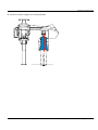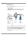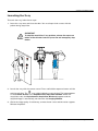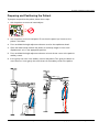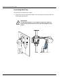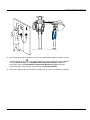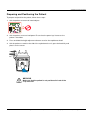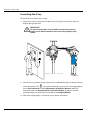
1
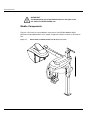
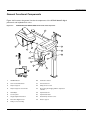
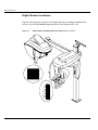
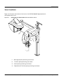
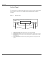
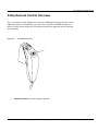
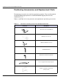
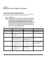
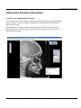
KODAK 8000C Digital Panoramic and Cephalometric
Extraoral Imaging System
User Guide
Notice
Congratulations on your purchase of the KODAK 8000C Digital Panoramic and Cephalometric Extraoral Imaging
System. The KODAK 8000C unit is the KODAK 8000 equipped with the cephalostat unit. Thank you for your
confidence in our products and we will do all in our power to ensure your complete satisfaction.
The User Guide for the KODAK 8000C Digital Panoramic and Cephalometric Extraoral Imaging System includes
information on the cephalometric features. For the panoramic features, see the KODAK 8000 Extraoral Imaging
System (SM722) User Guide. We recommend that you thoroughly familiarize yourself with this Guide in order to
make the most effective use of your system.
WARNING: We recommend that you consult the “Safety, Regulatory and the Technical
Specification User Guide” before using the KODAK 8000C Extraoral Imaging Systems.
The information contained in this Guide may be subject to modification without notice, justification or notification
to the persons concerned.
No part of this Guide may be reproduced without the express permission of Carestream Health, Inc.
The US Federal law restricts this device to sale by or on the order of a physician.
This document is originally written in English.
Manual Name: KODAK 8000C Digital Panoramic and Cephalometric Extraoral Imaging System User Guide
Part Number: SM735
Revision Number: 02
Print Date: 03/2010
The brand names and logos reproduced in this Guide are copyright.
KODAK is a trademark of KODAK used under License.
KODAK 8000C Digital Panoramic and Cephalometric Extraoral Imaging System, complies with Directive
93/42/CEE relating to medical equipment.
.
0086
Manufacturer
Care stream Hea lth, Inc.
150 Verona Street
Roche ster NY 14 608
Authorized Representative in the European Community
EC REP
TROPHY
4, Rue F. Pelloutier, Croissy-Beaubourg
77435 Marne la Vallée Cedex 2, France
Contents
1—About This Guide
Conventions in this Guide. . . . . . . . . . . . . . . . . . . . . . . . . . . . . . . . . . . . . . . . . . . . . . . . . . . . . . . . . . . . . . . . . . . . . 1–1
2—KODAK 8000C UNIT OVERVIEW
General Overview . . . . . . . . . . . . . . . . . . . . . . . . . . . . . . . . . . . . . . . . . . . . . . . . . . . . . . . . . . . . . . . . . . . . . . . . . . .
Mobile Components . . . . . . . . . . . . . . . . . . . . . . . . . . . . . . . . . . . . . . . . . . . . . . . . . . . . . . . . . . . . . . . . . . . . . .
General Functional Components . . . . . . . . . . . . . . . . . . . . . . . . . . . . . . . . . . . . . . . . . . . . . . . . . . . . . . . . . . . .
Digital Sensor Locations. . . . . . . . . . . . . . . . . . . . . . . . . . . . . . . . . . . . . . . . . . . . . . . . . . . . . . . . . . . . . . . . . . .
Laser Locations . . . . . . . . . . . . . . . . . . . . . . . . . . . . . . . . . . . . . . . . . . . . . . . . . . . . . . . . . . . . . . . . . . . . . . . . . .
Control Panel . . . . . . . . . . . . . . . . . . . . . . . . . . . . . . . . . . . . . . . . . . . . . . . . . . . . . . . . . . . . . . . . . . . . . . . . . . . . . . .
X-Ray Remote Control Overview . . . . . . . . . . . . . . . . . . . . . . . . . . . . . . . . . . . . . . . . . . . . . . . . . . . . . . . . . . . . . . .
Positioning Accessories and Replacement Parts . . . . . . . . . . . . . . . . . . . . . . . . . . . . . . . . . . . . . . . . . . . . . . . . . .
2–1
2–2
2–3
2–4
2–5
2–6
2–7
2–8
3—IMAGING SOFTWARE OVERVIEW
Computer System Requirements . . . . . . . . . . . . . . . . . . . . . . . . . . . . . . . . . . . . . . . . . . . . . . . . . . . . . . . . . . . . . . .
General Software Overview . . . . . . . . . . . . . . . . . . . . . . . . . . . . . . . . . . . . . . . . . . . . . . . . . . . . . . . . . . . . . . . . . . .
KODAK Dental Imaging Software . . . . . . . . . . . . . . . . . . . . . . . . . . . . . . . . . . . . . . . . . . . . . . . . . . . . . . . . . . .
Cephalometric Acquisition Interface Module . . . . . . . . . . . . . . . . . . . . . . . . . . . . . . . . . . . . . . . . . . . . . . . . . .
Cephalometric Acquisition Interface Module . . . . . . . . . . . . . . . . . . . . . . . . . . . . . . . . . . . . . . . . . . . . . . . . . . . . .
Cephalometric Acquisition Window Overview . . . . . . . . . . . . . . . . . . . . . . . . . . . . . . . . . . . . . . . . . . . . . . . . .
Cephalometric Program Pane . . . . . . . . . . . . . . . . . . . . . . . . . . . . . . . . . . . . . . . . . . . . . . . . . . . . . . . . . . .
Cephalometric Patient Pane . . . . . . . . . . . . . . . . . . . . . . . . . . . . . . . . . . . . . . . . . . . . . . . . . . . . . . . . . . . .
Cephalometric Parameter Pane. . . . . . . . . . . . . . . . . . . . . . . . . . . . . . . . . . . . . . . . . . . . . . . . . . . . . . . . . .
3–1
3–2
3–2
3–2
3–3
3–3
3–5
3–6
3–7
4—GETTING STARTED
Switching on the Unit . . . . . . . . . . . . . . . . . . . . . . . . . . . . . . . . . . . . . . . . . . . . . . . . . . . . . . . . . . . . . . . . . . . . . . . .
Starting the Imaging Software . . . . . . . . . . . . . . . . . . . . . . . . . . . . . . . . . . . . . . . . . . . . . . . . . . . . . . . . . . . . . . . . .
Creating a Patient Record. . . . . . . . . . . . . . . . . . . . . . . . . . . . . . . . . . . . . . . . . . . . . . . . . . . . . . . . . . . . . . . . . . . . .
Accessing the Cephalometric Acquisition Window . . . . . . . . . . . . . . . . . . . . . . . . . . . . . . . . . . . . . . . . . . . . . . . .
4–1
4–2
4–2
4–3
5—ACQUIRING CEPHALOMETRIC IMAGES
Acquiring a Lateral Image . . . . . . . . . . . . . . . . . . . . . . . . . . . . . . . . . . . . . . . . . . . . . . . . . . . . . . . . . . . . . . . . . . . . . 5–1
Preparing the Unit and Setting the Acquisition Parameters . . . . . . . . . . . . . . . . . . . . . . . . . . . . . . . . . . . . . . 5–1
Acquiring a Frontal AP or PA Image. . . . . . . . . . . . . . . . . . . . . . . . . . . . . . . . . . . . . . . . . . . . . . . . . . . . . . . . . . . . . 5–5
Preparing the Unit and Setting the Acquisition Parameters . . . . . . . . . . . . . . . . . . . . . . . . . . . . . . . . . . . . . . 5–5
Preparing and Positioning the Patient . . . . . . . . . . . . . . . . . . . . . . . . . . . . . . . . . . . . . . . . . . . . . . . . . . . . . . . . 5–6
Acquiring an Oblique Image . . . . . . . . . . . . . . . . . . . . . . . . . . . . . . . . . . . . . . . . . . . . . . . . . . . . . . . . . . . . . . . . . . . 5–9
Preparing the Unit and Setting the Acquisition Parameters . . . . . . . . . . . . . . . . . . . . . . . . . . . . . . . . . . . . . . 5–9
Preparing and Positioning the Patient . . . . . . . . . . . . . . . . . . . . . . . . . . . . . . . . . . . . . . . . . . . . . . . . . . . . . . . 5–10
Acquiring a Submento-Vertex Image . . . . . . . . . . . . . . . . . . . . . . . . . . . . . . . . . . . . . . . . . . . . . . . . . . . . . . . . . . . 5–12
Preparing the Unit and Setting the Acquisition Parameters . . . . . . . . . . . . . . . . . . . . . . . . . . . . . . . . . . . . . 5–12
Preparing and Positioning the Patient . . . . . . . . . . . . . . . . . . . . . . . . . . . . . . . . . . . . . . . . . . . . . . . . . . . . . . . 5–13
KODAK 8000C Digital Panoramic and Cephalometric Extraoral Imaging System User Guide (SM735)_Ed 02
iii
Contents
Acquiring a Carpus Image . . . . . . . . . . . . . . . . . . . . . . . . . . . . . . . . . . . . . . . . . . . . . . . . . . . . . . . . . . . . . . . . . . . . 5–16
Preparing the Unit and Setting the Acquisition Parameters . . . . . . . . . . . . . . . . . . . . . . . . . . . . . . . . . . . . . 5–16
Preparing and Positioning the Patient . . . . . . . . . . . . . . . . . . . . . . . . . . . . . . . . . . . . . . . . . . . . . . . . . . . . . . . 5–17
X-Ray Dose Emission Information . . . . . . . . . . . . . . . . . . . . . . . . . . . . . . . . . . . . . . . . . . . . . . . . . . . . . . . . . . . . . . 5–19
6—MAINTENANCE
Daily . . . . . . . . . . . . . . . . . . . . . . . . . . . . . . . . . . . . . . . . . . . . . . . . . . . . . . . . . . . . . . . . . . . . . . . . . . . . . . . . . . . . . . . 6–1
Monthly . . . . . . . . . . . . . . . . . . . . . . . . . . . . . . . . . . . . . . . . . . . . . . . . . . . . . . . . . . . . . . . . . . . . . . . . . . . . . . . . . . . . 6–1
Annually . . . . . . . . . . . . . . . . . . . . . . . . . . . . . . . . . . . . . . . . . . . . . . . . . . . . . . . . . . . . . . . . . . . . . . . . . . . . . . . . . . . . 6–1
7—TROUBLESHOOTING
Quick Troubleshooting . . . . . . . . . . . . . . . . . . . . . . . . . . . . . . . . . . . . . . . . . . . . . . . . . . . . . . . . . . . . . . . . . . . . . . . . 7–1
iv
Chapter 1
About This Guide
Conventions in this Guide
The following special messages emphasize information or indicate potential risk to
personnel or equipment:
WARNING
Warns you to avoid injury to yourself or others by following the
safety instructions precisely.
CAUTION
Alerts you to a condition that might cause serious damage.
IMPORTANT
Alerts you to a condition that might cause problems.
NOTE
Emphasizes important information.
TIP
Provides extra information and hints.
KODAK 8000C Digital Panoramic and Cephalometric Extraoral Imaging System User Guide (SM735)_Ed 02
1–1
Conventions in this Guide
1–2
About This Guide
Chapter 2
KODAK 8000C UNIT OVERVIEW
The KODAK 8000C digital panoramic and cephalometric unit is designed to carry out the
following radiological examinations:
•
•
•
•
•
•
•
•
Panoramic
Maxillary Sinus
Temporomandibular Joints (TMJ)
Lateral cephalometric
Frontal (PA or AP) cephalometric
Oblique cephalometric
Submento-vertex cephalometric
Carpus cephalometric
General Overview
The KODAK 8000C digital panoramic and cephalometric unit is composed of the
following functional components:
•
•
•
•
•
•
•
•
•
•
•
•
•
•
•
•
The unit head that contains all the electronic control
The rotative arm
The fixed arm with a control panel
The panoramic digital sensor
The x-ray source assembly
The x-ray remote control
The chin rest and bite block
The chin rest base
The panoramic chin rest and bite block
The temple supports
The hand grips
The cephalostat arm
The cephalostat head
The head clamps and ear cones
The nasion support
The acquisition software (see “Imaging Software Overview”)
The following figures illustrate the general overview of the KODAK 8000C digital
panoramic and cephalometric units.
KODAK 8000C Digital Panoramic and Cephalometric Extraoral Imaging System User Guide (SM735)_Ed 02
2–1
General Overview
IMPORTANT
The Cephalostat can be positioned either on the right or the
left side of the KODAK 8000 unit.
Mobile Components
Figure 2-1 illustrates the up and down movement of the KODAK 8000C digital
panoramic and cephalometric units mobile component and the rotation of the rotative
arm.
Figure 2–1
2–2
KODAK 8000C UNIT OVERVIEW
KODAK 8000 and KODAK 8000C Units Mobile Components
General Overview
General Functional Components
Figure 2-2 illustrates the general functional components of the KODAK 8000C digital
panoramic and cephalometric units.
Figure 2–2
KODAK 8000 and KODAK 8000C Units Functional Components
14
16
1
15
17
16
11
8
3
9
12
7
10
6
4
2
13
5
1
ON/OFF button
10
Collimator selector
2
Chin rest and bite block
11
Unit rotative arm
3
Temple supports
12
X-Ray remote control
4
Temple supports control knob
13
PC hosting the imaging and the acquisition
software
5
Hand Grips
14
Cephalostat arm
6
Control panel
15
Cephalostat head
7
Height adjustment buttons
16
Head clamps and ear cones
8
Panoramic digital sensor
17
Nasion support
9
X-Ray source assembly
KODAK 8000C Digital Panoramic and Cephalometric Extraoral Imaging System User Guide (SM735)_Ed 02
2–3
General Overview
Digital Sensor Locations
Figure 2-3 illustrates the locations of the digital panoramic and digital cephalometric
sensors of the KODAK 8000C digital panoramic and cephalometric units.
Figure 2–3
KODAK 8000 and KODAK 8000C Units Digital Sensor Locations
system
2–4
KODAK 8000C UNIT OVERVIEW
General Overview
Laser Locations
Figure 2-4 illustrates the location of the lasers of the KODAK 8000C digital panoramic
and cephalometric units.
Figure 2–4
KODAK 8000 and KODAK 8000C Units Laser Beam Locations
4
3
2
1
1
Mid-sagittal plane positioning laser beam
2
Frankfort plane positioning laser beam
3
Canine plane positioning laser beam
4
Cephalometric Frankfort plane positioning laser beam
KODAK 8000C Digital Panoramic and Cephalometric Extraoral Imaging System User Guide (SM735)_Ed 02
2–5
Control Panel
Control Panel
The control panel is an alphanumeric, digital soft touch console. It allows the operator to
control certain unit functions. It also displays the operating parameters and error
messages.
Figure 2–5
Unit Control Panel
8
Digital Panoramic and Cephalometric System
kV
1
2–6
mA
S
2
3
4
1
X-Ray emission LED: Yellow, indicates the x-rays are being emitted.
2
Display Screen: Displays the current acquisition parameters and the error messages.
3
Reset button: Resets the unit arm to the initial position to enable the patient to enter
and exit the unit.
4
Laser beam button: Activates the laser positioning beams to correctly position the
patient.
KODAK 8000C UNIT OVERVIEW
X-Ray Remote Control Overview
X-Ray Remote Control Overview
The x- ray remote control enables you to launch a radiological image acquisition via the
exposure button from outside the x-ray room. You must press and hold the exposure
button until the end of acquisition. Premature release of the exposure button interrupts
the acquisition.
Figure 2–6
X-Ray Remote Control
1
1
Exposure button: launches image acquisition.
KODAK 8000C Digital Panoramic and Cephalometric Extraoral Imaging System User Guide (SM735)_Ed 02
2–7
Positioning Accessories and Replacement Parts
Positioning Accessories and Replacement Parts
The following accessories are used when positioning a patient. They are delivered with
the KODAK 8000 digital panoramic and KODAK 8000C digital panoramic and
cephalometric unit.
Table 2-1 and Table 2-2 list the panoramic and cephalometric positioning accessories.
Table 2–1
Panoramic Positioning Accessories and Replacement Parts
Accessory
Description
Panoramic chin rest and TMJ x2
Maxillary sinus chin rest
TMJ x2 and TMJ x4 nose rest
Standard bite block
Bite block for edentulous patients
A set of right and left temple supports
x2
Single use sheaths for bite blocks
(500 pcs box)
2–8
KODAK 8000C UNIT OVERVIEW
Positioning Accessories and Replacement Parts
Table 2–2
Cephalometric Positioning Accessories and Replacement Parts
Accessory
Description
Head clamps with ear cones
x2
Nasion support
KODAK 8000C Digital Panoramic and Cephalometric Extraoral Imaging System User Guide (SM735)_Ed 02
2–9
Positioning Accessories and Replacement Parts
2–10
KODAK 8000C UNIT OVERVIEW
Chapter 3
IMAGING SOFTWARE OVERVIEW
Computer System Requirements
This section specifies the minimum computer system requirements for KODAK 8000C
digital panoramic and cephalometric extraoral imaging system software.
IMPORTANT
It is MANDATORY to check that the computer system configuration is
compatible with the computer system requirements for the KODAK
8000C software. If necessary you MUST update your computer
system configuration. KODAK 8000C MUST be connected to the
computer via a point-to-point Ethernet link and not via a LAN. DO NOT
place the computer and the peripheral equipment connected to it in
the immediate vicinity of the patient in the unit. Leave at least 1.5 m
distance from the unit. The computer and the peripheral equipment
must conform to the IEC 60950 standard.
Table 3–1
Minimum Computer System Requirements
Item
Viewing
Acquisition
CPU
2 GHz Intel Duo Core
2 GHz Intel Duo Core
RAM
2 GB
2GB
•
Hard disk drive
•
Graphic board
1.2 GB for software
installation
•
•
•
Operating system (32 bits)
•
•
•
RAM has a major impact on
system performance.
1.2 GB for software installation
80 GB free space to use the
software
Nvidia / ATI based board supporting
Open GL 1.2 with 256 MB of video
RAM on AGP x8 video bus (for
example: Nvidia GeForce 6800 GT)
Monitor
Comments
1 monitor
17" or larger
1024 x 768 minimum screen
resolution - 32 bits color
mode
Nvidia / ATI based board
The video RAM has major
supporting Open Glide 1.2 with
impact on system performance.
256 MB of video RAM on AGP x8
video bus (for example: Nvidia
GeForce 6800 GT)
•
•
•
Windows 2000 SP4
•
Windows XP Home / Pro
edition SP2
•
1 monitor
17"
1280 x 880 minimum
screen resolution
Your monitor is a vital
component in displaying quality
images. Low-quality screens will
prevent you from proper
diagnoses and treatment.
Windows XP Home / Pro
edition SP2
Windows Vista
Windows Vista
KODAK 8000C Digital Panoramic and Cephalometric Extraoral Imaging System User Guide (SM735)_Ed 02
3–1
General Software Overview
Table 3–1
Minimum Computer System Requirements (Continued)
Item
Viewing
Ethernet interface
1 Ethernet interface
Comments
Acquisition
1 Ethernet interfaces (100Mbits)
If there is a LAN connection a
second Ethernet interface is
required.
CD/DVD drive
A DVD-BURNER drive is required.
A DVD-BURNER drive is required.
Backup Media
Removable/portable, external hard
disk drive
Removable/portable, external hard We strongly recommend a daily
disk drive.
backup of x-ray images and
patient records.
General Software Overview
The KODAK 8000C digital panoramic and cephalometric extraoral imaging system
operates with the following software:
•
•
KODAK dental imaging software
Cephalometric acquisition interface module
KODAK Dental Imaging Software
The KODAK dental imaging software is a user-friendly working interface that was
designed and developed specifically for radiological diagnosis. It is the common imaging
platform for all our digital systems for dentistry.
The KODAK dental imaging software has the following features:
•
•
Patient record management using Patient Window features
Extraoral and intraoral image management using Imaging Window features
NOTE
For a complete information on the KODAK Dental Imaging
Software, see the “KODAK Dental Imaging Software, Quick Start
Guide”.
Cephalometric Acquisition Interface Module
The cephalometric acquisition interface module is a user-friendly working interface that
was designed and developed specifically for KODAK 8000C digital panoramic and
cepahlometric extraoral imaging system.
3–2
IMAGING SOFTWARE OVERVIEW
Cephalometric Acquisition Interface Module
Cephalometric Acquisition Interface Module
Cephalometric Acquisition Window Overview
The Cephalometric Acquisition Window is the main cephalometric interface with the
KODAK 8000C cephalometric extraoral imaging system that provides you with imaging
acquisition functions.
Figure 3–1
1
Cephalometric Acquisition Window
Information button:
• About: Identifies the Software and the Firmware versions
•
•
Reset of the Values: Resets to the manufacturing parameter settings
Memorize settings: Memorizes the user preference settings for each patient type (kV,
mA and seconds)
2
Preview Screen: Displays the acquired image in real time.
3
Selected Parameter Display: Displays the current acquisition parameter settings.
4
System Status Screen: Displays various alert or warning messages originating from the unit.
5
X-Rays ON / OFF button: Turns off the x-ray emissions to demonstrate the acquisition process
for the patient.
6
Generator Cooling indicator: Indicates the automatic cooling time (mm:ss) required for the
generator to reach 0 for a new acquisition.
KODAK 8000C Digital Panoramic and Cephalometric Extraoral Imaging System User Guide (SM735)_Ed 02
3–3
Cephalometric Acquisition Interface Module
7
Positioning laser button: Activates the laser positionning beams to correctly position the
patient.
8
Reset button: Resets the rotative arm in the start position.
9
Ready Indicator LED
• Green indicates the unit is ready to start acquisition.
•
Black indicates the unit is not ready to start acquisition.
10
Exit button: Closes the Acquisition Window.
11
X-Ray Emission indicator: Yellow, indicates the x-ray emission status.
12
Selector Button: Selects different acquisition setting options.
• Click Program to select examination type options.
•
•
Click Patient to select patient type parameters.
Click Parameters to select exposure parameter options.
The Selector button enables you to access the following 3 panes:
•
•
•
3–4
Program pane: Examination type options
Patient pane: Patient type parameter options
Parameters pane: Exposure parameter options
IMAGING SOFTWARE OVERVIEW
Cephalometric Acquisition Interface Module
Cephalometric Program Pane
The cephalometric Program pane enables you to choose different radiological exams as
well as different acquisition formats.
Figure 3–2
Cephalometric Program Pane
Radiological exam options:
Click
for a lateral exam.
Click
or
exam (AP and PA).
Click
for a frontal
Click
Click
for a submento-vertex exam.
for a carpus exam.
for an oblique exam.
NOTE
The above list of exam types are only a sample of exam options of
the Program pane.
KODAK 8000C Digital Panoramic and Cephalometric Extraoral Imaging System User Guide (SM735)_Ed 02
3–5
Cephalometric Acquisition Interface Module
Cephalometric Patient Pane
The cephalometric Patient pane enables you to choose different patient parameters. The
selection of the patient parameters influences the quality of the image. The selected
parameters must be based on the patient age and morphology.
Figure 3–3
1
2
3–6
Cephalometric Patient Pane
Patient type parameters:
Click
if the patient is a child.
Click
if the patient is adult.
Patient size parameters:
Click
if the patient is small.
Click
if the patient is medium.
Click
if the patient is large.
IMAGING SOFTWARE OVERVIEW
Cephalometric Acquisition Interface Module
Cephalometric Parameter Pane
The cephalometric Parameter pane enables you to choose exposure parameters for the
radiological image acquisition. If the default parameter setting is not adapted to your
patient type, you can manually adapt the parameter settings to the patient type and save
this setting as the default setting.
Figure 3–4
1
Cephalometric Parameter Pane
Radiation dose options:
kV: Kilovolt
mA: Milliampere
S: Second
2
Fine-tuning buttons:
Click
3
to fine-tune the kV, mA and the second.
Saving parameter button:
Click
to save the selected parameters.
KODAK 8000C Digital Panoramic and Cephalometric Extraoral Imaging System User Guide (SM735)_Ed 02
3–7
Cephalometric Acquisition Interface Module
3–8
IMAGING SOFTWARE OVERVIEW
Chapter 4
GETTING STARTED
Switching on the Unit
Before switching on the unit, check that:
•
•
The installation of the unit is complete.
The PC is switched on.
IMPORTANT
You must switch on the PC and wait for it to be ready for the
connection before switching on the unit.
To switch on the unit, follow these steps:
1. On the unit column, press the ON button.
2. You must wait for a minute for the connection between the unit and the PC to be
established. If you start the imaging software before the connection is established an
error message is displayed. Click OK, close the imaging software and wait for the
connection to be established.
3. You can now proceed to start the imaging software.
IMPORTANT
To increase the operating life of the x-ray tube, if the unit has not been
used for a month, you must follow the following procedures before
use.
1. In the Panoramic Acquisition Window, select the Parameter pane.
2. Select the following series of parameter settings:
•
•
•
70 kV - 6.3 mA
80 kV - 10 mA
85 kV - 10 mA
3. Leave the x-ray room and close the door. For each parameter setting, from the x-ray
remote control, press and hold the button to launch the x-ray
The unit is now ready to be used for acquisition.
KODAK 8000C Digital Panoramic and Cephalometric Extraoral Imaging System User Guide (SM735)_Ed 02
4–1
Starting the Imaging Software
Starting the Imaging Software
To start the imaging software, follow these steps:
1. On your desktop, double-click
.
OR
From your PC, click Start > All Programs > Kodak > Kodak Dental Software.
A blank Patient Window is displayed.
2. Create or open an existing patient record.
Creating a Patient Record
To create a patient record, follow these steps:
1. In the Patient Window, from the toolbar, click
OR
From the menu bar, select Patient > New.
2. Enter the required patient information. The Last name, the First name and the Date
of birth fields are required.
3. From the menu bar, select Picture > Insert Picture to add a *.tif or *.bmp picture of
the patient to the record. Select the picture from your directory and click Open.
4. Click OK to save. The patient record is automatically assigned a 7-digit number
starting with a letter (for example, M0000001).
5. Click
to access the Imaging Window.
6. Select an image acquisition.
4–2
GETTING STARTED
Accessing the Cephalometric Acquisition Window
Accessing the Cephalometric Acquisition Window
To access the Acquisition Windows, follow these steps:
1. In the Imaging Window, from the toolbar, click
Acquisition Window.
to access the Cephalometric
2. Prepare the acquisition parameters and launch an acquisition.
KODAK 8000C Digital Panoramic and Cephalometric Extraoral Imaging System User Guide (SM735)_Ed 02
4–3
Accessing the Cephalometric Acquisition Window
4–4
GETTING STARTED
Chapter 5
ACQUIRING CEPHALOMETRIC IMAGES
Acquiring a Lateral Image
Before acquiring a lateral image, check that you have:
•
•
•
•
Reset the unit rotative arm to the start position for patient to enter the unit.
Selected the patient record.
Accessed the Imaging Window.
Accessed the Cephalometric Acquisition Window.
Preparing the Unit and Setting the Acquisition Parameters
To set the acquisition parameters, follow these steps:
1. Raise and block the sensor manually.
2. On the x-ray source assembly, set the collimator selector to LA.
3. Position the head clamps manually for the lateral exam.
IMPORTANT
You must position the head clamps manually because they
are not positioned automatically from the Program pane exam
type selection. In this case, the relevant exam type selection
icon becomes active.
4. In the Cephalometric Acquisition Window, click the Program button to access the
Program pane. In the Program pane the
for a lateral exam is active.
5. Click the Patient button to access the Patient pane. Select the patient:
•
•
Type
Size
6. If the default parameter setting is not adapted to your patient type, click the
Parameter button and in the Parameter pane select the appropriate parameters. To
save the new parameter settings as the default settings, click
Memorize settings.
fand select
7. Preparing and Positioning the Patient
To prepare and position the patient, follow these steps:
KODAK 8000C Digital Panoramic and Cephalometric Extraoral Imaging System User Guide (SM735)_Ed 02
5–1
Acquiring a Lateral Image
1. Ask the patient to remove all metal objects.
2. Ask the patient to wear a lead apron. Ensure that the apron lays flat across the
patient’s shoulders.
3. Press and hold the height adjustment buttons to raise the cephalostat head.
4. Open the head clamps and ask the patient to stand up straight, in front of the
cephalometric unit, in the appropriate position.
5. Press and hold the height adjustment buttons to level the ear cones to the patient’s
auditory canals.
6. Insert gently one cone in the auditory canal of the patient. Turn gently the button to
close the arms. Insert gently the second cone in the auditory canal of the patient.
7. On the control panel, click
to turn ON the Frankfort laser positioning beam.
Align the patient with the Frankfort laser beam.
5–2
ACQUIRING CEPHALOMETRIC IMAGES
Acquiring a Lateral Image
8. Lower the nasion support to a vertical position.
KODAK 8000C Digital Panoramic and Cephalometric Extraoral Imaging System User Guide (SM735)_Ed 02
5–3
Acquiring a Lateral Image
Launching the X-ray
To launch the x-ray, follow these steps:
1. Leave the x-ray room and close the door. You must keep visual contact with the
patient during acquisition.
IMPORTANT
To stop the acquisition, if any problem, release the exposure
button of the remote control or press the red emergency stop
button.
2. Launch the x-ray with the remote control. Press and hold the exposure button until the
end of acquisition. The
turns yellow, indicating x-ray emission. The image appears
on the Preview Screen of the Cephalometric Acquisition Window. When the
acquisition ends, the Cephalometric Acquisition Window disappears and the
acquired image is transferred automatically to the Imaging Window.
3. Check the image quality, if satisfactory, remove the ear cones and the nasion support
and release the patient.
5–4
ACQUIRING CEPHALOMETRIC IMAGES
Acquiring a Frontal AP or PA Image
Acquiring a Frontal AP or PA Image
Before acquiring a frontal AP or PA image, check that you have:
•
•
•
•
Reset the unit rotative arm to the start position for patient to enter the unit.
Selected the patient record.
Accessed the Imaging Window.
Accessed the Cephalometric Acquisition Window.
Preparing the Unit and Setting the Acquisition Parameters
To acquire a frontal AP or PA image, follow these steps:
1. Raise and block the sensor manually.
2. On the x-ray source assembly, set the collimator selector to AP/PA.
3. Position the head clamps manually for the frontal AP or PA exam.
IMPORTANT
You must position the head clamps manually because they
are not positioned automatically from the Program pane exam
type selection. In this case, the relevant exam type selection
icon becomes active.
4. In the Cephalometric Acquisition Window, click the Program button to access the
Program pane. In the Program pane, click
for a frontal PA exam.
5. Click the Patient button to access the Patient pane. Select the patient:
•
•
Type
Size
6. If the default parameter setting is not adapted to your patient type, click the
Parameter button and in the Parameter pane select the appropriate parameters. To
save the new parameter settings as the default settings, click
Memorize settings.
fand select
KODAK 8000C Digital Panoramic and Cephalometric Extraoral Imaging System User Guide (SM735)_Ed 02
5–5
Acquiring a Frontal AP or PA Image
Preparing and Positioning the Patient
To prepare and position the patient, follow these steps:
1. Ask the patient to remove all metal objects.
2. Ask the patient to wear a lead apron. Ensure that the apron lays flat across the
patient’s shoulders.
3. Press and hold the height adjustment buttons to raise the cephalostat head.
4. Open and position the head clamps parallel to the cephalometric sensor. Ask the
patient to stand up straight in front of the cephalometric unit in the following
positions:
•
•
For a frontal AP, facing the generator
For a frontal PA, facing the cephalometric sensor
5. Press and hold the height adjustment buttons to level the ear cones to the patient’s
auditory canals.
6. Insert gently one cone in the auditory canal of the patient. Turn gently the button to
close the arms. Insert gently the second cone in the auditory canal of the patient.
7. On the control panel, click
to turn on the Frankfort laser positioning beam. Align
the patient with the Frankfort laser beam for the frontal AP only.
Figure 5–1
5–6
ACQUIRING CEPHALOMETRIC IMAGES
Frontal AP
Figure 5–2
Frontal PA
Acquiring a Frontal AP or PA Image
Figure 5–3
Frontal AP
Figure 5–4
Frontal PA
Launching the X-ray
To launch the x-ray, follow these steps:
1. Leave the x-ray room and close the door. You must keep visual contact with the
patient during acquisition.
IMPORTANT
To stop the acquisition, if any problem, release the exposure
button of the remote control or press the red emergency stop
button.
KODAK 8000C Digital Panoramic and Cephalometric Extraoral Imaging System User Guide (SM735)_Ed 02
5–7
Acquiring a Frontal AP or PA Image
Figure 5–5
Frontal AP
Figure 5–6
Frontal PA
2. Launch the x-ray with the remote control. Press and hold the exposure button until the
end of acquisition. The
turns yellow indicating x-ray emission. The image appears
on the Preview Screen of the Cephalometric Acquisition Window. When the
acquisition ends, the Cephalometric Acquisition Window disappears and the
acquired image is automatically transferred to the Imaging Window.
3. Check the image quality. If satisfactory, remove the ear cones and release the patient.
5–8
ACQUIRING CEPHALOMETRIC IMAGES
Acquiring an Oblique Image
Acquiring an Oblique Image
Before acquiring an oblique image, check that you have:
•
•
•
•
Reset the unit rotative arm to the start position for patient to enter the unit.
Selected the patient record.
Accessed the Imaging Window.
Accessed the Cephalometric Acquisition Window.
Preparing the Unit and Setting the Acquisition Parameters
To acquire an oblique image, follow these steps:
1. Raise and block the sensor manually.
2. On the x-ray source assembly, set the collimator selector to AP/PA.
3. Position the head clamps manually for the oblique exam with the desired angle.
IMPORTANT
You must position the head clamps manually because they
are not positioned automatically from the Program pane exam
type selection. In this case, the relevant exam type selection
icon becomes active.
4. In the Cephalometric Acquisition Window, click the Program button to access the
Program pane. In the Program pane:
•
The
•
Click
for an oblique exam is active.
to select the desired angle.
5. Click the Patient button to access the Patient pane. Select the patient type.
6. Click the Patient button to access the Patient pane. Select the patient:
•
•
Type
Size
7. If the default parameter setting is not adapted to your patient type, click the
Parameter button and in the Parameter pane select the appropriate parameters. To
save the new parameter settings as the default settings, click
Memorize settings.
fand select
KODAK 8000C Digital Panoramic and Cephalometric Extraoral Imaging System User Guide (SM735)_Ed 02
5–9
Acquiring an Oblique Image
Preparing and Positioning the Patient
To prepare and position the patient, follow these steps:
1. Ask the patient to remove all metal objects.
2. Ask the patient to wear a lead apron. Ensure that the apron lays flat across the
patient’s shoulders.
3. Press and hold the height adjustment buttons to raise the cephalostat head.
4. Open the head clamps and ask the patient to stand up straight in front of the
cephalometric unit, in the appropriate position.
5. Press and hold the height adjustment buttons to level the ear cones to the patient’s
auditory canals.
6. Insert gently one cone in the auditory canal of the patient. Turn gently the button to
close the arms. Insert gently the second cone in the auditory canal of the patient.
7. Lower the nasion support to a vertical position.
5–10
ACQUIRING CEPHALOMETRIC IMAGES
Acquiring an Oblique Image
Launching the X-ray
To launch the x-ray, follow these steps:
1. Leave the x-ray room and close the door. You must keep visual contact with the
patient during acquisition.
IMPORTANT
To stop the acquisition, if any problem, release the exposure
button of the remote control or press the red emergency stop
button.
2. Launch the x-ray with the remote control. Press and hold the exposure button until the
end of acquisition. The
turns yellow indicating x-ray emission. The image appears
on the Preview Screen of the Cephalometric Acquisition Window. When the
acquisition ends, the Cephalometric Acquisition Window disappears and the
acquired image is automatically transferred to the Imaging Window.
3. Check the image quality. If satisfactory, remove the ear cones and the nasion support.
Release the patient.
KODAK 8000C Digital Panoramic and Cephalometric Extraoral Imaging System User Guide (SM735)_Ed 02 5–11
Acquiring a Submento-Vertex Image
Acquiring a Submento-Vertex Image
Before acquiring a submento-vertex image, check that you have:
•
•
•
•
Reset the unit rotative arm to the start position for patient to enter the unit.
Selected the patient record.
Accessed the Imaging Window.
Accessed the Cephalometric Acquisition Window.
Preparing the Unit and Setting the Acquisition Parameters
To acquire a submento-vertex image, follow these steps:
1. Raise and block the sensor manually.
2. On the x-ray source assembly, set the collimator selector to AP/PA.
3. Position the head clamps manually for a frontal AP exam..
IMPORTANT
You must position the head clamps manually because they
are not positioned automatically from the Program pane exam
type selection. In this case, the relevant exam type selection
icon becomes active.
4. In the Cephalometric Acquisition Window, click the Program button to access the
Program pane. In the Program pane, click
for a submento-vertex exam.
5. Click the Patient button to access the Patient pane. Select the patient:
•
•
Type
Size
6. If the default parameter setting is not adapted to your patient type, click the
Parameter button and in the Parameter pane select the appropriate parameters. To
save the new parameter settings as the default settings, click
Memorize settings.
5–12
ACQUIRING CEPHALOMETRIC IMAGES
fand select
Acquiring a Submento-Vertex Image
Preparing and Positioning the Patient
To prepare and position the patient, follow these steps:
1. Ask the patient to remove all metal objects.
2. Ask the patient to wear a lead apron. Ensure that the apron lays flat across the
patient’s shoulders.
3. Press and hold the height adjustment buttons to raise the cephalostat head.
4. Open the head clamps and ask the patient to stand up straight in front of the
cephalometric unit, in the appropriate position.
5. Press and hold the height adjustment buttons to level the ear cones to the patient’s
auditory canals.
6. Insert gently one cone in the auditory canal of the patient. Turn gently the button to
close the arms. Insert gently the second cone in the auditory canal of the patient.
KODAK 8000C Digital Panoramic and Cephalometric Extraoral Imaging System User Guide (SM735)_Ed 02 5–13
Acquiring a Submento-Vertex Image
Launching the X-ray
To launch the x-ray, follow these steps:
1. Leave the x-ray room and close the door. You must keep visual contact with the
patient during acquisition.
IMPORTANT
To stop the acquisition, if any problem, release the exposure
button of the remote control or press the red emergency stop
button.
5–14
ACQUIRING CEPHALOMETRIC IMAGES
Acquiring a Submento-Vertex Image
2. Launch the x-ray with the remote control. Press and hold the exposure button until the
end of acquisition. The
turns yellow indicating x-ray emission. The image appears
on the Preview Screen of the Cephalometric Acquisition Window. When the
acquisition ends, the Cephalometric Acquisition Window disappears and the
acquired image is automatically transferred to the Imaging Window.
3. Check the image quality. If satisfactory, remove the ear cones and release the patient.
KODAK 8000C Digital Panoramic and Cephalometric Extraoral Imaging System User Guide (SM735)_Ed 02 5–15
Acquiring a Carpus Image
Acquiring a Carpus Image
Before acquiring a carpus image, check that you have:
•
•
•
•
Reset the unit rotative arm to the start position for patient to enter the unit.
Selected the patient record.
Accessed the Imaging Window.
Accessed the Cephalometric Acquisition Window.
Preparing the Unit and Setting the Acquisition Parameters
To acquire a carpus image, follow these steps:
1. Raise and block the sensor manually.
2. On the x-ray source assembly, set the collimator selector to AP/PA.
3. Position the head clamps manually for a frontal AP exam.
IMPORTANT
You must position the head clamps manually because they
are not positioned automatically from the Program pane exam
type selection. In this case, the relevant exam type selection
icon becomes active.
4. In the Cephalometric Acquisition Window, click the Program button to access the
Program pane. In the Program pane, click
for a carpus exam.
5. Click the Patient button to access the Patient pane. Select the patient:
•
•
Type
Size
6. If the default parameter setting is not adapted to your patient type, click the
Parameter button and in the Parameter pane select the appropriate parameters. To
save the new parameter settings as the default settings, click
Memorize settings.
5–16
ACQUIRING CEPHALOMETRIC IMAGES
fand select
Acquiring a Carpus Image
Preparing and Positioning the Patient
To prepare and position the patient, follow these steps:
1. Ask the patient to remove all metal objects.
2. Ask the patient to wear a lead apron. Ensure that the apron lays flat across the
patient’s shoulders.
3. Press and hold the height adjustment buttons to raise the cephalostat head.
4. Ask the patient to stand on the side of the cephalometric unit, open the hand fully and
place it on the sensor.
WARNING
Make sure that the patient is not positioned in front of the
x-ray emission.
KODAK 8000C Digital Panoramic and Cephalometric Extraoral Imaging System User Guide (SM735)_Ed 02 5–17
Acquiring a Carpus Image
Launching the X-ray
To launch the x-ray, follow these steps:
1. Leave the x-ray room and close the door. You must keep visual contact with the
patient during acquisition.
IMPORTANT
To stop the acquisition, if any problem, release the exposure
button of the remote control or press the red emergency stop
button.
2. Launch the x-ray with the remote control. Press and hold the exposure button until the
end of acquisition. The
turns yellow indicating x-ray emission. The image appears
on the Preview Screen of the Cephalometric Acquisition Window. When the
acquisition ends, the Cephalometric Acquisition Window disappears and the
acquired image is automatically transferred to the Imaging Window.
3. Check the image quality, if satisfactory, then release the patient.
5–18
ACQUIRING CEPHALOMETRIC IMAGES
X-Ray Dose Emission Information
X-Ray Dose Emission Information
Compliance with EURATOM 97/43 Directive
You can right-click on each image to display the estimated emitted dose received by the
patient. You can use this information to calculate the effective dose received by the
patient for the image.
The radiation emission dose is expressed in mGy.cm2. This dose is measured at the
primary collimator outlet. The dose is accurate to +/-30%. The primary slot is 0.5 mm
wide and 18.2 mm high.
KODAK 8000C Digital Panoramic and Cephalometric Extraoral Imaging System User Guide (SM735)_Ed 02 5–19
X-Ray Dose Emission Information
5–20
ACQUIRING CEPHALOMETRIC IMAGES
Chapter 6
MAINTENANCE
This section describes the maintenance tasks that you need to perform regularly for your
KODAK 8000C unit and the accessories.
WARNING
Switch off the unit, then, clean all accessible parts of the
machine with an alcohol-based non-corrosive product. Avoid
using liquids inside the unit. Follow the alcohol-based product
manufacturer recommendations for safety precautions.
CAUTION
You can use the usual disinfectant products, but we
recommend that you protect the unit from contamination by
using barriers available from dental distributors. Follow the
disinfectant product manufacturer recommendations for
safety precautions.
Daily
Carry out the following maintenance tasks:
Table 6–1
Daily Maintenance Tasks
Accessories
Head clamps and ear cones
Nasion support
Maintenance Tasks
Clean with medical-grade 76% alcohol disinfectant before the next
patient is x-rayed.
All components that come into
contact with the patient and the
operator
Clean all components with medical-grade 76% alcohol disinfectant
before the next patient is x-rayed.
Outer covers of the unit
Wipe the unit with a dry cloth at the end of each day's operation.
WARNING
Do not use detergents or solvents to clean the outer covers of
the unit.
Monthly
Wipe the outer covers of the unit with a soft, dry cloth.
Annually
We recommend a general inspection of the unit carried out by an authorized service
technician.
KODAK 8000C Digital Panoramic and Cephalometric Extraoral Imaging System User Guide (SM735)_Ed 02
6–1
Annually
6–2
MAINTENANCE
Chapter 7
TROUBLESHOOTING
Quick Troubleshooting
Occasionally, malfunctions can occur during use in the event of an incorrect action. An information (I) error code
is displayed on the Display Screen of the unit Control Panel and the message is displayed on the popup on the
Acquisition Window System Status Screen. In some cases, an audible warning is also issued.
The following table lists the information messages, their description and the action to take:
IMPORTANT
If an “E” message is displayed, the malfunction persists or
more serious conditions occur, contact a qualified technician.
When you call the qualified technician have the following
information ready:
•
•
•
Table 7–1
Model Number: KODAK 8000C
Error Code Number: E xx
Message displayed on the popup on the Acquisition
Window.
Information Messages
Information Error Code Information Message
Description
Action
I1
X-Ray tube cooling
Cooling in progress.
Wait until the Generator Cooling Indicator
on the Acquisition Window reaches zero.
I2
Thermal security
Cooling in progress.
Wait until the Generator Cooling Indicator
on the Acquisition Window reaches zero.
I3
Release handswitch
The acquisition has ended.
Release the exposure button of the x-ray
remote control.
I4
Ceph sensor position
The cephalometric sensor
position does not match the
selected radiological program.
Position correctly the sensor for:
Position manually the head clamps for the
selected exam.
•
•
LA, for lateral position
AP/PA, for all the other radiologica
programs
I5
Head clamps position
The head clamps position does
not match the selected exam
program.
I6
Wrong rotative arm
position
The exposure button of the x-ray
remote control is inactive
Press
because the rotative arm is not in position.
the start position.
to reset the rotative arm in start
KODAK 8000C Digital Panoramic and Cephalometric Extraoral Imaging System User Guide (SM735)_Ed 02
7–1
Quick Troubleshooting
Table 7–1
Information Messages (Continued)
Information Error Code Information Message
Description
Action
I7
Raise sensor
The panoramic sensor is
incorrectly positioned.
Check that the panoramic sensor is fully
retracted and locked in the upper position.
I8
Lower sensor
The panoramic sensor is
incorrectly positioned.
Check that the panoramic sensor is fully
retracted and locked in the lower position.
I9
Cooling Ceph sensor
The Cephalometric sensor is in
the cooling process.
Wait until the Ready Indicator LED is green.
I 10
Collimator position
The collimator is not correctly
Reposition the collimator.
positioned or does not match the
radiological program selection.
I 13
Mains too low
There is a problem with the
mains current input.
Call a qualified technician.
I 14
Mains too high
There is a problem with the
mains current input.
Call a qualified technician.
I 15
Interface inactive
The Acquisition Window cannot
be accessed.
1. Check that the unit is switched on.
2. Wait for the connection between the unit and
the PC.
3. Check that the Acquisition Window is not
masked by another application, in this case
close the masking application.
7–2
TROUBLESHOOTING