1
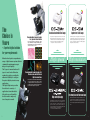
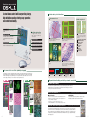
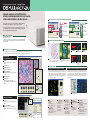
Digital Camera Digital Sight Series
Digital Camera System for Microscopy
The
Choice is
Yours
– A perfect digital solution
for your requirements
Nikon has developed a comprehensive
range of digital camera systems that are
optimized forcapturingmicroscopic
images of superb quality.
The 5 types of camera heads and 2
types of control units all function
seamlessly together, providing the
ultimate in flexibility to configure
the perfect digital system for many
applications.
The Digital Sight series provides
the solution for a variety of
applications, from industrial to
biological use, and from highlevel research to simple capture of inspection results.
Recommended camera heads
for general observation
These camera heads can be deployed in a broad
range of illumination techniques, including brightfield, darkfield, phase contrast and Nomarski DIC
(differential interference contrast).
Smooth and comfortable live images
Captures true to life images
Incorporates a 2-megapixel colorCCD that can smoothly
display SXGA live motion images at 15 fps*1 (max. 30 fps).
A well balanced camera head that enables display of
live images and capture of high definition images.
DS-5M incorporates a 5-megapixel color CCD that offers
a high-resolution image size of 2560x1920 pixels. It is
ideal for acquiring detailed microscopic images under a
variety of illumination techniques, including brightfield, phase contrast and (DIC).
*1 Using the DS-L1, with output to an external monitor.
High speed
High resolution
Recommended camera heads for
fluorescence and ultra low
light observations
These cameras are ideal for darkfield and fluorescence samples. Incorporating a Peltier
cooling mechanism, cooled types are able to
greatly reduce the thermal noise that can be
generated from long time exposures.
DS-2MBWc
DS-2MBW
High-sensitivity imaging
The 2MBWc incorporates a Peltier cooling mechanism
that minimizes thermal noise. Its 2-megapizel monochrome CCD boasts a sensitivity rating five times
greater than that of previous models, resulting in a
shortened total exposure time and reduced fading, and
allowing the realization of high frame rate.
For extremely clear color fluorescence images
With its built-in Peltier cooling mechanism, the temperature of the 5.0-megapixel color CCD can be maintained at 20 °C below room temperature . Thermal
noise is thus greatly reduced during long time exposure.
* A non-cooled model (DS-2MBW) is also available.
2
3
Stand-alone camera control unit
A stand-alone control unit incorporating a large,
high-definition monitor, offering easy operation
and useful functionality.
A wide variety of tool functions
Measurement tools
Other useful tools
(2) 0.60mm
Y : 621.72mm
X :835.43mm
(2) 0.51mm
Stand-alone type
Does not require a PC or an external
monitor.
6.3type LCD monitor
XGA (1024x768) screen resolution,
which allows simple, fast and accurate
focusing without the need to enlarge the
image.
Excellent output options
In addition to saving to a Compact
Flash TM card, the unit offers these
output options:
Analog RGB output
On-screen menus
Distance measurement
Easily measures the distance between any two
points specified by the user.
Vertical / horizontal measurement
Easily measures the distance between two horizontal and two vertical lines.
Screen patterns
Cross line and concentric circle patterns can be displayed.
Count marking
Up to 99 serial numbers per color can be marked to
provide a convenient way to identify points of
interest on-screen. They can be easily saved and
printed with the image.
Two-screen split display
A frozen image can be displayed
alongside the live image for easy
comparison.
Text input; line and figure drawing
Thumbnail image display
Superimpose function
Shading function
Focus indicator
Histogram
Digital zoom
Log text saving
Interval exposure
Direct printing (using PictBridge)
Intuitive, on-screen menu-based operation allows for simple set-up and capture.
USB Mass Storage Class
Network connectivity
Scene mode for one-click optimal photography
Using Nikon’s experience with digital imaging, optimal pre-programmed imaging modes have been
made available as menus, including preset camera conditions according to the sample types and illumination techniques used. Optimal images can be captured with a simple click. Users can also customize settings and save up to seven of these modes for quick retrieval.
Scale display
XY scale display
Point to line
distance
Circular measurement
Angle
(displays diameter, circumference,
center of gravity)
(intersection)
Expanded functionality provided by external connectivity
Network functionality
For fluorescence
/ darkfield observations
For wafer / IC chip
images
A 10/100Base-TX compatible Ethernet port is provided. By using an FTP client and
HTTP/FTP/Telnet server functionality, it is possible to transmit images to a network or
remotely control the camera from a network.
Direct Print function
* A separate license is needed in order to use the Direct Print function.
(option)
Real 10 mode
PictBridge
The Real 10 mode is selectable with a dedicated Mitsubishi CP900DW.
With this mode it is possible to print with a device magnification of 10x
for easy confirmation of sample size.
For brightfield
observations
For metal /
ceramic material images
* DS-5M/DS-5Mc
C-mount 0.7X TV Adaptor (optional) is necessary
DS-2Mv/DS-2MBW/DS-2MBWc C-mount 0.55X TV Adaptor (optional) is necessary
Interactive control of microscope and camera
4
For DIC /
phase contrast
observations
For printed circuit board
images
It is possible to print directly without a computer using a
PictBridge compatible printer and USB connection.
(option)
* A separate license is needed in order to use the interactive control.
In combination with the ECLIPSE 90i motorized microscope, it is possible to control the 90i from
the DS-L1 menu and to automatically detect and record the microscope status data with the
image, including the objective magnification and fluorescence filter in use. Automatic detection
of status is also possible when combined with the ECLIPSE 80i microscope configured with a digital imaging head.
Example of the microscope control menu
5
PC based camera control unit
Imaging Software
Single cable connection to a PC via USB2.0 interface.
Extremely versatile imaging software that is easy to operate,
and has a wealth of functions to suit almost any needs.
Scene mode for one-click optimal photography
This compact control unit can be connected quickly to a Windows® based PC
via its USB2.0 interface, without the need for a separate PCI board.
By controlling the camera with a PC, the user can expand their system, not only
to facilitate image capture but also results analysis, and image processing with
Nikon's ACT-2U Multi-functional imaging software.
Two USB ports are provided.
Connects to a PC via USB2.0 interface (USB2.0 offers both wide compatibility and high
data transfer speeds), which enables camera control and live image display at high
speed on a monitor.
For brightfield observations
For wafer /
IC chip images
For fluorescence /
darkfield observations
For metal / ceramic
material images
For DIC /
phase contrast observations
For printed circuit board
images
A variety of measurement tools
A variety of screen layouts
Scale display
Distance
measurement
XY scale display
Circular measurement
Angle
(displays diameter, circumference, center of gravity)
(intersection)
The configuration offers a standard screen layout and advanced layouts with a high
degree of image analysis functionality. Each layout can be customized according to need.
Standard Layout,
for straightforward operations
1
Area
The area inside an enclosure, outlined by
the mouse, is automatically calculated
1 Tool bar
Frequently used menu items are displayed as
buttons
Screen patterns
Grid or concentric circle patterns can be
displayed.
3
Key functions
2 Live image screen
3 Captured image screen
Display of the captured image
point to line
distance
Interactive control of the microscope and camera
Superimposition using the Merge function
2
4 Properties dialog box
For display/alteration of the camera’s current
settings
4
5 Image thumbnails
It is easy to produce merged images of multistained fluorescence samples using monochrome photography. When you acquire images after
initially setting the number of layers and display colors for each
reagent, colored images will be automatically displayed. By changing
the wavelength for each acquisition, this function easily allows a superimposed image to be produced.
In combination with the ECLIPSE 90i motorized microscope, it is possible to
control the 90i from the ACT-2U menu and to automatically detect and record
the microscope status data with the image, including the objective magnification and fluorescence filter in use. Automatic detection of status is also possible when combined with the ECLIPSE 80i microscope configured with a digital imaging head.
6 Capture window
Basic image capture control window
Scene function for one-click optimal photography
6
5
The control software of the 90i and digital imaging head can be easily
accessed from ACT-2U.
Advanced Layout,
adding useful functions
7 Toolbox
Tool buttons for processing and analysis of
images
7
8
Layout customizing window
8 Process view window
Analysis of focus, profile and histogram
Displays analytical settings for control and
focus
6
Settings box, which allows detailed
customizing of the screen layout, in
addition to the standard and advanced
layout.
Tool Bar:
1. Main Tool Bar
2. Tool Box
3. Annotation Tool Box
4. Graphic Annotation Setup
Recording Mode:
1. Average Recording
2. Time Lapse Recording
3. Variable Time Lapse Recording
4. Z Stack Recording
Image Processing:
1. Direction/Resize Control
2. Crop Control
3. Duplicate Control
4. Kirsch Filter
5. Sobel Filter
6. Laplace Filter
7. Low Pass Filter
8. Median Filter
Image Display:
1. 2D Image View
2. 2D Quadrant View
3. 3D Orthogonal View
4. 3D Tiling View
Image Analysis:
1. Focus Analysis
2. Profile Analysis
3. Histogram Analysis
4. Area ROI Analysis
5. Line ROI Analysis
Process View
Thumbnail Window
Experimental Information
Window
Printing:
1. Print Main Menu
2. Print Preview Mode
3. Layout Edit Mode
File Operation
Camera Control:
1. Hardware Connection Dialog Box
2. Control Window
3. Property Window
Microscope Control:
1. Hardware Connection Dialog Box
2. Microscope Control
3. Pixel Divide Function Setup
4. Fixed Scale Bar Setup
7
S y s t e m
L i n e u p
For industrial applications
For biological applications
Live image
observation
DS-2MBWc-U1 +ECLIPSE TE2000
Offers image display and capture
for low light florescence.
DS-2Mv-L1 +SMZ1000
Stand-alone type that offers high frame rate
for real-time observation
Combining a high sensitivity camera equipped with a cooling mechanism
that eliminates thermal noise, with software that boasts a wealth of analytical functions.
This system is most appropriate for high-level research that demands the
capture of weak fluorescence images clearly, with a high signal-to-noise ratio.
Maximum frame rate of 30 fps enables the system to be optimized for live
observation.
Stand-alone type featuring an XGA 6.3 type TFT monitor. Observation on a
larger screen is possible with an analog RGB output function
Images can be saved to the CF card simply by clicking the capture button.
DS-5Mc-U1 +ECLIPSE
Offering the potential for integrated
control of the camera and microscope
DS-2Mv-L1 +MM-40
Stand-alone type, with convenient built-in
measuring functions
Measurement
A single cable connection via USB interface is possible from
the control unit to a PC and the Eclipse 90i microscope.
Since the 90i can be controlled from the ACT-2U imaging
software, the operation of the microscope can be linked with
image recording. Auto-focus image capture during brightfield microscopy is possible, utilizing the image contrast
data detected by the camera.
Maximum frame rate of 30 fps enables the system to be optimized for live
observation.
Simple measurement functions are built into the camera control unit (DS-L1),
including distance, angle of intersection, circular measurements, area, etc.
Measurement results can be saved together with the image.
Images can be saved to the CF card simply by clicking the capture button.
DS-2Mv-L1 +ECLIPSE 80i
Delivering both smooth, live motion images
and beautiful captured images
DS-2Mv-U1 +Module Microscope
PC-based system that utilizes
multi-functional imaging software
Thanks to the high-frame-rate, 2-megapixel CCD camera, the movement of
live images is extremely smooth. Live images can be viewed on the large
LCD monitor built into the control unit.
This system is controlled by a PC via a USB2.0 connection.
ACT-2U imaging software enables the display of smooth
and easy-to-see live images as well as image acquisition, processing, analysis and simple measurements via
a PC.
Image
acquisition
Analysis
8
90i
DS-5M-L1 +ECLIPSE 50i
For easy image capture without a PC
DS-5M-L1 +Module Microscope
High definition stand-alone type with network functions
This stand-alone control unit, which does not require connection to a PC,
is a real space saver. Camera conditions most appropriate to the illumination techniques used can be chosen from a menu, allowing anybody to
easily capture beautiful images.
Incorporates a high-resolution 5-megapixel CCD, with a capture size of
2560x1920 pixels, minute details of images can be captured beautifully.
The camera control unit is equipped with a built-in large 6.3 type XGA TFT
LCD monitor for live image viewing.
Ethernet-based network functionality is built in. Images can be saved on
any given server as well as on the CF card.
DS-5M-U1 +Module Microscope
High definition PC-based system equipped with
multi-functional imaging software.
DS-2MBW-L1 +ECLIPSE TS-100F
A reasonably priced monochrome
configuration
Incorporates a high-resolution 5-megapixel CCD, with a capture size
of 2560x1920 pixels, true-to-life images can be captured with ease.
Controlled from a PC via a USB2.0 connection.
ACT-2U imaging software enables smooth and easy display of
images.
A launcher function is available. This allows saved images to be
easily transferred to other data application software for processing.
Easily takes high contrast monochrome images in observations such as
phase contrast and DIC. Using a high frame rate camera, live motion can
be quickly and easily confirmed.
*Exapmle of image created using image composition software
9
S y s t e m
S p e c i f i c a t i o n s
D i a g r a m
Camera Heads
DS-2MBWc
41
CCD
Recordable pixels
CCD cooling device
PC monitor
Ethernet
USB mass storage class
Direct print
printer
(Windows® 2000/XP)
USB HUB
RGB output
100Base-TX
USB2.0
Sensitivity
(Can be varied between ISO 50-2000
equivalent)
A/D conversion
12-bit
Live display mode (DS-L1) 1600 x 1200 (15fps), 800 x 560 (30fps), Center Scan (30fps)
Live display mode (DS-U1) 1600 x 1200 (4fps), 800 x 600 (15fps), 400 x 300 (20fps),
Center Scan 400 x 280
91
USB keyboard
* Frame rates are a guide only. Indicated rates assume USB data transfer speeds.
76
USB1.1
USB mouse
Exclusive
remote
controller
Lens mount
Exposure time
Dimensions
Weight
System composition
Optional accessories
Universal-type
AC power adapter
CF
Camera Control Unit
with DS-L1
800 x 600(12fps), 400 x 300(20fps),
Center Scan 400 x 280(30fps)
* Frame rates are a guide only. Indicated
rates assume USB data transfer speeds.
/
DS-5Mc (Cooled CCD Camera)
CCD
Recordable pixels
CCD cooling device
Sensitivity
A/D conversion
Live display mode (DS-L1)
Live display mode (DS-U1)
DS-5M (Standard CCD Camera)
2/3 in. high-density CCD: Total number of pixels: 5.24 million (effective 5.07 million)
2560 x 1920 pixels, 1280 x 960 pixels, 640 x 480 pixels
Peltier Device: Ambient temperature -20ºC
Equivalent to ISO64 (Can be varied between ISO 32-1250 equivalent)
12-bit
2560 x 1920 (3.8fps), 1280 x 960 (7.5fps), Center Scan (15fps) * Display reduced to SXGA/XGA with DS-L1
1280 x 960 (2fps), 640 x 480 (7.5fps), Center Scan 640 x 480 (15fps)
Lens mount
Exposure time
Dimensions
Weight
System composition
Optional accessories
C-mount
1/1000 to 600 sec
1/1000 to 60 sec
91.0 (W) x 76.0 (D) x 41.0 (H) mm
approx. 290g
approx. 260g
Camera Cable (3m)
For wide field of view observations 0.7x Relay lens (C-mount)
144.5
68.4
* Frame rates are a guide only. Indicated rates assume USB data transfer speeds.
180.5
204
203
Exclusive camera cable (3m)
77
USB
* Display reduced or enlarged to SXGA/XGA
C-mount
1/1000 to 600 sec
1/1000 to 60 sec
91.0 (W) x 76.0 (D) x 41.0 (H) mm
approx. 290g
approx. 260g
Camera Cable (3m)
For wide field of view observations 0.55x Relay lens (C-mount)
Camera Heads
(mm)
Compact
FlashTM
card
DS-2Mv
DS-2MBW
1/1.8 in. high-density CCD: Total number of pixels: 2.11 million (effective 1.98 million)
1600 x 1200 pixels, 800 x 600 pixels, 400 x 300 pixels
Peltier Device: Ambient
temperature -20ºC
Equivalent to ISO350 (Can be varied between ISO 160-6400 equivalent) Equivalent to ISO100
USB
Camera Control Units
/
DS-U1
DS-L1
Exposure control
Exposure correction
Digital zoom
Interval shooting
Exposure metering
Exposure metering range
White balance
Image adjustments
Program AE, Shutter-priority AE, Focus AE, Manual with AE lock function
13 steps
Correction range: ±2.0, Step: 1/3
5 to 2400%
Up to 16x (8 steps)
5 sec. - 12 hr. intervals
10 sec. - 6 hr. intervals
Average metering, Peak hold metering
3 selectable sizes
Set method, Color balance adjustable
Gamma correction, shading adjustment, black level adjustment, hue wheel variation,
color saturation adjustment
Storage format
BMP, TIFF, JPEG, JPEG2000,AVI
BMP, JPEG (4-step compression)
USB device port (computer control connector),
Interface
USB device port (Mass Storage Class support),
USB host port (microscope connector)
USB host port (USB mouse,
USB keyboard connection)
Power supply
AC100-240V 50/60Hz
Power consumption 43VA
138VA
Dimensions
180.5 (W) x 144.5 (D) x 68.4 (H) mm
203 (W) x 77(D) x 204 (H) mm
Control unit: approx. 1300g, AC adapter: approx. 350g
Weight
approx. 1000g
Operating environment 0-40˚C, 85% RH max. (without condensation)
System composition Power cord
AC adapter, Power cord, CompactFlashTM card
(128MB), Mouse
Networking
Ethernet (10/100Base-TX), DHCP compatible,
HTTP, TELNET or FTP server, FTP client
LCD monitor
6.3-in. TFT color LCD XGA (1024 x 768, 60Hz)
External monitor output
Analog RGB: SXGA (1280 x 1024, 60Hz),
XGA (1024 x 768, 60Hz)
Storage media
CompactFlashTM card (Type 1, Type II)
Direct printing
Direct printing possible without a computer
using special printer (PictBridge compatible)
Optional accessories
Exclusive remote controller, Direct Print license
key, Direct printer CP900DW (Mitsubishi),
Microscope
ACT-2U Imaging Software System Requirements
Windows 2000/XP
Camera Control Unit
Computer type
CPU
RAM
USB2.0
Hard disk
Operating system
Graphics
Others
MS-DOS PC supporting USB2.0
Pentium® 4, 1.7GHz or faster (Pentium® 4, 2.4GHz or faster recommended)
1GB or more
2 ports
100MB to install, 300MB or more free space to run (on launch disk)
Windows® 2000 Professional (SP4 or later, English or Japanese),
Windows® XP Professional (English or Japanese), pre-installed versions only
1280 x 1024 pixels or more, 16-bit color or more (24-bit color recommended), DirectX 9.0b support
CD-ROM drive (to install), Microsoft® USB2.0 driver
The above system requirements list does not constitute a guarantee that all computers and systems meeting these criteria will be able to run the software.
10
11
Company and product names included in this brochure are the
registered trademarks of the respective companies. Monitor images
are simulated. Specifications and equipment are subject to change
without any notice or obligation on the part of the manufacturer.
May 2005.
©2005 NIKON CORPORATION
WARNING
TO ENSURE CORRECT USAGE, READ THE
CORRESPONDING MANUALS CAREFULLY
BEFORE USING THE EQUIPMENT.
NIKON CORPORATION
Fuji Bldg., 2-3, Marunouchi 3-chome, Chiyoda-ku,
Tokyo 100-8331, Japan
www.nikon.com/
NIKON INSTECH CO., LTD.
Parale Mitsui Bldg.,8, Higashida-cho, Kawasaki-ku,
Kawasaki, Kanagawa 210-0005, Japan
Phone: +81-44-223-2175(Industrial dept.)
/+81-44-223-2167(Biological dept.) , fax: +81-44-223-2182
www.nikon-instruments.jp/eng/
ISO 9001
ISO 14001 Certified
NIKON INSTECH CO., LTD.
NIKON INSTRUMENTS EUROPE B.V.
Schipholweg 321, 1171PL Badhoevedorp, NL
Phone: +31-20-44-96-222, fax: +31-20-44-96-298
www.nikon-instruments.com/
Accredited by the
Dutch Council for
Accreditation
ISO 9001 Certified
NIKON CORPORATION
Instruments Company
Code No. 2CE-MRGH-1
ISO 14001 Certified
NIKON CORPORATION
Yokohama Plant
NIKON INSTRUMENTS INC.
1300 Walt Whitman Road, Melville, N.Y. 11747-3064, U.S.A.
Phone: +1-631-547-8500; +1-800-52-NIKON (within the U.S.A. only), fax: +1-631-547-0306
www.nikonusa.com/
NIKON FRANCE S.A.S.
NIKON CANADA INC.
FRANCE phone: +33-1-45-16-45-16, fax: +33-1-45-16-00-33
CANADA phone: +1-905-625-9910, fax: +1-905-625-0103
NIKON INSTRUMENTS (SHANGHAI) CO., LTD. NIKON GMBH
CHINA phone: +86-21-5836-0050, fax: +86-21-5836-0030 GERMANY phone: +49-211-9414-0, fax: +49-211-9414-322
NIKON INSTRUMENTS S.p.A.
(Beijing office)
CHINA phone: +86-10-5869-2255, fax: +86-10-5869-2277 ITALY phone: +39-55-3009601, fax: +39-55-300993
NIKON AG
NIKON SINGAPORE PTE LTD
SINGAPORE phone: +65-6559-3618, fax: +65-6559-3668 SWITZERLAND phone: +41-43-277-2860, fax: +41-43-277-2861
NIKON UK LTD.
NIKON MALAYSIA SDN. BHD.
MALAYSIA phone: +60-3-78763887, fax: +60-3-78763387 UNITED KINGDOM phone: +44-20-8541-4440, fax: +44-20-8541-4584
NIKON INSTRUMENTS KOREA CO., LTD.
KOREA phone: +82-2-2186-8410, fax: +82-2-555-4415
Printed in Japan (0506-10) Am/M
ISO 14001
En