1
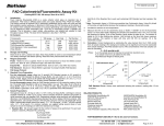
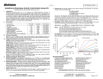
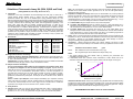
BioVision
(Catalog #K264-100; 100 assays; Store kit at -20 C)
I. Introduction:
Glutathione is the major intracellular low-molecular-weight thiol that plays a critical role in
cellular defense against oxidative stress in tissues and cells. Commercially available
glutathione detection kits, such as the DTNB-enzyme cycling glutathione assay kit or the
Monochlorobimane based assay kit hardly distinguish between reduced glutathione (GSH; FW:
307) and oxidized glutathione (GSSG; FW: 612). BioVision’s Glutathione Detection Kit provides
a unique, convenient tool for detecting GSH, GSSG, and total glutathione separately. In the
assay, OPA, reacts with GSH (not GSSG), generating fluorescence, so GSH can be
specifically quantified. Adding a reducing agent converts GSSG to GSH, so (GSH + GSSG)
can be determined. To measure GSSG specifically, a GSH Quencher is added to remove
GSH, preventing reaction with OPA (while GSSG is unaffected). Reducing agent is then added
to destroy excess quencher and convert GSSG to GSH. Thus, GSSG can be specifically
quantified. The kit provides a unique procedure and buffer formula to eliminate protein thiol
interference and to stabilize GSH and GSSG in solution. The assay is easy to perform and
detects 2-400 ng/µl of GSH, GSSG or total glutathione.
II. Kit Contents:
Glutathione Assay Buffer
PCA (Perchloric Acid, 6 N)
KOH (6 N)
OPA Probe (o-phthalaldehyde )
Reducing Agent Mix
GSH Quencher
GSH Standard (FW: 307)
100 Assays
Cap Code
Part Number
30 ml
2 ml
2 ml
0.2 ml
1 vial
20 µl
1 mg
WM
Red
Blue
Brown
Green
Purple
Yellow
K264-100-1
K264-100-2
K264-100-3
K264-100-4
K264-100-5
K264-100-6
K264-100-7
III. Reagent Reconstitution and Storage:
OPA Probe, Reducing Agent Mix, GSH Quencher: Add 0.85 ml H2O to the OPA probe, mix.
Dissolve both the Reducing Agent and the GSH Quencher in 1.05 ml of dH2O separately. Store
at –20oC. Use within two months.
GSH Standard: Accurately dissolve in 45 µl dH2O and add 5 µl PCA to stabilize the standard
GSH stock solution (20 µg/µl). Store at –20oC. Use within two months.
IV. Sample Collection and Storage:
Tissue and Cell Samples: GSH is labile and cell preparations will oxidize it rapidly. Keep
all samples and reagents ice cold and work as rapidly as possible. Prepare centrifuge
tubes with 20 µl PCA on ice to receive samples. Homogenize 2-4 x10⁶ cells or 40 mg tissue on
ice with 100 µl of ice cold Glutathione Assay Buffer. Take 60 µl of each homogenate to a
prechilled tube containing PCA and vortex several seconds to achieve a uniform emulsion.
Keep on ice for 5 min. Spin 2 min at 13,000 G at 4oC, collect supernatant (containing
glutathione) and discard the protein pellet. The sample can then be stored at –80oC, stable for
a month.
Serum or Other Liquid Samples: Freeze samples immediately upon acquisition and keep
frozen until ready for processing. Take 60 µl thawed sample to centrifuge tube containing 20 µl
ice cold PCA, vortex and keep on ice for 5 min. Spin for 2 min at 13,000 G at 4oC. Collect the
supernatant. The sample can then be stored at –80oC, stable for a month.
V. Assay Protocol:
1. Standard Curve: Add 10 µl of the 20 µg/µl standard GSH stock to 990 µl of Assay Buffer to
generate a 0.2 µg/µl working standard solution. Add 0, 2, 4, 6, 8, 10 µl to a 96-well plate to
generate 0, 0.4, 0.8, 1.2, 1.6, 2.0 µg/well GSH. Bring the volume to 90 µl with Assay Buffer.
BioVision Incorporated
155 S. Milpitas Boulevard, Milpitas, CA 95035 USA
Note: If the concentration of your assay samples is lower than the above standard range, the
standard can be further diluted 10-fold to generate 0, 40, 80, 120, 160, 200 ng/well GSH by
following the same procedure.
2. Preparation of Samples for Assays: Add 20 µl of ice cold 6N KOH to 40 µl of PCA preserved
samples (as prepared in Section IV) to precipitate PCA and neutralize the samples (pH should
be 5-10). Keep on ice for 5 min then spin 2 min at 13,000 G at 4oC. Transfer 10 µl of the
neutralized samples to a 96-well plate. You may choose to add several dilutions (e.g., 1-10 µl)
of your samples to ensure the readings are within the standard curve range.
A. To Detect GSH: Bring the sample volume to 90 µl with Assay Buffer.
B. To Detect Total Glutathione: Bring the sample well to 80 µl with Assay Buffer. Do a
background control without sample. Add 10 µl of Reducing Agent Mix to the wells, mix
well, incubate at room temperature for 10 min to convert GSSG to GSH.
C. To Detect GSSG: Bring the sample well volume to 70 µl with Assay Buffer. Do a
background control without sample. Add 10 µl of GSH Quencher, mix well, and incubate at
room temperature for 10 min to quench GSH. Then add 10 µl of Reducing Agent Mix to
destroy the excess GSH Quencher and convert GSSG to GSH.
3. Assay: Add 10 µl of OPA Probe into the standard and sample wells, mix well, incubate at room
temperature for 40 min. Read samples and standards on a fluorescence plate reader equipped
with Ex/Em = 340/420 nm. We suggest adjusting the plate reader settings so that the
background reading without glutathione is at about 50-150 RFU.
4. Calculations: Subtract background reading from sample readings. Plot RFU vs GSH
standard. Apply the sample readings to the standard curve to get glutathione amount in each
sample:
Glutathione Concentration = Ga/Sv
Where:
µg/ml
Ga = Glutathione amount from standard curve (in µg).
Sv = Original sample volume added to the sample wells (in ml).
Notes:
1. The original samples have been diluted 2-fold by performing the preservation and
neutralization procedures above.
2. If GSH/GSSG molar ratio is desired, molar concentration of GSH, GSSG need to be
calculated. GSH molecular weight: 307.32 g/mol; GSSG molecular weight: 612.63 g/mol.
4000
3000
RFU
Glutathione Fluorometric Assay Kit (GSH, GSSG
and Total)
o
Kit Component
For research use only
rev. 2/13
2000
1000
0
0
0.4
0.8
1.2
1.6
2
Glutathione Amount (µg/well)
Glutathione Standard Curve: Assay were prepared using standard GSH and GSSG following
kit instructions. Diamond (blue) represents GSH standard curve. Circle (pink) represents GSSG
standard curve produced by adding GSSG + GSH Quencher + Reducing Agent Mix. Triangle
(gray) represents GSSG standard without adding Reducing Agent mix.
FOR RESEARCH USE ONLY! Not to be used on humans.
Tel: 408-493-1800 | Fax: 408-493-1801
www.biovision.com | [email protected]
Page 1 of 2
BioVision
For research use only
rev. 2/13
GENERAL TROUBLESHOOTING GUIDE:
Problems
Cause
Solution
Assay not working
• Use of ice-cold assay buffer
• Assay buffer must be at room temperature
Samples with erratic readings
Lower/ Higher readings in Samples
and Standards
Readings do not follow a linear
pattern for Standard curve
Unanticipated results
• Omission of a step in the protocol
• Refer and follow the data sheet precisely
• Plate read at incorrect wavelength
• Check the wavelength in the data sheet and the filter settings of the instrument
• Use of a different 96-well plate
• Fluorescence: Black plates ; Luminescence: White plates; Colorimeters: Clear plates
• Use of an incompatible sample type
• Refer data sheet for details about incompatible samples
• Samples prepared in a different buffer
• Use the assay buffer provided in the kit or refer data sheet for instructions
• Samples were not deproteinized (if indicated in datasheet)
• Use the 10 kDa spin cut-off filter or PCA precipitation as indicated
• Cell/ tissue samples were not completely homogenized
• Use Dounce homogenizer (increase the number of strokes); observe for lysis under
microscope
• Samples used after multiple free-thaw cycles
• Aliquot and freeze samples if needed to use multiple times
• Presence of interfering substance in the sample
• Troubleshoot if needed, deproteinize samples
• Use of old or inappropriately stored samples
• Use fresh samples or store at correct temperatures till use
• Improperly thawed components
• Thaw all components completely and mix gently before use
• Use of expired kit or improperly stored reagents
• Always check the expiry date and store the components appropriately
• Allowing the reagents to sit for extended times on ice
• Always thaw and prepare fresh reaction mix before use
• Incorrect incubation times or temperatures
• Refer datasheet & verify correct incubation times and temperatures
• Incorrect volumes used
• Use calibrated pipettes and aliquot correctly
• Use of partially thawed components
• Thaw and resuspend all components before preparing the reaction mix
• Pipetting errors in the standard
• Avoid pipetting small volumes
• Pipetting errors in the reaction mix
• Prepare a master reaction mix whenever possible
• Air bubbles formed in well
• Pipette gently against the wall of the tubes
• Standard stock is at an incorrect concentration
• Always refer the dilutions in the data sheet
• Calculation errors
• Recheck calculations after referring the data sheet
• Substituting reagents from older kits/ lots
• Use fresh components from the same kit
• Measured at incorrect wavelength
• Check the equipment and the filter setting
• Samples contain interfering substances
• Troubleshoot if it interferes with the kit
• Use of incompatible sample type
• Refer data sheet to check if sample is compatible with the kit or optimization is needed
• Sample readings above/below the linear range
• Concentrate/ Dilute sample so as to be in the linear range
Note: The most probable list of causes is under each problem section. Causes/ Solutions may overlap with other problems.
BioVision Incorporated
155 S. Milpitas Boulevard, Milpitas, CA 95035 USA
Tel: 408-493-1800 | Fax: 408-493-1801
www.biovision.com | [email protected]
Page 2 of 2