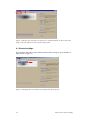
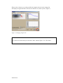
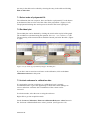
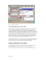
1
Downloaded from orbit.dtu.dk on: Dec 18, 2015
RisøScan 1.0 - User manual and toolset for retrospective validation
Helt-Hansen, Jakob
Publication date:
2004
Document Version
Publisher final version (usually the publisher pdf)
Link to publication
Citation (APA):
Helt-Hansen, J. (2004). RisøScan 1.0 - User manual and toolset for retrospective validation. (Denmark.
Forskningscenter Risoe. Risoe-R; No. 1487(EN)).
General rights
Copyright and moral rights for the publications made accessible in the public portal are retained by the authors and/or other copyright owners
and it is a condition of accessing publications that users recognise and abide by the legal requirements associated with these rights.
• Users may download and print one copy of any publication from the public portal for the purpose of private study or research.
• You may not further distribute the material or use it for any profit-making activity or commercial gain
• You may freely distribute the URL identifying the publication in the public portal ?
If you believe that this document breaches copyright please contact us providing details, and we will remove access to the work immediately
and investigate your claim.
Risø-R-1487(EN)
RisøScan 1.0
User Manual and Toolset for
Retrospective Validation
Jakob Helt-Hansen
Risø National Laboratory
Roskilde
Denmark
December 2004
Author: Jakob Helt-Hansen
Title: RisøScan 1.0 – User Manual
and Toolset for Retrospective
Risø-R-1487(EN)
December 2004
Validation
Department:
Radiation Research Department
Abstract (max. 2000 char.):
ISSN 0106-2840
ISBN 87-550-3387-3
The RisøScan software is used for dose measurements with
radiochromic films that color visibly. This report consists of two
documents for use with the RisøScan software. The User Manual
tells how to use the program and the Toolset for Retrospective
Validation describes how to perform a retrospective validation of
the software.
Contract no.:
Group's own reg. no.:
Sponsorship:
Cover :
Pages: 72
Tables: 30
References: 3
Risø National Laboratory
Information Service Department
P.O.Box 49
DK-4000 Roskilde
Denmark
Telephone +45 46774004
[email protected]
Fax +45 46774013
www.risoe.dk
Contents
RisøScan 1.0 - User manual 5
1 Introduction 7
1.1 Disclaimer and general precautions 8
1.2 Retrospective Validation 8
2 Requirements 9
3 Scanner 9
3.1 Reference 9
4 Installation 10
4.1 Uninstall 10
5 Open an image for analysis 11
5.1 Colour channel (RGB selection) 12
6 Selecting area of interest 13
6.1 General settings 14
7 Create a calibration 16
7.1 Select order of polynomial fit. 17
7.2 Residual plot 17
7.3 Include reference in calibration data 17
7.4 Save calibration 18
7.5 Possible calibration function conflict 19
8 Apply calibration to an image 19
8.1 Change image from response to dose 20
9 Apply reference to an image 21
10 Depth dose profile 22
10.1 Manual energy measurement 22
10.2 Automated energy measurement 24
10.3 Settings for energy measurement 25
11 Surface dose profile 26
12 3D surface dose profile 28
13 Creating documentation 29
13.1 Report generation (print to default windows printer) 29
13.2 Report generation (HTML file) 30
13.3 Graph values to ASCII file 30
13.4 Screen dump 31
14 Bibliography 32
www.risoe.dk
RisøScan 1.0 – Toolset for Retrospective Validation 35
1 Background. Relationship to FDA guidelines. 37
1.1 General FDA guidelines 37
1.2 Retrospective validation 37
1.3 Functional tests 37
2 Introduction 38
3 Definitions 39
4 Workflow 40
4.1 Open an image 40
4.2 Calibration 40
4.3 Dose measurement 41
4.4 Relationship between response value and dose 41
4.5 Electron beam energy measurement 42
5 Tests used for retrospective validation of RisøScan 1.0 43
5.1 TEST 1. Applying weighting factors to RGB value and conversion to Response
value. 43
5.2 TEST 2. Calibration procedure 44
5.3 TEST 3: Reference correction 49
5.4 TEST 4: Standard deviation 52
5.5 TEST 5: Minimum and Maximum values (Surface profile) 55
5.6 TEST 6: Depth dose profile 56
APPENDIX A. How to perform Test 1. 59
APPENDIX B. How to perform Test 2. 61
APPENDIX C. How to perform Test 3. 65
APPENDIX D. How to perform Test 4. 67
APPENDIX E. How to perform Test 5. 69
APPENDIX F. How to perform Test 6. 71
4
Report number ex. Risø-R-1234(EN))
RisøScan 1.0 - User manual
www.risoe.dk
6
Report number ex. Risø-R-1234(EN))
1 Introduction
RisøScan is a program for analysis of scanned images of radiochromic thin film
dosimeters that colours visibly such as Risø B3, Far West and GAF. The program is
developed and maintained at Risø High Dose Reference Laboratory. The software is
distributed and supported worldwide by the GEX Corporation.
RisøScan includes tools for:
• Selection of weights for red, green and blue colour channel of the
scanned image to obtain an optimal signal to noise ratio.
• Calibration (pixel response value versus dose).
• Inclusion of a reference to verify that scanner settings have been
chosen correctly. Minor variations due to scanner instability can be
corrected for.
• Depth dose profile analysis and calculation of electron radiation energy
based on range measurements in aluminium.
• Analysis of dose profiles including search for minimum and maximum
values.
• 3D surface plots of dose profiles.
• Report generation.
For customer service and technical questions contact:
Customer Service
GEX Corporation
7330 S. Alton Way, Suite 12-i
Centennial, Colorado 80112, USA
Website: www.gexcorp.com
Tel:
+1 303 400 9640
Fax:
+1 303 400 9831
e-mail: [email protected]
Technical questions may also be directed to
Risø National Laboratory
Att.: Jakob Helt-Hansen
Postbox 49
DK 4000 Roskilde, Denmark
Website: http://www.risoe.dk/nuk/risoescan/risoescan.htm
Tel:
+45 4677 4908
Fax:
+45 4677 4959
e-mail. [email protected]
www.risoe.dk
1.1 Disclaimer and general precautions
The RisøScan software program is intended for use in accordance with this manual.
The software was not designed to provide database or information storage in any
manner. The user is responsible for verification of the accuracy of the data contained
in the reports generated using the RisøScan software. Users are also responsible for
determining the suitability and performance of the software for their specific
applications.
Risø and GEX do not warrant the data input or output associated with this
software. All scan measurements and dose results and any subsequent usage
of the data derived from usage of this software are the sole responsibility of
the user. Further, by acceptance, implementation and use, the user assumes
any and all risks associated with such usage of this user manual and the
associated software product.
RisøScan users assume complete responsibility for the application of the software
product and agree to use the software program in accordance with the information
and instructions contained in this user manual. The examples given in this manual
are based on Risø recommended program uses; other uses of the program are
possible. Contact Risø or GEX to discuss your specific application needs.
Users of the RisøScan software program must provide and validate their own
computer and scanner system and have full and complete responsibility for any and
all verification and validation activities related to the use of the RisøScan software.
A Toolkit for Retrospective Validation is included with the software product to
facilitate user with Installation Qualification testing to demonstrate that the software
is functioning properly. Users must establish and perform their own validation of the
dosimetry system itself, which includes the PC, scanner, software and associated
procedures to be developed by the user to control image quality and dose
traceability.
1.2 Retrospective Validation
RisøScan ver. 1.0 has been black-box validated through the use of a “Toolkit
for Retrospective Validation”. The validation has been done retrospectively,
and should be repeated for any new installation and at time intervals specified
by the user. Use of the Retrospective Validation Toolkit ensures that the
software works as intended and meets “user needs and intended uses”
according to “General Principles of Software Validation; Final Guidance for
Industry and FDA Staff”, section 6.3 “Validation of Off-the-Shelf software and
automated equipment.”
8
Report number ex. Risø-R-1234(EN))
2 Requirements
Windows 98x, 2000 or XP.
200 Mb disc space.
256 Mb RAM.
500 MHz processor.
The program is best viewed with a screen resolution of 600x800 pixels. Other
resolutions may cause graphics to be misaligned.
3 Scanner
Most scanners use an auto-configuration of exposure parameters i.e. brightness,
contrast and gamma (not all scanners use the same terms). It is important to use a
scanner that can switch this feature off. Some scanner software allows the user to
save the exposure parameters for later use. Saving the exposure parameters
minimizes the risk of scanning with different settings for calibration and dose
measurements. If the scanner has an option for pre-heating the scanner lamp, it
should be used.
The surface on which the dosimeters are placed should be as homogeneous as
possible. High quality glossy paper used for ink-jet printers is recommended.
IMPORTANT
Use
2 *.bmp or *.png as file format for the scanned images.
The image file size depends on the size of the scanned area, and on the chosen
resolution. An image file size of 0.5 to 1.5 Mb is usually sufficient.
Large image files may cause RisøScan to halt (“Not enough memory to complete
this operation” error massage) or work extremely slow. Image file size should not
exceed 4 Mb.
3.1 Reference
Reproducibility of the instrument used to measure the dosimeters is essential in order
to maintain the measurement traceability. The stability of the scanner used in
connection with RisøScan is assured though measurement of the reference tablet
supplied with RisøScan. The reference tablet is measured at the time of the
measurement of the dosimeters for calibration, and at any subsequent measurement
of other dosimeters. Any differences between the two measurements are used for
correction of the measured result.
The reference tablet supplied with RisøScan is made from B3 dosimeter irradiated to
12, 25 and 50 kGy and laminated for protection. This reference can be used for
www.risoe.dk
measurements with B3 dosimeter films, Far West and GAF dosimeters. Other colour
charts can be used as long as they are long time colour stable.
Tests at Risø have proven the B3-reference to be stable for more than one year, and
it is recommended to re-measure or replace the reference once a year. Protect the B3reference from exposure to UV-light.
4 Installation
To install RisøScan and the Runtime-engine for LabView 6.1 from National
Instruments, run the Setup.exe file in the directory corresponding to the MicrosoftTM
Office-package installed on the computer. If there is no Office-package installed
either Setup file can be used.
4.1 Uninstall
To uninstall RisøScan remove the program by the “add/remove programs” in the
Control Panel. If RisøScan and Runtime-engine has been installed separately,
remove RisøScan by deleting the program and the Runtime-engine using
“add/remove programs” in the Control Panel.
10
Report number ex. Risø-R-1234(EN))
5 Open an image for analysis
When RisøScan starts, the main menu (Figure 1) is displayed. To the left is the
RisøScan logo or the image that is analyzed. To the right is a list of tasks.
To open an image click on Open Image and navigate to the image file.
Figure 1. Open image.
In the “Image properties” window (Figure 2) fill in the required information about
Scanning device: Write the name of the scanner used (e.g. “HP Scanjet 4P”)
Exposure adjustment units: Click the switch to select the scanner exposure units
(“Brightness, Contrast, Gamma” or “Shadow, Highlight and Midtone”). Write the
values used during the scanning of the image.
Scan resolution (DPI): Write the resolution used (e.g. 100 dpi) This information is
required to keep track of the length scale in the image.
Wedge angle (deg): If the image is a depth dose profile, fill in the angle of the
wedge used.
Dose unit: The dose unit to be used for the image (the unit used during calibration,
kGy or Gy).
Note: The dose unit used has to be specified at this stage.
It is recommended to write comments and to identify the operator.
www.risoe.dk
Figure 2. Image properties.
5.1 Colour channel (RGB selection)
Each pixel in the scanned image has a RGB value that specifies the amount of red,
green and blue. The signal to nose ratio can be enhanced by selecting a ratio of red,
green and blue that gives the highest contrast. For B3 and FWT dosimeters 100%
green and 0% red and blue gives the best result. (Figure 3). The values written here
will select the RGB value to be used for the analysis of the image. The same values
as used during calibration must be selected.
Figure 3. Weighting factors for red, green and blue colour channel.
12
Report number ex. Risø-R-1234(EN))
When all initial settings are selected, click on “Accept” to proceed. RisøScan returns
to the main menu with the selected image displayed. At this menu different tasks can
be selected.
6 Selecting area of interest
To select a area of interest on the scanned image left click and draw a rectangle to
select the area you want to analyse. This can be done anytime e.g. on the front menu
(Figure 4) or after a task has been selected.
Figure 4. A region of interest is marked while holding the left mouse button down.
IMPORTANT:
Press CTRL-Z or right-click to activate the selection (Acquisition mode). You can
de-activate again (Selection mode) by pressing CTRL-Z or right-click. That is, Ctrl-Z
and right-click toggles between Selection mode and Acquisition mode.
Note the difference in colour of the image frame:
Blue: Selection mode. Select area of interest.
Orange: Acquisition mode. Measurements can be performed on the selected part of
the image.
The mode of operation is also indicated just above the image to the right. (Figure 5).
www.risoe.dk
Figure 5. Whether the program is in Selection or Acquisition mode is shown above the
image to the left and by the colour of the image frame.
6.1 General settings
In the Display menu the general number format can be changed e.g. the number of
digits shown. (Figure 6)
Figure 6. Changing the way numbers are displayed in the program.
14
Report number ex. Risø-R-1234(EN))
When a task is chosen (e.g. surface profile) the length scale unit can be changed in
the Display>Length unit menu. The unit can either be cm, inch or pixels. (Figure 7).
Figure 7. Changing length scale.
IMPORTANT:
To return to the main menu press the blue “Done” button (Figure 7) or “File>Done”.
www.risoe.dk
7 Create a calibration
The image used to create a calibration should contain dosimeters irradiated to known
doses and a reference.
The recommended reference tablet is made from laminated B3 dosimeter films
irradiated to three different doses in the expected dose range (e.g. 12, 25, and 50
kGy).
To create a calibration, select Perform calibration on the main menu.
Left click on the image and select an area, which covers the irradiated dosimeters.
Right click (or click on Ctrl+M) to go into acquisition mode.
Make sure that the Create a Calibration / Point out calibration Reference values
switch is in the “Create a Calibration” position. (Figure 8)
Figure 8. Measuring dosimeters for creation of a calibration function.
Left click on the image and select an area of the first dosimeter. Write the dose to
which the dosimeter has been irradiated in the field above Add button. Click on the
return-key or the Add button. Continue the process with the rest of the dosimeters.
16
Report number ex. Risø-R-1234(EN))
An entry in the table can be edited by selecting the entry in the table and clicking
Edit or delete line.
7.1 Select order of polynomial fit.
The calibration data sets (response, dose) are fitted to a polynomial. Use the button
at the bottom of the screen to select the order of the polynomial. (Figure 9, left).
The polynomial relating dose and response is shown in the lower right figure.
7.2 Residual plot
The residual plot can be obtained by clicking the switch at the top left of the graph.
The residuals are calculated using the equation: (Dcalculated – Ddelivered)/Ddelivered * 100
The uncertainty of the measured dose should be entered just below the table. (Figure
9, right)
Figure 9. Left: Order of polynomial fit. Right: Residual plot.
If you don’t want to associate a reference to the calibration, click on the Save
calibration/reference at this point.
7.3 Include reference in calibration data
It is optional to associate a reference to a calibration, but it is strongly
recommended since it gives the opportunity to check that scanner settings are
correct, and minor variations due to instabilities of the scanner can be
corrected.
In selection mode, select the area covering the reference.
Right click to get into acquisition mode.
Set the Create a Calibration / Point out calibration Reference values button in
the “Point out calibration Reference values” position. (See Figure 8 or 10).
www.risoe.dk
Write the name of the reference in the Specify the name of the reference field, in
this example “Test Ref 1”. (Figure 10).
Select an area of each of the colours of the reference. Enter a name for each colour
field, e.g. “R1”, “R2” and “R3”. Click on Add to enter the response of each
reference colour in the table. The standard deviations in response value and in
percent are also displayed.
Figure 10. Measurement of reference values for the calibration function.
7.4 Save calibration
To save the calibration data together with the reference data, click on the Save
calibration/reference button.
In the pop-up window it is possible to check comments and scanner settings. (Figure
11).
Click on Save.
Write a name for the calibration and Click on Save.
IMPORTANT:
If a calibration is changed at any time after its initial save, then the changed calibration
should be saved under a new name. This avoids conflicts at later time of use.
18
Report number ex. Risø-R-1234(EN))
Figure 11. Saving a calibration.
7.5 Possible calibration function conflict
When a calibration is assigned to an image the calibration function is saved in the
*.info file belonging to the image. Then the image is opened at a later point the
program checks if the calibration function assigned to the image matches the one
saved on the hard drive. If the calibration function exists and is equal to the assigned
calibration function nothing happens. If the calibration function exists but is different
to the assigned function a warning will appear. In this case the user can either save
the calibration in the *.info file to the hard drive, use the calibration on the hard
drive (i.e. copy it to the *.info file) or use the calibration in the *.info file without
saving it to the hard drive. It is strongly recommended not to use the later option,
since there can be confusion about which calibration function is in use.
If the calibration function in the *.info file does not exist on the hard drive the user is
given the option to save it. An easy way to transfer a calibration between different
computers is therefore to copy the *.info file together with the image.
8 Apply calibration to an image
After an image has been loaded, a calibration can be selected by clicking on the
Select calibration button on the main menu (or File>Load Calibration).
Select the calibration to be assigned to the image and click on Select. (Figure 12)
www.risoe.dk
Figure 12. Assigning a calibration to an image.
8.1 Change image from response to dose
To display dose instead of pixel response (or vice versa) click on Ctrl+M or
Display>Map dose.
If an area of the image has been irradiated to a dose outside the limits of the
calibration, that area will be shown as white. An example is shown in Figure 13.
Figure 13. If a response value of a pixel is outside the range of the calibration, it will
show the dose value 0 – i.e. it appears white.
20
Report number ex. Risø-R-1234(EN))
9 Apply reference to an image
After a calibration function has been assigned to an image, the reference scanned
together with the image can be compared with the reference used for the calibration
function.
To measure the reference values for the image, click on the Reference for Image
button on the main menu.
Select the part of the image showing the reference and select Acquisition mode.
To the right of the screen (Figure 14) is shown the reference values obtained during
the calibration at the top, the reference values obtained for the image at the middle
and the ratio between the two at the bottom.
For each reference colour select an area. Click on the corresponding reference item
in the calibration reference table transferring the name of that reference item to the
image reference table. Click on the Add button.
Figure 14. Measuring image reference values.
The graph and the lower table show the ratio between the two reference values. If the
scanner settings are correct (= the settings used for the calibration), the deviation
should be less than one or two percent. If there are small deviations, a correction can
be made to the calibration function. To do this, click on the Enable reference button
(this can also be done on the main menu).
IMPORTANT:
reference
10TheDepth
dose ensures
profile traceability to the calibration scan. Use the reference in every scan
used for dose measurements.
www.risoe.dk
10 Depth dose profile
If a “wedge angle” has been selected during the “open image” task (see Figure 2),
the Depth Profile button can be selected.
The energy of a high-energy electron beam can be measured using the depth dose
profile in an aluminium wedge. Using properties of the descending slope of the
depth dose profile and equations from ICRU or ASTM/ISO the average and most
probable irradiation energy can be determined.
RisøScan can find the relevant properties of the descending slope in a manual or
automated way, i.e.:
- the starting point of the dosimeter film in the wedge,
- the maximum dose value,
- the linear fit of the descending slope with the highest coefficient of
determination,
- R50 and Rex
Select an area of the image, covering as much of the depth dose profile as possible
without going outside the edge of the dosimeter.
Make sure that “Intensity” displays “Dose”.
IMPORTANT:
Do not calculate energy if “intensity” is displayed as ”response”.
Since the calibration function is non-linear the calculated energy will be misleading.
10.1 Manual energy measurement
A. Point out the location of the start of the dosimeter film at the wedge surface
on the depth dose profile. To do so left click on the vertical red line in the
graph and move it. If the line cannot be found, click on the Guideline to
Centre button. (Figure 15)
B. To fine-tune the position of the wedge surface click on the Zoom In button.
(Figure 16).
C. Click on the Adjust for Off-set button. Note that the x-axis shifts, so “zero”
is at the wedge surface. Use the two vertical purple lines to mark the
beginning and end of the segment of the slope to be used in the calculation
of the energy. (Figure 17). If the vertical lines are not visible, click on the
Guidelines to Centre button.
D. Use the horizontal line to mark any dose off-set. (Figure 18).
E. The average and most probable energy are displayed below the graph to the
right.
22
Report number ex. Risø-R-1234(EN))
Figure 15. Finding the surface of the wedge.
Figure 16. Finding the surface of the wedge. Zoom in on depth dose curve.
Figure 17. Pointing out the segment of the depth dose curve to use for the energy
measurement. Moving the vertical lines changes the start and the end point.
www.risoe.dk
Figure 18. Defining the dose off-set by moving the horizontal line.
10.2 Automated energy measurement
A. Click on the Off-set and slope button. (Figure 19)
B. Adjust for dose off-set (if any) using the vertical green line.
C. The average and most probable energy are displayed below the graph to the
right.
If the identification of the wedge surface fails, point it out manually and then click
on Slope only to find the best fit to the slope.
Figure 19. The slope with the highest coefficient of determination is found using the
“Off-set and Slope” button.
24
Report number ex. Risø-R-1234(EN))
10.3 Settings for energy measurement
If the program chooses a wrong direction for the depth dose display, the direction
can be changed using the Scan direction button. The equations used to calculate the
energy can be changed using the Edit Equations button. (Figure 20).
Figure 20. Changing the equations for the energy calculations.
The automated energy measurement uses an algorithm to find the segment of the
slope to use. The algorithm makes a linear fit to every possible segment of the
descending curve, and then selects the segment, which gives the highest coefficient
of determination. There are some constrains in the algorithm that can be changed
from the menu: Display>Settings for slope algorithm. (Figure 21). Normally there
should be no reason to change the default values.
Figure 21. Settings for the slope algorithm.
www.risoe.dk
11 Surface dose profile
Clicking on the Surface Profile button on the main menu brings up the dose profile
analysis page. On the image to the right is a cross hair that can be moved around
while left clicking on it. On the right is the dose (or response value) profile along the
lines of the cross hair. The dose value at the centre of the cross hair is shown at the
lower left (Figure 22).
Figure 22. Dose profile along the lines of the cross hair is shown in the graphs and the
dose value at the centre is shown in the lower left corner.
By widening the cross hair the graphs will display an average along the width of the
cross hair, giving less noise in the dose profiles (Figure 23)-, but at the expense of
measurement resolution.
Minimum and maximum values can be found using the Find minimum or Find
maximum button. The search is performed averaging over an area equal to the
centre square of the cross hair. Choosing the width of the cross hair is a trade off
between resolution and noise reduction.
The average dose of the centre area of the cross hair (shown below the image in Fig
23) may be slightly different from the value shown on the graphs. This is because the
graph values are averaged over a one pixel wide line along the width of the cross
hair, while the centre average is obtained over the area of the cross hair centre.
It is possible to move the cross hair by dragging the vertical lines on the graphs.
26
Report number ex. Risø-R-1234(EN))
Figure 23. Widening the cross-hair gives less noise, but also less measurement
resolution. Compare the graphs with Figure 22.
It is possible to change the scale on the graphs by disabling Autoscale X or
Autoscale Y. By doing so, boxes for minimum and maximum values for the axes
will appear (Figure 24).
The axes of the two graphs will have the same scale if Interlock X or Interlock Y
are activated. By default, “Interlock Y” is enabled to make the dose values directly
comparable between the two graphs. The Interlock option may overwrite manual
settings of the axes scale.
Figure 24. Disabling “Autoscale” makes it possible to select the minimum and maximum
values on the axes. Note that if “Interlock” is enabled, it may overwrite manual settings.
www.risoe.dk
12 3D surface dose profile
The 3D Surface profile task shows the selected part of the image as a tree
dimensional plot with contour colours (Figure 25). Left clicking on the plot while
moving the mouse will rotate the plot.
Below the plot is a legend showing the units used in the plot and several options for
the layout.
The Maximum total number of pixels in x and y direction box shows the
maximum number of data points on each axis in the plot. The 3D plot takes some
time to compute, and making a plot using all pixels in the original image will not be
feasible. The number of data points is therefore reduced, lowering the resolution, but
speeding up the time to plot the 3D graph.
Figure 25. 3D surface dose profile.
28
Report number ex. Risø-R-1234(EN))
13 Creating documentation
RisøScan offers several ways to document measurements.
13.1 Report generation (print to default windows printer)
To create a report click Ctrl+P or File>Print. To print the report to the default
windows print set the switch to Print report to printer (see Figure 26).
The contents of the report is chosen by enabling one or more of the check boxes:
Print information about the Image: Information about creation date of the image,
operator etc.
Print information about the Calibration: The data and graphs used to establish the
calibration function.
Print information about the Reference: The data and graphs concerning the
reference values.
Print the Original Image: The scanned image as it is.
Print the Calibrated Image: The scanned image as it is shown in “Map dose”
mode, i.e. intensity equal dose.
Print only the selected part of the image: Only the selected part of the image is
shown.
Scale graphs by a factor of: The size of the graphics printed can be adjusted here.
Print the image as it appears on the screen: Prints the image including guidelines
etc. Is useful to document which part of the image is used for the measurement.
Figure 26. Send report to default windows printer.
www.risoe.dk
13.2 Report generation (HTML file)
The report can also be written in HTML file format by setting the switch to Write
report to HTML file (see Figure 27). Below the switch two boxes appear,
specifying the directory to write the HTML and graphics files and the name of the
HTML file.
Two check boxes give the option to show the HTML report in the default web
browser after the report is made, and to clean up the directory to which the HTML
file is written (this is useful if an old report is to be replaced).
Figure 27. Write the report as a HTML file.
13.3 Graph values to ASCII file
Clicking on Ctrl+W or File>Graph to file gives the opportunity to write data from
the graphs of the current task to an ASCII file. The user can decide the number
format, if comments should be included and – if there is more than one graph –
select what data to write. (Figure 28).
30
Report number ex. Risø-R-1234(EN))
Figure 28. Writing graph values to an ASCII file.
13.4 Screen dump
An effective way to document a measurement is to make a screen dump.
Clicking on Ctrl+S or File>Save will display a dialog box where it is possible to
write a comment. (Figure 29). Select the type of image file to create and click on
Save. Figure 30 shows an example of a screen dump.
Figure 29. A comment for the screen dump can be written in the dialog box.
www.risoe.dk
Figure 30. Example of a screen dump.
14 Bibliography
Helt-Hansen J and Miller A. (2004). RisøScan – A new Dosimetry Software. Rad
Phys Chem 71, 359–362, 2004.
Peter Sharpe and Arne Miller, Guidelines for the calibration of dosimeters for use in
radiation processing, NPL Report CIRM 29, National Physical Laboratory, UK,
1999.
32
Report number ex. Risø-R-1234(EN))
www.risoe.dk
34
Report number ex. Risø-R-1234(EN))
RisøScan 1.0 – Toolset for Retrospective
Validation
www.risoe.dk
36
Report number ex. Risø-R-1234(EN))
1 Background. Relationship to FDA guidelines.
1.1 General FDA guidelines
Software used in production of medical devices or in the quality system of the manufacture shall be validated according to FDA guidelines. The purpose of the validation
is “confirmation by examination and provision of objective evidence that software
specifications conform to user needs and intended uses, and that the particular
requirements implemented through software can be consistently fulfilled.” 1
Software validation usually consists of several separated tasks, many of which are
performed by the software developer. To prevent errors in the final program code the
developer shall use a set of documents to verify the successful completion of
separate tasks in the development process. These documents shall at least include a
quality manual describing the intended development process, a system requirements
specification document describing requirements to the functionality of the program,
a software design specification describing how the requirements are implemented
and a test scheme documenting that the output from modules of the program and the
program itself is as expected.
The amount of documentation needed to validate a specific piece of software
depends on the complexity of the program and the risks associated with its use.
1.2 Retrospective validation
As it is the case with RisøScan it is not always possible to present all development
documentation needed for a proper validation. In these cases one must rely on
“black-box” testing of the software together with documentation of correct operation
of the program. “Black-box” testing is also called functional or specification-based
testing since only specific functionality of the program is tested.
Whether the level of functional testing is enough to ensure that software works as
intended depends on a risk analysis made by the end user. Example: A researcher
using RisøScan for measuring the homogeneity of a dose profile may need very little
documentation, while medical device manufactures looking for dose distributions in
products might need substantial documentation.
The user of the program shall carry out this type of tests, and this report describes a
set of functional tests, intended to be used as part of a retrospective validation of
RisøScan.
1.3 Functional tests
The purpose of functional testing is to produce documentation that the software
works as intended. A typical functional test will use a defined input, and compare
output with an expected result obtained from an independent source. Objective
pass/fail criteria must be defined.
1
General Principles of Software Validation; Final Guidance for Industry and FDA Staff. U.S.
Department Of Health and Human Services, Food and Drug Administration, Center for Devices and
Radiological Health, 2002.
www.risoe.dk
2 Introduction
This document describes a series of tests that verify key functionality of the
program. The areas not addressed by the verification tests are: transfer of data from
the program to printer or ASCII files, the data displayed in the graphs in the dose
profile section of the program and finally the 3-D dose profile section.
The tests are only intended to verify that RisøScan functions as intended – the tests
will not validate the scanner nor the quality of scanned images of dosimeters.
The tests have been carried out at Risø and the results are given in this document as
baseline data. The functions and calculations have been tested independently using
Excel, and the two sets of data are compared. This comparison constitutes proof that
the tested functions of RisøScan work as intended.
The Excel spreadsheet used to calculate the independent test is distributed together
with this document, so the user can see how the results are obtained. Parts of the
spreadsheet may also serve as templates in validation documents made by the user.
The user of RisøScan should perform the tests, and compare the results with the data
provided in this document in order to verify that RisøScan consistently operates as
intended.
A few terms of glossary are defined in the first part of this document, followed by a
short description of the workflow in the program. This gives an overview of the parts
of the program on which the tests are focussed. Some of the mathematics used in the
program is also described. Finally follows the tests, and a step-by-step description on
how to execute them can be found in the appendixes.
38
Report number ex. Risø-R-1234(EN))
3 Definitions
“RGB value”: A scanned image is composed of pixels, and each pixel has a value
for the red, green and blue content (RGB value). The RGB value is thus a triplet of
values (one for each colour), each being an integer value between 0 and 255.
“Pixel signal”: To enhance the signal to noise ratio, the RGB value is assigned a
weighting factor for each of the three colours. The weighting factors have a value
between 0 and 1. The pixel signal (S in the equation below) is defined as the sum of
the weighted RGB value normalised by the sum of the weighting factors. The pixel
signal has a value between 0 and 255. The pixel signal is rounded to nearest integer.
⎡ w R + wG G + w B B ⎤
S=⎢ R
⎥
⎣ wR + wG + wB ⎦
(1)
where
S: pixel signal
R: value for red colour
G: value for green colour
B: value for blue colour
wR: weighting factor red colour
wG: weighting factor green colour
wB: weighting factor blue colour
Definition of “response value”: White has the RGB value (255,255,255) and black
(0,0,0). This means that the pixel signal will be a high value for light colours and a
low value for dark colours, contrary to the dose that will be higher the darker the
dosimeter is. To comply with intuition and to make the calibration curves more
similar to those obtained at spectrophotometers or densitometers, the response value
of a pixel is defined as
⎡ w (255 − R ) + wG (255 − G ) + w B (255 − B) ⎤
I = 255 − S = ⎢ R
⎥
w R + wG + w B
⎣
⎦
where
I: response value
S: pixel signal
www.risoe.dk
(2)
4 Workflow
The data-flow in RisøScan has a low degree of complexity as sketched in Figure 1.
Tests are designed to verify each step in the data-flow. As input is used computer
generated images with known content. The image format is WindowsTM Bitmap.
Output values from RisøScan are compared against calculations made with
WindowsTM Excel.
Dose profile
Open Image
Colour channel
Calculate
dose
Depth dose
profile
Measure
reference
Figure 1. Data flow in RisøScan.
4.1 Open an image
When a scanned image is opened, a binary file will be assigned to the image. The
file has the same name as the image, but with the extension *.info and contains
information about the weighting factors for the RGB value, the assigned calibration,
the reference values etc.
The info-file also contains data about the size and creation date of the image file.
This means that if an image file is altered it will be detected when the image is reopened, and a warning message will appear.
When an image is opened, the user provides information about the weighting factors,
scanner settings and, if the image is from an energy measurement with a wedge, the
angle of the wedge. This information is stored in the info-file.
It is verified that response values in an image are calculated correctly from the pixel
signal by using Test 1.
4.2 Calibration
An image of dosimeters irradiated to known doses is used to create a calibration
function. An area of each dosimeter is selected and the average response value is
assigned to the known dose. A calibration function (1st to 4th order polynomial) is
chosen by the user, and the residual is calculated.
It is verified that the calibration function is calculated correctly by using Test 2.
40
Report number ex. Risø-R-1234(EN))
The image of a reference colour chart is recorded by the scanner with every image.
The response values for the different colours of the reference chart are stored
together with the calibration function when a calibration is saved.
4.3 Dose measurement
When an image with dosimeters irradiated to unknown doses is opened, the user will
again supply information about the scanner etc. When a calibration is chosen these
data will be compared to the information given at the time, when the calibration was
made. If there are discrepancies, a warning massage will be displayed.
The response values of the reference colour chart is measured and compared with
those from the calibration image. Any disagreement between the reference values for
the calibration and the image of the unknown dosimeters is displayed in percent.
The difference between the measurements of the reference values can be used to
correct the calibration function as described in the next paragraph to account for
minor fluctuations between scans.
4.4 Relationship between response value and dose
A calibration is saved as dose- and response values and the order of the polynomial
used for fitting the response value as a function of dose. The coefficients of the
polynomial are re-calculated each time the image using the calibration is opened.
The polynomial fit is written as:
n
I ( D) = ∑ ai x i
(3)
i =0
where
I(D): response value as function of dose.
n: order of the polynomial.
a: coefficients of the polynomial fit.
To find a dose value for a given response value, the polynomial fit is shifted down
by the response value and the first real positive root is found:
n
D( I ) = min Re root(∑ a i x i − I )
(4)
i =0
If a reference correction is used, Eq. 4 will be rewritten as
⎛ n
R
(I ) ⎞
⎟
D( I ) = min Re root ⎜⎜ ∑ a i x i − I ⋅ calibration
R dosimeter ( I ) ⎟⎠
⎝ i =0
(5)
where
Rcalibration: linear extrapolation between the response values of the reference chart
measured for the calibration.
Rdosimeter: linear extrapolation between the response values of the reference chart
measured for the unknown dosimeter.
www.risoe.dk
The first real positive root is found using the LabView subroutine “Eval y=f(x)
optimal step.vi” by National Instruments.
It is verified that dose is be correctly calculated using a given calibration function by
using Test 2.
It is verified that the correction based on a comparison of reference values is
calculated correctly by using Test 3.
A measurement in RisøScan will usually consist of a collection of pixel signals and
the response or dose values are displayed as an average value with a standard
deviation.
It is verified that average and standard deviation are calculated correctly by using
Test 4.
It is verified that minimum and maximum signal in an image can be found correctly
by using Test 5.
4.5 Electron beam energy measurement
The energy of an electron beam can be measured using the depth dose profile
through e.g. an aluminium wedge. The average (EA) and most probable (EP) energy
can be found based on a measurement of the depth where 50% of maximum dose is
reached (R50) and the extrapolated range (Rex) based on the descending slope of the
depth dose profile.
Differentiating the first part of the curve and finding the first local minimum value
locate the starting point on the dosimeter, often marked by a line or scratch. To this
value is added a length of 1 pixel, i.e. the offset in depth is located just after the
offset mark.
To find the maximum dose value (used for R50) each data point on the depth dose
curve is averaged together with the two nearest data points on each side, to minimize
the influence of noise. The maximum value is found as the first local maximum in
the negative direction of the depth dose curve.
The linear equation best fitting the descending slope of the depth dose profile is
found using a “brute force” method. A linear fit is performed for every possible
segment of the depth dose profile with the constraints that the segment is going
through R50 and lies within the depth of maximum and minimum dose. The segment
with the highest coefficient of determination is chosen. The method is not very
sensitive to noise and does not depend on any mathematical fitting of the depth dose
curve itself.
If the program is asked to find the offset on the dose axis, it will find the minimum
value of a segment of the later part of the depth dose curve where there is no abrupt
changes. To the minimum value is added a user defined constant times the square
root of the standard deviation of the mean standard error on the linear fit of the slope.
The user will have to make sure that offsets in depth and dose are chosen correctly.
It is verified that the algorithms used to find the best slope of a depth dose profile
work correctly and that EA, EP, R50 and Rex are calculated correctly by using Test 6.
42
Report number ex. Risø-R-1234(EN))
5 Tests used for retrospective validation of
RisøScan 1.0
Each test is based on artificial data, that is, the images to be analysed are generated
by software and are not images of real dosimeter films. The RGB values for each
pixel in each test image are known precisely, and the analysis of a test image must
therefore lead to the predicted values without any uncertainty. Any deviations from
the predicted values are interpreted as a malfunction of RisøScan.
Note: To save a measurement using RisøScan for documentation purposes, one can
make a screen dump by selecting “Save” in the File menu or pressing Ctrl+S.
5.1 TEST 1. Applying weighting factors to RGB value and
conversion to Response value.
Refer to page 1 for definitions of RGB and response values.
Refer to Appendix A on how to perform this test.
The purpose of the test is to verify that the response value is calculated correctly for
a given set of weighting factors of the RGB value.
Each pixel in a bitmap image consists of an “unsigned long integer” that is split up
into four unsigned “bytes”. The first byte is not used, the last three bytes (of value 0255) describe the amount of red, green and blue in that pixel – these three bytes are
called the RGB value. To maximise the signal to noise ratio in a measurement, the
three bytes are reduced to one byte using a linear equation (Eq. 2):
⎡ w (255 − R ) + wG (255 − G ) + w B (255 − B) ⎤
I = 255 − S = ⎢ R
⎥
w R + wG + w B
⎣
⎦
(2)
where
I: response value
S: pixel signal
R: value for red colour
G: value for green colour
B: value for blue colour
wR: weighting factor red colour
wG: weighting factor green colour
wB: weighting factor blue colour
The test image (Figure 2) consists three squares with following RGB values:
(205,155,105), (105,205,155) and (155,105,205). The response value in the red,
green and blue colour is (50,100,150), (150,50,100) and (100,150,50).
The image is opened using different weighting factors for the RGB value, and the
response values of each of the squares are measured using the tool in the “Surface
profile” section of the program.
www.risoe.dk
Figure 2. Image for Test 1.
Table 1 and 2 shows the expected and measured response values:
Table 1. Expected response values calculated using Equation 2 in Excel.
Weighting factors
Expected response value (Eq. 2)
(wR, wG, wB)
Square 1
Square 2
Square 3
0,0,1
150
100
50
0,1,0
100
50
150
0,1,1
125
75
100
1,0,0
50
150
100
1,0,1
100
125
75
1,1,0
75
100
125
1,1,1
100
100
100
Table 2. Measured response values using RisøScan.
Weighting factors
Response value (RisøScan results)
(wR, wG, wB)
Square 1
Square 2
Square 3
0,0,1
150
100
50
0,1,0
100
50
150
0,1,1
125
75
100
1,0,0
50
150
100
1,0,1
100
125
75
1,1,0
75
100
125
1,1,1
100
100
100
The exact agreement between response values calculated in Excel and by RisøScan
verifies that RisøScan functions.
5.2 TEST 2. Calibration procedure
Refer to Appendix B on how to perform this test.
A key feature in the RisøScan software is the calibration: A polynomial fits the
response values as a function of dose. This test is used to verify that the polynomial
fit and the residuals are calculated correctly, and that dose measurements are carried
out according to the polynomial fit.
44
Report number ex. Risø-R-1234(EN))
The test image (Figure 3) consists of 12 squares. Only the green RGB-value is used
(red and blue bit equal zero). The squares have the following response values:
20,40,60,80,100,120,140,160,180,200,220,240.
Six additional squares have the response values: 19,20,90,190,240,241. The four
middle values are used as reference values, while the first and the last are used for
checks of “out of calibration range”.
Figure 3. Image for Test 2.
The following dose values are assigned to each of the 12 response values (Table 3):
Table 3. Dose values assigned to the response values of test image 2.
Response value Dose
20
40
60
80
100
120
140
160
180
200
220
240
www.risoe.dk
1
3
5
7
10
13
16
20
25
30
41
80
Using the data above, the polynomial fits of 2nd, 3rd. and 4th order were calculated by
Excel (Figure 3):
250
Response value
200
150
100
50
0
0
20
Data points
40
Dose
4th order fit
60
3rd order fit
80
100
2nd order fit
Figure 4. Calibration function for 2nd, 3rd and 4th order fit calculated by Excel.
The coefficients of the polynomial fit (2nd to 4th order) is shown in Table 4 and 5.
Table 4. Coefficients of the polynomial fit calculated using Excel.
Coefficient
Order
4
3
2
1
0R^2
2
-6.4472E-02 7.8276E+00 2.3304E+01 9.8940E-01
3
-9.4789E-04 -1.7198E-01 1.0557E+01 1.0806E+01 9.9960E-01
4 -5.5924E-06 1.7212E-03 -2.0279E-01 1.0949E+01 9.7642E+00 9.9964E-01
Results from RisøScan:
Table 5. Coefficients of the polynomial fit calculated using RisøScan.
Coefficient
Order
4
3
2
1
0R^2
2
-6.4472E-02 7.8276E+00 2.3304E+019.8940E-01
3
-9.4789E-04 -1.7198E-01 1.0557E+01 1.0806E+019.9960E-01
4 -5.5924E-06 1.7212E-03 -2.0279E-01 1.0949E+01 9.7642E+009.9964E-01
The exact agreement between coefficients calculated by Excel and RisøScan verifies
that RisøScan functions.
46
Report number ex. Risø-R-1234(EN))
Selecting the 4th order fit the following residuals are calculated in Excel and
RisøScan, respectively (Table 6 and 7):
Table 6. Residuals calculated using Excel.
Dose
Image response Calculated response Calculated residual, %
1
20
20.5
2.56
3
40
40.8
2.08
5
60
59.6
-0.58
7
80
77.0
-3.69
10
100
100.6
0.64
13
120
121.4
1.21
16
140
139.7
-0.21
20
160
160.5
0.31
25
180
181.4
0.80
30
200
197.7
-1.17
41
220
220.6
0.27
80
240
240.0
0.00
Table 7. Residuals calculated using RisøScan.
Dose
1
3
5
7
10
13
16
20
25
30
41
80
Image response Measured residual, %
20
2.56
40
2.08
60
-0.58
80
-3.69
100
0.64
120
1.20
140
-0.21
160
0.31
180
0.80
200
-1.17
220
0.27
240
0.00
The 4th order polynomial fit is assigned as a calibration function to the image and the
expected dose value for each square is calculated using an Excel-Macro supplied by
National Physical Lab, UK.
www.risoe.dk
Table 8 shows the calculated doses using Excel, and table 9 shows the measured
dose values using RisøScan.
Table 8. Dose values calculated using Excel.
Response value Assigned Dose
20
40
60
80
100
120
140
160
180
200
220
240
Calculated dose
(Excel)
Difference, %
1
#VALUE!
#VALUE!
3
2.92
-2.83
5
5.04
0.77
7
7.36
5.09
10
9.91
-0.86
13
12.78
-1.70
16
16.05
0.32
20
19.90
-0.52
25
24.61
-1.55
30
30.85
2.83
41
40.60
-0.98
80
#VALUE!
#VALUE!
19
(0)
#VALUE!
241
(0)
#VALUE!
“#VALUE!” means that the calculated dose is not within the dose range [1,80].
#VALUE!
#VALUE!
Table 9. Dose values measured using RisøScan.
Response value Assigned Dose
20
40
60
80
100
120
140
160
180
200
220
240
19
241
Dose measurement
(RisøScan)
Difference, %
1
0.00
3
2.92
5
5.04
7
7.36
10
9.91
13
12.78
16
16.05
20
19.90
25
24.61
30
30.85
41
40.60
80
0.00
-100.00
-2.67
0.80
5.14
-0.90
-1.69
0.31
-0.50
-1.56
2.83
-0.98
-100.00
(0)
(0)
#DIV/0!
#DIV/0!
0.00
0.00
The agreement (within 0.2%) between dose calculated by Excel and RisøScan
verifies that RisøScan functions. The small differences seen in “difference, %” are
due to different curve fitting algorithms.
48
Report number ex. Risø-R-1234(EN))
5.3 TEST 3: Reference correction
Refer to Appendix C how to perform this test.
Different lamp temperature of the scanner and drift in electronics may lead to small
variations in multiple recordings of the same dosimeter. To correct for this, a
reference colour chart is scanned together with the dosimeters used for the
calibration. At later scans the same reference is measured and assuming that the
reference colours are stable, a correction for changes in response values can be
made. The purpose of this test is to evaluate if the correction is executed correctly.
The image (Figure 5) used in Test 3 is similar to the one in Test 2 except that the
pixel response values are changed according to Table 10 below. (In general:
response values between 20 and 90 are shifted 5% up, between 190 and 240 are
shifted 5% down, between 91 and 189 a linear fit is used to calculate the shift).
Figure 5. Image for Test 3.
www.risoe.dk
Table 10. Change in response values.
Response Change in
Changed response
value
response value, % value
19
4.75
20
20
5
21
40
5
42
60
5
63
80
5
84
100
4
104
120
2
122
140
0
140
160
-2
157
180
-4
173
200
-5
190
220
-5
209
240
-5
228
241
-4.77
230
Reference values and out of range check
19
20
90
190
240
241
4.75
5
5
-5
-5
-4.77
20
21
95
181
228
230
“Changed response value” rounded to nearest integer.
The calibration created in Test 2 is assigned to the image used in Test 3. The
reference values are pointed out, and the reference correction enabled.
Each of the squares is measured using the tool in the “Surface profile” section of the
program. The squares are measured without and with the use of the reference
correction and the results are shown in Table 11 and 12.
50
Report number ex. Risø-R-1234(EN))
Table 11. Dose values calculated using Excel.
Original
Dose without Dose with
response value correction
correction
20
1.05
1.00
40
3.12
2.90
60
5.37
5.02
80
7.85
7.33
100
10.46
9.89
120
13.09
12.71
140
16.05
16.05
160
19.27
19.92
180
22.83
24.59
200
27.47
30.67
220
34.56
40.24
240
47.21
74.99
Out of range check
19
241
1.00
49.48
1.00
80.00
Table 12. Dose values measured using RisøScan.
Original
Dose without
response value correction
20
40
60
80
100
120
140
160
180
200
220
240
Dose with
correction
1.05
0.00
3.12
2.91
5.37
5.02
7.85
7.31
10.46
9.87
13.09
12.70
16.05
16.07
19.27
19.96
22.83
24.60
27.47
30.69
34.56
40.44
47.21
0.00
Out of range check
19
241
0.00
49.48
0.00
0.00
The agreement (within 0.5%) between dose calculated by Excel and RisøScan
verifies that RisøScan functions. The small differences are due to different curve
fitting algorithms. The large deviations in the “out of range check” and in the result
for response value 240 is due to the fact that just a small difference in calculated
pixel value can be decisive whether or not the calculated dose in inside or outside the
calibration range.
www.risoe.dk
5.4 TEST 4: Standard deviation
Refer to Appendix D on how to perform this test.
This test is designed to verify that the average and standard deviation of a
measurement is calculated correctly. The test is divided into two parts (A and B).
The test image for both tests is composed of three sets of two squares. The average
response values for the three sets are 50, 100 and 200. The standard deviation for
each set of two squares is either 5 (absolute response value) or 2% of the average
pixel response value.
In Test 4A the spread about the average pixel response is simply obtained by
alternating the pixel response by the standard deviation, so that neighbouring pixels
have a different value (Figure 6). I.e. the pixel response value is described by
ri , j = r + (−1) i + j ⋅ σ
(6)
where
ri,j: response value of pixel (i,j)
i,j: pixel index
r : average pixel response value
σ: standard deviation.
Figure 6. Image for Test 4A.
In the second test (Test 4B, Figure 7) the pixel values are generated by a algorithm
which approximate a Gaussian probability distribution with density:
P ( x) =
⎛ − x2 ⎞
⎟
exp⎜⎜
2 ⎟
σ 2π
⎝ 2σ ⎠
1
(7)
where
σ²: variance
The noise in the pixel value distribution is sampled by using the following algorithm,
which as a result has a number x distributed according to Equation 7.
52
Report number ex. Risø-R-1234(EN))
Let random(1) and random(2) be two random numbers in the interval ]0;1].
Calculate
r = σ − 2 ⋅ ln(random(1))
ϕ = 2π ⋅ random(2)
x = r ⋅ cos(ϕ )
(8)
The pixel response value is then
r = r + [x ]
The standard deviation, σ, is either set to 5 (top row in image) or 0.02*50=1,
0.02*100=2 or 0.02*200=4 (lower row).
Figure 7. Image for Test 4B.
In both tests a set of 10000 data points rounded to nearest number are generated
using the algorithms above. These data are analysed in Excel.
Since the response values are randomly distributed small differences between Excel
and RisøScan results are expected.
www.risoe.dk
Table 13. Test 4A. Average value and standard deviation calculated using Excel.
Expected
mean,
response
Test 4A
Excel
Expected std.dev., Mean, response
Std.dev., response
response
50
50
100
100
200
200
5.0
1.0
5.0
2.0
5.0
4.0
50.0
50.0
100.0
100.0
200.0
200.0
5.0
1.0
5.0
2.0
5.0
4.0
Table 14. Test 4A. Average value and standard deviation calculated using RisøScan.
Expected
mean,
response
Test 4A
RisøScan
Expected std.dev., Mean of centre,
Std.dev., response
response
response
50
50
100
100
200
200
5.0
1.0
5.0
2.0
5.0
4.0
50.0
50.0
100.0
100.0
200.0
200.0
5.0
1.0
5.0
2.0
5.0
4.0
Table 15. Test 4B. Average value and standard deviation calculated using Excel.
Expected
mean,
response
Test 4B
Excel
Expected std.dev., Mean, response
Std.dev., response
response
50
50
100
100
200
200
5.0
1.0
5.0
2.0
5.0
4.0
49.7
50.0
99.7
100.0
199.8
200.1
5.1
1.0
4.9
2.1
5.0
4.0
Table 16. Test 4B. Average value and standard deviation calculated using RisøScan.
Expected
mean,
response
Test 4B
RisøScan
Expected std.dev., Mean of centre,
Std.dev., response
response
response
50
50
100
100
200
200
54
5.0
1.0
5.0
2.0
5.0
4.0
50.0
50.0
100.0
100.0
200.0
200.0
4.9
1.1
5.0
2.0
5.0
3.9
Report number ex. Risø-R-1234(EN))
The exact agreement in Test 4A and the good agreement in Test 4B between mean
value and standard deviation calculated by Excel and RisøScan verifies that
RisøScan functions. Small differences in Test 4B are expected due to the random
distribution of pixel values.
5.5 TEST 5: Minimum and Maximum values (Surface profile)
Refer to Appendix E on how to perform this test.
The “surface profile” tool in the RisøScan software has the ability to find minimum
and maximum values. Varying the width of the centre square of the cross hair
chooses the resolution of the search. A low resolution is useful to minimize the effect
of noise in the image. When a search is initiated all possible selections – with the
size of the cross hair centre square – are evaluated. The average pixel response is
calculated and the location are stored, if the value is below/above the previous
minimum/maximum value.
The purpose of the test is to verify that the minimum/maximum search tool works as
expected, and that it takes search the resolution chosen for the search into account.
The test image (Figure 8) consists of one square with response value 100. In this
square is placed three sets of two smaller squares each. The first set has the size of
1x1 pixel with the response values 200 and 0. Second set has the size of 5x5 pixel
with the response values 190 and 10. Third set has the size of 51x51 pixel with the
response values 180 and 20.
Figure 8. Image for Test 5.
A search for minimum and maximum values is carried out with a resolution of 1x1
pixel, 5x5 pixel and 51x51 pixel. The search result should place the cross hair at the
1x1, 5x5 and 51x51 squares, respectively. The last search can take a while.
Table 17. Expected and measured response values using minimum and maximum search
tool in RisøScan.
Resolution, pixels
1x1
1x1
5x5
5x5
51x51
51x51
www.risoe.dk
Value sought Expected value Measured value Placement OK?
Minimum
0
0
9
Maximum
200
200
9
Minimum
10
10
9
Maximum
190
190
9
Minimum
20
20
9
Maximum
180
180
9
The exact agreement between expected and measured value and placement verifies
that RisøScan functions.
5.6 TEST 6: Depth dose profile
Refer to Appendix F on how to perform this test.
The energy of a high-energy electron beam can be measured using the depth dose
profile in an aluminium wedge. Using properties of the descending slope of the
depth dose profile and equations from ICRU or ASTM/ISO the average and most
probable irradiation energy can be determined.
RisøScan has the option of finding the relevant properties of the descending slope in
a automated way, and the purpose of the test is to verify:
- that the starting point of the wedge is found,
- that the maximum (dose) value is found,
- that the linear fit with the lowest coefficient of determination is found,
- that R50 and Rex are measured and
- that E50 and Ep are calculated correctly.
The test image is an artificial depth dose curve as depicted in the Figure 9 and 10.
Rresponse value
200
150
100
50
0
0.00
0.50
1.00
1.50
2.00
2.50
3.00
Depth, cm
Figure 9. Response values of the test dose profile.
Figure 10. Image for Test 6.
The curve consists of 11 segments of different lengths and slopes. The test image is
designed so it scales with a scan resolution of 200 dpi and an aluminium wedge
angle of 16.0 degrees.
56
Report number ex. Risø-R-1234(EN))
Table 18 shows the composition of the test image.
Table 18. Composition of the depth dose profile image.
Length, pixels Length, cm
Depth, cm
Response
0
0.00
0.00
54
0.69
0.19
56
0.71
0.20
57
0.72
0.20
171
2.17
0.60
229
2.91
0.80
286
3.63
1.00
345
4.38
1.21
486
6.17
1.70
628
7.98
2.20
769
9.77
2.69
857
10.88
3.00
99
99
119
99
99
139
153
149
99
20
10
10
The segment from pixel 486 to 628 (marked by yellow in Table 18) is the longest
segment in the depth dose profile - one pixel longer than the segment from 345 to
486. It is therefore the segment from pixel 486 to 628, which should be covered by
the linear regression found by the automated measurement performed by RisøScan.
The equation for average energy (R50, Eav) is set to be Eav = 6.2*R50
The equation for most probable energy (Rex, Ep) is set to be Ep = 5.09*Rex+0.2
The “Offset and Slope” function is used to perform an automated measurement.
Results calculated by Excel and RisøScan is shown in Table 19 and Table 20,
respectively.
Table 19. Results from depth dose analysis calculated using Excel.
Excel
X-axis shifted (cm)
Data points for L.R. starts at (cm depth)
Data points for L.R. ends at (cm depth)
Ymax
Yoffset
R50 (cm)
Rex (cm)
Average energy, E50
Most probable energy, Eex
L.R. = linear regression
www.risoe.dk
-0.2
1.5
2.0
153
10
1.61
2.06
9.99
10.69
Table 20. Results from depth dose analysis measured using RisøScan.
X-axis shifted (cm)
Data points for L.R. starts at (cm depth)
Data points for L.R. ends at (cm depth)
Ymax
Yoffset
R50 (cm)
Rex (cm)
Average energy, E50
Most probable energy, Eex
RisøScan
-0.2
1.5
2.0
153
11
1.60
2.05
9.95
10.64
The agreement within 0.5% between values calculated by Excel and RisøScan
verifies that RisøScan functions. The small differences are due to round off errors.
58
Report number ex. Risø-R-1234(EN))
APPENDIX A. How to perform Test 1.
1. Open the image “Test1.bmp” Notice that the image is shown in grey scale colour
only.
2. In the “Image properties” window set the Scan Resolution (DPI) = 100. Set the
weighting factors for the RGB values (upper right corner of the window, see
Figure A1). Start with the first value (0,0,1) shown in the first column of table
A1. Press “Accept”.
3. From the main window, select the “Surface profile” button.
4. Press Ctrl+A to select the whole image.
5. Press Ctrl+Z (or right-click) to go into acquisition mode.
6. Click on the first square, and read the response value in the lower left corner.
See Figure A2. Note the response value in Table A1. If needed save a screen
dump using Ctrl+S.
7. Click on the second square and third square. Note the response values in Table
A1 and make a screen dump if necessary.
8. In the File menu, select “Open image”. Re-open the image “Test1.bmp”
9. In the “Image properties” window set the weighting factors for the RGB values
to the next value in the first column of table A1. Press “Accept”.
10. Repeat the instructions from line 4, until all the weighting factors in table 1 has
been evaluated.
11. Compare the result noted in table A1 with the data in table 2 in the main text of
this document. Agreement in all values indicates that RisøScan functions as
intended.
12. Press “Done” to return to the main window.
Figure A1. Weighting factors are typed in the upper right corner when an image is
opened.
www.risoe.dk
Figure A2. Press Ctrl-A to select the whole image. Then, if the program is in “Selection
mode” instead of “Acquisition mode” (see upper left corner of the screen) press Ctrl-Z
to change. The response is written in the lower right corner of the screen.
Table A1
Weighting factors Measured response value
(wR, wG, wB)
Square 1
Square 2
Square 3
0,0,1
0,1,0
0,1,1
1,0,0
1,0,1
1,1,0
1,1,1
60
Report number ex. Risø-R-1234(EN))
APPENDIX B. How to perform Test 2.
1.
2.
3.
4.
5.
6.
7.
8.
9.
10.
11.
12.
Open the image “Test2.bmp”
In the “Image properties” window set the Scan Resolution (DPI) = 100. Set the
weighting factors for the RGB values to (0,1,0). Press “Accept”.
From the main window, select the “Perform calibration” button.
Press Ctrl+A to select the whole image.
Press Ctrl+Z (or right-click) to go into acquisition mode.
While holding down the left button, select an area inside the first square (upper
left). The response value of the square is shown in box above the “Add” button.
See Figure B1.
Type in the first dose value listed in Table B1 (i.e. 1). Press Add.
Repeat the instructions from line 6 for every of the remaining 11 big squares.
Select the 2nd order fit just left of the calibration function graph. See Figure B1.
Read the coefficients of the polynomial fit either on the screen just above the
calibration function graph (R^2 value is displayed in the upper right corner of
the graph) or by printing the values (Press Ctrl+P). Note the values in Table B2
and make a screen dump (Ctrl+S) if necessary.
Compare with Figure 3 and Table 5 in the main text of this document.
Agreement in all values indicates that RisøScan functions as intended.
Repeat the instructions from line 9 choosing the third and fourth order fit.
Instructions in line 13-17 tell how to store the reference values. These are needed in
Test 3. The middle four rectangles at the bottom line of the image are used for this.
The first and the last are used for “out of range” tests.
13. Make sure that the 4th order fit is selected.
14. Select “Point out calibration Reference values” just below the image. See Figure
B2.
15. While holding down the left button, select an area inside the second rectangle.
The response value of that square is shown in the line below the table to the
right.
16. Type the letter “A”, as this will be the first of the reference values. Press Add.
17. Repeat the instructions from line 15 for the next three rectangles (exclude the
last one). Give them the names “B”, “C” and “D”.
18. Select the “Create a calibration” button to return to the calibration.
19. Press “Save calibration/reference” to save the calibration. Give the calibration
the name “Test calibration” (or something similar as long as it is not confused
with a real calibration).
20. When back in the calibration window, in the File menu select “Load
calibration”. This is necessary to be able to read “dose” values from the image.
21. Press the “Residual” button to view the residuals. Press Ctrl+P to make a print.
Make sure the “Print information about calibration” button is checked. Note the
residuals in Table B3.
22. Compare the residuals in Table B3 with those listed in Table 7. Agreement in
all values indicates that RisøScan functions as intended.
23. Press “Done” to return to the main window.
24. Press “Surface profile”. Make sure the program is in acquisition mode and the
whole image is selected – if not press Ctrl+A and Ctrl+Z.
www.risoe.dk
25. Show the image intensity as dose: Press Ctrl+M or “Display>Map dose”.
26. Click on each of the big squares and read the dose. Note the doses in Table B4.
Make a screen dump (Ctrl+S) if necessary.
27. Click on the first and last of the rectangles and note the dose value in table B4.
Make a screen dump (Ctrl+S) if necessary.
28. Compare Table B4 with Table 9. Agreement in all values indicates that
RisøScan functions as intended.
29. Press “Done” to return to the main window.
Figure B1. After a section of a square has been selected the dose value is written in the
box right above the press “Add” button. It is checked that the response value shown next
to it has the right value according to Table B1. Then the “Add” button is pressed. After
the order of the polynomial has been chosen the coefficients are shown just above the
graph.
62
Report number ex. Risø-R-1234(EN))
Figure B2. The button used to toggle between the input for the calibration and the
reference values is located to the left under the image. The four middle rectangle are
used for reference values and the letters A to D are assigned to each rectangle.
Table B1.
Response value Dose
20
40
60
80
100
120
140
160
180
200
220
240
1
3
5
7
10
13
16
20
25
30
41
80
Table B2. Polynomial coefficients.
Coefficient
Order
4
2
3
4
www.risoe.dk
3
2
1
0R^2
Table B3. Residuals. 4th order polynomial used. The data is obtained using a print
out (Ctrl+P).
Dose
Image response Measured residual, %
1
3
5
7
10
13
16
20
25
30
41
80
Table B4. Dose values.
Response value Assigned Dose
20
40
60
80
100
120
140
160
180
200
220
240
19
241
64
Dose measurement
(RisøScan)
1
3
5
7
10
13
16
20
25
30
41
80
(0)
(0)
Report number ex. Risø-R-1234(EN))
APPENDIX C. How to perform Test 3.
1. Open the image “Test3.bmp”
2. In the “Image properties” window set the DPI = 100. Set the weighting factors
for the RGB values to (0,1,0). Press “Accept”.
3. From the main window, select the “Select calibration” button.
4. Select the calibration, which was saved during Test 2. Press “Select” button.
5. From the main window, select the “Reference for image” button.
6. Press Ctrl+A to select the whole image.
7. Press Ctrl+Z (or right-click) to go into acquisition mode.
8. While clicking, select an area inside the second rectangle at the bottom of the
image. This is the first reference value. See Figure C1.
9. Press the add button.
10. Select an area inside the next rectangle.
11. Press the second line in the upper table of calibration reference values (“B”) –
this will transfer the name of the reference to the table of image reference values
(middle one).
12. Press add.
13. Repeat the instructions from line 10 for the next two reference values. (Don’t use
the last rectangle).
14. Make sure that the lower table with correction values now have 6 entries (4
reference values plus 0 and 255). Do not press the “Enable reference” button.
15. Press “Done” to return to the main window.
16. Press “Surface profile”. Make sure the program is in acquisition mode and the
whole image is selected – if not press Ctrl+A and Ctrl+Z.
17. Show the image intensity as dose: Press Ctrl+M or “File>Map dose”.
18. Click on each of the big squares and read the dose. Note the dose in Table C1
and make a screen dump (Ctrl+S) if necessary.
19. Click on the first and last of the rectangles. Note the dose value in Table C1 and
make a screen dump (Ctrl+S) if necessary.
20. Press “Done” to return to the main window.
21. Press the “Enable reference” button.
22. Repeat the instructions from line 16 to 19, to read the doses with the reference
correction enabled.
23. Press “Done” to return to the main window.
24. Compare Table C1 with Table 12. Agreement in all values indicates that
RisøScan functions as intended.
www.risoe.dk
Figure C1. Each of the four reference values is measured. The response value for each
rectangle is assigned the letters A to D. There are 6 entries in the lower table when all
reference values are measured.
Table C1. Reference correction.
Original
Dose without
response value correction
20
40
60
80
100
120
140
160
180
200
220
240
Dose with
correction
Out of range check
19
241
66
Report number ex. Risø-R-1234(EN))
APPENDIX D. How to perform Test 4.
1. Open the image “Test4A.bmp”
2. In the “Image properties” window set the DPI = 100. Set the weighting factors
for the RGB values to (0,1,0). Press “Accept”.
3. From the main window, select the “Surface profile” button.
4. Press Ctrl+A to select the whole image.
5. Press Ctrl+Z (or right-click) to go into acquisition mode.
6. Change the “Width of selection” (use the slider) so centre square of the cross
hair fills out most of one of the sample squares (set the width to e.g. 75 pixels).
7. Move the cross hair to the first sample square.
8. Read the response value and the standard deviation in the line below the image.
See Figure D1. Note the “Mean of centre” and “Std.var.” values in Table D1.
Make a screen dump (Ctrl+S) if necessary.
9. Move the cross hair to the sample square below, read values, move to sample
square upper middle, read values, and so forth. Six reading all in all. See Figure
D1.
10. In the File menu select “Open image” and open the image “Test4B.bmp”
11. In the “Image properties” window set the DPI = 100. Set the weighting factors
for the RGB values to (0,1,0). Press “Accept”.
12. Press Ctrl+A to select the whole image.
13. Press Ctrl+Z (or right-click) to go into acquisition mode.
14. Repeat the instructions from line 7 to 9, except the measurements are now
written in Table D2
15. Compare Table D1 with Table 13 and Table D2 with Table 15. Agreement in all
values indicates that RisøScan functions as intended. Small deviations between
Table D2 and Table 15 are acceptable.
Figure D1. The squares are measured in this order. Average value and standard
deviation is displayed in the line in the lower left corner.
www.risoe.dk
Table D1.
Test 4A
RisøScan
Pixel response Expected std.dev., Average, response Std.dev., response
response
50
50
100
100
200
200
5.0
1.0
5.0
2.0
5.0
4.0
Table D2.
Test 4B
RisøScan
Pixel response Expected std.dev., Average, response Std.dev., response
response
50
50
100
100
200
200
68
5.0
1.0
5.0
2.0
5.0
4.0
Report number ex. Risø-R-1234(EN))
APPENDIX E. How to perform Test 5.
1. Open the image “Test5.bmp”
2. In the “Image properties” window set the DPI = 100. Set the weighting factors
for the RGB values to (0,1,0). Press “Accept”.
3. From the main window, select the “Surface profile” button.
4. Press Ctrl+A to select the whole image.
5. Press Ctrl+Z (or right-click) to go into acquisition mode.
6. Make sure that the “Width of selection” is 1 pixel (use the slider).
7. Press “Find minimum” button. See Figure E1.
8. Read the response value in the text below the image. Note the response value in
Table E1, make a screen dump if necessary.
9. Note in Table E1 if the cross hair has been placed over the correct part of the
image.
10. Press “Find maximum” button.
11. Read the response value in the text below the image. Note the response value
and placement in Table E1.
12. Repeat the instructions from line 6, changing the “width of selection” to 5 and
later 51 pixels.
13. Compare Table E1 with Table 16. Agreement in all values indicates that
RisøScan functions as intended.
Figure E1. Minimum and maximum values are found using the buttons to the right. The
width of the cross hair is adjusted with the slider in the middle.
www.risoe.dk
Table E1.
Resolution, pixels
1x1
1x1
5x5
5x5
51x51
51x51
70
Value sought Expected value Measured value Placement OK?
Minimum
0
Maximum
200
Minimum
10
Maximum
190
Minimum
20
Maximum
180
Report number ex. Risø-R-1234(EN))
APPENDIX F. How to perform Test 6.
1. Open the image “Test6.bmp”
2. In the “Image properties” window set the DPI = 200, Angle between surface and
image = 16.0. Set the weighting factors for the RGB values to (0,1,0). Press
“Accept”.
3. From the main window, select the “Depth profile” button.
4. Press Ctrl+A to select the whole image.
5. Press Ctrl+Z (or right-click) to go into acquisition mode.
6. Make sure that the equations for average energy (R50, E50) is set to be 6.2*x and
the equation for most probable energy (Rex, Eex) is set to be 5.09*x+0.2. The
equations are displayed in the box below the graph.
7. If you are using another length unit than centimetres, select “cm” as length unit
(Display>Length unit). (The calculation of average and most probable energy is
independent of the length scale, but the “cm” setting is necessary here to
evaluate the position of the segment selected for the linear regression).
8. In the “Display” menu select “Settings for slope algorithm” make sure that the
first setting is 1. Press “Accept”. See Figure F1.
9. Press “Offset and Slope” button. See Figure F2.
10. The x-axis has been shifted –0.2 cm if the starting point of the wedge (thin spike
in the response profile) is placed at 0.0 cm depth. Note the shift in Table F1.
11. Read the interval of the linear fit as the positions of the vertical lines in the
graph. Note the start and end point of the linear fit (L.R.) in Table F1.
12. Read the y-axis offset and the maximum response value (Y max) in the upper
right corner of the graph. Note the values in Table F1.
13. Read the values for R50, E50 and Rex, Eex in the table below the graph. Note the
values in Table E1.
14. Make a screen dump if necessary.
15. Compare Table F1 with Table 19. Agreement in all values indicates that
RisøScan functions as intended.
Figure F1. Off-set on the y-axis should be set to one.
www.risoe.dk
Figure F2. The test is initiated by pressing the “Offset and Slope” button. Y-axis offset
and maximum response value is found in the upper right corner of the graph. Vertical
purple sliders on the graph mark the interval covered by the linear regression. The rest
of the data is displayed in the table.
Table F1. Depth dose analysis.
RisøScan
X-axis shifted (cm)
Data points for L.R. starts at (cm depth)
Data points for L.R. ends at (cm depth)
Ymax
Yoffset
R50 (cm)
Rex (cm)
Average energy, E50
Most probable energy, Eex
L.R. = linear regression
72
Report number ex. Risø-R-1234(EN))
Mission
To promote an innovative and environmentally sustainable
technological development within the areas of energy, industrial
technology and bioproduction through research, innovation and
advisory services.
Vision
Risø’s research shall extend the boundaries for the
understanding of nature’s processes and interactions right
down to the molecular nanoscale.
The results obtained shall set new trends for the development
of sustainable technologies within the fields of energy, industrial
technology and biotechnology.
The efforts made shall benefit Danish society and lead to the
development of new multi-billion industries.
www.risoe.dk