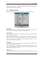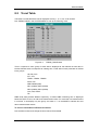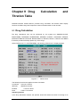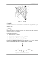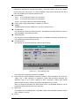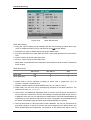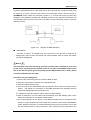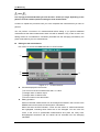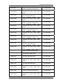
1
PM-6000 Portable Multi-parameter Patient Monitor Operation Manual Copyright SHENZHEN MINDRAY®BIO-MEDICAL ELECTRONICS CO., LTD. 2002 Version: 2.2 Issued date: 2004/08/08 Serial No: PM-6000 Statement SHENZHEN MINDRAY® BIO-MEDICAL ELECTRONICS CO., LTD. (hereinafter called Mindray) owns all rights to this unpublished work and intends to maintain this work as confidential. Mindray may also seek to maintain this work as an unpublished copyright. This publication is to be used solely for the purposes of reference, operation, maintenance, or repair of Mindray equipment. No part of this can be disseminated for other purposes. In the event of inadvertent or deliberate publication, Mindray intends to enforce its rights to this work under copyright laws as a published work. Those having access to this work may not copy, use, or disclose the information in this work unless expressly authorized by Mindray to do so. Mindray shall not be liable for errors contained herein nor for incidental or consequential damages in connection with the furnishing, performance, or use of this material. This publication may refer to information and protected by copyrights or patents and does not convey any license under the patent rights of Mindray, nor the rights of others. Mindray does not assume any liability arising out of any infringements of patents or other rights of third parties. Content of this manual is subject to changes without prior notice. PROPERTY OF SHENZHEN MINDRAY®BIO-MEDICAL ELECTRONICS CO., LTD. ALL RIGHTS RESERVED Responsibility on the manufacturer party Mindray is responsible for safety, reliability and performance of this equipment only in the condition that: • all installation, expansion, change, modification and repair of this equipment are conducted by Mindray qualified personnel; and, • applied electrical appliance is in compliance with relevant National Standards; and, • the monitor is operated under strict observance of this manual. Patient Monitor user’s manual (V 2.2) I Note This equipment is not intended for family usage. Warning Failure to follow this message may result in death or serious injury to the patient or operator, or may cause damage to the equipment. Warning To use this equipment safely, the user must observe all special application instructions or precautions in this operation manual. However, these special instructions or precautions cannot replace the medical procedures executed presently. Do not solely rely on the audible alarming system to monitor a patient. Setting a very low volume or completely turning off the volume may cause catastrophe to the patient. The most reliable method to monitor the patient is to at the same time of using the monitoring equipment, the physician should himself closely observe the situation of the patient. This equipment is designed to be used only by trained health care personnel in health care institution. To reduce the risk of electrical shock, do not open any casing of the equipment. Service can be performed only by qualified personnel. This equipment may interfere with the ultrasound imaging system. If there is interference, interference signals may appear on the screen of the ultrasound imaging system. To eliminate the interference, the distance between this equipment and the ultrasound imaging system should be as far as possible. It is dangerous to subject the electric contact and the connector to normal saline, other liquid or conductive adhesive. The electric contact and the connector such as cable connector, power and parameter module plug-in connector, and frame connector must be kept clean and dry. Once being wetted, they must be thoroughly dried before use. If to decontaminate the electric contact or the connector, please contact biomedical department or Mindray company. Warning This monitor is not a device for treatment purpose. It is important for the hospital or organization that employs this equipment to carry out a reasonable maintenance schedule. Neglect of this may result in machine breakdown or injury of human health. II Patient Monitor user’s manual (V 2.2) Warranty Workmanship & Materials Mindray guarantees new equipment other than accessories to be free from defects in workmanship and materials for a period of 18 months (six months for multi-site probes and SpO2 sensor) from date of shipment under normal use and service. Mindray's obligation under this warranty is limited to repairing, at Mindray’s option, any part which upon Mindray's examination proves defective. THIS WARRANTY IS EXCLUSIVE AND IS IN LIEU OF ALL OTHER WARRANTIES, EXPRESSED OR IMPLIED, INCLUDING WARRANTIES OF MERCHANT ABILITY OR FITNESS FOR ANY PARTICULAR PURPOSE. Exemptions Mindray's obligation or liability under this warranty does not include any transportation or other charges or liability for direct, indirect or consequential damages or delay resulting from the improper use or application of the product or the substitution upon it of parts or accessories not approved by Mindray or repaired by anyone other than a Mindray authorized representative. This warranty shall not extend to any instrument which has been subjected to misuse, negligence or accident; any instrument from which Mindray's original serial number tag or product identification markings have been altered or removed, or any product of any other manufacturer. Safety, Reliability and Performance Mindray is not responsible for the effects on safety, reliability and performance of the PM-6000 Modular Patient Monitor if: ■ assembly operations, extensions, re-adjusts, modifications or repairs are carried out by persons other than those authorized by Mindray. ■ the Patient Monitor is not used in accordance with the instructions for use, or the electrical installation of the relevant room does not comply with NFPA 70: National Electric Code or NFPA 99: Standard for Health Care Facilities (Outside the United States, the relevant room must comply with all electrical installation regulations mandated by the local and regional bodies of government). Patient Monitor user’s manual (V 2.2) III Return Policy Return Procedure In the event that it becomes necessary to return a unit to Mindray, the following procedure should be followed: 1. Obtain return authorization. Contact the Mindray Service Department and obtain a Customer Service Authorization (Mindray) number. The Mindray number must appear on the outside of the shipping container. Return shipments will not be accepted if the Mindray number is not clearly visible. Please provide the model number, serial number, and a brief description of the reason for return. 2. Freight policy. The customer is responsible for freight charges when equipment is shipped to Mindray for service (this includes customs charges). Company Contact Address: Mindray Building, Keji 12th Road South, Hi-tech Industrial Park, Nanshan, Shenzhen, P. R. China 518057, Phone: +86 755 2658 2888 Fax: +86 755 2658 2680 Free hot line: +86 800 830 3312 EC Representative IV Name: Shanghai International Holding Corp. GmbH(Europe) Address: Eiffestrasse 80 D-20537 Hamburg Germany Phone: +49 40 2513174 Fax: +49 40 255726 Patient Monitor user’s manual (V 2.2) Preface This manual gives detailed description to PM-6000 Modular Patient Monitor concerning its performance, operation, and other safety information. Reading through this manual is the first step for the user to get familiar with the equipment and make the best out of it. Following symbols indicates some important facts that you have to pay special attention to: Warning Caution Note Points to be noted to avoid injury to the patient and the operator. Points to be noted to avoid damage to the equipment. Points to be noted. This manual is intended for persons who are trained in the use of this field and have adequate experience in operation of monitoring equipment. Patient Monitor user’s manual (V 2.2) V Content Content Chapter 1 Introduction................................................................................................... 1-1 1.1 General Information................................................................................................... 1-3 1.2 Screen Display .......................................................................................................... 1-7 1.3 Button Functions ..................................................................................................... 1-10 1.4 Interfaces................................................................................................................. 1-13 1.5 Spefication ............................................................................................................... 1-14 Chapter 2 Getting Started .............................................................................................. 2-1 2.1 Open the Package and Check .................................................................................. 2-1 2.2 Connect the Power Cables........................................................................................ 2-1 2.3 Power on the Monitor ................................................................................................ 2-2 2.4 Connect Patient Sensors........................................................................................... 2-2 2.5 Check the Recorder .................................................................................................. 2-2 Chapter 3 System Menu.................................................................................................. 3-1 3.1 Patient Information Setup.......................................................................................... 3-1 3.2 Default Setup............................................................................................................. 3-3 3.3 Review....................................................................................................................... 3-4 3.4 System Setup ............................................................................................................ 3-4 3.5 Selection Setup ....................................................................................................... 3-11 3.6 Monitor Version........................................................................................................ 3-12 3.7 Drug Calculation ...................................................................................................... 3-13 3.8 Maintenance ............................................................................................................ 3-13 3.9 DEMO Function ....................................................................................................... 3-16 Chapter 4 Face Select ..................................................................................................... 4-1 4.1 Select Operating Screen ........................................................................................... 4-1 4.2 Standard Screen........................................................................................................ 4-1 4.3 Trend Screen ............................................................................................................. 4-2 4.4 oxyCRG Screen......................................................................................................... 4-3 4.5 Viewbed Screen ........................................................................................................ 4-4 Chapter 5 Alarm............................................................................................................... 5-1 5.1 Alarm Modes.............................................................................................................. 5-1 5.2 Alarm verification during power on ............................................................................ 5-4 5.3 Alarm Cause .............................................................................................................. 5-4 5.4 SILENCE and PAUSE ............................................................................................... 5-5 5.5 Parameter Alarm........................................................................................................ 5-6 5.6 When an Alarm Occurs ............................................................................................. 5-6 Chapter 6 Freeze............................................................................................................. 6-1 6.1 General...................................................................................................................... 6-1 6.2 Enter/Exit Freeze Status............................................................................................ 6-1 6.3 Frozen Menu ............................................................................................................. 6-2 Patient Monitor user’s manual (V 2.2) 1 Content 6.4 Reviewing Frozen Waveform .................................................................................... 6-3 6.5 Recording Frozen Waveform..................................................................................... 6-3 Chapter 7 Recording ...................................................................................................... 7-1 7.1 General Information on Recording ............................................................................ 7-1 7.2 Recording Type ......................................................................................................... 7-1 7.3 Recording Startup...................................................................................................... 7-4 7.4 Recorder Operations and Status Messages ............................................................. 7-5 Chapter 8 Trend and Event............................................................................................ 8-1 8.1 Trend Graph .............................................................................................................. 8-1 8.2 Trend Table................................................................................................................ 8-3 8.3 NIBP Recall ............................................................................................................... 8-4 8.4 Alarm Event Recall .................................................................................................... 8-5 8.5 Power-off data storage(Optional) .............................................................................. 8-7 Chapter 9 Drug Calculation and Titration Table ........................................................... 9-1 9.1 Drug Calculation ........................................................................................................ 9-1 9.2 Titration Table ............................................................................................................ 9-3 Chapter 10 Patient Safety.............................................................................................. 10-1 Chapter 11 Care / Cleaning............................................................................................ 11-1 11.1 System Check........................................................................................................ 11-1 11.2 General Cleaning................................................................................................... 11-1 11.3 Cleaning Agents..................................................................................................... 11-2 11.4 Sterilization ............................................................................................................ 11-3 11.5 Disinfection ............................................................................................................ 11-3 Chapter 12 ECG/RESP Monitoring ............................................................................... 12-1 12.1 What Is ECG Monitoring........................................................................................ 12-1 12.2 ECG/RESP Module ............................................................................................... 12-1 12.3 Precautions during ECG Monitoring...................................................................... 12-3 12.4 Monitoring Procedure ............................................................................................ 12-3 12.5 ECG Screen Hot Keys........................................................................................... 12-8 12.6 ECG Menu............................................................................................................. 12-9 12.7 ECG Alarm Information and Prompt.................................................................... 12-14 12.8 ST Segment Monitoring (optional)....................................................................... 12-15 12.9 Arr. Monitoring (optional) ..................................................................................... 12-19 12.10 Measuring RESP............................................................................................... 12-24 12.11 Maintenance and Cleaning................................................................................ 12-28 Chapter 13 SpO2 Monitoring .......................................................................................... 13-1 13.1 PART 1 (MASIMO SpO2 board configuration) ...................................................... 13-1 13.1.1 Precautions......................................................................................................... 13-3 13.1.2 Monitoring Procedure ......................................................................................... 13-5 13.1.3 Sensors and Accessories: .................................................................................. 13-9 13.1.4 Alarm Description and Prompt.......................................................................... 13-12 13.1.5 Masimo Information.......................................................................................... 13-14 13.2 PART 2 (MINDRAY SpO2 board configuration) .................................................. 13-15 13.2.1 SpO2 Module.................................................................................................... 13-15 2 Patient Monitor user’s manual (V 2.2) Content 13.2.2 Precautions during SpO2/Pulse Monitoring ..................................................... 13-17 13.2.3 Monitoring Procedure ....................................................................................... 13-18 13.2.4 Limitations for Measurement ............................................................................ 13-20 13.2.5 SpO2 Menu ...................................................................................................... 13-20 13.2.6 Alarm Description and Prompt.......................................................................... 13-22 13.2.7 Maintenance and Cleaning............................................................................... 13-24 Chapter 14 NIBP Monitoring........................................................................................... 14-1 14.1 Introduction............................................................................................................ 14-1 14.2 NIBP Module ......................................................................................................... 14-2 14.3 NIBP Monitoring .................................................................................................... 14-3 14.4 NIBP SETUP menu ............................................................................................... 14-7 14.5 NIBP Alarm Message .......................................................................................... 14-11 14.6 Maintenance and Cleaning.................................................................................. 14-13 Chapter 15 TEMP Monitoring ......................................................................................... 15-1 15.1 TEMP Monitoring................................................................................................... 15-1 15.2 TEMP Module........................................................................................................ 15-2 15.3 TEMP SETUP Menu.............................................................................................. 15-3 15.4 TEMP Alarm message........................................................................................... 15-4 15.5 Care and Cleaning ................................................................................................ 15-5 Chapter 16 IBP Monitoring .............................................................................................. 16-1 16.1 General.................................................................................................................. 16-1 16.2 IBP Module ............................................................................................................ 16-1 16.3 Precautions during IBP Monitoring........................................................................ 16-2 16.4 Monitoring Procedure ............................................................................................ 16-3 16.5 IBP Menu............................................................................................................... 16-4 16.6 Alarm Information and Prompts........................................................................... 16-12 16.7 Maintenance and Cleaning.................................................................................. 16-14 16.8 ICP Transducer ICT/B(Optional Accessory) ................................................... 16-15 Chapter 17 CO Measuring............................................................................................... 17-1 17.1 General.................................................................................................................. 17-1 17.2 CO Module ............................................................................................................ 17-1 17.3 Monitoring Procedure ............................................................................................ 17-3 17.4 CO SETUP Menu .................................................................................................. 17-7 17.5 Hemodynamic Calculation..................................................................................... 17-9 17.6 Alarm Information and Prompt............................................................................. 17-10 17.7 Maintenance and Cleaning.................................................................................. 17-11 Chapter 18 CO2 Measuring............................................................................................. 18-1 18.1 General.................................................................................................................. 18-1 18.2 CO2 Module .......................................................................................................... 18-2 18.3 Monitoring Procedure ............................................................................................ 18-3 18.4 CO2 Menu ............................................................................................................. 18-5 18.5 Alarm Information and Prompt............................................................................. 18-10 18.6 Maintenance and Cleaning.................................................................................. 18-12 Chapter 19 AG Measuring ............................................................................................... 19-1 Patient Monitor user’s manual (V 2.2) 3 Content 19.1 General ......................................................................................................... 19-1 19.2 AG Module .................................................................................................... 19-2 19.3 Measuring principle and operating process................................................... 19-4 19.4 Menus ........................................................................................................... 19-6 19.5 Alarm information and prompts ................................................................... 19-12 19.6 Technical specifications............................................................................... 19-15 19.7 Anesthetic Gas Accessories ........................................................................ 19-17 19.8 Maintenance and cleaning ................................................................................ 19-17 Chapter 20 Accessories and Ordering Information ..................................................... 20-1 20.1 ECG Accessories .................................................................................................. 20-1 20.2 SpO2 Accessories ................................................................................................. 20-2 20.3 NIBP Accessories .................................................................................................. 20-2 20.4 TEMP Accessories ................................................................................................ 20-3 20.5 IBP Accessories..................................................................................................... 20-3 20.6 CO Accessories ..................................................................................................... 20-4 20.7 CO2 Accessories................................................................................................... 20-4 20.8 AG Accessories ..................................................................................................... 20-4 Appendix I EC Declaration of Conformance ......................................................................... 1 Appendix II Product Specification ......................................................................................... 1 Appendix III EMC ..................................................................................................................... 1 Appendix IV System Alarm Prompt ....................................................................................... 1 4 Patient Monitor user’s manual (V 2.2) Chapter 1 Instruction For the overall information about the monitor, refer to General For the knowledge about the various displayed information on the screen, refer to Screen Display For the operating method, refer to Button Function and Basic Operation For the position of the various interface, refer to External Interface For the specifications of the monitor, refer to Specifications For safety precautions of the monitor, please refer to Patient Safety. Warning PM-6200 is intended for clinical monitoring application with operation only granted to appropriate medical staff. Warning Monitor can only monitoring one patient at a time. Warning There could be hazard of electrical shock by opening the monitor casing. All servicing and future upgrading to this equipment must be carried out by personnel trained and authorized by Mindray. Warning Possible explosion hazard if used in the presence of flammable anesthetics or other flammable substance in combination with air, oxygen-enriched environments, or nitrous oxide. Warning You must verify if the device and accessories can function safely and normally before use. Warning You must customize the alarm setups according to individual patient situation and make sure that alarm sound can be activated when alarm occurs. Warning Do not use cellular phone in the vicinity of this device. High level electromagnetic radiation emitted from such devices may greatly affect the monitor performance. Warning Patient Monitor user’s manual (V 2.2) 1-1 Introduction Do not touch the patient, table, or the device during defibrillation. Warning Devices connected to the monitor shall form an equipotential system (protectively earthed). Warning When used with Electro-surgery equipment, you (doctor or nurse) must give top priority to the patient safety. Warning Do not place the monitor or external power supply in any position that might cause it to fall on the patient. Do not lift the monitor by the power supply cord or patient cable, use only the handle on the monitor. Warning Consult IEC-601-1-1 for system interconnection guidance. The specific requirements for system interconnection are dependent upon the device connected to the monitor and the relative locations of each device from the patient, and the relative location of the connected device to the medically used room containing the monitor. In all circumstance the monitor must be connected to a grounded AC power supply. The monitor is referred to as an IEC 601/F device in the summary of situations table contained in IEC 601-1-1. Warning Dispose of the packaging material, observing the applicable waste control regulations and keeping it out of children’s reach. Warning This equipment is accord with the standard CISPR11(EN55011) class A. Warning Grounding: Connect the oximeter only to a three-wire, grounded, hospital-grade receptacle. The three-conductor plug must be inserted into a properly wired three-wire receptacle; if a three-wire receptacle is not available, a qualified electrician must install one in accordance with the governing electrical code. Do not under any circumstances remove the grounding conductor from the power plug. Do not use extension cords or adapters of any type. The power cord and plug must be intact and undamaged. If there is any doubt about the integrity of the protective earth conductor arrangement, operate the oximeter on internal battery power until the AC power supply protective conductor is fully functional. 1-2 Patient Monitor user’s manual (V 2.2) Instruction Note The software was developed per IEC601-1-4. The possibility of hazards arising from errors in the software program is minimized. Caution At the end of its service life, the product described in this manual, as well as its accessories, must be disposed of in compliance with the guidelines regulation the disposal of such products. If you have questions concerning disposal of the product, please contact MINDRAY or its representatives. Caution If you have any doubt to the grounding layout and its performance, you must use the built-in battery to power the monitor. 1.1 General PM-6000 is a modular multi-parameter patient monitor that has abundant monitoring functions and is used for the clinical monitoring of adult, pediatric and neonate. The user may increase the parameter and functional module at any time according to different requirements. In addition, the system also supports the module exchange between inside and outside of the hospital and the transmission of parameter alarm setup. PM-6000 can be connected to the central monitoring system via the Mindray network so as to form a network monitoring system. PM-6000 Modular Patient Monitor is year 2000 compliant. The Patient Monitor (Figure 1-1) can monitor vital physiological signals as ECG, Respiratory Rate, SpO2, NIBP, Dual-TEMP, fourfold-IBP, CO, CO2 and AG. Heart Rate (HR) 2-channel ECG waveforms ECG Arrhythmia and S-T segment analysis(optional),Pace Respiratory Rate (RR) RESP Respiration Waveform Oxygen Saturation (SpO2), Pulse Rate (PR) SpO2 SpO2 Plethysmogram Systolic Pressure (NS), Diastolic Pressure (ND), Mean Pressure NIBP (NM) Channel-1 Temperature (T1), Channel-2 Temperature (T2), TEMP Temperature Difference between two channels (TD) Channel-1 SYS, DIA, MAP Channel-2 SYS, DIA, MAP IBP Channel-3 SYS, DIA, MAP Channel-4 SYS, DIA, MAP fourfold-IBP waveforms Blood Temperature (TB) CO Cardiac Output (CO) End Tidal CO2 (EtCO2) CO2 Inspired Minimum CO2 (InsCO2) Air Way Respiration Rate (AwRR) Inhale and exhale CO2 (FiCO2, EtCO2) AG Patient Monitor user’s manual (V 2.2) 1-3 Introduction Inhale and exhale N2O (FiN2O, EtN2O) Inhale and Exhale O2 (FiO2, EtO2) Inhale and exhale anesthetic agent (FIAA, ETAA, Note: AA refers to one of anesthetic agents listed below: HAL (Halothance) ISO (Isoflurance) ENF (Enflurance) SEV (Sevoflurance) DES (Desflurance) Airway Respiration Rate (respiratory times per minute, unit: rpm) AwRR MAC(Minimum alveolar concentration) Waveforms of four anesthetic gases including CO2, N2O, O2, AA System Construction: This Monitor has three independent parts: ■ Display: 12.1’’ color LCD or CRT ■ Module Box: allows horizontal or vertical placement. ■ Module: includes parameter module and function module. The display can be placed independently as a separate system or placed together with the module box forming a compact system. ③ ④ ② ⑤ ① Figure 1-1 System Construction The visible LED are CLASS 1 LED PRODUCT according EN 60825-1 A11 Oct 1996. The power switch “POWER” of the monitor lies at the right side of the module box(①). The three lamps (②) on the right side of the module box are used to indicate the power situation. After powering on the monitor, these three lamps light on. When the monitor is in function and there is alarm occurring, both the alarm lamp on the screen (③) and the ALARM lamp on the button module (④) light on or flash in a specific way. The socket for each sensor is on the front panel of corresponding module. The output end of the recorder is on the front panel of the recorder module (⑤). The battery door is on the rear panel of the monitor. The analog interface, VGA interface, TFT interface are all on the right side of the rear panel of the monitor. The power socket is on the left side of the rear panel, refer to “1.4 Rear Panel” Display: ■ The system may use either the 12.1’’ color LCD provided by Mindray or the ordinary purchased display. ■ The color LCD provided by Mindray has 6 system buttons and a rotary knob used for 1-4 Patient Monitor user’s manual (V 2.2) Instruction direct operation performed by the user. ■ The display provided by the Mindray possesses the device used for visual and audio alarm. ■ The ordinary purchased display does not have system buttons and alarm device. Therefore, the user should buy the button module to realize the direct operation by the user and visual and audio alarm function. Module box ■ The module box can maximally hold 8 standard modules at the same time. ■ The module box can be placed either horizontally or vertically. Modules: The modules currently provided by the system include: ■ 0# module: blank module (used for decoration, standard module) ■ 1# module: button/alarm module (standard module) ■ 2# module: ECG/RESP+TEMP(2 channel, standard module) ■ 3# module: SPO2 module (standard module) ■ 4# module: NIBP module (standard module) ■ 5# module: IBP(1,2 channel)module (standard module) ■ 6# module: CO module (standard module) ■ 7# module: EtCO2 module (standard module) ■ 8# module: Anaesthetic gas module (occupies a space of three slots) ■ 9# module: Recorder module(occupies a space of two slots) ■ 11# module:IBP(3, 4 channel)module (standard module) 1# module 5# module 2# module 6# module Figure 1-2 3# module 7# module 4# module 9# module module Patient Monitor user’s manual (V 2.2) 1-5 Introduction These modules are independent from each other, hence easy for the expansion of the new parameter module. Note The manufacturer will not inform each customer of the appearance of the new module. Method to load/unload the module: (1) Step 1 unload the module (2) step 2 unload the module (3) Step 3 unload the module Figure 1-3 (4) load the module methods to load/unload the module Unload the module: as per the Figure 1-3 (1)-(3), first use one hand to hold the module to be unloaded, then use the other hand to press down the plate under the module, and finally take out the module carefully. Load the module: Align the module to be loaded with the empty slot, and then push the module into the slot as per the method shown in the Figure 1-3 (4). When the sound “click” is heard, it indicates the module has been successfully loaded. Note You can load/unload a module when the monitor is operating. After loading/unloading a module, the system will refresh the screen and display parameters and waveforms according to the module newly loaded. For a module that has been unloaded, its measured data will be still stored in the system. You can view these data by using Trend Graph, Trend Table and NIBP Recall menus. Warning If the user does not load/unload the module as per the method illustrated above, when the top rake of the device is large, the module may slide out from the slot. In this case, the user may be injured and the module may be damaged. 1-6 Patient Monitor user’s manual (V 2.2) Instruction 1.2 Screen Display The display of PM-6000 modular multi-parameter monitor is a color LCD, which can display the collected patient parameters, waveforms, alarm information as well as bed number, time and monitor status, etc. The operating screen of the PM-6000 is the standard monitoring screen. Main system screen: ■ Standard monitoring screen ① ② ③ Figure 1-4 standard monitoring screen In the standard monitoring screen, the screen has three areas: information area( ① ), waveform/menu area(②), and parameter area(③). Information area(①): Information area lies on the top part of the screen, which is used to display the current status of the monitor and the patient. The information area contains following data: Bed No.: used to indicate the bed number of the patient being monitored. This information is displayed all the time. Adult: used to indicate the patient type. This information is displayed all the time. 2000-01-01: used to indicate the date. “21:09:12” is the time. This information is displayed all the time. Patient name: displayed to the right side of the data. When the operator is entering the patient name, the name is displayed in this position. When the operator does not enter the patient name, the position displays blank. Patient sex: displayed to the right side of the patient name. Other information displayed in the information area appears or disappears together with the status being reported, which is divided into following parts based on the contents: Patient Monitor user’s manual (V 2.2) 1-7 Introduction ■ Monitor Prompt Information: informing the status of monitor or sensor, which always appears to the right side of the time. When this information appears, it will cover the patient sex and name. ■ flag for alarm PAUSE. Press “SILENCE” button once (less than 1 second) to mute all alarm sounds are muted for the time being and the flag appears at the same time. Press the button again to terminate the PAUSE status. The duration for PAUSE status can be 1 minute, 2 minutes or 3 minutes. ■ flag for alarm SILENCE. Press “SILENCE” button once (more than 1 second) to manually mute the alarm sound and this flag appears at the same time. The SILENCE status terminates when you discharge the status or new alarm occurs. ■ flag for Alarm Volume Off. It appears indicating that you have closed the alarm sound permanently. This status terminates when you discharges the status. Note If symbol appears, the system will no longer give audible alarm sound. You must be very careful in using this function. Two ways can be used to discharge this status. One is set the alarm volume to an option other than OFF in the USER MAINTAIN menu. The other method is to press SILENCE button to make the flag turn to . And then press SILENCE again and the system will restore the normal alarm status. ■ Patient parameter alarm information: this information is fixedly displayed in the extreme right side of the screen. ■When the waveform on the screen is frozen, the corresponding prompt “FROZEN” window appears on the lower part of the screen. ■There is a status bar on the bottom of the screen. Some status information will be displayed in it. For example, “Loading XX module”, “Module unloaded”, “The recorder does not exit”, and the information such as the lead system detected by the hardware is different from the current user setup. Waveform/menu area(②): The waveform area can maximally display 8 waveforms. The displaying order of the waveforms on the screen can be adjusted. For the maximum configuration, the waveforms provided by the system for selection are: 2 ECG waveforms, SpO2 waveform, 4 IBP waveforms, RESP waveform, CO2 waveform, O2 waveform and anaesthetic gas waveform. All the waveforms in the system are listed out in the “ TRACE SETUP” menu. The user may select the waveform to be displayed and adjust their displaying positions. The specific method is illustrated in the part: SYSTEM MENU. The name of the waveform is displayed on the upper left part of the waveform. The user may 1-8 Patient Monitor user’s manual (V 2.2) Instruction choose ECG lead based on the requirements. The gain of the channel and the filter way are also displayed on each ECG waveform. A 1mV scale bar is also displayed to the right side of ECG waveform. The IBP waveform scale can also be selected according to the actual requirement. Its range is described in the part: IBP MONITORING. In the IBP waveform area, the waveform scale is displayed. The three dotted lines for each IBP waveform form up to down represent respectively the upper limit scale, reference scale and lower limit scale. The values of these three scales can be set. The specific method is given in the part: IBP MONITORING. When menu is wanted during screen operation, the menu always occupies the fixed position in the middle part of the waveform area, therefore part of waveform can not be viewed temporarily. After exiting the menu, the system will restores the original screen. The user may set up the rate to refresh the waveform. The method to adjust the refreshing rate of each waveform is discussed in the setup description of each parameter. Parameter area(③): The parameter area lies to the right side of the waveform area, whose position basically corresponds to the waveform. The parameters displayed in the parameter area include: ECG — heart rate or pulse rate (unit: beats/minute) — The ST analyzing result of channel 1 and 2: ST1, ST2 (unit: mV) — PVCs(unit: times/minute) NIBP — From left to right, there are Systolic pressure, Mean pressure and Diastolic pressure(unit: mmHg or kPa) SpO2 — SpO2(unit: %) — Pulse Rate(unit: beats/minute)(When “BOTH” item is selected) CO ─ CO(unit: liter/minute); ─ TB(unit: ℃ or ℉) IBP — The blood pressure of channel 1, 2, 3 and 4. From left to right, there are Systolic pressure, Mean pressure and Diastolic pressure(unit: mmHg or kPa). RESP — Respiration Rate(unit: times/minute) CO2 — EtCO2(unit: mmHg or kPa) — INS CO2 (unit: mmHg or kPa) — AwRR (times/minute) TEMP — Temperature of channel 1 and 2: T1, T2 and the difference between them TD. (unit: ℃ or ℉) AG Patient Monitor user’s manual (V 2.2) 1-9 Introduction ─ AwRR (times/minute) ─ MAC Alarm lamp and alarm status: In normal status: the alarm lamp is not on. When alarm exists, the alarm lamp flashes or lights on. The color of the lamp corresponds to the alarm level. Refer to related chapter: Alarm. For the details of alarm information and prompt information, refer to the related content of each parameter in related chapter. 1.3 Button Function and Basic Operation This section discusses the following information: Button functions of PM-6000 The method to use the rotary knob to realize the operation on the screen Index for basic operation Button function of PM-6000: Two types of buttons are available on PM-6000: hard buttons and soft buttons. Hard buttons include system button panel and module button panel. The system button panel is on the screen or on the button module. The module button is on each module. Soft buttons are on the screen, which have to be selected by the operator through using the rotary knob. Figure 1-5 module button panel and screen button panel System buttons: SILENCE Push this button to suspend alarm for maximum 3 minutes (with 1 minute, 2 minutes and 3 minutes selectable). In Alarm PAUSE status, a symbol appears in the Message Area. Push this button to mute all kinds of sounds (including alarm sound, heart beat, pulse tone, key sound). At the same time, a symbol appears in the Message Area. Push this button again to restore all kinds of sounds and the 1-10 Patient Monitor user’s manual (V 2.2) Instruction symbol disappears from the screen. Note If new alarm occurs in Alarm Pause/Silence status, the system will discharge Pause/Silence status automatically. For specific rules, see Chapter Alarm. Note The system will begin to give alarm information again once there exist alarm-triggering event. Nevertheless, remember pushing SILENCE button can permanently shut off audible alarm sound of ECG LEAD OFF and SPO2 SENSOR OFF alarms. PAUSE Press this button, the system accesses the Alarm Pause status. All the alarm sounds are muted. However, other sounds such as heart beat, button sound and pulse sound will still exist. At this time, the “ALARM PAUSE xx s” appears in the parameter alarm information area. Press this button again or when the pause time runs out, the system terminates the alarm pause status and returns to the normal monitoring status, then the alarm sound resumes and the prompt “ALARM PAUSE xx s” disappears from the screen. Note After the end of Alarm Pause status, whether the alarm sound is restored depends on whether the alarm exists. However, the technical alarms such as lead off will not be restored. That is to say the user may also close the technical alarms such as lead off through accessing and then exiting the Alarm Pause status. ALARM SETUP Press this button to let the system rapidly access the sub-menu of ALARM SETUP. In this menu, the user may set up all the information about the alarms. MAIN Whatever level of menu the system is in, press the button and the system will always return to the main screen. FREEZE Press this button and the system will access the FREEZE status. In this status the user may review the waveform of 40 seconds. Also, the frozen waveform can be printed out. In the FREEZE status, press this button again to discharge the FREEZE status. For detailed information, refer to related chapter: Freeze. MENU Press this button to call up the SYSTEM MENU, in which the user may set up system information and perform review operation. For detailed information, refer to related chapter: System Menu and related chapter: Trend and Event. Rotary knob Patient Monitor user’s manual (V 2.2) 1-11 Introduction The user may use the rotary knob to select the menu item and modify the setup. It can be rotated clockwise or counter-clockwise and pressed like other buttons. The user may use the knob to realize the operations on the screen and in the system menu and parameter menu. Method to use the knob to operate on the screen: The rectangular mark on the screen that moves with the rotation of the knob is called “cursor”. Operation can be performed at any position at which the cursor can stay. When the cursor is in the waveform area, the user may immediately modify the current setup. When the cursor is in the parameter area, the user may open the setup menu of the corresponding parameter module so as to set up the menu items of the module. Operating method: ■ Move the cursor to the item where the operation is wanted ■ Press the knob ■ One of the following four situations may appear: 1.The cursor with background color may become into the frame without background color, which implies that the content in the frame can change with the rotation of the knob. 2.Menu or measuring window may appear on the screen, or the original menu is replaced by the new menu. 3.A check mark “√” appears at the position, indicating that the item is confirmed. 4.The system immediately executes a certain function. Module buttons: ECG module: SETUP: setup button of ECG/RESP, TEMP : blank button SPO2 module: SETUP: SPO2setup button : blank button NIBP module: SETUP: NIBP setup button START/STOP: button to start/stop measurement IBP(1, 2) module: SETUP:IBP setup button ZERO :IBP zero button CAL :IBP calibrate button CO module: SETUP: CO setup button MEASURE: button used to access the CO MEASURMENT window 1-12 Patient Monitor user’s manual (V 2.2) Instruction CO2 module: SETUP: CO2 setup button STANDBY/START: Standby/Measure button Recorder module: REC/STOP: button to start/stop recording IBP(3, 4)module: SETUP: IBP setup button ZERO: IBP zero button CAL : IBP calibrate button Note When using the color LCD provided by Mindray, the system buttons and alarm function are integrated on the screen. The user may operate the buttons on the display directly to realize the functions of the monitor. When using the ordinary purchased display, the user should buy the button module to realize the functions of the monitor. The monitor supports the use of both hard buttons on the screen and the button module. All the operations of the system can be realized through the combined operation of the buttons and the rotary knob. 1.4 Rear panel ⑦ ① ⑥ ② ⑤ Figure 1-6 ⑧ rear panel ③ ④ ① FUSE: Standard-T 3.0A ② Power supply: 100/250 (VAC), 50/60 (Hz) ③ Analog output: used to output the analog signals, able to be connected to oscillometer and pen recorder. ④ VGA MONITOR: able to be connected to the external VGA monitor. ⑤ LVDS MONITOR: able to be connected to the display provided by Mindray. Patient Monitor user’s manual (V 2.2) 1-13 Introduction ⑥ Network interface: connected to the Mindray Hypervisor III via a standard RJ45 Socket. ⑦ EXTEND: used to connect to an external device. ⑧ Equipotential grounding terminal: able to be connected to the hospital’s grounding system. This symbol means “BE CAREFUL". Refer to the manual (this manual). Note Monitor must be connected with specific network equipment such as Harb during using net function. Warning Accessory equipment connected to the analog and digital interfaces must be certified according to the respective IEC standards (e.g. IEC 60950 for data processing equipment and IEC 60601-1 for medical equipment). Furthermore all configurations shall comply with the valid version of the system standard IEC 60601-1-1. Everybody who connects additional equipment to the signal input part or signal output part configures a medical system, and is therefore responsible that the system complies with the requirements of the valid version of the system standard IEC 60601-1-1. If in doubt, consult the technical service department or your local representative. 1.5 Specifications 1.5.1 Products classification Anti-electroshock type Class I equipment EMC type Class A Anti-electroshock degree ECG (RESP), SpO2, NIBP, IBP, TEMP, CO,CO2 CF Harmful liquid proof degree Ordinary equipment (sealed equipment without liquid proof) Working system 1.5.2 Continuous running equipment Specifications ■ Applicable patient type: Adult, pediatric and neonate ■ Parameters monitored by PM-6000: ECG, RESP, NIBP, SpO2, PR (pulse rate), Dual-TEMP, 4 IBP, CO, TB (blood temperature), CO2, ANAESTHETIC GAS, etc. ■ Display 1-14 Patient Monitor user’s manual (V 2.2) Instruction 12.1”TFT ■ Analog output 1 channel, signal amplitude: 1V/mV, amplitude error <5%, output impedance: 100ohm, signal delay <20ms. ■ Power supply: Operate on AC power. AC power: 100~250VAC, 50/60Hz, Pmax=110VA. 1.5.3 Environmental requirements ■ Working environment: PM-6000 can only be used in the hospital or by the trained physician during transporting the patient into or out of the hospital. This device can not be used at home. The operating environment must satisfy following conditions: Surrounding temperature: 0℃ ∼ 40℃ Humidity: 15% - 85% -500 ∼ 4600 meters Altitude: ■ Conditions for transportation and storage: Without specific description, the transportation and storage of PM-6000 must satisfy following conditions: Surrounding temperature: -20℃ ∼ 60℃ Humidity: 10% - 93%, relative humidity Altitude: -500 ∼ 13100 meters Patient Monitor user’s manual (V 2.2) 1-15 Chapter 2 Getting Started ■ Open the package and check ■ Connect the power cables ■ Power on the monitor ■ Connect patient sensors ■ Check the recorder Note To ensure that the monitor works properly, please read Chapter Patient safety, and follow the steps before using the monitor. 2.1 Open the Package and Check Open the package and take out the monitor and accessories carefully. Keep the package for possible future transportation or storage. Check the components according to the packing list. ■ Check for any mechanical damage. ■ Check all the cables, modules and accessories. If there is any problem, contact the distributor immediately. 2.2 Connect the Power Cables Connection procedure of the AC power line: Make sure the AC power supply complies with following specification: 100~250 VAC, 50/60 Hz. Apply the power line provided with the monitor. Plug the power line to INPUT interface of the monitor(Socket ③ in Figure 1-7).. Connect the other end of the power line to a grounded 3-phase power output. Note Connect the power line to the jack special for hospital usage. Note Mindray does not provide MULTIPLE PORTABLE SOCKET-OUTLETS. IF use it, please do not place it on the floor. Mindray advises that every one monitor uses one MULTIPLE PORTABLE SOCKET-OUTLETS. Connect to the ground line if necessary. Refer to Chapter Patient Safety for details. Patient Monitor user’s manual (V 2.2) 2-1 Getting Started 2.3 Power on the Monitor Press POWER(① in Figure 1-1) to power on the monitor. Then a beep will be heard and at the same time the indicator will flash twice in yellow and red. After 10 seconds or so, the system will enter monitoring screen after self-test, and you can perform normal monitoring now. During self-test, the software version will display. Warning If any sign of damage is detected, or the monitor displays some error messages, do not use it on any patient. Contact biomedical engineer in the hospital or Mindray Customer Service Center immediately. Note If the monitor finds any fatal error during self-test, it will alarm. Note Check all the functions that may be used to monitor and make sure that the monitor is in good status. Note The battery must be recharged to the full electricity after each use to ensure adequate electricity reserve. Note The interval between twice press of POWER should be more than 1 minute. 2.4 Connect Patient Sensors Connect all the necessary patient sensors between the monitor and the patient. Note For information on correct connection, refer to Chapter 12~19. 2.5 Check the Recorder If your monitor is equipped with a recorder, open the recorder door to check if paper is properly installed in the output slot. If no paper present, refer to Chapter Recording for details. 2-2 Patient Monitor user’s manual (V 2.2) Chapter 3 System Menu ■ New patient enrolment ■ Recording ■ Trend Graph/Table and Alarm Review ■ System Setup ■ Drug Calculation ■ Maintenance The Patient Monitor features flexible configurations. You can configure various aspects of the monitor, including the parameters to be monitored, sweeping speed of the waveforms, audio signal volume, and printout text. Press the “MENU” button on the Button Module or the display panel to call up “SYSTEM MENU”. The configuration is realized through operations on the SYSTEM MENU, as shown below. Figure 3-1 SYSTEM MENU Trend graph/table review, NIBP review and alarm review are discussed in Chapter: Trend and Event. 3.1 Patient Information Setup Note To clear current patient data, refer to New Patient for details. Pick the [PATIENT SETUP] item in the “SYSTEM MENU” to call up the following menu. Patient Monitor user’s manual (V 2.2) 3-1 System Menu Figure 3-2 PATIENT SETUP You can setup following patient information: DEPT. Department in which the patient receives treatment. PAT NO Patient No. BED NO Patient bed number (Range: 1-100) DOCTOR Name of the doctor. NAME Patient name (Valid characters: A-Z, 0-9 and space bar; Max. length: 12 characters) SEX Patient gender (Available options: "F" for Female, "M" for Male) PAT TYPE Patient type (Available options: ADU, PED, and NEO) ADMIT Hospitalization starting date (format: year\month\ day) BIRTH Patient date of birth (format: year\month\day) HT. (cm/inch ) Patient height (turning the knob with the increase/decrease of 0.5 cm/inch each time)The other HT. unit in the other menus accord with the unit which you choosed here. WT. (kg/Ib) Patient weight (turning the knob with the increase/decrease of 0.5 kg/Ib each time)The other WT. unit in the other menus accord with the unit which you choosed here. BLOOD Patient blood type (Pick A, B, O, AB, or N. "N" represents unknown blood type) NEW PATIENT Admission of new patient Also in this menu, you may select the [NEW PATIENT] item to access the “CONFIRM TO 3-2 Patient Monitor user’s manual (V 2.2) System Menu UPDATE PATIENT” dialog box as shown below, in which you can decide whether to monitor a new patient. Figure 3-3 Confirm To Update Patient Menu Pick [YES] to delete all information of the patient being currently monitored and exit the menu. Pick [NO] to give up updating the patient and the system will keep the information of the current patient and exit the menu. Note If you select [YES], the system will delete all information of the patient being currently monitored. 3.2 Default Setup Note After selecting any item in this sub-menu, the selected item will replace the current setup of the system and accordingly become the system default configuration. Figure 3-4 DEFAULT Menu Patient Monitor user’s manual (V 2.2) 3-3 System Menu In this sub-menu, you can select both the factory default and the user-defined default. Also in this sub-menu, you can save the current system configuration as the user-defined default configuration. But at this time, the system will automatically save all the setups in the parameter menu, ECG gain and filter way as the user-defined default configuration according to the patient type. Also, the dialog box as shown below will pop up. Figure 3-5 CONFIRM DEFAULT CONFIG Note After selecting any item in the DEFAULT menu and exiting the box, the “CONFIRM DEFAULT CONFIG” Dialog box will pop up, in which you can select [YES] to confirm your selection or [NO] to give up your selection. Warning All configurations in the system will be replaced by “default configurations”. 3.3 Trend Review, Measurement Review and Alarm Event Review In the “SYSTEM MENU”, there are [TREND GRAPH], [TREND TABLE], [NIBP RECALL] and [ALARM RECALL] items. Please refer to Chapter 7: Trend and Event for detailed information. 3.4 System Setup Select the [system setup] item in the [system menu]: 3-4 Patient Monitor user’s manual (V 2.2) System Menu Figure 3-6 System setup In the [System setup] menu , users can setup the following items. 3.4.1 Face select Select “FACE SELECT” item in “SYSTEM SETUP” menu to access “FACE SELECT” dialog box as shown below, in which four selections are available: STANDARD SCREEN, TREND SCREEN, oxyCRG SCREEN and VIEWBED SCREEN. Only one selection can be chosen for each time. Figure 3-7 FACE SELECT 3.4.2 Alarm setup The system provides three levels of alarm volume. You can select any of them as per the clinical requirement. The procedures are: Select the [ALARM SETUP] item in the “SYSTEM SETUP” sub-menu of the “SYSTEM SETUP” menu. The menu as shown below will pop up, in which you can set up the alarm volume and other alarm information. For detailed information, refer to Chapter Alarm. Patient Monitor user’s manual (V 2.2) 3-5 System Menu Figure 3-8 Alarm Setup You can highlight the [ALARM VOL] item and then turn the knob to set up the alarm volume. There are three options: LOW, MED and HIGH. 3.4.3 Time Setup Select the [TIME SETUP] item in the “SYSTEM SETUP” menu. The menu as shown below will pop up. System time is in the format of year, month, day, hour, minute and second. Use cursor to highlight the item that you want to modify and turn the knob to select time. Then select [EXIT]. Note You shall set up the system time upon turning on the monitor (if you need to set up the system time); otherwise, when you review the content with time information, the system may not display the correct time. Figure 3-9 System Time Setup When this monitor is linked to the Central Station, its system time will keep consistent with 3-6 Patient Monitor user’s manual (V 2.2) System Menu that of the Central Station. Method to adjust time:Once link is successfully established, the Central Station will send its current time to the monitor. The monitor will automatically adjust its system time accordingly. Besides, the Central Station will keep on sending its current time to the monitor once per hour to maintain consistent time between them. However, the monitor will not adjust its time if it is different from the Central Station only in second. Please note that if you are setting up the system time when link is just established successfully, the monitor will immediately close the setup menu of system time. The setup button of system time in the system setup menu is disabled when the monitor is linked to the Central Station. That means you cannot open the setup menu of system time.(If the Central Station has no this function, you can skip over this paragraph.) 3.4.3 Recorder setup Select the [RECORD] in the “SYSTEM SETUP” menu to call up the following menu: Figure 3-10 Record Setup In this menu, the user can set up to output two waveforms. The waveforms that can be selected include: ECG1The first to the seventh ECG waveform on the screen (there are seven ECG ECG6 waveforms in full leads display)(If no ECG waveform is currently displayed on the screen, this item cannot be picked). SPO2 SpO2 Plethysmogram. IBP1 IBP2 Displayed waveform of Generated by IBP(1,2) module on the screen (If no IBP IBP3 IBP4 Displayed waveform of Generated by IBP(3,4) module on the screen (If no IBP waveform is currently displayed on the screen, this item cannot be picked). waveform is currently displayed on the screen, this item cannot be picked). Patient Monitor user’s manual (V 2.2) 3-7 System Menu RESP RESP waveform (If no RESP waveform is currently displayed on the screen, this item cannot be picked). CO2 Displayed waveform either of anesthetic gas or generated by CO2 module. N2O Displayed waveform of anesthetic gas. O2 Displayed waveform of anesthetic gas. AA Refers to displayed waveform anesthetic agent. OFF No display for this waveform. RT REC TIME this item has two options, CONTINUAL and 8s. “CONTINUAL” means once pushing the “REC/STOP” button on the recorder panel or the monitor panel, the recorder will continuously print out the waveform or parameter until this button is pushed again. TIMING REC TIME OFF used to set up the time interval between two recordings. 10 selections are available: “OFF, 10min, 20min, 30min, 40min, 50min, 1hour, 2hours, 3hours and 4hours”. The system will start the recording process according to the selected time interval. The recording time is always 8 seconds. Note RT REC TIME takes priority over TIMING REC TIME OFF. REC RATE: this item has two options, 25.0 and 50.0 mm/s. REC GRID: used to decide output format: OFF is without grid, and ON is with grid. CLEAR REC TASK: used to clear the alarm event that has been generated and is waiting for recording out. Note If two same waveforms are selected, the system will automatically change one of the waveform to a different one. 3.4.4 Module Setup Select the [MODULE SETUP] item in the “SYSTEM SETUP” menu to call up the following menu: 3-8 Patient Monitor user’s manual (V 2.2) System Menu Figure 3-11 Module Setup You can choose the parameters to be monitored in this menu. This can avoid the interference from the parameters that need not attention. 3.4.5 Analog output setup The monitor can output an analog waveform, whose time delay is less than 30ms. The output terminal is on the rear panel. Select “ANALOG” item in “SYSTEM SETUP” menu to call up the ANALOG menu. The first item is for setting up On/Off of the switch of the analog output. The second item is for selecting the waveform name to be output. Select “EXIT” item to return to the previous menu. Figure 3-12 Analog 3.4.6 Tracing Waveforms Selection Select the [TRACE SETUP] in the “SYSTEM SETUP” menu to call up the following menu. Patient Monitor user’s manual (V 2.2) 3-9 System Menu Figure 3-13 Tracing Waveforms Selection You can choose the waveform to be displayed in this menu. 3.4.7 Event Setup The monitor has four types of events. You can specify their representations by yourself. Select the [MARK EVENT] item in the “SYSTEM SETUP” to call up the following menu: Figure 3-14 MARK EVENT Menu How to mark the event: Use the rotary knob to select one from event A, B, C and D. The @ symbol will appear in the frame of the event being selected. Once making a wrong selection, you can push the knob on the event again to give up the selection. Select [EXIT] to exit the menu and consequently the selection will come into effect. Event function has following significance: To classify the records into different categories, such as those having influence on patients 3-10 Patient Monitor user’s manual (V 2.2) System Menu and those having influence on parameter monitoring including dose taking, injection, therapy status. Event will be displayed on the trend graph/table in order to assist the analysis on the patient parameters when the event happens. 3.5 Selection Setup Select the [SELECTION] item in the “SYSTEM SETUP” to call up the following menu. Figure 3-15 Selection Setup Key Volume: Select the [KEY VOL] item in the “SELECTION” menu. Turn the knob to select the volume. There are four selections available, which are “OFF, LOW, MED, HIGH”. Help Function: The system provides On-line Help to menu operations. You can choose any help information as per your need. The method is: Select the [SELECTION] item in the “SYSTEM MENU” to access the “SELECTION” sub-menu, in which you can highlight the [HELP] item and turn the knob to select “ON” or “OFF”. When it is “ON”, you can browse the on-line help information. When it is “OFF”, the system will turn off the on-line help function. Scan type: The system can display all waveforms about monitored patient on the screen either in “Refresh” or “Scroll” way. The method is: Select “SELECTION” item in “SYSTEM MENU” to access “SELECTION” sub-menu, in which there is the item “SCAN TYPE”. The user may decide the way to display the waveform by choosing either “REFRESH” or “SCROLL”. Alarm Limits: The system can display the alarm limits. You can choose this function as per your need. The method is: Patient Monitor user’s manual (V 2.2) 3-11 System Menu Select the [SELECTION] in the “SYSTEM MENU” to call up the “SELECTION” menu. You can set the “ALM LIMIT” switch to “ON” or “OFF”. Night Mode You can use the “NIGHT” item to set On/Off of night mode. When “ON” is picked, the monitor will silence the beep and pulse volume in order to ensure comfortable environment for the patient. Select the “SELECTION” item in “SYSTEM SETUP” MENU, “SELECT” menu pops up, in which the user may set up the “NIGHT” to “ON” or “OFF”. When it is “ON”, the system automatically adjusts the screen brightness to the “DARK” level (when the system has brightness adjustment function), and turns off the volume of heart beat and pulse to help patient sleep. When the system is alarming, the user can not select “NIGHT” mode. If any parameter alarm is triggered or any operation is executed under the NIGHT mode, night mode will be automatically shut off. Brightness You can user the “BRIGHTNESS” item to select the screen brightness. There are totally 8 options to select. 3.6 Monitor Version Select the [VERSION] item in the “SYSTEM MENU” to know the software version of the monitor. Figure 3-16 Monitor Version Select the [DEVICE CONFIG LIST] to know the configuration of the monitor. 3-12 Patient Monitor user’s manual (V 2.2) System Menu Figure 3-17 Device Configuration List 3.7 Drug Calculation You can use the drug calculation and titration table function of PM-6000 to calculate the concentration of 15 kinds of drugs. Refer to Chapter: Drug Calculation and Titration Table for detailed information. 3.8 Maintenance Select the [MAINTAIN] item in the “SYSTEM MENU” to call up the “ENTER MAINTAIN PASSWORD” dialog box as shown below, in which you can enter password and then customize maintenance settings. You cannot execute factory maintenance function, which is only available for the service engineers of MINDRAY company. Figure 3-18 Enter Maintain Password Patient Monitor user’s manual (V 2.2) 3-13 System Menu Input the password into the “ENTER MAINTAIN PASSWORD” box and press [CONFIRM], the “USER MAINTAIN” menu will pop up, in which you can set up following items. Figure 3-19 User Maintain For the [LANGUAGE] language, you can set the screen language to “Chinese” or “English”. For the [LEAD NAMING] item, you can select “AHA” or “EURO”. To know the difference between these two styles, refer to Chapter: ECG/RESP Monitoring. For the [ALM SOUND] item, you can set the alarm volume to “ON” or “OFF”. For the [NET TYPE] item, two selections are available: HYPER III and CMS. For the [LOCAL NET NO] item, it refers to the net No. SYSTEM KEY SETUP: press this item to access “SYSTEM KEY SETUP” submenu, in which the user may define the functions of the six extension keys (F1~F6) on the external keyboard. The user can define that the extension key has the same function as the button on any of the modules including the button module (except rotary knob). Pressing this extension is the same as pressing the corresponding button on the module. All the buttons on all the modules (not including rotary knob on the button module) are given as the selections for the extension function, which are not related to whether the corresponding module is loaded or not. 3-14 Patient Monitor user’s manual (V 2.2) System Menu Figure 3-20 SYSTEM KEY SETUP Warning When the alarm volume is set to “OFF”, you will not hear the alarm sound if new alarm occurs. Therefore, you must be very careful in using this selection. If setting the alarm volume to “OFF” when the system is in Silence or Pause status, the system will automatically discharge Silence or Pause status. If you select “Silence” or “Pause” when the alarm volume is set to “OFF”, the system will restore the alarm volume before the alarm volume is set to “OFF” and enter Silence or Pause status. Note After the alarm volume is set to OFF, a symbol will appear in the Technical Alarm Area. Note Setting Alarm Volume to “OFF” is valid only when the monitor is turned on for this time. After turning on the monitor next time, this setup will restore its value of the previous time when the system is turned on. COLOR SELF-DEFINE: is used by the user to define the color of the waveform displayed on the screen. Five colors can be chosen from: green, cyan, red, yellow and white. Patient Monitor user’s manual (V 2.2) 3-15 System Menu Figure 3-21 Color Self-define 3.9 DEMO function Select the [DEMO] item in the “SYSTEM MENU” to call up the “ENTER DEMO PASSWORD”. After entering the password, the system enters DEMO status. The purpose of waveform demonstration is only to demonstrate the machine performance, and for training purpose. In clinical application, this function is not forbidden because the DEMO will mislead the medical staff to treat the DEMO waveform and parameter as the actual data of the patient, which may result in the delay of treatment or mistreatment. Therefore before entering this menu, you shall enter password. Figure 3-22 Input Demo Key 3-16 Patient Monitor user’s manual (V 2.2) Chapter 4 Face Select This monitor has four different operating screens, which are “Standard Screen”, “Trend Screen”, “oxyCRG Screen”, and “Viewbed Screen”. When required, you can select different operating screens for necessary information. Let’s probe into these four operating screens one by one. 4.1 Select Operating Screen In the “SYSTEM MENU”, select the “FACE SELECT” option in the “SYSTEM SETUP” menu to call up the dialog box as shown in the figure below. There are four options in this dialog, which are “STANDARD SCREEN”, “TREND SCREEN”, “oxyCRG SCREEN” and “VIEWBED SCREEN”. Only one item can be selected at one time. Figure 4-1 4.2 FACE SELECT Standard Screen In the “FACE SELECT” menu, Select the “STANDARD SCREEN” option to enter the Standard Screen. The Standard Screen displays to us the parameters in the Parameter area and the waveforms being monitored. This screen is the basic operating screen of the monitor. Patient Monitor user’s manual (V 2.2) 4-1 Face Select Figure 4-2 4.3 STANDARD SCREEN Trend Screen Enter TREND SCREEN In the “FACE SELECT” menu, select the“TREND SCREEN”option to enter the Trend Screen. Figure 4-3 TREND SCREEN Position of trend graph 4-2 Patient Monitor user’s manual (V 2.2) Face Select Trend graph is located to the right of the corresponding waveform in the Waveform area. Its color is the same as that of the corresponding parameter. Trend length Dynamic trend length is 2 hours. On the trend graph, the scale of the right end of the X-axis is 0 hour while the left end is 2-hour. Select trend parameter If multiple parameters are located at the same position on the trend graph, by selecting the corresponding hot key of a parameter on the trend graph, you can have the trend graph of this parameter displayed on the screen. For example, in ECG trend graph, you can select hot keys such as HR, ST or PVCs, then the system will display their corresponding trend graphs respectively. Close trend screen In the “FACE SELECT” menu, select options of other operating screens to close the Trend Screen. 4.4 oxyCRG Screen Enter oxyCRG screen In the “FACE SELECT” menu, select the “oxyCRG SCREEN” to enter the oxyCRG Screen. Figure 4-4 oxyCRG SCREEN Trend graph of oxyCRG screen Located at the lower part of the screen, oxyCRG screen consists of three trends: HR Patient Monitor user’s manual (V 2.2) 4-3 Face Select Trend, SpO2 Trend and RR Trend or Compressed Resp. Waveform. Select OxyCRG trend length Three are three hot keys at the bottom part of the oxyCRG Screen, which are 4MIN/2MIN/1MIN, RR/RESP WAVE, and REC. By using hot keys for trend time, you may select to display trend graphs of three different lengths, i.e., 1 min, 2 min and 4 min. Select RR trend or Compressed Resp. Waveform By using the hot keys for RR/RESP WAVE, you may select either RR trend graph or compressed Resp. Wave. They occupy the same position. Therefore, if select “RR”, the position displays the dynamic trend of RR. If select “RESP WAVE”, the position displays the compressed Resp. Wave. Record Select the “REC” hot key in the “OxyCRG Screen”, you may use the recorder to output the three waveforms in the oxyCRG at the same time. Close OxyCRG In the FACE SELECT menu, select options of other operating screens to close the OxyCRG Screen. 4.5 Viewbed Screen If another monitor is connected on the same LAN of this monitor, you can use this monitor to view any measured waveform and information about all measured parameters from another monitor. Enter Viewbed Screen Select the “VIEWBED SCREEN” option in the “FACE SELECT” menu. Viewbed Screen window occupies the space of the bottom four waveforms. 4-4 Patient Monitor user’s manual (V 2.2) Face Select Figure 4-5 VIEWBED SCREEN Hot key of Viewbed There are two hot keys in the Viewbed Screen: Select Bed Number and Select Waveform. The hot key of Select Bed Number displays the bed numbers and patient names of other monitors currently connected on the LAN. You can select a monitor to be monitored according to the patient name and bed number. If at this time no other monitors are connected on the same LAN of this monitor, the hot key of Bed Number will therefore display “N/A”. After you use this hot key to select a monitor to be viewed, the system will toggle to the display of the selected monitor for your view. The selected waveform is one of those listed in the hot key of Select Waveform. The hot key of Select Waveform is used to select a waveform generated by the monitor being viewed. If the hot key of Select Waveform displays “N/A”, it indicates that the bedside monitor being viewed has no waveforms. You can use this hot key to select and therefore view different waveforms of the monitor being viewed. Alarm indicator of Viewbed On the upper right side of the Viewbed Screen, there is an Alarm Indicator used to tell the alarm status of the monitor being viewed. The activity of this alarm indicator is identical with that of the alarm lamp on the panel of the monitor being viewed. That is to say, if the monitor being viewed occurs medium/low level alarm, this alarm indicator illuminates yellow; if it occurs high level alarm, this alarm indicator illuminates red. If the monitor being viewed has no alarm or the alarm is screened, the icon for this alarm indicator will not be displayed. Parameter area of Viewbed Screen Under the hot key of Select Bed Number is the Parameter area, in which parameters Patient Monitor user’s manual (V 2.2) 4-5 Face Select of all monitors being viewed are displayed. Waveform area of Viewbed Screen Under the hot key of Select Waveform is the Waveform area. The Sweep manner (refreshing or scrolling) of the waveform is identical with that of this monitor. The feature description of the displayed waveform is given above the waveform. Sweep speed is also identical with that set up for the same waveform on this monitor. Technical Information area Technical Information area is to the right of patient name in Viewbed Screen. This area displays related technical information to Viewbed, such as due to network failure or network too busy, Viewbed is disabled. Close Viewbed Screen In the FACE SELECT menu, select options of other operating screens to close the Viewbed Screen. Rules for automatically selecting monitor to be viewed and waveform When you turn on the monitor or enter Viewbed Screen, the system will automatically select a networked bedside monitor and a waveform of this monitor for you to view. If the monitor being currently viewed is disconnected, the viewed monitor will automatically close, clear displays of all alarms, parameters and waveforms. However in this situation, the Viewbed Screen still displays. If you want to view another monitor, you must select again through using hot keys. If a measure module of the viewed monitor is plugged out or closed, its corresponding waveform will disappear and the waveform in the Waveform area will not be refreshed. Instead this Waveform area will display empty. At this time, if you want to view other waveforms of this monitor, you need to select again. 4-6 Patient Monitor user’s manual (V 2.2) Chapter 5 Alarm This chapter gives general information about the alarm and corresponding remedies. Alarm setup and prompt messages are provided in respective parameter setup sections. Warning When the monitor is powered on, the system may verify the audio and visual alarm function. Upon turning on the monitor, a “Dang” will be heard and at the same time the indicator will flash twice in yellow and red. This is used to verify the audio and visual alarm function of the system. Therefore, the user should be carefully observe the status. If the audio and visual alarm function is not normal, it indicates that the monitor cannot be used to monitor a patient. Please contact Mindray company or service center. 5.1 Alarm Modes 5.1.1 Alarm Level Each alarm, either technical or physiological, has its own level. For alarm of higher level, when it occurs, the system will give prompt in a more alert way. Some alarm’s level can be set by the user via software. Others can not by changed once defined by the system. Alarms in The monitor are divided into three levels, that is, high, medium and low. High-level alarm indicates the patient’s life is in danger or the monitor under using has serious problem in technical respect. It is the most serious alarm. Medium-level alarm means serious warning. Low-level alarm is a general warning. Alarms are classified into three categories, which are physiological alarm, technical alarm and general alarm. Physiological alarm refer to those alarms triggered by patient’s physiological situation which could be considered dangerous to his or her life, such as heart rate (HR) exceeding alarm limit (parameter alarms). Technical alarm refer to system failure which can make certain monitoring process technically impossible or make monitoring result unbelievable. Technical alarm is also called System Error Message. General alarm belongs to those situations that can not be categorized into these two cases but still need to pay some attention. The monitor has preset the alarm level for the parameters. You can also modify the alarm level using the method described in this chapter. Alarm level of the System Error Message (technical alarm) is pre-set in the system. All technical alarm level and general alarm level, some of the physiological alarm level are pre-set in the system and can not be changed by user. Patient Monitor user’s manual (V 2.2) 5-1 Alarm 5.1.2 Alarm Modes When alarm occurs, The monitor may raise the user’s attention in at least three ways, which are audio prompt, visual prompt and description. Audio and visual prompt is given by the button module or TFT display device, the speaker on the module (or on the display device) and the alarm indicator. Description is displayed on the screen. Physiological alarm is displayed in the Physiological Alarm area. Most of technical alarms are displayed in the Technical Alarm area. Technical alarms related to NIBP measurement are displayed in the NIBP Technical Alarm area at the bottom of NIBP parameter area. Note The Physiological Alarm area is on the upper right part of the screen. The Technical Alarm area is to the left side of the Physiological Alarm area. Note If the monitor is connected to the external alarm prompt system (e.g. the alarm speaker and indicator connected onto the rear panel of the monitor), when alarm occurs, the external alarm prompt system responds in the same way as the button module of the monitor. Note The concrete presentation of each alarm prompt is related to the alarm level. Alarm prompt of the parameter exceeding the alarm limit. When physiological alarm of the monitored parameter exceeds the alarm limit, besides using the above-mentioned three ways to give the alarm prompt, the monitor also gives alarm by making the monitored parameter flash in the frequency of 1Hz. If at this time the upper and lower limits of the parameter are displayed, they will flash in the same frequency (1Hz). Screen Display When an alarm occurs, the parameter triggering the alarm flashes. “*” signal appears on the screen indicating the occurrence of alarm. Red “***” indicates high-level alarm, yellow “**” indicates medium-level alarm, and yellow “*” indicates low-level alarm. Technical alarm will not prompts “*” signal. Lamp light The high/medium/low-level alarms are indicated by the system in following different visual ways: Alarm level High Medium Low Visual prompt Alarm indicator flashes in red with high frequency. Alarm indicator flashes in yellow with low frequency. Alarm indicator lights on in yellow. Alarm Sound The high/medium/low-level alarms are indicated by the system in following different audio 5-2 Patient Monitor user’s manual (V 2.2) Alarm ways: Alarm level High Medium Low Audio prompt Mode is “DO-DO-DO------DO-DO, DO-DO-DO------DO-DO”, which is triggered once every 8 seconds. Mode is “DO-DO-DO”, which is triggered once every 24 seconds. Mode is “DO-”, which is triggered once every 24 seconds. Note When alarms of different levels occur at the same time, the monitor prompts the one of the highest level. 5.1.3 Alarm Setup The setup of the alarms can be realized in the alarm menu. Press the “ALARM SETUP” button on the button module to call up “ALARM SETUP” menu (default menu) as shown below. In the “ALM SEL” item, the user may set up the information about common alarm setup (represented by “COMMON ALM SETUP”) and the alarm setup of each parameter. Figure 5-1 ALARM SETUP COMMON ALM SETUP Select “COMMON ALM SETUP” selection in “ALM SEL” item. This operation may call up the dialog box as the default one. ALARM VOL: which has three selections: OFF, LOW, MED and HIGH. ALM REC TIME: which has three selections: 8S, 16S, 32S. ALM STOP TIME: refers to the alarm suspension time span, which has three selections: 1MIN, 2MIN, 3MIN. PARA ALM TYPE: which has two selections: LATCH, UNLATCH. LATCH refers to the situation once alarm occurs, the system will alarm always until the intervention of the operator (press PAUSE or SILENCE on the button module). UNLATCH refers to the situation that once the alarm condition is discharged, the alarm will disappear automatically. Alarm setup of each parameter Patient Monitor user’s manual (V 2.2) 5-3 Alarm In “ALARM SETUP” menu select “ALM SEL” item to set up the alarm information of following parameters. They are HR, ST, PVC, SPO2, NIBP, IBP (1, 2), RESP, TEMP. For example: Method to set up alarm information of HR: Step 1: Select “HR ALM SETUP” in “ALM SEL” item to call up the dialog box “ALARM SETUP” for HR only. Step 2: Five items are available for the user to set up, which are HR ALM (on/off of the alarm switch), ALM LEV(alarm level), ALM REC(alarm recording switch), ALM HI (higher limit of HR alarm), ALM LO (lower limit of HR alarm). When use the knob to select each item and press the knob, a pull-down list appears for the user to choose his desired selection. The method for setting the alarm information of other parameters is the same as HR. 5.2 Alarm verification during power on During the monitor power on, audible and visual alarm capability will be tested by the system. Every time when the monitor powers on, alarm beeps “DANG-”, and the LED indicator on button module or on the display device flashes yellow and red once. If no beeps heard or no alarm indicator flashing viewed, do not use this device to monitor any patient, and notify Customer Service Center. Warning Always verify the audible and visual alarms when the monitor powers on. 5.3 Alarm Cause Alarm occurs when: 1.Physiological alarm is evoked; 2. Alarm for error of the system (technical alarm) is evoked; 3. General alert occurs. Conditions that activate the parameter alarms: When the measurement value exceeds the alarm limit and the alarm is set “ON”. Alarm will not activate if the alarm is set “OFF”. Conditions that activate the system alarms (technical alarm): Upon the system error, the monitor prompts alarm immediately and proceeds corresponding remedy, stops all monitoring and eliminates the final results in order to avoid faulted treatment. If more than one error occur, they will be displayed by turns. General alert In some circumstances, alerts will behave as physiological alarm but in normal sense, we don’t regard them as real patient health related items. 5-4 Patient Monitor user’s manual (V 2.2) Alarm 5.4 SILENCE and PAUSE SILENCE/CLOSE Push the SILENCE button on the panel for more than 1 second, the system will shut off all sounds. Push the SILENCE button again, the system can exit the SILENCE status and accordingly suspend the alarm as per the previously defined time duration. When the system is in the SILENCE status, any new alarm will terminate the SILENCE status and make the system restore the normal alarm status. Note When the symbol appears indicating the alarm sound is shut off and accordingly the system will not give alarm sound. Therefore, you must be very careful in using this function. There are two methods to terminate this status. One is to set the alarm volume to “ON” in the MAINTAIN menu. The second method is to push the PAUSE button shortly to make the symbol become ; push the PAUSE button again and the system will restore the normal alarm status again. The third method is to twice push the PAUSE button , the system will restore the normal alarm status again. PAUSE Push the PAUSE button on the panel shortly, the system will shut off all alarm sound and visual prompt as well as description of physiological alarm, and enter the PAUSE status. The countdown of PAUSE status is displayed in the Physiological Alarm area, in which area the symbol is also displayed. The time duration of the PAUSE status can be set to 1min, 2min or 3min. You can select in the [ALM PAUSE TIME] item in the “SYSTEM MENU\MAINTAIN”. After pushing the PAUSE button again, the system will restore the normal status. Besides, the occurrence of any new technical alarm will also terminate the PAUSE status and let the system restore the normal status. The symbol disappears, too. Note After the system goes back to the normal status, the existence of alarm depends on whether the alarm condition is complied with. After pushing the SILENCE button, the system will permanently shut off the alarm sound for LEAD OFF/SENSOR OFF alarm. 5.5 Parameter Alarm The setup for parameter alarms is in their menus. In the menu for a specific parameter, you can check and set the alarm limit, alarm status. The setup is isolated from each other. When a parameter alarm is off, a symbol “ ” displays near the parameter. If the alarms Patient Monitor user’s manual (V 2.2) 5-5 Alarm are turned off individually, they must be turned on individually. For the parameters whose alarm is set to ON, the alarm will be triggered when at least one of them exceeds alarm limit. The following actions take place: 1. Alarm message displays on the screen as described in alarm mode; 2. The monitor beeps in its corresponding alarm class and volume; 3. Alarm lamp flashes; 4. Store all parameter values during the alarm and 4,8 or 16 second waveform prior to and after alarm. 5. If alarm recording is on, the recorder starts alarm recording. For further information on alarm recording, please refer to Chapter Recording. 5.6 When an Alarm Occurs Note When an alarm occurs, you should always check the patient's condition first. The alarm message appears at the top of the screen on the right side. It is needed to identify the alarm and act appropriately, according to the cause of the alarm. 1. Check the patient's condition. 2. Identify the module in alarm. 3. Identify the cause of the alarm. 4. Silence the alarm, if necessary. 5. When cause of alarm has been over, check that the alarm is working properly. You will find the alarm messages for the individual parameter in Chapter 12~19 of this manual. 5-6 Patient Monitor user’s manual (V 2.2) Chapter 6 Freeze General Freeze & Unfreeze Review & Record Frozen Waveforms 6.1 General When monitoring a patient, you may freeze the waveforms of interest so as to view them carefully. Generally you can review maximally 40 seconds of a frozen waveform. If required, you may also use recorder to print out a frozen waveform. The Freeze function of this monitor has following features: Freeze status can be activated on any operating screen; At the same time of entering the Freeze status, the system exits all other operating menus. Besides, the system freezes all waveforms in the Waveform area of the Basic Screen, or Full-lead ECG waveforms and the extra waveform (if available) on the Full-lead ECG screen. Nevertheless the Parameter area refreshes normally. In the Freeze status, it does not affect the display and refresh of the Trend Graph area on the trend screen, the display and refresh of oxyCRG on the Dynamic Refresh screen, or the display and refresh of the Viewbed window on the Viewbed screen. The frozen waveforms can be reviewed or recorded. 6.2 Enter/Exit Freeze Status Enter Freeze Status In the Non-Freeze status, press the “FREEZE” button on the button module of the monitor to let the system exit the Menu being currently displayed (if available), then enter the Freeze status and display the popup “FROZEN” menu. In the Freeze status, except Viewbed waveforms, all other waveforms are frozen. In other words, the system will no longer refresh all other waveforms. Exit Freeze Status In the Freeze status, executing any of the following operations will command the system to exit the Freeze status: Select the “EXIT” option on the “FROZEN” menu; Press the “FREEZE” button on the button module again; Patient Monitor user’s manual (V 2.2) 6-1 Freeze Press the non-immediate-to-execute button (such as a button once pressed, a menu will pop up for you to further select an option )on the module and system buttons of MAIN and MENU; Plug in/out the module; Execute any operation that may trigger the adjustment of the screen or display of a new menu. After exiting the Freeze status, the system will discharge the Freeze status, clear screen waveforms and resume to display real-time waveforms. In the Screen Refresh mode, the system begins scanning waveforms from the extreme left one. In the Screen Scroll mode, the system begins displaying and scrolling waveforms from the extreme right one. 6.3 FROZEN Menu Press the “FREEZE” button on the button module, the FROZEN menu will appear on the bottom part of the screen. At the same time, the system enters the Freeze status. Figure 6-1 WAVE 1: FROZEN menu used to select the first frozen waveform to record. The pull-down list of this item gives you the names of all frozen waveforms displayed on the screen. WAVE 2: used to select the second frozen waveform to record. The pull-down list of this item gives you the names of all waveforms displayed on the screen. RECALL:used to review frozen waveforms. REC: after selected, the system begins recording the frozen waveforms selected in“WAVE 1” and“WAVE 2”. EXIT: after pressed, the system closes the FROZEN menu and exits the Freeze status。 Note Pressing the “FREEZE” button repeatedly in short time period may result in discontinuous waveforms on the screen. 6-2 Patient Monitor user’s manual (V 2.2) Freeze 6.4 Reviewing Frozen Waveform By moving the waveform, you may review a waveform of 40 seconds before the moment when it is frozen. For a waveform less than 40 seconds, the remaining part is displayed as a straight line. Use the rotary snob on the button module to move the cursor to the “RECALL” option on the FROZEN menu. Press the knob, the option displays “L-RIGHT”. By turning the knob left or right, frozen waveforms on the screen will move left or right correspondingly. There is an arrow indicating upward under the right side of the last waveform. There is also a time scale beside the arrow. “0S” is used to mark the moment when waveforms are frozen. With waveforms moving right, this time mark will in turn change into -1S, -2S, -3S… These time marks are applied to all waveforms on the screen. 6.5 Recording Frozen Waveform In the Freeze status, you may output displayed frozen waveforms via the recorder. Maximum 2 waveforms can be output at one time. On the FROZEN menu, the pull-down lists of both “WAVE 1” and “WAVE 2” give you all names of frozen waveforms on the screen, from which you may select two. Select the “REC” option on the FROZEN menu to output parameters generated upon the freezing moment and the two selected frozen waveforms. If one of the two selected waveforms is closed or not available, only parameters and the other waveform are recorded. If these two selected waveforms are all closed or not available, only parameters are recorded. As for the function of recording frozen waveforms, you can only record the waveforms displayed upon the freezing moment. The recording time length is the same as the length of the waveform displayed on the screen. For example, if the speed of a waveform is relatively fast, then it needs shorter time to record it. When recording frozen waveforms, the system is still in the Freeze status. After completion of recording, if required, you may select once more the waveform to be output and select “REC” option again to record the whole selected waveforms. You may also record frozen waveforms by pressing the “REC/STOP” button on the recorder module. If the recorder does not exist, selecting the “REC” option can only call out the prompt “Recorder does not exist” in the STATUS bar. For more detailed information about recording, please refer to the chapter of “Recording”. Patient Monitor user’s manual (V 2.2) 6-3 Chapter 7 Recording General information on recording Instructions for configuring and recording Recording messages 7.1 General Information on Recording A thermal dot matrices recorder with 48mm wide printout paper is used for PM-6000 Modular Patient Monitor. Performance of the Recorder Waveform record is printed out at a rate of 25 or 50 mm/s. It can record up to 2 waveforms. Output with grid selectable. English / Chinese printout. The real time recording time and waveform are user-configurable. Auto recording interval is set by the user, the waveform is in accordance with the real time recording. The alarm recording waveform is automatically selected by the monitor. 7.2 Recording Type The monitor provides several stripe recording types: Continuous real-time recording 8 second real-time recording Auto 8 second recording Alarm recording Waveform freeze recording Trend graph/table recording ARR events review recording Alarm event recording NIBP review recording CO2 Measurement review recording AG Measurement review recording CO Measurement curve recording Hemodynamic Calculation result recording Drug Calculation and titration table recording Monitor information recording Patient Monitor user’s manual (V 2.2) 7-1 Recording OxyCRG review recording Real-time Recording Real-time recording starts as you press the REC/STOP button on the recorder module. The waveforms for continuous real-time recording and continuous 8 second recording are automatically set by the monitor (usually the first two waveforms displayed on the screen). You can also configure it through the menu. Refer to related section for details. In RECORD menu, the user can choose two waveforms to be printed out. The User can setup one waveform off. Thus, the real time record will print out one waveform. If two waveforms are off, the real time record will print out measure parameters only. Note If certain recording is in process, and another parameter demands alarm recording, it will only be executed after the earlier recording is finished. Auto recording The monitor starts the recorder for 8 seconds according to interval time set in the “TIMING REC TIME” of the “RECORDER SETUP” menu. Refer to Charter Recorder Setup for details. Alarm Recording 1 Parameter Alarm The monitor records waveforms 4, 8, or 16 seconds prior to and after the alarm (totally 8, 16 or 32 seconds) (which can be selected in System Menu). All parameter values during the alarm will also be recorded.。 When parameter alarm occurs, two recorded waveforms can be printed out. In order to avoid repeated printout of alarm waveforms: ■If more than two parameter alarms are switched on and triggered simultaneously, the recorder will print out those of the highest level. If of the same alarm level, the latest alarm will be printed out. ■If an alarm occurs during the alarm of another parameter, it will be printed out after the current recording is finished. ■If many alarms occur at the same time, some of waveforms will be stored for printout in turn. 2. ST Segment Alarm The monitor records 2-channel ECG waveforms 4, 8, or 16 seconds prior to and after the alarm (totally 8, 16, or 32 seconds) (which can be selected in the ECG SETUP menu). All parameter values during the alarm will also be recorded. 3. Arrhythmia Alarm The monitor records 2-channel ECG waveforms 4 seconds prior to and after the alarm (totally 7-2 Patient Monitor user’s manual (V 2.2) Recording 8 seconds). All measurement results during the alarm will also be recorded. Freeze Waveform Recording The monitor prints out the selected waveforms under the FREEZE mode. In this way you can snap the abnormal waveforms on the screen and record it. Trend Graph / Table Recording The monitor can print out the trend graph and table in the current TREND GRAPH or TREND TABLE window. Arrhythmia Review Recording The monitor can print out the alarm Arrhythmia event in the current ARR RECALL window. Alarm Review Recording The monitor can print out the alarm events include waves and parameters in the current ALARM RECALL window. NIBP Review Recording The monitor can print out all the NIBP review events in NIBP RECALL window. CO Measurement Curve Recording The monitor can print out CO Measurement curve in WINDOWS FOR CO MEASUREMENT. Hemodynamic Calculation result recording The monitor can print out parameters and results in HEMOD WINDOWS. Monitor Information The monitor can print out messages in the current STATUS window. Titration Table The monitor can print out the messages in the current TITRATION window. Notes on Recording Recording texts: Real time Report Periodic Report Para Alarm Report: XXX (name of the alarm parameter) Arrhythmia Report: XXX (Arrhythmia type) Freeze Wave Report Trend Graph Trend Table Para Alarm Review NIBP Test Review CO Test Curve HEMOCAL PARAMETERS Patient Monitor user’s manual (V 2.2) 7-3 Recording Status Report Titration Table Alarm parameters, alarm time and freeze time Patient bed number, name, sex, height, weight, date of birth, admission date Parameter name and value Recording time Waveform name Waveform scale (for ECG waveform) ECG lead, scale, filter mode, (if having ECG waveforms, it will be printed out within the first second or when changing the lead, gain and filter mode during real-time recording. ) IBP scale (the first second of IBP waveform) CO2 scale (the first second of CO2 waveform) Date and time Company name 7.3 Recording Startup You can start the recording in the following ways: Continuous real-time recording Press REC/STOP to start/stop the recording. 8 second real-time recording Press REC/STOP to start recording. It will automatically stop in 8 seconds. Auto recording Record the two waveforms selected in RECORD menu according to the setup time interval in RECORD menu. When alarm recording is set ON, it automatically starts when alarm occurs. Alarm recording Frozen waveform recording Trend graph recording ---After accessing FREEZE menu, use knob to select two waveforms to be output. Then press REC button in the menu to print out the waveforms. If two waveforms are off, the measure parameters in frozen are printed out only. Pick ”REC” button in the “TREND GRAPH” menu when viewing the trend graph to print out the currently displayed trend graph. Trend table recording Pick ”REC” button in the “TREND TABLE” menu when viewing the trend table to printout the currently displayed trend table. Arrhythmia review recording Access ARR RECALL window from ARR ANALYSIS of ECG SETUP menu and Pick ”WAVE” button to access “ARR WAVE RECALL” menu. Then press “REC” button to output 7-4 Patient Monitor user’s manual (V 2.2) Recording the Arr. waveform and related information currently displayed on the screen. Access the “ALARM RECALL” window from “ALARM RECALL CONDITION” menu from “SYSTEM MENU” and pick “REC” button to print out the alarm review waveform and related information currently displayed in the “ALARM RECALL” window. Alarm review recording NIBP review recording Access the “NIBP RECALL” window from “SYSTEM MENU” and pick “REC” button to print out the NIBP information currently displayed in the window. CO measurement curve record Press the MEASURE button on the CO module to call up the “WINDOWS FOR CO MEASUREMENT” window. Pick ”REC” button to print out the CO value and measurement curve. Access the MEASURE button on the CO module to call up the “WINDOWS FOR CO MEASUREMENT” window. Pick the “EDIT” button in the window to call up the “WINDOWS FOR C.O. EDIT” window, in which pick the “HEMO CALCULATE” button to access the “HEMOD WINDOW” window. Then pick the “REC” button to print out the calculated result. Access the “ENTER MAINTAIN PASSWORD” menu from the “MAINTAIN” menu. Then pick the “STATUS” button to access the ‘’STATUS” window. Pick “REC” button to print out the status monitor information currently displayed in the window. Hemodynamic result recording Calculation Monitor information recording Titration table recording Access the “DRUG CALC” menu from the “SYSTEM MENU” menu. Pick the “TITRATION” button in the menu to access the “TITRATION” window. Pick the “REC” button to print out the titration currently displayed in the window. OxyCRG recording In oxyCRG screen, pick the “REC” button to print out the oxyCRG currently displayed in the window. Note You can press REC/STOP button on the recorder module to stop the current recording process. Access the “RECORD” menu from the “SYSTEM SETUP” menu. Then pick the “CLEAR REC TASK” button to stop all recording tasks. 7.4 Recorder Operations and Status Messages Record Paper Requirement Only standard 50 (+0/-1) mm thermosensitive record paper can be used, otherwise the recorder may not function, the recording quality may be poor, and the thermosensitive printhead may be damaged. Patient Monitor user’s manual (V 2.2) 7-5 Recording Function Properly ■ When the recorder is working, the record paper goes out steadily. Do not pull the paper, or the recorder will be damaged. ■ Do not operate the recorder without record paper. Paper Out When "RECORDER OUT OF PAPER" alarm is displayed, the recorder cannot start. Please insert record paper properly. Inserting Paper ■ Open the recorder catch. ■ Pull down the switch on the left axis of the recorder. ■ Insert a new roll of paper into the paper cassette, printing side facing the thermosensitive printhead. ■ When the paper can be seen from the other side, pull it out. Ensure proper position and tidy margin. ■ Pull back the switch on the left axis of the recorder. ■ Give out the paper from the recorder outlet. ■ Close the recorder catch. Note Be careful when inserting paper. Avoid damaging the thermosensitive printhead. Unless when inserting paper or shooting troubles, do not leave the recorder catch open. Removing Paper Jam When the recorder functions or sounds improperly, open the recorder catch to check for a paper jam. Removing the paper jam in the following way: ■ Cut the record paper from the feeding edge. ■ Pull up the switch on the left axis of the recorder. ■ Pull the paper from below. ■ Re-insert the paper. Recorder Status Message (Technical Alarms) Message RECORDER HOT Cause HEAD REC HEAD IN WRONG POS. RECORDER OUT OF PAPER 7-6 Alarm Level Remedy The thermal terminal is too hot. low The thermal head is not in recording place. low Push down the switch on the left axis of the recorder. low Insert a new record paper. Record paper runs out. Patient Monitor user’s manual (V 2.2) Stop operation roll of Recording RECORDER COMM ERR RECORDER PAPER JAM RECORDER INITIALIZING Operating status error low Recording continuously for more than 30m The recorder is initialization process. in low Reset the recorder. Re-insert paper. low Wait for the completion of initialization TOO MANY REC TASKS Too many alarm events take place simultaneously. Low Send recording after a while. RECORDER PAPER W.P. The paper is in wrong position. low Insert the record paper again. low Wait for the completion of printing out Low Gives recording order after the recorder restores to the normal status or the failure is removed. Low Stop recording until the recorder restores normal status. Low Stop recording until the recorder restores normal status. Shut down the monitor and re-start it again. RECORDER BUSY In the status of printing out Recorder stops working. REC NOT AVAILABLE The voltage of recorder is too high. the The voltage of recorder is too low. the RECORDER VLT LOW RECORDER S. COMM ERR Unrecoverable serial port communication error. Low RECORDER SELFTEST ERR Possibly caused by the RAM, ROM, CPU or WATCHDOG. Low RECORDER VLT HIGH RECORDER INIT ERR Error occurs initialization during RECORDER INIT ERR1 Error occurs initialization during RECORDER INIT ERR2 Error occurs initialization during RECORDER INIT ERR3 Error occurs initialization during RECORDER INIT ERR4 Error occurs initialization during RECORDER INIT ERR7 Error occurs initialization during RECORDER INIT ERR8 Error occurs initialization during order Reset the recorder. low low low low low low low Shutdown and re-start Shutdown and re-start Shutdown and re-start Shutdown and re-start Shutdown and re-start Shutdown and re-start Shutdown and re-start If after shutdown and re-start, error still exists, contact out service engineers. Patient Monitor user’s manual (V 2.2) 7-7 Chapter 8 Trend and Event The monitor provides 72-hour trend data of all parameters, storage of 400 NIBP measurement results and 60 alarm events. This chapter gives detailed instruction for review of all data. 8.1 Trend Graph ■ The latest 1-hour trend is displayed every 1 or 5 seconds; ■ The latest 72-hour trend is displayed every 1, 5 or 10 minutes; Pick "TREND GRAPH" in the SYSTEM MENU to call up the following menu: Figure 8-1 TREND GRAPH Menu The uppermost part is the name of the parameter, in which y-axis stands for value and x-axis time. " " Indicates the value of the parameter, which it points to, is below the x-axis, with corresponding time displayed beyond the trend graph. Other trends except NIBP trend are displayed as continuous curves. In NIBP trend graph, "σ" indicates systolic value, "τ" indicates diastolic value, and "*" indicates mean value. To select trend graph of a specific parameter: Pick PARA SELECT item (the first selection of the upper line) and select a requested parameter name by turning the knob. To select 1-hour or 72-hour trend graph: Pick RESOLUTION item (the latter selection of the upper line), choose 1 or 5 sec for 1-hour trend graph and 1, 5 or 10 min for 72-hour trend graph. Patient Monitor user’s manual (V 2.2) 8-1 Trend and Event To view other trend curves: When " " appears on the right part of the screen, pick "L-RIGHT" (the button at the extreme left of the lower line), turn the knob clockwise to view later trend curves. When " " appears on the left part of the screen, pick the same item, turn the knob counterclockwise to view earlier trend curve. To change the display scale Pick the “ZOOM” button in the lower line to adjust the y-axis scale and thus change the trend curve in proportion. The value beyond maximum value will be represented by the maximum value. To obtain trend data of a specific time The time to which the cursor points will change as the knob is turned. Parameter at this time is displayed below the x-axis. When " " appears on the right part of the screen, the trend graph pages down for later trend curve as the cursor moves here. When " " appears on the left part of the screen, the trend graph pages up for earlier trend curve as the cursor moves here. To print out the trend curve Press REC button to print out the trend curve of current selected parameter. Mark event If an event is marked A, B, C, or D, then the corresponding event type will display on the axis time of the trend graph. The event sign (A, B, C or D) is displayed in a frame. Operation example To view the NIBP trend graph of the last 1 hour: Press the MENU button on the button module. Pick TREND GRAPH item. Pick the first item and switch to NIBP by turning the knob. Adjust the second item to be 1 or 5 sec. Pick the ZOOM button and turn the knob to view changes of the trend graph time and trend curve. Stop at requested trend time section for careful review. Pick the ZOOM button to adjust the display scale if necessary. For measurement result of a specific time, pick CURSOR to move the cursor to the point, corresponding time and value will display on above and below respectively. For printout of trend graph, pick REC to start report printing of NIBP trend of this hour. Pick EXIT to return to trend graph display. 8-2 Patient Monitor user’s manual (V 2.2) Trend and Event 8.2 Trend Table The latest 72-trend table data can be displayed at every 1, 5, 10, 30, or 60 minutes. Pick TREND TABLE in the SYSTEM MENU to call up the following menu: Figure 8-2 TREND_TABLE Menu Time in response to each group of trend data is displayed at the leftmost list with date in bracket. Marked event corresponds to marking time. Trend data of each parameter is divided into 9 groups. HR, RR, PVC ST1, ST2 TEMP1, TEMP2, TEMPD SPO2, PR NIBP NS/ND (NM) IBP1 (S/D/M), IBP2 (S/D/M) IBP3 (S/D/M), IBP4 (S/D/M) CO2, INS, AwRR TB NIBP trend data presents different specificity. A certain NIBP measuring time is displayed below the TEST AT item, as well as the measurement value. For more than one measurement in one time, it can display only one group, and mark a "*" on the MORE to indicate two and above measurement results. To choose trend table of different resolution Pick the leftmost item and change the time interval of trend data. Patient Monitor user’s manual (V 2.2) 8-3 Trend and Event To view other trend data: When " " appears on the upper part of the screen, pick UP-DOWN button and turn the knob clockwise to view later trend data. When " " appears on the lower part of the screen, pick the same item and turn the knob counterclockwise to view earlier trend data. To obtain trend data of different parameter Pick L-RIGHT to select one from the 9 groups of parameters. A ">" by the rightmost item indicates following page available. And "<" by the leftmost item indicated previous page available. To print out the trend data Pick REC to print out the trend data of current displayed parameter. Mark event If an event is marked A, B, C, or D, the corresponding event type will display on the axis time of the trend table. Operation example To view a NIBP trend table: ■ Press MENU button on the button module to access “SYSTEM MENU”. ■ Pick TREND TABLE. Pick L-RIGHT and switch to NIBP by turning the knob. Pick the first item from the left and select requested time interval. Pick UP-DOWN and turn the knob to view NIBP trend data of different time. For printout of trend table, pick REC to start report printing of all trend data including NIBP of this time span. ■ Pick EXIT to return to SYSTEM MENU. 8.3 NIBP Recall The monitor can review the latest 400 NIBP measurement data. Pick NIBP RECALL in the SYSTEM MENU to invoke the result and time of the latest 10 measurements, as shown in the figure below. 8-4 Patient Monitor user’s manual (V 2.2) Trend and Event Figure 8-3 NIBP RECALL Data is listed chronologically from the latest to the earliest. 10 measurements can be displayed in one screen. Pick UP-DOWN to view other trend curve up to 400 results. Pick REC to print out all measurement data of NIBP RECALL. 8.4 Alarm Event Recall PM-6000 can display the latest 60 alarm events. Select “ALARM RECALL” in the SYSTEM MENU to access ALARM RECALL CONDITION menu as shown below. Figure 8-4 ALARM RECALL CONDITION Menu In this menu, the user may select the conditions for alarm review, including: Patient Monitor user’s manual (V 2.2) 8-5 Trend and Event 1. Start and End time of review: The user may select the start time of review in the item of START. Then the user may select the end time of review. Two selections are available: current time and the user-defined time. For user-defined end time, the user can use the knob to select. 2. ALARM RECALL EVENT In the pull-down list of ALARM RECALL EVENT, the user can select the parameter whose alarm events he wants to review. The selections include ALL(alarm events of all parameters), ECG, REST, SPO2, NIBP, IBP, TEMP, CO2, CO, HR_H>180(the value of HR is higher than the upper alarm limit), HR_L<60(the value of HR is below the lower alarm limit), SPO2<90%, IBP_H>200mmHg, IBP_L<40mmHg, RR_H>40, RR_L<10, TEMP_H>40℃, TEMP_L<34℃. After setting up all the review conditions, press the “ALARM RECALL” button to access “ALARM RECALL” window. ALARM RECALL The ALARM RECALL window is as shown below, in which following data are displayed: ① Time span (Format: month-day-year hour: minute- month-day-year hour: minute). ② Event type. ③ Serial number (Format: NO. xx of XX ). ④ The value at the time of alarm. NIBP result is with time. ⑤ Two 8/16/32-second waveforms. ① ② ③ ④ ⑤ Figure 8-5 ALARM RECALL Menu To view all waveforms during the alarming process Pick L-RIGHT and turn the knob to view all 8/16/32-second waveforms stored. 8-6 Patient Monitor user’s manual (V 2.2) Trend and Event To view other alarm events Events of up to 60 are listed chronologically from the latest to the earliest. Pick UP-DOWN button and turn the knob to view later or earlier events. Recording Pick REC to print our all data and waveform of this event. 8.5 Power-off data storage (optional) PM-6000 has the function to store the data after power-off. Once power-off accident happens, the PM-6000 can save the 72-hour trend data, the latest 400 NIBP data, the latest 60 alarm events and latest 60 Arr. events. That means even power-off accident takes place, these data can be safely saved. And after the monitor is turned on next time, the user can view all saved data via menus including Trend Graph, Trend Table, NIBP Recall, Alarm Recall, and Arr. Recall, etc. Note If the user executes New Patient operation, all measured data and trends will be cleared. Warning When monitoring a new patient, the new measured data will be saved together with the measured data of the previous patient. To save appropriate data, please execute New Patient operation. Patient Monitor user’s manual (V 2.2) 8-7 Chapter 9 Drug Titration Table Calculation and PM-6000 Modular Patient Monitor provides Drug calculation and titration table display functions for fifteen drugs and outputs the content of titration table on the recorder. 9.1 Drug Calculation The drug calculations that can be performed by the system are AMINOPHYLLINE, DOBUTAMINE, DOPAMINE, EPINEPHRINE, HEPARIN, ISUPREL, LIDOCAINE, NIPRIDE, NITROGLYCERIN and PITOCIN. Besides DRUG A, DRUG B, DRUG C, DRUG D and DRUG E are also provided to flexibly replace any of the drugs. Select “DRUG CALC” in SYSTEM MENU, the following “DRUG CALC” display appears: Figure 9-1 DRUG CALC The following formulas are applied for dose calculation: Concentrat = Amount / Volume INF Rate =DOSE/Concentrat Duration Dose = Amount / Dose = Rate × Concentrat Operating method: In the Drug Calculation window, the operator should first select the name of the drug to be Patient Monitor user’s manual (V 2.2) 9-1 Drug Calculation and Titration Table calculated, and then confirm the patient weight. Afterwards, the operator should also enter other known values. Turn the knob to select the value of the item to be calculated. Turn the knob to change the value. When it is the required value, press the knob to view the calculation result. Each item has its calculation range. If the result exceeds the range, display “---.--” Note For the drug calculation, the prerequisite is that the operator must first of all enter the patient weight and drug name. The system first gives a group of random initial values, which cannot be used by the operator as the calculation reference. Instead, he should enter a new group of values at the doctor’s instruction. Note Each drug has its fixed unit or unit series. Operator must select the proper unit at the doctor’s instruction. If the result exceeds the system-defined range, it will display “---”. Note After entering a value, a conspicuous prompt will appear in the menu warning the operator to confirm the correctness of the entered value. The correct value is the guarantee for the reliability and safety of the calculated results. Note In neonate mode, Drip Rate and Drop Size items are disabled. Note For each entered value, the system will always give a dialog box asking for the user’s confirmation. You must be careful when answering each box. The calculated result is reliable only after the entered value is confirmed to be correct. Select the drug name: Turn the knob to pick the DRUG NAME item in DRUG CALC menu. The user may select the drug name in the pull-down list, including AMINOPHYLLINE、DOBUTAMINE、DOPAMINE、 EPINEPHRINE、HEPARIN、ISUPREL、LIDOCAINE、NIPRIDE、NITROGLYCERIN、PITOCIN、 Drug A、Drug B、Drug C、Drug D and Drug E. Calculation for only one type can be generated each time. NOTE: A、B、C、D、E are only codes for drugs instead of their real names. The units for these five drugs are fixed. The operator may select the appropriate units according to the convention of using these drugs. The rules for expressing the units are: “mg” series units are fixedly used for drug A, B and C: g, mg, mcg. “unit” series units are fixedly used for drug D: unit, k unit, m unit. “mEq” is fixedly used for drug E. 9-2 Patient Monitor user’s manual (V 2.2) Drug Calculation and Titration Table Patient weight: After accessing the DRUG CALC window, the operator should enter the patient weight into the first or the second item. The entered weight will be used as the independent data only for the calculation of drug concentration. Note This drug calculation function acts only as a calculator. That means the patient weight in Drug Calculation menu and the patient weight in Patient Information menu are independent from each other. Therefore if the Weight in Drug Calculation changes, the Weight in Patient Information does not change. In this way, we can say, the Drug Calculation menu is independent from other menus in the system. Any change of it will not affect other information about the patient being currently monitored. 9.2 Titration Table Access titration table: Select TITRATION item in DRUG CALC menu to enter titration table display. Titration table display for drug is as following: Figure 9-2 TITRATION Method to operate the titration table: 1. In the TITRATION table, turn the knob to pick BASIC item. Press and turn the knob to select either FLOW RATE or DOSE or DROP RATE. 2. Then turn the knob to pick STEP item. Press and turn the knob to select step. 1 ~ 10 are available for selection with the increment being 1. Patient Monitor user’s manual (V 2.2) 9-3 Drug Calculation and Titration Table 3. Turn the knob to pick DOSE TYPE item. Press and turn the knob to select the unit in the pull-down list. 4. Use UP-DOWN item in the table to view the data in previous or following pages. 5. Turn the knob to pick REC item. After pressing the knob, the recorder prints out the data displayed in the current titration table. 6. Turn the knob to pick EXIT to return to DRUG CALC menu. Total amount, dose, volume, flow-rate, drop rate and patient weight and drug name are displayed on the top of the titration table. Meaning of each English identifier is: AMOUNT:drug amount VOLUME:liquid volume DOSE/min:drug dose FLOW RATE:flow rate DROP RATE:drop rate WEIGHT:patient weight 9-4 Patient Monitor user’s manual (V 2.2) Chapter 10 Patient Safety The PM-6000 Modular Patient Monitor is designed to comply with the International National Safety requirements for medical electrical equipment. This device has floating inputs and is protected against the effects of defibrillation and electrosurgery. If the correct electrodes are used and applied in accordance with the manufacturer instructions, the screen display will recover within 10 seconds after defibrillation. This symbol indicates that the instrument is IEC60601-1 Type CF equipment. The unit displaying this symbol contains an F-Type isolated (floating) patient applied part providing a high degree of protection against shock, and is suitable for use during defibrillation. Warning Do not touch the patient, bed or instrument during defibrillation. Environment Follow the instructions below to ensure a completely safe electrical installation. The environment where the PM-6000 Modular Patient Monitor will be used should be reasonably free from vibration, dust, corrosive or explosive gases, extremes of temperature, humidity, and so on. For a cabinet mounted installation, allow sufficient room at the front for operation and sufficient room at the rear for servicing with the cabinet access door open. The PM-6000 Modular Patient Monitor operates within specifications at ambient temperatures between 0℃ and 40℃, approximately 15 minutes after switch on. Ambient temperatures that exceed these limits could affect the accuracy of the instrument and cause damage to the modules and circuits. Allow at least 2 inches (5cms) clearance around the instrument for proper air circulation. Power Source Requirements Refer to Appendix Ⅱ Production Specification. Grounding the PM-6000 Modular Patient Monitor To protect the patient and hospital personnel, the cabinet of the PM-6000 Modular Patient Monitor must be grounded. Accordingly, the PM-6000 Modular Patient Monitor is equipped with a detachable 3-wire cable which grounds the instrument to the power line ground (protective earth) when plugged into an appropriate 3-wire receptacle. If a 3-wire receptacle is not available, consult the hospital electrician. If completeness of the protective grounding wire Patient Monitor user’s manual (V 2.2) 10-1 Patient Safety is in doubt, the equipment must be operated with internal power supply. Warning Do not use a 3-wire to 2-wire adapter with this instrument. Connect the grounding wire to the equipotential grounding terminal on the main system. If it is not evident from the instrument specifications whether a particular instrument combination is hazardous or not, for example due to summation of leakage currents, the user should consult the manufacturers concerned or else an expert in the field, to ensure that the necessary safety of all instruments concerned will not be impaired by the proposed combination. Equipotential Grounding Protection class 1 instruments are already included in the protective grounding (protective earth) system of the room by way of grounding contacts in the power plug. For internal examinations on the heart or the brain, the PM-6000 Modular Patient Monitor must have a separate connection to the equipotential grounding system. One end of the equipotential grounding cable (potential equalization conductor) is connected to the equipotential grounding terminal on the instrument rear panel and the other end to one point of the equipotential grounding system. The equipotential grounding system assumes the safety function of the protective grounding conductor if ever there is a break in the protective grounding system. Examinations in or on the heart (or brain) should only be carried out in medically used rooms incorporating an equipotential grounding system. Check each time before use that the instrument is in perfect working order. The cable connecting the patient to the instrument must be free of electrolyte. Warning If the protective grounding (protective earth) system is doubtful, the monitor must be supplied by inner power only. Condensation Make sure that during operation, the instrument is free of condensation. Condensation can form when equipment is moved from one building to another, thus being exposed to moisture and differences in temperature. Warning Possible explosion hazard if used in the presence of flammable anesthetics. Explanation of Symbols in the Monitor This symbol means 'BE CAREFUL '. Refer to the manual.. 10-2 Patient Monitor user’s manual (V 2.2) Patient Safety This symbol indicates that the instrument is IEC60601-1 Type CF equipment. The unit displaying this symbol contains an F-Type isolated (floating) patient applied part providing a high degree of protection against shock, and is suitable for use during defibrillation. Equipotential grounding system. Protective earth ground. Power on Power off This item is compliant with Medical Device Directive 93/42/EEC of 14 June 1993, a directive of the European Economic Community. Patient Monitor user’s manual (V 2.2) 10-3 Chapter 11 Care / Cleaning 11.1 System Check Before using the monitor, do the following: ■ check if there is any mechanical damage; ■ check all the outer cables, inserted modules and accessories; ■ check all the functions of the monitor to make sure that the monitor is in good condition. If you find any damage on the monitor, stop using the monitor on patient, and contact the biomedical engineer of the hospital or Mindray Customer Service immediately. The overall check of the monitor, including the safety check, should be performed only by qualified personnel once every 6 to 12 month, and each time after fix up. You should check the synchronism of the defibrillator in the frequency described in the hospital regulations. At least every 3 months, it should be checked by a qualified customer service technician. All the checks that need to open the monitor should be performed by qualified customer service technician. The safety and maintenance check can be conducted by persons from Mindray. You can obtain the material about the customer service contract from the local Mindray office. Warning If the hospital or agency that is responding to using the monitor does not follow a satisfactory maintenance schedule, the monitor may become invalid, and the human health may be endangered. Note To ensure maximum battery life, it is recommended that, at least once a month, the monitor be run on battery until it turns itself off and then recharged. Warning Refer the battery replacement only to Mindray service technician. 11.2 General Cleaning Warning Before cleaning the monitor or the sensor, make sure that the equipment is switched off and disconnected from the power line. Patient Monitor user’s manual (V 2.2) 11-1 Care / Cleaning The PM-6000 Modular Monitor must be kept dust-free. Regular cleaning of the monitor shell and the screen is strongly recommended. Use only non-caustic detergents such as soap and water to clean the monitor shell. Note Please pay special attention to the following items: 1. Avoid using ammonia-based or acetone-based cleaners such as acetone. 2. Most cleaning agents must be diluted before use. Follow the manufacturer's directions carefully to avoid damaging the monitor. 3. Don't use the grinding material, such as steel wool etc. 4. Don't let the cleaning agent enter into the chassis of the system. 5. Don't leave the cleaning agents at any part of the equipment. 11.3 Cleaning Agents Examples of disinfectants that can be used on the instrument casing are listed below: ■ Diluted Ammonia Water ■ Diluted Sodium Hyoichlo (Bleaching agent). Note The diluted sodium hyoichlo from 500ppm(1:100 diluted bleaching agent) to 5000ppm (1:10 bleaching agents) is very effective. The concentration of the diluted sodium hyocihlo depends on how many organisms (blood, mucus) on the surface of the chassis to be cleaned. ■ Diluted Formaldehyde 35% -- 37% ■ Hydrogen Peroxide 3% ■ Alcohol ■ Isopropanol Note PM-6000 monitor and sensor surface can be cleaned with hospital-grade ethanol and dried in air or with crisp and clean cloth. Note Mindray has no responsibility for the effectiveness of controlling infectious disease using these chemical agents. Please contact infectious disease experts in your hospital for details. 11-2 Patient Monitor user’s manual (V 2.2) Care / Cleaning 11.4 Sterilization To avoid extended damage to the equipment, sterilization is only recommended when stipulated as necessary in the Hospital Maintenance Schedule. Sterilization facilities should be cleaned first. Recommended sterilization material: Ethylate, and Acetaldehyde. Appropriate sterilization materials for ECG lead, blood pressure cuff are introduced in Chapter ECG/RESP Monitoring, Chapter NIBP Monitoring respectively. Caution Follow the manufacturer’s instruction to dilute the solution, or adopt the lowest possible density. Do not let liquid enter the monitor. No part of this monitor can be subjected to immersion in liquid. Do not pour liquid onto the monitor during sterilization. Use a moistened cloth to wipe up any agent remained on the monitor. 11.5 Disinfection To avoid extended damage to the equipment, disinfection is only recommended when stipulated as necessary in the Hospital Maintenance Schedule. Disinfection facilities should be cleaned first. Appropriate disinfection materials for ECG lead, SpO2 sensor, blood pressure cuff, TEMP probe, IBP sensor and CO cable are introduced in Chapters 12~17 respectively. Caution Do not use EtO gas or formaldehyde to disinfect the monitor. Patient Monitor user’s manual (V 2.2) 11-3 Chapter 12 ECG/RESP Monitoring 12.1 What Is ECG Monitoring Monitoring the ECG produces a continuous waveform of the patient's cardiac electric activity to enable an accurate assessment of his current physiological state. Only proper connection of the ECG cables can ensure satisfactory measurement. On the Normal Display, PM-6000 provides display of 2-channel ECG waveforms. ■ The patient cable consists of 2 parts ; The cable that connects to the monitor; The lead set that connects to the patient. ■ Using a 5-lead set, the ECG can derive up to two waveforms from two different leads. For requested lead, you may choose from the left side of ECG waveform. ■ The monitor displays the Heart Rate (HR), ST segment and Arrhythmia analysis. ■ All of the parameters above can be set as alarm parameters. Note In the default settings of PM-6000, the ECG waveforms are the first two waveforms from top in the Waveform Area. 12.2 ECG/TEMP Module ECG/TEMP module is a standard module incorporating functions of ECG, RESP and TEMP monitoring. For TEMP monitoring, refer to Chapter 15. ① ③ ② ④ ⑤ ⑦ ⑥ Figure 12-1 ECG/TEMP module Patient Monitor user’s manual (V 2.2) 12-1 ECG/RESP Monitoring ① Indicator: the indicator that indicates the working status of the module. When the module is working, this indicator is on. Note When loading the module, this indicator is on at all times, indicating that the module communicates with the host system triumphantly. If the indicator is still flashing, it means that the ECG/TEMP module or the slot has failure. In this case, the operator should re-load the module. If the failure still exists, please contact the Mindray service engineer for repair. ② SETUP:the button used to set up the related items of ECG/REST/TEMP. Press SETUP button, the ECG/RESP/TEMP SETUP menu appears on the screen. The user may modify any item in the menu through using the rotary knob on the control panel. ③ Blank Button ④ Two sockets for TEMP sensor: Before monitoring the patient, the user should first safely insert the TEMP sensor cable into this socket. ⑤ A socket for ECG :Befor monitoring the patient, the user should first safely insert the ECG lead cable into this socket. means “BE CAREFUL“. Refer to the attached document (this manual) of the ⑥ monitor. means that the ECG/TEMP module is IEC 60601-1 Type CF equipment. ⑦ Note When not using the ECG/TEMP module to monitor the patient, the user may unload the module, which however must be stored in a clean and dry environment, otherwise the life cycle of the module will be unfavorably affected. Press “SETUP” button, the ECG/RESP/TEMP SETUP appears on the screen. Then the user may access the setup menu respectively for ECG, RESP or TEMP through using the rotary knob on the control panel. Figure 12-2 12-2 ECG/RESP/TEMP SETUP menu Patient Monitor user’s manual (V 2.2) ECG/RESP Monitoring 12.3 Precautions during ECG Monitoring Warning Do not touch the patient, table nearby, or the equipment during defibrillation. Warning Use only the original PM-6000 ECG cable for monitoring. Warning When connecting the cables and electrodes, make sure no conductive part is in contact with the ground. Verify that all ECG electrodes, including neutral electrodes, are securely attached to the patient. Warning When apply the ECG cable with no resistances to Mindray patient monitor or other patient monitors which themselves with no current limit resistance, it can’t be applied to defibrillation. Note Interference from a non-grounded instrument near the patient and ESU interference can cause inaccuracy of the waveform. 12.4 Monitoring Procedure 12.4.1 1. Preparation Prepare the patient's skin prior to placing the electrodes. ■ The skin is a poor conductor of electricity, therefore preparation of the patient's skin is important to facilitate good electrode contact to skin. ■ Shave hair from sites, if necessary. ■ Wash sites thoroughly with soap and water. (Never use ether or pure alcohol, because this increases skin impedance). ■ Rub the skin briskly to increase capillary blood flow in the tissues and remove skin scurf and grease. 2. Attach clip or snap to electrodes prior to placement. 3. Put the electrodes on the patient. Before attaching, apply some conductive jelly on the electrodes if the electrodes are not electrolyte self-supplied. 4. Connect the electrode lead to the patient's cable. 5. Make sure the monitor is ready with power supply. Warning Patient Monitor user’s manual (V 2.2) 12-3 ECG/RESP Monitoring Check everyday whether there is skin irritation resulted from the ECG electrodes. If so, replace electrodes every 24 hours or change their sites. Note For protecting environment, the electrodes must be recycled or disposed of properly. Warning Verify lead fault detection prior to the start of monitoring phase. Unplug the ECG cable from the socket, the screen will display the error message “ECG LEAD OFF” and the audible alarm is activated. 12.4.2 Installing ECG lead Placing the Electrodes for ECG Monitoring Electrode placement for 5-lead set (Figure 12-3) ■ Red (R) electrode - Be placed near the right shoulder, directly below the clavicle. ■ Yellow (L) electrode - Be placed near the left shoulder, directly below the clavicle. ■ Black (N) electrode - Be placed on the right hypogastrium. ■ Green (F) electrode - Be placed on the left hypogastrium. ■ White (C) electrode - Be placed on the chest as illustrated in the F Figure 12-4 Note: the following table gives the corresponding lead names used in Europe and America respectively. (Lead names are represented by R, L, N, F and C respectively in Europe, whose corresponding lead names in America are RA, LA, RL, LL and V.) America 12-4 Euro Lead names color Lead names color RA White R Red LA Black L Yellow LL Red F Green RL Green N Black V brown C White Patient Monitor user’s manual (V 2.2) ECG/RESP Monitoring Figure 12-3 Electrode placement for 5-lead set Note To ensure patient safety, all leads must be attached to the patient. For 12-lead set, attach the C-electrode to one of the indicated positions as below (Figure 12-4): ■ V1 On the 4th intercostal space at the right sterna margin. ■ V2 On the 4th intercostal space at the left sterna margin. ■ V3 Midway between V2 and V4 electrodes. ■ V4 On the 5th intercostal space at the left clavicular line. ■ V5 On the left anterior axillary line, horizontal with V4 electrode. ■ V6 On the left middle axillary line, horizontal with V4 electrode. ■ V3R-V7R On the right side of the chest in positions corresponding to those on the left. ■ VE Over the xiphoid position. ■ V7 On the 5th intercostal space at the left posterior axillary line of back. ■ V7R On the 5th intercostal space at the right posterior axillary line of back. Patient Monitor user’s manual (V 2.2) 12-5 ECG/RESP Monitoring Figure 12-4 C-electrode placement for 5-lead set Recommended ECG Lead Placement for Surgical Patients Warning When using Electrosurgery equipment, leads should be placed in a position in equal distance from Electrosurgery electrotome and the grounding plate to avoid cautery. Electrosurgery equipment wire and ECG cable must not be tangled up. The placing of the ECG leads will depend on the type of surgery that is being performed. For example, with open chest surgery the electrodes may be placed laterally on the chest or on the back. In the operating room, artifacts can sometimes affect the ECG waveform due to the use of ES (Electrosurgery) equipment. To help reduce this you can place the electrodes on the right and left shoulders, the right and left sides near the stomach, and the chest lead on the left side at mid-chest. Avoid placing the electrodes on the upper arms, otherwise the ECG waveform will be too small. Warning When using Electrosurgery equipment, never place an electrode near the grounding plate of the Electrosurgery device, otherwise there will be a great deal of interference with the ECG signal. ■ Using 5-lead ECG set The default setting is ECG CH1 corresponding to Channel II, and ECG CH2 to Channel I, you can modify the setting to meet your needs. You can set them to correspond to any two from I, II, III, AVR, AVL, AVF and V. If you set both to the same value, one of them will be adjusted to another option automatically. (Figure 12-5) 12-6 Patient Monitor user’s manual (V 2.2) ECG/RESP Monitoring Figure 12-5 ECG lead Note If a ECG waveform is not accurate, while the electrodes are tightly attached, try to change the lead. Note Interference from a non-grounded instrument near the patient and ESU interference can cause inaccuracy of the waveform. Normal QRS complex should be: Tall and narrow with no notches. With tall R-wave completely above or below the baseline. With pacer spike no higher than R-wave height. With T-wave less than one-third of the R-wave height. With P-wave much smaller than the T-wave. For getting 1 mv calibrated ECG wave, choose ECG CAL button in ECG SETUP -> OTHER SETUP menu. A message "when CAL, can't monitor! " prompts on the screen. R T P Q Figure 12-6 S Standard ECG Waveform Patient Monitor user’s manual (V 2.2) 12-7 ECG/RESP Monitoring Warning Do not touch the patient, table nearby, or the equipment during defibrillation. 12.5 ECG Screen Hot Keys ① ② ③ ④ ⑤ Figure 12-7 the hot key for ECG ① Leads of channel 1: 1) The selectable leads are I, II, III, aVR, aVL, aVF, V. 2) When the ECG is 5-lead, the selectable leads are: I, II, III, aVR, aVL, aVF, V; when ECG is 3-lead, the selectable leads are: I,II,III. 3) Leads on the ECG wave must not have the same name. Otherwise, the system will automatically change the ECG waveform name that has the same name as the waveform being currently adjusted to another name. ② Waveform gain of channel 1: used to adjust the size of ECG waveforms Select gain value for each channel from ×0.25,×0.5,×1,×2, and auto. Under "auto" mode, the monitor chooses an appropriate level automatically. A 1mv scale displays on each ECG channel's right side. The height of 1mV bar is directly proportional to the waveform amplitude. Note When the input signals are too large, the peak of the waveform may be not able to be displayed. In this case the user may manually change the setup method of ECG waveform according to the actual waveform so as to avoid the occurrence of the unfavorable phenomena. ③ Filter method: used for displaying clearer and more detailed waveform There are three filter modes for selection. DIAGNOSTI, MONITOR and SURGERY modes may reduce perturbance and interference from Electrosurgery equipment. The filter method is 12-8 Patient Monitor user’s manual (V 2.2) ECG/RESP Monitoring the item applicable for both channels, which is always displayed at the waveform place of the channel 1 ECG waveform. Note Only in Diagnosis mode, the system can provide non-processed real signals. In Monitor or Sugery mode, ECG waveforms may have distortion of different extent. In either of the latter two modes, the system can only show the basic ECG and the results of ST analysis may also be greatly affected. In Surgery mode, results of ARR analysis may be somewhat affected. Therefore, it is suggested that in the environment having relative small interference, you’d better monitor a patient in Diagnosis mode. ④ Leads of channel 2: refer to ① for detailed information. ⑤ Waveform gain of channel 2: refer to ② for detailed information. Note Pacemaking signal detected is marked by a "|" above the ECG waveform. 12.6 ECG Menu ECG SETUP Menu There are two ways for the user to access the ECG SETUP menu. Pick the SETUP button on the ECG/TEMP module to call up the ECG/RESP/TEMP SETUP menu. Then select ECG SETUP item to access the ECG SETUP menu as shown below. Turn the rotary knob on the control panel to move the cursor on the screen to the ECG hot key in the parameter area and then press the knob to directly access the ECG SETUP menu. Patient Monitor user’s manual (V 2.2) 12-9 ECG/RESP Monitoring Figure 12-8 ECG SETUP menu ECG alarm setting • HR ALM: pick "ON" to enable prompt message and data record during the ECG alarm; pick "OFF" to disable the alarm function, and there will be a beside “ECG”. • ALM LEV: selectable from HIGH, MED, LOW. Level HIGH represents the most serious case. • ALM REC: pick "ON" to enable report printing upon ECG alarm. • ALM HI: used to set up the upper limit of ECG alarm. • ALM LO: used to set up the lower limit of ECG alarm. ECG alarm is activated when the heart beat exceeds set ALM HI value or falls below ALM LO value. ECG alarm limits: Max. ALM HI Min. ALM LO Step HR ADU 300 15 1 HR PED 350 15 1 HR NEO 350 15 1 Note Please set the alarm limits according to clinical condition of individual patient. The upper limit shall not exceed 20 beat/min higher than the patient's heart rate. HR FROM ■ ECG, SpO2, AUTO and BOTH may detect heart rate. AUTO distinguishes heart rate source according to the quality of signal. By picking ECG, the monitor prompts HR and activates HR beep. By picking SpO2, the monitor prompts PULSE and activates pulse beep. BOTH mode displays HR and PR simultaneously, when this item is picked, PR 12-10 Patient Monitor user’s manual (V 2.2) ECG/RESP Monitoring parameter is displayed to the right side of SpO2. As for the sound of HR or PR in BOTH mode, HR is given the priority, i.e., if HR is available, whose sound will be sent out, but if HR is not available, then the sound will be for PR. ■ HR CHANNEL "CH1" to count the heart rate by CH 1 waveform "CH2" to count the heart rate by CH 2 waveform "AUTO" the monitor selects a channel automatically ■ LEAD TYPE: used to select either 5 LEADS or 3 LEADS. ■ SWEEP Available options for ECG SWEEP are 12.5, 25.0, and 50.0 mm/s. ■ ST ANALYSIS Pick this item to access ST ANALYSIS menu, the detailed information about the menu is to be discussed in the following section. ■ ARR ANALYSIS Pick this item to access ARR ANALYSIS menu, the detailed information about the menu is to be discussed in the following section. ■ OTHER SETUP Pick this item to access ECG SETUP menu as shown below: Figure 12-9 ECG SETUP menu In the sub-menu, following functions are available: ECG DISPLAY: Select NORMAL DISPLAY to display 2 ECG waveforms for 5-lead (for 3-lead, only 1 ECG waveform is displayed.). Select MULTI-LEADS DISPLAY, the waveform area on the screen displays 6 ECG waveforms. Select HALF-SCAN MULTI-LEADS, there are 4 ECG waveforms are displayed on the screen. Note: If 3 LEADS is selected in the ECG SETUP menu, only NORMAL DISPLAY can be selected for ECG DISPLAY item in the sub-menu. BEAT VOL Four selections are available: OFF, LOW, MED, HIGH. HIGH indicates maximum volume. OFF indicates no sound. PACE Patient Monitor user’s manual (V 2.2) 12-11 ECG/RESP Monitoring "ON" detected signal will be marked by a "|" above the ECG waveform "OFF" for non-pacemaking patient Note If monitoring a patient with the pacemaker, set “PACE” to On. If monitoring a patient without pacemaker, set “PACE” to Off. If “PACE” is on, the system will not perform some types of ARR analysis. For detailed information, please refer to the section: ARR ALARM. In the table, the ARR type marked by All types applies to the analysis in all situations, marked by Non-paced applies only to the analysis in the situation when the patient does not use pacemaker. CASCADE: switch for ECG cascade. CASCADE: wave of each channel is displayed in two lines. This function effects only when NORMAL DISPLAY is selected for ECG DISPLAY. ECG CAL: pick this item to start calibrating ECG. The method to end CAL: re-select the CAL key in the menu or re-select the lead name on the screen. ADJUST WAVE POS: used to adjust the position of the waveform on the screen. Pick to access ADJUST WAVE POS dialog box. The user may use CH NAME item to select the channel to be adjusted, UP-DOWN to adjust the position of the selected channel on the screen, BACK TO DEFAULT to let the wave go back to the default position on the screen. Figure 12-10 ADJUST WAVE POS menu SETUP TRANSFER: used to access the sub-menu of ECG SETUP TRANSFER, in which the user may set up following information. 12-12 Patient Monitor user’s manual (V 2.2) ECG/RESP Monitoring Figure 12-11 ECG SETUP TRANSFER Refers to real-time transfer. two selections are available: 1. OFF: when choose the OFF selection, the system will not automatically save the modifications to the ECG/TEMP module into the ECG/TEMP module. 2. ON: when choose the ON selection, the system first saves all the setups into the ECG/TEMP module and then saves the modifications to the module into the ECG/TEMP module immediately. For the newly loaded ECG/TEMP module, default is OFF ◆ SAVE SETUP INTO MODULE: means that when the RT TRANSFER is off (when it is ON, this function is unnecessary), the setups in the system are transferred into the module. ◆ LOAD MODULE SETUP: means that when the RT TRANSFER is off (when it is ON, this function is unnecessary), the setups in the module are loaded into the system. When using this function, the warning “Menu item will adopt the value from the module. Yes?” will be appearing on the screen. And the system will execute the operation after the confirmation of the user. DEFAULT: pick this item to access the ECG DEFAULT CONFIG dialog box, in which the user may select whether the FACTORY DEFAULT CONFIG or the USER DEFAULT CONFIG is to be used. After selecting any of the items and exiting the dialog box, the system will pop up the dialog box asking for the user’s confirmation. Warning For pacemaker patient, the pacing impulse analysis function must be switched on, otherwise, the pacing impulse may be counted as normal QRS complex, which results in failure of “ECG LOST” error detection. ■ For monitor with ST segment & Arrhythmia analysis software, refer to ST Segment Monitoring and Arrhythmia Analysis for details. Note When Pacer Switch is On, the Arrhythmia events related to PVCs will not be monitored. Patient Monitor user’s manual (V 2.2) 12-13 ECG/RESP Monitoring At the same time, the ST analysis will not be performed either. 12.7 ECG Alarm Information and Prompt Alarm Message Alarms occurring in the process of ECG measurement contain two types: physiological alarm and technical alarm. Prompt message may also appear in the mean time. For the audio and visual features during the appearance of these alarms and prompt messages in the process of ECG measurement, please refer to the related description in Chapter Alarm. In the screen, physiological alarm messages and the prompt messages able to trigger alarms (general alerts) all displayed in the alarm area of the monitor while technical alarms and prompt messages unable to trigger alarms are then displayed in the information area of the monitor. This section does not describe the content about Arr. and ST analysis. Among physiological alarms, those belonging to the type that the parameter has exceeded the limits may activate the recorder to automatically output the parameters and related measured waveforms when the alarms occur on the condition that the alarm record switch in the related menu is On. Tables below describe respectively the possible various alarms those may occur during the measurement. Physiological alarms: Message ECG LOST HR TOO HIGH HR TOO LOW Cause No ECG signal of the patient is detected. HR measuring value is above the upper alarm limit HR measuring value is below the lower alarm limit Alarm level HIGH User-selectable User-selectable Technical alarms: Message Alarm level Remedy ECG electrodes fall off the skin or ECG cables fall off the monitor. LOW Make sure that all electrodes, leads and patient cables are properly connected. ECG module failure HIGH Cause ECG LEAD OFF ECG V LEAD OFF or ECG C LEAD OFF ECG LL LEAD OFF or ECG F LEAD OFF ECG LA LEAD OFF or ECG L LEAD OFF ECG RA LEAD OFF or ECG R LEAD OFF ECG INIT ERR ECG INIT ERR1 ECG INIT ERR2 12-14 Patient Monitor user’s manual (V 2.2) Stop using measuring function provided by ECG module, notifies biomedical engineer ECG/RESP Monitoring or Mindray staff. ECG INIT ERR3 service ECG INIT ERR4 ECG INIT ERR5 ECG INIT ERR6 ECG INIT ERR7 ECG INIT ERR8 ECG COMM STOP Occasional communication failure ECG COMM ERR Occasional communication failure HR ALM LMT ERR ECG NOISE Functional safety failure ECG measuring signal is greatly interfered. HIGH If failure persists, notify biomedical engineer or Mindray service staff. HIGH If failure persists, notify biomedical engineer or Mindray service staff. HIGH Stop using HR alarm function, notify biomedical engineer or Mindray service staff. LOW Make sure the patient is quiet, the electrodes are properly connected and AC power system is well grounded. Prompt messages (include general alerts): Message Cause Alarm Level HR EXCEED HR measuring value exceeds the measurement range. HIGH 12.8 ST Segment Monitoring (optional) ST Segment Monitoring (Optional) ■ ST segment monitoring function is shutoff by default. You can switch it to ON when necessary. NOTE: When setting ST ANALYSIS on, the monitor will automatically switch to “DIAGNOSTIC” mode. You can set it to “MONITOR” mode or “OPERATE” mode as required. However at this time ST value has been severely distorted. ■ It is available to measure the variance of ST segment with ST analysis at the waveform tracks for selected lead. The corresponding ST measurement result displays numerically at ST1 and ST2 in the Parameter Area. The trend can be viewed with table or graphic form. ■ Measurement unit of ST segment: mv. ■ Measurement symbol of ST segment: "+" = elevating, "-" = depressing. Patient Monitor user’s manual (V 2.2) 12-15 ECG/RESP Monitoring ■ Measurement range of ST segment: -2.0 mv, ~ + 2.0 mv. Pick the ST ANALYSIS item in the ECG SETUP menu to access the ST ANALYSIS sub-menu as shown below. ST ANALYSIS menu Figure 12-12 ST ANALYSIS menu ST analysis alarm setting ν ST ANAL: the switch for ST analysis. Set it to ON to activate the ST analysis or OFF to disable the ST analysis. ν ST ALM: pick "ON" to enable prompt message and data record during the ST analysis alarm; pick "OFF" to disable the alarm function, and there will be a beside ST. ST alarm is activated when the result exceeds set ST HI value or falls below ST LO value. ν ALM LEV: used to set up the ST alarm level. There are three selections: HIGH, MED and LOW. ν ALM REC: pick "ON" to enable report printing upon ST analysis alarm. ν ALM HI: used to set up the upper limit of ST alarm. The max. higher limit is 2.0. The minimum higher limit is 0.2 larger than the set lower limit. ν ALM LOW: used to set up the lower limit of ST alarm. The minimum lower limit is –2.0. The max. lower limit is 0.2 lower than the set higher limit. ST analysis alarm limits: ST ■ Max. ST HI Min. ST LO Step 2.0 mv -2.0 mv 0.1 DEF POINT pick this item to access the DEF POINT window, in which the position of ISO and ST point can be set up. 12-16 ISO Base point. Default is 78 ms. ST Measurement point. Patient Monitor user’s manual (V 2.2) ECG/RESP Monitoring Figure 12-13 DEF POINT window The operator can adjust the position of both ISO and ST measurement points. The reference point is the position where the peak of R-wave locates (see Figure 12-14). R Wave T P } Q ISO -78 ms ST Value S ST +109 ms Figure 12-14 DEF Point The ST measurement for each beat complex is the vertical difference between the two measurement points. Note the ST measurement point should be adjusted if the patient's HR or ECG morphology changes significantly. Patient Monitor user’s manual (V 2.2) 12-17 ECG/RESP Monitoring Adjusting ISO, ST These two points can be adjusted turning the knob. When adjusting ST measurement point, the system will show the ST Measurement Point Window. The QRS complex template displays in the window (If the template is not established, a horizontal line will display. If the channel is not at ON position, a horizontal line will also display). It is adjustable of the highlight bar in the window. You may select ISO or ST, then switch the knob left or right to move the cursor line. When the cursor is at the required position, you may select the base point or the measurement point. Note Abnormal QRS complex is not considered in ST segment analysis. ST Alarm Message Note: The alarm limits for two ST measurements are identical. No setting of alarm limits can be made only for one channel. Among physiological alarms, those belonging to the type that the parameter has exceeded the limits may activate the recorder to automatically output the parameters and related measured waveforms when the alarms occur on the condition that the alarm record switch in the related menu is On. Tables below describe the possible physiological alarms, technical alarms and prompt messages during ST measurement. Physiological alarms: Message Cause Alarm Level ST1 TOO HIGH ST measuring value of channel 1 is above the upper alarm limit. User-selectable STI TOO LOW ST measuring value of channel 1 is below the lower alarm limit. User-selectable ST2 TOO HIGH ST measuring value of channel 2 is above the upper alarm limit. User-selectable ST2 TOO LOW ST measuring value of channel 2 is below the lower alarm limit. User-selectable Technical alarms: Message ST ALM LMT ERR Cause Functional safety failure Alarm Level Remedy HIGH Stop using ST alarming function, notify biomedical engineer or Mindray service staff. Prompt messages (include general alerts): Message 12-18 Cause Patient Monitor user’s manual (V 2.2) Alarm Level ECG/RESP Monitoring ST measuring value of channel 1 exceeds the measurement range. ST measuring value of channel 2 exceeds the measurement range. ST1 EXCEED ST2 EXCEED HIGH HIGH 12.9 Arr. Monitoring (optional) Arrhythmia Analysis The arrhythmia algorithm is used to monitor ECG of neonate and adult patient in clinical, detect the changing of heart rate and ventricular rhythm, and also save arrhythmia events and generate alarming information. Arrhythmia algorithm can monitor paced and non-paced patients. Qualified personnel can use arrhythmia analysis to evaluate patient’s condition (such as heart rate, PVCs frequency, rhythm and ectopic beat) and decide the treatment. Besides detecting changing of ECG, arrhythmia algorithm can also monitor patients and give proper alarm for arrhythmia. ■ The arrhythmia monitoring is shutoff by default. You can enable it when necessary. ■ This function can call up the doctor's attention to the patient's heart rate by measuring and classifying the arrhythmia and abnormal heart beat and triggering the alarm. ■ The monitor can conduct up to 13 different arrhythmia analyses. ■ The monitor can store the latest 60 alarm events when taking arrhythmia analysis to a peculiar buffer. The operator can edit these arrhythmia events through the menu below. Pick the item ARR ANALYSIS in ECG SETUP menu to access the ARR ANALYSIS sub-menu. ARR ANALYSIS Menu Figure 12-15 ARR ANALYSIS Menu ■ ARR ANAL: Pick "ON" during monitoring. Default set is "OFF". ■ PVCs ALM: pick "ON" to enable prompt message and data record when alarm occurs; pick "OFF" to disable the alarm function, and there will be a ■ beside “PVCs”. ALM LEV: selectable from HIGH, MED, LOW. Level HIGH represents the most serious case. Patient Monitor user’s manual (V 2.2) 12-19 ECG/RESP Monitoring ALM REC: pick "ON" to enable report printing upon PVCs alarm. ■ • PVCs alarm is activated when the PVCs exceeds set PVCs ALM HI value. PVCs alarm upper limits: PVCs Max Min Step 10 1 1 PVCs alarm and prompt message: Among physiological alarms, those belonging to the type that the parameter has exceeded the limits may activate the recorder to automatically output the parameters and related measured waveforms when the alarms occur on the condition that the alarm record switch in the related menu is On. Tables below describe the possible physiological alarms, technical alarms and prompt messages occurring during pvcs measurement. Physiological alarms: Message Cause Alarm Level PVCs TOO HIGH PVCs measuring value is above upper alarm limit. User-selectable Technical alarms: Message PVCs ALM LMT ERR Cause Functional failure safety Alarm Level Remedy HIGH Stop using PVCs alarming function, notify biomedical engineer or Mindray service staff. ■ ARR RELEARN Pick this item to start a learning procedure. ■ ARR ALARM Pick this item to access the ARR ALARM dialog box to set arrhythmia alarm parameters. Set ALM to ON/OFF to enable/disable the alarm function; Set REC to ON/OFF to enable/disable alarm record function, turn the knob under LEV column to set alarm level to HIGH, MED or LOW. 12-20 Patient Monitor user’s manual (V 2.2) ECG/RESP Monitoring Figure 12-16 ARR ALARM Menu You can pick ALL ALM ON to enable alarm function of all arrhythmia types and pick ALL ALM OFF to disable this function. Likewise, you can pick ALL REC ON to enable recording function for all arrhythmia types and pick ALL REC OFF to disable this function. Changing the ALM LEV can reset alarm level of all arrhythmia types to the same value. ■ ARR RECALL Pick this item to review and edit the ARR analysis result. Figure 12-17 ARR RECALL Menu The latest arrhythmia events (up to 60) are displayed. UP-DOWN Observe other event lists of other page. CURSOR Select the Arr. event, whose name is displayed in a protruding frame. Patient Monitor user’s manual (V 2.2) 12-21 ECG/RESP Monitoring DELETE Delete the selected Arr. event. RENAME Rename the selected Arr. event, whose name is displayed in a sunken frame. Switch the knob until the name you want appears. WAVE To display the Arrhythmia waveform, time and parameter value. UP-DOWN To observe waveforms of other Arrhythmia events. L_RIGHT To observe 8-second waveform of Arrhythmia events. REC To print out displayed Arrhythmia event. EXIT To return to ARR RECALL menu of Arrhythmia event. Figure 12-18 ARR WAVE RECALL Menu Note If there are more than 60 Arrhythmia events, the latest will be retained. For a monitor having the function of power-off data storage, the system can save the latest 60 Arr. Events once power-off accident happens. ARR ALARM The alarm is triggered when an Arrhythmia occurs. If the ALM is ON, the alarm sounds and the alarm indicator flashes. If the REC is ON, the alarm record will be printed out (4 seconds prior to and after the alarm, with the ECG waveforms of analysis channel). Physiological alarms: Prompt ASYSTOLE 12-22 Applicable Patient Type All patients Occurring Condition Alarm Level No QRS is detected for 4 consecutive seconds User-selectable Patient Monitor user’s manual (V 2.2) ECG/RESP Monitoring VFIB /VTAC VT>2 COUPLET Without pacemaker Without pacemaker Without pacemaker BIGEMINY Without pacemaker Without TRIGEMINY pacemaker R ON T Without pacemaker PVC Without pacemaker TACHY All patients BRADY All patients MISSED BEATS Without pacemaker PNP With pacemaker PNC With pacemaker Fibrillatory wave for consecutive 4 seconds; or The number of continuous Vent beats is larger than the upper limit of cluster Vent beats (>5). The RR interval is less than 600ms. 3 < the number of cluster PVCs < 5 2 consecutive PVCs User-selectable User-selectable User-selectable Vent Bigeminy User-selectable Vent Trigeminy User-selectable A type of single PVC under the condition that HR<100,R-R interval is less than 1/3 the average interval, followed by a compensating pause of 1.25X the average R-R interval(the next R wave advances onto the previous T wave). Single PVCs not belonging to the type of above mentioned PVCs. 5 consecutive QRS complex , RR interval is less than 500ms. 5 consecutive QRS complex, RR interval is longer than 1.5s. When HR is less than 100 beats/min., no heart beat is tested during the period 1.75 times of the average RR interval; or When HR is larger than 100 beats/min., no beat is tested with 1 second. No QRS complex and pacing pulse are availabe during the period 1.75 times of the average R-R interval (only considering patients with pacemaker.) When pacing pulse is available, no QRS exists during the period 1.75 times of the average RR interval (only considering patients with pacemaker.) User-selectable User-selectable User-selectable User-selectable User-selectable User-selectable User-selectable Patient type: All patients: refers to perform Arr.analysis on patients either with pacemakers or without pacemakers. Without pacemaker: refers to perform Arr. Analysis only on the patients without pacemakers. With pacemaker: refers to perform Arr. Analysis only on the patients with pacemakers. Prompt message: Message Cause Alarm Level ARR LEARNING The QRS template building required for Arr. Analysis is in process. No alarm Patient Monitor user’s manual (V 2.2) 12-23 ECG/RESP Monitoring Note Arrhythmia name displays in the Alarm Message Area. 12.10 Measuring RESP 12.10.1 How to measure RESP? The monitor measures respiration from the amount of thoracic impedance between two ECG electrodes. The change of impedance between the two electrodes, (due to the thoracic movement), produces a respiratory waveform on the screen. 12.10.2 Setting Up RESP measurement For RESP monitoring, it is not necessary for additional electrodes, however, the placing of electrodes is important. Some patients, due to their clinical condition, expand their chest laterally, causing a negative intrathoracic pressure. In these cases it is better to place the two RESP electrodes laterally in the right axillary and left lateral chest areas at the maximum point of breathing movement to optimize the respiratory waveform. Note The RESP monitoring is not recommended to be used on patients who are very active, as this can cause false alarms. Checklist for RESP Monitoring 1. Prepare the patient's skin prior to placing the electrodes. 2. Attach snap or clip to the electrodes and attach the electrodes to the patient as described below. 3. 12-24 Switch on the monitor. Patient Monitor user’s manual (V 2.2) ECG/RESP Monitoring 12.10.3 Installing electrode for RESP measurement Placing the Electrodes for Respiratory Monitoring Figure 12-19 Electrodes placement (5-lead) Note Place the red and green electrodes diagonally to optimize the respiration waveform. Avoid the liver area and the ventricles of the heart in the line between the RESP electrodes so as to avoid cardiac overlay or artifacts from pulsating blood flow. This is particularly important for neonates. 12.10.4 RESP menu RESP SETUP Menu There are two ways for the user to access the RESP SETUP menu. Pick the SETUP button on the ECG/TEMP module to call up the ECG/RESP/TEMP SETUP menu. Then pick the RESP SETUP item to call up the following menu: Turn the rotary knob on the control panel to move the cursor on the screen to the RESP hot key in the parameter area and then press the knob to directly access the RESP SETUP menu. Patient Monitor user’s manual (V 2.2) 12-25 ECG/RESP Monitoring Figure 12-20 RESP SETUP Menu RESP alarm setting • ALM: pick "ON" to enable prompt message and data record during the RESP alarm; pick "OFF" to disable the alarm function, and there will be a beside “RESP”. • ALM REC: pick "ON" to enable report printing upon RESP alarm. • ALM LEV: selectable from HIGH, MED and LOW. Level HIGH represents the most serious case. • ALM HI: used to set up the upper alarm limit. • ALM LO: used to set up the lower alarm limit. RESP alarm is activated when the respiration rate exceeds set ALM HI value or falls below ALM LO value. RESP alarm limits: Max. RR HI Min. RR LO Step RESP ADU 120 0 1 RESP NEO/PED 150 0 1 • APNEA ALM: to set the standard of judging an apnea case. It ranges from 10 to 40 seconds, increases / decreases by 5. • SWEEP: Available options for RESP SWEEP are 6.25, 12.5 and 25.0 mm/s. • WAVE AMP: The user may set up the displaying amplitude of the RESP waveform. The selections are 0.25, 0.5, 1, 2, 3, 4, 5. • HOLD TYPE: AUTO/MANUAL adjustable. When it is AUTO mode, HOLD HI and HOLD LO menus cannot be used and the monitor automatically calculates the RESP RATE. When it is MANUAL mode, the user can adjust the two dashed lines in the RESP WAVEFORM area respectively by using HOLD HI and HOLD LO menus. The position of the dashed lines will be used to calculate the upper and lower limits of RESP RATE by the monitor. • HOLD HI and HOLD LO: When the HOLD TYPE is MANUAL, the user can use the knob to pick either HOLD HI or HOLD LO and turn the knob to adjust the two dashed lines in the RESP WAVEFORM area respectively. The positions of the dashed lines will be used to 12-26 Patient Monitor user’s manual (V 2.2) ECG/RESP Monitoring calculate the upper and lower limits of RESP RATE by the monitor. • SETUP TRANSFER: used to access the sub-menu of ECG SETUP TRANSFER, in which the user may set up following information. ▲ RT TRANSFER Refers to real-time transfer. Three selections are available: 1. OFF: when choose the OFF selection, the system will not automatically save the modifications to the ECG/TEMP module. 2. ON: when choose the ON selection, the system first saves all the setups into the ECG/TEMP module and then saves the modifications to the ECG/TEMP module into the ECG/TEMP module immediately. For the newly loaded ECG/TEMP module, default is OFF ▲ SAVE SETUP INTO MODULE: means that when the RT TRANSFER is off (when it is ON, this function is unnecessary), the setups in the system are transferred into the module. ▲ LOAD MODULE SETUP: means that when the RT TRANSFER is off (when it is ON, this function is unnecessary), the setups in the module are loaded into the system. When using this function, the warning “Menu item will adopt the value from the module. Yes?” will be appearing on the screen. And the system will execute the operation after the confirmation of the user. • DEFAULT: pick this item to access the RESP DEFAULT CONFIG dialog box, in which the user may select whether the FACTORY DEFAULT CONFIG or the USER DEFAULT CONFIG is to be used. After selecting any of the items and exiting the dialog box, the system will pop up the dialog box asking for the user’s confirmation RESP Alarm Message Among physiological alarms, those belonging to the type that the parameter has exceeded the limits may activate the recorder to automatically output the parameters and related measured waveforms when the alarms occur on the condition that the alarm record switch in the related menu is On. Tables below describe the possible physiological alarms, technical alarms and prompt messages occurring during RESP measurement. Physiological alarms: Message RR TOO HIGH RR TOO LOW RESP APNEA Cause Alarm Level RESP measuring value is above upper alarm limit. RESP measuring value is below lower alarm limit. RESP can not be measured within specific time interval. User-selectable User-selectable HIGH Technical alarms: Message Cause Alarm Level Patient Monitor user’s manual (V 2.2) Remedy 12-27 ECG/RESP Monitoring RESP ERR ALM LMT Functional failure safety HIGH Stop using RESP alarming function, notify biomedical engineer or Mindray service staff. Prompt message (general alerts): Message Cause Alarm Level RR EXCEED RR measuring value exceeds the measure range. HIGH 12.11 Maintenance and Cleaning Care and Cleaning Warning Before cleaning the monitor or the sensor, make sure that the equipment is switched off and disconnected from the power line. If there is any sign that the ECG cable may be damaged or deteriorated, replace it with a new one instead of continuing its application on the patient. ■ Cleaning: Use fine-hair cloth moistened in mild soap liquid or cleaning agent containing 70% ethanol to clean the equipment. ■ Sterilization To avoid extended damage to the equipment, sterilization is only recommended when stipulated as necessary in the Hospital Maintenance Schedule. Sterilization facilities should be cleaned first. Recommended sterilization material: Ethylate: 70% alcohol, 70% isopropanol Acetaldehyde ■ Disinfection To avoid extended damage to the equipment, disinfection is only recommended when stipulated as necessary in the Hospital Maintenance Schedule. Disinfection facilities should be cleaned first. 12-28 Patient Monitor user’s manual (V 2.2) Chapter 13 SpO2 Monitoring 13.1 PART 1 (MASIMO SpO2 board configuration) General description SpO2 is a non-invasive measurement of the functional oxygen saturation. The measurement is taken by placing a sensor on a patient , usually on the fingertip for adults, and the hand or foot for neonates. The sensor is connected to the patient monitor with pulse oximetry measurement module (Masimo Set, which is called MS-7). The monitor displays the calculated data from MS-7 in three ways: 1)as a percent value for arterial oxygen saturation (SpO2) ; 2)as a pulse rate (PR) and 3)as a plethysmographic waveform on the screen. Principles of Operation This MS-7 is based on three principles: Oxyhemoglobin and deoxyhemoglobin differ in their absorption of red and infrared light(spectrophotometry). The volume of arterial blood in tissue and the light absorbed by the blood changes during the pulse(plethysmography) Arterio-venous shunting is highly variable and that fluctuating absorbance by venous blood is a major component of noise during the pulse. This MS-7 determines SpO2 by passing red and infrared light into a capillary bed and measuring changes in light absorption during the pulsatile cycle. Red and infrared light-emitting diodes (LEDs) in oximetry sensors serve as the light sources, a photodiode serves as the photodetector. Traditional pulse oximeter assumes that all pulsations in the light absorbance signal are caused by oscillations in the arterial blood volume. This assumes that the blood flow in the region of the sensor passes entirely through the capillary bed rather than through any arterio-venous shunts. The traditional pulse oximeter calculates the ratio of pulsatile absorbance (AC) to the mean absorbance (DC) at each of two wavelengths, 660 nm and 940 nm: S(660) = AC(660)/DC(660) Patient Monitor user’s manual (V 2.2) 13-1 SpO2 Monitoring S(940) = AC(940)/DC(940) This traditional instrument then calculates the ratio of these two arterial pulse-added absorbance signals: R = S(660)/S(940) This value of R is used to find the saturation SpO2 in a look-up table built into the instrument’s software. The values in the look-up table are based upon human blood studies against a laboratory co-oximeter on healthy adult volunteers in induced hypoxia studies. This MS-7 assumes that arterio-venous shunting is highly variable and that fluctuating absorbance by venous blood is the major component of noise during the pulse. The MS-7 decomposes S(660) and S(940) into an arterial signal plus a noise component and calculates the ratio of the arterial signals without the noise: S(660) = S1 + N1 S(940) = S2 + N2 R = S1/S2 Again, R is the ratio of two arterial pulse-added absorbance signals and its value is used to find the saturation SpO2 in an empirically derived equation into the software. The values in the empirically derived equation are based upon human blood studies against a laboratory co-oximeter on healthy adult volunteers in induced hypoxia studies. The above equations are combined and a noise reference (N’) is determined: N’ = S(660) - S(940) x R If there is no noise N’ = 0: then S(660) = S(940) x R which is the same relationship for the traditional pulse oximeter. The equation for the noise reference is based on the value of R, the value being seeked to determine the SpO2. This instrument’s software sweeps through possible values of R that correspond to SpO2 values between 1% and 100% and generates an N’ value for each of these R values. The S(660) and S(940) signals are processed with each possible N’ noise reference through an adaptive correlation canceler (ACC) which yields an output power for each possible value of R (i.e., each possible SpO2 from 1% to 100%). The result is a Discrete Saturation Transform (DST™) plot of relative output power versus possible SpO2 value as shown in the following figure where R corresponds to SpO2 = 97%: 13-2 Patient Monitor user’s manual (V 2.2) SpO2 Monitoring Discrete Saturation Transform DST Relative Correlation Canceler Energy Output TM 65 70 75 80 85 90 95 97 100 %SpO2 The DST plot has two peaks: the peak corresponding to the higher saturation is selected as the SpO2 value. This entire sequence is repeated once every two seconds on the most recent four seconds of raw data. The SpO2 value therefore corresponds to a running average of arterial hemoglobin saturation that is updated every two seconds. 13.1.1 Precautions Warning This pulse wave from M-7 should NOT be used as an apnea monitoring. Warning This monitor with M-7 should be considered an early warning device for SpO2. As a trend towards patient deoxygenation is indicated, blood samples should be analyzed by a laboratory co-oximeter to completely understand the patient’s condition. Warning If an alarm condition (other than exceptions listed herein) occurs while the alarm silence period is set to off, the only alarm indications will be visual displays and symbols related to the alarm condition. Warning Patient Monitor user’s manual (V 2.2) 13-3 SpO2 Monitoring Measure the monitor’s leakage current whenever an external device is connected to the serial port. Leakage current must not exceed 100 microamperes. Warning To ensure patient electrical isolation, connect only to other equipment with electronically isolated circuits. Warning Do not connect to an electrical outlet controlled by a wall switch or dimmer. Warning As with all medical equipment, carefully route patient cabling to reduce the possibility of patient entanglement or strangulation. Warning Interfering Substances: Carboxyhemoglobin may erroneously increase readings. The level of increase is approximately equal to the amount of carboxyhemoglobin present. Dyes, or any substance containing dyes, that change usual arterial pigmentation may cause erroneous readings. Warning Do not use this instrument and the sensors during magnetic resonance imaging (MRI) scanning. Induced current could potentially cause burns. The monitor may affect the MRI image, and the MRI unit may affect the accuracy of the oximetry measurements. Warning Pulse oximetry can overestimate the SpO2 value in the presence of Hb-CO, Met-Hb or dye dilution chemicals. Warning Verify sensor cable fault detection before beginning of monitoring phase. Unplug the SpO2 sensor cable from the socket, the screen will display the error message “SpO2 SENSOR OFF” and the audible alarm is activated. Warning Do not use the sterile supplied SpO2 sensors if the packaging or the sensor is 13-4 Patient Monitor user’s manual (V 2.2) SpO2 Monitoring damaged and return them to the vendor. Warning Prolonged and continuous monitoring may increase jeopardy of unexpected change of dermal condition such as abnormal sensitivity, rubescence, vesicle, repressive putrescence, and so on. It is especially important to check the sensor placement of neonate and patient of poor perfusion or immature dermogram by light collimation and proper attaching strictly according to changes of the skin. Check per 2~3 hours the sensor placement and move it when the skin deteriorates. More frequent examinations may be required for different patients. Note Do not perform SpO2 measuring and NIBP measuring in same arm at one time, because obstruction of blood flow during NIBP measuring may adversely affect the reading of SpO2 value. Note Make sure the nail covers the light window; The wire should be on the backside of the hand. Note SpO2 value always displays at the same position. Pulse Rate will display when HR FROM is set at "SpO2", “BOTH” in the ECG SETUP menu. Note SpO2 waveform is not proportional to the pulse volume. 13.1.2 Monitoring Procedure SpO2 Module: Patient Monitor user’s manual (V 2.2) 13-5 SpO2 Monitoring ① ② ③ ④ ⑤ ⑥ Figure 13-1 SPO2 module ① Indicator: the indicator that indicates the working status of the module. When the module is working, this indicator is on. Note When loading the module, this indicator is on at all times, indicating that the module communicates with the host system triumphantly. If the indicator is still flashing, it means that the SpO2 module or the slot has failure. In this case, the operator should re-load the module. If the failure still exists, please contact the Mindray service engineer for repair. ② SETUP:the button used to set up the related items of SpO2. Press SETUP button, the SPO2 SETUP menu appears on the screen. The user may modify any item in the menu through using the rotary knob on the control panel. ③ Blank Button ④ A socket for SpO2 sensor: Before monitoring the patient, the user should first safely insert the watertrap into this socket. ⑤ means “BE CAREFUL“. Refer to the attached document (this manual) of the monitor. ⑥ means that the SpO2 module is IEC 60601-1 Type CF equipment. Note When not using the SpO2 module to monitor the patient, the user may unload the module, which however must be stored in a clean and dry environment, otherwise the 13-6 Patient Monitor user’s manual (V 2.2) SpO2 Monitoring life cycle of the module will be unfavorably affected. Note This monitor with MS-7 is intended for continuous noninvasive monitoring of functional oxygen saturation of arterial hemoglobin (SpO2) and pulse rate (measured by an SpO2 sensor) for adult, pediatric, and neonatal patients in a hospital and mobile environment. Limitations for Measurement If the accuracy of any measurement does not seem reasonable, first check the patient’s vital signs by alternate means and the check the instrument for proper functioning. Inaccurate measurements may be caused by: • • incorrect sensor application or use significant levels of dysfunctional hemoglobins (e.g., carboxyhemoglobin or methemoglobin) • intravascular dyes such as indocyanine green or methylene blue. • exposure to excessive illumination, such as surgical lamps (especially ones with a xenon light source), bilirubin lamps, fluorescent lights, infrared heating lamps, or direct sunlight (exposure to excessive illumination can be corrected by covering the sensor with a dark or opaque material) • excessive patient movement • venous pulsations • placement of a sensor on an extremity with a blood pressure cuff, arterial catheter, or intravascular line the monitor can be used during defibrillation, but the readings may be inaccurate for a short time. Loss of pulse signal can occur in any of the following situation: the sensor is too tight • • there is excessive illumination from light sources such as a surgical lamp, a bilirubin lamp, or sunlight • a blood pressure cuff is inflated on the same extremity as the one with a SpO2 sensor attached. • the patient has hypotension, severe vasoconstriction, severe anemia, or hypothermia • • there is arterial occlusion proximal to the sensor the patient is in cardiac arrest or is in shock SpO2 plethysmogram measurement: (MASIMO SETTM ONLY) 1. Switch on the monitor. 2. Attach the sensor to the appropriate site of the patient finger. 3. Plug the connector of the sensor extension cable into the SpO2 socket on the PM-9000. Patient Monitor user’s manual (V 2.2) 13-7 SpO2 Monitoring Figure 13-2 mounting of the sensor SpO2 SETUP Menu Pick the SpO2 hot key on the screen to call up the SpO2 SETUP menu as shown below. Figure 13-3 SpO2 SETUP menu Warning Setting the SpO2 upper alarm limit to 100% is equivalent to switching off the alarm on upper limit. High oxygen levels may predispose a premature infant to retrolental fibroplasia. Therefore, the upper alarm limit for oxygen saturation must be carefully selected in accordance with commonly accepted clinical practices. SpO2 alarm setting • ALM: pick "ON" to enable prompt message and data record during the SpO2 alarm; pick "OFF" to disable the alarm function, and there will be a beside “SpO2”. • ALM REC: pick "ON" to enable report printing upon SpO2 alarm. • ALM LEV: used to set up alarm level, selectable from HIGH, MED and LOW. HIGH represents the most serious case. 13-8 Patient Monitor user’s manual (V 2.2) SpO2 Monitoring • SpO2 ALM HI and SpO2 ALM LO:SpO2 alarm is activated when the result exceeds set SpO2 ALM HI value or falls below SpO2 ALM LO value. Use the knob to pick the SpO2 ALM HI or SpO2 ALM LO item and turn the knob to select the desired alarm limit. • PR ALM HI and PR ALM LO: PR alarm is activated when the pulse rate exceeds set PR ALM HI value or falls below PR ALM LO value. Use the knob to pick the PR ALM HI or PR ALM LO item and turn the knob to select the desired alarm limit. Warning Check alarm limits each time the monitor is used to ensure that they are appropriate for the patient being monitored. SpO2 and PR alarm limits: Max. Upper Limit Min. Lower Limit Step SpO2 100 0 1 PR 254 0 1 ■ SWEEP Available options are 12.5, 25.0 mm/s. ■ PR SOUND Pulse beep volume. Options are OFF, HIGH, MED, LOW. ■ AVG TIME 2~4S,4~6S, 8S,10S,12S,14S, 16S represent times that SpO2 average value is counted. ■ SENSITIVITY MODE Available options are normal and high. ■ DEFAULT: Pick this item to access the SpO2 DEFAULT CONFIG dialog box, in which the user may select whether the FACTORY DEFAULT CONFIG or the USER DEFAULT CONFIG is to be used. After selecting any of the items and exiting the dialog box, the system will pop up the dialog box asking for the user’s confirmation. 13.1.3 Sensors and Accessories: Before use, carefully read the LNOP sensor Directions for Use. Use only Masimo oximetry sensors for SpO2 measurements. Other oxygen transducers or sensors may cause improper Radical Pulse Oximeter performance. Tissue damage can be caused by incorrect application or use of an LNOP sensor, for example by wrapping the sensor too tightly. Inspect the sensor site as directed in he sensor Directions for Use to ensure skin integrity and correct positioning and adhesion of he sensor. Caution Do not use the damaged sensors. Do not use a sensor with exposed optical Patient Monitor user’s manual (V 2.2) 13-9 SpO2 Monitoring components. Do not immerse the sensor in water, solvents, or cleaning solutions (the sensors and connectors are not waterproof ). Do not sterilize by irradiation, steam, or ethylene oxide. Caution Do not use damaged patient cables. Do not immerse the patient cables in water, solvents, or cleaning solutions (the sensors and connectors are not waterproof ). Do not sterilize by irradiation, steam, or ethylene oxide. Selecting a Masimo sensor: When selecting a sensor, consider, the patient’s weight, the adequacy of perfusion, the available sensor sites, and the duration of monitoring. For more information refer to the following table or contact Masimo. Use only Masimo sensors and sensor cables. Select an appropriate sensor, apply it as directed, and observe all warnings and cautions presented in the directions for use accompanying the sensor. High ambient light sources such as surgical lights(especially those with a xenon light source), bilirubin lamps, fluorescent lights, infrared heating lamps, and direct sunlight can interfere with the performance of an SpO2 sensor. To prevent interference from ambient light, ensure that the sensor is properly applied, and cover the sensor site with opaque material, if required. Failure to take this precaution in high ambient light conditions may result in inaccurate measurements. SENSOR LNOP ADT LNOP PDT LNOP NEO LNOP NEO PT LNOP DCI LNOP DCIP LNOP DCSC LNOP Ear sensor LNOP YI Multi-site USAGE SINGLE USE SINGLE USE SINGLE USE SINGLE USE REUSABLE REUSABLE REUSABLE REUSABLE REUSABLE PATIENT WEIGHT ADULTS > 30 kg Adults >10 kg and <50 kg Neonate <10 kg Neonate <1 kg,or with poor skin integrity Adults and Pediatrics >30 kg Pediatrics >10 kg and <50 kg Adult and Pediatrics >30 kg,for spot check applications Adult/Pediatric >30 kg Adult/Pediatric/Infant/Neonatal > 1 kg Cleaning and reuse of Masimo LNOP sensors Reusable sensors can be cleaned per the following procedure: Remove the sensor from the patient. Disconnect the sensor from the monitor. Wipe the entire sensor clean with a 70%isopropyl alcohol pad. Allow the sensor to air dry before returning it to operation. Reattachment of single use adhesive sensors LNOP single use sensors may be reapplied to the same patient if the emitter and 13-10 Patient Monitor user’s manual (V 2.2) SpO2 Monitoring detector windows are clear and the adhesive still adheres to the skin. The adhesive can be partially rejuvenated by wiping with a 70%isopropyl alcohol wipe and allowing the sensor to thoroughly air dry prior to replacement on the patient. Warning To avoid cross contamination only use Masimo LNOP single use sensors on the same patient. Note If the sensor fails to track the pulse consistently, the sensors may be incorrectly positioned. Reposition the sensor or choose a different monitoring site. Caution Do not reprocess any LNOP single use sensors. MASIMO SET PATIENT CABLES: Reusable patient cables of various lengths are available. All cables that display the Masimo SET logo are designed to work with any Masimo LNOP sensor and with any pulse oximeter or multiparameter instrument displaying the Masimo SET logo. Only use Masimo oximetry patient cables for SpO2 measurements. Other patient cables may cause improper Radical pulse oximeter performance. Caution Carefully route patient cables to reduce the possibility of patient entanglement or strangulation. Caution Do not soak or immerse patient cables in any liquid solution. Do not sterilize patient cables by irradiation steam or ethylene oxide see the cleaning instructions in the directions for use for reusable Masimo patient cables. Maintenance Warning Before cleaning the monitor or the sensor, make sure that the equipment is switched off and disconnected from the power line. For cleaning: Use a cotton ball or a soft mull moistened with hospital-grade ethanol to wipe the Patient Monitor user’s manual (V 2.2) 13-11 SpO2 Monitoring surface of the sensor, and then dry it with a cloth. This cleaning method can also be applied to the luminotron and receiving unit. The cable can be cleaned with 3% hydrogen dioxide, 7% isopropanol, or other active reagent. However, connector of the sensor shall not be subjected to such solution. 13.1.4 Alarm Description and Prompt SpO2 Alarm Message Among physiological alarms, those belonging to the type that the parameter has exceeded the limits may activate the recorder to automatically output the parameters and related measured waveforms when the alarms occur on the condition that the alarm record switch in the related menu is On. TABLES BELOW DESCRIBE THE POSSIBLE PHYSIOLOGICAL ALARMS, TECHNICAL ALARMS AND PROMPT MESSAGES OCCURRING DURING SPO2 MEASUREMENT. Physiological alarm: Message Cause Alarm Level SPO2TOO HIGH SpO2 measuring value is above upper alarm limit. User-selectable SpO2 TOO LOW SpO2 measuring value is below lower alarm limit. User-selectable PR TOO HIGH PR measuring value is above upper alarm limit. User-selectable PR TOO LOW PR measuring value is below lower alarm limit. User-selectable Technical alarms: Message SpO2 NO Possible Cause(s) Sensor not fully inserted into the connector. Alarm Level Recommendation LOW May be an incorrect sensor, or a defective sensor or cable. Insert sensor into the connector. Disconnect and reconnect sensor. Refer to the instructions for the sensor being used. SENSOR Sensor inserted upside down. SpO2 SENSOR OFF SpO2 SENSOR FAULT 13-12 SpO2 sensor may be disconnected from the patient or the monitor. This message appears when the sensor is faulty LOW LOW Disconnect and reconnect he sensor with the logos matching. Disconnect the sensor. and reconnect Reattach sensor. HIGH Patient Monitor user’s manual (V 2.2) Stop using the measuring function of SpO2 module, notify biomedical engineer or our service staff. SpO2 Monitoring SpO2 UNRECOGNIZED SENSOR Masimo board does not recognize the sensor. LOW Make sure that the monitor and the patient are in correct connection with the cables. SpO2 INCOMPATIBLE SENSOR This message is displayed when the masimo sensor is finding incompatible sensor. LOW Make sure that the monitor use incompatible sensor. SpO2 INTERFERENCE Outside signal or energy preventing reading. LOW Remove outside interference. SpO2 PULSE SEARCH Unit is searching for the patients pulse. LOW If values are not displayed within 30 seconds, disconnect and reconnect sensor. If pulse search continues, remove sensor and replace on a better perfused site. SpO2 LOW PERFUSION Signal too small. LOW Move sensor perfused site. Too much light on patient(sensor). Inadequate tissue covering sensor detector. LOW Remove or reduce lighting. Cover sensor from light. Reposition sensor. Low signal quality. LOW Ensure proper sensor application. Mover sensor to a better perfused site. HIGH Stop using the measuring function of SpO2 module, notify biomedical engineer or our service staff. HIGH Stop using the measuring function of SpO2 module, notify biomedical engineer or our service staff. HIGH Stop using the measuring function of SpO2 module, notify biomedical engineer or our service staff. HIGH Stop using the measuring function of SpO2 module, notify biomedical engineer or our service staff. SpO2 TOO MUCH LIGHT SpO2 LOW SIGNAL IQ SpO2 BOARD FAULT This message appears when the Masimo Set board malfunctions. SpO2 COMMUNICATION ERROR This message is displayed when the front end module is having problems communicating ( ie: framing errors or bad checksums) with the Masimo board. SpO2 COMMUNICATION STOP This message is displayed when the host can not receive the data from Masimo board for 5 seconds SpO2 INIT ERR This message is displayed when the SpO2 module initialization error happened. Patient Monitor user’s manual (V 2.2) to better 13-13 SpO2 Monitoring Prompt message (include general alerts): Message Cause SpO2 EXCEED PR EXCEED Alarm Level SpO2 measuring value exceeds the range. HIGH PR measuring value exceeds the range. HIGH 13.1.5 Masimo Information The MASIMO SET® Product ® Masimo Patents This device is covered under one or more the following U.S. Patents: 5,482,036; 5,490,505; 5,632,272; 5,685,299; 5,758,644; 5,769,785; 6,002,952; 6,036,642; 6,067,462; 6,206,830, 6,157,850 and international equivalents. U.S.A. and international patents pending. No Implied License Possession or purchase of this device does not convey any express or implied license to use the device with replacement parts which would, alone, or in combination with this device, fall within the scope of one or more of the patents relating to this device. 13-14 Patient Monitor user’s manual (V 2.2) SpO2 Monitoring 13.2 PART 2 (MINDRAY SpO2 board configuration) What is SpO2 Monitoring SpO2 Plethysmogram measurement is employed to determine the oxygen saturation of hemoglobin in the arterial blood. If, for example, 97% hemoglobin molecules in the red blood cells of the arterial blood combine with oxygen, then the blood has a SpO2 oxygen saturation of 97%. The SpO2 numeric on the monitor will read 97% .The SpO2 numeric shows the percentage of hemoglobin molecules which have combined with oxygen molecules to form oxyhemoglobin. The SpO2/PLETH parameter can also provide a pulse rate signal and a plethysmogram wave. 13.2.1 SpO2 Module ① ② ③ ④ ⑤ ⑥ Figure 13-2 SPO2 module ① Indicator: the indicator that indicates the working status of the module. When the module is working, this indicator is on. Note When loading the module, this indicator is on at all times, indicating that the module communicates with the host system triumphantly. If the indicator is still flashing, it means that the SpO2 module or the slot has failure. In this case, the operator should re-load the module. If the failure still exists, please contact the Mindray service engineer for repair. ② SETUP:the button used to set up the related items of SpO2. Press SETUP button, the Patient Monitor user’s manual (V 2.2) 13-15 SpO2 Monitoring SPO2 SETUP menu appears on the screen. The user may modify any item in the menu through using the rotary knob on the control panel. ③ Blank Button ④ A socket for SpO2 sensor: Before monitoring the patient, the user should first safely insert the watertrap into this socket. ⑤ means “BE CAREFUL“. Refer to the attached document (this manual) of the monitor. ⑥ means that the SpO2 module is IEC 60601-1 Type CF equipment. Note When not using the SpO2 module to monitor the patient, the user may unload the module, which however must be stored in a clean and dry environment, otherwise the life cycle of the module will be unfavorably affected. How the SpO2 / PLETH Parameter Works ■ Arterial oxygen saturation is measured by a method called pulse oximetry. It is a continuous, non-invasive method based on the different absorption spectra of reduced hemoglobin and oxyhemoglobin. It measures how much light, sent from light sources on one side of the sensor, is transmitted through patient tissue (such as a finger or an ear), to a receiver on the other side. The sensor measurement wavelengths are nominally 660nm for the Red LED and 940nm for Infrared LED. Maximum optical power output for LED is 4 mW. ■ The amount of light transmitted depends on many factors, most of which are constant. However, one of these factors, the blood flow in the arteries, varies with time, because it is pulsating. By measuring the light absorption during a pulsation, it is possible to derive the oxygen saturation of the arterial blood. Detecting the pulsation gives a PLETH waveform and pulse rate signal. ■ The SpO2 value and the PLETH waveform can be displayed on the main screen. Warning Pulse oximetry can overestimate the SpO2 value in the presence of Hb-CO, Met-Hb or dye dilution chemicals. SpO2 / Pulse Monitoring Warning ES (Electrosurgery) equipment wire and SpO2 cable must not be tangled up. 13-16 Patient Monitor user’s manual (V 2.2) SpO2 Monitoring Warning Do not put the sensor on extremities with arterial catheter or venous syringe. Note Do not perform SpO2 measuring and NIBP measuring in same arm at one time, because obstruction of blood flow during NIBP measuring may adversely affect the reading of SpO2 value. 13.2.2 Precautions during SpO2/Pulse Monitoring Note Make sure the nail covers the light window; The wire should be on the backside of the hand. Note SpO2 value always displays at the same position. Pulse Rate will display when HR FROM is set at "SPO2", “BOTH” in the ECG SETUP menu. Note SpO2 waveform is not proportional to the pulse volume. Warning Verify sensor cable fault detection before beginning of monitoring phase. Unplug the SpO2 sensor cable from the socket, the screen will display the error message “SENSOR OFF” and the audible alarm is activated. Warning Do not use the sterile supplied SpO2 sensors if the packaging or the sensor is damaged and return them to the vendor. Warning Prolonged and continuous monitoring may increase jeopardy of unexpected change of dermal condition such as abnormal sensitivity, rubescence, vesicle, repressive putrescence, and so on. It is especially important to check the sensor placement of neonate and patient of poor perfusion or immature dermogram by light collimation and proper attaching strictly according to changes of the skin. Check per 2~3 hours the Patient Monitor user’s manual (V 2.2) 13-17 SpO2 Monitoring sensor placement and move it when the skin deteriorates. More frequent examinations may be required for different patients. 13.2.3 Monitoring Procedure Adult SpO2 plethysmogram measurement 4. Switch on the monitor. 5. Attach the sensor to the appropriate site of the patient finger. 6. Plug the connector of the sensor extension cable into the SpO2 socket on the SpO2 module. Figure 13-3 mounting of the adult sensor Neonate SpO2 plethysmogram measurement The process of neonate SpO2 plethysmogram measurement is the same as for adult. The neonate SpO2 sensor and install method will be introduced. 1. Neonate SpO2 sensor Neonate SpO2 sensor consists of Y-form SpO2 sensor and its sheath. Insert the LED and PD ends of the Y-form SpO2 sensor respectively into the upper and lower grooves on the sheath (figure 13-3). Figure 13-4 shows us the neonate SpO2 sensor after insertion. Y-form SpO2 sensor Neonate SpO2 sensor sheath Figure 13-4 13-18 Neonate SpO2 sensor (1) Patient Monitor user’s manual (V 2.2) SpO2 Monitoring Figure 13-5 neonate SpO2 sensor (2) 2. Mounting of the neonate SpO2 sensor Wind the SpO2 sensor around a hand or foot. Hold the sensor, pull the belt and fit one of its sides with “V” edge into the “V” groove on the corresponding side of the sheath. Appropriately elongate the belt (about 20mm) and fit the “V” edge of the other side of the belt into the “V” groove of the other side of the sheath and then loosen the belt. After the “V” edges of the two sides of the belt fit well into the “V” grooves on the two sides of the sheath, put the belt into the first lock bar to fasten the belt. See figure 13-5. If the belt is too much long, you may put it into the second lock bar. You must position the SpO2 sensor in this way so as to make the photoelectric component face the correct position. In the mean time, note not to elongate the belt too much, which may cause venous pulsation and therefore result in inaccurate measurement and also blocking the blood circulation severely. Figure 13-6 mounting of the neonate sensor Note If the sensor can not be positioned accurately to the part to be measured, it may result in inaccurate SpO2 reading, or even that the SpO2 cannot be measured because no pulse is detected. If this is true, you must position the sensor again. The excessive patient movement may result in inaccurate reading. In this situation, you must keep the patient quiet or change the part for monitoring to reduce the adverse influence of excessive movement. Warning In the process of extended and continuous monitoring, you should check the Patient Monitor user’s manual (V 2.2) 13-19 SpO2 Monitoring peripheral circulation and the skin every 2 hours. If any unfavorable changes take place, you should change the measured position in time. In the process of extended and continuous monitoring, you should periodically check the position of the sensor. In case that the position of the sensor moves during monitoring, the measurement accuracy may be affected. 13.2.4 Limitations for Measurement Measurement Limitations In operation, the accuracy of oximetry readings can be affected by: High-frequency electrical noise, including noise created by the host system, or noise from external sources, such as electrosurgical apparatus, which is admitted by the host system. Do not use oximeters and oximetry sensors during magnetic resonance imaging (MRI) scanning. Induced current could potentially cause burns. Intravascular dye injections Excessive patient movement Improper sensor application Sensor temperature (maintain between 28°C and 42°C for best operation ) Placement of the sensor on an extremity that has a blood pressure cuff, arterial catheter, or intravascular line Significant concentrations of dysfunctional hemoglobin, such as carboxyhemoglobin and methemoglobin External illumination more than 5,000 lumens/square meter (typical office lighting) Venous pulsations It is recommended to use SpO2 sensors described in chapter Accessories and Ordering Information. Shock, anemia, low temperature and the use of vasoconstrictive drug can cause the blood flow in the artery to reduce to a value exceeding the measurement range. Measurement is also dependent on the properties of HbO2 and deoxyhemoglobin to absorb lights of special wavelengths. If there are materials that can absorb the lights of the same wavelengths, false or low SpO2 value may be resulted. These materials include COHb, MetHb, indigo carmine, etc. 13.2.5 SpO2 Menu SPO2 SETUP Menu There are two ways to access the SPO2 SETUP menu: Pick the SETUP button on the SpO2 module to access the SPO2 SETUP menu as shown below. Turn the rotary knob on the control panel to move the cursor on the screen to the SPO2 hot key in the parameter area and then press the knob to directly access the SPO2 13-20 Patient Monitor user’s manual (V 2.2) SpO2 Monitoring SETUP menu. Figure 13-7 SPO2 SETUP menu Warning Setting the SpO2 upper alarm limit to 100% is equivalent to switching off the alarm on upper limit. High oxygen levels may predispose a premature infant to retrolental fibroplasia. Therefore, the upper alarm limit for oxygen saturation must be carefully selected in accordance with commonly accepted clinical practices. SpO2 alarm setting • ALM: pick "ON" to enable prompt message and data record during the SpO2 alarm; pick "OFF" to disable the alarm function, and there will be a beside “SpO2”. • ALM REC: pick "ON" to enable report printing upon SpO2 alarm. • ALM LEV: used to set up alarm level, selectable from HIGH, MED and LOW. HIGH represents the most serious case. • SpO2 alarm is activated when the result exceeds set SPO2 ALM HI value or falls below SPO2 ALM LO value. Use the knob to pick the SPO2 ALM HI or SPO2 ALM LOW item and turn the knob to select the desired alarm limit. • PR alarm is activated when the pulse rate exceeds set PR ALM HI value or falls below PR ALM LO value. Use the knob to pick the PR ALM HI or PR ALM LOW item and turn the knob to select the desired alarm limit. SpO2 and PR alarm limits: Max. Upper Limit Min. Lower Limit Step SpO2 100 0 1 PR 254 0 1 ■ SWEEP Available options for SPO2 SWEEP are 12.5, 25.0 mm/s. Patient Monitor user’s manual (V 2.2) 13-21 SpO2 Monitoring ■ PR SOUND Pulse beep volume. Options are OFF, HIGH, MED, LOW. ■ AVG TIME 4S, 8S,16S represent times that SpO2 average value is counted. ■ SETUP TRANSFER Pick this item to access SPO2 SETUP TRANSFER dialog box Figure 13-8 SPO2 SETUP Transfer menu Whose functions are: RT TRANSFER Refers to real-time transfer. Three selections are available: 1. OFF: when choose the OFF selection, the system will not automatically save the modifications to the SpO2 module into the SpO2 module. 2. ON: when choose the ON selection, the system first saves all the setups into the SpO2 module and then saves the modifications to the module into the SpO2 module immediately. For the newly loaded SpO2 module, default is OFF SAVE SETUP INTO MODULE: means that when the RT TRANSFER is off (when it is ON, this function is unnecessary), the setups in the system are transferred into the module. LOAD MODULE SETUP: means that when the RT TRANSFER is off (when it is ON, this function is unnecessary), the setups in the module are loaded into the system. When using this function, the warning “Menu item will adopt the value from the module. Yes?” will be appearing on the screen. And the system will execute the operation after the confirmation of the user. ■ DEFAULT: pick this item to access the SPO2 DEFAULT CONFIG dialog box, in which the user may select whether the FACTORY DEFAULT CONFIG or the USER DEFAULT CONFIG is to be used. After selecting any of the items and exiting the dialog box, the system will pop up the dialog box asking for the user’s confirmation. 13.2.6 Alarm Description and Prompt SpO2 Alarm Message Among physiological alarms, those belonging to the type that the parameter has exceeded 13-22 Patient Monitor user’s manual (V 2.2) SpO2 Monitoring the limits may activate the recorder to automatically output the parameters and related measured waveforms when the alarms occur on the condition that the alarm record switch in the related menu is On. TABLES BELOW DESCRIBE THE POSSIBLE PHYSIOLOGICAL ALARMS, TECHNICAL ALARMS AND PROMPT MESSAGES OCCURRING DURING SPO2 MEASUREMENT. Physiological alarm Message SPO2 TOO HIGH SpO2 TOO LOW PR TOO HIGH PR TOO LOW Cause Alarm Level SpO2 measuring value is above upper alarm limit. User-selectable SpO2 measuring value is below lower alarm limit. PR measuring value is above upper alarm limit. PR measuring value is below lower alarm limit. Technical alarmsMessage SPO2 SENSOR OFF SPO2 INIT ERR Cause SpO2 sensor may be disconnected from the patient or the monitor. SpO2 failure module SPO2 COMM STOP SpO2 module failure or communication error SPO2 COMM ERR SpO2 module failure or communication error SPO2 ALM LMT ERR Functional failure safety PR ALM LMT ERR Functional safety User-selectable User-selectable User-selectable Alarm Level Remedy LOW Make sure that the monitor and the patient are in correct connection with the cables. HIGH HIGH HIGH HIGH HIGH Patient Monitor user’s manual (V 2.2) Stop using the measuring function of SpO2 module, notify biomedical engineer or Mindray service staff. Stop using the measuring function of SpO2 module, notify biomedical engineer or Mindray service staff. Stop using the measuring function of SpO2 module, notify biomedical engineer or Mindray service staff. Stop using the measuring function of SpO2 module, notify biomedical engineer or Mindray service staff. Stop using the 13-23 SpO2 Monitoring failure measuring function of SpO2 module, notify biomedical engineer or Mindray service staff. Prompt message (include general alerts): Message Cause Alarm Level SPO2 EXCEED SpO2 measuring value exceeds the range. HIGH PR EXCEED PR measuring value exceeds the range. HIGH SEARCH PULSE SpO2 module is searching for pulse. NO PULSE SpO2 module cannot detect SpO2 signal for a long time. No alarm HIGH 13.2.7 Maintenance and Cleaning Care and Cleaning Warning Before cleaning the monitor or the sensor, make sure that the equipment is switched off and disconnected from the power line. Warning Do not subject the sensor to autoclaving. Do not immerse the sensor into any liquid. Do not use any sensor or cable that may be damaged or deteriorated. For cleaning: Use a cotton ball or a soft mull moistened with hospital-grade ethanol to wipe the surface of the sensor, and then dry it with a cloth. This cleaning method can also be applied to the luminotron and receiving unit. The cable can be cleaned with 3% hydrogen dioxide, 70% isopropanol, or other active reagent. However, connector of the sensor shall not be subjected to such solution. 13-24 Patient Monitor user’s manual (V 2.2) Chapter 14 NIBP Monitoring 14.1 Introduction ■ Reference to the European standard EN 1060-1: Specification for Non-invasive sphygmomanometers Part 1, General requirements. ■ The Non-invasive Blood Pressure (NIBP) module measures the blood pressure using the oscillometric method. ■ It is applicable for adult, pediatric, and neonatal usage. ■ There are three modes of measurement available: manual, automatic and continuous. Each mode displays the diastolic, systolic and mean blood pressure. In the MANUAL mode, only one measurement is conducted for each time. In the AUTO mode, the measurement is cycled; you can set the interval time to 1/2/3/4/5/10/15/30/60/90/120/180/240/480 minutes. In the continuous mode, the monitor measures the blood pressure as many times as possible in five minutes. Warning 1. You must not perform NIBP measurements on patients with sickle-cell disease or under any condition which the skin is damaged or expected to be damaged. 2. For a thrombasthemia patient, it is important to determine whether measurement of the blood pressure shall be done automatically. The determination should be based on the clinical evaluation. 3. Ensure that the correct setting is selected when performing measurements on children. It may be dangerous for the children to use an over pressure level. Patient Monitor user’s manual (V 2.2) 14-1 NIBP Monitoring 14.2 NIBP Module ① ③ ② ④ ⑥ ⑤ Figure 14-1 NIBP module ① Indicator: the indicator that indicates the working status of the module. When the module is working, this indicator is on. Note When loading the module, this indicator is on at all times, indicating that the module communicates with the host system triumphantly. If the indicator is still flashing, it means that the NIBP module or the slot has failure. In this case, the operator should re-load the module. If the failure still exists, please contact the Mindray service engineer for repair. ② SETUP:the button used to set up the related items of NIBP. Press SETUP button, the NIBP menu appears on the screen. The user may modify any item in the menu through using the rotary knob on the control panel. ③ START/STOP: the button is used to start/stop the NIBP measurement. ④ A socket for NIBP: a socket for NIBP air hose. Before monitoring the patient, the user should first safely insert the NIBP air hose into this socket. ⑤ means “BE CAREFUL“. Refer to the attached document (this manual) of the monitor. ⑥ means that the NIBP module is IEC 60601-1 Type CF equipment. Note When not using the NIBP module to monitor the patient, the user may unload the module, which however must be stored in a clean and dry environment, otherwise the life cycle of the module will be unfavorably affected. 14-2 Patient Monitor user’s manual (V 2.2) NIBP Monitoring 14.3 NIBP Monitoring 14.3.1 NIBP Measuring Warning ■ Before starting a measurement, verify that you have selected a setting appropriate for your patient (adult, pediatric or neonate.) ■ Do not apply the cuff to a limb that has an intravenous infusion or catheter in place. This could cause tissue damage around the catheter when infusion is slowed or blocked during cuff inflation. Warning Make sure that the air conduit connecting the blood pressure cuff and the monitor is neither blocked nor tangled. 1. Plug in the air hose and switch on the system. 2. Apply the blood pressure cuff to the patient's arm or leg following the instructions below (Figure 14-2). ■ Ensure that the cuff is completely deflated. ■ Apply the appropriate size cuff to the patient, and make sure that the symbol "Φ" is over the appropriate artery. Ensure that the cuff is not wrapped too tightly around the limb. Excessive tightness may cause discoloration and eventual ischemia of the extremities. Figure 14-2 Applying Cuff Note The width of the cuff should be either 40% of the limb circumference (50% for neonates) or 2/3 of the upper arm length. The inflatable part of the cuff should be long enough to encircle 50-80% of the limb. The wrong size of cuff can cause erroneous readings. If the cuff size is in question, then use a larger cuff. Patient Monitor user’s manual (V 2.2) 14-3 NIBP Monitoring Size of reusable cuff for neonate/children/adult Patient Type Limb perimeter Cuff width Hose Infant 10 ~19 cm 8 cm Child 18 ~ 26 cm 10.6 cm 1.5 m Adult 25 ~ 35 cm 14 cm or Large Adult 33 ~ 47 cm 17 cm 3m Thigh 46 ~ 66 cm 21 cm Size of disposable cuff for neonate/children/adult Size No. ■ Limb perimeter Cuff width 1 3.1 ~ 5.7 cm 2.5 cm 2 4.3 ~ 8.0 cm 3.2 cm 3 5.8 ~ 10.9 cm 4.3 cm 4 7.1 ~ 13.1 cm 5.1 cm Hose 1.5 m or 3m Make sure that the cuff edge falls within the range of mark <->. If it does not, use a larger or smaller cuff that fits better. 3. Connect the cuff to the air hose. The limb chosen for taking the measurement should be placed at the same level as the patient's heart. If this is not possible you should apply the following corrections to the measured values: ■ If the cuff is placed higher than the heart level, add 0.75 mmHg (0.10 kPa) for each cm of difference. ■ If it is placed lower than the heart level, deduct 0.75 mmHg (0.10 kPa) for each cm of difference. 4. Check whether the patient mode is appropriately selected. Access PATIENT SETUP menu from SYSTEM MENU and pick PAT TYPE item and turn the knob to select the required patient type. 5. Press the SETUP button on the NIBP module to call up the NIBP SETUP menu. 6. Press the START/STOP button on the NIBP module to start a measurement. Operation Hints 1. To start auto measuring: Access NIBP SETUP menu and pick the INTERVAL item, in which the user may choose the selections other than MANUAL to set up the time interval for auto measurement. After that, press START/STOP button on the NIBP module to start the auto measuring according to the selected time interval. Warning Prolonged non-invasive blood pressure measurements in Auto mode may be 14-4 Patient Monitor user’s manual (V 2.2) NIBP Monitoring associated with purport, ischemia and neuropathy in the limb wearing the cuff. When monitoring a patient, examine the extremities of the limb frequently for normal color, warmth and sensitivity. If any abnormality is observed, stop the blood pressure measurements. 2. To stop auto measuring: During auto measuring press START/STOP button on the NIBP module at any time to stop auto measurement. 3. To start a manual measuring: Access NIBP SETUP menu and pick the INTERVAL item. Select the MANUAL selection. Then press the START/STOP button on the NIBP module to start a manual measurement. During the idle period of auto measuring process, press the START/STOP button on the NIBP module at any time to start a manual measurement. Then press the START/STOP button on the NIBP module to stop manual measurement and the system continues executes auto-measuring program according to selected time interval. 4. To start a manual measuring during the AUTO mode: Press START/STOP button on the NIBP module. 5. To stop a manual measuring Repress the START/STOP button on the NIBP module again. 6. To perform continuous measuring: Access NIBP SETUP menu and pick the CONTINUAL item to start the continuous measurement. The monitor will measure as many times of NIBP as possible within 5 minutes. Warning Prolonged non-invasive blood pressure measurements in continual mode may be associated with purport, ischemia and neuropathy in the limb wearing the cuff. When monitoring a patient, examine the extremities of the limb frequently for normal color, warmth and sensitivity. If any abnormality is observed, stop the blood pressure measurements. 7. To stop continuous measuring: During continuous measuring press START/STOP button on the NIBP module at any time to stop continuous measurement. Note If you are in doubt about the accuracy of any reading(s), check the patient's vital signs by an alternative method before checking the functioning of the monitor. Warning Patient Monitor user’s manual (V 2.2) 14-5 NIBP Monitoring If liquid is inadvertently splashed on the equipment or its accessories, or may enter the conduit or inside the monitor, contact local Customer Service Center. Measurement Limitations To different patient conditions, the oscillometric measurement has certain limitations. The measurement is in search of regular arterial pressure pulse. In those circumstances when the patient's condition makes it difficult to detect, the measurement becomes unreliable and measuring time increases. The user should be aware that the following conditions could interfere with the measurement, making the measurement unreliable or longer to derive. In some cases, the patient's condition will make a measurement impossible. Patient Movement Measurements will be unreliable or may not be possible if the patient is moving, shivering or having convulsions. These motions may interfere with the detection of the arterial pressure pulses. In addition, the measurement time will be prolonged. Cardiac Arrhythmia's Measurements will be unreliable and may not be possible if the patient's cardiac arrhythmia has caused an irregular heartbeat. The measuring time thus will be prolonged. Heart-lung Machine Measurements will not be possible if the patient is connected to a heart-lung machine. Pressure Changes Measurements will be unreliable and may not be possible if the patient's blood pressure is changing rapidly over the period of time during which the arterial pressure pulses are being analyzed to obtain the measurement. Severe Shock If the patient is in severe shock or hypothermia, measurements will be unreliable since reduced blood flow to the peripheries will cause reduced pulsation of the arteries. Heart Rate Extremes Measurements can not be made at a heart rate of less than 40 bpm and greater than 240 bpm. Fatty patient The thick fat layer around the extremity may prevent the oscillation in the artery from coming to the cuff, which therefore leads to decreased accuracy. 14.3.2 NIBP monitoring screen NIBP measurement result and corresponding message are displayed as follows: 14-6 Patient Monitor user’s manual (V 2.2) NIBP Monitoring Time of measurement Measurement value Measurement mode Message NIBP 16: 50 108 84 mmHg BP unit 70 NS Alarm limit MANUAL Current cuff pressure Manual measure …… CUFF:100 14.4 NIBP SETUP menu There are two ways for the users to access the NIBP SETUP menu: Pick the SETUP button on the NIBP module to access the NIBP SETUP menu as shown below. Turn the rotary knob on the control panel to move the curse on the screen to the NIBP hot key in the parameter area and then press the rotary knob to directly access the NIBP SETUP menu. Figure 14-3 ν NIBP SETUP Menu NIBP alarm setting • ALM: pick "ON" to enable prompt message and data record during the NIBP alarm; pick "OFF" to disable the alarm function, and there will be a beside “NIBP”. • ALM LEV: selectable from HIGH, MED to LOW. HIGH represents the most serious case. • ALM REC: pick "ON" to enable report printing upon NIBP alarm. • SYS ALM HI, SYS ALM LOW, MEAN ALM HI, MEAN ALM LO, DIA ALM HI, DIA ALM LO are for the user to set up the alarm limit for each type of pressure. NIBP alarm is activated when the pressure exceeds set upper alarm limits or falls below lower alarm limits. Patient Monitor user’s manual (V 2.2) 14-7 NIBP Monitoring NIBP alarm limits: Adult Mode SYS 40-270 mmHg DIA 10-215 mmHg Mean 20-230 mmHg Pediatric Mode SYS 40-200 mmHg DIA 10-150 mmHg Mean 20-165 mmHg Neonatal Mode ■ SYS 40-135 mmHg DIA 10-100 mmHg Mean 20-105 mmHg RESET Restore measurement status. Pick this item to restore initial settings of the pressure pump. When the pressure does not work properly and the system fails to give message for the problem, pick this item to activate self-test procedure, thus restore the system from abnormal performance. ■ CONTINUAL Start continuous measuring. When this item is picked, the menu will disappear automatically. ■ INTERVAL Interval time for automatic measuring. Available selections: 1/2/3/4/5/10/15/30/60/90/120/180/240/480 minutes. Press START/STOP button on the NIBP module to start the first auto measuring. Pick MANUAL selection in INTERVAL item to set up the measuring mode to MANUAL. ■ UNIT Pick this item to set measurement unit. (Option: mmHg or kPa) ■ CALIBRATE Calibrate the cuff pressure reading with a calibrated reference manometer. Pick the CALIBRATE item to start the calibration and the item will change into STOP CAL, which if picked, the system will stop calibration. Warning The calibration of the NIBP measurement is necessary for every two years (or as frequently as dictated by your Hospital Procedures Policy). The performance should be checked according to the following details. Procedure of the Pressure Transducer Calibration: Replace the cuff of the device with a rigid metal vessel with a capacity of 500 ml ± 5%. 14-8 Patient Monitor user’s manual (V 2.2) NIBP Monitoring Connect a calibrated reference manometer with an error less than 0.8 mmHg and a ball pump by means of a T-piece connector and hoses to the pneumatic system. Set the monitor in CALIBRATE mode. Inflate the pneumatic system to 0, 50 and 200 mmHg by ball pump separately. The difference between the indicated pressure of the reference manometer and the indicated pressure of the monitor will not exceed 3 mmHg. Otherwise, please contact our customer service. Figure 14-4 ■ Diagram of NIBP calibration PNEUMATIC This item is used for air leakage test. Turn the knob to pick the item to start the air leakage test. Then the item will change into STOP PENUM, which if picked, the system will stop air leakage test. Warning This pneumatic test other than being specified in the EN 1060-1 standard is to be used by the user to simply determine whether there are air leaks in the NIBP airway. If at the end of the test the system gives the prompt that the NIBP airway has air leaks, please contact the manufacturer for repair. Procedure of the air leakage test: 1) Connect the cuff securely with the socket for NIBP air hole. 2) Wrap the cuff around the cylinder of an appropriate size. 3) Access the NIBP SETUP menu. 4) Turn the knob to the PNEUMATIC item and press the knob. Then the prompt “Pneum testing…” will appear on the bottom of the NIBP parameter area indicating that the system has started performing pneumatic test. 5) Inflate the pneumatic system to about 180mmHg automatically. 6) After 20 seconds or so, the system will automatically open the deflating valve, which marks the completion of a pneumatic measurement. 7) If no prompt appears on the bottom of the NIBP parameter area, it indicates that the airway is in good situation and no air leaks exist. However if the prompt “PNEUMATIC LEAK” appears in the place, it indicates that the airway may have air leaks. In this case, the user should check for loose connection. After confirming secure connections, the Patient Monitor user’s manual (V 2.2) 14-9 NIBP Monitoring user should re-perform the pneumatic test. If the failure prompt still appears, please contact the manufacturer for repair. Figure 14-5 ■ Diagram of NIBP air leakage test SETUP TRANSFER Pick this item to access NIBP SETUP TRANSFER dialog box. Figure 14-6 NIBP SETUP menu Whose functions are: RT TRANSFER Refers to real-time transfer. Three selections are available: 1. OFF: when choose the OFF selection, the system will not automatically save the modifications to the NIBP module into the NIBP module. 2. ON: when choose the ON selection, the system first saves all the setups into the NIBP module and then saves the modifications to the module into the NIBP module immediately. For the newly loaded NIBP module, default is OFF SAVE SETUP INTO MODULE: means that when the RT TRANSFER is off (when it is ON, this function is unnecessary), the setups in the system are transferred into the module. LOAD MODULE SETUP: means that when the RT TRANSFER is off (when it is ON, this function is unnecessary), the setups in the module are loaded into the system. When using this function, the warning “Menu item will adopt the value from the module. Yes?” will be appearing on the screen. And the system will execute the operation after the confirmation of the user. ■ DEFAULT: pick this item to access the NIBP DEFAULT CONFIG dialog box, in which the user may select whether the FACTORY DEFAULT CONFIG or the USER DEFAULT CONFIG is to be used. After selecting any of the items and exiting the dialog box, the system will pop up the dialog box asking for the user’s confirmation. 14-10 Patient Monitor user’s manual (V 2.2) NIBP Monitoring 14.5 NIBP Alarm Message Among physiological alarms, those belonging to the type that the parameter has exceeded the limits may activate the recorder to automatically output the parameters and related measured waveforms when the alarms occur on the condition that the alarm record switch in the related menu is On. Tables below describe the possible physiological alarms, technical alarms and prompt messages occurring during NIBP measurement. Physiological alarms: Message Cause Alarm Level NIBP SYS measuring value is upper alarm limit. NIBP SYS measuring value is NS TOO LOW lower alarm limit. NIBP DIA measuring value is ND TOO HIGH upper alarm limit. NIBP DIA measuring value is ND TOO LOW lower alarm limit. NIBP MAP measuring value is NM TOO HIGN upper alarm limit. NIBP MAP measuring value is NM TOO LOW lower alarm limit. Technical alarms 1: (display in information area) Alarm Message Cause Level NS TOO HIGH above below above below above below User-selectable User-selectable User-selectable User-selectable User-selectable User-selectable Remedy Stop using alarming functions of NIBP module and notify biomedical engineer or Mindray service staff. Stop using alarming functions of Functional safety NM ALM LMT ERR HIGH NIBP module and notify biomedical failure engineer or Mindray service staff. Stop using alarming functions of Functional safety ND ALM LMT ERR HIGH NIBP module and notify biomedical failure engineer or Mindray service staff. Technical alarms 2: (display in the area below the NIBP value) Alarm Remedy Message Cause Level Sensor or other Stop using measuring function of NIBP SELF TEST hardware of NIBP HIGH NIBP module, notify biomedical ERR module is incorrect. engineer or Mindray service staff. If failure persists, stop using measuring function of NIBP module, Communication with HIGH NIBP COMM ERR notify biomedical engineer or NIBP module is failed. Mindray service staff. Cuff is no properly LOOSE CUFF wrapped or no cuff LOW Properly wrap the cuff exists. Cuff, hose or Check and replace the leaking parts, AIR LEAK connector is LOW if required, notify biomedical damaged. engineer or Mindray service staff. Stable pressure value Check if the hoses are tangled, if AIR PRESSURE is not available. e.g. LOW failure persists, notify biomedical ERROR hoses are tangled. engineer or Mindray service staff. NS ALM LMT ERR Functional safety failure HIGH Patient Monitor user’s manual (V 2.2) 14-11 NIBP Monitoring WEAK SIGNAL Cuff is too loose or patient pulse is too weak. RANGE EXCEEDED Measuring range exceeds the specified upper limit. EXCESSIVE MOTION OVER PRESSURE SIGNAL STURATED PNEUMATIC LEAK LOW Use other method to measure blood pressure. HIGH Reset NIBP module, if failure persists, stop using measuring function of NIBP module, notify biomedical engineer or Mindray service staff. LOW Make sure that the patient under monitoring is motionless. HIGH Measure again, if failure persists, stop using measuring function of NIBP module and notify biomedical engineer or Mindray service staff. Excessive motion LOW Stop the patient from moving. During pneumatic test, leak is detected. LOW Affected by arm motion, signal noise is too large or pulse rate is not regular. Pressure has exceeded the specified upper safety limit. Check and replace the leaking parts, if required, notify biomedical engineer or Mindray service staff. Stop using measuring function of NIBP module, notify biomedical engineer or Mindray service staff. Operation of blood pressure pump HIGH system is failed. Cuff type does not CUFF TYPE ERR comply with the LOW Select appropriate cuff type patient type. Measuring time has exceeded 120 Measure again or use other NIBP TIME OUT HIGH seconds (adult) or 90 measuring method. seconds (neonatal). NIBP ILLEGALLY Abnormal module HIGH Reset again RESET reset Problem happens when measuring the Check the cuff. Make sure that the curve. The system MEASURE FAIL cannot perform HIGH patient under monitoring is measurement, motionless. Measure again. analysis or calculation. Prompt message: (display in the prompt area below NIBP value) NIBP SYSTEM FAILURE Message Manual measure… During manual measuring mode. Cont measuring… During continuous measuring mode. Auto measuring… During automatic measuring mode. Please start Measurement over Calibrating… 14-12 Cause After selecting interval between measurements in MENU Press START/STOP key during measuring to stop measurement During calibrating Calibration over Calibration over Pneum testing… During pneumatic test Pneum test over pneumatic test over Patient Monitor user’s manual (V 2.2) Alarm Level No alarm NIBP Monitoring Resetting... NIBP module in resetting Reset failed NIBP module reset failed 14.6 Maintenance and Cleaning Warning ■ Do not squeeze the rubber tube on the cuff. ■ Do not allow liquid to enter the connector socket at the front of the monitor. ■ Do not wipe the inner part of the connector socket when cleaning the monitor. ■ When the reusable cuff is not connected with the monitor, or being cleaned, always place the cover on the rubber tube to avoid liquid permeation. Reusable Blood Pressure Cuff The cuff can be sterilized by means of conventional autoclaving, gas, or radiation sterilization in hot air ovens or disinfected by immersion in decontamination solutions, but remember to remove the rubber bag if you use this method. The cuff should not be dry-cleaned. The cuff can also be machine-washed or hand-washed, the latter method may prolong the service life of the cuff. Before washing, remove the latex rubber bag, and for machine-washing, close the Velcro fastening. Allow the cuff to dry thoroughly after washing, then reinsert the rubber bag. Figure 14-7 Replace Rubber Bag in Cuff Patient Monitor user’s manual (V 2.2) 14-13 NIBP Monitoring To replace the rubber bag in the cuff, first place the bag on top of the cuff so that the rubber tubes line up with the large opening on the long side of the cuff. Now roll the bag lengthwise and insert it into the opening on the long side of the cuff. Hold the tubes and the cuff and shake the complete cuff until the bag is in position. Thread the rubber tubes from inside the cuff, and out through the small hole under the internal flap. Disposable Blood Pressure Cuffs Disposable cuffs are intended for one-patient use only. Do not use the same cuff on any other patient. Do not sterilize or use autoclave on disposable cuffs. Disposable cuffs can be cleaned using soap solution to prevent infection. Note For protecting environment, the disposable blood pressure cuffs must be recycled or disposed of properly. 14-14 Patient Monitor user’s manual (V 2.2) Chapter 15 TEMP Monitoring 15.1 TEMP Monitoring TEMP monitoring setup If you are using disposable TEMP probes you need to plug the TEMP cable into the monitor and then connect the probe to the cable. With a reusable TEMP probe you can plug the probe directly into the monitor. Apply the TEMP probe(s) securely to the patient. Switch on the system. Warning Verify probe cables fault detection before beginning of monitoring phase. Unplug the temperature probe cable of the channel 1 from the socket, the screen will display the error message “T1 SENSOR OFF” and the audible alarm is activated. The other channel is the same. Warning Handle the TEMP probes with due care. The probe and cable should be rolled up to a loose loop when not in use. Over straining will result in mechanical damage of the probes. Warning The calibration of the temperature measurement is necessary for every two years (or as frequently as dictated by your Hospital Procedures Policy). When you need calibrate the temperature measurement, contact the manufacture please. Warning If use two TEMP probes, them must be made by the same manufacture. Note Disposable TEMP probe can only be used once for one patient. Note Patient Monitor user’s manual (V 2.2) 15-1 TEMP Monitoring Disposable TEMP probe can only be used once for one patient. The self-test of the temperature measurement is performed automatically once per hour during the monitoring. The test procedure lasts about 2 seconds and does not affect the normal measurement of the temperature monitoring. 15.2 TEMP Module TEMP module refers also to the ECG/TEMP module. It is a standard module incorporating functions of ECG, RESP and TEMP monitoring. For ECG, RESP monitoring, refer to Chapter 12. Figure 15-1 TEMP module ① Indicator: the indicator that indicates the working status of the module. When the module is working, this indicator is on. Note When loading the module, this indicator is on at all times, indicating that the module communicates with the host system triumphantly. If the indicator is still flashing, it means that the ECG/TEMP module or the slot has failure. In this case, the operator should re-load the module. If the failure still exists, please contact the Mindray service engineer for repair. ② SETUP:the button used to set up the related items of ECG/REST/TEMP. Press SETUP button, the ECG/RESP/TEMP SETUP menu appears on the screen. The user may modify any item in the menu through using the rotary knob on the control panel. 15-2 Patient Monitor user’s manual (V 2.2) TEMP Monitoring ③ Blank Button ④ Two sockets for TEMP sensor: Before monitoring the patient, the user should first safely insert the TEMP sensor cable into this socket. ⑤ A socket for ECG ⑥ means “BE CAREFUL“. Refer to the attached document (this manual) of the monitor. ⑦ means that the ECG/TEMP module is IEC 60601-1 Type CF equipment. Note When not using the ECG/TEMP module to monitor the patient, the user may unload the module, which however must be stored in a clean and dry environment, otherwise the life cycle of the module will be unfavorably affected. 15.3 TEMP SETUP Menu There are two ways to access the TEMP SETUP menu: Press the SETUP button on the ECG/TEMP module to access the ECG/RESP/TEMP SETUP menu. Pick the TEMP SETUP to call up the TEMP SETUP menu as shown below. Turn the rotary knob on the control panel to move the cursor on the screen to the TEMP hot key in the parameter area and then press the knob to directly access the TEMP SETUP menu. Figure 15-2 TEMP SETUP Menu TEMP alarm setting • ALM: pick "ON" to enable prompt message and data record during the TEMP alarm; pick "OFF" to disable the alarm function, and prompt the symbol beside TEMP numeric. Patient Monitor user’s manual (V 2.2) 15-3 TEMP Monitoring • ALM LEV: used to set up the alarm level, selectable from HIGH, MED or LOW. • ALM REC: used to start/stop recording TEMP alarms. Pick "ON" to enable report printing upon TEMP alarm. • Alarm for T1, T2 and TD occurs when the measured temperature exceeds set alarm high limit or falls below alarm low limit. T1 is Channel-1 temperature, T2 is Channel-2 temperature, TD is the temperature difference between the above two. TEMP alarm limits: Max. TEMP HI Min. TEMP LO Step 50 0 0.1 50 0 To set temperature unit (ºC or ºF). 0.1 T1, T2 TD ■ UNIT ■ SETUP TRANSFER see chapter ECG/RESP Monitoring for ECG SETUP TRANSFER. ■ DEFAULT see chapter ECG/RESP Monitoring for ECG DEFAULT CONFIG. 15.4 TEMP Alarm message Among physiological alarms, those belonging to the type that the parameter has exceeded the limits may activate the recorder to automatically output the parameters and related measured waveforms when the alarms occur on the condition that the alarm record switch in the related menu is On. Tables below describe the possible physiological alarms, technical alarms and prompt messages occurring during TEMP measurement. Physiological alarms: Message T1 TOO HIGH TI TOO LOW T2 TOO HIGH T2 TOO LOW TD TOO HIGH Cause Alarm Level Measuring value of channel 1 is above upper alarm limit. Measuring value of channel 1 is below lower alarm limit. Measuring value of channel 2 is above upper alarm limit. Measuring value of channel 2 is below lower alarm limit. Difference between two channels is larger than upper limit. User-selectable User-selectable User-selectable User-selectable User-selectable Technical alarms: Alarm Message T1 SENSOR OFF 15-4 Cause Temperature cable of channel 1 may be disconnected from the monitor. Alarm Level Remedy LOW Make sure that the cable is properly connected. Patient Monitor user’s manual (V 2.2) TEMP Monitoring T2 SENSOR OFF Temperature cable of channel 2 may be disconnected from the monitor. T1 ALM LMT ERR Functional failure safety T2 ALM LMT ERR Functional failure safety TD ALM LMT ERR Functional failure safety LOW Make sure that the cable is properly connected. HIGH Stop using alarming function of TEMP module, notify biomedical engineer or Mindray service staff. HIGH Stop using alarming function of TEMP module, notify biomedical engineer or Mindray service staff. HIGH Stop using alarming function of TEMP module, notify biomedical engineer or Mindray service staff. Prompt message: Message T1 EXCEED T2 EXCEED Cause Alarm Level Measuring value of channel 1 is beyond measuring range. Measuring value of channel 2 is beyond measuring range. HIGH HIGH 15.5 Care and Cleaning Warning Before cleaning the monitor or the probe, make sure that the equipment is switched off and disconnected from the power line. Reusable TEMP Probes 1 The TEMP probe should not be heated above 100℃ (212℉). It should only be subjected briefly to temperatures between 80℃ (176℉) and 100℃ (212℉). 2 The probe must not be sterilized in steam. 3 Only detergents containing no alcohol can be used for disaffection. 4 The rectal probes should be used, if possible, in conjunction with a protective rubber cover. 5 To clean the probe, hold the tip with one hand and with the other hand rubbing the probe down in the direction of the connector using a moist lint-free cloth. Note Disposable TEMP probe must not be re-sterilized or reused. Note Patient Monitor user’s manual (V 2.2) 15-5 TEMP Monitoring For protecting environment, the disposable TEMP probe must be recycled or disposed of properly. 15-6 Patient Monitor user’s manual (V 2.2) Chapter 16 IBP Monitoring 16.1 General This chapter introduces IBP measurement, maintenance and cleaning of relevant accessories. This chapter mainly describes the information about IBP module, including the specifications of the module, the method to use the module to measure some indexes of invasive blood pressure. Also information about how to maintain and clean the module is briefed in the chapter. Using the PM-6000, the user may simultaneously monitor maximum 4-channel IBP of 2 modules, i.e., one module has IBP1 and IBP2, the other module has IBP3 and IBP4. 16.2 IBP Module Figure 16-1 IBP module ① Indicator: the indicator that indicates the working status of the module. When the module is working, this indicator is on. Note When loading the module, this indicator is on at all times, indicating that the module communicates with the host system triumphantly. If the indicator is still flashing, it means that the IBP module or the slot has failure. In this case, the operator should re-load the module. If the failure still exists, please contact the Mindray service engineer for repair. Patient Monitor user’s manual (V 2.2) 16-1 IBP Monitoring ② SETUP:the button used to set up the related items of IBP. Press SETUP button, the IBP(1,2) SETUP menu appears on the screen. The user may modify any item in the menu through using the rotary knob on the control panel. ③ ZERO: used to zero the IBP module. ④ CAL: used to calibrate the IBP module. ⑤ Two sockets for IBP: socket for IBP sensor. Before monitoring the patient, the user should first safely insert the IBP sensor cable into this socket. ⑥ means “BE CAREFUL“. Refer to the attached document (this manual) of the monitor. ⑦ means that the IBP module is IEC-601-1 Type CF equipment. Note When not using the IBP module to monitor the patient, the user may unload the module, which however must be stored in a clean and dry environment, otherwise the life cycle of the module will be unfavorably affected. PM-6000 Modular Patient Monitor measures direct blood pressure (SYS, DIA and MAP) of one selected blood vessel through two channels, and displays two BP waveforms. The available pressure labels are: Label Definition ART: Arterial Blood Pressure PA: Pulmonary Arterial Pressure CVP: Center Venous Pressure RAP: Right Atrial Pressure LAP: Left Atrial Pressure ICP: Intracranial Pressure(ICT/B Transducer information Refer to 16.8) P1-P4 Expand Pressure 16.3 Precautions during IBP Monitoring Warning Parts and accessories used must meet the safety requirements of the medical electrical equipment standards. Warning Do not contact the metal part connected to the electrical appliance when connecting or using the accessory. 16-2 Patient Monitor user’s manual (V 2.2) IBP Monitoring Warning When the monitor is used with HF surgical equipment, do not let the transducer and cable contact the HF surgical equipment to prevent the patient from burning caused by leakage current. Warning Disposable IBP transducer or domes should not be reused. Note Use only the pressure transducer specified in this operation manual. The specified transducer(except for ICT/B transducer) has the function of protecting against the electric shock (especially the leakage current) and the influence of cardiac defibrillator. It can be used in surgical operation. When the patient is in the defibrillation, the pressure waveform may become temporarily distorted. However the monitor will work normally after defibrillation with the operation mode and user configuration being not affected. Warning Inspect the transducer cable is in normal condition before monitoring. Unplug the transducer of the channel 1, the monitor should display the error message “IBP: SENSOR 1 OFF” and trigger audible alarm. The other channel should act the same. Note Periodically calibrate the transducer either new or used according to the Hospital Regulation. Warning If any kind of liquid, other than the solution to be infused in the pressure line or transducer, is splashed on the equipment or its accessories, especially enters the transducer or the monitor, contact the Service Center of the Hospital immediately. 16.4 Monitoring Procedure Preparation before IBP measurement: 1.Plug the pressure cable into corresponding socket and check that the monitor is switched on. 2.Any entrapped air should be removed from the pressure system (pressure line and transducer) by filling with normal saline. Patient Monitor user’s manual (V 2.2) 16-3 IBP Monitoring 3.Connect the arterial catheter to the pressure line, ensure any entrapped air removed. Warning If any entrapped air in pressure system, re-fill system with normal saline. 4.Position the transducer at the same level of the patient’s heart, approximately mid-axillary line. 5.Ensure the correct label name has been selected, Refer to the next section for details. 6.Zero the transducer. Refer to the next section for details. Figure 16-2 16.5 IBP Monitoring IBP Menu There are two ways to access the IBP (1,2) SETUP menu. z Pick the SETUP button on the IBP module to call up the IBP (1,2) SETUP menu as shown below. z Turn the rotary knob on the control panel to move the cursor on the screen to the IBP hot key in the parameter area and then press the knob to directly access the IBP (1,2) SELECT menu. And then pick the IBP SETUP button to access the IBP (1,2) SETUP menu. 16-4 Patient Monitor user’s manual (V 2.2) IBP Monitoring Figure 16-3 IBP SETUP Menu Options that could be set up are: ■ ■ ALM: Select “ON” to enable alarm and data storage during IBP alarm. Select “OFF” to disable physiological alarm and display the symbol beside “IBP” numeric. ■ ALM LEV: Set up the alarm level. Three levels are available: HIGH, MED, LOW. ■ ALM REC: Select “ON” to enable recording once IBP alarm occurs. Select ”OFF” to disable recording function. ■ AMP ADJUST: adjust waveform amplitude. Two selections are available: MANUAL, AUTO. Set it to AUTO, the pressure names of IBP become P1 and P2, and the IBP scale is adjusted by system automatically. Set it to MANUAL, the pressure names of IBP can choose one of ART, PA, CVP, RAP, LAP, ICP, P1, P2, and the IBP scale is adjusted by the user via SCALE ADJUST item. ■ SWEEP: Select the scanning speed of the IBP wave. Two selections are available: 12.5 mm/s or 25 mm/s. ■ UNIT: Select the pressure unit (mmHg or kPa). ■ FILTER: Select filtering mode of system. Three selections are available: NORMAL (filter the waveform at the frequency of 16Hz), SMOOTH (filter the waveform at the frequency of 8Hz) and NO FILTER (display the original waveform). The default is NO FILTER. ■ ALM LIMIT SETUP: Access the sub-menu of IBP ALM LIMIT SETUP, in which user may set up the upper and lower alarm limit of systolic pressure, diastolic pressure and mean pressure respectively for channel 1 and channel 2. ■ SCALE ADJUST: Access the sub-menu of IBP SCALE ADJUST, in which user may adjust the position of the high, reference and low scales for the two waveforms displayed on the screen. ■ EXPAND PRESSURE: Access the sub-menu of IBP EXPAND PRESSURE, user could select the pressure type to be represented by P1 and P2. ■ SETUP TRANSFER: used to access the sub-menu of IBP (1,2) SETUP TRANSFER as shown below. ■ DEFAULT: Access the IBP DEFAULT CONFIG dialog, in which user could select Patient Monitor user’s manual (V 2.2) 16-5 IBP Monitoring FACTORY DEFAULT CONFIG or USER DEFAULT CONFIG. After selecting an option and exiting the dialog, the system will pop up a dialog asking for confirmation. ■ EXIT: Exit the menu and return to the upper menu Figure 16-4 IBP(1,2) SETUP TRANSFER in which the user may set up following information. z RT TRANSFER Refers to real-time transfer. Three selections are available: 1. OFF: when choose the OFF selection, the system will not automatically save the modifications to the IBP module into the IBP module. 2. ON: when choose the ON selection, the system first saves all the setups into the IBP module and then saves the modifications to the module into the IBP module immediately. For the newly loaded IBP module, default is OFF z SAVE SETUP INTO MODULE: means that when the RT TRANSFER is off (when it is ON, this function is unnecessary), the setups in the system are transferred into the module. z LOAD MODULE SETUP: means that when the RT TRANSFER is off (when it is ON, this function is unnecessary), the setups in the module are loaded into the system. When using this function, the warning “Menu item will adopt the value from the module. Yes?” will be appearing on the screen. And the system will execute the operation after the confirmation of the user. DEFAULT: pick this item to access the IBP(1, 2) DEFAULT CONFIG dialog box, in which the user may select whether the FACTORY DEFAULT CONFIG or the USER DEFAULT CONFIG is to be used. After selecting any of the items and exiting the dialog box, the system will pop up the dialog box asking for the user’s confirmation. EXIT: used to exit the menu and return to the main screen. Warning Before setting the alarm limits, confirm to choose the correct label. 16-6 Patient Monitor user’s manual (V 2.2) IBP Monitoring Figure 16-5 IBP ALM LIMIT SETUP When the value exceeds the alarm limits, an alarm will occur. IBP alarm limits: ART Max. Alarm High (mmHg) 300 Min. Alarm Low (mmHg) 0 Step (mmHg) 1 PA 120 -6 1 CVP 40 -10 1 RAP 40 -10 1 LAP 40 -10 1 ICP 40 -10 1 P1 300 -50 1 P2 300 -50 1 Pressure Label IBP Zeroing Press the ZERO button on the IBP module to call up IBP PRESSURE ZERO menu as shown below: Figure 16-6 IBP PRESSURE ZERO Patient Monitor user’s manual (V 2.2) 16-7 IBP Monitoring Note User should ensure that the transducer has been zeroed before measurement; otherwise the device does not have valid zero value, which may result in inaccurate measuring data. Zero Transducer Select “CH1 ZERO”, IBP1 is zeroed, Select “CH2 ZERO”, IBP2 is zeroed Cautions: Close the transducer stopcock to the patient before zeroing. Open the venting stopcock to atmosphere. The transducer should be placed at the same level of the patient’s heart, approximately mid-axillary line. Zero procedure should be performed before starting the monitoring or at least once a day (or each time after connecting/disconnecting the cable). Information related to zero; (For this example, CH1 is used) ■ “SUCCESSFUL ZERO” Indicate the zero procedure has finished, open the transducer stopcock to the patient and close the venting stopcock to atmosphere. ■ “SENSOR OFF, FAIL” Verify that the transducer for CH1 does not fall off, then execute zeroing. If problem still exists, contact the serviceman. ■ “IN DEMO FAIL” Ensure that the monitor is not in DEMO mode. Contact the serviceman if necessary. ■ “PRESSURE OVER RANGE, FALL” Ensure that the venting stopcock is opened to atmosphere, then execute zeroing. If the problem still exists, replace the transducer and contact the serviceman. ■ “PULSATILE PRESSURE, FALL” Ensure that the transducer is not opened to the patient and the stopcock is vented to atmosphere. Then execute zeroing. If the problem still exists, contact the serviceman. IBP Calibration Pick IBP PRESSURE CALIBRATE in the IBP (1,2) SELECT menu to popup IBP PRESSURE CALIBRATE menu as shown below: 16-8 Patient Monitor user’s manual (V 2.2) IBP Monitoring Figure 16-7 IBP Calibration Menu Calibrate the transducer: Turn the knob to select the item CH1 CAL VALUE, press and turn the knob to select the pressure value to be calibrated for channel 1. Then turn the knob to select CALIBRATE to start calibrating channel 1. Turn the knob to select the item CH2 CAL VALUE, press and turn the knob to select the pressure value to be calibrated for channel 2. Then turn the knob to select CALIBRATE to start calibrating channel 2. The pressure calibration of PM-6000 Figure 16-8 IBP Calibration Caution: Mercury calibration should be performed by the biomedical engineering department either whenever a new transducer is used or as periodically as requested by your Patient Monitor user’s manual (V 2.2) 16-9 IBP Monitoring Hospital regulation. The purpose of the calibration is to ensure that the system gives you accurate measurements. Before starting a mercury calibration, a zero procedure must be performed. If you need to perform this procedure yourself you will need the following pieces of equipment: • Standard sphygmomanometer • 3-way stopcock • Tubing approximately 25 cm long The Calibration Procedure: (SEE Figure 16-8) Warning You must never perform this procedure while patient is being monitored. 1. Disconnect transducer with patient.(when patient is monitored) 2. By using of tube, one end of T-type connector links to 3-way stopcock of transducer, another end links to inflation orb and the third end links to sphygmomanometer. 3. Vent the stopcock of transducer to atmosphere and run zeroing procedure. Open the stopcock to the sphygmomanometer side after successful zeroing. 4. Select the calibrated channel in “IBP calibration” menu and preset the calibration pressure of this channel. 5. Inflate sphygmomanometer and obtain the value of pressure to preset value in menu. 6. Repeatedly adjust the calibrating pressure value in the menu or the pressure value of sphygmomanometer until they are equal. 7. Push CALIBRATION button, the monitor starts calibrating process. 8. 9. Wait for the result of calibration; determine the action according to the prompt message. After calibration, disconnect the tube of sphygmomanometer and the T-type connector; then connect the transducer to the patient by following specified steps. Related information to calibration(for this example,CH1 is used) • “SUCCESSFUL CALIBRATION” CH1 work properly, User could perform IBP monitoring via CH1. • “SENSOR OFF , FALL” Check the connection of CH1 transducer, Ensure no “SENSOR OFF, FALL” message prompts, and Execute calibration. If problem still exists, contact serviceman. • “IN DEMO, FAIL” Ensure that the monitor is not in DEMO mode, Execute calibration. If problem still exists, contact serviceman. • “PRESSURE OVER RANGE, FAIL” Make sure that you have selected transducer value in IBP CAL, then proceed calibration. • “PULSATILE PRESSURE, FALL” 16-10 Patient Monitor user’s manual (V 2.2) IBP Monitoring Ensure that the pressure value of the sphygmomanometer is constant. Execute calibration. If problem still exists, contact serviceman. Changing the Label IBP SCALE ADJUST submenu: Figure 16-9 IBP SCALE ADJUST Menu The waveform and corresponding scale appears in the IBP Waveform Area with 3 dotted lines representing Higher Scale, Reference Scale, and Lower Scale from the top to the bottom. Values of the three scales can be set according to the instruction given below. ■ IBP label: Selectable from ART, PA, CVP, RAP, LAP, ICP, P1, P2; ■ HI: IBP value of Higher scale , the range of which is the measurable range of current pressure. Note The HI value must be higher than the LO value. ■ LO: IBP value of Lower scale , the range of which is the measurable range of current pressure. Note The LO value must be lower than the HI value. ■ VAL: IBP value of Reference scale (between HI and LO). . Note HI scale, LO scale, Reference scale and IBP waveform are displayed simultaneously on the screen, user could obviously view the change of the waveform after the scale has been adjusted. Patient Monitor user’s manual (V 2.2) 16-11 IBP Monitoring 16.6 Alarm Information and Prompts Alarm Messages Physiological alarm, caused by the parameter value exceeds the limits, will activate the recorder to automatically outputting the parameters and related measuring waveforms once the alarm occur while ALARM REC in related menu switch ON. Tables below describe the possible physiological alarms, technical alarms and prompt messages occurring during IBP measurement. Physiological alarms: Message IS1 TOO HIGH IS1 TOO LOW ID1 TOO HIGH ID1 TOO LOW IM1 TOO HIGH IM1 TOO LOW IS2 TOO HIGH IS2 TOO LOW ID2 TOO HIGH ID2 TOO LOW IM2 TOO HIGH IM2 TOO LOW Cause Alarm Level SYS measuring value of channel 1 above upper alarm limit. SYS measuring value of channel 1 below lower alarm limit. DIA measuring value of channel 1 above upper alarm limit. DIA measuring value of channel 1 below lower alarm limit. MAP measuring value of channel 1 above upper alarm limit. MAP measuring value of channel 1 below lower alarm limit. SYS measuring value of channel 2 above upper alarm limit. SYS measuring value of channel 2 below lower alarm limit. DIA measuring value of channel 2 above upper alarm limit. DIA measuring value of channel 2 below lower alarm limit. MAP measuring value of channel 2 above upper alarm limit. MAP measuring value of channel 2 below lower alarm limit. is is is is is is is is is is is is User-selectable User-selectable User-selectable User-selectable User-selectable User-selectable User-selectable User-selectable User-selectable User-selectable User-selectable User-selectable Technical alarms: Message IBP1 SENSOR OFF IBP2 SENSOR OFF IBP(1,2) INIT ERR 16-12 Alarm Level Remedy of channel off from LOW Make sure that cable is properly connected. of channel off from LOW Make sure that cable is properly connected. HIGH Stop using measuring function of Cause IBP cable 1 falls monitor. IBP cable 2 falls monitor. IBP module failure Patient Monitor user’s manual (V 2.2) IBP Monitoring IBP module, notify biomedical engineer or Mindray service staff. IBP(1,2) INIT ERR1 IBP(1,2) INIT ERR2 IBP(1,2) INIT ERR3 IBP(1,2) INIT ERR4 IBP(1,2) INIT ERR5 IBP(1,2) INIT ERR6 IBP(1,2) INIT ERR7 IBP(1,2) INIT ERR8 IBP(1,2) COMM STOP IBP(1,2) module failure or communication failure HIGH IBP(1,2) COMM ERR IBP(1,2) communication error HIGH IBP1 ALM LMT ERR Functional failure safety HIGH IBP2 ALM LMT ERR Functional failure safety HIGH Stop using ALARM function of IBP module, notify biomedical engineer or Our service staff. Stop using ALARM function of IBP module, notify biomedical engineer or Our service staff. Stop using ALARM function of IBP module, notify biomedical engineer or Our service staff. Stop using ALARM function of IBP module, notify biomedical engineer or Our service staff. Prompt message: Message IBP1 SYS EXCEED IBP1 DIA EXCEED IBP1 MEAN EXCEED IBP2 SYS EXCEED IBP2 DIA EXCEED IBP2 MEAN EXCEED Cause Systolic value of channel 1 is beyond measurement range. Diastolic measuring value of channel 1 is beyond measurement range. Mean measuring value of channel 1 is beyond measurement range. Systolic value of channel 2 is beyond measurement range. Diastolic measuring value of channel 2 is beyond measurement range. Mean measuring value of channel 2 is beyond measurement range. Alarm Level HIGH HIGH HIGH HIGH HIGH HIGH IBP1 NEED ZERO-CAL IBP channel 1 has not been zeroed. LOW IBP2 NEED ZERO-CAL IBP channel 2 has not been zeroed. LOW Patient Monitor user’s manual (V 2.2) 16-13 IBP Monitoring 16.7 Maintenance and Cleaning 16.7.1 Care and cleaning Warning Before cleaning the monitor or the transducer, turn off the power and disconnect from power line. Cleaning of IBP Transducer (Reusable) After the IBP monitoring operation is completed, remove the tubing and the dome from the transducer and wipe the transducer diaphragm with water. To clean the transducer and the cable, soak or wipe them by using soap or the detergents listed below: Cetylcide Wavicide-01 Wescodyne Cidex Lysol Vesphene Do not immerse the connector in any liquid. After cleaning, dry the transducer thoroughly before storing. Slight discoloration or temporary increase of surface stickiness of the cable should not be considered abnormal If adhesive tape residue must be removed from the transducer cable, double seal tape remover is effective and will cause a minimum of damage to the cable if used sparingly. Acetone, Alcohol, Ammonia and Chloroform, or other strong solvents are not recommended because over time the vinyl cabling will be damaged by these agents. Note The disposable transducers or domes must not be re-sterilized or re-used. Note For protecting environment, the disposable transducers or domes must be reclaimed or disposed properly Sterilization ■ Chemical solution Sterilization Remove obvious contamination by using the cleaning procedure described previously. Select a sterilant that has been found effective to your hospital or institution for chemical solution sterilization of operating room equipment. Buffered glutaraldehyde (e.g. Cidex or Hospisept) has been found to be effective. Do not use quaternary cationic detergents 16-14 Patient Monitor user’s manual (V 2.2) IBP Monitoring such as zephiran chloride. If the whole unit is to be sterilized, immerse the transducer but not the electrical connector into the sterilant for the recommended sterilizing period. Ensure that the dome has been removed. Then rinse all transducer parts except the electrical connector with sterilized water or saline. The transducer must be thoroughly dried before storing. ■ Gas Sterilization For more complete asepsis, use gas sterilization. Remove obvious contamination by using the cleaning procedure described previously. To inhibit the formation of ethylene glycol when ethylene oxide gas is used as the disinfectant, the transducer should be completely dry. Follow the operating instructions provided by the manufacturer of the gas disinfectant. Warning The sterilize temperature must not exceed 70°C (158°F). Plastics in the pressure transducer may deform or melt above this temperature. 16.8 ICP Transducer ICT/B (Optional Accessory) 16.8.1 Introduction The ICT/B is one of catheter tip transducers manufactured by Gaeltec. It is designed for measuring intracranial pressure by the epidural method. There are many advantages of catheter tip measurement including simplicity of use and excellent frequency response without artefacts. The ICT/B has an atmospheric reference pressure channel that connects the back of the sensing area to the ambient air pressure via the luer fitting on the connector. All measurements are differential with respect to ambient air pressure. Measured pressure Calibration sleeve Luer fitting for zero check by balloon inflation sensor Reference channel to ambient air pressure Extension cable Temperature compensation and bridge balance network Figure 16-10 Transducer drive Output signal ICT/B transducer A significant feature of the ICT/B is the ability to check the zero drift of the ICT/B and pressure Patient Monitor user’s manual (V 2.2) 16-15 IBP Monitoring monitor in-vivo. Not only does this allow for accurate measurements, but also allow moving the patient with the ICT/B in the epidural space and reconnection to another monitor quickly and easily. There is a flat silicone rubber membrane, or balloon, covering the pressure sensing diaphragm. Two internal tubes connect the two sides of the diaphragm to a female luer fitting on the connector shell. By introducing approximately 0.2 to 0.3ml of air from a 1ml syringe the pressure in these tubes will be greater than the ICP being measured. The exact amount of air is not critical, subject to the permitted maximum. When this air is injected, the pressure will cause the balloon to be lifted from the surface of the sensor and the same pressure will be applied to the back of the sensor. The strain gauge senses equal pressure above and below which is equivalent to having zero pressure applied. Thus by injecting a small volume of air, one undeflects the pressure sensor and checks the zero of the transducer and amplifier. 16.8.2 Cautions Note Gaeltec catheter tip pressure transducers are designed for use by trained physicians practicing a specialized branch of medicine. Use of the transducers should be restricted to those trained to perform the procedures. Note All pressure transducers must be used with patient monitors which meet the current safety standards for the country in which they are used and which are intended for use with strain gauge pressure transducers. The PATIENT MONITOR must provide electrical isolation between the transducer and any mains powered equipment to which the monitor is connected. Warning Disconnect the catheter from the monitor before defibrillation or electrosurgery. Warning Do not plug the female luer on the proximal end of the catheter during ethylene oxide sterilization or damage to the transducer may result. Warning Do not immerse or soak the electrical connector end in any kind of fluid or liquid. Warning The total volume of air injected to check the zero or baseline must not exceed 0.5ml or the membrane over the sensor may be ruptured. 16-16 Patient Monitor user’s manual (V 2.2) IBP Monitoring Warning Do not press with thumb and forefinger on the tip of the ICT/B. Enormous pressures will be generated this way and the device will be subject to possible damage. To see if the ICT/B is operating, gently touch the sensor tip. Note Carefully check for cuts on the silicone of the catheter and sensor tip before use. z PLACING THE ICT/B Note Burr hole edges must be rounded where the catheter makes an ''S'' bend into the epidural space. Evacuate all bone chips. Note The catheter should be protected by suitable means where sutures are placed. This will prevent damage to the catheter when pulling sutures tight. Warning Do not use haemostats or forceps as these will damage the device. Note When removing the catheter, care should be taken not to nick the device while cutting sutures. Pull slowly on the catheter to remove the ICT/B. 16.8.3 Connection to the pressure monitor Note Although the catheter tip pressure transducer sensor is electrically isolated from the patient, it is recommended that pressure monitors with patient isolation be used for safety. Consult the manufacturer of the monitoring equipment for questions relating to monitor safety. z Calibration The ICT/B is supplied with minimal zero offset and the sensitivity is set at 5 uV/V/mmHg. In order to set up the amplifier and recorder accurately the controls should be zeroed at ambient pressure and then a known pressure applied, for instance using the calibration tube, syringe and manometer, or immersion to a known depth in a water column. The gain of the system is Patient Monitor user’s manual (V 2.2) 16-17 IBP Monitoring then set to the required level. The procedure should be repeated to check that the zero baseline has not changed due to the change in gain. Figure 16-11 ICT/B calibration Tightening the collet on calibration tube over the sliding calibration sleeve will seal around the ICT/B catheter. Using the male luer fitting a connection can be made to a reference pressure, such as a syringe and manometer. The output of the transducer and amplifier system can be reliably and quickly confirmed. z Zeroing Checking the Zero when the ICT/B is in the Epidural Space Using a 1ml syringe, inject approximately 0.3cc of air into the female luer connector on the proximal end of the ICT/B. Leave the syringe attached and note the value on the pressure monitor or scope. The ICP will decrease to zero or a value very close to it. If the monitor/transducer combination has drifted from zero, reset the zero control to zero value on the meter. Remove the syringe and the monitor will immediately begin to measure intracranial pressure. Warning The total volume of air injected from a 1ml syringe to check the zero must not exceed 0.5ml or the membrane over the sensor may be ruptured. Connecting to a new monitor when the ICT/B is in the Epidural Space 9 Set correct pressure range on monitor. 9 Inject 0.3cc of air from a 1ml syringe. 9 Adjust the monitor for zero reading. 9 With the air still injected, set the calibration number on the monitor, if applicable. 16-18 Patient Monitor user’s manual (V 2.2) IBP Monitoring 9 Remove the syringe and the ICP will be displayed immediately. Note Always leave the luer fitting open to ambient pressure during measurement. Warning Disconnect the catheter from the monitor before defibrillation or electrosurgery. 16.8.4 Practicing with the ICT/B It is a good idea to obtain experience using the ICT/B and monitor combination before actually using the device with a patient. Set up the monitor and the ICT/B as already described. Use either a water column or the calibration tube to apply a known pressure of from 10 to 25mmHg to the ICT/B. Recall that 13.6cm of water is about equal to 10mmHg. With the known pressure applied to the ICT/B, inject approximately 0.3cc of air into the female luer using a 1ml syringe and note that the monitor does indeed immediately go to zero. Also note that if the ICT/B is moved rapidly up and down in a column of water pressure waves of high fidelity are seen. The ICT/B has a very high frequency response and you will observe excellent pressure waves in actual practice. It can also be confirmed that the exact amount of air injected to check the zero is not important. 16.8.5 Note to the neurosuregeon The ICT/B is intended for the measurement of epidural pressures. Use of the transducer for the measurement of intraventricular pressures is not recommended. The ICT/B is designed for the measurement of positive pressures only. Note Catheter Tip Pressure Transducers must be used under the supervision of a suitably qualified Physician. z Method of Application of the ICT/B The application of the ICT/B may be accomplished through a variety of surgical techniques. Therefore, the surgeon is best advised to use the method which his own practice and discretion dictate to be best for the patient. The following are some general guidelines. The ICT/B may be inserted during surgery or through a burr hole. When in place, the catheter tip transducer should have its pressure sensing surface facing against the dura, under the cranium. There are 2.5cm marks on the back of the catheter and these are visible when the sensor is facing in the proper direction. The site of placement should be away from any craniotomy flap, preferably via a contralateral burr hole. The dura mater should be carefully stripped at least 2cm under the skull and 180° in arc Patient Monitor user’s manual (V 2.2) 16-19 IBP Monitoring before insertion. Failure to do this will result in wedging of the pressure sensor and inaccurate readings. Protect the catheter :Use thick sutures and put tape around the catheter before suturing. Remove all bone chips. Use bone wax. Use of a tunnel is recommended. A short length of a large bore catheter can be passed first to act Note that both edges are smoothed. as a guide for the sensor tip. Remove the guide This will help to prevent damage to from the burr hole end after the ICT/B has been the catheter. passed to the burr hole from the tunnel entrance. Strip the dura at least 2 cm under the inner table of the skull. Strip in an arc of at least 180° Figure 16-12 ICT/B application Reseat the transducer tip after a few days since the dura may rapidly tighten and change its physical characteristics. If possible, round the bone at the point where the catheter makes its first bend into the burr hole and round the bone where the catheter makes the second bend under the cranium. This will help to prevent tearing the catheter or tip during insertion or removal. A tear will require the device to be returned for repair, an inconvenience that may be avoided by smoothing areas of bone in contact with the catheter transducer. The catheter is led out through the wound in the manner of a drain. It may make sharp bends without disturbing the operation of the ICT/B. Care should be taken though, not to pinch the catheter by bending onto itself at acute angles for this will seal and possibly damage the internal lumens required for proper operation. The catheter should be restrained from moving once the tip is in place. It may be fixed to the scalp by encircling sutures or with a silicone rubber suture collar available from manufacturers of such items as peritoneal shunt systems. The latter method is preferred as it will help prevent damage to the catheter by sutures or during removal of sutures. Another method is by first approaching the burr hole through a tunnel under the skin (entering the tunnel from a point distal to the burr hole by making a small incision in the skin). The ICT/B can be guided in the tunnel by using a disposable tube removable from the burr hole side. This latter method is to be preferred from both a mechanical stability point of view and from 16-20 Patient Monitor user’s manual (V 2.2) IBP Monitoring the reported low incidence of infection. The catheter can then be led out in the manner of a drain and the burr hole incision sutured. The physician is urged to examine the ICT/B for physical damage to the silicone rubber covering anywhere on the tip or catheter before use. If damage is suspected, do not use the catheter and return it to Manufactory for repair. Proper function before insertion into the epidural space should be confirmed by gently touching the tip of the transducer and observing a deflection on the operating room pressure monitor. Once the ICT/B has been inserted into the epidural space, the physician should check the proper function again, by injecting 0.3cc of air to check the zero of the ICT/B. The monitor should respond correctly as previously described. z Review of techniques to prevent damage to the catheter. 1. In preparing the burr hole, it is imperative that the hole be rounded at the edges where the catheter makes an "S" bend into the epidural space. 2. Evacuate all bone chips. 3. A small pledgelet of woven bandage should be placed around the catheter where sutures will be placed. This will prevent damage to the catheter when pulling sutures tight. Otherwise you may cut the catheter. 4. Use some bone wax on the edges of bone where the catheter and tip make contact with bone. 5. When removing the catheter, care should be taken not to cut the device while cutting sutures. Remove the ICT/B by pulling slowly on the catheter. 6. The dura mater should be stripped sufficiently so that the tip of the sensor is not forced or wedged into place. 7. Do not use haemostats or forceps, they will damage the device. Do not squeeze the sensor between thumb and forefinger. 16.8.6 Cleaning and sterilization Warning Do not autoclave Warning Do not use radiation sterilization Warning Do not use ultrasonic cleaning Warning Do not use chlorinated hydrocarbons Warning Patient Monitor user’s manual (V 2.2) 16-21 IBP Monitoring Do not use toluene Warning Do not use sodium hypochlorite solution Note The ICT/B is supplied non-sterile. It must be cleaned and sterilised before each use. Inspect for cuts or damage to silicone coating before immersing in any liquid. Be careful not to get liquid on the connector pins or inside the connector via the luer fitting Wash the catheter with soap solution being careful not to poke the sensing area. Do not use synthetic detergents or oil based soaps as this may result in a foreign body reaction. Transducers may be cleaned gently with alcohol wipes. Do not soak in alcohol. Sterilisation is by means of cold aqueous solutions of detergicide (e.g. Cidex), formalin or by ethylene oxide gas. Warning Do not use the sterilizing cap during ETO gas sterilizing. Note Immediately after removal of the catheter from the patient, Checking for cuts in the silicone rubber. Use a 1ml syringe to inject 0.5cc of air into the luer and immerse the catheter in water. If small bubbles are seen from any part of the catheter or tip, wipe dry and sterilize. Return to Manufactory for repair. Note It is recommended that each institution establish the efficacy of its sterilization procedure by a method which includes the sterilization of an intentionally contaminated product. Note There are only two chemical sterilization techniques recognized by the U.S. Department of Agriculture as effective and truly sporicidal, gas sterilization by ethylene oxide and liquid sterilization by a glutaraldehyde. z Ethylene oxide (ETO) Procedure Unplug the female luer on the proximal connector before the ETO sterilization cycle. Failure to do this will result in damage to the ICT/B and render it unusable. The luer must be open to allow free passage of ETO gas both internally and externally. Package the ICT/B in a coil in disposable ETO packaging. Include an approved 16-22 Patient Monitor user’s manual (V 2.2) IBP Monitoring sterilization indicator. Sterilize - "Normal Cycle" in an accepted commercially available hospital sterilizer. Follow the manufacturer's instructions for the sterilizer. Use the following as a guide only. In an actual hospital sterilization facility, the following parameters were found to provide acceptable sterilization via ETO: z Sterilizer make and model - AMSCO Eagle 2000 Prevacuum - 15 minutes, 24 inches Hg Relative humidity - 40% Temperature - 140°F ETO mix - 12:88 Gas pressure - 8 psi Exposure time - 1 hour 45 minutes Post Vacuum - 15 minutes, 24 inches Hg Aeration Cycle - 12 hours Calculated ETO Concentration - 600 mg/l Liquid Sterilization Procedure Prevent liquids from entering the female luer on the electrical connector. A male plug may be used to do this. This plug must be removed during normal use and ETO sterilization. 1. Rinse and cold soak the catheter transducer in a solution of glutaraldehyde such as Cidex, following the chemical manufacturer's instructions. Note that disinfecting does not equal sterilisation and the strength of the glutaraldehyde must be confirmed by the chemical manufacturer's instructions. 2. After sterilisation of the catheter and just before use, rinse the device with pyrogen-free, sterile distilled water or saline solution as recommended by the manufacturer of the sterilising agent. z Care of the ICT/B The metal sensor is very robust and can withstand severe shocks and vibrations. It can be irreversibly damaged by contact with sharp objects or overpressure, for instance by squeezing the tip between finger and thumb. The silicone coating on the sensing area allows a small amount of water absorption. During this process, which may take an hour or more, the baseline may drift a few mmHg. The device should be allowed to stabilize in water or saline before use for a few hours. Liquids entering the back of the sensor will cause damage to the sensor. Cuts to the outer coating should be avoided and repaired immediately if any are found. Return to manufactory or apply a temporary repair using a suitable silicone sealant to the damage. The most common reason for failure of the ICT/B pressure transducer is physical damage to the device's silicone catheter and/or tip. The cuts are usually caused by sharp bone segments and are not always visible to the naked eye. If such damage remains undetected fluids may enter the device and damage the sensing element. Check for damage as described in the Cleaning section of this manual. Patient Monitor user’s manual (V 2.2) 16-23 IBP Monitoring 16.8.7 SOME COMMONLY ASKED QUESTIONS QUESTION Is the ICT/B a single use device? If it is damaged, what shall we do? Does the air used for checking the zero get into the patient? What happens if we autoclave the ICT/B? We inject air to check the zero but the baseline on the scope always returns to ICP even if we leave the syringe attached. What is wrong? Readings were taken with the syringe left attached until we noticed it. Can we rely on these readings? Someone new on the staff began injecting water into the luer but we caught it just as a little went into the ICT/B. Is the device ruined? Why will the ICT/B be damaged if we ETO sterilize it with the luer plugged? 16.8.8 ANSWER No, it is designed to be reused many times. Sterilize first. Then, obtain a purchase order for repair and send it back to Manufactory for repair. No. Air used for checking the zero stays in the fine lumens and tip of the ICT/B until the syringe is removed. It will have to be returned to Manufactory for repair. There is a leak in the catheter or sensor tip. Remove, wipe clean with alcohol and then sterilize. Return the device to Manufactory for repair. No. They are incorrect. All pressure readings must be made with the proximal female luer open to atmosphere. Probably not. Return to Manufactory for repair. When you plug the luer you are sealing the internal lumens at normal atmospheric pressure. Part of the ETO cycle is a partial vacuum. Thus, the trapped air at atmospheric pressure will expand and rupture the balloon. TROUBLE-SHOOTING TROUBLE CAUSE REMEDY You inject air to check the zero and cal but the baseline reappears with the waveform showing. The catheter or tip is cut and cannot hold zero long enough. Readings cannot be trusted. Remove the ICT/B and use a spare. The waveforms will be accurate if that is all you need. The monitor indicates ‘damaged gauge’ or ‘over range’ and if you inject air or not, you cannot see the waveform. Either the tip is ‘wedged’ or the tip sensor was overpressured against the dura during insertion. Therefore the monitor is seeing a transducer that has a very high initial zero and finds this zero out of its range. If the tip is wedged, pull back a few millimeters to free it. This will allow the monitor to be zeroed. If this does not help, the transducer has been strained and must come out and be returned for repair. Everything was alright for several hours and then the Although overpressured or wedged, the sensor zero must have been just within the range Try raising the pressure scale to 90, 120 or 300mmHg and then setting 16-24 Sometimes raising the scale on the monitor will allow it to manage a transducer with a high zero offset. Try raising the pressure scale to 90, 120 or 300mmHg and then setting zero. If this works, the only thing that you will sacrifice is the waveform resolution. Return the catheter for repair when the measurement is finished. Patient Monitor user’s manual (V 2.2) IBP Monitoring ‘damaged gauge’ or ‘over-range’ light came on. of the monitor. As conditions changed, the total pressure (=zero amount+ICP), pushed the monitor beyond its capabilities. zero. The transducer can be zeroed and we have good pressure waves but the ICP reads constantly near zero mmHg. The sensor face must be flat (planar) against the dura. If its facing the inner table of the skull for example, then you will get pressure waves and be able to zero it but not obtain actual ICP readings. It is important that the transducer face be placed against an intact section of dura. If required, use a contralateral burr hole. If indeed placed properly, the brain may have moved away from the skull substantially enough so that there is poor contact between the skull, transducer and the dura. This may happen soon after the transducer is placed but may correct itself in a short time. We read negative ICP but get good waveforms on the monitor Not proper zeroing. The ICT/B cannot read negative pressure. Rezero the monitor /transducer combination. Make sure that you are not plugging the female luer during readings. The waveform on the monitor makes large cyclical swings If you are using a respirator or some device that applies pressure even indirectly it may affect ICP. The transducer is responding normally by showing this accurately. / Patient Monitor user’s manual (V 2.2) 16-25 Chapter 17 CO Measuring 17.1 General The Cardiac Output(CO) measurement is performed by using the rmodilution method. The PM-6000 Modular Patient Monitor can determine blood temperature, measure cardiac output, and perform hemodynamic calculations. You can have iced injectate using either the flow through system or individual syringes of injectate. You can perform up to six measurements before editing the average cardiac output (C.O.) and cardiac index (C.I.). Prompt message on the screen will tell you when to inject. 17.2 CO Module ① ③ ② ④ ⑤ ⑥ Figure 17-1 CO module ① Indicator: the indicator that indicates the working status of the module. When the module is working, this indicator is on. Note When loading the module, this indicator is on at all times, indicating that the module communicates with the host system triumphantly. If the indicator is still flashing, it means that the CO module or the slot has failure. In this case, the Patient Monitor user’s manual (V 2.2) 17-1 CO Measuring operator should re-load the module. If the failure still exists, please contact the Mindray service engineer for repair. ② SETUP:the button used to set up the related items of CO. Press SETUP button, the CO SETUP menu appears on the screen. The user may modify any item in the menu through using the rotary knob on the control panel. ③ MEASURE: press the MEASURE button to activate CO measuring program. ④ A socket for CO sensor: Used to connect CO sensor. Before monitoring the patient, the user should first safely insert the CO sensor cable into this socket. ⑤ means “BE CAREFUL“. Refer to the attached document (this manual) of the monitor. ⑥ means that the CO module is IEC 60601-1 Type CF equipment. Note When not using the CO module to monitor the patient, the user may unload the module, which however must be stored in a clean and dry environment, otherwise the life cycle of the module will be unfavorably affected. 17.3 Monitoring Procedure 17.3.1 C.O. Measurement Procedure 1. Plug the C.O. interface cable into the C.O. socket on the CO module. 2. Attach the injectate probe connector and catheter thermistor connector to the appropriate parts of the cardiac output interface cable. (Figure 17-2). 3. Pick C.O SELECT Menu and if necessary change the computation constant to the one appropriate to the catheter and volume of fluid used. Note If to replace the Catheter thermistor, please enter the catheter computation coefficient into the CO .CONST item according to the instruction. 4. Pick the MEASURE button on the CO module to access the WINDOW FOR CO MEASUREMENT. Note You should appropriately set the injectate switch, because the CO calculation will be according to the ON or OFF of the injectate switch at the completion of measurement. No change shall be made after the switch is set off. 17-2 Patient Monitor user’s manual (V 2.2) CO Measuring Figure 17-2 CO sensor connection 5. You can perform more than one measurement as required. 6. After completion of the measurement(s), access the WINDOW FOR CO EDIT window to edit measured data. The procedure in detail is described in the following pages. Warning Make sure that the computational constant for the measurement is appropriate to the catheter used. Patient Monitor user’s manual (V 2.2) 17-3 CO Measuring Note The blood temperature alarm will not function during CO measurement. It will resume automatically when the measurement is over. 17.3.2 CO Measuring Warning Make sure that appurtenance applied is in conformity with relevant Medical Device Safety Requirements. Warning Appurtenance should be avoided from contact with conductive metal body when being connected or applied. CO Measurement Window Press the MEASURE button on the CO module to access WINDOWS FOR CO MEASUREMENT window. If CO transducer is not connected, the monitor will prompt “No Sensor, unable to measure CO” in the window. ④ ⑤ ① ⑥ ⑦ ⑧ ② ③ ⑨ Figure 17-3 Contents in CO Measurement Window Contents displayed in the CO Window: ① Measurement curve ② Prompt message ③ Start time of the measurement ④ CO: Cardiac Output ⑤ CI: Cardiac Index ⑥ BSA: Body Surface Area 17-4 Patient Monitor user’s manual (V 2.2) CO Measuring ⑦ TB: Blood Temperature ⑧ TI: Injectate Temperature. If necessary, change can be performed in the “SETUP” menu. ⑨ Function keys: START Start a measurement. STOP If the blood temperature cannot resume in a considerably long time, the measurement could not stop automatically. Use this button to stop the measurement and display the C.O. C.I. calculation result. CANCEL Cancel the processing measurement or cancel the result after measurement. REC Print out the curve. Scale Y Change the scale Y (temperature) value. Three modes are available: 0 - 0.5℃, 0 - 1.0℃, and 0 - 2.0℃. Adjust the scale by the temperature differences. A smaller scale results in a larger curve. Scale X Change the scale X (time) value. Two modes are available: 0 - 30s and 0 - 60s. If you start measurement in the 0 - 30s mode, it will be switched to 0 - 60s mode automatically if the measurement can not finish within 30 seconds. After the switch, no further adjustment can be made to the Scale X. EDIT Enter the WINDOW FOR CO EDIT. EXIT Press to exit the WINDOWS FOR CO MEASUREMENT. Measuring CO Measurement should be taken when the message “Ready for new measurement” appears on the screen (② in the Figure 17-3). Press the START button, and then start injection. The thermodilution curve, current blood temperature and the injectate temperature are displayed during the measurement. Curve drawing will stop automatically when the measurement completes, and then the C.O. (Cardiac output) and the C.I. (Cardiac Index) (④ and ⑤ in the Figure 17-3) will be calculated and displayed on the screen. The monitor will also display the CO in the Parameter Area, as well as the remaining time to the next measurement (② in the Figure 17-3). To ensure the accuracy of the measurement, it is suggested that a reasonable interval should take place between two consecutive measurements. The length of the interval can be set in the CO SETUP menu (Time unit: second). The interval time counter (② in the Figure 17-3) is displayed on the screen. The next measurement can not be performed until the time reduces to zero and a prompt message “Ready for new measurement” appears. Note It is strongly recommended that the user must push the injector within four seconds after pressing the START button. Patient Monitor user’s manual (V 2.2) 17-5 CO Measuring Note It is strongly recommended that you wait at least 1 minute (or longer depending on the patient’s clinical condition) before starting the next measurement. Continue to repeat this procedure until you have completed the measurements you want to perform. You can perform a maximum of 6 measurements before editing. If you perform additional measurements the oldest measurement each time will be deleted. If any of the curves in the editing window is not selected for calculation (excluded from the averaging calculations), the place will be taken by the new measurement. Editing the CO measurement Pick “EDIT” to access the WINDOW FOR C.O. EDIT window: ① ② ④ ③ Figure 17-4 z CO Edit Windows Contents displayed in the window: ①Six curves of the six measurements and C.O. value ②Average value of cardiac output ③Average value of cardiac index ④Function button in the edit window z Editing operation: Values of selected measurements can be averaged and stored in the CO item in the HEMOD menu as the basis for Hemodynamic calculations. When first enter the EDIT Window, curves and CO values of valid measurements are highlighted, indicating these values are to be averaged. You can move the cursor to the curve of questionable measurements and press the rotary knob, dis-highlighted waveforms and CO values will be excluded from the averaging calculation. 17-6 Patient Monitor user’s manual (V 2.2) CO Measuring Note Dis-highlighted curves can be picked and included into the averaging calculation. 17.3.3 Blood Temperature Monitoring Blood temperature monitoring can function when C.O. measurement is not taken. The blood temperature is measured by the thermistor situated in the distal end of the flotation catheter in the pulmonary artery. (See the Figure 17-5). The blood temperature alarm function will not work during the C.O. measurement. When the measurement ends, the function will automatically resume. The current blood temperature is displayed in the CO Parameter Area. Thermodilutio Cathetern Pulmonary Artery Balloon Thermister Right Atrium Right Ventricle Figure 17-5 Thermodilutio Cathetern Site 17.4 CO SETUP Menu CO Setup and Adjustment There are two ways for the user to access the CO SETUP menu. z Pick the SETUP button on the CO module to call up the CO SETUP menu as shown below. z Turn the rotary knob on the control panel to move the cursor on the screen to the CO hot key in the parameter area and then press the knob to call up the C.O. SELECT menu, pick the C.O. SETUP button to access the C.O. SETUP menu. Patient Monitor user’s manual (V 2.2) 17-7 CO Measuring Figure 17-6 CO Setup Menu TB Alarm setup z ALM: Select “ON” to enable alarm prompt and data storage during TB alarm. Select “OFF” to disable audio alarm and prompt the symbol beside TB numeric. Warning During the cardiac output measurement procedure the blood temperature alarms will be inactive. z ALM REC: Select “ON” to enable recording during the TB alarm. z ALM LEV: selectable from level HIGH, level MED to level LOW. Level HIGH represents the most serious case. z TB ALM HI and TB ALM LO: used to set up the upper and lower alarm limit for TB. Alarm occurs when the measured TB exceeds set alarm high limit or falls below alarm low limit. TB alarm limits: TB Max. Alarm High 43℃ Min. Alarm Low 23℃ Step 0.1℃ CO.CONST It represents the computation constant related to the catheter and injectate volume. After replacing the catheter, you should adjust this constant according to the instruction. Warning Make sure that the computational constant for the measurement is appropriate to the catheter used. INT TIME(s): It refers to the minimum time interval between two measurements. It is in second unit. The adjustment range is 5 to 300 seconds with the increment being 5 17-8 Patient Monitor user’s manual (V 2.2) CO Measuring seconds INJ. TEMP FROM: Pick “ON” or “OFF” to select from two ways of obtaining the injectate temperature. ON: the system obtains the injectate temperature through sampling. OFF: directly display the injectate temperature obtains from the INJ.TEMP item. INJ. TEMP: When the INJ. TEMP FROM is OFF, the user can set the injectate temperature between 0 – 27℃ with the increment being 0.1℃. TEMP UNIT: “C” for Celsius degree, “F” for Fahrenheit degree. SETUP TRANSFER: used to access the sub-menu of CO SETUP TRANSFER, in which the user may set up following information. z RT TRANSFER Refers to real-time transfer. Three selections are available: 1. OFF: when choose the OFF selection, the system will not automatically save the modifications to the CO module into the CO moduel. 2. ON: when choose the ON selection, the system first saves all the setups into the CO module and then saves the modifications to the module into the CO module immediately. For the newly loaded CO module, default is OFF z SAVE SETUP INTO MODULE: means that when the RT TRANSFER is off (when it is ON, this function is unnecessary), the setups in the system are transferred into the module. z LOAD MODULE SETUP: means that when the RT TRANSFER is off (when it is ON, this function is unnecessary), the setups in the module is loaded into the system. When using this function, the warning “Menu item will adopt the value from the module. Yes?” will be appearing on the screen. And the system will execute the operation after the confirmation of the user. DEFAULT: pick this item to access the CO DEFAULT CONFIG dialog box, in which the user may select whether the FACTORY DEFAULT CONFIG or the USER DEFAULT CONFIG is to be used. After selecting any of the items and exiting the dialog box, the system will pop up the dialog box asking for the user’s confirmation. EXIT: used to exit the menu and return to the main screen. 17.5 Hemodynamic Calculation Hemocalculation Pick HEMO CALCULATE item in the WINDOWS FOR C.O. EDIT to call up the HEMOD WINDOWS that displays input parameter value and lists calculation results. Patient Monitor user’s manual (V 2.2) 17-9 CO Measuring Figure 17-7 HEMOD Windows Turn the rotary knob, you can change the value of the selected parameter. Pick “CALCULATE” after all parameter values being input, the calculation results will be displayed in the window. Pick “REC” can print out all the calculation results. Input parameter value: PAWP: Pulmonary Artery Wedge Pressure CVP: Central Venous Pressure CO: Cardiac Output HR: Heart Rate AP MAP: Mean Artery Pressure LV_D: Left Ventricular Diameter PA MAP: Mean Pulmonary Artery Pressure HT: Height WT: Weight 17.6 Alarm Information and Prompt CO Alarm Message Among physiological alarms, those belonging to the type that the parameter has exceeded the limits may activate the recorder to automatically output the parameters and related measured waveforms when the alarms occur on the condition that the alarm record switch in the related menu is On. Tables below describe the possible physiological alarms, technical alarms and prompt messages occurring during co measurement. 17-10 Patient Monitor user’s manual (V 2.2) CO Measuring Physiological alarms: Message TB TOO HIGH TB TOO LOW Cause Alarm Level TB measuring value is above upper alarm limit. TB measuring value is below lower alarm limit. User-selectable User-selectable Technical alarms: Message Cause Alarm Level Remedy TB SENSOR OFF TB measuring cable falls off the monitor. LOW Make sure that cable is properly connected. CO module failure HIGH Stop using measuring function of CO module, notify biomedical engineer or Mindray service staff. CO INIT ERR CO INIT ERR1 CO INIT ERR2 CO INIT ERR3 CO INIT ERR4 CO INIT ERR5 CO INIT ERR6 CO INIT ERR7 CO INIT ERR8 CO COMM STOP CO COMM ERR TB ALM LMT ERR CO module failure or communication failure CO module failure or communication failure Functional failure safety HIGH HIGH HIGH Stop using measuring function of CO module, notify biomedical engineer or Mindray service staff. Stop using measuring function of CO module, notify biomedical engineer or Mindray service staff. Stop using TB alarming function, notify biomedical engineer or Mindray service staff. Prompt message (general alerts): Message TB EXCEED Cause TB measuring value beyond measuring range. Alarm Level is HIGH 17.7 Maintenance and Cleaning Care and Maintenance Warning Before cleaning the monitor or the transducer, make sure that the equipment is Patient Monitor user’s manual (V 2.2) 17-11 CO Measuring switched off and disconnected from the power line. CO Cable Cleaning 1. If adhesive tape residue must be removed from the transducer cable, double seal tape remover is effective and will cause a minimum of damage to the cable if used sparingly. Acetone, Alcohol, Ammonia, Chloroform, or other strong solvents are not recommended because they will eventually damage the vinyl cabling. 2. Sponge the cable with warm water and soap, or another suitable cleaning solution, and dry. Do not immerse them in water. 3. Check each cable for corrosion, cracks and deterioration. Gas Sterilization For more complete asepsis, use gas sterilization. Remove obvious contamination by using the cleaning procedure described previously. To inhibit the formation of ethylene glycol when ethylene oxide gas is used as the disinfectant, the transducer should be completely dry. Follow the operating instructions provided by the manufacturer of the gas disinfectant. Warning Do not autoclave the cable or heat it above 75℃ (167℉). The cable should be stored in an environmental temperature between -20℃ to 75 ℃ (-68℉ to 167℉). It should be hung up or laid flat to prevent damage to the cable. 17-12 Patient Monitor user’s manual (V 2.2) Chapter 18 CO2 Measuring 18.1 General This chapter offers some relevant data concerning CO2 monitoring. PM-6000 provides two kinds of CO2 measuring methods as per the requirements of users, which are MainStream and SideStream. This module can be applied in operation room, monitor units etc, it can measure the CO2 partial pressure or concentration of patient Air Way, obtain EtCO2, Inspired Maximum CO2 (InsCO2), Air Way Respiration Rate (AwRR), and display CO2 concentration waveforms. The parameter symbols displayed on the screen are defined as following: CO2:EtCO2 INS: InsCO2。 AWRR: Air Way Respiration (AwRR)(Resp. times/MIN)。 Warning Do not perform CO2 sidestream measurement in the presence of flammable anaesthetic agent. Do not measure CO2 in the presence of foggy drug. Use the specified gas sampling tube. Mindray Company shall not be responsible for any damage caused by other than specified sampling tube. The device can only be operated by personnel having taken professional training and familiar with this manual. Warning CO2 module shall be avoided from crash and vibration. Patient Monitor user’s manual (V 2.2) 18-1 CO2 Measuring 18.2 CO2 Module ① ③ ② ④ ⑥ ⑤ Figure 18-1 CO2 module ① Indicator: the indicator that indicates the working status of the module. When the module is working, this indicator is on. Note When loading the module, this indicator is on at all times, indicating that the module communicates with the host system triumphantly. If the indicator is still flashing, it means that the CO2 module or the slot has failure. In this case, the operator should re-load the module. If the failure still exists, please contact the Mindray service engineer for repair. ② SETUP:the button used to set up the related items of CO2. Press SETUP button, the CO2 SETUP menu appears on the screen. The user may modify any item in the menu through using the rotary knob on the control panel. ③ STANDBY/START:Standby/Measure button. ④ A socket for CO2 watertrap: Before monitoring the patient, the user should first safely insert the watertrap into this socket. ⑤ means “BE CAREFUL“. Refer to the attached document (this manual) of the monitor. ⑥ means that the CO2 module is IEC 60601-1 Type CF equipment. Note When not using the CO2 module to monitor the patient, the user may unload the 18-2 Patient Monitor user’s manual (V 2.2) CO2 Measuring module, which however must be stored in a clean and dry environment, otherwise the life cycle of the module will be unfavorably affected. 18.3 Monitoring Procedure Principle of CO2 measurement is primarily based on the fact that CO2 molecule can absorb 4.3um infrared ray. Absorption intensity is proportional to CO2 concentration of patient sample, the CO2 concentration will compute from the detecting CO2 absorption intensity of patient sample. The relation between partial pressure and percentage of CO2 concentration is given below : P(mmHg) = Percentage(%) * Pamp(ambient pressure) Of CO2 MainStream and CO2 SideStream modules, whichever is selected by the user, Autorun measuring mode is adopted. Rate for waveform sampling is 31 msec/time. The operating series for the two modules are respectively: MainStream work sequence: After the system is powered on, CO2 module automatically begins warming-up for about 45S to 90S. Then the sensor motor is activated. After 5S to 10S, the light source of infrared ray is opened. After 10S, the system enters the normal measuring status. SideStream work sequence: Except the procedures that after being powered on, the system needs not warming-up and the air pump should be activated, other procedures are the same as those in MainStream sequence. CO2 measurement setups: 1. Verify the type of the configured CO2 module (MainStream or SideStream); 2. For MainStream, connect the sensor to the receptacle of CO2 module. For SideStream, plug the water trap onto its fixing chassis. Add a permanently used nafion tube between the sampling line and the watertrap to further remove the influence of water vapor. 3. After CO2 module is activated and enters the normal status, for MainStream, “MAIN” is displayed following CO2 waveform identifier, and for SideStream, “SIDE” is displayed following the identifier of CO2 waveform. Patient Monitor user’s manual (V 2.2) 18-3 CO2 Measuring Figure 18-2 Sidestream Connectio Figure 18-3 Mainstram Connection 1. Do not use the supplied CO2 Water trap set (for side stream including water trap and sample line and cannula) and Air adapter (for main stream) if the packaging or the sensor is damaged and return them to the vendor. 2. “CO2 WARM UP” or “CO2 SENSOR START UP” displayed on the screen indicates that the sensor is in warm-up or starting-up. After the information disappears from the screen, the standard measurement can then be generated. 3. For MainStream, if the sensor is not connected, it will display “CO2 SENSOR OFF” on the screen. If connect the sensor, the information will disappear and the information “CO2 WARM UP” will be on the screen. For SideStream, if water trap is not inserted, “CO2 WaterTrap Off” is displayed on the screen. If connect water trap, the information will disappear. 4. Monitor has water trap beside it, which is used to prevent the moisture or water drops produced by patient’s respiration from entering the module. The sample line and the water trap are one-off consumables that can not be repeatedly used by different patients. Warning For mainstream measurement, the sensor and the airway connector must be braced to avoid oppressing the endotracheal sleeve. 18-4 Patient Monitor user’s manual (V 2.2) CO2 Measuring 18.4 CO2 Menu 18.4.1 Parameter Setup and Adjustment There are two ways to access the CO2 SETUP menu: Pick the SETUP button on the CO2 module to call up the CO2 SETUP menu as shown below. Turn the rotary knob on the control panel to move the cursor on the screen to the CO2 hot key in the parameter area and then press the knob to directly access the CO2 SETUP menu. Figure 18-4 CO2 Setup Menu Following functions can be realized via CO2 SETUP menu. ALM: select “ON” to enable and store alarm prompt when CO2 parameters have alarms. Select “OFF” to disable alarm and display beside CO2. The default is “ON”. ALM REC: select “ON” to generate output from the recorder ever since CO2 parameter alarm occurs. The default is “OFF”. ALM LEV: select from HIGH, MED and LOW. Level HIGH represents the most serious alarm, followed by Level MED and Level LOW with a decrease of seriousness. Change in “ALM LEV” can only affect the physiological alarm levels of CO2 parameters including EtCO2 upper limit, EtCO2 lower limit, InsCO2 upper limit, AwRR upper limit and AwRR lower limit. The default alarm level is “MED”. CO2 ALM HI: to adjust the upper alarm limit of EtCO2. If the measuring value is larger than CO2 upper alarm limit, “CO2 TOO HIGH” appears on the screen. After the measuring value returns to the normal one, the information disappears. CO2 ALM LO: to adjust the lower alarm limit of EtCO2. If the measuring value is smaller than CO2 lower alarm limit, “CO2 TOO LOW” appears on the screen. After the measuring value returns to the normal one, the information disappears. Patient Monitor user’s manual (V 2.2) 18-5 CO2 Measuring INS ALM HI: to adjust the upper alarm limit of InsCO2. If the measuring value is larger than InsCO2 upper alarm limit, “INS TOO HIGH” appears on the screen. After the measuring value returns to the normal one, the information disappears. AWRR ALM HI: to adjust the upper alarm limit of AwRR. If the measuring value is larger than the upper alarm limit of AwRR, “AWRR TOO HIGH” appears on the screen. After the measuring value returns to the normal one, the information disappears. AWRR ALM LO: to adjust the lower alarm limit of AwRR. If the measuring value is smaller than the lower alarm limit of AwRR, “AWRR TOO LOW” appears on the screen. After the measuring value returns to the normal one, the information disappears. UNIT: to change the display units of CO2 and InsCO2 parameters. “mmHg” and “kPa” are available for selection. APNEA ALM: After selecting the alarm time for APNEA alarm (having 7 options, which are 10S, 15S, 20S, 25S, 30S, 35S, and 40S), the “CO2 APNEA” information will appear on the screen after the corresponding selected time. The alarm level is HIGH. SWEEP: to adjust the display rate of CO2 waveforms with “6.25 mm/s”, “12.5 mm/s”, or “25.0 mm/s” selectable. Exit: to close CO2 SETUP menu. Note “APNEA ALM” cannot be closed. When various alarms occur simultaneously, the alarm information of highest level will be displayed on the screen. OTHER SETUP: pick this item in the menu to call up CO2 more setup sub-menu. Figure 18-5 CO2 More Setups Menu Now we introduce you to the functions of each item in CO2 SETUP submenu. WAVE SCALE: to adjust full scale size of CO2 waveform display area with “LOW” or “HIGH” selectable. The default value is “LOW”. PUMP RATE: to adjust the pump rate of the air pump of CO2 module with “100ml/min”, 18-6 Patient Monitor user’s manual (V 2.2) CO2 Measuring “150ml/min”, or “200ml/min” selectable. The default value is “100ml/min”. This item only functions to “SideStream”. WORK MODE: to change the work mode of CO2 with “MEASURE” mode or “STANDBY” mode selectable. The default is “STANDBY” mode. When it is required to monitor CO2, select “MEASURE” mode. “STANDBY” mode disables the air pump in SideStream module, the sensor and the IR (infrared ray) source in MainStream module, thus decreases the power consumption and extends the life cycles of IR source and the whole CO2 module. Note When not using CO2 monitoring function, it is suggested not to connect MainStream sensor or SideStream water trap and to adjust to “STANDBY” mode. COMPENSATE: to perform different compensate operations as per the selection of the user. The selections are GENERAL, O2, N2O/DESand ALL. Work conditions for calculating compensate are shown in following table. Here is the operation method. First, select the gas compensates to be used, including general compensate, O2 compensate, Desf compensate and full compensates. Then, determine whether to make VA compensate and BTPS compensate. Work Conditions for CO2 Calculation Compensate: Calculation Compensate Method General O2 O2 Modification Desflurance OFF ON OFF N2O/Desflurane Modification OFF OFF ON Full ON ON Work Conditions O2≤60%,no N2O O2>50%,no N2O O2≤60%,&N2O 或 Desflurane≥12% O2>60%,& N2O WATERVAPOR: determine whether to make watervapor compensate. Water vapor compensation accounts for the effect of water vapor on the CO2 IR(Infra-Red) absorption characteristics. It is used in both mainstream and sidestream measurement modes. The user may disable this compensation under certain situations. During normal sidestream operation, CO2 measurements are adjusted mathematically to compensation for this effect. The host may choose to disable this compensation when performing dry gas meaasurements in which the gas does not contain water vapor.Dry gas procedures may include steady state measurements and calibration procedures.Steady state measurements are performed only when background CO2,or CO2 present in the immediate environment,is measured. An example of a steady state measurement is measuring the CO2 content inside an incubator. Calibration procedures use calibrated gas which is free of water vapor,or dry,as well. The water vapor compensation is ON by default and may be enabled or disabled via a host system command. BTPS:The end user may want choose whether to correct values for gas that is at body temperature, ambient pressure and is saturated with water vapor (BTPS) or has that is at Patient Monitor user’s manual (V 2.2) 18-7 CO2 Measuring ambient temperature and pressure and is dry (ATPD). BTPS compensation (Body Temperature and Pressure, Saturated) is a user selectable compensation that accounts for the differences between the airway sample and “deep lung” CO2. Since the intent is to report “deep lung” CO2, where the sample is at 37 ℃ and fully saturated, BTPS compensates for the variance of water vapor content due to temperature. The BTPS compensation of CO2 module is on by default. Note 1. If Compensate item is not correctly set as per the operation conditions, the result will be far from the actual value, thus leading to severe misdiagnosis. 2. The default of Water Vapor Compensate is on. Turn it off when measuring dry gas, such as when performing regular maintenance or measurement validation by using dry calibrated gas. 3. The default of BTPS is on. Turn it on when measuring the VA saturated “damp” gas under the body temperature and ambient pressure and turn it off when measuring the “dry” gas under the ambient temperature and pressure. 4. Operate by strictly observing the Compensate operation method. SETUP TRANSFER:used to access the sub-menu of CO2 SETUP TRANSFER, in which the user may set up following information. RT TRANSFER Refers to real-time transfer. Three selections are available: 1. OFF: when choose the OFF selection, the system will not automatically save the modifications to the CO2 module into the CO2 module. 2. ON: when choose the ON selection, the system first saves all the setups into the CO2 module and then saves the modifications to the module into the CO2 module immediately. For the newly loaded CO2 module, default is OFF SAVE SETUP INTO MODULE: means that when the RT TRANSFER is off (when it is ON, this function is unnecessary), the setups in the system are transferred into the module. LOAD MODULE SETUP: means that when the RT TRANSFER is off (when it is ON, this function is unnecessary), the setups in the module are loaded into the system. When using this function, the warning “Menu item will adopt the value from the module. Yes?” will be appearing on the screen. And the system will execute the operation after the confirmation of the user. DEFAULT: pick this item to access the CO2 DEFAULT CONFIG dialog box, in which the user may select whether the FACTORY DEFAULT CONFIG or the USER DEFAULT CONFIG is to be used. After selecting any of the items and exiting the dialog box, the system will pop up the dialog box asking for the user’s confirmation. EtCO2 upper alarm limit: when parameter value exceeds this limit, there will be alarm for exceeding the upper limit. 18-8 Patient Monitor user’s manual (V 2.2) CO2 Measuring Default: Adult: 50 mmHg Pediatric: 50 mmHg Neonatal: 45 mmHg EtCO2 lower alarm limit: when parameter value is smaller than the lower limit, there will be alarm for exceeding lower limit. Default: Adult: 15 mmHg Pediatric: 20 mmHg Neonatal: 30 mmHg InsCO2 upper alarm limit: when parameter value exceeds this limit, there will be alarm for exceeding upper limit. Default: Adult: 4 mmHg Pediatric: 4 mmHg Neonatal: 4 mmHg AwRR upper alarm limit: when parameter value exceeds this limit, there will be alarm for exceeding upper limit. Default: Adult: 30 rpm Pediatric: 30 rpm Neonatal: 100 rpm AwRR lower alarm limit: when parameter value is smaller than the limit, there will be alarm for exceeding lower limit. Default: Adult: 8 rpm Pediatric: 8 rpm Neonatal: 30 rpm APNEA Time: Selections are 10S to 40S, Default: 20S. Work Mode: MainStream: Standby, Measurement; SideStream: Standby, Measurement. Default: Measurement Compensate Method: MainStream: General /O2 /N2O/DES/ALL SideStream: General /O2 /N2O/DES/ALL Default Methods: General Pump Rate: 100 – 200 ml/min. Default: 100 ml/min Unit: mmHg/kPa. Default: mmHg Waveform Sweep: 25.0/12.5/6.25 (mm/s) Patient Monitor user’s manual (V 2.2) 18-9 CO2 Measuring Default: 25.0 mm/s Waveform Scale: LOW/HIGH Default: LOW Besides, for alarm function of CO2 module, refer to Chapter Alarm, for its recording function, refer to Chapter Recording, and for information about alarm event review, graphic and tabular trend of CO2 parameters, refer to Chapter Trend and Event. 18.5 Alarm Information and Prompt Among physiological alarms, those belonging to the type that the parameter has exceeded the limits may activate the recorder to automatically output the parameters and related measured waveforms when the alarms occur on the condition that the alarm record switch in the related menu is On. Tables below describe the possible physiological alarms, technical alarms and prompt messages occurring during CO2 measurement. Physiological alarms: Message CO2 TOO HIGH CO2 TOO LOW INS TOO HIGH AWRR TOO HIGH AWRR TOO LOW CO2 APNEA Cause Alarm Level EtCO2 measuring value is above upper alarm limit. EtCO2 measuring value is below lower alarm limit. InsCO2 measuring value is above alarm limits. AwRR measuring value is above upper alarm limit. AwRR measuring value is below lower alarm limit. In specific time interval, no RESP can be detected using CO2 module. User-selectable User-selectable User-selectable User-selectable User-selectable HIGH Technical alarms: Message CO2 SENSOR OFF CO2 NO WATERTRAP Alarm Level Cause Mainstream sensor is not properly connected or has fallen off. Sidestream water trap is not properly connected or has fallen off. CO2 WATERTRAP OCCLUDE Sidestream occluded. CO2 SIGNAL LOW Measuring module technical failure CO2 SIGNAL TOO LOW CO2 BAROMTRC TOO LARGE CO2 PNEUMATIC LEAK 18-10 water trap is LOW LOW LOW LOW LOW MED MED Patient Monitor user’s manual (V 2.2) Remedy Make sure that mainstream sensor is properly connected. Make sure that sidestream water trap is soundly connected. Make sure that sidestream water trap functions smoothly. If necessary, re-start the monitor. If failure persists, stop using measuring function of CO2 module, notify biomedical engineer or Mindray service staff. CO2 Measuring CO2 SIGNAL NOISY LOW CO2 SIGNAL SATURATE CO2 CALCULATION ERR staff. LOW HIGH CO2 SENSOR FAULT HIGH CO2 SENSOR TEMP HIGH CO2 SENSOR TEMP LOW CO2 WATCHDOG TIMEOUT HIGH HIGH HIGH CO2 INT COMM ERR HIGH CO2 SYSTEM ROM ERR CO2 FLASH CRC ERR HIGH HIGH CO2 INT RAM ERR HIGH CO2 FLASH CHECK ERR HIGH CO2 EXT RAM ERR HIGH CO2 STACK OVER HIGH CO2 PUMP FAULT HIGH CO2 REVERSE FLOW CO2 FORWARD FLOW HIGH HIGH CO2 MALFUNCTION HIGH CO2 BAROMETRIC HIGH CO2 BAROMETRIC LOW HIGH HIGH CO2 COMM ERR CO2 module communication failure HIGH CO2 INIT ERR CO2 module is not properly connected or failed. HIGH CO2 COMM STOP Measuring module failure or communication failure. HIGH CO2 ALM LMT ERR Functional safety failure HIGH INS ALM LMT ERR Functional safety failure HIGH AWRR ALM LMT ERR Functional safety failure HIGH Stop using measuring function of CO2 module, notify biomedical engineer or Mindray service staff. Stop using measuring function of CO2 module, notify biomedical engineer or Mindray service staff. Stop using measuring function of CO2 module, notify biomedical engineer or Mindray service staff. Prompt message: Patient Monitor user’s manual (V 2.2) 18-11 CO2 Measuring Message CO2 STANDBY STATUS CO2 WARM UP CO2 SENSOR START UP Cause Turn from measuring mode to standby mode, making the module in energy-saving status. Shows that the sensor is in warming-up stage. Shows that the sensor has just entering start-up stage. Alarm Level No alarm No alarm No alarm 18.6 Maintenance and Cleaning Care and Maintenance 1. Sample line is for one-off use in SideStream module. Do not sterilize or clean for reuse on another patient. 2. Airway adapter is for one-off use in MainStream module. Do not sterilize or clean for reuse on another patient. 3. When the sample system of Sidestream module occurring occlusion, first check kinks for sampling line. If no kinks are found, then check water trap after disconnecting sample line from the Watertrap. If the occlusion message on the screen disappears, the sampling line must be replaced. If the occlusion message on the screen remains, the Watertrap must be replaced. 4. No routine calibration required in both Mainstream and Sidestream CO2 module. 18-12 Patient Monitor user’s manual (V 2.2) Chapter 19 Anesthetic Gas Measurement 19.1 General AG module is used to measure respiratory and anesthetic gases of a patient during anesthesia. This module provides et (end tidal) values and inspired values of various gases listed below. z --- here it represents the measured EtCO2 value (maximum expired CO2 gas value---maximum expired gas value tested during expiring period) z N2O --- z O2 z AwRR --- nitrous oxide optional function --- respiring time per minute The system can simultaneously display the waveforms of 4 anesthetic gases: CO2, N2O, O2 and an anesthetic waveform. The default is to display CO2 waveform. Parameters that can be displayed simultaneously are CO2, N2O, O2 and AA (it refers to anesthetic: DES, ISO, ENF, SEV, HAL). In addition, inspired and expired values are displayed at the same time plus MAC (Minimal Alveolar Concentration) or BAL (Balance gas) and AwRR. Definitions of parameter: CO2 : carbon dioxide N2O : nitrous oxide O2 : oxygen AwRR : air way respiration rate (respiring time per minute) Halothame : HAL Isoflurane : ISO Enflurane : ENF Sevoflurane : SEV Desflurane : DES Patient Monitor user’s manual (V 2.2) 19-1 Anesthetic Gas Measurement AwRR AG value MAC Figure 19-1 AG measurement display Note The system can only display the waveform and value of one anesthetic agent at one time. 19.2 AG Module 19.2.1 AG module The outlook of AG module is as shown in the figure below: ① ③ ② ⑤ ④ ⑥ Figure 19-2 19-2 AG module Patient Monitor user’s manual (V 2.2) Anesthetic Gas Measurement ① Indicator: used to indicate the working status of the module. It lights on when the module is operating. Note When AG module is being loaded, this indicator should always light on indicating that the module is communicating with the master system. If the indicator flashes, it indicates that either the AG module or the communication port has failure. In this situation, you should load the module once again. If the failure persists, you should contact Mindray service engineer. ② SETUP: used to set up anesthetic-related items. Press SETUP key to call out the popup “AG SETUP” menu. ③ MEASURE/STANDBY: press this key to make the module either access the standby status or start a measurement. ④ Watertrap socket: prior to monitoring a patient, you should first of all safely plug the watertrap into this socket. ⑤ EXHAUST OUTLET: Exhaust Line to Scavenging System. ⑥ ATTENTION: consult accompanying documents (this operation manual). When not using this AG module, you may plug out it and preserve it in a clean and dry place so as to ensure its intended life cycle. 19.2.2 Plug in/out AG module This AG module has relatively big volume and heavy weight. It occupies a space of three slots. The method of plugging in/out the AG module is different from that of plugging in/out other modules. The detailed procedures are: (1) (2) (3) (4) Figure 19-3 Plug in/out AG module Patient Monitor user’s manual (V 2.2) 19-3 Anesthetic Gas Measurement Unload AG module: according to figure (1), (2) and (3). Use one hand to hold the AG module and another hand to press down the press plates at the left and right sides of the bottom of the module. And then press down the press plate on the middle bottom of the module. After the module comes out, use two hands to safely hold the module and put it out gently. Load AG module: place the AG module into empty slots (totally three slots), push the module into slots in the way as shown in figure (4). You should push the module for consecutive two times. The sound of “Ka” “Ka” indicates the completion of loading the AG module. Note When loading AG module, if CO2 module works normally, the system will automatically unload CO2 module. When loading CO2 module, if AG module works normally, the system will automatically unload AG module. AG module and CO2 module cannot work at the same time. 19.3 Measuring principle and operating process Principle for measuring anesthetic gas: Anesthetic gas can absorb infrared ray. By using this principle, we can measure the concentration of anesthetic gas. Gases that can be measured using “AG module” are all able to absorb infrared ray. Besides, each gas has it own absorption characteristic. First the gas to be measured is driven into a sample cell. Then optic infrared filter selects the infrared ray with special wavelength to penetrate this gas. For a given volume, the higher the gas concentration is, the more infrared rays are absorbed. This means that the higher the concentration of the absorbed infrared is, the fewer infrared rays there are to have penetrated the gas. We may first measure the quantity of the infrared rays that have penetrated the gas and then calculate the gas concentration via specialized formula. If you desire to measure multiple gases, you should install various infrared filters in the AG module. 19-4 Patient Monitor user’s manual (V 2.2) Anesthetic Gas Measurement Principle for measuring oxygen: Within the range of wavelengths mentioned above, oxygen does not absorb infrared rays. Therefore we have to measure oxygen concentration by taking advantage of its paramagnetic characteristic. Inside the sensor of the oxygen module, there are two glass balls filled up with Nitrogen. These two glass balls are suspended into symmetric non-uniform magnetic field, pointing into the direction away from the most intensive part of the field. This device is surrounded by oxygen having paramagnetic characteristic. By this means, this device is actually further pushed out of the field by the oxygen having relatively more intensive paramagnetic characteristic. The force moment acted on this device is proportional to the paramagnetic intensity of the surrounding gas, and therefore also proportional to oxygen concentration. Figure 19-4 connection diagram for measuring AG gas Warning Ensure tight connection when installing the filter. Any leakage in the system will result in incorrect reading because this leakage will make the surrounding environmental air mix up with patient gas. Warning To protect the module against contamination, always use bacteria filter because without it, bacteria and liquid may directly enter the AG module and lead to system contamination, clog or incorrect reading. In order to prevent clog, dispose the filter each time after it is used on a patient. Do not try to disinfect or clean a used filter. Only use the sample line recommended especially for the system. Using other sample line may reduce the performance and reliability of the AG module. If the sample line is tangled up, do not use it because the line in this condition may have clog or leakage. Patient Monitor user’s manual (V 2.2) 19-5 Anesthetic Gas Measurement 19.4 Menus 19.4.1 AG SETUP menu There are two ways to enter the AG SETUP menu: 1. Use the rotary knob to select the “GAS” hot key in the Parameter area to call out the “AG SETUP” menu. 2. Press the “SETUP” button on the AG module to call out the “AG SETUP” menu. Figure 19-5 AG SETUP menu Detailed information about each item in the AG SETUP menu is: AGENT: used to select the name of the anesthetic to be monitored. CO2 UNIT: used to select the display unit of CO2. O2 UNIT: used to select the display unit of O2. N2O UNIT: used to select the display unit of N2O. AA UNIT: used to select the display unit of anesthetic. PUMP RATE: used to select the appropriate pump rate. O2 COMPEN: O2 compensation switch. When the O2 concentration is larger than 60% and O2 is not being monitored, turn on this switch. SWEEP: used to select the speed to scan the screen waveforms. WORK MODE: to monitor the anesthetic gas, select the “MEASURE” option. Otherwise, select the “STANDBY” option. 19-6 Patient Monitor user’s manual (V 2.2) Anesthetic Gas Measurement ALARM SETUP>>: used to enter the ALARM SETUP submenu. CALIBRATE>>: used to enter the CALIBRATE submenu. O2 calibrate>>: used to enter the “O2 CALIBRATE” submenu. ADJUST WAVE AMP>>: used to enter the “ADJUST WAVE AMP” submenu, in which you may select the appropriate waveform amplitude for display. SETUP TRANSFER>>: used to enter the “AG SETUP TRANSFER” submenu. DEFAULT>>: used to enter the “AG DEFAULT CONFIG” submenu. You can use the information in this submenu to initialize all menus. 19.4.2 ALARM SETUP menu In the ALARM SETUP menu, select the “ALARM” item to pop up the “ALARM SETUP” menu. Figure 19-6 ALARM SETUP menu ALM: when this switch is “ON”, if anesthetic gas has alarm, the system will give alarm prompt and save the alarm information. When this switch is “OFF”, the system will beside GAS in the Parameter area. not trigger alarm. Instead it will display ALM LEV: there are three options: “HIGH”, “MED” and “LOW”. “HIGH” refers to the most serious alarm, followed by “MED” and “LOW” in the order of descending seriousness. The default alarm level is “MED”. ALM REC: if it is “ON”, when anesthetic gas parameter has alarm, the recorder will output the alarm information. The default is “OFF”. EtCO2 ALM HI: used to adjust the upper alarm limit of EtCO2. When the measured value is larger than EtCO2 upper alarm limit, the “EtCO2 HIGH” message is Patient Monitor user’s manual (V 2.2) 19-7 Anesthetic Gas Measurement displayed on the screen. In the UNLATCH mode, this message disappears when the measured value is below the upper alarm limit. EtCO2 ALM LO: used to adjust the lower alarm limit of EtCO2. When the measured value is smaller than EtCO2 lower alarm limit, the “EtCO2 LOW” message is displayed on the screen. In the UNLATCH mode, this message disappears when the measured value is above the lower alarm limit. FiCO2 ALM HI: used to adjust the upper alarm limit of FiCO2. When the measured value is larger than FiCO2 upper alarm limit, the “FiCO2 HIGH” message is displayed on the screen. In the UNLATCH mode, this message disappears when the measured value is below the upper alarm limit. FiCO2 ALM LO: used to adjust the lower alarm limit of FiCO2. When the measured value is smaller than FiCO2 lower alarm limit, the “FiCO2 LOW” message is displayed on the screen. In the UNLATCH mode, this message disappears when the measured value is above the lower alarm limit. EtO2 ALM HI: used to adjust the upper alarm limit of EtO2. When the measured value is larger than EtO2 upper alarm limit, the “EtO2 HIGH” message is displayed on the screen. In the UNLATCH mode, this message disappears when the measured value is below the upper alarm limit. EtO2 ALM LO: used to adjust the lower alarm limit of EtO2. When the measured value is smaller than EtO2 lower alarm limit, the “EtO2 LOW” message is displayed on the screen. In the UNLATCH mode, this message disappears when the measured value is above the lower alarm limit. FiO2 ALM HI: used to adjust the upper alarm limit of FiO2. When the measured value is larger than FiO2 upper alarm limit, the “FiO2 HIGH” message is displayed on the screen. In the UNLATCH mode, this message disappears when the measured value is below the upper alarm limit. FiCO2 ALM LO: used to adjust the lower alarm limit of FiO2. When the measured value is smaller than EiO2 lower alarm limit, the “FiO2 LOW” message is displayed on the screen. In the UNLATCH mode, this message disappears when the measured value is above the lower alarm limit. AwRR ALM HI: used to adjust the upper alarm limit of AwRR. When the measured value is larger than AwRR upper alarm limit, the “AwRR HIGH” message is displayed on the screen. In the UNLATCH mode, this message disappears when the measured value is below the upper alarm limit. AwRR ALM LO: used to adjust the lower alarm limit of AwRR. When the measured value is smaller than AwRR lower alarm limit, the “AwRR LOW” message is displayed on the screen. In the UNLATCH mode, this message disappears when the measured value is above the lower alarm limit. OTHER SETUP>>: used to enter the other ALARM SETUP menus. EXIT: used to close this “ALARM SETUP” menu. 19-8 Patient Monitor user’s manual (V 2.2) Anesthetic Gas Measurement After selecting “OTHER SETUP>>” item in the ALARM SETUP menu, the following ALARM SETUP menu pops up. Figure 19-7 ALARM SETUP menu for other parameters EtN2O ALM HI: used to adjust the upper alarm limit of EtN2O. When the measured value is larger than EtN2O upper alarm limit, the “EtN2O HIGH” message is displayed on the screen. In the UNLATCH mode, this message disappears when the measured value is below the upper alarm limit. EtN2O ALM LO: used to adjust the lower alarm limit of EtN2O. When the measured value is smaller than EtN2O lower alarm limit, the “EtN2O LOW” message is displayed on the screen. In the UNLATCH mode, this message disappears when the measured value is above the lower alarm limit. FiN2O ALM HI: used to adjust the upper alarm limit of FiN2O. When the measured value is larger than FiN2O upper alarm limit, the “FiN2O HIGH” message is displayed on the screen. In the UNLATCH mode, this message disappears when the measured value is below the upper alarm limit. FiN2O ALM LO: used to adjust the lower alarm limit of FiN2O. When the measured value is smaller than FiN2O lower alarm limit, the “FiN2O LOW” message is displayed on the screen. In the UNLATCH mode, this message disappears when the measured value is above the lower alarm limit. EtAA ALM HI: used to adjust the upper alarm limit of EtAA. When the measured value is larger than EtAA upper alarm limit, the “EtAA HIGH” message is displayed on the screen. In the UNLATCH mode, this message disappears when the measured value is below the upper alarm limit. EtAA ALM LO: used to adjust the lower alarm limit of EtAA. When the measured value is smaller than EtAA lower alarm limit, the “EtAA LOW” message is displayed on the screen. In the UNLATCH mode, this message disappears when the measured value is above the lower alarm limit. FiAA ALM HI: used to adjust the upper alarm limit of FiAA. When the measured value Patient Monitor user’s manual (V 2.2) 19-9 Anesthetic Gas Measurement is larger than FiAA upper alarm limit, the “FiAA HIGH” message is displayed on the screen. In the UNLATCH mode, this message disappears when the measured value is below the upper alarm limit. FiAA ALM LO: used to adjust the lower alarm limit of FiAA. When the measured value is smaller than FiAA lower alarm limit, the “FiAA LOW” message is displayed on the screen. In the UNLATCH mode, this message disappears when the measured value is above the lower alarm limit. APNEA ALM: used to set up the apnea alarm time. Note Never turn off APNEA alarm. When various alarms happen together, the screen only displays the alarm message of the highest alarm level. 19.4.3 ADJUST WAVE AMP menu In the “AG SETUP” menu, select the “ADJUST WAVE AMP>>” item to pop up the “ADJUST WAVE AMP” menu as shown in the figure below: Figure 19-8 ADJUST WAVE AMP menu CO2 WAVE AMP: used to adjust the display amplitude of CO2 waveform. N2O WAVE AMP: used to adjust the display amplitude of N2O waveform. O2 WAVE AMP: used to adjust the display amplitude of O2 waveform. AA WAVE AMP: used to adjust the display amplitude of anesthetic waveform. EXIT: used to exit this menu. 19-10 Patient Monitor user’s manual (V 2.2) Anesthetic Gas Measurement 19.4.4 SETUP TRANSFER menu In the “AG SETUP” menu, select the “TSETUP RANSFER” item to pop up the “AG SETUP TRANSFER” menu. Figure 19-9 AG SETUP TRANSFER menu Refers to RT TRANSFER, there are two selections are available: OFF: when choose the OFF selection, the system will not automatically save the modifications to the AG module into the AG module. ON : when choose the ON selection, the system first saves all the setups into the AG module and then saves the modifications to the module into the AG module immediately. For the newly loaded AG module, default is OFF. SAVE SETUP INTO MODULE: means that when the RT TRANSFER is off (when it is ON, this function is unnecessary), the setups in the system are transferred into the module. LOAD MODULE SETUP: means that when the RT TRANSFER is off (when it is ON, this function is unnecessary), the setups in the module are loaded into the system. When using this function, the warning “Menu items will adopt the value from the module, Yes?” will be appearing on the screen. And the system will execute the operation after the confirmation of the user. 19.4.5 DEFAULT menu In the “AG SETUP” menu, select the “DEFAULT” item to pop up the “AG DEFAULT CONFIG” menu as shown in the figure below: Patient Monitor user’s manual (V 2.2) 19-11 Anesthetic Gas Measurement Figure 19-10 AG DEFAULT CONFIG FACTORY DEFAULT CONFIG: use the factory default configuration to initialize menu items. USER DEFAULT CONFIG: use the user default configuration to initialize menu items. EXIT: used to exit this menu. 19.5 Alarm information and prompts When the alarm record switch in a related menu is on, those physiological alarms caused by the parameter value exceeding the alarm limits will trigger the recorder to automatically output this parameter value and its related measured waveforms. Physiological and technical alarms and prompts that may appear during AG monitoring are listed in following tables: Physiological alarms: Message Cause Alarm level FiCO2 HIGH value The measured FiCO2 exceeds the setup upper alarm limit. User selectable FiCO2 LOW The measured FiCO2 value is below the setup lower alarm limit. User selectable EtCO2 HIGH value The measured EtCO2 exceeds the setup upper alarm limit. User selectable EtCO2 LOW The measured EtCO2 value is below the setup lower alarm limit. User selectable FiO2 HIGH The measured FiO2 value exceeds the setup upper alarm limit. User selectable FiO2 LOW The measured FiO2 value is below the setup lower alarm limit. User selectable EtO2 HIGH value The measured EtCO2 exceeds the setup upper alarm limit. User selectable 19-12 Patient Monitor user’s manual (V 2.2) Anesthetic Gas Measurement EtO2 LOW The measured EtCO2 value is below the setup lower alarm limit. User selectable FiN2O HIGH value The measured FiN2O exceeds the setup upper alarm limit. User selectable FiN2O LOW The measured FiN2O value is below the setup lower alarm limit. User selectable EtN2O HIGH value The measured EtN2O exceeds the setup upper alarm limit. User selectable EtN2O LOW The measured EtN2O value is below the setup lower alarm limit. User selectable FiDES HIGH The measured FiDES value exceeds the setup upper alarm limit. User selectable FiDES LOW The measured FiDES value is below the setup lower alarm limit. User selectable EtDES HIGH The measured EtDES value exceeds the setup upper alarm limit. User selectable EtDES LOW The measured EtDES value is below the setup lower alarm limit. User selectable FiHAL HIGH The measured FiHAL value exceeds the setup upper alarm limit. User selectable FiHAL LOW The measured FiHAL value is below the setup lower alarm limit. User selectable EtHAL HIGH The measured EtHAL value exceeds the setup upper alarm limit. User selectable EtHAL LOW The measured EtHAL value is below the setup lower alarm limit. User selectable FiISO HIGH The measured FiISO value exceeds the setup upper alarm limit. User selectable FiISO LOW The measured FiISO value is below the setup lower alarm limit. User selectable EtISO HIGH The measured EtISO value exceeds the setup upper alarm limit. User selectable EtISO LOW The measured EtISO value is below the setup lower alarm limit. User selectable FiSEV HIGH The measured FiSEV value exceeds the setup upper alarm limit. User selectable FiSEV LOW The measured FiSEV value is below the setup lower alarm limit. User selectable EtSEV HIGH The measured EtSEV value exceeds the setup upper alarm limit. User selectable Patient Monitor user’s manual (V 2.2) 19-13 Anesthetic Gas Measurement EtSEV LOW The measured EtSEV value is below the setup lower alarm limit. User selectable FiENF HIGH The measured FiENF value exceeds the setup upper alarm limit. User selectable FiENF LOW The measured FiENF value is below the setup lower alarm limit. User selectable EtENF HIGH The measured EtENF value exceeds the setup upper alarm limit. User selectable EtENF LOW The measured EtENF value is below the setup lower alarm limit. User selectable AwRR HIGH The measured AwRR value exceeds the setup upper alarm limit. User selectable AwRR LOW The measured AwRR value is below the setup lower alarm limit. User selectable GAS APNEA ALM Respiration cannot be detected during specified time interval. HIGH Technical alarms: Cause Alarm Level The AG watertrap falls off from the monitor. Medium Replace the AG watertrap Medium AG WATERTRAP TYPE WRONG The type of the AG watertrap being used is not suitable. Medium AG INIT FAIL AG module has failure. High AG COMM STOP AG module failure or communication failure High AG OCCLUSION The actual PUMP rate of the AG module is <20ml/min, which exceeds 1 second. High AG COMM ERROR AG module has communication failure. High AG HARDWARE ERROR AG module has hardware failure. High AG DATA LIMIT ERROR AG module failure High AG USA ERROR AG module failure High AG ZREF FAIL AG module fails to zero. High AG CAL FAIL AG module fails to calibrate. High FiCO2 ALM LMT ERR Functional safety failure High EtCO2 ALM LMT ERR Functional safety failure High FiO2 ALM LMT ERR Functional safety failure High EtO2 ALM LMT ERR Functional safety failure High Message AG NO WATERTRAP CHANGE WATERTRAP 19-14 AG Patient Monitor user’s manual (V 2.2) Anesthetic Gas Measurement FiN2O ALM LMT ERR Functional safety failure High EtN2O ALM LMT ERR Functional safety failure High FiAA ALM LMT ERR Functional safety failure High EtAA ALM LMT ERR Functional safety failure High AwRR ALM LMT ERR Functional safety failure High Prompt: Message Cause Alarm level AG IS STARTING Loading the AG module No alarm AG WARM UP AG module is operating in the Warm-up status. No alarm AG STANDBY The AG module is operating in the Standby status. No alarm 19.6 Technical specifications Method infrared absorption characteristic Patient mode: Adult/Pediatric, Neonate Flow rate: Adult/Pediatric: High: 200 ml/min Medium: 150 ml/min Neonate: Low: 120 ml/min High: 120 ml/min Medium: 90 ml/min Low: 70 ml/min Measure range: CO2 0-76 mmHg O2 0-100% N2O 0-100% AwRR 0-100 rmp Halothame 0-5% Isoflurane 0-5% Enflurane 0-5% Sevoflurane Desflurane 0-8% 0-18% Patient Monitor user’s manual (V 2.2) 19-15 Anesthetic Gas Measurement Gas CO2 Concentration[%REL] Inaccuracy[%ABS] 0-1 ±0.1 1-5 ±0.2 5-7 ±0.3 7-10 ±0.5 >10 Unspecified 0-20 ±2 20-100 ±3 0-25 ±1 25-80 ±2 80-100 ±3 0-1 ±0.15 1-5 ±0.2 >5 Unspecified 0-1 ±0.15 1-5 ±0.2 5-8 ±0.4 >8 Unspecified 0-1 ±0.15 1-5 ±0.2 5-10 ±0.4 10-15 ±0.6 15-18 ±1 >18 Unspecified N2O O2 HAL,EN F, ISO SEV DES Updating frequency: once per second Start time: ≤40 seconds Calibrate: Test gas measurement once per year. Refer to the “Service Manual” for the method. Responding time: Delay time: ≤350ms (10% - 90 %) Sampling rate is 200ml/min for adult/pediatric and 120ml/min for neonate. When using Artema watertrap and sampling tube, the delay time is ≤4 seconds. 19-16 Patient Monitor user’s manual (V 2.2) Anesthetic Gas Measurement 19.7 Anesthetic Gas Accessories Sampling elbow PRYON P/N: 000.91400 Airway adapter for Adult PRYON P/N: 000.91060 DRYLINE Water Trap Adult ARTEMA P/N:60-13100-00 DRYLINE Water Trap Neonate ARTEMA P/N:60-13200-00 DRYLINE Sample Line Adult 1.5m ARTEMA P/N:60-15100-00 DRYLINE Sample Line Adult 2.5m ARTEMA P/N:60-15200-00 DRYLINE Sample Line Neonate 2.5m ARTEMA P/N:60-15300-00 19.8 Maintenance and cleaning AG module For detailed cleaning information about “AG Module”, refer to the chapter of “Maintenance and Cleaning” in this operation manual. Bacteria filter The bacteria filter is one-off type, i.e., one bacteria filter can only be used by one patient. Sample line The sample line is one-off type. Gas exhaust outlet The gas exhaust outlet is reusable. You need to replace it only when it is damaged or becomes loosely connected. This tube can be cleaned and disinfected. Cleaning: use cloth moistened with warm soap water to clean the tube. Do not immerse the tube into the liquid. Disinfection: use cloth moistened with cool chemical disinfector (ramification mainly containing aldehyde, ethanol or ramification mainly containing ethanol) to clean the tube. Do not immerse the tube into the liquid. After cleaning, use wet cloth to wipe off the disinfector and then use dry cloth to wipe the tube. Watertrap Replace the watertrap periodically every month or when prompt message appears requiring the maintenance. Occlusion handling If the AG module passage is occluded, the screen will display the message “AG OCCLUSION”. Following are a few examples of occlusion, which you may remove one by one until this message disappears. Patient Monitor user’s manual (V 2.2) 19-17 Anesthetic Gas Measurement Entrance Occlusion If the part at the entrance such as filter, sample line or airway connector is occluded by condensed water, the screen will display the message telling that the airway is occluded. The optimal method to remove clogs of this kind is: check for clogs in entrance parts: a. replace the bacteria filter at the entrance; b. check the sample pipe for clogs and/or entangle. If necessary, replace it. c. Check the airway connector for water. If necessary, drain off the water and install the connector again. Internal Occlusion If the interior of the AG module is contaminated by condensed water, the screen will also display the message telling that the airway is occluded. The optimal method to remove clogs of this kind is: Step 1: as usual, check the entrance or the exit for clogs and remove them. Step 2: if occlusion still persist after step 1, you should consider the existence of interior occlusion. In this situation, contact Mindray service engineer. Calibrate the reading accuracy Check the module once per year and confirm if it is necessary to calibrate the reading accuracy. Please consult the biomedical engineer in your hospital or Mindray service engineer for related information. Note Calibration of the reading accuracy of the AG module must be performed by trained and qualified personnel. 19-18 Patient Monitor user’s manual (V 2.2) Chapter 20 Accessories and Ordering Information This chapter lists the recommendation accessories used in this device. Warning The accessories list below are specified to be used in this device of Shenzhen Mindray Bio-Medical Electronics Co., Ltd. The device will be possibly damaged or lead some harm if any other accessories are used. 20.1 ECG Accessories Cables 12PIN 5-lead ECG cable and lead wires assembly, AHA(Mindray) P/N:0010-30-12248 12PIN 5-lead ECG cable and lead wires assembly, IEC(Mindray) P/N:0010-30-12249 12PIN 3-lead ECG cable and lead wires assembly, AHA(Mindray) P/N:0010-30-12250 12PIN 3-lead ECG cable and lead wires assembly, IEC(Mindray) P/N:0010-30-12251 12PIN 5-lead ECG cable and lead wires assembly, Deffibrilation, AHA(Mindray) P/N:0010-30-12252 12PIN 5-lead ECG cable and lead wires assembly, Deffibrilation, IEC(Mindray) P/N:0010-30-12253 12PIN 3-lead ECG cable and lead wires assembly, Deffibrilation, AHA(Mindray) P/N:0010-30-12254 12PIN 3-lead ECG cable and lead wires assembly, Deffibrilation, IEC(Mindray) P/N:0010-30-12255 12PIN separable ECG trunk cable assembly,white(Mindray) P/N:0010-30-12258 12PIN separable ECG trunk cable assembly,green(Mindray) P/N:0010-30-12259 12PIN separable ECG trunk cable assembly, Deffibrilation, white(Mindray) P/N:0010-30-12260 12PIN separable ECG trunk cable assembly, Deffibrilation, green(Mindray) P/N:0010-30-12261 Lead wires 3-lead ECG lead wire LL-22363 P/N:9000-10-07445 EURO 5-lead ECG lead wire (KENDALL) P/N:9000-30-07338 3-lead ECG lead wire, EURO (KENDALL) P/N:9000-30-07470 Patient Monitor user’s manual (V 2.2) 20-1 Accessories and Ordering Information 5-lead separable ECG lead wire, AHA, PINCH (Mindray) P/N:0010-30-12262 3-lead separable ECG lead wire, AHA, PINCH (Mindray) P/N:0010-30-12263 5-lead separable ECG lead wire, IEC, PINCH (Mindray) P/N:0010-30-12264 3-lead separable ECG lead wire, IEC, PINCH (Mindray) P/N:0010-30-12265 5-lead separable ECG lead wire, AHA, SNAP (Mindray) P/N:0010-30-12266 3-lead separable ECG lead wire, AHA, SNAP (Mindray) P/N:0010-30-12267 5-lead separable ECG lead wire, IEC, SNAP (Mindray) P/N:0010-30-12268 3-lead separable ECG lead wire, IEC, SNAP (Mindray) P/N:0010-30-12269 Electrodes ECG electrode (Medi-Trace 230) P/N:0010-10-12080 ECG electrode (Medi-Trace 210’KENDALL’) P/N:0010-10-12304 ECG electrode (2249 3M) P/N:0509-10-00094 Pediatric ECG electrode (2248 3M) P/N:900E-10-04879 Pediatric electrode (2258-3 3M) P/N:900E-10-04880 20.2 SpO2 Accessories MASIMO ACCESSORIES: 1269 LNOP-DCI ADULT SENSOR(KIT) P/N:0010-10-12274 1269 LNOP-DCI ADULT REUSABLE SENSOR P/N:0010-10-12101 12PIN SPO2 cable P/N:6200-30-11692 NELLCO ACCESSORIES: DS-100A finger sensor P/N:9000-10-05161 OXI-P/I pediatric sensor P/N:9000-10-07308 NELLCO Adult/Neonate wrapping SPO2 sensor (OXI-A/N) P/N:9000-10-07336 MINDRAY ACCESSORIES: 512B finger SPO2 sensor P/N:512B-30-90134 Finger SPO2 sensor P/N:512D-30-90200 518A multi-function SPO2 sensor P/N:518A-30-90226 12PIN SPO2 sensor trunk cable,ver E P/N:0010-21-11957 20.3 NIBP Accessories 20-2 Reusable cuff Patient Monitor user’s manual (V 2.2) Accessories and Ordering Information Patient Type Limb perimeter PN Factory PN Infant 10-19 cm 0010-30-12157 CM1201 Child 18-26 cm 0010-30-12158 CM1202 Adult 25-35 cm 0010-30-12159 CM1203 Large Adult 33-47 cm 0010-30-12160 CM1204 Thigh 46-66 cm 0010-30-12161 CM1205 Adult 25-35 cm 0010-30-12059 W.A.BAUM Child 18-26 cm 0010-30-12060 W.A.BAUM Infant 10-19 cm 0010-30-12061 W.A.BAUM Pediatric 6-11 cm 0010-30-12067 W.A.BAUM Adult 25-35 cm 509B-30-08845 CM1203-1 Disposable cuff Size No. Limb perimeter PN Factory PN. 1 3.1~5.7 cm 900E-10-04876 Pediatric PHILIPS M1866A 2 4.3 ~ 8.0 cm 900E-10-04875 Pediatric PHILIPS M1868A 3 5.8 ~ 10.9 cm 900E-10-04874 Pediatric PHILIPS M1870A 4 7.1 ~ 13.1 cm 900E-10-04873 Pediatric PHILIPS M1872A 20.4 TEMP Accessories NAME Temperature Probe YSI PN YSI 401 PN 0509-10-00095 Temperature Probe YSI 402 6000-10-01969 Skin TEMP probe (Adult) Skin TEMP probe (Pediatric) YSI 409B 900E-10-04881 YSI-427 0010-10-12124 Application Adult esophageal and rectal temperature measurements Pediatric esophageal and rectal temperature measurements Can easily be taped to the skin to give surface temperature readings Can easily be taped to the skin to give surface temperature readings 20.5 IBP Accessories ICP encephalic pressure transducer ICT/B’’GADTEC” P/N:0010-10-12151 12PIN ICP cable assembly P/N:0010-21-12206 12PIN IBP cable assembly PX1800/896019021EDWARDS,ver A P/N:0010-21-12180 Disposable IBP transducer DT-4812'OHMEDA' or 'BD' P/N:6000-10-02107 Reusable IBP transducer P23XL`OHMEDA` P/N:900E-10-04882 Disposable IBP pressure transducer PX260/EDWARDS P/N:0010-10-12176 Edwards two disposable IBP pressure transducer PX2X2 P/N:0010-10-12208 Patient Monitor user’s manual (V 2.2) 20-3 Accessories and Ordering Information 20.6 CO Accessories IT sensor OHMEDA P /N: SP4042 'BD' P/N:6000-10-02079 IT sensor cap OHMEDA P/N: SP5045 'BD' P/N:6000-10-02080 anti-pressure three-circles injector 12CC P/N:6000-10-02081 12PIN CO cable assembly P/N:6200-30-09744 20.7 CO2 Accessories 008-0781-00/EtCO2 DryerSample Line with TEE(HH) P/N:0010-10-12082 008-0784-00/EtCO2 DryerSample Line (HH)(No TEE) P/N:0010-10-12083 008-0780-00/EtCO2 DryerSample Line with TEE(LH) P/N:0010-10-12084 008-0782-00/EtCO2 Dryer P/N:0010-10-12085 008-0789-00/Adult Nasal Sampling Line (soft) P/N:0010-10-12086 008-0786-00/Adult O2 Delivery Sampling Line P/N:0010-10-12087 008-0790-00/Pediatric Nasal Sampling Line,Soft P/N:0010-10-12088 008-0785-00/Pediatric O2 Deliver Nasal SampleLine P/N:0010-10-12089 008-0788-00/Infant EtCO2 Nasal Sampling Line,Soft P/N:0010-10-12090 008-0783-00/Infant O2 Deliver Nasal SampleLine P/N:0010-10-12091 2.5mm,008-0766-00/InfantConnecter with Sideport P/N:0010-10-12092 008-0779-00/Adult/Pediatric Sampling TEE P/N:0010-10-12093 Sampling LineNeonate2.5m purchase No:60-15300-00 P/N:9200-10-10555 Sampling ELBOW (BOX OF 50) (P/N 000.91167) P/N:9000-10-07297 Sampling SATRAIGHT CTEE connector P/N:9200-10-10593 DRYLINE WATER TRAP NEONATER(60-13200-00) P/N:9200-10-10574 MAINSTREAM SENSOR II (P/N 000.59000) P/N:9000-10-07299 AIRWAY ADAPTER,ADULT (BOX OF 10)(P/N 000.91060) P/N:9000-10-07301 AIRWAY ADAPTER,LDS(BOX OF 10)(P/N 000.91070) P/N:9000-10-07302 20.8 Anesthetic Gas Accessories DRYLINE Airway Adapter,Straight P/N:60-14100-00 P/N:9000-10-07486 DRYLINE Airway Adapter,Elbow P/N:60-14200-00 P/N:9000-10-07487 Aion DRYLINE Water Trap,Adult, purchase No:60-13100-00 P/N:9200-10-10530 20-4 Patient Monitor user’s manual (V 2.2) Accessories and Ordering Information Sampling Line,Adult 2.5m, Adult, purchase No:60-15200-00 Patient Monitor user’s manual (V 2.2) P/N:9200-10-10533 20-5 Appendix I EC Declaration of Conformance Manufacturer Shenzhen Mindray Bio-Medical Electronics Co., Ltd. Address Mindray Building, Keji 12th Road South, Hi-tech Industrial Park, Nanshan, Shenzhen, 518057, P. R. China. European Shanghai International Holding Corp. GmbH (Europe) Representative Eiffestrasse 80 D-20537 Hamburg Germany Product Patient Monitor Model Code PM-6000 Standard Configuration including: ECG/RESP/Temp, NIBP, SpO2 Options: 1. Thermal Recorder 2. 4-channel IBP 3. Cardiac Output 4. EtCO2(Sidestream) 5. EtCO2(Mainstream) 6. Anaesthetic Gases Classification (MDD, Annex IX): IIb We herewith declare that the above mentioned products meet the provisions of the following EC Council Directives and Standards. All supporting documentations are retained under the premises of the manufacturer and the notified body. Patient Monitor user’s manual (V 2.2) 1 EC Declaration of Conformance DIRECTIVES General applicable directives: Medical Device Directive: COUNCIL DIRECTIVE 93/42/EEC of 14 June 1993 concerning medical devices (MDD 93/42/EEC). Standards: Harmonized Standards (published in the Official Journal of the European Communities) applicable to this product are: ISO14971:2000+A1:2003, EN1041:1998, ISO1000:1992+A1:1998, ISO10993-1:2003, EN980:2003, IEC60878:2003, ISO3744:1994, EN540:1993, EN60601-1:1990+A1:1993+A2:1995+A13:1996, EN60601-1-1:2001, EN60601-1-2:2001, EN60601-1-4:2000, EN475:1995, EN60601-2-27:1994, EN865:1997, EN60601-2-30:2000, EN864:1996, EN60601-2-34:2000, EN12470-4:2000, EN1060-1:1995, EN1060-3:1997, IEC60601-2-49:2001, ANSI/AAMI SP-10:1996. Notified Body: TÜV Product Service GmbH, Ridlerstrasse 65 D- 80339 Münich, Germany. 2 Patient Monitor user’s manual (V 2.2) Appendix II Product Specification 1 Classification Anti-electroshock type Class I equipment with internal power supply Anti-electroshock degree ECG(RESP), SpO2, NIBP, IBP, TEMP, CO, CO2 CF AG BF EMC Class A Harmful liquid proof degree Ordinary equipment (sealed equipment without liquid proof) Disinfection/sterilizing method Refer to Operation manual for details. Working system Continuous running equipment 2 Specifications 2.1 Size and Weight Size Weight Main box 340 x 120 x 250 mm (W*H*D) Single module 40 x 100 x 155 mm (W*H*D) Main box 7.3 (kg) 2.2 Environment Temperature Working 0 ~ 40 (°C) Welch Allyn Sidestream CO2 +5ºC ~ +35ºC Welch Allyn Mainstream CO2 +10ºC ~ +40ºC Artema AION Anesthesia Gases +10ºC ~ +40ºC Storage -20 ~ 60 (°C) Humidiity Working 15% - 85 %(noncondensing) Storage 10% - 93 % (noncondensing) Patient Monitor user’s manual (V 2.2) 1 Product Specification Altitude Working -500 to 4,600m Transport and Storage -500 to 13,100m Power Supply 100~250 (V) AC, 50/60 (Hz) Pmax = 110VA FUSE T 1.6A 2.3 Display Device 12.1 (in.) Color TFT, 800 x 600 Resolution, 3 LED Messages 8 Waveforms Maximum 1 Alarm LED (Yellow/Red) 1 Power LED (Green) 3 Sound Mode corresponding Alarm Mode 2.4 Signal Interface External Display Standard VGA ECG Output BNC Amplitude 1 V/mV Accuracy <5% Impedance 100 (ohm) Signal Delay < 20 (ms) 2.5 Recorder Record Width 48 (mm) Paper Speed 25/50(mm/s) Trace 2 Recording types: Continuous real-time recording 8 second real-time recording Auto 8 second recording Parameter alarm recording Waveform freeze recording Trend graph/table recording ARR events review recording Alarm event review recording NIBP review recording CO2 Measurement review recording 2 Patient Monitor user’s manual (V 2.2) Product Specification AG Measurement review recording CO Measurement curve recording Hemodynamic Calculation result recording Drug Calculation and titration table recording Monitor information recording OxyCRG review recording 2.6 Recall Trend Recall Short 1 hour, 1 second or 5 second Resolution Long 72 (hrs), 1 min, 5min or 10 min Resolution Alarm Event Recall 60 alarm events of all parameters and 8, 16 or 32 seconds of corresponding waveform. NIBP Measurement Recall 400 NIBP measurement data. 2.7 ECG Lead Mode 5 Leads ( R, L, F, N, C or RA, LA, LL, RL, V ) Lead selection I, II, III, avR, avL, avF, V, CAL Waveform 2 ch Lead mode 3 Leads ( R, L, F or RA, LA, LL) Lead selection I, II, III, CAL Waveform 1 ch Gain ×0.25, ×0.5, ×1, ×2, auto HR and Alarm Range Adult 15 ~ 300 (bpm) Neo/Ped 15 ~ 350 (bpm) Accuracy ± 1% or ± 1bpm, use the greater Resolution 1 (bpm) Sensitivity > 200 (uV) P-P Differential Input Impedance > 5 (Mohm) CMRR Monitor > 105 dB Surgery > 105 dB Diagnostic > 90 dB Electrode offset potential ±300mV Leakage Current < 10 (uA) Baseline Recovery < 3 (S) After Defi. ECG Signal Range ± 8 (mV) p-p Bandwidth Surgery 1 ~ 15 Hz Patient Monitor user’s manual (V 2.2) 3 Product Specification Monitor 0.5 ~ 35 Hz Diagnostic 0.05 ~ 100 Hz 1 (mV) p-p, ±5% Accuracy Calibration Signal ST Segment Monitoring Range Measure and Alarm -2.0 ~ +2.0 (mV) ARR Detecting Type ASYSTOLE, VFIB/VTAC, COUPLET, BIGEMINY, TRIGEMINY, R ON T, VT>2, PVC, TACHY, BRADY, MISSED BEATS, PNP, PNC Alarm Available Review Available 2.8 RESPARATION Method Impedance between RA-LL Differential input Impedance: >2.5 MOhm Measuring Impedance Range: 0.3~5.0Ω Base line Impedance Range: 200- 2500Ω(use 1kΩ of ECG cable) Bandwidth 0.2 ~ 2 Hz(-3db) Resp.Rate Measuring and Alarm Range Adult 0 ~ 120 (rpm) Neo/Ped 0 ~ 150 (rpm) Resolution 1 (rpm) Accuracy ±2 (rpm) Apean Alarm 10 ~ 40 (s) 2.9 NIBP Method Oscillometric Mode Manual, Auto, STAT Measuring Interval in AUTO Mode 1, 2, 3, 4, 5, 10, 15, 30, 60, 90, 120, 180, 240, 480 min Measuring Period in STAT Mode 5 (Min) Pulse Rate Range 40 ~ 240 (bpm) Alarm Type SYS, DIA, MEAN Measuring and alarm range Adult Mode SYS 40 ~ 270 mmHg DIA 10 ~ 215 mmHg MEAN 20 ~ 230 mmHg Pediatric Mode 4 Patient Monitor user’s manual (V 2.2) Product Specification SYS 40 ~ 200 mmHg DIA 10 ~ 150 mmHg MEAN 20 ~ 165 mmHg Neonatal Mode SYS 40 ~ 135 mmHg DIA 10 ~ 100 mmHg MEAN 20 ~ 105 mmHg Resolution Pressure 1mmHg Cuff pressure accuracy ± 3mmHg Accuracy Pressure Maximum Mean error ±5mmHg Maximum Standard deviation 8mmHg Overpressure Protection dult Mode 297±3 mmHg Pediatric Mode 240±3 mmHg Neonatal Mode 147±3 mmHg 2.10 SPO2 Measuring Range 0 ~ 100 % Alarm Range 0 ~ 100 % Resolution 1% Accuracy 70% ~ 100% ±2 % 0% ~ 69% unspecified Actualization interval about 1(Sec.) Alarm Delay 10 (Sec.) Pulse Rate Measuring and Alarm Range 0~254bpm Resolution 1bpm Accuracy ±2bpm or ±2%, use the greater MASIMO Specification: Range Saturation(%SpO2) Pulse Rate(bmp) 1%~100% 25~240 Accuracy Saturation(%SpO2) - During No Motion Conditions Adults/pediatric 70%~100%±2% 0%~69% unspecified Neonates 70%~100%±3% Patient Monitor user’s manual (V 2.2) 5 Product Specification 0%~69% unspecified Saturation(%SpO2) - During Motion Conditions Adults/ pediatric/ Neonates 70%~100%±3% 0%~69% unspecified Pulse(bpm) - During No Motion Condition 25 to 240 ± 3BPM Pulse(bpm) - During Motion Condition 25 to 240 ± 5BPM Resolution Saturation(%SpO2) 1% Pulse Rate(bpm) 1 2.11 TEMPERATURE Channel 2 Measuring and Alarm Range 0 ~ 50 °C Resolution 0.1°C Accuracy ±0.1°C (0°C~50°C ,exclusive of probe errors) Actualization interval about 1(Sec.) Average Time Constant < 10 (Sec.) 2.12 IBP Channel 2 Label ART, PA, CVP, RAP, LAP, ICP, P1, P2 Measuring and alarm range ART 0 ~ 300 (mmHg) PA -6 ~ 120 (mmHg) CVP/RAP/LAP/ICP -10 ~ 40 mmHg P1/P2 -50 ~ 300 mmHg Press Sensor Sensitivity 5 (uV/V/mmHg) Impedance 300 ~ 3000 (Ohm) Resolution 1 (mmHg) Accuracy ±2% or ±1mmHg, use the greater Actualization interval about 1(Sec.) 2.13 CO Method Thermodilution Technique Measuring range 6 CO 0.1 ~ 20 (L/min) TB 23 ~ 43 (°C) TI 0 ~ 27 (°C) Patient Monitor user’s manual (V 2.2) Product Specification Resolution CO 0.1 (L/min) TB 0.01 (°C) TI 0.1 (°C) Accuracy CO ±2% or ± 0.1L/min TB ±0.1 (°C) TI ±0.1 (°C) Calculation CO, Hemodynamic Calculation Alarm Range 23-43 (°C) 2.14 CO2 Method Infra-red Absorbation Technique Measuring mode Sidestream or Mainstream (optional) Side-stream mode sampling gas flow rate 100, 150, 200 ml/min (option) Measuring range CO2 0~99 mmHg INS CO2 0-99 mmHg AwRR 0~150 bpm CO2 1 mmHg Resolution INS CO2 1mmHg AwRR 1 rpm CO2 ±2 mmHg, 0 ~ 40 mmHg ±5% of reading, 41 ~ 76 mmHg Accuracy ±10% of reading, 77 ~ 99 mmHg ±2 rpm AwRR Actualization interval about 1(Sec.) Start-up Time: < 30 sec typical in sidestream mode < 80 seconds in mainstream mode from 25°C ambient, 5W supplied to sensor heater (Mainstream sensor temperature controlled to 42°C) Mainstream Response Time 100 msec (10% to 90 %) Sidestream Rise Time 240 msec (10% to 90 %) Sidestream Delay Time 1.12 seconds maximum with 7-feet long sampling line and ID 0.055 inch ,Sample rate is 175 ml/min Alarm range CO2 Ins CO2 AwRR 0 – 99 mmHg 0 – 99 mmHg 0 – 150 rpm Patient Monitor user’s manual (V 2.2) 7 Product Specification Suffocation Alarm Delay AwRR 2.15 10 ~ 40 Sec. AG Method Infrared Absorption Technique Measuring mode Side-stream Warm-up time 30 Sec Iso accuracy mode 10 Min Full accuracy mode Side-stream mode sampling gas flow rate Adult: 120,150,250 ml/min (option) Neonate: 70,90,120 ml/min (option) Gas Sort CO2, N2O, O2 (Option), Des. , Iso., Enf., Sev., Hal. Measuring range CO2 0- 10% ( 0-76 mmHg) N2O 0-100% Des 0- 18% Sev 0- 8% Enf, Iso, Hal 0- 5% O2 0- 100%(Option) awRR 2-100 rpm Resolution CO2 1 mmHg awRR 1 rpm Accuracy CO2 N2O Des Sev Enf, Iso, Hal 8 Accuracy range ±0.1% 0 – 1% ±0.2% 1 – 5% ±0.3 5 – 7% ±0.4 7 – 10% Unspecified > 10% ±2% 0 - 20% ±3% 20 - 100% ±0.15% 0 – 1% ±0.2% 1- 5% ±0.4% 5 - 10% ±0.6% 10 - 18% Unspecified >18% ±0.15% 0 – 1% ±0.2% 1- 5% ±0.4% 5 - 8% Unspecified > 8% ±0.15% 0-1% Patient Monitor user’s manual (V 2.2) Product Specification O2 (Option) awRR ±0.2% 1-5% Unspecified >5% ±1% 0- 25% ±2% 25-80% ±3% 80-100% ±1 rpm Alarm range CO2 0-10% (0-76 mmHg) awRR 2-100 rpm Suffocation Alarm Delay awRR Updating frequency: : 20-40 Sec. once per second no specified calibrate regulations AG calibrate stability after being used for consecutive 12 months, the deviation from precision is < 1% Descending time: 240ms (10% - 90 %) Delay time 1.12 seconds maximum with 7-feet long sampling line and ID 0.055 inch ,Sample rate is 175 ml/min Patient Monitor user’s manual (V 2.2) 9 Appendix III EMC The monitor meets the requirements of EN 60601-1-2:2001 Note The monitor needs special precautions regarding EMC and needs to be installed and put into service according to the EMC information provided below. Note Portable and mobile RF communications equipment can affect this monitor. See tables 1,2,3, and 4 below. TABLE 1 Guidance and manufacturer’s declaration — electromagnetic emmissions The monitor is intended for use in the electromagnetic environment specified below. The customer or the user of the monitor should assure that it is used in such an environment. Emissions test Compliance RF emissions CISPR 11 Group1 RF emissions CISPR 11 Class A Harmonic Emissions IEC Voltage Fluctuations/ Flicker Emissions IEC 61000-3-3 Class A Electromagnetic environment — guidance The MONITOR uses RF energy only for its internal function. Therefore, its RF emissions are very low and are not likely to cause any interference in nearby electronic equipment. The MONITOR is suitable for use in all establishments other than domestic and those directly connected to the public low-voltage power supply network that supplies buildings used for domestic purposes Class A TABLE 2 Guidance and manufacturer’s declaration — electromagnetic immunity The monitor is intended for use in the electromagnetic environment specified below. The customer or the user of the monitor should assure that it is used in such an environment. Immunity test IEC 60601 Compliance level Electromagnetic Test level environment — guidance Patient Monitor user’s manual (V 2.2) 1 EMC ± 2KV, ± 4KV, ±6KV contact Electrostatic Discharge (ESD) IEC 61000-4-2 Electrical fast Transient/burst IEC 61000-4-4 ± 2kV, ± 4KV, ± 6KV, ± 8KV air ±2kV, ±4KV, ± 6KV, ±8KV air ±2 kV for power supply lines ±2 kV ± 1 kV differential mode ±2 kV common mode <5% UT (>95% dip in UT ) for 0.5 cycle Surge IEC 61000-4-5 Voltage dips, Short interruptions and voltage variation on power supply input lines IEC 61000-4-11 40% UT (60% dip in UT ) for 5 cycle 70% UT (30% dip in UT ) for 25 cycle <5% UT (>95% dip in UT ) for 5 sec Power frequency (50/60 HZ) magnetic field IEC 61000-4-8 ±2KV, ±4KV, ± 6KV contact 3 A/m ±1 kV Mains power quality should be that of a typical commercial or hospital environment. ±2 kV <5% UT (>95% dip in UT ) for 0.5 cycle 40% UT (60% dip in UT ) for 5 cycle 70% UT (30% dip in UT ) for 25 cycle <5% UT (>95% dip in UT ) for 5 sec 3 A/m Floors should be wood, concrete or ceramic tile. If floors are covered with synthetic material, the relative humidity should be at least 30%. Mains power quality should be that of a typical commercial or hospital environment. Mains power quality should be that of a typical commercial or hospital environment. If the user of the monitor requires continued operation during power mains interruptions, it is recommended that the monitor be powered from an uninterruptible power supply or a battery. Power frequency magnetic fields should be at levels characteristic of a typical location in a typical commercial of hospital environment. NOTE — UT is the a.c. mains voltage prior to application of the test level. TABLE 3 Guidance and manufacturer’s declaration — electromagnetic immunity Portable and mobile RF communications equipment should be used no closer to any part of the monitor, including cables, than the recommended separation distance calculated from the equation applicable to the frequency of the transmitter. NOTE 1 — At 80 MHz and 800 MHz, the higher frequency range applies. NOTE 2 — These guidelines may not apply in all situations. Electromagnetic propagation is affected by absorption and reflection from structures, objects and people. The monitor is intended for use in the electromagnetic environment specified below. The customer or the user of the monitor should assure that it is used in such an environment. Immunity test IEC 60601 Test level Conducted RF IEC 61000-4-6 3 Vrms 150kHz to 80MHz 2 Compliance level 3 Vrms Electromagnetic environment — guidance Recommended separation distance: d = 1.2 x P Patient Monitor user’s manual (V 2.2) EMC Radiated RF IEC 61000-4-3 3 V/m 80MHz to 2.5 GHz 3 V/m Recommended separation distance: d = 1.2 x P 80MHz to 800MHz d = 2.3 x P 800MHz to 2.5GHz Where P is the maximum output power rating of the transmitter in watts (W) according to the transmitter manufacturer and d is the recommended separation distance in meters (m). Field strengths from fixed RF transmitters, as determined by an electromagnetic site survey, a should be less than the compliance level in each frequency range. b Interference may occur in the vicinity of equipment marked with the following symbol: a Field strengths from fixed transmitters, such as base stations for radio (cellular/cordless) telephones and land mobile radios, amateur radio, AM and FM radio broadcast, and TV broadcast cannot be predicted theoretically with accuracy. To assess the electromagnetic environment due to fixed RF transmitters, an electromagnetic site survey should be considered. If the measured field strength in the location in which the monitor is used exceeds the applicable RF compliance level above, the monitor should be observed to verify normal operation. If abnormal performance is observed, additional measures may be necessary, such as reorienting or relocating the monitor. b Over the frequency range 150kHz to 80 MHz, field strengths should be less than 3V/m. TABLE 4 Recommended separation distances between portable and mobile RF communications equipment and the monitor The monitor is intended for user in an electromagnetic environment in which radiated RF disturbance are controlled. The customer or the user of the monitor can help prevent electromagnetic interference by maintain a minimum distance between portable and mobile RF communication equipment (transmitters) and the monitor as recommended below, according to the maximum output power of the communication equipment. Rated maximum output power of transmitter W Separation distance according to frequency of transmitter m 800 MHz to 2.5 150 kHz to 80 MHz 80 MHz to 800 MHz GHz d = 1.2 x P d = 1.2 x P 0.01 0.12 0.12 0.23 0.1 0.38 0.38 0.73 1 1.2 1.2 2.3 d = 2.3 x P 10 3.8 3.8 7.3 100 12 12 23 For transmitters rated at a maximum output power not listed above, the recommended separation distanced d in meters (m) can be estimated using the equation applicable to the frequency of the transmitter, where P is the maximum output power rating of the transmitter in watts (W) according to the transmitter manufacturer. NOTE 1 — At 80 MHz and 800 MHz, the separation distance for the higher frequency range applies. NOTE 2 — These guidelines may not apply in all situations. Electromagnetic propagation is affected by absorption and reflection from structures, objects and people. Patient Monitor user’s manual (V 2.2) 3 Appendix IV System Alarm Prompt PROMPT CAUSE MEASURE "SLOT X LOADING MODULE CONFLICT" Already load another module of the same type. Do not load more than one module of the same parameter. X represents the numerical code of 1-8 slots. "XX TOO HIGH" XX value exceeds higher alarm limit. the "XX TOO LOW" XX value is below the lower alarm limit. Check if the alarm limits are appropriate and the current situation of the patient. XX represents the value of parameter such as HR, ST1, ST2, RR, SpO2, IBP, NIBP, etc in the system. "ECG WEAK SIGNAL" “NO PULSE” "RESP APNEA" "CO2 APNEA" The ECG signal of the patient is too small so that the system can not perform ECG analysis. The pulse signal of the patient is too small so that the system can not perform pulse analysis. The respiration signal of the patient is too small so that the system cannot perform RESP analysis. The respiration signal of the patient is too small so that the system cannot perform RESP analysis. "ASYSTOLE" Patient suffers from Arr. Of ASYSTOLE. "VFIB/VTAC" Patient suffers from Arr. of VFIB/VTAC. "COUPLET" Patient suffers from Arr. of COUPLET. "BIGEMINY" Patient suffers from Arr. Of BIGEMINY. "TRIGEMINY" Patient suffers from Arr. of TRIGEMINY. "R ON T" Patient suffers from Arr. of R ON T. Check if the electrodes and lead wires are connected correctly and the current situation of the patient. Check the connection of the sensor and the current situation of the patient. Check the connection of the linking wire and the current situation of the patient. Check the connection of CO2 sensor and the current situation of the patient. Check the current situation of the patient. Check the connection of the electrodes and lead wires. Check the current situation of the patient. Check the connection of the electrodes and lead wires. Check the current situation of the patient. Check the connection of the electrodes and lead wires. Check the current situation of the patient. Check the connection of the electrodes and lead wires. Check the current situation of the patient. Check the connection of the electrodes and lead wires. Check the current situation of the patient. Check the connection of Patient Monitor user’s manual (V 2.2) 1 System Alarm Prompt "PVC" Patient suffers from Arr. of PVC. "TACHY" Patient suffers from TACHY. " BRADY" Patient BRADY. "VT>2" Patient suffers from Arr. of VT>2. “MISSED BEATS” Patient suffers from Arr. of MISSED BEATS. "PNP" The pacemaker paced. "PNC" No pacemaker signal is captured. "ECG LEAD OFF" "ECG V LEAD OFF"; "ECG LL LEAD OFF"; "ECG LA LEAD OFF"; "ECG RA LEAD OFF"; "ECG C LEAD OFF"; "ECG F LEAD OFF"; "ECG L LEAD OFF"; "ECG R LEAD OFF"; suffers from is not the electrodes and lead wires. Check the current situation of the patient. Check the connection of the electrodes and lead wires. Check the current situation of the patient. Check the connection of the electrodes and lead wires. Check the current situation of the patient. Check the connection of the electrodes and lead wires. Check the current situation of the patient. Check the connection of the electrodes and lead wires. Check the current situation of the patient. Check the connection of the electrodes and lead wires. Check the connection of the pacemaker. Check the connection of electrodes and lead wires. Check the current situation of the patient. Check the connection of the pacemaker. Check the connection of electrodes and lead wires. Check the current situation of the patient. ECG lead is not connected correctly. The V lead wire of ECG is not connected correctly. The LL lead wire of ECG is not connected correctly. The LA lead wire of ECG is not connected correctly. The RA lead wire of ECG is not connected correctly. Check the connection of ECG lead wire. Check the connection of V lead wire. Check the connection of LL lead wire. Check the connection of LA lead wire. Check the connection of RA lead wire. The C lead wire of ECG not connected correctly. The F lead wire of ECG not connected correctly. The L lead wire of ECG not connected correctly. The R lead wire of ECG not connected correctly. Check wire. Check wire. Check wire. Check wire. is is is is the connection of C lead the connection of F lead the connection of L lead the connection of R lead SPO2 SENSOR OFF SpO2 sensor may be disconnected from the patient or the monitor. Make sure that the monitor and the patient are in correct connection with the cables. SPO2 INIT ERR SpO2 module failure Stop using the measuring function of SpO2 module, notify biomedical engineer or Mindray service staff. SPO2 INIT ERR 1 SPO2 INIT ERR 2 SPO2 INIT ERR 3 2 Patient Monitor user’s manual (V 2.2) System Alarm Prompt SPO2 INIT ERR 4 SPO2 INIT ERR 5 SPO2 INIT ERR 6 SPO2 INIT ERR 7 SPO2 INIT ERR 8 SPO2 COMM STOP SpO2 module failure or communication error Stop using the measuring function of SpO2 module, notify biomedical engineer or Mindray service staff. SPO2 COMM ERR SpO2 module failure or communication error Stop using the measuring function of SpO2 module, notify biomedical engineer or Mindray service staff. Functional safety failure Stop using the measuring function of SpO2 module, notify biomedical engineer or Mindray service staff. Functional safety failure Stop using the measuring function of SpO2 module, notify biomedical engineer or Mindray service staff. Sensor not fully inserted into the connector. May be an incorrect sensor, or a defective sensor or cable. Insert sensor into the connector. Disconnect and reconnect sensor. Refer to the instructions for the sensor being used. Sensor down. Disconnect and reconnect he sensor with the logos matching. SPO2 ALM LMT ERR PR ALM LMT ERR MASIMO Alarm information: SpO2 NO SENSOR SpO2 SENSOR OFF inserted upside SpO2 sensor may disconnected from patient or the monitor. be the Disconnect and reconnect the sensor. Reattach sensor. This message appears when the sensor is faulty Stop using the measuring function of SpO2 module, notify biomedical engineer or our service staff. SpO2 UNRECOGNIZED SENSOR Masimo board does not recognize the sensor. Make sure that the monitor and the patient are in correct connection with the cables. SpO2 INCOMPATIBLE SENSOR This message is displayed when the masimo sensor is finding incompatible sensor. Make sure that the monitor use incompatible sensor. SpO2 INTERFERENCE Outside signal or energy preventing reading. Remove outside interference. SpO2 SEARCH Unit is searching for the patients pulse. If values are not displayed within 30 seconds, disconnect and reconnect sensor. If pulse search SpO2 FAULT SENSOR PULSE Patient Monitor user’s manual (V 2.2) 3 System Alarm Prompt continues, remove sensor and replace on a better perfused site. SpO2 LOW PERFUSTION SpO2 TOO MUCH LIGHT Move sensor to better perfused site. Signal too small. Too much light on patient(sensor). Inadequate tissue covering sensor detector. Remove or reduce lighting. Cover sensor from light. Reposition sensor. Ensure proper sensor application. Mover sensor to a better perfused site. Stop using the measuring function of SpO2 module, notify biomedical engineer or our service staff. SpO2 LOW SIGNAL IQ Low signal quality. SpO2 BOARD FAULT This message appears when the Masimo Set board malfunctions. SpO2 COMMUNICATION ERROR This message is displayed when the front end module is having problems communicating ( ie: framing errors or bad checksums) with the Masimo board. Stop using the measuring function of SpO2 module, notify biomedical engineer or our service staff. SpO2 COMMUNICATION STOP This message is displayed when the host can not receive the data from Masimo board for 5 seconds Stop using the measuring function of SpO2 module, notify biomedical engineer or our service staff. SpO2 INIT ERR This message is displayed when the SpO2 module initialization error happened. Stop using the measuring function of SpO2 module, notify biomedical engineer or our service staff. TEMP1 sensor is connected correctly. TEMP2 sensor is connected correctly. Check the connection of TEMP1 sensor. Check the connection of TEMP2 sensor. "TEMP1 SENSOR OFF" "TEMP2 SENSOR OFF" "IBP1 LEAD OFF" "IBP2 LEAD OFF" "IBP3 LEAD OFF" "IBP4 LEAD OFF" "IBP1 NEED ZERO-CAL" "IBP2 NEED ZERO-CAL" "IBP3 NEED ZERO-CAL" "IBP4 NEED ZERO-CAL" 4 not not IBP1 sensor is not connected correctly. IBP2 sensor is not connected correctly. IBP3 sensor is not connected correctly. IBP4 sensor is not connected correctly. Zero calibrating must be done before measuring in IBP1 Zero calibrating must be done before measuring in IBP2 Zero calibrating must be done before measuring in IBP3 Zero calibrating must be done before measuring in Check sensor. Check sensor. Check sensor. Check sensor. the connection of IBP1 the connection of IBP2 the connection of IBP3 the connection of IBP4 Do zero calibrating for IBP1 Do zero calibrating for IBP2 Do zero calibrating for IBP3 Do zero calibrating for IBP4 Patient Monitor user’s manual (V 2.2) System Alarm Prompt IBP4 TB sensor is not connected correctly. CO2 sensor is not connected correctly. Check the connection of TB sensor. Check the connection of CO2 sensor. "ECG NOISE" Rather large interference signals appear in the ECG signals. Check the connection of ECG lead wire. Check the current situation of the patient. Check if the patient moves a lot. "XX INIT ERR X" XX has error X during initialization. "XX COMM STOP" XX cannot communicate with the host. "XX COMM ERR" XX cannot communicate normally with the host. "TB SENSOR OFF" "CO2 SENSOR OFF" Re-start up the monitor or re-plug in/out the module. If the error still exists, contact the manufacturer. XX represents all the parameter modules in the system such as ECG, NIBP, SpO2, IBP, CO module, etc. "XX ALM LMT ERR" "XX RANGE EXCEEDED" The alarm limit of XX parameter is modified by chance. The measured value of XX parameter has exceeded the measuring range of the system. Contact repair. the manufacturer for Contact repair. the manufacturer for XX represents the parameter name in the system such as HR, ST1, ST2, RR, SpO2, IBP, NIBP, etc. "CO2 NO WATERTRAP" CO2 watertrap is connected correctly. "CO2 WATERTRAP OCCLUDE" CO2 watertrap is clogged. "CO2 SIGNAL LOW" CO2 signals are poor. "CO2 SIGNAL TOO LOW" CO2 signals are too poor. "CO2 BAROMETRIC TOO LARGE" "CO2 PNEUMATIC LEAK" "CO2 SIGNAL NOISY" "CO2 SIGNAL SATURATE" "CO2 CALCULATION ERR" "CO2 PUMP FAULT" "CO2REVERSE FLOW" "CO2 FORWARD FLOW" "CO2 MALNUFUNCTION" "CO2 BAROMETRIC HIGH" "CO2 BAROMETRIC LOW" not CO2 modules has failure. Check the connection of CO2 watertrap. Replace the filter net or CO2 air hose. Check if the water in the CO2 watertrap is too much. Check for leaks in the airway. Check if the airway is clogged. Check if the watertrap is too old. After excluding the above problems, replace another CO2 air hose or watertrap. If it still cannot work normally, contact the manufacturer for repair. Contact the manufacturer for repair. Patient Monitor user’s manual (V 2.2) 5 System Alarm Prompt "CO2 WATCHDOG ERROR" "CO2 INT COMM ERR" “CO2 SYSTEM ROM ERR” “CO2 FLASH CRC ERR” “CO2 EXT RAM ERR” “CO2 INT RAM ERR” “CO2 FLASH CHECK ERR” “CO2 STACK OVER” "CO2 SENSOR FAULT" "CO2 SENSOR TEMP HIGH" "CO2 SENSOR TEMP LOW" "REAL NEEDSET" "REAL EXIST" CLOCK CLOCK NOT "SYSTEM WD FAILURE" "SYSTEM SOFTWARE ERR" "SYSTEM CMOS FULL" "SYSTEM CMOS ERR" "SYSTEM EPGA FAILURE" "SYSTEM FAILURE2" "SYSTEM FAILURE3" "SYSTEM FAILURE4" "SYSTEM FAILURE5" "SYSTEM FAILURE6" "SYSTEM FAILURE7" "SYSTEM FAILURE8" "SYSTEM FAILURE9" "SYSTEM FAILURE10" "SYSTEM FAILURE11" "SYSTEM FAILURE12" "KEYBOARD AVAILABLE"; NOT "KEYBOARD COMM ERR"; "KEBOARD ERROR"; "KEYBOARD ERR1"; "KEYBOARD ERR2"; 6 When the system displays 2000-1-1, the system gives this prompt reminding the user that the current system time is not right. Re-set up the system time. It is better to set up the time just after the start-up and prior to monitoring the patient. After modifying the time, the user had better re-start up the monitor to avoid storing error time. The system has no cell battery or the battery has run out of the capacity. Install or replace rechargeable battery. The system has serious error. Re-start up the system. If the failure still exists, contact the manufacturer. The keys on the keyboard cannot be used. Check the keys to see whether it is pressed manually or by other object. If the key is not pressed abnormally, contact the manufacturer for repair. The keyboard has failure, which cannot be used. Contact repair. Patient Monitor user’s manual (V 2.2) the manufacturer the for System Alarm Prompt "NET INIT ERR(G.)" "NET INIT ERR(Ram)" "NET INIT ERR(Reg)" "NET INIT ERR(Mii)" "NET INIT ERR(Loop)" "NET ERR(Run1)" "NET ERR(Run2)" "NET ERR(Run3)" "5V TOO HIGH" "5V TOO LOW" "POWER ERR3" "POWER ERR4" "12V TOO HIGH" "12V TOO LOW" "POWER ERR7" "POWER ERR8" "3.3V TOO HIGH" "3.3V TOO LOW" "CELL BAT TOO HIGH" "CELL BAT TOO LOW" The network part in the system has failure. The system cannot be linked to the net. Contact repair. The power part of system has failure. If the prompt appears repeatedly, contact the manufacturer for repair. the Cell battery has problem. The cell battery has low capacity or the cell battery is not installed or the connection is loose. Place the paper into the recorder. The paper in the recorder is jammed. Place the recorder correctly and try again. Re-load the module or in the recorder setup menu, execute the function of clearing record task. The function can make the host and the recorder connect again. If the failure still exists, contact the manufacturer for repair. The recorder module has voltage failure. "RECORDER HEAD HOT" The continuous recording time may be too long. "RECORDER W.P." COMM PAPER "REC NOT AVAILABLE" Replace the battery. If the failure still exists, contact the manufacturer. No paper is in the recorder. "RECORDER VLT HIGH" "RECORDER VLT LOW" "RECORDER S. COMM ERR" for The handle for pressing the paper is not pressed down. During the selftest, the system fails connecting with the recorder module. "RECORDER ERR" manufacturer Execute ‘Clear Record Task’ function in the recorder setup menu to re-connect the host and the recorder. If the failure still exists, contact the manufacturer for repair. Contact the manufacturer for repair. After the recorder becomes cool, use the recorder for output again. If the failure still exists, contact the manufacturer for repair. Press down the recorder handle for pressing the paper. "RECORDER SELFTEST ERR" "REC HEAD IN WRONG POSITION" "RECORDER OUT OF PAPER" "RECORDER PAPER JAM" the The communication of the recorder is abnormal. The paper roll of the recorder is not placed in the correction position. Place the paper roll in the correct position. Cannot communicate with the recorder. Re-load the module or in the recorder setup menu, execute the function of clearing record task. The function can make the host and the recorder connect Patient Monitor user’s manual (V 2.2) 7 System Alarm Prompt again. If the failure still exists, contact the manufacturer for repair. Execute the reset program in the NIBP menu. If the failure still exists, contact the manufacturer for repair. "NIBP INIT ERR" NIBP initialization error "NIBP SELFTEST ERR" "NIBP RESET" ILLEGALLY During NIBP measurement, illegal reset occurs. Check the airway of NIBP to see if there are clogs. Then measure again, if the failure still exists, contact the manufacturer for repair. Execute the reset program in the NIBP menu. If the failure still exists, contact the manufacturer for repair. "NIBP COMM ERR" The NIBP communication part has problem. "LOOSE CUFF" The NIBP cuff is connected correctly. "AIR LEAK" The NIBP cuff is not connected correctly or there are leaks in the airway. Check the connection of each part or replace with a new cuff. If the failure still exists, contact the manufacturer for repair. "AIR ERROR" Problem happens when measuring the curve. The system cannot perform measurement, analysis or calculation. Check the connection of each part or replace with a new cuff. If the failure still exists, contact the manufacturer for repair. "WEAK SIGNAL" Problem happens when measuring the curve. The system cannot perform measurement, analysis or calculation. Check if the setup of patient type is correct. Check the connection of each part or replace with a new cuff. If the failure still exists, contact the manufacturer for repair "RANGE EXCEEDED" Problem happens when measuring the curve. The system cannot perform measurement, analysis or calculation. Check the connection of each part or replace with a new cuff. If the failure still exists, contact the manufacturer for repair. PRESSURE not "EXCESSIVE MOTION" The patient arm moves. "OVER PRESSURE" Perhaps folds exist in the airway. "SIGNAL SATURATED" "NIBP TIME OUT" 8 Problem happens when measuring the curve. The system cannot perform measurement, analysis or calculation. Problem happens when measuring the curve. The system cannot perform measurement, analysis or calculation. Re-connect the NIBP cuff. Check the connection of each part and the patient situation. Measure again, if the failure still exists, contact the manufacturer for repair. Check for the smoothness in the airway and patient situation. Measure again, if the failure still exists, contact the manufacturer for repair. Check the connection of each part and the patient situation. Measure again, if the failure still exists, contact the manufacturer for repair. Check the connection of each part and the patient situation. Measure again, if the failure still exists, contact the manufacturer for repair. Patient Monitor user’s manual (V 2.2) System Alarm Prompt "CUFF TYPE ERR" Perhaps the used cuff does not fit the setup patient type. "PNEUMATIC LEAK" NIBP airway has leaks. "MEASURE FAIL" "NIBP SYSTEM FAILURE" Check the connection The AG watertrap falls off watertrap sensor. from the monitor. AG NO WATERTRAP CHANGE WATERTRAP AG WATERTRAP WRONG Problem happens when measuring the curve. The system cannot perform measurement, analysis or calculation. Problem happens when measuring the curve. The system cannot perform measurement, analysis or calculation. AG TYPE AG COMM STOP AG module failure or communication failure AG COMM ERROR AG HARDWARE ERROR AG DATA LIMIT ERROR AG USA ERROR AG ZREF FAIL FiCO2 ALM LMT ERR EtCO2 ALM LMT ERR FiO2 ALM LMT ERR EtO2 ALM LMT ERR FiN2O ALM LMT ERR EtN2O ALM LMT ERR FiAA ALM LMT ERR EtAA ALM LMT ERR AwRR ALM LMT ERR AG Check if the watertrap type is correct. Check the connection of each part or replace with a new The type of the AG watertrap. If the failure still exists, watertrap being used is contact the manufacturer for repair. not suitable. AG module has failure. AG OCCLUSION of Replace the AG watertrap AG INIT FAIL AG CAL FAIL Check if the patient type is set up correctly. Check the connection of each part or replace with a new cuff. If the failure still exists, contact the manufacturer for repair. Check the connection of each part or replace with a new cuff. If the failure still exists, contact the manufacturer for repair. Check the connection of each part and the patient situation. Measure again, if the failure still exists, contact the manufacturer for repair. Check the connection of each part and the patient situation. Measure again, if the failure still exists, contact the manufacturer for repair. The actual PUMP rate of the AG module is <20ml/min, which exceeds 1 second. AG module has communication failure. AG module has hardware failure. AG module failure AG module failure AG module fails to zero. AG module fails to calibrate. Functional safety failure Functional safety failure Functional safety failure Functional safety failure Functional safety failure Functional safety failure Functional safety failure Functional safety failure Functional safety failure Check the connection of each part and the patient situation. Measure again, if the failure still exists, contact the manufacturer for repair. Refer to the chapter of “Maintenance and Cleaning” of the “Anesthetic Gas Measurement” in this operation manual. Check the connection of each part and the patient situation. Measure again, if the failure still exists, contact the manufacturer for repair. Patient Monitor user’s manual (V 2.2) 9 P/N: 6200-20-09733