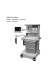
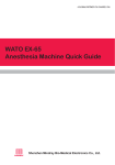
Download Mindray Wato EX-65 Anaesthesia Machine
Transcript
WATO EX-65 Anesthesia Machine Operator’s Manual CE Marking The product bears CE mark indicating its conformity with the provisions of the Council Directive 93/42/EEC concerning medical devices and fulfils the essential requirements of Annex I of this directive. The product is in radio-interference protection Group I Class B in accordance with EN55011. The product complies with the requirement of standard EN60601-1-2 “Electromagnetic Compatibility – Medical Electrical Equipment”. Revision History This manual has a revision number. This revision number changes whenever the manual is updated due to software or technical specification change. Contents of this manual are subject to change without prior notice. Revision 1.0 is the initial release of the document. Revision number: 1.0 Release time: 2009-1 © Copyright 2009 Shenzhen Mindray Bio-Medical Electronics Co., Ltd. All rights reserved. WARNING z Federal Law (USA) restricts this device to sale by or on the order of a physician. I Intellectual Property Statement SHENZHEN MINDRAY BIO-MEDICAL ELECTRONICS CO., LTD. (hereinafter called Mindray) owns the intellectual property rights to this product and this manual. This manual may refer to information protected by copyrights or patents and does not convey any license under the patent rights of Mindray, nor the rights of others. Mindray intends to maintain the contents of this manual as confidential information. Disclosure of the information in this manual in any manner whatsoever without the written permission of Mindray is strictly forbidden. Release, amendment, reproduction, distribution, rental, adaption and translation of this manual in any manner whatsoever without the written permission of Mindray is strictly forbidden , and WATO are the registered trademarks or trademarks owned by Mindray in China and other countries. All other trademarks that appear in this manual are used only for editorial purposes without the intention of improperly using them. They are the property of their respective owners. Contents of this manual are subject to changes without prior notice. II Manufacturer’s Responsibility All information contained in this manual is believed to be correct. Mindray shall not be liable for errors contained herein nor for incidental or consequential damages in connection with the furnishing, performance, or use of this manual. Mindray is responsible for the effects on safety, reliability and performance of this product only if: all installation operations, expansions, changes, modifications and repairs of this product are conducted by Mindray authorized personnel; and the electrical installation of the relevant room complies with the applicable national and local requirements; and the product is used in accordance with the instructions for use. Warranty This warranty is exclusive and is in lieu of all other warranties, expressed or implied, including warranties of merchantability or fitness for any particular purpose. Exemptions Mindray's obligation or liability under this warranty does not include any transportation or other charges or liability for direct, indirect or consequential damages or delay resulting from the improper use or application of the product or the use of parts or accessories not approved by Mindray or repairs by people other than Mindray authorized personnel. This warranty shall not extend to Any Mindray product which has been subjected to misuse, negligence or accident; or Any Mindray product from which Mindray's original serial number tag or product identification markings have been altered or removed; or Any product of any other manufacturer. III Return Policy In the event that it becomes necessary to return a unit to Mindray, follow the instructions below. 1. Return authorization. Contact the Customer Service Department and obtain a Customer Service Authorization number. This number must appear on the outside of the shipping container. Returned shipments will not be accepted if the number is not clearly visible. Please provide the model number, serial number, and a brief description of the reason for return. 2. Freight policy The customer is responsible for freight charges when this product is shipped to Mindray for service (this includes customs charges). 3. Return address Please send the part(s) or equipment to the address offered by the Customer Service Department. Contact Information Manufacturer: Shenzhen Mindray Bio-Medical Electronics Co., Ltd. Address: Mindray Building, Keji 12th Road South, Hi-tech Industrial Park, Nanshan, Shenzhen 518057 P.R. China Tel: +86 755 26582479 +86 755 26582888 Fax: +86 755 26582934 +86 755 26582500 Website: www.mindray.com EC-Representative: Shanghai International Holding Corp. GmbH (Europe) Address: Eiffestraße 80, Hamburg 20537, Germany Tel: 0049-40-2513175 Fax: 0049-40-255726 IV Preface Manual Purpose This manual contains the instructions necessary to operate the product safely and in accordance with its function and intended use. Observance of this manual is a prerequisite for proper product performance and correct operation and ensures patient and operator safety. This manual is based on the maximum configuration and therefore some contents may not apply to your product. If you have any question, please contact us. This manual is an integral part of the product. It should always be kept close to the equipment so that it can be obtained conveniently when needed. Intended Audience This manual is geared for clinical professionals who are expected to have a working knowledge of medical procedures, practices and terminology as required for monitoring of critically ill patients. Illustrations All illustrations in this manual serve as examples only. They may not necessarily reflect the setup or data displayed on your anesthesia machine. Conventions Italic text is used in this manual to quote the referenced chapters or sections. [ ] is used to enclose screen texts. → is used to indicate operational procedures. V FOR YOUR NOTES VI Contents 1 Safety ................................................................................................................................. 1-1 1.1 Safety Information .......................................................................................................... 1-1 1.1.1 Dangers .............................................................................................................. 1-2 1.1.2 Warnings............................................................................................................. 1-2 1.1.3 Cautions ............................................................................................................. 1-3 1.1.4 Notes .................................................................................................................. 1-4 1.2 Equipment Symbols ........................................................................................................ 1-5 2 The Basics ......................................................................................................................... 2-1 2.1 System Description ......................................................................................................... 2-1 2.1.1 Intended Use....................................................................................................... 2-1 2.1.2 Contraindications ............................................................................................... 2-1 2.1.3 Components ....................................................................................................... 2-2 2.2 Equipment Appearance ................................................................................................... 2-3 2.2.1 Front View.......................................................................................................... 2-3 2.2.2 Rear View........................................................................................................... 2-7 2.3 Batteries ........................................................................................................................ 2-12 3 System Controls and Basic Settings................................................................................ 3-1 3.1 Display Control ............................................................................................................... 3-1 3.2 Display Screen ................................................................................................................ 3-3 3.3 Basic Settings.................................................................................................................. 3-5 3.3.1 Adjust Screen Brightness ................................................................................... 3-5 3.3.2 Adjust Sound Volume......................................................................................... 3-5 3.3.3 Set System Time................................................................................................. 3-6 3.3.4 Set Language...................................................................................................... 3-6 3.3.5 Set Unit .............................................................................................................. 3-6 3.3.6 Restore Default Configurations.......................................................................... 3-6 3.3.7 Set the IP Address of Anesthesia Information System (CIS) ............................. 3-7 4 Operations and Ventilation Setup................................................................................... 4-1 4.1 Turn on the System ......................................................................................................... 4-1 4.2 Turn off the System......................................................................................................... 4-1 4.3 Input Fresh Gas ............................................................................................................... 4-2 4.3.1 Set O2, N2O and Air Inputs ................................................................................. 4-2 4.3.2 Set Anesthetic Agent .......................................................................................... 4-3 4.4 Set Ventilation Mode....................................................................................................... 4-4 4.4.1 Set Manual Ventilation Mode............................................................................. 4-4 4.4.2 Make Settings before Starting Mechanical Ventilation Mode............................ 4-5 1 4.4.3 Volume Control Ventilation (VCV).................................................................... 4-5 4.4.4 Pressure Control Ventilation (PCV) ................................................................... 4-8 4.4.5 Synchronized Intermittent Mandatory Ventilation (SIMV)...............................4-11 4.4.6 Pressure Support Ventilation (PSV) ................................................................. 4-17 4.5 Start Mechanical Ventilation ......................................................................................... 4-21 4.6 Set the Timer ................................................................................................................. 4-22 4.6.1 Start the Timer.................................................................................................. 4-22 4.6.2 Stop the Timer .................................................................................................. 4-22 4.6.3 Reset the Timer ................................................................................................ 4-22 4.7 Stop Mechanical Ventilation ......................................................................................... 4-23 5 User Interface and Parameter Monitoring .................................................................... 5-1 5.1 Screen Layout ................................................................................................................. 5-1 5.1.1 Standby Screen................................................................................................... 5-2 5.1.2 Normal Screen.................................................................................................... 5-3 5.1.3 Special Screen .................................................................................................... 5-4 5.2 Screen Setup.................................................................................................................... 5-5 5.3 Parameter Monitoring ..................................................................................................... 5-5 5.3.1 O2 Concentration Monitoring ............................................................................ 5-5 5.3.2 Anesthetic Agent (AA) Concentration Monitoring ............................................ 5-7 5.3.3 CO2 Concentration Monitoring ......................................................................... 5-8 5.3.4 Pressure Monitoring ........................................................................................... 5-9 5.3.5 Tidal Volume Monitoring ................................................................................. 5-10 5.3.6 Tidal Volume Compensation ............................................................................ 5-12 5.3.7 Volume Monitoring .......................................................................................... 5-13 5.3.8 Breath Rate Monitoring.................................................................................... 5-13 5.3.9 BIS Monitoring ................................................................................................ 5-14 5.4 Display Electronic Flowmeter....................................................................................... 5-16 5.5 Spirometry Loop ........................................................................................................... 5-16 6 Preoperative Test.............................................................................................................. 6-1 6.1 Preoperative Test Schedules............................................................................................ 6-1 6.1.1 Test Intervals ...................................................................................................... 6-1 6.2 Inspect the System .......................................................................................................... 6-2 6.3 Power Failure Alarm Test................................................................................................ 6-2 6.4 Pipeline Tests .................................................................................................................. 6-3 6.4.1 O2 Pipeline Test ................................................................................................. 6-3 6.4.2 N2O Pipeline Test .............................................................................................. 6-4 6.4.3 Air Pipeline Test ................................................................................................. 6-4 6.5 Cylinder Tests.................................................................................................................. 6-4 6.5.1 Check the Cylinder in Full Status....................................................................... 6-4 6.5.2 O2 Cylinder High Pressure Leak Test ................................................................ 6-5 6.5.3 N2O Cylinder High Pressure Leak Test ............................................................. 6-5 6.6 Flow Control System Tests ............................................................................................. 6-5 2 6.6.1 Without O2 Sensor ............................................................................................. 6-5 6.6.2 With O2 Sensor .................................................................................................. 6-7 6.7 Vaporizer Back Pressure Test .......................................................................................... 6-8 6.8 Breathing System Tests ................................................................................................... 6-9 6.8.1 Bellows Test ....................................................................................................... 6-9 6.8.2 Breathing System Leak Test in Mechanical Ventilation Status ........................ 6-10 6.8.3 Breathing System Leak Test in Manual Ventilation Status................................6-11 6.8.4 APL Valve Test ..................................................................................................6-11 6.9 Alarm Tests.................................................................................................................... 6-12 6.9.1 Prepare for Alarm Tests.................................................................................... 6-12 6.9.2 Test the O2 Concentration Monitoring and Alarms.......................................... 6-13 6.9.3 Test the Low Minute Volume Alarm ................................................................ 6-13 6.9.4 Test the Apnea Alarm ....................................................................................... 6-14 6.9.5 Test the Sustained Airway Pressure Alarm....................................................... 6-14 6.9.6 Test the High Paw Alarm.................................................................................. 6-14 6.9.7 Test the Low Paw Alarm .................................................................................. 6-15 6.9.8 Test the AG Module Alarm .............................................................................. 6-15 6.10 Preoperative Preparations............................................................................................ 6-15 6.11 Inspect the AGSS ........................................................................................................ 6-16 7 User Maintenance............................................................................................................. 7-1 7.1 Repair Policy................................................................................................................... 7-1 7.2 Maintenance Schedule .................................................................................................... 7-2 7.3 Breathing System Maintenance....................................................................................... 7-3 7.4 Flow Sensor Calibration.................................................................................................. 7-3 7.5 O2 Sensor Calibration ..................................................................................................... 7-5 7.5.1 21% O2 Calibration............................................................................................ 7-5 7.5.2 100% O2 Calibration.......................................................................................... 7-6 7.6 Water Build-up in the Flow Sensor ................................................................................. 7-7 7.6.1 Prevent Water Build-up ...................................................................................... 7-7 7.6.2 Clear Water Build-up.......................................................................................... 7-8 7.7 Airway Pressure Gauge Zeroing ..................................................................................... 7-8 7.8 AGSS Transfer Tube Maintenance................................................................................ 7-10 8 CO2 Monitoring ............................................................................................................... 8-1 8.1 Introduction..................................................................................................................... 8-1 8.2 Identify CO2 Module ...................................................................................................... 8-2 8.3 Use a Sidestream CO2 Module ....................................................................................... 8-3 8.3.1 Prepare to Measure CO2 .................................................................................... 8-3 8.3.2 Make CO2 Settings ............................................................................................ 8-4 8.3.3 Measurement Limitations................................................................................... 8-6 8.3.4 Troubleshooting.................................................................................................. 8-6 8.3.5 Scavenge the Sample Gas .................................................................................. 8-7 8.3.6 Zero the Sensor .................................................................................................. 8-7 3 8.3.7 Calibrate the Sensor ........................................................................................... 8-7 8.4 Use a Microstream CO2 Module .................................................................................... 8-8 8.4.1 Prepare to Measure CO2 .................................................................................... 8-8 8.4.2 Make CO2 Settings ............................................................................................ 8-8 8.4.3 Measurement Limitations..................................................................................8-11 8.4.4 Scavenge the Sample Gas .................................................................................8-11 8.4.5 Zero the Sensor ................................................................................................ 8-12 8.4.6 Calibrate the Sensor ......................................................................................... 8-12 8.4.7 Oridion Information ......................................................................................... 8-12 8.5 Use a Mainstream CO2 Module.................................................................................... 8-13 8.5.1 Prepare to Measure CO2 .................................................................................. 8-13 8.5.2 Make CO2 Settings .......................................................................................... 8-14 8.5.3 Measurement Limitations................................................................................. 8-16 8.5.4 Zero the Sensor ................................................................................................ 8-16 8.5.5 Calibrate the Sensor ......................................................................................... 8-16 9 AG and O2 Concentration Monitoring .......................................................................... 9-1 9.1 Introduction..................................................................................................................... 9-1 9.2 Understand MAC Values................................................................................................. 9-2 9.3 Identify AG Modules....................................................................................................... 9-3 9.4 Prepare to Measure AG ................................................................................................... 9-3 9.5 Make AG Settings ........................................................................................................... 9-5 9.5.1 Set Anesthetic Agent .......................................................................................... 9-5 9.5.2 Set Pump Rate .................................................................................................... 9-5 9.5.3 Set O2 Compensation......................................................................................... 9-5 9.5.4 Set Working Mode.............................................................................................. 9-5 9.5.5 Set CO2 Unit ...................................................................................................... 9-6 9.5.6 Restore Defaults ................................................................................................. 9-6 9.5.7 Set CO2 Waveform ............................................................................................ 9-6 9.6 Change Anesthetic Agent ................................................................................................ 9-6 9.7 Measurement Limitations................................................................................................ 9-7 9.8 Troubleshooting .............................................................................................................. 9-7 9.9 Scavenge the Sample Gas ............................................................................................... 9-8 9.10 Calibrate the AG Module .............................................................................................. 9-8 10 BIS Monitoring............................................................................................................. 10-1 10.1 Introduction................................................................................................................. 10-1 10.2 Identify the BIS Module.............................................................................................. 10-1 10.3 Safety Information ...................................................................................................... 10-2 10.4 Understand BIS Parameters ........................................................................................ 10-3 10.5 Prepare to Measure BIS .............................................................................................. 10-4 10.6 Continuous Impedance Check..................................................................................... 10-5 10.7 Cyclic Impedance Check............................................................................................. 10-6 10.8 BIS Sensor Check Window ......................................................................................... 10-6 4 10.9 Set BIS Smoothing Rate.............................................................................................. 10-7 10.10 Restore Defaults ........................................................................................................ 10-7 10.11 Set BIS Related Waveforms ...................................................................................... 10-8 11 Alarms ............................................................................................................................11-1 11.1 Introduction ..................................................................................................................11-1 11.1.1 Alarm Categories.............................................................................................11-1 11.1.2 Alarm Levels ...................................................................................................11-2 11.2 Alarm Indicators...........................................................................................................11-2 11.2.1 Alarm Lamp.....................................................................................................11-2 11.2.2 Audible Alarm Tones.......................................................................................11-3 11.2.3 Alarm Message................................................................................................11-3 11.2.4 Flashing Alarm Numeric .................................................................................11-3 11.2.5 Alarm Status Symbols .....................................................................................11-3 11.3 Set Alarm Volume ........................................................................................................11-4 11.4 Set Alarm Limits ..........................................................................................................11-4 11.4.1 Set Ventilator Alarm Limits.............................................................................11-4 11.4.2 Set CO2 Alarm Limits.....................................................................................11-4 11.4.3 Set AG Alarm Limits.......................................................................................11-5 11.4.4 Set BIS Alarm Limits ......................................................................................11-5 11.5 Set Alarm Level............................................................................................................11-5 11.6 Set Cardiopulmonary Bypass (CPB) Alarm .................................................................11-5 11.7 Set MV&TVe Alarm.....................................................................................................11-6 11.8 Set Apnea Alarm...........................................................................................................11-6 11.9 Alarm Silence ...............................................................................................................11-7 11.9.1 Set 120 s Alarm Silence...................................................................................11-7 11.9.2 Cancel 120 s Alarm Silence.............................................................................11-7 11.10 When an Alarm Occurs ..............................................................................................11-7 12 Trend and Logbook ...................................................................................................... 12-1 12.1 Trend Graph ................................................................................................................ 12-1 12.2 Trend Table.................................................................................................................. 12-2 12.3 Alarm Logbook ........................................................................................................... 12-3 13 Installations and Connections ..................................................................................... 13-1 13.1 Install the Breathing System ....................................................................................... 13-1 13.1.1 Breathing System Diagrams........................................................................... 13-2 13.1.2 Circuit Adapter Diagram ................................................................................ 13-3 13.1.3 Install the Breathing system ........................................................................... 13-4 13.1.4 Install the Bag Arm ........................................................................................ 13-6 13.1.5 Install the Bellows.......................................................................................... 13-7 13.1.6 Install the Flow sensor.................................................................................... 13-9 13.1.7 Install the O2 Sensor .................................................................................... 13-10 13.1.8 Install the Sodalime Canister........................................................................ 13-12 5 13.2 Install the Breathing Tubes........................................................................................ 13-19 13.3 Install the Manual Bag .............................................................................................. 13-20 13.4 Install the Vaporizer .................................................................................................. 13-21 13.4.1 Assemble the Vaporizer ................................................................................ 13-21 13.4.2 Fill the Vaporizer.......................................................................................... 13-25 13.4.3 Drain the Vaporizer ...................................................................................... 13-27 13.5 Install/Replace the Gas Cylinder............................................................................... 13-29 13.6 Install Modules.......................................................................................................... 13-31 13.6.1 Install the CO2 Module ................................................................................ 13-31 13.6.2 Install the AG Module .................................................................................. 13-31 13.6.3 Install the BIS Module ................................................................................. 13-32 13.7 Pneumatic Connectors............................................................................................... 13-32 13.7.1 Connect the Pipeline Gas Supplies............................................................... 13-33 13.7.2 Install the Gas Cylinder................................................................................ 13-34 13.8 CIS Connector........................................................................................................... 13-34 13.9 Scavenging ................................................................................................................ 13-34 13.10 AGSS Transfer and Receiving System.................................................................... 13-35 13.10.1 Components................................................................................................ 13-35 13.10.2 Assemble the AGSS ................................................................................... 13-36 13.10.3 Waste Gas Disposal System ....................................................................... 13-37 14 Cleaning and Disinfection............................................................................................ 14-1 14.1 Clean and Disinfect the Anesthesia Machine Housing................................................ 14-2 14.2 Disassemble the Breathing System Cleanable Parts ................................................... 14-2 14.2.1 O2 Sensor ....................................................................................................... 14-3 14.2.2 Manual Bag .................................................................................................... 14-4 14.2.3 Breathing Tubes ............................................................................................. 14-5 14.2.4 Airway Pressure Gauge .................................................................................. 14-6 14.2.5 Bag Arm ......................................................................................................... 14-6 14.2.6 Bellows Assembly .......................................................................................... 14-7 14.2.7 Flow Sensor.................................................................................................... 14-8 14.2.8 Expiratory Check Valve Assembly................................................................. 14-9 14.2.9 Inspiratory Check Valve Assembly ................................................................ 14-9 14.2.10 Sodalime Canister ...................................................................................... 14-10 14.2.11 Water Collection Cup ..................................................................................14-11 14.2.12 Breathing system........................................................................................ 14-12 14.2.13 AGSS Transfer and Receiving System....................................................... 14-13 14.3 Clean&Disinfect and Re-install the Breathing System ............................................. 14-15 14.3.1 Breathing system.......................................................................................... 14-17 14.3.2 Water Collection Cup ................................................................................... 14-17 14.3.3 Manual Bag .................................................................................................. 14-17 14.3.4 Breathing Mask ............................................................................................ 14-18 14.3.5 Inspiratory and Expiratory Check Valves Assembly .................................... 14-18 14.3.6 Bellows Assembly ........................................................................................ 14-18 6 14.3.7 Sodalime Canister ........................................................................................ 14-19 14.3.8 Breathing Tubes and Y Piece........................................................................ 14-20 14.3.9 Flow Sensor.................................................................................................. 14-20 14.3.10 O2 Sensor ................................................................................................... 14-21 14.3.11 AGSS Transfer and Receiving System ....................................................... 14-21 15 Accessories .................................................................................................................... 15-1 A Theory of Operation....................................................................................................... A-1 A.1 Pneumatic Circuit System ............................................................................................. A-1 A.2 Electrical System Structure ........................................................................................... A-4 B Product Specifications.....................................................................................................B-1 B.1 Safety Specifications ......................................................................................................B-1 B.2 Environmental Specifications.........................................................................................B-2 B.3 Power Requirements.......................................................................................................B-2 B.4 Physical Specifications...................................................................................................B-3 B.5 Pneumatic Circuit System Specifications.......................................................................B-4 B.6 Breathing System Specifications ....................................................................................B-5 B.7 Ventilator Specifications.................................................................................................B-7 B.8 Ventilator Accuracy ........................................................................................................B-9 B.9 Anesthetic vaporizer .....................................................................................................B-10 B.10 AGSS Transfer and Receiving System Specifications................................................B-10 B.11 O2 Sensor Specifications............................................................................................B-11 B.12 CO2 Module Specifications .......................................................................................B-14 B.13 AG Module Specifications .........................................................................................B-17 B.14 BIS Module Specifications.........................................................................................B-21 C EMC ................................................................................................................................ C-1 D Alarm Messages.............................................................................................................. D-1 D.1 Physiological Alarm Messages...................................................................................... D-1 D.2 Technical Alarm Messages ............................................................................................ D-4 E Symbols and Abbreviations ............................................................................................E-1 E.1 Symbols .......................................................................................................................... E-1 E.2 Abbreviations.................................................................................................................. E-3 F Factory Defaults............................................................................................................... F-1 F.1 CO2 Module.................................................................................................................... F-1 F.2 AG Module...................................................................................................................... F-2 F.3 BIS Module ..................................................................................................................... F-3 F.4 Ventilator ......................................................................................................................... F-4 7 FOR YOUR NOTES 8 1 Safety 1.1 Safety Information DANGER z Indicates an imminent hazard that, if not avoided, will result in death or serious injury. WARNING z Indicates a potential hazard or unsafe practice that, if not avoided, could result in death or serious injury. CAUTION z Indicates a potential hazard or unsafe practice that, if not avoided, could result in minor personal injury or product/property damage. NOTE z Provides application tips or other useful information to ensure that you get the most from your product. 1-1 1.1.1 Dangers There are no dangers that refer to the product in general. Specific “Danger” statements may be given in the respective sections of this manual. 1.1.2 Warnings WARNING z Before putting the system into operation, the operator must verify that the equipment, connecting cables and accessories are in correct working order and operating condition. z The equipment must be connected to a properly installed power outlet with protective earth contacts only. If the installation does not provide for a protective earth conductor, disconnect it from the power line. z Use AC power source before the batteries are depleted. z To avoid explosion hazard, do not use the equipment in the presence of flammable anesthetic agent, vapors or liquids. z Do not open the equipment housings. All servicing and future upgrades must be carried out by the personnel trained and authorized by us only. z Do not rely exclusively on the audible alarm system for patient monitoring. Adjustment of alarm volume to a low level may result in a hazard to the patient. Remember that alarm settings should be customized according to different patient situations and always keeping the patient under close surveillance is the most reliable way for safe patient monitoring. z The physiological parameters and alarm messages displayed on the screen of the equipment are for doctor’s reference only and cannot be directly used as the basis for clinical treatment. z Dispose of the package material, observing the applicable waste control regulations and keeping it out of children’s reach. z To avoid explosion hazard, do not use flammable anesthetic agent such as ether and cyclopropane for this equipment. Only non-flammable anesthetic agents which meet the requirements specified in IEC 60601-2-13 can be applied to this equipment. This anesthesia machine can be used with halothane, enflurane, isoflurane, sevoflurane and desflurane. Only one of the five anesthetic agents can be used at a time. z Do not touch the patient, table, or instruments during defibrillation. 1-2 WARNING z Use appropriate electrodes and place them according to the instructions provided by the manufacturer. The display restores to normal within 10 seconds after defibrillation. 1.1.3 Cautions CAUTION z To ensure patient safety, use only parts and accessories specified in this manual. z At the end of its service life, the equipment, as well as its accessories, must be disposed of in compliance with the guidelines regulating the disposal of such products. z Magnetic and electrical fields are capable of interfering with the proper performance of the equipment. For this reason make sure that all external devices operated in the vicinity of the equipment comply with the relevant EMC requirements. Mobile phone, X-ray equipment or MRI devices are a possible source of interference as they may emit higher levels of electromagnetic radiation. z This system operates correctly at the electrical interference levels identified in this manual. Higher levels can cause nuisance alarms that may stop mechanical ventilation. Pay attention to false alarms caused by high-intensity electrical fields. z Before connecting the equipment to the power line, check that the voltage and frequency ratings of the power line are the same as tube indicated on the equipment’s label or in this manual. z Always install or carry the equipment properly to avoid damage caused by drop, impact, strong vibration or other mechanical force. z The anesthesia machine keeps stable with a 10º tilt in typical configuration. Do not hang articles on both sides of the anesthesia machine for fear of getting tilted. 1-3 1.1.4 Notes NOTE z Put the equipment in a location where you can easily see the screen and access the operating controls. z Keep this manual close to the equipment so that it can be obtained conveniently when needed. z The software was developed in compliance with IEC 60601-1-4. The possibility of hazards arising from software errors is minimized. z This manual describes all features and options. Your equipment may not have all of them. 1-4 1.2 Equipment Symbols Attention: Consult accompanying documents (this manual) Dangerous voltage Alternating current Fuse Battery Equipotential Operating state Autoclavable Material description Not autoclavable Power On Power Off Reset Standby Alarm silence key MV&TVe alarm key Normal screen key O2 flush button ACGO On ACGO Off Bag position/ manual ventilation Mechanical ventilation Lock Unlock Network connector Flow control USB connector O2 sensor connector Air supply connector N2O supply connector Upward (Pop-off valve) Sample gas return port (to the AGSS) 1-5 VGA connector O2 supply connector Table top light AGSS outlet Cylinder PEEP outlet Manufacture date Vaporizer Manufacturer Isolation transformer Serial number European community representative APL valve CAUTION HOT Maximum level of the sodalime canister Lock or unlock as the arrow shows Gas input direction Unlock the lifting device Lock the lifting device Do Not Crush Approximate Please align! Max. weight: 11.3 kg Pipeline Max. weight: 30 kg CE marking Type BF applied part. Defibrillation-proof protection against electric shock. The anesthesia machine is driven by Air. The following definition of the WEEE label applies to EU member states only. This symbol indicates that this product should not be treated as household waste. By ensuring that this product is disposed of correctly, you will help prevent bringing potential negative consequences to the environment and human health. For more detailed information with regard to returning and recycling this product, please consult the distributor from whom you purchased it. * For system products, this label may be attached to the main unit only. 1-6 2 The Basics 2.1 System Description 2.1.1 Intended Use The anesthesia machine is intended to provide breathing anesthesia for adult, pediatric and infant patients during surgery. The anesthesia machine must only be operated by qualified anesthesia personnel who have received adequate training in its use. WARNING z This anesthesia machine is intended for use by qualified anesthesia personnel only or under their guidance. Anyone unauthorized or untrained must not perform any operation on it. z This anesthesia machine is not suitable for use in an MRI environment. 2.1.2 Contraindications The anesthesia machine is contraindicated for use on patients who suffer pneumothorax or severe pulmonary incompetence. 2-1 2.1.3 Components The anesthesia machine consists of a main unit, vaporizer (five optional anesthetic agents: enflurane, isoflurane, sevoflurane, desflurane and halothane), anesthetic ventilator, electronic flowmeter assembly, breathing system etc. The anesthesia machine provides monitoring and displaying of respiratory mechanics (RM) parameters (airway resistance and compliance) and spirometry loops as well. It is configured with the following ventilation modes: volume control ventilation (VCV), pressure control ventilation (PCV), pressure support ventilation (PSV), synchronized intermittent mandatory ventilation—volume control (SIMV-VC) and synchronized intermittent mandatory ventilation—pressure control (SIMV-PC). The anesthesia machine can be externally connected to a patient monitor which is in compliance with the requirements of relevant international standard and can be configured with anesthesia information system (CIS). The anesthesia machine features the following: Automatic leak detection Breathing system gas leak compensation and automatic compliance compensation Cylinder and pipeline connections available for gas supplies Electronic flowmeter and electronic PEEP Timer which counts the duration between the start and end of an operation Table top light Information displayed in big numerics User-adjustable display screen Alarm events storage and review, fault status and maintenance information recording Auxiliary O2 supply and active anesthesia gas scavenging system (AGSS) N2O cut-off Modular AG, CO2 and BIS modules Sample gas return to the AGSS Setting CPB alarm mode 2-2 2.2 Equipment Appearance 2.2.1 Front View ——Display and control panel 2-3 1. Brake 2. Pipeline pressure gauge (s) Displays the pipeline pressure or the cylinder pressure after relief. 3. Total flowmeter The medium level of flowtube float indicates the current flow of the mixed gas. 4 Flow control (s) When the system switch is set to the ON position: 5 Turn the control counterclockwise to increase the gas flow. Turn the control clockwise to decrease the gas flow. Electronic flowmeter Displays the current flow of the corresponding gas. 6. Ventilator control panel 7. Control knob 8. Display 9. Vaporizer A. Concentration control Push and turn the concentration control to set the concentration of anesthetic agent. B. Locking lever Turn the locking lever clockwise to lock the vaporizer in position. 10. Gas supply connector (s) O2 , N2O and AIR connectors are provided. 11. System switch Set the switch to the position to enable gas flow and to turn on the system. Set the switch to the position to disable gas flow and to turn off the system. 12. Cylinder pressure gauge (s) High-pressure pressure gauge (s) that displays cylinder pressure before relief. 13. O2 flush button Push to supply high flows of O2 to the breathing system. 14. Auxiliary electrical outlet Three auxiliary electrical outlets are provided when the anesthesia machine is configured with an isolation transformer. 15 Drawer lock 16. Worktable (with drawer) 2-4 ——Breathing system 2-5 1. O2 sensor connector 2. Inspiration connector 3. Expiration connector 4. Inspiratory check valve 5. Expiratory check valve 6. Bellows housing 7. Sample gas return port (to the AGSS) 8. Manual bag port 9. Bag/mechanical ventilation switch Select the position to use bag for manual ventilation. Select the position to use ventilator for mechanical ventilation. 10. APL (airway pressure limit) valve Adjusts breathing system pressure limit during manual ventilation. The scale shows approximate pressures. Above 30 cmH2O, you will feel clicks as the v turns. Turn clockwise to increase. 11. O2 sensor connector 12. Rotary handle 13. Sodalime canister The sodalime inside the canister absorbs the CO2 the patient exhales, which enables cyclic use of the patient exhaled gas. 2-6 2.2.2 Rear View ——Power supply 2-7 1. Cylinder connector (s) 2. Equipotential stud 3. Fan 4. Mains inlet 5. Network connector 6. CIS 12 V power supply connector 7. Speaker 8. Auxiliary O2 supply 9. ACGO (Auxiliary Common Gas Outlet) switch Set the switch to the position to stop mechanical ventilation. Then, fresh gas is sent to the externally connected manual breathing system through the ACGO outlet and the technical alarm of [ACGO On] is triggered. The system monitors airway pressure and O2 concentration instead of volume. Set the switch to the position to apply mechanical or manual ventilation to the patient through the breathing system. 10. Module slot CO2, AG and BIS modules mentioned in this manual can be inserted into the slot and identified. The CO2 and AG modules cannot be used simultaneously. 11. AGSS outlet 12. AGSS Transfer and Receiving System 2-8 ——Anesthesia information system (CIS) 2-9 This rear view is based on the situation that the anesthesia machine is configured with anesthesia information system (CIS). 1. Display 2. Rail 3. Mounting bracket 4. Keyboard 5. CIS main unit A B C F A. E D C Reset key : Press to restart the CIS. B. CIS switch : Press to switch on/off the CIS. C. USB connector D. Network connector E. Electrical outlet F. Display connector 2-10 WARNING z Connect to the AC mains in compliance with B.3 Power Requirements. Failure to do so may cause damage to the equipment or affect its normal operation. z Make sure that the jacket on the electrical outlet is already fixed to avoid power cord off during surgery. NOTE z If the equipment cannot be powered by the AC mains, check if the fuse inside the electrical outlet is normal. If AC mains supply still fails after the fuse is replaced, contact the service personnel. z When the auxiliary electrical outlet does not work normally, check if the corresponding fuse is burned. z Equipment connected to the auxiliary electrical outlet shall be authorized. Otherwise, leakage current above the allowable limit will result, which may endanger the patient or operator, and damage the anesthesia machine or externally connected equipment. When the anesthesia machine is configured with only one auxiliary electrical outlet, this electrical outlet is only used for connecting the adapter for Desflurane vaporizer. When the anesthesia machine is configured with multiple auxiliary electrical outlets, the equipment connected shall comply with the voltage and current specifications of the auxiliary electrical outlets. z All analog or digital products connected to this system must be certified passing the specified IEC standards (such as IEC 60950 for data processing equipment and IEC 60601-1 for medical electrical equipment). All configurations shall comply with the valid version of IEC 60601-1-1. The personnel who are responsible for connecting the optional equipment to the I/O signal port shall be responsible for medical system configuration and system compliance with IEC 60601-1-1 as well. 2-11 2.3 Batteries NOTE z Use batteries at least once every month to extend their life. Charge the batteries before their capacities are worn out. z Inspect and replace batteries regularly. Battery life depends on how frequent and how long it is used. For a properly maintained and stored lithium battery, its life expectancy is approximately 3 years. For more aggressive use models, life expectancy can be shortened. We recommend replacing lithium batteries every 3 years. z The operating time of a battery depends on equipment configuration and operation. For example, starting module monitoring frequently will shorten the operating time of the batteries. z In case of battery failure, contact us or have your service personnel replace it. Do not replace the battery without permission. The anesthesia machine is designed to operate on battery power whenever AC power becomes interrupted. When the anesthesia machine is connected to the AC power source, the batteries are charged regardless of whether or not the anesthesia machine is currently on. In case of power failure, the anesthesia machine will automatically be powered by the internal batteries. When AC power source is restored within the specified time, power supply is switched from battery to AC automatically to ensure continuous system use. On-screen battery icon indicates the battery statuses as follows: : indicates that the batteries operate normally. The solid portion represents the current charge level of the batteries in proportion to its maximum charge level. : indicates low battery and the batteries need to be charged. : indicates too low battery and the batteries need to be charged immediately. The capacity of the internal battery is limited. If the battery capacity is too low, a high-level alarm will be triggered and the [Low Battery Voltage!] message displayed in the technical alarm area. In this case, apply AC power to the anesthesia machine. 2-12 3 System Controls and Basic Settings 3.1 Display Control 1. 2. Alarm lamp High level alarms: the lamp quickly flashes red. Medium level alarms: Low level alarms: the lamp turns yellow without flashing. the lamp slowly flashes yellow. Menu shortcut key(s) Push the menu shortcut key to access the corresponding menu. 3. Control knob Push the control knob to select a menu option or confirm your setting. Turn the control knob clockwise or counterclockwise to scroll through the menu options or change your settings. 3-1 4. 5. MV&TVe alarm key In case of manual ventilation mode: Push the key to switch off MV and TVe overrange alarms and apnea alarm. Push the key again to switch on MV and TVe overrange alarms and apnea alarm. In case of mechanical ventilation mode: Push the key to switch off MV and TVe overrange alarms. Push the key again to switch on MV and TVe overrange alarms. Normal screen key Push the key to close all menus displayed. 6. Standby key Push the key to enter or exit standby mode. 7. Alarm silence key To set alarm silence state, push this key to enter 120 s alarm silenced status. The alarm silence symbol and 120 s countdown time appear in the upper right corner of the screen. 8. 9. To clear alarm silence, push this key again. Operating state LED On: when the anesthesia machine is operating. Off: when the anesthesia machine is turned off. AC power LED On: when the anesthesia machine is connected to the AC power source. Off: when the anesthesia machine is not connected to the AC power source. 10. Battery LED On: when the anesthesia machine is equipped with batteries and is connected to the AC power source, and the batteries are being charged. Off: when the anesthesia machine is not equipped with batteries or is switched off. Flash: when the anesthesia machine is being battery powered. 11. Ventilator parameter setup shortcut key(s) Push the parameter setup shortcut key to change the corresponding setting. Turn the control knob to change the specific setting and push the control knob to activate the selected setting. 12. Display screen Refer to 3.2Display Screen for details. 3-2 3.2 Display Screen This anesthesia machine adopts a high-resolution color TFT LCD to display various parameters and graphs, such as ventilation parameters and pressure/flow/volume waveforms. Depending on how your anesthesia machine is configured, it may display gas module parameters and waveforms, BIS parameters, BIS trend waveform, spirometry loops etc.The following is a standard display screen. For descriptions of other screens, refer to5 User Interface and Parameter Monitoring. 1 2 3 4 6 5 7 8 9 10 11 12 18 13 14 15 17 16 1. Ventilation mode prompt area Displays the current ventilation mode. If manual ventilation is selected for the bag/mechanical ventilation switch, is displayed in this area. If mechanical ventilation is selected for the bag/mechanical ventilation switch, the currently selected mechanical ventilation mode is displayed. 2. Lung icon area is displayed when SIMV-VC or SIMV-PC mode is selected and The icon inspiration triggering is performed currently. 3. MV&TVe alarm off icon Displays the MV&TVe alarm off icon when MV&TVe alarm is switched off. 3-3 4. Physiological alarm area Displays physiological alarm messages. 5. Apnea alarm off icon area Displays apnea alarm off icon when apnea alarm is switched off in non-mechanical ventilation mode. 6. Alarm silence icon area Displays alarm silence icon and 120 s countdown time. 7. System time area Displays system time of the anesthesia machine. 8. Technical alarm area Displays technical alarm messages. When multiple alarms occur, they are displayed cyclically. 9. Power supply state icon area Displays power source or battery icon.The icon is displayed when the anesthesia machine is powered by AC power source. The battery icon is displayed when the anesthesia machine is battery powered to indicate battery capacity. For details, refer to2.3 Batteries. 10. [Vent Mode] shortcut key Used to select mechanical ventilation mode. 11. [Alarm Setup] shortcut key Used to change the alarm settings for the anesthetic ventilator, gas modules or BIS module. 12. [Screens] shortcut key Used to set user screen. 13. [User Setup] shortcut key Used to change the settings for TV compensation, O2 monitoring source, gas module, BIS module, screen, sound etc. 14. [Maintenance] shortcut key Used to perform leak test, calibrate O2 sensor and flow sensor, view trend graph, trend table and alarm logbook, and set language, system time, pressure unit, IP address etc. 15. Timer setup shortcut key Used to start, stop and reset the timer. 16. Parameter setup shortcut keys area Used to set the parameters related to the selected mechanical ventilation mode. The arrangement of the shortcut keys in this area varies depending on the selected mechanical ventilation mode. For details, refer to 4 Operations and Ventilation Setup. 17 System prompt message area 3-4 Displays information about system operating state. 18 Parameter&graph area Displays the parameters, waveforms, spirometry loops, or electronic flowmeter graphs which the anesthesia ventilator, gas module or BIS module monitors. Different types of screens are displayed based on the actual system configuration or screen layout settings. For details, refer to 5 User Interface and Parameter Monitoring. 3.3 Basic Settings This chapter covers only general settings of the anesthesia machine, such as language, screen brightness, system time etc. Parameter settings and other settings can be referred to in the respective sections. 3.3.1 Adjust Screen Brightness 1. Select the [User Setup] shortcut key and select [Screen and Audio Setup >>]. 2. Select [Screen Brightness] and select the appropriate value (ranging from 1 to 10) for screen brightness. The value 10 is for the brightest and 1 the least bright. If the anesthesia machine is battery powered, you can select less brightness to save battery capacity. 3.3.2 Adjust Sound Volume 3.3.2.1 Key Sound Volume 1. Select the [User Setup] shortcut key and select [Screen and Audio Setup >>]. 2. Select [Key Sound Volume] and select the appropriate value (ranging from 0 to 10) for key sound volume. The value 0 is for audio off and 10 for the loudest. 3.3.2.2 Alarm Sound Volume 1. Select the [User Setup] shortcut key and select [Screen and Audio Setup >>]. 2. Select [Alarm Sound Volume] and select the appropriate value (ranging from 1 to 10) for alarm sound volume. The value 1 is for the lowest and 10 for the loudest. 3-5 3.3.3 Set System Time 1. Select the [Maintenance] shortcut key → [User Maintenance >>] → [Set System Time >>]. 2. Set [Date] and [Time]. 3. Select [Date Format] and toggle between [YYYY-MM-DD], [MM-DD-YYYY] and [DD-MM-YYYY]. 4. Select [Time Format] and toggle between [24 h] and [12 h]. CAUTION z Changing date and time will affect the storage of trends and log information. It may also cause loss of data. 3.3.4 Set Language 1. Select the [Maintenance] shortcut key and select [User Maintenance >>]. 2. Select [Language] and select the desired language. 3. Restart the anesthesia machine to activate the language setting. 3.3.5 Set Unit 1. Select the [Maintenance] shortcut key and select [User Maintenance >>]. 2. Select [Paw Unit] and toggle between cmH2O, hPa and mbar. If the anesthesia machine is configured with CO2 or AG module, you can set the display units of FiCO2 and EtCO2. For details, refer to 8 CO2 Monitoring. 3.3.6 Restore Default Configurations 3.3.6.1 Restore the Factory Default Configuration of the Ventilator To restore the factory default configuration of the ventilator, do as follows: 1. Select the [Maintenance] shortcut key → [User Maintenance >>] → [Ventilator Defaults]. 3-6 2. Select [Ok] from the pop-up menu. After [Ok] is selected, the following settings restore their default values: User screen Ventilator parameters Alarm limits of ventilator-related parameters O2 monitoring source Alarm sound volume and key sound volume Screen brightness Paw display unit 3.3.6.2 Restore the Factory Default Configuration of the Gas Module If the anesthesia machine is configured with CO2 or AG module, you can directly restore the factory default configuration of the corresponding module. For details, refer to 8 CO2 Monitoring and 9 AG and O2 Concentration Monitoring. 3.3.6.3 Restore the Factory Default Configuration of the BIS Module If the anesthesia machine is configured with BIS module, you can directly restore the factory default configuration of the corresponding module. For details, refer to 10 BIS Monitoring. 3.3.7 Set the IP Address of Anesthesia Information System (CIS) To set the IP address of anesthesia information system (CIS), do as follows: 1. Select the [Maintenance] shortcut key → [User Maintenance >>] → [Set IP Address >>]. 2. In the [Set IP Address] menu, set the correct IP address of the CIS. 3. Select [Ok] to activate the IP address setting. 3-7 FOR YOUR NOTES 3-8 4 Operations and Ventilation Setup WARNING z Before using this anesthesia machine on the patient, make sure that the system is correctly connected and in good condition, and that all the tests described in 6 Preoperative Test are already completed. In case of test failure, do not use the system. Have a qualified service representative repair the system. 4.1 Turn on the System 1. Connect the power cord to the AC power source. Make sure that the AC power LED is illuminated. 2. Set the system switch to ON. Make sure that both the operating state LED and battery LED are illuminated (the battery is being charged or fully charged). 3. The alarm lamp flashes yellow and red once in turn and then a beep is given. 4. The display shows the start-up screen and then enters the standby screen after half a minute. WARNING z Do not use the anesthesia machine if it generates alarms during start-up or fails to operate normally. Contact your service personnel or us. 4.2 Turn off the System To turn off the system, do as follows: 1. Confirm that system use is finished. 2. Set the system switch to OFF. NOTE z For the first mechanical ventilation of each patient, do not exit the standby screen if mechanical ventilation related parameters are not set properly. Adjust fresh gas and anesthetic agent concentrations (if necessary) on the standby screen and set ventilation parameters properly based on the patient’s conditions before applying mechanical ventilation. 4-1 4.3 Input Fresh Gas 4.3.1 Set O2, N2O and Air Inputs 1. Connect the gas supplies correctly and ensure adequate gas pressure. 2. You can control the O2, N2O and Air flows in the fresh gas through the O2, N2O and Air flow controls. Readings of the gas flow can be seen on the respective electronic flowmeter. On the left hand of the electronic flowmeters is the total flowmeter showing the flow of the mixed gas. The O2 and N2O flow controls constitute a chain linkage: Turn the N2O flow control counterclockwise to increase the N2O flow to some extent. Then continuing turning the N2O flow control will cause the O2 flow control to turn counterclockwise together to increase the O2 flow, keeping the O2 concentration in the mixed gas above 25%. Turn the O2 flow control clockwise to decrease the O2 flow to some extent. Then continuing turning the O2 flow control will cause the N2O flow control to turn clockwise together to decrease the N2O flow, keeping the O2 concentration in the mixed gas above 25%. NOTE z This anesthesia machine can be used alone as a ventilator. You can adjust O2 concentration in the breathing system through the O2 flow control. z The O2 concentration in the fresh gas may be quite different from that in the breathing system. z The total flowmeter is calibrated based on 100% O2. The accuracy of the flowmeter may degrade with other gas or mixed gas. z When viewing the readings on the total flowmeter, keep your visual angle at the same level of the float. The reading of a same scale may vary when viewed at a different angle. z If the readings shown on the electronic flowmeters differ from that on the total flowmeter, the former shall prevail and the latter is an approximate value. 4-2 4.3.2 Set Anesthetic Agent NOTE z You do not need to perform this operation if inspiratory anesthetic agent is not used. z This anesthesia machine can be mounted with vaporizers corresponding with halothane, enflurane, isoflurane, sevoflurane and desflurane. Only one of the five vaporizers can be opened at a time because the vaporizers are featured with interlock. 4.3.2.1 Select the Desired Anesthetic Agent 1. Determine the anesthetic agent to be used and then fill the vaporizer. For details, refer to 13.4.2 Fill the Vaporizer. 2. Mount the vaporizer filled with anesthetic agent onto the anesthesia machine. For details, refer to 13.4 Install the Vaporizer. 4.3.2.2 Adjust the Concentration of Anesthetic Agent Push and turn the concentration control on the vaporizer to set the appropriate concentration of anesthetic agent. NOTE z Inspect the color of the sodalime in the canister before using the anesthetic agent. Replace the sodalime immediately if obvious color change is detected. z For details about how to use the anesthetic agent, refer to the Vaporizer Instructions for Use. 4-3 4.4 Set Ventilation Mode 4.4.1 Set Manual Ventilation Mode 1. Turn the APL valve control to adjust the pressure in the breathing system within the appropriate range. 2. Set the bag/mechanical ventilation switch to the position. The ventilation mode prompt area displays the icon for manual ventilation mode. Besides, the system prompt message area displays [Manual Vent.]. 3. Push the O2 flush button to inflate the bag if necessary. In the manual ventilation mode, you can use the APL valve to adjust the breathing system pressure limit and gas volume in the manual bag. When the pressure in the breathing system reaches the pressure limit set for the APL valve, the valve opens to release excess gas. The following figures show the Paw waveform and flow waveform in the manual ventilation mode. 4-4 NOTE z When using the anesthesia machine on the patient, make sure that manual ventilation mode is available. 4.4.2 Make Settings before Starting Mechanical Ventilation Mode 1. Make sure that the system is Standby. 2. Set the appropriate Plimit value in the parameter setup shortcut keys area. 3. Check the ACGO switch to make sure that it is OFF. 4. Set the bag/mechanical ventilation switch to the 5. If necessary, push the O2 flush button position. to inflate the bellows. NOTE z The default mechanical ventilation mode of the anesthesia machine is VCV. Other mechanical ventilation modes are optional. For the ventilation mode not configured for your anesthesia machine, operations of the corresponding menu options are disabled. 4.4.3 Volume Control Ventilation (VCV) 4.4.3.1 Description Volume control ventilation (hereinafter referred to as VCV) mode is a basic fully-mechanical ventilation mode. In the VCV mode, each time mechanical ventilation starts, gas is delivered to the patient at a constant flow, which reaches the preset TV within the gas delivery time. To ensure a certain amount of TV, the resulted airway pressure (Paw) changes based on patient pulmonary compliance and airway resistance. In the VCV mode, as long as Paw is less than Plimit and the gas delivery flow is kept constant, expirations starts immediately after Plimit is reached. In the VCV mode, you need to set [Plimit] to prevent high airway pressure from injuring the patient. In this mode, you can select to set [TIP :TI] to improve patient pulmonary gas distribution and [PEEP] to improve expiration of end-tidal carbon dioxide and to increase oxygenation of breathing process. 4-5 To ensure the set tidal volume gas delivery, the ventilator adjusts gas flow based on the measured inspiratory volume, dynamically compensates for the loss of tidal volume arising from breathing system compliance and system leakage and eliminates the effect of fresh gas as well. This is called tidal volume compensation. In the VCV mode, if tidal volume compensation is turned off or failed, the anesthesia machine can continue delivering gas stably but cannot compensate for the effects of fresh gas flow and breathing system compliance losses. 4.4.3.2 Waveforms The following figures show the Paw waveform and flow waveform in the VCV mode. Generally, in the VCV mode, the flow waveform is at a constant flow during inspiration and the Paw waveform rises in the same period. 4.4.3.3 Start VCV Mode 1. Select the [Vent Mode] shortcut key to open the [Vent Mode Setup] menu. 2. Select [VCV] in the [Vent Mode Setup] menu. 3. After confirming the selection, the[TV] shortcut key (the first key from the left in the parameter setup shortcut keys area) is highlighted. 4. Make sure that TV is appropriately set for the patient. Push the control knob to confirm the setting so as to start VCV mode. 4-6 NOTE z When it is necessary to switch over to VCV mode, confirm the setting of TV first. Otherwise, the system works in the previous ventilation mode. If the setting of TV is not confirmed for 10 s, the screen returns to the previous mode automatically. z Before activating a new mechanical ventilation mode, make sure that all related parameters are set appropriately. 4.4.3.4 Parameter Setup Shortcut Keys Area in VCV Mode When selection of VCV mode is confirmed, the parameter setup shortcut keys area at the bottom of the screen is automatically switched over to the parameter setup area in this mode. The following figure shows all related parameters to be set in VCV mode. 1. [TV]: Tidal volume 2. [Rate]: Breath rate 3. [I:E]: Ratio of inspiratory time to expiratory time 4. [TIP:TI]: Percentage of inspiratory plateau time in inspiratory time 5. [Plimit]: Pressure limit level 6 [PEEP]: Positive end-expiratory pressure 4.4.3.5 Set Parameters in VCV Mode You can use the shortcut keys and control knob to set the parameters in VCV mode. The following takes setting of TV as an example. 1. Select the [TV] shortcut key. 2. Push the control knob and turn it to set [TV] to the appropriate value. 3. Push the control knob to confirm the setting. 4. Set other parameters in this mode in the similar way. 4-7 NOTE z If the parameter value is adjusted outside of the range, the system prompt message area displays [Parameter Settings Outside the Safety Range]. z Confirm the adjustment of one parameter before adjusting another parameter. If you want to restore the value before adjustment, you have to reset the parameter value. 4.4.3.6 Parameter Range and Default Value in VCV Mode Parameter Range Step Default 20 to 100 ml: 5 ml TV 20 to 1500 ml 100 to 300 ml: 10 ml 500 ml 300 to 1500 ml: 25 ml Rate 4 to 100 BPM 1 BPM 12 BPM I:E 4:1 to 1:8 0.5 1:2 Plimit 10 to 100 cmH2O 1 cmH2O 30 cmH2O PEEP OFF, 4 to 30 cmH2O 1 cmH2O OFF 4.4.4 Pressure Control Ventilation (PCV) 4.4.4.1 Description Pressure control ventilation (hereinafter referred to as PCV) mode is a basic fully-mechanical ventilation mode. In the PCV mode, each time mechanical ventilation starts, Paw rises rapidly to the preset Plimit. Then gas flow slows down through the feedback system to keep Paw constant until expiration starts at the end of inspiration. The tidal volume delivered in the PCV mode changes based on patient pulmonary compliance and airway resistance. In the PCV mode, you need to set Plimit to prevent high airway pressure from injuring the patient. In the PCV mode, you can also select to set [PEEP] to improve expiration of end-tidal carbon dioxide and to increase oxygenation of breathing process. 4-8 4.4.4.2 Waveforms The following figures show the Paw waveform and flow waveform in the PCV mode. Generally, in the PCV mode, the Paw waveform rises sharply during inspiration and stays at the plateau for a relatively long time without peak. The flow waveform declines in the same period. In the PCV mode, tidal volume is measured instead of preset. 4.4.4.3 Start PCV Mode 1. Select the [Vent Mode] shortcut key to open the [Vent Mode Setup] menu. 2. Select [PCV] in the [Vent Mode Setup] menu. 3. After confirming the selection, the [Pinsp] shortcut key (the first key from the left in the parameter setup shortcut keys area) is highlighted. 4. Make sure that Pinsp is appropriately set for the patient. Push the control knob to confirm the setting so as to start PCV mode. NOTE z When it is necessary to switch over to PCV mode, confirm the setting of Pinsp first. Otherwise, the system works in the previous ventilation mode. If the setting of Pinsp is not confirmed for 10 s, the screen returns to the previous mode automatically. 4-9 4.4.4.4 Parameter Setup Shortcut Keys Area in PCV Mode When selection of PCV mode is confirmed, the parameter setup shortcut keys area at the bottom of the screen is automatically switched over to the parameter setup area in this mode. The following figure shows all related parameters to be set in PCV mode. 1. [Pinsp]: Pressure control level of inspiration 2. [Rate]: Breath rate 3. [I:E]: Ratio of inspiratory time to expiratory time 4. [TIP:TI]: Percentage of inspiratory plateau time in inspiratory time (this shortcut key is disabled in PCV mode) 5. [Plimit]: Pressure limit level 6 [PEEP]: Positive end-expiratory pressure 4.4.4.5 Set Parameters in PCV Mode You can use the shortcut keys and control knob to set the parameters in PCV mode. The following takes setting of Pinsp as an example. 1. Select the [Pinsp] shortcut key. 2. Push the control knob and turn it to set [Pinsp] to the appropriate value. 3. Push the control knob to confirm the setting. 4. Set other parameters in this mode in the similar way. NOTE z If the parameter value is adjusted outside of the range, the system prompt message area displays [Parameter Settings Outside the Safety Range]. z Confirm the adjustment of one parameter before adjusting another parameter. If you want to restore the value before adjustment, you have to reset the parameter value. 4-10 4.4.4.6 Parameter Range and Default Value in PCV Mode Parameter Range Step Default Pinsp 5 to 60 cmH2O 1 cmH2O 15 cmH2O Rate 4 to 100 BPM 1 BPM 12 BPM I:E 4:1 to 1:8 0.5 1:2 Plimit 10 to 100 cmH2O 1 cmH2O 30 cmH2O PEEP OFF, 4 to 30 cmH2O 1 cmH2O OFF 4.4.5 Synchronized Intermittent Mandatory Ventilation (SIMV) This anesthesia machine supports two modes of SIMV: SIMV-volume control (SIMV-VC) and SIMV–pressure control (SIMV-PC). 4.4.5.1 Description SIMV-VC SIMV-VC means to deliver volume controlled breathing to the patient by phase at the preset intermission. In the SIMV-VC mode, the ventilator waits for patient’s next inspiration based on the specified time interval. The sensitivity depends on [Trigger Level] (optional flow and pressure). If [Trigger Level] is reached within the trigger waiting time (called synchronous [Trigger Window]), the ventilator delivers volume controlled breathing synchronously with the preset tidal volume and inspiratory time. If the patient does not inspire within the [Trigger Window], the ventilator delivers volume controlled breathing to the patient at the end of [Trigger Window].Spontaneous breathing outside of [Trigger Window] can acquire pressure support. SIMV-PC SIMV-PC means to deliver pressure controlled breathing to the patient by phase at the preset intermission. In the SIMV-PC mode, the ventilator waits for patient’s next inspiration based on the specified time interval. The sensitivity depends on [Trigger Level] (optional flow and pressure). If [Trigger Level] is reached within the trigger waiting time (called synchronous [Trigger Window]), the ventilator delivers pressure controlled breathing synchronously with the preset tidal volume and inspiratory time. If the patient does not inspire within the [Trigger Window], the ventilator delivers pressure controlled breathing to the patient at the end of [Trigger Window].Spontaneous breathing outside of [Trigger Window] can acquire pressure support. If [Trigger Level] is reached outside of [Trigger Window], the ventilator delivers pressure-supported ventilation based on the preset [Psupp]. 4-11 4.4.5.2 Waveforms SIMV-VC: The following figures show the Paw waveform and flow waveform in the SIMV-VC mode. 【SIMV-VC】+【PSV】 SIMV-PC: The following figures show the Paw waveform and flow waveform in the SIMV-PC mode. 【SIMV-PC】+【PSV】 4-12 4.4.5.3 Start SIMV Mode You can select [SIMV-VC] or [SIMV-PC] as required. To start SIMV-VC, do as follows: 1. Select the [Vent Mode] shortcut key to open the [Vent Mode Setup] menu. 2. Select [SIMV-VC >>] in the [Vent Mode Setup] menu. 3. Select [Ok] directly in the [SIMV-VC Setup] menu. Or, you can set [Trigger Level] and [PSV Insp Termination Level] before selecting [Ok]. After [Ok] is selected, the [] shortcut key (the first key from the left in the parameter setup shortcut keys area) is highlighted. 4. Make sure that TV is appropriately set for the patient. Push the control knob to confirm the setting so as to start SIMV-VC mode. NOTE z You can not set [Trigger Window] when entering the [SIMV-VC >>] menu for the first time. z When it is necessary to switch over to SIMV-VC mode, confirm the setting of TV first. Otherwise, the system works in the previous ventilation mode. If the setting of TV is not confirmed for 10 s, the screen returns to the previous mode automatically. To start SIMV-PC, do as follows: 1. Select the [Vent Mode] shortcut key to open the [Vent Mode Setup] menu. 2. Select [SIMV-PC >>] in the [Vent Mode Setup] menu. 3. Select [Ok] directly in the [SIMV-PC Setup] menu. Or, you can set [Trigger Level] and [PSV Insp Termination Level] before selecting [Ok]. After [Ok] is selected, the [] shortcut key (the first key from the left in the parameter setup shortcut keys area) is highlighted. 4. Make sure that Pinsp is appropriately set for the patient. Push the control knob to confirm the setting so as to start SIMV-PC mode. NOTE z You can not set [Trigger Window] when entering the [SIMV-PC >>] menu for the first time. z When it is necessary to switch over to SIMV-PC mode, confirm the setting of Pinsp first. Otherwise, the system works in the previous ventilation mode. If the setting of Pinsp is not confirmed for 10 s, the screen returns to the previous mode automatically. 4-13 4.4.5.4 Parameter Setup Shortcut Keys Area in SIMV Mode When selection of SIMV mode is confirmed, the parameter setup shortcut keys area at the bottom of the screen is automatically switched over to the parameter setup area in this mode. The specific parameters vary depending on SMIV modes, namely, SIMV-VC and SIMV-PC. Their unique difference lies in the first parameter, which is TV for SIMV-VC and Pinsp for SIMV-PC. Parameter setup shortcut keys in SIMV-VC mode 1. [TV]: 2. [SIMV Rate]: Frequency of SIMV 3. [Tinsp]: Time of inspiration 4. [Finsp]: Flow of inspiration 5. [Plimit]: Pressure limit level 6. [Psupp]: Pressure support level 7. [PEEP]: Positive end-expiratory pressure Parameter setup shortcut keys in SIMV-PC mode 1. [Pinsp]: 2. [SIMV Rate]: Frequency of SIMV 3. [Tinsp]: Time of inspiration 4. [Finsp]: Flow of inspiration 5. [Plimit]: Pressure limit level 6. [Psupp]: Pressure support level 7 [PEEP]: Positive end-expiratory pressure Tidal volume Pressure control level of inspiration 4-14 NOTE z When SIMV mode, either SIMV-VC or SIMV-PC, is selected, pressure support ventilation (PSV) mode is used for triggering outside of the trigger window. Therefore, you also need to set the parameters in PSV mode appropriately, [Psupp], [Finsp] and [PSV Insp Termination Level]. 4.4.5.5 Set Parameters in SIMV Mode Similar to setting the parameters in VCV and PCV modes, you can use the shortcut keys and control knob to set the parameters in SIMV mode. The following takes setting of TV as an example. 1. Select the [TV] shortcut key. 2. Push the control knob and turn it to set [TV] to the appropriate value. 3. Push the control knob to confirm the setting. 4. Set other parameters in this mode in the similar way. NOTE z If the parameter value is adjusted outside of the range, the system prompt message area displays [Parameter Settings Outside the Safety Range]. z Confirm the adjustment of one parameter before adjusting another parameter. If you want to restore the value before adjustment, you have to reset the parameter value. In SIMV (SIMV-VC or SIMV-PC) mode, you also need to set: [Trigger Window] 1. Select the [Vent Mode] shortcut key →[SIMV-VC >>] or [SIMV-PC >>] → [Trigger Window]. 2. Push the control knob and turn it to set [Trigger Window] to the appropriate value. 3. Push the control knob to confirm the setting. 4. Select [Ok] to activate the current setting. 5. To cancel the current setting and exit the current menu, select [Cancel], Normal Screen key. 4-15 or push the [Trigger Level] 1. In the SIMV-VC mode, select the [Vent Mode] shortcut key → [SIMV –VC >>] → [Trigger Level]. Or, in the SIMV-PC mode, select the [Vent Mode] shortcut key → [SIMV-PC >>] → [Trigger Level]. Or, in the PSV mode, select the [Vent Mode] shortcut key → [PSV >>] → [Trigger Level]. 2. Select [Pressure] or [Flow] for trigger type. 3. Turn the control knob to set [Trigger Level] to the appropriate value. 4. Push the control knob to confirm the setting. 5. Select [Ok] to activate the current setting. 6. To cancel the current setting and exit the current menu, select [Cancel], Normal Screen key. [PSV Insp Termination Level] 1. In the SIMV-VC mode, select the [Vent Mode] shortcut key → [SIMV –VC >>] → [PSV Insp Termination Level]. Or, in the SIMV-PC mode, select the [Vent Mode] shortcut key → [SIMV-PC >>] → [PSV Insp Termination Level]. Or, in the PSV mode, select the [Vent Mode] shortcut key → [PSV >>] → [PSV Insp Termination Level]. 2. Push the control knob and turn it to set [PSV Insp Termination Level] to the appropriate value. 3. Push the control knob to confirm the setting. 4. Select [Ok] to activate the current setting. 5. To cancel the current setting and exit the current menu, select [Cancel], Normal Screen key. 4-16 or push the or push the 4.4.5.6 Parameter Range and Default Value in SIMV Mode Parameter TV Range Step TV 20 to 1500 ml Default SIMV mode 20 to 100 ml: 5 ml SIMV-VC 100 to 300 ml: 10 ml 300 to 1500 ml: 25 ml Pinsp 5 to 60 cmH2O 1 cmH2O 15 cmH2O SIMV-PC SIMV Rate 4 to 60 BPM 1 BPM 10 BPM SIMV-VC Tinsp 0 to 4.5 s 0.1 1.5 s SIMV-PC Finsp 20 to 85 L/min 1 L/min 60 L/min Plimit 10 to 100 cmH2O 1 cmH2O 30 cmH2O Psupp 5 to 60 cmH2O 1 cmH2O 15 cmH2O PEEP OFF, 4 cmH2O 1 cmH2O OFF Trigger Window 5 to 90 % 5% 25 % Pressure -20 to -1 cmH2O 1 cmH2O -2 cmH2O Flow 0.5 to 15 L/min 0.5 L/min 3 L/min 5 to 60 % 5% 25 % Trigger Level PSV Insp Termination Level to 30 4.4.6 Pressure Support Ventilation (PSV) 4.4.6.1 Description Pressure support ventilation (hereinafter referred to as PCV) mode is an auxiliary breathing mode which needs patient’s spontaneous breathing to trigger mechanical ventilation. When the patient’s spontaneous inspiration reaches the preset Trigger Level, the ventilator begins to deliver gas at the preset Finsp to make Paw rise to the preset Psupp rapidly. After that, the ventilator slows down the flow through the feedback system to keep Paw constant. When the inspiration flow drops to the preset PSV Insp Termination Level, the ventilator stops delivering gas and the patient is allowed to expire, waiting for next inspiration trigger. If inspiration is not triggered within the set time (Backup Mode Active), the system automatically switches to the backup ventilation mode—PCV. In the PSV mode, you do not need to set TV. TV depends on the patient’s inspiratory force and pressure support level, compliance and resistance of the patient and of the whole system. The PSV mode is used only when the patient is driven by reliable breathing because breathing must be fully triggered by the patient during ventilation. 4-17 When PSV mode is applied alone, the PCV backup mode is available. If within the preset time (Backup Mode Active), no spontaneous breathing occurs or spontaneous breathing is not strong enough to reach Trigger Level, the PCV backup mode is activated automatically when the time for Backup Mode Active is up to enable mechanical ventilation forcibly. The PSV mode can be used jointly with SIMV-VC or SIMV-PC. 4.4.6.2 Waveforms The following figures show the Paw waveform and flow waveform in the PSV mode. 4.4.6.3 Start PSV Mode 1. Select the [Vent Mode] shortcut key to open the [Vent Mode Setup] menu. 2. Select the [Vent Mode] shortcut key and then [PSV >>] to open the [PSV Setup] menu. 3. Select [Ok] directly in the [PSV Setup] menu. Or, you can set [Backup Mode Active], [Trigger Level], and [PSV Insp Termination Level] followed by selecting [Ok]. After [Ok] is selected, the [Psupp] shortcut key (the second key from the right in the parameter setup shortcut keys area) is highlighted. 4. Make sure that Psupp is appropriately set for the patient. Push the control knob to confirm the setting so as to start PSV mode. NOTE z When it is necessary to switch over to PSV mode, confirm the setting of Psupp first. Otherwise, the system works in the previous ventilation mode. If the setting of Psupp is not confirmed for 10 s, the screen returns to the previous mode automatically. z Before activating a new mechanical ventilation mode, make sure that all related parameters are set appropriately. 4-18 4.4.6.4 Parameter Setup Shortcut Keys Area in PSV Mode When selection of PSV mode is confirmed, the parameter setup shortcut keys area at the bottom of the screen is automatically switched over to the parameter setup area in this mode. The following figure shows all related parameters to be set in PSV mode. 1. [Pinsp]: Pressure control level of inspiration 2. [Rate]: Breath rate 3. [I:E]: Ratio of inspiratory time to expiratory time 4. [Finsp]: Flow of inspiration 5. [Plimit]: Pressure limit level 6. [Psupp]: Pressure support level 7 [PEEP]: Positive end-expiratory pressure NOTE z The first three parameter setup shortcut keys in PSV mode are enabled for the PCV backup mode. If PCV is not triggered when start-up time for the backup mode is up, the system is switched over from PSV mode to PCV mode automatically. 4.4.6.5 Set Parameters in PSV Mode You can use the shortcut keys and control knob to set the parameters in PSV mode. The following takes setting of Psupp as an example. 1. Select the [Psupp] shortcut key. 2. Push the control knob and turn it to set [Psupp] to the appropriate value. 3. Push the control knob to confirm the setting. 4. Set other parameters in this mode in the similar way. NOTE z If the parameter value is adjusted outside of the range, the system prompt message area displays [Parameter Settings Outside the Safety Range]. z Confirm the adjustment of one parameter before adjusting another parameter. If you want to restore the value before adjustment, you have to reset the parameter value. 4-19 In PSV mode, you also need to set: [Trigger Level] 1. Select the [Vent Mode] shortcut key → [PSV >>] → [Trigger Level]. 2. Select [Pressure] or [Flow] for trigger type. 3. Turn the control knob to set [Trigger Level] to the appropriate value. 4. Push the control knob to confirm the setting. 5. Select [Ok] to activate the current setting. 6. To cancel the current setting and exit the current menu, select [Cancel], Normal Screen key. [PSV Insp Termination Level] or push the Inspiration termination level refers to the percentage of inspiration flow to the maximum inspiration flow during inspiration in the PSV mode. To set [PSV Insp Termination Level], do as follows: 1. Select the [Vent Mode] shortcut key → [PSV >>] → [PSV Insp Termination Level]. 2. Push the control knob and turn it to set [PSV Insp Termination Level] to the appropriate value. 3. Push the control knob to confirm the setting. 4. Select [Ok] to activate the current setting. 5. To cancel the current setting and exit the current menu, select [Cancel], Normal Screen key. [Backup Mode Active] or push the When PSV mode is applied alone, the PCV backup mode is available. If within the preset time (Backup Mode Active), no spontaneous breathing occurs or the spontaneous breathing is not strong enough to reach Trigger Level, the PCV backup mode is activated automatically when the time for Backup Mode Active is up to enable mechanical ventilation forcibly. To set [Backup Mode Active], do as follows: 1. Select the [Vent Mode] shortcut key → [PSV >>] → [Backup Mode Active]. 2. Push the control knob and turn it to set [Backup Mode Active] to the appropriate value. 3. Push the control knob to confirm the setting. 4. Select [Ok] to activate the current setting. 5. To cancel the current setting and exit the current menu, select [Cancel], Normal Screen key. 4-20 or push the 4.4.6.6 Parameter Range and Default Value in PSV Mode Parameter Range Step Default Ventilation mode Pinsp 5 to 60 cmH2O 1 cmH2O 15 cmH2O Rate 4 to 60 BPM 1 BPM 10 BPM PCV (backup ventilation mode) I:E 4 to 100 BPM 1 BPM 12 BPM Finsp 20 to 85 L/min 1 L/min 60 L/min Plimit 10 to 100 cmH2O 1 cmH2O 30 cmH2O Psupp 5 to 60 cmH2O 1 cmH2O 15 cmH2O PEEP OFF, 4 cmH2O 1 cmH2O OFF Backup Mode Active 5 to 30 s 5s 30 s Pressure -20 to -1 cmH2O 1 cmH2O -2 cmH2O Flow 0.5 to 15 L/min 0.5 L/min 3 L/min 5 to 60 % 5% 25 % Trigger Level PSV Insp Termination Level to 30 PSV 4.5 Start Mechanical Ventilation After settings of the related parameters are already made, you can enter mechanical ventilation mode by pushing the Standby key on the panel and then selecting [Ok] from the pop-up menu to exit the standby status. The system will then work in the selected mechanical ventilation mode. NOTE z Before starting a new mechanical ventilation mode, make sure that all related parameters are set appropriately. 4-21 4.6 Set the Timer 4.6.1 Start the Timer To start the timer, select the timer setup shortcut key and select [Start]. NOTE z During timing, if you select [Start] from the [Timer Setup] menu again, timing continues normally instead of restart. 4.6.2 Stop the Timer To stop the timer, select the timer setup shortcut key and then [Stop]. The timer setup shortcut key displays the time when timing stops. NOTE z When timing stops, if you select [Start] from the [Timer Setup] menu, the timer starts timing from the time when timing last stopped. 4.6.3 Reset the Timer To reset the timer, select the timer setup shortcut key and then [Reset]. The timer setup shortcut key displays [00:00:00]. NOTE z In timing status, if you select [Reset] from the [Timer Setup] menu, the timer is stopped and reset. 4-22 4.7 Stop Mechanical Ventilation To stop mechanical ventilation, do as follows: 1. Make sure that the breathing system is set up and the APL valve is set properly before stopping mechanical ventilation. The APL valve adjusts the breathing system pressure limit during manual ventilation. Its scale shows approximate pressure. 2. Set the bag/mechanical ventilation switch to the position. This selects manual ventilation and stops mechanical ventilation (ventilator). 4-23 FOR YOUR NOTES 4-24 5 User Interface and Parameter Monitoring 5.1 Screen Layout Depending on module and functional configurations, user screens differ in parameter&graph area and parameter setup shortcut keys area. User screens fall into four categories: Standby screen Normal screen Big numerics screen Measured values screen The standby screen is switched over through the Standby key on the panel. You can easily switch between the other three types of screen by using the [Screens] shortcut key. NOTE z This manual describes all functions and modules. Some of the operations may be inapplicable to your equipment. z All illustrations in this manual serve as examples only. They may not necessarily reflect the setup or data displayed on your anesthesia machine. 5-1 5.1.1 Standby Screen When the anesthesia machine is not in use for a short period of time, entering standby status can help save power and extend service life of the machine. The anesthesia machine enters standby status automatically after start-up. To enter standby status, you can also push the key in operating mode and then select [Ok] from the pop-up menu. The following figure shows the standby screen. In standby status, the following changes occur to the system: Displaying monitored parameters and waveforms is disabled. The system is in standby status. The ventilator stops supplying gases. The parameters can be set. When the standby status exits, the system will operate based on the final settings in standby status. Physiological alarms are cleared automatically. Technical alarms function normally. The gas module enters standby status. To exit standby, push the key in standby mode and then select [Ok] from the pop-up menu. 5-2 5.1.2 Normal Screen On the normal screen, parameter/graph area and waveform area are divided. Parameter/graph area Waveform area The structure of these two areas varies depending on the configurations. 5.1.2.1 Parameter&graph Area This area displays parameters and spirometry loops or electronic flowmeters as well. The parameter&graph combinations displayed vary depending on the configurations. 1. Parameter information displayed includes: Ventilator parameters The following parameters may be displayed simultaneously depending on the configurations of gas module and BIS module: CO2 parameters AG parameters BIS parameters 2. Graph information displayed includes: Spirometry loops Electronic flowmeters For details, refer to the respective sections of this chapter. 5.1.2.2 Waveform Area This area displays waveforms monitored. The waveform combinations vary depending on the configurations. The waveforms displayed include: Paw waveform Flow waveform Volume waveform CO2 waveform AG module related waveforms BIS module related waveforms For details, refer to the respective sections of this chapter. 5-3 5.1.3 Special Screen Special screen includes big numerics screen and measured values screen. The screen layout is: Parameter&graph area Big numerics/measured values sharing area 5.1.3.1 Parameter&graph Area This area may display: CO2 parameters AG parameters BIS parameters Electronic flowmeters For details, refer to the respective sections of this chapter. 5.1.3.2 Big Numerics/Measured Values Sharing Area This area displays either big numerics or measured values. When screen layout is set to big numerics, this area is displayed as shown below. 5-4 When screen layout is set to measured values screen, this area displays Paw waveform and ventilation parameters as shown below. 5.2 Screen Setup To set the desired screen style, 1. Select the [Screens] shortcut key and select [Screens]. 2. You can toggle between [Normal Screen], [Big Numerics] and [Measured Values]. 5.3 Parameter Monitoring 5.3.1 O2 Concentration Monitoring If your anesthesia machine is configured with an O2 sensor, select [Maintenance] → [User Maintenance >>] → [Set O2 Sensor Monitoring >>]. Then select [ON] from the pop-up menu to monitor the patient’s FiO2. Select [OFF] if you do not need to use the O2 sensor monitoring function which the anesthesia machine has. You can make the following settings when [O2 Sensor Monitoring] is set to [ON]. 5.3.1.1 Switch on O2 Sensor or O2 Module 1. Select the [User Setup] shortcut key and select [O2 Monitoring Source >>]. 2. Select [O2 Sensor] or [O2 Module] as desired. Select [OFF] if you do not need to use the O2 sensor or O2 module. 3 Select to exit the current menu. 5-5 5.3.1.2 Set FiO2 Alarm Limits 1. Select the [Alarm Setup] shortcut key and select [Ventilator >>]. 2. Set FiO2 high and low alarm limits in the [Ventilator Alarm Limits] menu. When the measured FiO2 exceeds the alarm limit, an alarm is generated. 3 Select to exit the current menu. NOTE z When the O2 sensor is used for the first time or is to be replaced, test that O2 concentration is accurately monitored. Calibrate the O2 sensor if a great error is detected. z When [OFF] is selected for [O2 Sensor Monitoring], O2 sensor calibration is disabled. If [O2 Module] is selected for [O2 Monitoring Source], the functions related to O2 module can still be performed. z When [ON] is selected for [O2 Sensor Monitoring] and [OFF] for [O2 Monitoring Source], FiO2 is displayed as invalid value. In this case, O2 sensor calibration, FiO2 alarm limit setting, and alarm related to FiO2 and O2 sensor are all disabled. z As required by the relevant international rules and regulations, O2 concentration monitoring needs to be performed when the anesthesia machine is used on the patient. If your anesthesia machine is not configured with such monitoring function, use a qualified monitor for O2 concentration monitoring. 5.3.1.3 Display FiO2 If your anesthesia machine is configured with O2 module or O2 sensor, the monitored FiO2 parameter is displayed. If AG module is configured, FiO2 is displayed together with AA concentration parameters. For details, refer to 5.3.2.1Display AG Parameters. If CO2 module is configured, FiO2 is displayed together with CO2 parameters. For details, refer to 5.3.3.1Display CO2 Parameters. If no gas module is configured, FiO2 is displayed together with tidal volume, breath rate etc. For details, refer to 5.3.5.1Display Tidal Volume and Breath Rate Parameters. 5-6 5.3.1.4 Display O2 Waveform If the AG module which your anesthesia machine is configured with incorporates an O2 module, an O2 waveform is displayed as shown below. 5.3.2 Anesthetic Agent (AA) Concentration Monitoring If your anesthesia machine is configured with AG module, you can monitor FiAA and EtAA by setting up the AG module. For details, refer to 9 AG and O2 Concentration Monitoring. 5.3.2.1 Display AG Parameters If your anesthesia machine is configured with AG module, AG related parameters are displayed as shown below. [FiN2O]: Fraction of inspired nitrous oxide [EtN2O]: End-tidal nitrous oxide [FiEnf]: Fraction of inspired enflurane (displaying the concentration of the actually selected anesthetic agent) [EtEnf]: End-tidal enflurane (displaying the concentration of the actually selected anesthetic agent) [EtCO2]: End-tidal carbon dioxide [FiCO2]: Fraction of inspired carbon dioxide [MAC]: Minimum alveolar concentration [FiO2]: Fraction of inspired oxygen 5-7 NOTE z As required by the relevant international rules and regulations, anesthetic agent concentration monitoring needs to be performed when the anesthesia machine is used on the patient. If your anesthesia machine is not configured with such monitoring function, use a qualified monitor for anesthetic agent concentration monitoring. 5.3.3 CO2 Concentration Monitoring If your anesthesia machine is configured with CO2 module, you can monitor FiCO2 and EtCO2 by setting up the CO2 module. If your anesthesia machine is configured with AG module, the system can also monitor FiCO2 and EtCO2. 5.3.3.1 Display CO2 Parameters If your anesthesia machine is configured with CO2 module, CO2 related parameters are displayed as shown below. [FiO2]: Fraction of inspired oxygen [EtCO2]: End-tidal carbon dioxide [FiCO2]: Fraction of inspired carbon dioxide 5.3.3.2 Display CO2 Waveform If your anesthesia machine is configured with CO2 or AG module, a CO2 waveform is displayed as shown below. 5-8 5.3.3.3 Other Settings For details, refer to 8CO2 Monitoring and 9AG and O2 Concentration Monitoring. NOTE z As required by the relevant international rules and regulations, CO2 concentration monitoring needs to be performed when the anesthesia machine is used on the patient. If your anesthesia machine is not configured with such monitoring function, use a qualified monitor for CO2 concentration monitoring. 5.3.4 Pressure Monitoring 5.3.4.1 Display Pressure Parameters On the normal screen, the pressure related parameters are displayed as shown below. [Ppeak]: Peak pressure [Pplat]: Plateau pressure [PEEP]: Positive end-expiratory pressure 5.3.4.2 Display Paw Waveform 5.3.4.3 Set Paw Waveform 1. Select the Paw waveform area to access the [Paw Waveform Setup] menu. 2. Select [Waveform] and select [Paw]. 3. Select [Sweep] and toggle between [6.25 mm/s] and [12.5 mm/s].The greater the value is, the faster the waveform sweeps. 5-9 4. Select to exit the current menu. 5. Set waveform scale. The Paw waveform scale is automatically adjusted based on the set Plimit. You can set the Paw waveform scale appropriately by setting Plimit. 5.3.4.4 Set Paw Unit 1. Select the [Maintenance] shortcut key and select [User Maintenance >>]. 2. Select [Paw Unit] and toggle between [cmH2O], [hPa] and [mbar]. 3 Select to exit the current menu. 5.3.4.5 Review Ppeak Trend For details about reviewing Ppeak trend, refer to 12 Trend and Logbook. 5.3.5 Tidal Volume Monitoring NOTE z The tidal volume marked on the bellows housing is only a rough indicator. It may be inconsistent with the tidal volume actually measured. This is a normal phenomenon. z As required by the relevant international rules and regulations, tidal volume monitoring needs to be performed when the anesthesia machine is used on the patient. If your anesthesia machine is not configured with such monitoring function, use a qualified monitor for tidal volume monitoring. 5.3.5.1 Display Tidal Volume and Breath Rate Parameters If your anesthesia machine is configured with CO2 or AG module, tidal volume and breath rate related parameters are displayed as shown below. 5-10 If your anesthesia machine is not configured with CO2 or AG module, tidal volume and breath rate related parameters are displayed as shown below. [MV]: Minute ventilation [TVe]: Expired tidal volume [Rate]: Breath rate [FiO2]: Fraction of inspired oxygen 5.3.5.2 Display Flow Waveform 5.3.5.3 Set Flow Waveform 1. Select the flow waveform area to access the [Flow Waveform Setup] menu. 2. Select [Waveform] and select [Flow]. 3. Select [Sweep] and toggle between [6.25 mm/s] and [12.5 mm/s]. The greater the value is, the faster the waveform sweeps, the wider the waveform is. 4. Select [Scale] and toggle between [30], [60] and [120]. The unit is L/mm. The flow ranges corresponding to the waveform scales are:: 5. [30]: -30 to +30 L/min. [60]: -60 to +60 L/min. [120]: -120 to +120 L/min. Select to exit the current menu. 5-11 5.3.5.4 Set MV and TVe Alarm Limits 1. Select the [Alarm Setup] shortcut key and select [Ventilator >>]. 2. Set MV high and low alarm limits in the [Ventilator Alarm Limits] menu. 3. Set TVe high and low alarm limits as required. 4 Select to exit the current menu. 5.3.5.5 Review TVe and MV Trends For details about reviewing TVe and MV trends, refer to 12 Trend and Logbook. 5.3.6 Tidal Volume Compensation Tidal volume compensation compensates for lack of tidal volume due to the effects of Fresh gas flow, or Loss of gas compression, or Breathing system compliance, or Small amount of leakage, or Combination of the factors above , to achieve the consistency between actually delivered tidal volume and the set tidal volume. By default, the system automatically performs tidal volume compensation. If the measured tidal volume is quite different from the tidal volume indicated by the bellows, you can turn off tidal volume compensation. By changing the set tidal volume or switching over to pressure ventilation mode, you can achieve the consistency between the tidal volume indicated by the bellows and the tidal volume required. To turn off tidal volume compensation, do as follows: 1. Select the [User Setup] shortcut key. 2. Select [TV Comp] and select [OFF]. 3. Select to exit the current menu. If the current ventilation mode is VCV or SIMV-VC, the system prompts [TV Comp Off] when tidal volume compensation is turned off. In the volume ventilation mode, tidal volume compensation is turned off automatically if the fresh gas pressure is too high, or the flow sensor has a great measurement deviation, or there is significant leakage in the breathing system. In this case, the system prompts [TV Comp Off] and the menu item of [TV Comp] turns grey indicating that this option is disabled. You need to troubleshoot the problem. After the fault is troubleshot, the system prompts [TV Comp Available]. You can set [TV Comp] to [ON] to restore the TV compensation function. 5-12 5.3.7 Volume Monitoring 5.3.7.1 Display Volume Waveform 5.3.7.2 Set Volume Waveform 1. Select the waveform area to access the waveform setup menu. 2. Select [Waveform] and select [Volume]. 3. Select [Sweep] and toggle between [6.25 mm/s] and [12.5 mm/s]. The greater the value is, the faster the waveform sweeps. 4. Select [Scale] and toggle between [500], [1000], and [1500].The volume ranges corresponding to the waveform scales are: [500]: 0 to 500 ml. [1000]: 0 to 1000 ml. [1500]: 0 to 1500 ml. 5. Select to exit the current menu. 5.3.8 Breath Rate Monitoring 5.3.8.1 Display Breath Rate Refer to 5.3.5.1Display Tidal Volume and Breath Rate Parameters. 5.3.8.2 Set Breath Rate Alarm Limits 1. Select the [Alarm Setup] shortcut key and select [Ventilator >>]. 2. Set Rate high and low alarm limits in the [Ventilator Alarm Limits] menu. 3 Select to exit the current menu. 5-13 5.3.9 BIS Monitoring 5.3.9.1 Display BIS Parameters If your anesthesia machine is configured with BIS module, on the normal screen, BIS related parameters are displayed as shown below. [BIS]: Bispectral index [SQI]: Signal quality index [EMG]: Electromyograph If your anesthesia machine is configured with BIS module, on the special screen, BIS related parameters are displayed as shown below. Non-Extend sensor [BIS]: Bispectral index [SQI]: Signal quality index [EMG]: Electromyograph [SR]: Suppression ratio [SEF]: Spectral edge frequency [TP]: Total power [BC] : Burst count Extend sensor 5-14 5.3.9.2 Display BIS EEG Waveform If your anesthesia machine is configured with BIS module, BIS EEG and BIS Trend waveforms are displayed as shown below. BIS EEG waveform: BIS Trend waveform: 5.3.9.3 Set BIS EEG Waveform 1. Select the waveform area to access the waveform setup menu. 2. Select [Waveform] and select [BIS EEG]. 3. Select [Sweep] and set waveform sweep speed to an appropriate value. The greater the value is, the faster the waveform sweeps, the wider the waveform is. 4. Select [Scale] and set waveform scale to an appropriate value. 5. Select [Filters] and toggle between [ON] and [OFF]. 6. Select to exit the current menu. 5.3.9.4 Other Settings For details, refer to 10 BIS Monitoring. 5-15 5.4 Display Electronic Flowmeter Gas flow can be displayed either in a standard-resolution mode or high-resolution mode. These two resolution modes vary in scale and accuracy. Switchover between the standard-resolution mode and the high-resolution mode can be performed manually based on gas flow. The default is high-resolution mode. The scale range of the standard-resolution mode is 0 to 10 L/min and that of the high-resolution mode 0 to 6 L/min. To select the electronic flowmeter with desired resolution, select the electronic flowmeter area to access the [Display Selection] menu. When spirometry loop and gas modules are configured, you can select to display either spirometry loop or electronic flowmeter. In this case, select the electronic flowmeters area to open the [Display Selection] menu and then select the desired spirometry loop or electronic flowmeter. 5.5 Spirometry Loop Spirometry loops reflect patient lung function and ventilation as well, such as compliance, over-inflation, breathing system leak and airway blockage. The system provides two spirometry loops: P-V (Paw-volume) loop and F-V (flow-volume) loop. Only one loop is displayed at a time: P-V loop or F-V loop. To switch over between the two loops, select spirometry loop area and select the desired loop. The scales of volume flow and Paw are adjusted automatically. The following figures show an F-V loop and a P-V loop. 5-16 6 Preoperative Test 6.1 Preoperative Test Schedules 6.1.1 Test Intervals Perform the preoperative tests listed below at these events: 1. Before each patient. 2. When required after a maintenance or service procedure. The following table indicates when a test must be done. Test Item Test Intervals Pipeline tests Every day before the first patient Cylinder tests Flow control system tests Inspect the system Before each patient Alarm tests Power failure alarm test Breathing system tests O2 Flush Test Preoperative preparations Inspect the AGSS NOTE z Read and understand the operation and maintenance of each component before using the anesthesia machine. z Do not use the anesthesia machine if a test failure occurs. Contact us immediately. z A checklist of the anesthetic system should be provided including aneshetic gas delivery system, monitoring device, alarm system and protective device which are intended to be used for the anesthetic system, whether they are used alone or assembled together. 6-1 6.2 Inspect the System NOTE z Make sure that the breathing system is correctly connected and not damaged. z The top shelf weight limit is 30 kg. Make sure that: 1. The anesthesia machine is undamaged. 2. All components are correctly attached. 3. The breathing system is correctly connected, and the breathing tubes are undamaged. 4. The vaporizers are locked in position and contain sufficient agent. 5. The gas supplies are connected and the pressures are correct. 6. Cylinder valves are closed on models with cylinder supplies. 7. The necessary emergency equipment is available and in good condition. 8. Equipment for airway maintenance and tracheal intubation is available and in good condition. 9. Applicable anesthetic and emergency drugs are available. 10. The casters are not damaged or loose and the brake (s) is set and prevents movement. 11. Make sure the breathing system is locked (in the position). 12. The AC mains indicator and the battery indicator come on when the power cord is connected to the AC power source. If the indicators are not on, the system does not have electrical power. 13. The anesthesia machine is switched on or off normally. 6.3 Power Failure Alarm Test 1. Set the system switch to the position. 2. Disconnect the AC mains. 3. Make sure that the AC mains indicator is extinguished and the battery indicator is flashing. Meanwhile, the prompt message [Battery in Use] is displayed. 4. Reconnect the AC mains. 6-2 5. Make sure that the AC mains indicator is illuminated and the battery indicator stops flashing and continues illuminated. Meanwhile, the prompt message [Battery in Use] disappears. 6. Set the system switch to the position. 6.4 Pipeline Tests NOTE z Do not leave gas cylinder valves open if the pipeline supply is in use. Cylinder supplies could be depleted, leaving an insufficient reserve supply in case of pipeline failure. 6.4.1 O2 Pipeline Test 1. Close all cylinder valves and connect an O2 supply if the anesthesia machine is equipped with cylinders. 2. Set the system switch to the 3. Set the flow controls to mid range. 4. Make sure that all pipeline pressure gauges show 280 to 600 kPa. 5. Disconnect the O2 supply. 6. As O2 pressure decreases, alarms for [O2 Supply Failure] and [Drive Gas Pressure Low] should occur. 7. Make sure that the O2 gauge goes to zero. position. 6-3 6.4.2 N2O Pipeline Test Connect an O2 supply before doing the N2O pipeline test. For details, refer to 6.4.1O2 Pipeline Test NOTE z When doing the N2O pipeline test, connect O2 supply first to enable N2O flow control. z Different from O2 pipeline supply, when N2O supply is disconnected, no alarms related to N2O pressure occur as N2O pressure decreases. 6.4.3 Air Pipeline Test For details about Air pipeline test, refer to 6.4.1O2 Pipeline Test NOTE z Different from O2 pipeline supply, when Air supply is disconnected, no alarms related to Air pressure occur as Air pressure decreases. 6.5 Cylinder Tests You do not need to perform cylinder tests if the anesthesia machine is not equipped with cylinders. 6.5.1 Check the Cylinder in Full Status 1. Set the system switch to the position and connect the cylinders to be checked. 2. Open each cylinder valve. 3. Make sure that each cylinder has sufficient pressure. If not, close the applicable cylinder valve and install a full cylinder. 4. Close all cylinder valves. 6-4 6.5.2 O2 Cylinder High Pressure Leak Test 1. Set the system switch to the 2. Turn off the O2 flowmeter. 3. Open the O2 cylinder valve. 4. Record the current cylinder pressure. 5. Close the O2 cylinder valve. 6. Record the cylinder pressure after one minute. position and stop O2 pipeline supply. If the cylinder pressure decreases more than 5000 kPa (725 psi), there is a leak. Install a new cylinder gasket as described in 13.5Install/Replace the Gas Cylinder. Repeat steps 1 through 6. If the leak continues, do not use the cylinder supply system. 6.5.3 N2O Cylinder High Pressure Leak Test Refer to 6.5.2 O2 Cylinder High Pressure Leak Test to do the N2O cylinder high pressure leak test. For N2O cylinder, a pressure decrease of more than 700 kPa (100 psi) in one minute represents a leak. 6.6 Flow Control System Tests 6.6.1 Without O2 Sensor WARNING z Sufficient O2 in the fresh gas may not prevent hypoxic mixtures in the breathing system. z If N2O is available and flows through the system during this test, use a safe and approved procedure to collect and remove it. z Incorrect gas mixtures can cause patient injury. If the O2-N2O Link system does not supply O2 and N2O in the correct proportions, do not use the system. NOTE z Slowly open the cylinder valves to avoid damage. Do not adopt flow controls forcibly. 6-5 NOTE z After doing the cylinder tests, close all cylinder valves if cylinder supplies are not used. z Turn the flow controls slowly. Do not turn further when the flow indicated on the flowmeter is outside of the range to avoid damaging the control valve. When the flow control is turned to the minimum, the reading indicated on the flowmeter should be zero. To do the flow control system tests: 1. Connect the pipeline supplies or slowly open the cylinder valves. 2. Turn all flow controls fully clockwise (minimum flow). 3. Set the system switch to the 4. Do not use the system if low battery or other ventilator failure alarms occur. 5. Adjust all gas flows to minimum. 6. Test the O2-N2O Link system with flow increasing: position. Turn the N2O and O2 flow controls fully clockwise (minimum flow). Then turn the N2O flow control counterclockwise and set the N2O flow control to the rates shown in the table. The O2 flow must meet the requirement listed in the following table. Step N2O flow (L/min) O2 flow (L/min) 1 0.6 ≥0.2 2 1.5 ≥0.5 3 3.0 ≥1.0 4 7.5 ≥2.5 7. Test the O2-N2O Link system with flow decreasing: Turn the N2O and O2 flow controls and set the N2O flow to 9.0 L/min and the O2 flow to above 3 L/min respectively. Then slowly turn the O2 flow control clockwise and set the N2O flow control to the rates shown in the table. The O2 flow must meet the requirement listed in the following table. Step N2O flow (L/min) O2 flow (L/min) 1 7.5 ≥2.5 2 3.0 ≥1.0 3 1.5 ≥0.5 4 0.6 ≥0.2 8. Disconnect the O2 pipeline supply or close the O2 cylinder valve. 6-6 NOTE z When O2 supply is disconnected, alarms for [O2 Supply Failure] and [Drive Gas Pressure Low] occur as O2 pressure decreases. 9. Set the system switch to the position. 6.6.2 With O2 Sensor Do as described in 6.9.2 Test the O2 Concentration Monitoring and Alarms before testing. To do the flow control system tests: 1. Connect the pipeline supplies or slowly open the cylinder valves. 2. Turn all flow controls fully clockwise (minimum flow). 3. Set the system switch to the 4. Do not use the system if low battery or other ventilator failure alarms occur. 5. Adjust all gas flows to minimum. position. Steps 6 and 7 are only for systems with N2O. WARNING z During steps 6 and 7, the O2 sensor used must be correctly calibrated and the Link system should be kept engaged. z Adjust only the test control (N2O in step 6 and O2 in step 7). z Test the flows in sequence (N2O then O2). 6. Test the O2-N2O Link system with flow increasing: 7. Turn the N2O and O2 flow controls fully clockwise (minimum flow). Slowly turn the N2O flow control counterclockwise. Make sure that the O2 flow increases. The measured O2 concentration must be ≥21% through the full range. Test the O2-N2O Link system with flow decreasing: Turn the N2O flow control and set the N2O flow to 9.0 L/min. Turn the O2 flow control and set the O2 flow to 3 L/min or higher. Slowly turn the O2 flow control clockwise. 6-7 Make sure that the N2O flow decreases. The measured O2 concentration must be ≥21% through the full range. 8. Disconnect the O2 pipeline supply or close the O2 cylinder valve. 9. Make sure that: N2O and O2 flows stop. The O2 flow stops last. Air flow continues if Air supply is available. Gas supply alarms occur on the ventilator. 10. Turn all the flow controls fully clockwise (minimum flow). 11. Reconnect the O2 pipeline supply or open the O2 cylinder valve. 12. Set the system to Standby. 6.7 Vaporizer Back Pressure Test WARNING z Use the Selectatec series vaporizers only. Make sure that the vaporizers are locked when doing the test. z During the test, the anesthetic agent comes out of the fresh gas outlet. Use a safe and approved procedure to remove and collect the agent. z To prevent damage, turn the flow controls fully clockwise (minimum flow or OFF) before using the system. Before the test, make sure that the vaporizers are correctly installed. For details about vaporizer installation, refer to 13.4 Install the Vaporizer. 1. Connect the O2 pipeline supply or open the O2 cylinder valve. 2. Turn the O2 flow control and set the O2 flow to 6 L/min. 3. Make sure that the O2 flow stays constant. 4. Adjust the vaporizer concentration from 0 to 1%. Make sure that the O2 flow must not decrease more than 1 L/min through the full range. Otherwise, install a different vaporizer and try this step again. If the problem persists, the malfunction is in the anesthesia system. Do not use this system. 5. Test each vaporizer as per the steps above. 6-8 NOTE z Do not perform test on the vaporizer when the concentration control is between “OFF” and the first graduation above “0” (zero) as the amount of anesthetic drug outputted is very small within this range. 6.8 Breathing System Tests WARNING z Objects in the breathing system can stop gas flow to the patient. This can cause injury or death. Make sure that there are no test plugs or other objects in the breathing system. z Do not use a test plug that is small enough to fall into the breathing system. 1. Make sure that the breathing system is correctly connected and not damaged. 2. Make sure that the check valves in the breathing system work correctly: The inspiratory check valve opens during inspiration and closes at the start of expiration. The expiratory check valve opens during expiration and closes at the start of inspiration. 6.8.1 Bellows Test 1. Set the system to Standby. 2. Set the bag/mechanical ventilation switch to the mechanical ventilation position. 3. Set all flow controls to minimum. 4. Close the breathing system at the patient connection. 5. Push the O2 flush button to fill the bellows, folding bag rising to the top. 6. Make sure that the pressure must not increase to more than 15 cmH2O on the airway pressure gauge. 7. The folding bag should not fall. If it falls, it has a leak. You need to reinstall the bellows. 6-9 6.8.2 Breathing System Leak Test in Mechanical Ventilation Status NOTE z Breathing system leak test must be performed when the system is in standby status. z Before doing the breathing system leak test, make sure that the breathing system is correctly connected and the breathing tubes not damaged. 1. Make sure that the system is Standby. If not, press the pop-up menu to enter standby status. 2. Connect the Y piece on the breathing tube to the leak test plug on the breathing system. Occlude the gas outlet of the Y piece. 3. Turn the O2 flow control to set O2 flow to 0.15-0.2 L/min. 4. Push the O2 flush button to fill the bellows, folding bag rising to the top. 5. Select the [Maintenance] shortcut key and then select [Breathing System Leak Test >>]. 6. Select [Start] to start the breathing system leak test. The screen prompts [Performing leak test]. 7. After a successful test, the screen shows [Leak Test Passed!]. Otherwise, the message [Leak Test Failure! Please try again.] is displayed. In this case, you need to check that the breathing system is correctly connected and the tubes are not damaged before doing the leak test again. 8. Select key and select [Ok] from the to exit the current menu. NOTE z During the leak test, if you select [Stop], test is stopped. Then the message [Leak Test Stopped! Leak test is unfinished.] is displayed. This indicates invalid test instead of test failure. z In case of leak test failure, check all possible leak sources, including bellows, breathing tubes, and sodalime canister. Check that they are correctly connected and their connectors are undamaged. When checking the sodalime canister, check if there is sodalime attaching the sealing component of the canister. If there is, clear the sodalime z Do not use the anesthesia machine if breathing system leak occurs. Contact your service personnel or us. 6-10 6.8.3 Breathing System Leak Test in Manual Ventilation Status 1. Make sure that the system is Standby. If not, press the pop-up menu to enter standby status. key and select [Ok] from the 2. Set the bag/mechanical ventilation switch to the bag position. 3. Connect the manual bag to the manual bag port. 4. Turn the APL valve control to fully close the APL valve (75 cmH2O). 5. Turn the O2 flow control to set the O2 flow to 0.15-0.2 L/min. 6. Connect the Y piece on the breathing tube to the leak test plug on the manual bag port. Occlude the gas outlet of the Y piece. 7. Push the O2 flush button to let the pressure increase to approximately 30 cmH2O on the airway pressure gauge. 8. Release the flush button. A pressure decrease on the airway pressure gauge indicates a leak. Look for and please contact your service personnel. 6.8.4 APL Valve Test 1. Make sure that the system is Standby. If not, press the pop-up menu to enter Standby. key and select [Ok] from the 2. Set the bag/mechanical ventilation switch to the bag position. 3. Connect the manual bag to the manual bag port. 4. Connect the Y piece on the breathing tube to the leak test plug on the manual bag port. 5. Turn the APL valve control to let the pressure of APL valve stay at 30 cmH2O. 6. Push the O2 flush button to inflate the manual bag. 7. Make sure that the reading on the airway pressure gauge is with the range of 20 to 40 cmH2O. 8. Turn the APL valve control to the MIN position. 9. Set the O2 flow to 3 L/min. Turn any other gases off. 10. Make sure that the reading on the airway pressure gauge is less than 5 cmH2O. 11. Push the O2 flush button. Make sure that the reading on the airway pressure gauge does not exceed 10 cmH2O. 12. Turn the O2 flow control to set the O2 flow to minimum. Make sure that the reading on the airway pressure gauge does not decrease below 0 cmH2O. 6-11 6.9 Alarm Tests The anesthesia machine performs a self test after started. The alarm lamp flashes yellow and red once in turn and then a beep is given. Then the display shows the start-up screen and enters the standby screen after 30 seconds. This means that audio and visual alarm indicators begin to work normally. 6.9.1 Prepare for Alarm Tests 1. Connect a test lung or manual bag to the Y piece patient connection. 2. Set the bag/mechanical ventilation switch to the mechanical ( 3. Set the system switch to the 4. Set the system to Standby. 5. Set the ventilator controls as follows: ) position. position. Ventilation mode: select the [Vent Mode] shortcut key and then [VCV]. [TV]: 500 ml. [Rate]: 12 BPM. [I:E]: 1:2. [Plimit]: 30 cmH2O. [PEEP]: OFF. 6. Push the O2 flush button to fill the bellows, folding bag rising to the top. 7. Turn the O2 flow control to set the O2 flow to 0.5 to 1 L/min. 8 Press the 9. Make sure that: key and select [Ok] from the pop-up menu to exit the standby status. The ventilator displays the correct data. The folding bag inside the bellows inflates and deflates normally during mechanical ventilation. 6-12 6.9.2 Test the O2 Concentration Monitoring and Alarms NOTE z This test is not required if no O2 sensor is configured. 1. Set the bag/mechanical ventilation switch to the bag 2. Remove the O2 sensor. After two to three minutes, make sure that the sensor measures approximately 21% O2 in room air. 3. Select the [Alarm Setup] shortcut key and then [Ventilator >>]. Set the FiO2 low alarm limit to 50%. 4. Make sure that a low FiO2 alarm occurs. 5. Set the FiO2 low alarm limit back to a value less than the measured FiO2 value and make sure that the alarm cancels. 6. Put the O2 sensor back in the breathing system. 7. Select the [Alarm Setup] shortcut key and then [Ventilator >>]. Set the FiO2 high alarm limit to 50%. 8. Connect the manual bag to the manual bag port. Push the O2 flush button to fill the manual bag. Make sure that the sensor measures approximately 100% O2. 9. Make sure that a high FiO2 alarm occurs. position. 10. Set the FiO2 high alarm limit to 100% and make sure that the alarm cancels. 6.9.3 Test the Low Minute Volume Alarm 1. Make sure that MV alarm is switched on. 2. Select the [Alarm Setup] shortcut key and then [Ventilator >>]. Set the MV low alarm limit to 8.0 L/min. 3. Make sure that a low MV alarm occurs. 4. Select the [Alarm Setup] shortcut key and then [Ventilator >>]. Set the MV low alarm limit to 2.0 L/min. 6-13 6.9.4 Test the Apnea Alarm 1. Connect the manual bag to the manual bag port 2. Set the bag/mechanical ventilation switch to the bag 3. Turn the APL valve control to set the APL valve to the minimum position. 4. Inflate the manual bag to make sure that a complete breathing cycle occurs. 5. Stop inflating the manual bag and wait for more than 20 seconds to make sure that the apnea alarm occurs. 6. Inflate the manual bag to make sure that the alarm cancels. position. 6.9.5 Test the Sustained Airway Pressure Alarm 1. Connect the manual bag to the manual bag port. 2. Turn the O2 flow control to set the O2 flow to minimum. 3. Turn the APL valve control to set the APL valve to 30 cmH2O position. 4. Set the bag/mechanical ventilation switch to the bag 5. Push the O2 flush button for approximately 15 seconds. Make sure that the sustained airway pressure alarm occurs. 6. Open the patient connection and make sure that the alarm cancels. position. 6.9.6 Test the High Paw Alarm 1. Set the bag/mechanical ventilation switch to the mechanical 2. Select the [Alarm Setup] shortcut key and then [Ventilator >>]. 3. Set the Paw low alarm limit to 0 cmH2O and Paw high alarm limit to 5 cmH2O. 4. Make sure that a high Paw alarm occurs. 5. Set the Paw high alarm limit to 40 cmH2O. 6. Make sure the high Paw alarm cancels. 6-14 position. 6.9.7 Test the Low Paw Alarm 1. Set the bag/mechanical ventilation switch to the mechanical position. 2. Select the [Alarm Setup] shortcut key and then [Ventilator >>]. 3. Set the Paw low alarm limit to 2 cmH2O. 4. Disconnect the manual bag from the Y piece patient connection. 5. Wait for 20 seconds. View the alarm area and make sure that a low Paw alarm occurs. 6. Connect the manual bag to the manual bag port. 7. Make sure the low Paw alarm cancels. 6.9.8 Test the AG Module Alarm 1. Refer to 13.6.2Install the AG Module and then refer to9.4Prepare to Measure AG. 2. Disconnect the gas sampling tube and connect the tube to the standard gas bag filled with AA (5% CO2 must be contained). AA stands for any of the five anesthetic agents: Des (desflurane), Iso (isoflurane), Enf (enflurane), Sev (sevoflurane), or Hal (halothane). 3. Select the [Alarm Setup] shortcut key and then [Gas Module >>] 4. Set the EtAA high alarm limit to be lower than the concentration of the standard gas. 5. Make sure that a high EtAA alarm occurs. 6. Set the EtAA low alarm limit to be higher than the concentration of the standard gas. 7. Make sure that a low EtAA alarm occurs. 6.10 Preoperative Preparations 1. Make sure that the ventilator parameters and alarm limits are set to applicable clinical levels. For details, refer to 4 Operations and Ventilation Setup. 2. Make sure that the system is Standby. 3. Make sure that the equipment for airway maintenance, manual ventilation and tracheal intubation, and applicable anesthetic and emergency drugs are available. 4. Set the bag/mechanical ventilation switch to the bag position. 5. Connect the manual bag to the manual bag port. 6. Turn off all vaporizers. 7. Turn the APL valve control to fully open the APL valve (MIN position). 8. Turn all flow controls to set all gas flows to minimum. 9. Make sure that the breathing system is correctly connected and not damaged. 6-15 WARNING z Before connecting a patient, flush the anesthesia machine with 5 L/min of O2 for at least one minute. This removes unwanted mixtures and by-products from the system. 6.11 Inspect the AGSS Assemble the AGSS as described in 13.10.2Assemble the AGSS and then turn on the waste gas disposal system. Check if the float can rise and exceed the “MIN” mark. If any blockage, tackiness, or damage occurs to the float, disassemble and assemble the float again or replace the float. NOTE z Do not block the AGSS pressure compensation openings during the inspection. If the float cannot rise, the possible reasons are: 1. The float is tacky. Turn over the AGSS and check if the float moves up and down freely. 2. The float is rising slowly. The filter may be blocked. Check if the filter is blocked as described in 14.2.13.1Filter. 3. The waste gas disposal system is not working or the pump rate is less than 50 L/min at which the AGSS works normally. Check the waste gas disposal system as described in 13.10.3 Waste Gas Disposal System. 6-16 7 User Maintenance 7.1 Repair Policy WARNING z Only use lubricants approved for anesthesia or O2 equipment. z Do not use lubricants that contain oil or grease. They burn or explode in high O2 concentrations. z Obey infection control and safety procedures. Used equipment may contain blood and body fluids. z Movable parts and removable components may present a pinch or a crush hazard. Use care when moving or replacing system parts and components. Do not use malfunctioning anesthesia machine. Have all repairs and service done by an authorized service representative. Replacement and maintenance of tube parts listed in this manual may be undertaken by a competent, trained individual having experience in the repair of devices of this nature. After repair, test the anesthesia machine to ensure that it is functioning properly, in accordance with the specifications. NOTE z No repair should ever be attempted by anyone not having experience in the repair of devices of this nature. z Replace damaged parts with components manufactured or sold by us. Then test the unit to make sure that it complies with the manufacturer’s published specifications. z Contact us for service assistance. z For further information about the product, contact us. We can provide documents about some parts depending on the actual condition. 7-1 7.2 Maintenance Schedule NOTE z These schedules are the minimum frequency based on typical usage of 2000 hours per year. You should service the equipment more frequently if you use it more than the typical yearly usage. Minimum frequency Daily Biweekly Monthly During cleaning and setup Annually Maintenance Clean the external surfaces. 21%O2 calibration (O2 sensor in breathing system). Drain the vaporizers. 100% O2 calibration (breathing system O2sensor). Clear water built up inside the waterstraps of CO2 module and AG module. Inspect the parts and seals for damage. Replace or repair as necessary. Replace the seal on the vaporizer manifold and that on the breathing system port. Contact us for details. CO2 module calibration. AG module calibration. Every three years Replace the built-in lithium-ion batteries. Contact us for details. Before installing the cylinder, use a new cylinder gasket on cylinder yoke. Empty the water collection cup If there is water built up in it. Replace the sodalime in the canister if sodalime color change is detected. As necessary Replace the O2 sensor if a great deviation of the measured value by the O2 sensor occurs and the problem persists after multiple calibrations. Replace the flow sensor if the seal for the flow sensor is damaged, the membrane inside the flow sensor is cracked or distorted, or the flow sensor is cracked or distorted. Replace the transfer tube if it is damaged. 7-2 7.3 Breathing System Maintenance When cleaning the breathing system, replace any parts that are visibly cracked, chipped, distorted or worn. For details, refer to 13 Installations and Connections and 14 Cleaning and Disinfection. 7.4 Flow Sensor Calibration NOTE z Do not perform calibration while the unit is connected to a patient. z During calibration, do not operate the pneumatic parts. Do not move or press the breathing tubes especially. z During calibration, the drive gas pressure must be kept above 0.3 MPa. Otherwise calibration failure may result. To calibrate the flow sensor, do as follows: 1. Make sure that the supply gas pressure is normal. 2. Turn off all fresh gas inputs. 3. Set the bag/mechanical ventilation switch to the 4. Remove the folding bag from the bellows and reinstall the bellows housing. 7-3 position. 5. Plug the Y piece into the leak test plug to close the breathing system. 6. Remove the water collection cup. For details, refer to 14.2.11 Water Collection Cup. Make sure that the system is Standby. If not, press the key and then select [Ok] from the pop-up menu to enter standby status. Select the [Maintenance] shortcut key and then select [Flow Sensor Cal. >>] to open the [Flow Sensor Cal.] menu. Select [Start] from the menu to start to calibrate the flow sensor. The screen prompts [Calibrating]. 9. During the calibration, if you select [Stop], calibration is stopped. Then the message [Calibration Stopped! Calibration is unfinished.] is displayed. This indicates invalid calibration instead of calibration failure. 10. After a successful calibration, the screen shows [Calibration Completed!]. Otherwise, the message [Calibration Failure! Please try again.] is displayed. In this case, you need to do the calibration again. 11. Select to exit the current menu. 7-4 NOTE z In case of calibration failure, check for sensor malfunctioning alarm and then troubleshoot it if there is. If it still fails or great measurement error occurs after calibration, select [Defaults] to restore the factory default calibration values. If the measurement error is still great, replace the flow sensor and repeat the above operation. If the measurement error is still great, contact your service personnel or us. z Do not calibrate the flow sensor when the system is connected to the patient. 7.5 O2 Sensor Calibration WARNING z Do not perform calibration while the unit is connected to a patient. z The O2 sensor must be calibrated at the same environment pressure at which it will be used to monitor oxygen delivery in the breathing system. Otherwise, the measured value may be outside of the stated range. z Disassemble the O2 sensor before calibrating it. Re-install the O2 sensor after making sure that there is no water build-up in the O2 sensor and its installation part. z The O2 calibration is not required if no O2 sensor is configured or used. 7.5.1 21% O2 Calibration NOTE z Perform O2 calibration when the measured value of O2 concentration has a great deviation or when the O2 sensor is replaced. z The O2 calibration must be performed when the system is Standby. z If the calibration fails, check for technical alarm and troubleshoot it if there is. Then do the calibration again. z In case of repeated calibration failures, replace the O2 sensor and do the calibration again. If it still fails, contact your service personnel or us. z Obey the relevant stipulations about biohazard when disposing the discarded O2 sensor. Do not burn it. 7-5 To calibrate at 21% O2, do as follows: 1. Make sure that the system is Standby. If not, press the key and then select [Ok] from the pop-up menu to enter standby status. 2. Select the [Maintenance] shortcut key → [O2 Sensor Cal. >>] → [21% O2 Cal. >>] to open the [O2 21% Cal.] menu. 3. Remove the O2 sensor from the breathing system and leave it exposed to room air for two to three minutes. For details about how to disassemble the flow sensor, refer to 14.2.1 O2 Sensor. 4. Select [Start] from the menu to start to calibrate at 21% O2. The screen prompts [Calibrating]. 5. During the calibration, if you select [Stop], calibration is stopped. Then the message [Calibration Stopped! Calibration is unfinished.] is displayed. This indicates invalid calibration instead of calibration failure. 6. After a successful calibration, the screen shows [Calibration Completed!]. Otherwise, the message [Calibration Failure! Please try again.] is displayed. In this case, you need to do the calibration again. 7. Select to exit the current menu. 7.5.2 100% O2 Calibration NOTE z If the calibration fails, check for technical alarm and troubleshoot it if there is. Then do the calibration again. z In case of repeated calibration failures, replace the O2 sensor and do the 21% O2 calibration again. Calibrate at 100% O2 again after 21% O2 calibration is completed. If it still fails, contact your service personnel or us. To calibrate at 100% O2, do as follows: 1. Make sure that 21% O2 calibration is already completed successfully and that no [O2 Supply Failure] alarm occurs. 2. Make sure that the system is Standby. If not, press the key and then select [Ok] from the pop-up menu to enter standby status. 3. Select the [Maintenance] shortcut key → [O2 Sensor Cal. >>] → [100% O2 Cal. >>] to open the [O2 100% Cal.] menu. 4. Make sure that the patient is disconnected from the system. 7-6 5. Position the patient O2 sensor connector to the air. 6. Turn on ACGO. 7. Turn on the O2 inlet and adjust the flow above 8 L/min. Turn off other gas supplies. 8. After two to three minutes, select [Start] from the menu to start to calibrate at 100% O2. The screen prompts [Calibrating]. 9. During the calibration, if you select [Stop], calibration is stopped. Then the message [Calibration Stopped! Calibration is unfinished.] is displayed. This indicates invalid calibration instead of calibration failure. 10. After a successful calibration, the screen shows [Calibration Completed!]. Otherwise, the message [Calibration Failure! Please try again.] is displayed. In this case, you need to do the calibration again. 11. Select to exit the current menu. 12. Turn off ACGO. 7.6 Water Build-up in the Flow Sensor 7.6.1 Prevent Water Build-up Water comes from the condensation of exhaled gas and a chemical reaction between CO2 and the sodalime in the sodalime canister. At lower fresh gas flows more water builds up because: More CO2 stays in the sodalime canister to react and produce water. More moist, exhaled gas stays in the breathing system and sodalime canister to produce condensed water. Check the inspiratory and expiratory flow sensors when abnormal flow waveform or unstable tidal volume fluctuation is detected. Check the sensor for water. If there is water build-up, clear it before use. To prevent water build-up, solutions are: 1. Water condensation in the flow sensor can be eased using a filter between the flow sensor and the patient. 2. Check the water collection cup for water before using the anesthesia machine. If there is water build-up, clear it without delay. 7-7 7.6.2 Clear Water Build-up The water built up inside the flow sensor will result in inaccurate measured value of tidal volume and trigger the [TV Comp Disabled] alarm. If there is water built up inside the flow sensor, remove the sensor and clear the water. Then reinstall the sensor for use. WARNING z Check water build-up inside the flow sensor every time before system use. Pooled water in the flow sensor causes erroneous readings. z Make sure that all breathing system parts are dry ever time when the breathing system is cleaned and disinfected. 7.7 Airway Pressure Gauge Zeroing If manual or mechanical ventilation stops and the pointer of airway pressure gauge fails to go to zero, the airway pressure gauge will indicate incorrect pressure. In this case, you need to zero the airway pressure gauge as follows. 1. Stop manual or mechanical ventilation. Connect a breathing tube to the breathing system and let the breathing tube patient connection open to the air. Make sure that the folding bag falls to the bottom. 2. Remove the lens by digging out the lens buckle with a small flathead screwdriver. 7-8 3. Use a screwdriver to adjust the zeroing screw, letting the pressure gauge pointer go to zero. Zeroing point 4. Set the bag/mechanical ventilation switch to the mechanical position. 5. Plug the Y piece into the leak test plug to close the breathing system. 6. Push the O2 flush button repeatedly to sweep the pointer across the pressure gauge. 7. Remove the plug from the patient connection and release the O2 flush button. Check if the pointer goes to zero. 8. Repeat the steps above if the pointer fails to go to zero. 9. If the pointer goes to zero, re-install the lens onto the gauge. If not, replace the airway pressure gauge. 7-9 7.8 AGSS Transfer Tube Maintenance Check the tube of the AGSS transfer system. Replace it if it is damaged. 7-10 8 CO2 Monitoring 8.1 Introduction CO2 monitoring is a continuous, non-invasive technique for determining the concentration of CO2 in the patient’ airway by measuring the absorption of infrared (IR) light of specific wavelengths. The CO2 has its own absorption characteristic and the amount of light passing the gas probe depends on the concentration of the measured CO2. When a specific band of IR light is passed through respiratory gas samples, some of IR light will be absorbed by the CO2 molecules. The amount of IR light transmitted after it has been passed through the respiratory gas sample is measured with a photodetector. From the amount of IR light measured, the concentration of CO2 is calculated. There are two methods for measuring CO2 in the patient’s airway: 1. Mainstream measurement Uses a CO2 sensor attached to an airway adapter directly inserted into the patient’s breathing system. 2. Sidestream/microstream measurement Samples expired patient gas at a constant sample flow from the patient’s airway and analyzes it with a CO2 sensor built into the CO2 module. The measurement provides: 1. CO2 waveform. 2. End-tidal CO2 (EtCO2) value: the CO2 value measured at the end of the expiration phase. 3. Fraction of inspired CO2 (FiCO2): the CO2 value measured during inspiration. NOTE z Perform CO2 monitoring when using this anesthesia machine to ensure patient safety. If your anesthesia machine is not configured with CO2 module, use the anesthesia machine with CO2 monitoring function in compliance with the relevant international standard to perform CO2 monitoring. 8-1 8.2 Identify CO2 Module Sidestream CO2 module, microstream CO2 module and mainstream CO2 module are shown below from left to right. 1 3 1 1 2 2 3 4 5 1. CO2 setup key 2. Measure/standby key 3. Gas outlet 4. CO2 watertrap fixer 5. Sampling tube connector 6. CO2 sensor connector 6 If you measure CO2 using AG module, refer to 9 AG and O2 Concentration Monitoring . 8-2 2 8.3 Use a Sidestream CO2 Module NOTE z This section is only applicable to the anesthesia machine configured with sidestream CO2 module. 8.3.1 Prepare to Measure CO2 1. Attach the watertrap to the watertrap fixer and then connect the CO2 components as shown below. Watertrap fixer Sampling tube Watertrap 2. By default, the CO2 module is in measure mode. The [CO2 Startup] message appears on the screen when the CO2 module is plugged in. 3. After start-up is finished, the message [CO2 Warmup] is displayed. The CO2 module is in ISO accuracy mode. If you perform CO2 measurements during warm-up, the measurement accuracy may be compromised. 4. After warm-up is finished, the CO2 module enters full accuracy mode. NOTE z To extend the lifetime of the watertrap and CO2 module, disconnect the watertrap and set the working mode of the module to standby when CO2 monitoring is not required. 8-3 CAUTION z The watertrap collects water drops condensed in the sampling line and therefore prevents them from entering the module. If the collected water reaches a certain amount, you should drain it to avoid airway blockage. z The watertrap has a filter preventing bacterium, vapor and patient secretions from entering the module. After a long-term use, dust or other substances may compromise the performance of the filter or even block the airway. In this case, replace the watertrap. Replacing the watertrap once a month is recommended. Or, replace the watertrap when it is detected leaky, damaged or contaminated. 8.3.2 Make CO2 Settings By selecting the [User Setup] shortcut key and then [Gas Module Setup >>], you can make CO2 settings described below. 8.3.2.1 Set Working Mode The default working mode of the CO2 module is [Measure] when the anesthesia machine is turned on for the first time. If the current CO2 module is Standby, you must push the key or select the [User Setup] shortcut key → [Gas Module Setup >>] → [Working Mode] → [Measure] to start the CO2 module. When the anesthesia machine restarts, the CO2 module automatically continues with the previously selected working mode. During standby, the working components of the CO2 module such as gas pump and infrared source are automatically turned off to extend the service life of the module. 8.3.2.2 Set Pump Rate You can set patient [Pump Rate] to either [High] or [Low]. WARNING z Please consider the patient’s actual bearing capability and select the appropriate pump rate when setting the pump rate. 8.3.2.3 Set Unit In the [Gas Module Setup >>] menu, select [Unit] and toggle between [mmHg], [%], and [kPa]. 8-4 8.3.2.4 Set Gas Compensations WARNING z Make sure that the appropriate compensations are used. Inappropriate compensations may cause inaccurate measured values and result in misdiagnosis. 1. Access the [Gas Module Setup >>] menu. 2. Set the following compensations based on the actual conditions: [O2 Comp] [N2O Comp] [Des Comp] The total of the concentrations of the above three gas compensations cannot be greater than 100%. 8.3.2.5 Set Humidity Compensation The CO2 module is configured to compensate CO2 readings for either Body Temperature and Pressure, Saturated Gas (BTPS), to account for humidity in the patient’s breath, or Ambient Temperature and Pressure, Dry Gas (ATPD). 1. Access the [Gas Module Setup] menu and select [Humidity Comp]. 2. Select either [Wet] for BTPS or [Dry] for ATPD, depending on which compensation applies. ,. For CO2, the humidity compensation can be set to [Wet] or [Dry]: 1. Dry: Pco 2 (mmHg) = CO2 (vol%) × Pamb / 100 2. Wet: PCO 2 ( mmHg ) = CO2 (vol %) × ( Pamb − 47 ) / 100 where, PCO 2 = partial pressure, vol % = CO2 concentration, Pamb = ambient pressure, and unit is mmHg. For CO2 module, humidity compensation is switched on or off based on the actual situations. 8.3.2.6 Restore Defaults Select [Defaults] from the [Gas Module Setup >>] menu. Then all the menu options except [Working Mode] are restored to the factory default configurations. 8-5 8.3.2.7 Set CO2 Waveform 1. Select the waveform area to access the waveform setup menu. 2. Select [Waveform] and select [CO2]. 3. Select [Sweep] and set waveform sweep speed to an appropriate value. The greater the value is, the faster the waveform sweeps, the wider the waveform is. 4. Select [Scale] and toggle between: [40], [60] and [80] if the unit is mmHg; [5.0], [7.5] and [10.0] if the unit is % or kPa. 5. Select to exit the current menu. For details about displaying the CO2 waveform, refer to 5.3.3.2Display CO2 Waveform. 8.3.3 Measurement Limitations Measurement accuracy may degrade due to: Leakage or internal leakage of the sample gas. Mechanical shock Cyclic pressure which is greater than 10 kPa (100 cmH2O) Other interference source (if available) 8.3.4 Troubleshooting When the sampling system of the CO2 module works incorrectly, check if the sampling tube is kinked. If not, remove the sampling tube from the watertrap. Then, if a prompt message indicating airway malfunction appears on the screen, it means that the watertrap is occluded. In this case, you must replace the watertrap. If no such prompt message is displayed, it means that the sampling tube is occluded. Then you must replace the sampling tube. 8-6 8.3.5 Scavenge the Sample Gas Metal chip Exhaust tube To scavenge the sample gas to the waste gas disposal system, depress the metal chip and then plug the exhaust tube to the ports marked (sample gas return to the AGSS) on the anesthesia machine as shown in the above picture. WARNING z When using CO2 module to perform CO2 measurements on the patient who is receiving or has recently received anesthetic agents, connect the gas outlet to the waste gas disposal system to prevent the medical staff from breathing in the anesthetic agent. 8.3.6 Zero the Sensor Zeroing the sensor aims to eliminate the effect of baseline drift on the readings during the measurement so as to ensure measurement accuracy. For CO2 module, a zero calibration is carried out automatically when necessary. You can also start a manual zero calibration when deemed necessary. To manually start a zero calibration, enter the [Gas Module Setup >>] menu and then select [Zero]. You do not need to disconnect the sensor from the breathing system when performing the zeroing. 8.3.7 Calibrate the Sensor For CO2 module, a calibration should be performed once a year or when the measured value has a great deviation. 8-7 8.4 Use a Microstream CO2 Module NOTE z This section is only applicable to the anesthesia machine configured with microstream CO2 module. 8.4.1 Prepare to Measure CO2 1. Plug the sampling tube into the sampling tube connector and then connect the CO2 components as shown below. Sampling tube connector Sampling tube 2. By default, the microstream CO2 module is in measure mode. The [CO2 Warmup] message appears on the screen when the CO2 module is plugged in. 3. After warm-up is finished, you can perform CO2 measurements. 8.4.2 Make CO2 Settings By selecting the [User Setup] shortcut key and then [Gas Module Setup >>], you can make CO2 settings described below. 8-8 8.4.2.1 Set Working Mode The default working mode of the CO2 module is [Measure] when the anesthesia machine is turned on for the first time. If the current CO2 module is Standby, you must push the key or select the [User Setup] shortcut key → [Gas Module Setup >>] → [Working Mode] → [Measure] to start the CO2 module. When the anesthesia machine restarts, the CO2 module automatically continues with the previously selected working mode. During standby, the working components of the CO2 module such as gas pump and infrared source are automatically turned off to extend the service life of the module. 8.4.2.2 Set Unit In the [Gas Module Setup >>] menu, select [Unit] and toggle between [mmHg], [%], and [kPa]. 8.4.2.3 Set Humidity Compensation The CO2 module is configured to compensate CO2 readings for either Body Temperature and Pressure, Saturated Gas (BTPS), to account for humidity in the patient’s breath, or Ambient Temperature and Pressure, Dry Gas (ATPD). 1. Access the [Gas Module Setup] menu and select [Humidity Comp]. 2. Select either [Wet] for BTPS or [Dry] for ATPD, depending on which compensation applies. ,. For CO2, the humidity compensation can be set to [Wet] or [Dry]: 1. Dry: Pco 2 (mmHg) = CO2 (vol%) × Pamb / 100 2. Wet: PCO 2 ( mmHg ) = CO2 (vol %) × ( Pamb − 47 ) / 100 where, PCO 2 = partial pressure, vol % = CO2 concentration, Pamb = ambient pressure, and unit is mmHg. For microstream CO2 module, humidity compensation is switched on or off based on the actual situations 8-9 8.4.2.4 Set Maximum Hold In the CO2 parameter area, EtCO2 and FiCO2 values are refreshed in real-time. To set EtCO2 and FiCO2: 1. Access the [Gas Module Setup >>] menu. 2. Select [Max Hold] and select: [Single Breath]: EtCO2 and FiCO2 are calculated based on each breath. [10 s], [20 s] and [30 s]: EtCO2 and FiCO2 refer to the highest and the lowest CO2 values measured respectively within the configured time period (10 s, 20 s or 30 s). 8.4.2.5 Restore Defaults Select [Defaults] from the [Gas Module Setup >>] menu. Then all the menu options except [Working Mode] are restored to the factory default configurations. 8.4.2.6 Set CO2 Waveform 1. Select the waveform area to access the waveform setup menu. 2. Select [Waveform] and select [CO2]. 3. Select [Sweep] and set waveform sweep speed to an appropriate value. The greater the value is, the faster the waveform sweeps, the wider the waveform is. 4. Select [Scale] and toggle between: [40], [60] and [80] if the unit is mmHg; [5.0], [7.5] and [10.0] if the unit is % or kPa. 5. Select to exit the current menu. For details about displaying the CO2 waveform, refer to 5.3.3.2Display CO2 Waveform. 8.4.2.7 Set Automatic Standby Time For microstream CO2 module, you can set a period of time after which the CO2 module enters the standby mode if no patient breath is detected since the last patient breath detected. To set the automatic standby time, access the [Gas Module Setup >>] menu and select [Auto Standby (min)]. 8-10 8.4.3 Measurement Limitations Measurement accuracy may degrade due to: Leakage or internal leakage of the sample gas. Mechanical shock Cyclic pressure which is greater than 10 kPa (100 cmH2O) Other interference source (if available) 8.4.4 Scavenge the Sample Gas Metal chip Exhaust tube To scavenge the sample gas to the waste gas disposal system, depress the metal chip and then (sample gas return to the AGSS) on the plug the exhaust tube to the ports marked anesthesia machine as shown in the above picture. WARNING z When using microstream CO2 module to perform CO2 measurements on the patient who is receiving or has recently received anesthetic agents, connect the gas outlet to the waste gas disposal system to prevent the medical staff from breathing in the anesthetic agent. 8-11 8.4.5 Zero the Sensor Zeroing the sensor aims to eliminate the effect of baseline drift on the readings during the measurement so as to ensure measurement accuracy. For microstream CO2 module, a zero calibration is carried out automatically when necessary. You can also start a manual zero calibration when deemed necessary. To manually start a zero calibration, enter the [Gas Module Setup >>] menu and then select [Zero]. You do not need to disconnect the sensor from the breathing system when performing the zeroing. 8.4.6 Calibrate the Sensor For microstream CO2 module, a calibration should be performed once a year or when the measured value has a great deviation. 8.4.7 Oridion Information This trademark is registered in Israel, Japan, German and America already. Oridion Patents This device and the CO2 sampling consumables designed for use herewith are covered by one or more of the following USA patents: 4,755,675; 5,300,859; 5,657,750; 5,857,461 and international equivalents. USA and international patents are pending. No Implied License Possession or purchase of this device does not convey any express or implied license to use the device with unauthorized CO2 sampling consumables, which would, alone, or in combination with this device, fall within the scope of one or more of the patents relating to this device and/or CO2 sampling consumable. 8-12 8.5 Use a Mainstream CO2 Module NOTE z This section is only applicable to the anesthesia machine configured with mainstream CO2 module. 8.5.1 Prepare to Measure CO2 1. Connect the sensor to the CO2 module. 2. By default, the mainstream CO2 module is in measure mode. The [CO2 Warmup] message appears on the screen when the CO2 module is plugged in. 3. After warm-up is finished, connect the sensor to the airway adapter. 4. Perform a zero calibration by referring to8.5.4Zero the Sensor. 5. After the zero calibration is finished, connect the airway as shown below. Connect to the anesthesia machine Sensor Airway adapter Connect to the patient 5. Make sure that there are no leakages in the airway and then perform CO2 measurements. NOTE z Always position the sensor with the adapter in an upright position to avoid collection of fluids on the windows of the adapter. Large concentrations of fluids at this point will obstruct gas analysis. 8-13 8.5.2 Make CO2 Settings By selecting the [User Setup] shortcut key and then [Gas Module Setup >>], you can make CO2 settings described below. 8.5.2.1 Set Working Mode The default working mode of the CO2 module is [Measure] when the anesthesia machine is turned on for the first time. If the current CO2 module is Standby, you must push the key or select the [User Setup] shortcut key → [Gas Module Setup >>] → [Working Mode] → [Measure] to start the CO2 module. When the anesthesia machine restarts, the CO2 module automatically continues with the previously selected working mode. During standby, the working components of the CO2 module such as gas pump and infrared source are automatically turned off to extend the service life of the module. 8.5.2.2 Set Unit In the [Gas Module Setup >>] menu, select [Unit] and toggle between [mmHg], [%], and [kPa]. 8.5.2.3 Set Gas Compensations WARNING z Make sure that the appropriate compensations are used. Inappropriate compensations may cause inaccurate measured values and result in misdiagnosis. 1. Access the [Gas Module Setup >>] menu. 2. Set the following compensations based on the actual conditions: [Balance Gas] [Room Air]: when air predominates in the ventilation gas mixture. [N2O]: when N2O predominates in the ventilation gas mixture. [O2 Comp] [OFF]: when the amount of O2 in the ventilation gas mixture is less than 30% Other options: selects an appropriate value according to the amount of O2 in the ventilation gas mixture. 8-14 [AG Comp]: enters the concentration of anesthetic gas (if there is) in the ventilation gas mixture to compensate for the effect of anesthetic gas upon the readings. The total of the concentrations of O2 compensation and AG compensation cannot be greater than 100%. 8.5.2.4 Set Maximum Hold In the CO2 parameter area, EtCO2 and FiCO2 values are refreshed in real-time. To set EtCO2 and FiCO2: 1. Access the [Gas Module Setup >>] menu. 2. Select [Max Hold] and select: [Single Breath]: EtCO2 and FiCO2 are calculated based on each breath. [10 s]and [20 s]: EtCO2 and FiCO2 refer to the highest and the lowest CO2 values measured respectively within the configured time period (10 s or 20 s). 8.5.2.5 Restore Defaults Select [Defaults] from the [Gas Module Setup >>] menu. Then all the menu options except [Working Mode] are restored to the factory default configurations. 8.5.2.6 Set CO2 Waveform 1. Select the waveform area to access the waveform setup menu. 2. Select [Waveform] and select [CO2]. 3. Select [Sweep] and set waveform sweep speed to an appropriate value. The greater the value is, the faster the waveform sweeps, the wider the waveform is. 4. Select [Scale] and toggle between: [40], [60] and [80] if the unit is mmHg; [5.0], [7.5] and [10.0] if the unit is % or kPa. 5. Select to exit the current menu. For details about displaying the CO2 waveform, refer to 5.3.3.2Display CO2 Waveform. 8-15 8.5.3 Measurement Limitations Measurement accuracy may degrade due to: Leakage or internal leakage of the sample gas. Mechanical shock Cyclic pressure which is greater than 10 kPa (100 cmH2O) Other interference source (if available) 8.5.4 Zero the Sensor Zeroing the sensor aims to eliminate the effect of baseline drift on the readings during the measurement so as to ensure measurement accuracy. For mainstream CO2 module, zero the sensor when: 1. The adapter is replaced. 2. The sensor is re-connected to the module. 3. The message [CO2 Zero Required] is displayed. In this case, check the airway adapter for blockage. If a blockage is detected, clear or replace the adapter. To zero the sensor, do as follows: 1. Connect the sensor to the CO2 module. 2. Access the [Gas Module Setup >>] menu and set [Working Mode] to [Measure]. The message [CO2 Warmup] is displayed. 3. After warm-up is finished, connect the sensor to a clean, dry airway adapter. The adapter should be vented to the air and isolated from CO2 sources, including ventilator, the patient’s breathing and your own breathing. 4. Select [Zero] from the [Gas Module Setup >>] menu and the screen displays [CO2 Zero Running]. 5. A typical zeroing takes about 15 to 20 seconds. This message disappears after zeroing is completed. WARNING z When zeroing the sensor during the measurement, disconnect the sensor from the breathing system first. 8.5.5 Calibrate the Sensor For the mainstream CO2 module, calibration is not required. Contact us if calibration is necessary. 8-16 9 AG and O2 Concentration Monitoring 9.1 Introduction The anaesthetic gas (AG) module measures the patient’s anesthetic and respiratory gases, and incorporates the features of the O2 module and BIS module as well. The AG (anesthesia gas) module determines the concentrations of certain gases using the infrared (IR) light absorption measurement. The gases that can be measured by the AG module absorb IR light. Each gas has its own absorption characteristic. The gas is transported into a sample cell, and an optical IR filter selects a specific band of IR light to pass through the gas. For multiple gas measurement, there are multiple IR filters. This means that higher concentration of IR absorbing gas causes a lower transmission of IR light. The amount of IR light transmitted after it has been passed though an IR absorbing gas is measured. From the amount of IR light measured, the concentration of gas present can be calculated. Oxygen does not absorb IR light as other breathing gases and is therefore measured relying on its paramagnetic properties. Inside the O2 sensor are two nitrogen-filled glass spheres mounted on a strong rare metal taut-band suspension. This assembly is suspended in a symmetrical non-uniform magnetic field. In the presence of paramagnetic oxygen, the glass spheres are pushed further away from the strongest part of the magnetic field. The strength of the torque acting on the suspension is proportional to the oxygen concentration. From the strength of the torque, the concentration of oxygen is calculated. The measurement provides: 1. An EtCO2 waveform; 2. Measured parameters: EtCO2, FiCO2, EtN2O, FiN2O, EtAA, FiAA and MAC, where, AA stands for any of the five anesthetic agents: Des (desflurane), Iso (isoflurane), Enf (enflurane), Sev (sevoflurane), or Hal (halothane), NOTE z Perform AG monitoring when using this anesthesia machine to ensure patient safety. If your anesthesia machine is not configured with AG module, use the monitor with CO2 monitoring function in compliance with the relevant international standard to perform AG monitoring. 9-1 9.2 Understand MAC Values Minimum alveolar concentration (hereinafter referred to as MAC) is a basic index indicating the depth of inhaled anesthesia. The ISO 21647 defines MAC as follows: alveolar concentration of an inhaled anesthetic agent that, in the absence of other anesthetic agents and at equilibrium, prevents 50% of subjects from moving in response to a standard surgical stimulus. The following table lists 1 MAC of various inhaled anesthetic agents. Anesthetic agent Des Iso Enf Sev Hal N2O 1 MAC 7.3% 1.15% 1.7% 2.1% 0.77% 105%* *:1 MAC nitrous oxide can only be reached in a hyperbaric chamber. NOTE z The data shown in this table are from ISO 21647, which are published by the U.S. Food and Drug Administration for a healthy 40-year-old male patient. z In actual applications, the effects of age, weight and other factors on the inhaled anesthetic agent should be considered. When one or more than one anesthetic agents are used, the formula for calculating MAC is: N −1 MAC = ∑ i =0 EtAgent i AgentVol i Where, N stands for the number of all anesthetic agents (including N2O) which the AG module can measure, EtAgenti for the concentration of end-tidal anesthetic agent and AgentVoli for the 1MAC value corresponding to the anesthetic agent. For example, if the AG module detects 4% Des, 0.5% Hal and 50% N2O in the patient end-tidal mixed gas, the MAC value is calculated as follows: MAC = 4.0% 0.5% 50% + + = 1.67 7.3% 0.77% 105% NOTE z The MAC value calculation formula is applicable to adults only. 9-2 9.3 Identify AG Modules There are two types of AG modules available. 1. M-type, which cannot identify anesthesia gas automatically. 2. A-type, which can identify anesthesia gas automatically. Measure/standby key AG setup key Indicator Gas outlet AG watertrap fixer BIS sensor connector For details about BIS, refer to 10 BIS Monitoring. NOTE z The AG module is configured with the function of compensating barometric pressure automatically. 9.4 Prepare to Measure AG 1. Select the appropriate watertrap according to patient type and attach it to the watertrap fixer. 2. Connect one end of the gas sampling tube to the watertrap. 3. Connect the other end of the gas sampling tube to the patient via the airway adapter. 9-3 4. Connect the exhaust tube to the gas outlet on the module to scavenge the sample gas to the waste gas disposal system. AG module Airway adapter Exhaust tube Gas sampling tube Connect to the patient 5. By default, the AG module is in measure mode. The message [AG Startup] appears on the screen when the AG module is plugged in. 6. After start-up is finished, the message [AG Warmup] is displayed. The AG module is in ISO accuracy mode. If you perform AG measurements during warm-up, the measurement accuracy may be compromised. 7. After warm-up is finished, the AG module enters full accuracy mode. CAUTION z Position the airway adapter properly so that the part connecting to the gas sampling tube is pointing upwards. This prevents condensed water from entering the gas sampling tube and causing an occlusion as a result. z The watertrap collects water drops condensed in the sampling tube and therefore prevents them from entering the module. If the collected water reaches a certain amount, you should drain it to avoid airway blockage. z The watertrap has a filter preventing bacterium, vapor and patient secretions from entering the module. After a long-term use, dust or other substances may compromise the performance of the filter or even block the airway. In this case, replace the watertrap. Replacing the watertrap once a month is recommended. WARNING z Do not apply adult watertraps to neonatal patients. Otherwise, patient injury could result. z Make sure that all connections are reliable. Any leak in the system can result in erroneous readings due to patient breathing gas mixed with ambient air. 9-4 9.5 Make AG Settings By selecting the [User Setup] shortcut key and then [Gas Module Setup >>], you can make AG settings described below. 9.5.1 Set Anesthetic Agent As M-type AG module cannot identify the type of anesthetic agent automatically, you need to set [Agent] to select the correct type of anesthetic agent before venting the anesthetic agent. 9.5.2 Set Pump Rate In the [Gas Module Setup] menu, you can select [Pump Rate] and then select either:[High],[Med] or [Low]. 9.5.3 Set O2 Compensation If the AG module is not integrated with O2 module, you need to select to set O2 compensation based on the actual conditions. Access the [Gas Module Setup >>] menu and select [O2 Comp]. The options include: [OFF]: when the amount of O2 in the ventilation gas mixture is less than 30%; Other options: selects an appropriate value according to the amount of O2 in the ventilation gas mixture. If the AG module is integrated with O2 module, the system calculates compensation directly using the O2 concentration detected by the O2 module. In this case, [O2 Comp] is set to [OFF] permanently and is not user adjustable. 9.5.4 Set Working Mode The default working mode of the AG module is [Measure] when the anesthesia machine is turned on. If the current AG module is Standby, you must push the key or select the [User Setup] shortcut key → [Gas Module Setup >>]→ [Working Mode] → [Measure] to start the AG module. When the anesthesia machine restarts, the AG module automatically continues with the previously selected working mode. 9-5 When [Working Mode] is set to [Measure], the message [AG Startup] appears on the screen. After start-up is finished, the message [AG Warmup] is displayed. The AG module is in ISO accuracy mode .After warm-up is finished, the .AG module enters full accuracy mode. 9.5.5 Set CO2 Unit In the [Gas Module Setup >>] menu, select [CO2 Unit] and toggle between [mmHg], [%], and [kPa]. 9.5.6 Restore Defaults Select [Defaults] from the [Gas Module Setup] menu. Then all the options in this menu except [Working Mode] are restored to the factory default configurations. 9.5.7 Set CO2 Waveform 1. Select the waveform area, open the corresponding menu. 2. Select [CO2] for [Waveform]. 3. Select [Sweep] and set waveform sweep speed to an appropriate value. The greater the value is set to, the faster the waveform sweeps, the wider the waveform is. 4. Select [Scale] to change the select CO2 waveform scale. Options include: 5. [40], [60] and [80] if the unit is mmHg; [5.0], [7.5] and [10.0] if the unit is % or kPa. Select to exit the current menu. For details about displaying the CO2 waveform, refer to 5.3.3.2Display CO2 Waveform. 9.6 Change Anesthetic Agent If the anesthetic agent used changes, the AG module is capable of detecting the gas mixture during the transition period. The time required for anesthetic agent exchange depends upon the type of anesthesia (low flow or high flow) and the features of the anesthetic agents used (pharmacokinetics). During the exchange, the anesthesia machine gives no prompt message and the MAC values displayed may be inaccurate. The M-type AG module cannot identify anesthetic agent automatically. Therefore, you need to change the setting of [Agent] to let the agent set consistent with the agent applied. 9-6 The A-type AG module can identify anesthetic agent automatically. When one anesthetic agent decreases below the threshold value and another anesthetic agent plays the dominant role, the anesthesia machine can identify such exchange automatically and displays the name and data of the dominant anesthetic agent. 9.7 Measurement Limitations Measurement accuracy may degrade due to: Leakage or internal leakage of the sample gas. Mechanical shock Cyclic pressure which is greater than 10 kPa (100 cmH2O) Other interference source (if available) 9.8 Troubleshooting If the gas inlet (including watertrap, sampling tube and airway adapter) is occluded by condensed water, airway occlusion will be prompted on the screen. To remove the occlusion: Check the airway adapter for occlusion and replace if necessary. Check the sampling tube for occlusion or kinking and replace if necessary. Check the watertrap for water build-up. Empty the watertrap. If the problem persists, replace the watertrap. If the problem persists, internal occlusions may exist. Contact your service personnel. 9-7 9.9 Scavenge the Sample Gas Metal chip Exhaust tube To scavenge the sample gas to the waste gas disposal system, depress the metal chip and then plug the exhaust tube to the ports marked (sample gas return to the AGSS) on the anesthesia machine as shown in the above picture. WARNING z When using the AG module to perform AG measurements on the patients who are receiving or have recently received anesthetic agents, connect the outlet to the waste gas disposal system to prevent the medical staff from breathing in the anesthetic agents. 9.10 Calibrate the AG Module Calibrate the AG module once a year or when the measured value has a great deviation. Contact us for calibration service. 9-8 10 BIS Monitoring 10.1 Introduction Bispectral index (BIS) monitoring is for use on adult and pediatric patients within a hospital or medial facility providing patient care to monitor the state of the brain by data acquisition of EEG signals. The BIS, a processed EEG variable, may be used as an aid in monitoring the effects of certain anesthetic agents. Use of BIS monitoring to help guide anesthetic administration may be associated with the reduction of the incidence of awareness with recall during general anesthesia or sedation. The BISx equipment must be used under the direct supervision of a licensed healthcare practitioner or by personnel trained in its proper use. The measurement provides: 1. BIS EEG and BIS Trend waveforms; 2. Measured parameters: BIS, SQI, EMG, SR, SEF and TP. 10.2 Identify the BIS Module Check sensor key BIS setup key Indicator BIS cable connector 10-1 10.3 Safety Information For patients with neurological disorders, patients taking psychoactive medication, and children under one year of age, BIS values should be interpreted cautiously. WARNING z The conductive parts of sensors and connectors should not come into contact with other conductive parts, including earth. z To reduce the hazard of burns in the high-frequency surgical neutral electrode connection, the BIS sensor should not be located between the surgical site and the electro-surgical unit return electrode. z The BIS sensor must not be located between defibrillator pads when a defibrillator is used on a patient. z The BIS component using on our monitor is purchased from Aspect Medical System. It is important to recognize this index is derived using solely that company's proprietary technology. Therefore, it is recommended that clinicians have reviewed applicable information on its utility and/or risks in published articles and literature/web site information from Aspect Medical Systems, Inc. or contact that company itself at www.aspectmedical.com, if you have clinical-based BIS questions relating to this module portion of the patient monitor. Failure to do so could potentially result in the incorrect administration of anesthetic agents and/or other potential complications of anesthesia or sedation. We recommend that clinicians also review the following practice advisory (that includes a section on BIS monitoring): The American Society of Anesthesiologists, Practice Advisory for Intraoperative Awareness and Brain Function Monitoring (Anesthesiology 2006;104:847-64). Clinicians are also recommended to maintain current knowledge of FDA or other federal-based regulatory, practice or research information on BIS and related topics. z The Bispectral Index is a complex technology, intended for use only as an adjunct to clinical judgment and training. z The clinical utility, risk/benefit and application of the BIS component have not undergone full evaluation in the pediatric population. 10-2 10.4 Understand BIS Parameters BIS monitoring provides the following parameters for display as shown below. Non-Extend sensor 1. Extend sensor Bispectral Index (BIS) The BIS numeric reflects the patient’s level of consciousness. Typically, it ranges from 40 to 60 for a patient under general anesthesia during surgery. 2. BIS numeric Description 100 The patient is widely awake. 70 The patient is underdosed but still unlikely to become aware. 60 The patient is under general anesthesia and loses consciousness. 40 The patient is overdosed and in deep hypnosis. 0 The EEG waveform is displayed as a flat line, and the patient has no electrical brain activity. Signal Quality Index (SQI) The SQI numeric reflects signal quality and provides information about the reliability of the BIS, SEF, TP, and SR numerics during the last minute. It ranges from 0 to 100%. 3. 0 to15%: the numerics cannot be derived. 15 to 50%: the numerics cannot be reliably derived. 50 to 100%: the numerics are reliable. Electromyograph (EMG) EMG bar graph reflects the electrical power of muscle activity and high frequency artifacts. The minimum possible EMG is about 25 dB. 4. EMG <55 dB: this is an acceptable ECG. EMG≤30 dB: this is an optimal EMG. Suppression Ratio (SR) SR numeric is the percentage of time over the last 63-second period during which the EEG is considered to be in a suppressed state. 10-3 5. Spectral Edge Frequency (SEF) The SEF is the frequency below which 95% of the total power is measured. 6. Total Power (TP) TP numeric which only monitors the state of the brain indicates the power in the frequency band 0.5-30Hz. The useful range is 40-100db. 7. Burst Count (BC) A burst means a period (at least 0.5 second) of EEG activity followed and preceded by inactivity. The BC numeric helps you quantify suppression by measuring the number of EEG bursts per minute. This parameter is intended for the BIS module with the Extend Sensor only. 10.5 Prepare to Measure BIS 1. Connect the BISx model to the BIS module. BIS module BISx Patient cable BIS sensor 2. Use the attachment clip to secure the BISx model near, but not above the level of the patient’s head. 3. Connect the BISx model to the patient cable. 4. Attach the BIS sensor to the patient following the instructions supplied with sensor. NOTE z Make sure that the patient’s skin is dry. A wet sensor or salt bridge could result in erroneous BIS and impedance values. 10-4 5. Connect the BIS sensor to the patient cable. As soon as a valid sensor is detected, the impedances of all electrodes are measured automatically and the impedance value for each electrode is displayed in the sensor check window. CAUTION z Do not attach the BISx model to the patient’s skin for a long time. Otherwise, the BISx heats while on the patient and may cause discomfort. 10.6 Continuous Impedance Check By default, this check is switched on. It checks: The combined impedance of the signal electrodes plus the reference electrode. This is done continuously and does not affect the EEG wave. As long as the impedances are within the valid range, no prompt message about this check is given. The impedance of the ground electrode. This is done every ten minutes and takes approximately four seconds. It causes an artifact in the EEG wave, and the message [BIS Ground Checking] is displayed during the check. If the ground electrode does not pass this check, another check is initiated. This continues until the ground electrode passes the check.. If the continuous impedance check interferes with other measurements, it can be switched off. To do this: 1. Select the [User Setup] shortcut key and then [BIS Module Setup >>]. 2. Select [Cont. Imped. Check] and then [OFF]. CAUTION z Switching off the continuous impedance check off will disable automatic prompt to the user of impedance value changes, which may lead to incorrect BIS values. Therefore, this should only be done if the check interferes with or disturbs other measurements. 10-5 10.7 Cyclic Impedance Check This measures the exact impedance of each individual electrode. It causes a disturbed EEG wave, and a prompt message is displayed on the screen. The cyclic impedance check is automatically initiated when a sensor is connected. To manually start a cyclic impedance check manually, you can either: Select [Cyc. Imped. Check] in the [BIS Module Setup] menu and then select [ON]. Press the Select [Start Sensor Check] in the BIS sensor check window. key on the BIS module. The cyclic impedance check stops automatically if the impedances of all electrodes are within the valid range. To manually stop a cyclic impedance check, you can either: Select [Cyc. Imped. Check] in the [BIS Module Setup] menu and then select [OFF]. Press the Select [Stop Sensor Check] in the BIS sensor check window. key on the BIS module. 10.8 BIS Sensor Check Window To open the sensor check window, select [Sensor Check >>] in the [BIS Module Setup] menu. The graphic in the BIS sensor check window automatically adapts to show the type of sensor you are using, show three or four electrodes as required. Each symbol in the graphic represents an electrode and illustrates the most recently-measured impedance status of the electrodes:① is the reference electrode; ② the ground electrode; ③ and ④ are signal electrodes 1 2 3 4 10-6 1. Measure electrode impedance 2. Time of the most recent impedance check 3. Start/stop cyclic impedance checks 4. Show sensor information The measured electrode-to-skin impedance and electrode status are displayed above each electrode: Status Description Action [Lead off] Electrode falls off and has no skin contact. Reconnect electrode, or check the sensor-to-skin contact. If necessary, clean and dry skin. [Noise] The EEG signal is too noisy. Impedance cannot be measured. Check the sensor-to-skin contact. If necessary, clean and dry skin. [High] The impedance is above the limit. [Pass] The impedance is within valid range. No action necessary. Although BIS may still be measured when the electrode status is [Noise] or [High], all electrodes should be in [Pass] status for the best performance. 10.9 Set BIS Smoothing Rate Select [Smoothing Rate] from the [BIS Module Setup >>] menu and toggle between [10 s], [15 s] and [30 s]. The smoothing rate defines how the anesthesia machine averages the BIS value. A smaller smoothing rate indicates increased responsiveness to changes in the patient’s state. A bigger smoothing rate indicates a smoother BIS trend with decreased sensitivity to artifacts. 10.10 Restore Defaults Select [Defaults] from the [BIS Module Setup] menu. Then all the options in this menu except [Cont. Imped. Check] and [Cyc. Imped. Check] are restored to the factory default configurations. 10-7 10.11 Set BIS Related Waveforms To set BIS EEG waveform: 1. Select the waveform area, open the corresponding menu. 2. Select [BIS EEG] for [Waveform]. 3. Select [Sweep] and set waveform sweep speed to an appropriate value. The greater the value is set to, the faster the waveform sweeps, the wider the waveform is. 4. Select [Scale] and set waveform scale to an appropriate value. 5. Select [Filter] and toggle between [ON] and [OFF]. 6. Select to exit the current menu. To set BIS Trend waveform: 1. Select the waveform area, open the corresponding menu. 2. Select [BIS Trend] for [Waveform]. 3. Select [Trend Length] and toggle between [6 min], [12 min], [30 min] and [60 min]. 4. Select to exit the current menu. For details about displaying the BIS related waveforms, refer to 5.3.9BIS Monitoring. 10-8 11 Alarms 11.1 Introduction Alarms, triggered by a vital sign that appears abnormal or by technical problems of the anesthesia machine, are indicated to the user by visual and audible alarm indications. NOTE z When the anesthesia machine is started, the system detects whether alarm lamp and audible alarm tones function normally. If yes, the equipment gives a beep and the alarm lamp flashes yellow and red once in turn. If not, do not use the equipment and contact us immediately. z When multiple alarms of different levels occur simultaneously, the anesthesia machine will select the alarm of the highest level and give visual and audible alarm indications accordingly. 11.1.1 Alarm Categories By nature, the anesthesia machine’s alarms fall into three categories: physiological alarms, technical alarms and prompt messages. 1. Physiological alarms Physiological alarms, also called patient status alarms, are triggered by a monitored parameter value that violates set alarm limits or an abnormal patient condition. Physiological alarm messages are displayed in the physiological alarm area. 2. Technical alarms Technical alarms, also called system status alarms, are triggered by a device malfunction or a patient data distortion due to proper operation or mechanical problems. Technical alarm messages are displayed in the technical alarm area. 3. Prompt messages As a matter of fact, prompt messages are not alarm messages. Apart from the physiological and technical alarm messages, the anesthesia machine will show some messages telling the system status. Messages of this kind are included into the prompt message category and usually displayed in the prompt message area. 11-1 11.1.2 Alarm Levels By severity, the anesthesia machine’s alarms fall into three categories: high level alarms, medium level alarms and low level alarms. 1. High level alarms Indicates that the patient is in a life threatening situation and an emergency treatment is demanded. 2. Medium level alarms Indicates that the patient’s vital signs appear abnormal and an immediate treatment is required. 3. Low level alarms Indicates that the patient’s vital signs appear abnormal and an immediate treatment may be required. The level for all technical alarms and some physiological alarms are preset before the anesthesia machine leaves the factory and can not be changed. But for some physiological alarms, the level is user adjustable. 11.2 Alarm Indicators When an alarm occurs, the anesthesia machine will indicate it to the user through visual or audible alarm indications. Alarm lamp Alarm message Flashing numeric Audible alarm tones 11.2.1 Alarm Lamp If an alarm occurs, the alarm lamp will flash. The flashing color and frequency match the alarm level as follows: High level alarms: the lamp quickly flashes red. Medium level alarms: the lamp slowly flashes yellow. Low level alarms: the lamp turns yellow without flashing 11-2 11.2.2 Audible Alarm Tones The anesthesia machine uses different alarm tone patterns to match the alarm level: High level alarms: triple+double+triple+double beep. Medium level alarms: triple beep. Low level alarms: single beep. 11.2.3 Alarm Message When an alarm occurs, an alarm message will appear in the technical or physiological alarm area. The alarm message uses a different background color to match the alarm level: High level alarms: red Medium level alarms: yellow Low level alarms: yellow The prompt messages displayed in the technical alarm area have no background color. For physiological alarms, the asterisk symbols (*) before the alarm message match the alarm level as follows: High level alarms: *** Medium level alarms: ** Low level alarms: * 11.2.4 Flashing Alarm Numeric If an alarm triggered by an alarm limit violation occurs, the numeric of the measure parameter in alarm will flash once every second. 11.2.5 Alarm Status Symbols Apart from the aforementioned alarm indicators, the anesthesia machine still uses the following symbols telling the alarm status: : indicates alarm silenced. : indicates MV&TVe alarm switched off. : indicates apnea alarm switched off. 11-3 11.3 Set Alarm Volume 1. Select the [User Setup] shortcut key. 2. Select [Screen and Audio Setup >>] and then [Alarm Sound Volume] to select an appropriate value ranging from 1 to 10. The value 1 is for the lowest and 10 for the loudest. WARNING z Do not rely exclusively on the audible alarm system when using the anesthesia machine. Adjustment of alarm volume to a low level may result in a hazard to the patient. Always keep the patient under close surveillance. 11.4 Set Alarm Limits NOTE z An alarm is triggered when the parameter value is higher than the [High Limit] or lower than the [Low Limit]. z When using the anesthesia machine, always keep an eye to whether the alarm limits of a specific parameter are set to the appropriate values. 11.4.1 Set Ventilator Alarm Limits 1. Select the [Alarm Setup] shortcut key and then [Ventilator >>]. 2. Set [High Limit] and [Low Limit] respectively for each parameter. 3. Select to exit the current menu. 11.4.2 Set CO2 Alarm Limits 1. Select the [Alarm Setup] shortcut key and then [Gas Module >>]. 2. Set [High Limit] and [Low Limit] respectively for each parameter. 3. Select to exit the current menu. 11-4 11.4.3 Set AG Alarm Limits 1. Select the [Alarm Setup] shortcut key and then [Gas Module >>]. 2. Set [High Limit] and [Low Limit] respectively for each parameter. 3. Select to exit the current menu. 11.4.4 Set BIS Alarm Limits 1. Select the [Alarm Setup] shortcut key and then [Gas Module >>]. 2. Set [High Limit] and [Low Limit] respectively for each parameter. 3. Select to exit the current menu. 11.5 Set Alarm Level To set the alarm level for CO2 or AG, select the [Alarm Setup] shortcut key →[Gas Module >>] → [Alarm Level]. The CO2 or AG alarm level toggles between [High] and [Med]. To set the alarm level for BIS, select the [Alarm Setup] shortcut key →[BIS Module >>] →[Alarm Level]. The BIS alarm level toggles between [High], [Med] and [Low]. NOTE z For this anesthesia machine, only alarm levels for the parameters related to CO2, AG, or BIS module can be set. Alarm level for other parameters is factory configured. 11.6 Set Cardiopulmonary Bypass (CPB) Alarm In non-mechanical ventilation mode: 1. Select the [Alarm Setup] shortcut key and select [Ventilator >>]. 2. Select [CPB] and toggle between [ON] and [OFF].The systems prompts [CPB] when [CPB] is set to [ON]. 3. In mechanical ventilation mode, the system automatically sets [CPB] to [OFF]. Such setting is not user adjustable. 11-5 WARNING z Take care to set [CPB] to [ON] because some physiological alarms are not triggered under this setting. These disabled physiological alarms include: apnea alarm, Volume Apnea>2 min, Paw too low, TVe too high, TVe too low, MV too high, MV too low, Rate too high, Rate too low, EtCO2 too low, FiCO2 too low, EtN2O too low, FiN2O too low, EtHal too low, FiHal too low, EtEnf too low, FiEnf too low, EtIso too low, FiIso too low, EtSev too low, FiSev too low, EtDes too low and FiDes too low. 11.7 Set MV&TVe Alarm 1. Push the MV&TVe alarm key when MV&TVe alarm is turned on. The message [MV&TVe Alarm Off] is prompted and the icon is displayed on the screen. 2. Push the MV&TVe alarm key again and the message [MV&TVe Alarm On] is prompted. WARNING z MV&TVe alarm is not triggered when MV&TVe alarm is turned off. Exert care when using MV&TVe alarm. 11.8 Set Apnea Alarm In non-mechanical ventilation mode: 1. Push the MV&TVe alarm key when apnea alarm is turned on. The message [Apnea Alarm Off] is prompted and the icon 2. is displayed on the screen. Push the MV&TVe alarm key again and the message [Apnea Alarm On] is prompted. When apnea alarm is turned off, if the anesthesia machine detects breathing waveforms, the system automatically turns on apnea alarm. 11-6 11.9 Alarm Silence 11.9.1 Set 120 s Alarm Silence Pressing the 120 s alarm silence key will set the system to alarm silenced status. Sound alarm will be disabled. Besides, the alarm silence symbol and 120 s countdown time will appear in the upper right corner of the screen. NOTE z In the 120 s alarm silenced status, all the alarm indicators work normally except audible alarm tones. z In the 120 s alarm silenced status, if an alarm occurs, the current silenced status is finished automatically and audible alarm tones are restored. z When the 120 s countdown time is up, the 120 s alarm silenced status will be finished and audible alarm tones restored. z If the system is already in the alarm silenced status when the alarm of [O2 Supply Failure] occurs, alarm silenced status will be finished automatically and a high-level technical alarm will be generated. In this case, the 120 s alarm silence key is disabled. It returns to normal when the alarm of [O2 Supply Failure] disappears. 11.9.2 Cancel 120 s Alarm Silence In the alarm silenced status, pressing the 120 s alarm silence key or triggering a new alarm will finish the current silenced status and restore audible alarm tones. Besides, the alarm silence symbol and 120 s countdown time will disappear from the upper right corner of the screen. 11.10 When an Alarm Occurs When an alarm occurs, do as follows: 1. Check the patient’s condition. 2. Determine the alarming parameter or alarm category. 3. Identify the alarm source. 4. Take proper actions to eliminate the alarm condition. 5. Make sure the alarm condition is corrected. For details about how to troubleshoot alarms, refer to D Alarm Messages. 11-7 FOR YOUR NOTES 11-8 12 Trend and Logbook 12.1 Trend Graph A trend graph is used to review the trend of parameter values within a specific time period. The trend is reflected through a curve. Every point on the curve corresponds to the parameter value at a specific time point. You can review TVe, MV, Ppeak, FiO2, EtCO2, Plat, PEEP, Pmean, Rate and BIS data within a maximum of 24-hour operating time. When the anesthesia machine is restarted, the trend graph is recorded anew. Select the [Maintenance] shortcut key → [Trend and Logbook >>] → [Trend Graph >>] to access the window as shown below. A G B F E D B C A. Y-axis B. Parameter value C. Parameter combo box D. X-axis E. Trend graph F. Cursor G. Cursor time To select the parameter for recall, highlight the parameter combo box. Push the control knob to select the desired parameter from TVe, MV, Ppeak, FiO2 and EtCO2. Select or on both sides of [Browse] to move the cursor one page to the left or right to navigate through the trend graph at large resolution. Select or on both sides of [Move Cursor] to move the cursor one step to the left or right to navigate through the trend graph at small resolution. The time indicating your current position is displayed above the cursor. It changes automatically as the cursor moves. Select [Resolution] and toggle between [5 s], [30 s], [1 min], [2 min] and [4 min] to view the trend graph. 12-1 12.2 Trend Table A trend table is used to recall the patient’s physiological parameter data at a specific time point. The parameter data are reflected through a table. You can recall TVe, MV, Ppeak, FiO2, EtC02, Plat, PEEP, Pmean, Rate and BIS data at the selected resolution within a maximum of 24-hour operating time. When the anesthesia machine is restarted, the trend table is recorded anew. Select the [Maintenance] shortcut key → [Trend and Logbook >>] → [Trend Table >>] to access the window as shown below. Select [Resolution] and toggle between [30 s], [1 min], [5 min] and [30 min] to view the trend table. To browse the trend table: Select [Left] or [Right] to scroll left or right to view more measured values. Select [Prev Page] or [Next Page] to scroll up or down to view more measured values. 12-2 12.3 Alarm Logbook For alarm logbook, the system provides up to 100 events, which are stored in chronological order. When a new event occurs after 100 events are already stored, the new event overwrites the earliest one. To access the Alarm Logbook window, select the [Maintenance] shortcut key → [Trend and Logbook >>] → [Alarm Logbook >>]. The alarm logbook records all physiological alarm messages which are arranged in chronological order. The latest event is placed at the foremost. In the [Alarm Logbook] window, you can: 1. Select [Prev] or [Next] to review the previous or next item. 2. Move the cursor to the position. Push the knob and enter the number of alarm message you want to review. NOTE z The stored alarm logbook is not deleted when the anesthesia machine suffers power failure or is switched off. 12-3 FOR YOUR NOTES 12-4 13 Installations and Connections WARNING z Continuous use of desiccated sodalime may endanger patient safety. Adequate precautions should be taken to ensure that the sodalime in the sodalime canister does not become desiccated. Turn off all gases when finished using the system. z When electrosurgical equipment is used, keep the electrosurgical leads away from the breathing system, the O2 sensor and other parts of the anesthesia machine. Keep backup manual ventilation and simple respirator with mask available in case the electrosurgical equipment prevents safe use of the ventilator. In addition, make sure of the correct operations of all life support and monitoring equipment. z Do not use antistatic or conductive masks or breathing tubes. They can cause burns if they are used near high frequency electrosurgical equipment. z This equipment must be installed by the factory authorized engineer. z This anesthesia machine has waste gas exhaust ports. The operator of the machine should pay attention to the disposal of the residual breathing gas scavenged. CAUTION z The operational environment and the power source of the equipment shall comply with the requirements in B.2 Environmental Specifications and B.3 Power Requirements. 13.1 Install the Breathing System NOTE z Pay attention to the disposal of the breathing system after equipment use, the detection of the sodalime in the canister and the anesthetic agent in the vaporizer to ensure the normal operation of the equipment. 13-1 13.1.1 Breathing System Diagrams 1 2 3 10 4 5 11 6 12 7 13 8 9 14 15 16 22 21 20 19 18 13-2 17 1 Bellows housing 12 Expiration connector 2 Bag arm 13 Inspiration connector 3 Bag/mechanical ventilation switch 14 Water collection cup 4 APL valve 15 Locking hook 5 Inspiratory check valve 16 Drive gas connector 6 Expiratory check valve 17 Guide pin hole 7 Plug for O2 sensor (O2 sensor optional) 18 Locking catch retainer 8 Rotary handle 19 Pressure sampling connector(s) 9 Sodalime canister 20 APL valve gas outlet 10 Airway pressure gauge 21 Fresh gas inlet 11 Leak test plug 22 ACGO connector 13.1.2 Circuit Adapter Diagram 2 1 6 7 3 8 4 9 10 5 11 7 Locking catch Pressure sampling connector(s) 8 ACGO connector 3 Heating module 9 Fresh gas inlet 4 Drive gas connector 10 APL valve gas outlet 5 Circuit switch 11 Circuit support guide(s) 6 Circuit adapter base 1 Bag/mechanical switch 2 ventilation linked 13-3 NOTE z The heating module does not work when the anesthesia machine is battery powered. z Do not overbear the bag arm, such as depressing it forcibly or hanging heavy objects onto it. z When the difference between the reading on the airway pressure gauge and the Paw value displayed is great, please contact us. 13.1.3 Install the Breathing system 1. Set the locking catches on the circuit adapter to the 2. Align the guide pin holes on the circuit block with the matching guide pins on the circuit adapter. 13-4 position. 3. Push the breathing system into the circuit adapter with force to let the breathing system connected to the adapter seamlessly. 4. Set the locking catches on the circuit adapter to the position and make sure that the breathing system is safely locked. WARNING z Set the locking catches to the position after the breathing system is installed onto the circuit adapter and make sure that the breathing system is reliably locked. If not, the breathing system will be disconnected from the circuit adapter during use, which can cause serious fresh gas leak and inaccurate tidal volume measurement. 13-5 NOTE z If it is hard to push the breathing system into or out of the circuit adapter, you need to apply some lubricant (M6F-020003--- : “Dupont Krytox high-performance fluorine lubricating grease”)to the seal on the pneumatic connector to reduce friction. 13.1.4 Install the Bag Arm 1. Align the bag arm with the connector on the breathing system. 13-6 2. Turn the locking nut clockwise to tighten the bag arm. 13.1.5 Install the Bellows 1. Attach the bottom ring of the folding bag to the bellows base on the breathing system and make sure that the bag is tightly connected to the base. Folding bag Bellows base Seal 13-7 2. Align the bellows housing bayonet tabs with the slots on the breathing system and then lower the bellows housing. Make sure that the housing is depressing the seal evenly. 3. Hold the bellows housing tightly and turn it clockwise until it stops. Make sure that the side of the housing marked with scale is facing the operator. WARNING z Before installing the bellows housing, check that the sealing component on the breathing system is in position. If not, you must install the sealing component properly before installing the bellows housing. 13-8 13.1.6 Install the Flow sensor 1. Make sure that the direction of arrow on the flow sensor is same to that on the breathing system and the side with silkscreen is facing upward. 2. Insert the flow sensor horizontally. 3. Align the inspiration/expiration connectors and their locking nuts with the flow sensor connectors. 13-9 4. Tighten the locking nuts clockwise. WARNING z Tighten the locking nuts when installing the flow sensor. Failure to do so may result in invalid measurement. z Exert care when moving the anesthesia machine to prevent the flow sensor from getting damaged. z The end of inspiration/expiration connectors which connects the breathing tube shall be kept downward to prevent condensed water from entering the breathing system. 13.1.7 Install the O2 Sensor WARNING z Before installing the O2 sensor, check that the seal on the sensor is in good condition. If no seal is installed or the seal is damaged, replace the O2 sensor. z When installing the O2 sensor, turn it tightly to avoid breathing system leak. z Install the O2 sensor manually. Using a wrench or other tool may damage the O2 sensor. 13-10 1. Align the threads of the O2 sensor with the O2 sensor connector marked breathing system and turn the sensor clockwise to tighten it. 2. Insert one end of the O2 sensor cable into the sensor jack 13-11 on the 3. Insert the other end of the O2 sensor cable into the O2 sensor connector marked the circuit adapter. on 13.1.8 Install the Sodalime Canister WARNING z Obey applicable safety precautions. z Do not use the sodalime canister with chloroform or trichloroethylene. z Disposable sodalime canister is a sealed unit which should not be opened or refilled. z Avoid skin or eye contact with the contents of the sodalime canister. In the event of skin or eye contact, immediately rinse the affected area with water and seek medical assistance. z Changing the sodalime during ventilation may result in breathing system leakage if the anesthesia machine does not have BYPASS function. z If the anesthesia machine has BYPASS function, be sure to install and lock the sodalime canister in place. Failure to do so will result in repeated inhalation of the patient’s expired CO2. z CO2 concentration monitoring is strongly recommended when the anesthesia machine has BYPASS function. z Before installing a sodalime canister, inspect the color of the sodalime in the canister to determine when to change the sodalime. 13-12 WARNING z Inspect sodalime color during the surgery or at the end of a case. During non-use, sodalime may go back to the original color. Refer to the sodalime labelling for more information about color changes. z Adequate precautions should be taken to ensure that the sodalime in the sodalime canister does not become desiccated. Turn off all gases every time when finished using the system. If the sodalime completely dries out, it may give off carbon monoxide (CO) when exposed to anesthesia agents. For safety, replace the sodalime. z Clean the sodalime canister and change the sodalime canister sponge regularly. Otherwise, the sodalime powder built up inside the sodalime canister will go into the breathing system. z Clean the mouth of the sodalime canister regularly. Sodalime particles sticking on the mouth may cause breathing system leak. z Before installing the sodalime canister, inspect the canister mouth, canister support and seal for sodalime particles. If there is, clear it to prevent breathing system leakage. NOTE z The sodalime canister should only be used with air, oxygen, nitrous oxide, halothane, enflurane, isoflurane, sevoflurane and desflurane. z Change sodalime when necessary to prevent the build up of non-metabolic gases when the system is not in use. z Before installing the sodalime canister, check that the seal between the breathing system and the sodalime canister is in good condition. If not, replace the seal immediately. 13-13 13.1.8.1 Assemble the Sodalime Canister 1. The following figures show the components of a sodalime canister: A. Sodalime B. Canister support C. Sodalime canister D. Canister handle E Canister support buckle A Press the buckle as shown in the figure to remove the canister support. B C D E 13-14 2. Before installing the sodalime canister, inspect the canister mouth, canister support and seal for sodalime particles. If there is, please clear it. Canister seal Canister support Canister mouth 3. Align the sodalime canister with the mounting slot. Canister mounting slot 4. Push the sodalime canister into the mounting slot. 13-15 5. Turn the rotary handle clockwise for 90 degrees. 6. Let the rotary handle fall to lock the sodalime canister. 13-16 CAUTION z Remember to do a breathing system leak test after reinstalling the sodalime canister. 13.1.8.2 Change the Sodalime NOTE z A gradual color change of the sodalime in the canister indicates absorption of carbon dioxide. The color change of the sodalime is only a rough indicator. Use carbon dioxide monitoring to determine when to change the sodalime. z Follow local regulations regarding disposal of hospital waste when the sodalime has changed color. If left standing for several hours, it may regain its original color giving a misleading indication of activity. z MedisorbTM sodalime is recommended. 1. Disassemble the sodalime canister by referring to 13.1.8.1Assemble the Sodalime Canister in the reverse order. 2. Pour out the sodalime which has changed color. 3. Press the canister support buckle to remove the canister support. Replace the sodalime canister sponge. Canister support Sodalime canister sponge 13-17 4. Pour new sodalime into the sodalime canister. When pouring, prevent the sodalime from falling on the venthole of the canister support, which may increase airway resistance. 5. Install the canister support into the canister. Depress the canister support buckle to lock the canister. 6. Assemble the sodalime canister. WARNING z Do not reuse the sodalime canister sponge, which must be replaced every time the sodalime canister is replaced. z The sodalime canister sponge must be in place to prevent dust and particles from entering the breathing system. z When re-installing the sodalime canister after changing the sodalime, make sure that the canister is locked reliably and installed in position. 13-18 NOTE z The sodalime which is poured in cannot exceed the on the sodalime canister. level marked 13.2 Install the Breathing Tubes NOTE z When installing the breathing tube, hold the tube connector at both ends of the tube to prevent damage of the tube. z Do not reuse the filter to prevent cross-contamination. z Install the filter as described in this manual to prevent dust and particles from entering the patient’s lungs and prevent cross-contamination. 1. The following figure shows the filter at the patient connection. 2. Connect the two ends of the breathing tubes to the inspiration/expiration connectors on the breathing system. 13-19 3. Connect the filter to the Y piece. 13.3 Install the Manual Bag Connect the manual bag to the manual bag port on the breathing system. The anesthesia machine is configured with bag arm: The anesthesia machine is not configured with bag arm: 13-20 13.4 Install the Vaporizer WARNING z If the vaporizer is incompatible with the anesthesia machine, the performance of the anesthetic agent in the vaporizer will be degraded. Use the vaporizer matching the anesthesia machine. NOTE z For details about how to install and use the vaporizer, refer to the Vaporizer Instructions for Use. 13.4.1 Assemble the Vaporizer A. B. C. 1. Mount the vaporizer onto the manifold. 13-21 Locking lever Interlock bolts Locking shaft 2. Push and turn the locking lever A clockwise to lock the vaporizer in position. 3. Make sure that the top of the vaporizer is horizontal. If not, remove the vaporizer and reinstall it. 4. In case of reinstalling the vaporizer, try to lift each vaporizer straight up off the manifold rather than pulling forward. Do not rotate the vaporizer on the manifold. 5. If a vaporizer lifts off the manifold, install it again and complete steps 1 through 3. If the vaporizer lifts off a second time, do not use the system. 13-22 6. With a Desflurane vaporizer: Make sure that the vaporizer is connected to an electrical outlet. Plug in the electrical input cable. Push the adapter into the mounting box. 13-23 7. Lift the hand-pull block, rotate it counterclockwise for 270 degrees and then release it to fix the adapter onto the mounting box. Connect the power cord at the other end of the adapter to the power source. Try to turn on more than one vaporizer at the same time. NOTE z For details about how to use the Desflurane vaporizer, refer to Instructions for Use of Desflurane vaporizer. 8. Test each possible combination. If more than one vaporizer turns on at the same time, remove the vaporizers, install them again, and complete steps 1 through 7. 13-24 13.4.2 Fill the Vaporizer WARNING z Make sure that the correct anesthetic agent is used. The vaporizer is designed with the specific anesthetic agent named on it and further indicated by color coded labelling. The concentration of the anesthetic agent actually output will vary if the vaporizer is filled with the wrong agent. 13.4.2.1 Pour Fill System 1. Check that the vaporizer concentration control A is in the 0 (zero) position. Check that the drain screw C is fully tightened. 2. Unscrew the filler cap B. 3. Allow the liquid to flow into the vaporizer slowly. Pay attention to the liquid level during filling. Stop filling when the maximum level mark is reached. 4. Tighten filler cap B properly. 13-25 13.4.2.2 Quik-Fil System 1. Check that the vaporizer concentration control is in the off (“0”) position. 2. Remove the protective cap from the anesthetic agent bottle filler, checking that the bottle and filler mechanism are not damaged. 3. Remove the vaporizer filler block cap and insert the bottle nozzle into the filler block. Rotate the bottle to align the bottle filler keys with the slots in the filler block. 4. Note the liquid level in the vaporizer sight glass and press the agent bottle firmly into the vaporizer filler against the spring valve assembly. Allow the liquid to flow into the vaporizer until the maximum level mark is reached, paying continuous attention to the level in the sight glass and the air return bubbles flowing into the bottle. 5. Release the bottle when the vaporizer is full and the continuous stream of bubbles ceases. 6. Withdraw the bottle from the vaporizer filler and replace the vaporizer filler block cap and the protective cap on the agent bottle NOTE z The vaporizer volume is 250 ml at the maximum liquid level and 35 ml at the minimum liquid level. 13-26 13.4.3 Drain the Vaporizer WARNING z Do not reuse the agent drained from the vaporizer. Treat as a hazardous chemical. 13.4.3.1 Pour Fill System 1. Check that the vaporizer concentration control A is in the 0 (zero) position. 2. Unscrew the filler cap B. 3. Place a bottle marked with the drug name on the vaporizer under the drain tube in the base of the filler block. Undo the drain screw C to allow the liquid to run into the bottle. 13-27 13.4.3.2 Quik-Fil System NOTE z To avoid spillage, check that the bottle to be used for draining has sufficient capacity for the volume of liquid to be drained. WARNING z The filler cap must be refitted before using the vaporizer. 1. Remove the protective cap from an empty bottle. Insert the bottle nozzle into the drain funnel. Rotate the bottle to align the bottle filler keys with the index slots in the drain funnel, and screw the drain funnel onto the empty bottle. 2. Remove the vaporizer filler block cap. 3. Fully insert the drain funnel into the keyed drain slot, and unscrew the drain plug. Continue to drain the vaporizer until empty. Close the drain plug and tighten, and withdraw the drain funnel. 4. Unscrew the drain funnel from the bottle and refit the bottle cap and the vaporizer filler block cap. 13-28 13.5 Install/Replace the Gas Cylinder To install/change a gas cylinder, do as follows: 1. Turn the handle of the cylinder valve clockwise. Close the cylinder valve on the cylinder to be replaced Handle of the cylinder valve 2. Turn the tee handle counterclockwise. Tee handle 3. Fully loosen the tee handle to open the yoke gate. 13-29 4. Remove the used cylinder and the used gasket. Gasket 5. Point the cylinder outlet away from all items that can be damaged by a release of high pressure gas. 6. Quickly open and close the cylinder valve. This removes dirt from the cylinder outlet. 7. Install a new gasket. 8. Align the cylinder post with the index pins. 9. Close the yoke gate and tighten the tee handle. 10. Do a high pressure leak test. For details, refer to section 6.5 Cylinder Tests. WARNING z Do not leave gas cylinder valves open if the pipeline supply is in use. Cylinder supplies could be depleted, leaving an insufficient reserve supply in case of pipeline failure. z Use a new gasket when installing or replacing the cylinder. 13-30 13.6 Install Modules Push the module into the slot with force until you hear a click, indicating the module is installed in place. To remove the module, lift the wrench at the bottom of the module and then drag the module outward. After inserting the module, make sure that the indicator on the module is lit up. If not, re-plug the module. 13.6.1 Install the CO2 Module 13.6.2 Install the AG Module 13-31 13.6.3 Install the BIS Module 13.7 Pneumatic Connectors This anesthesia machine provides two types of connectors —pipeline connectors (for O2, N2O and AIR) and cylinder connectors (for O2 and N2O). For the pipeline connectors, four types of configuration are available: O2 O2 and N2O O2 and AIR O2, N2O and AIR For the cylinder connectors, three types of configurations are available: O2 O2 and N2O O2 and O2 For details, refer to 2.2 Equipment Appearance WARNING z Use medical grade gas supplies only. Other types of gas supplies may contain water, oil, or other contaminants. z When the central piping system fails, one or more equipment connected may stop work. Make sure that cylinders are available. z When gas supplies are cut off, there is still pressure inside the pipeline. Remember to release the gas inside the pipeline before removing the tube. z If the [Drive Gas Pressure Low] alarm occurs when the gas supply pressure is greater than 200 kPa, contact your service personnel or us. 13-32 WARNING z The anesthesia machine stops gas delivery when the supply gas pressure is lower than 200 kPa. 13.7.1 Connect the Pipeline Gas Supplies The anesthesia machine provides three (O2, N2O and AIR) pipeline supply connectors which are connected to three tubes of different colors and cannot be exchanged. Connect the pipeline gas supplies as follows: 1. Check that the seal at the tube connector is in good condition before connecting the gas supply tube. If damaged, do not use the tube. Replace the seal to avoid leakage. 2. Align the tube connector with the matching gas supply connector at the back of the anesthesia machine and then insert it. Seal 3. Make sure that the tube is properly connected and tighten the tube nut. 13-33 13.7.2 Install the Gas Cylinder For details, refer to 13.5Install/Replace the Gas Cylinder 13.8 CIS Connector The anesthesia machine can be connected to an anesthesia information system (CIS), which is to be installed, serviced and updated by Mindray authorized or approved personnel. For details, refer to the Instructions for Use accompanying the CIS. 13.9 Scavenging The scavenging assembly is located on the left side of the work table. There are two outlets labeled AGSS and PEEP as shown below. 1. The PEEP outlet gives off the exhaust gas indoors directly. 2. The outside diameter of the AGSS connector is 30 mm. with 1:20 taper ratio. Please connect to the AGSS or waste gas disposal system. WARNING z The PEEP outlet gives off a small amount of O2 continuously. Do not occlude this outlet. Otherwise, the anesthetic ventilator cannot work normally. z Before performing an operation on the patient, equip the anesthesia machine with anesthesia gas scavenging system which complies with ISO 8835-3 to purify the air in the operating room. z If your anesthesia machine is not configured with active AGSS, do not connect the waste gas exhaust port of the anesthesia machine to the active hospital’s waste gas disposal system. 13-34 13.10 AGSS Transfer and Receiving System 13.10.1 Components 1. Top cover 1 The AGSS outlet on the top cover is connected with the AGSS active scavenging tube. 3 2. Filter screen 4 3. Sight glass 5 4. Float 6 5. “MIN” mark 6. AGSS inlet 7. Pressure compensation opening 8. 30 mm male conical connector 9. Hook 2 7 8 9 10 10. Transfer tube 11. Gas reservoir 11 12 30 mm female conical connector Connected with the AGSS waste gas outlet on the left side of the anesthesia machine. 13-35 12 13.10.2 Assemble the AGSS 1. Mount the AGSS bracket onto the lower left decorative plate of the anesthesia machine. Install M4 socket head screws and spring washers Install fixed pins 2. Mount the AGSS system already equipped with hook onto the AGSS bracket. Connect the 30 mm male conical connector of the transfer tube to the gas inlet of the receiving system. Connect the AGSS outlet to the hospital’s waste gas disposal system using the AGSS active scavenging tube. AGSS active scavenging tube which is connected with the hospital’s waste gas disposal system Transfer tube 13-36 3. Connect the 30 mm female conical connector of the transfer tube to the AGSS waste gas outlet on the anesthesia machine. NOTE z Remove the AGSS transfer and receiving system from the main unit when transporting or moving the anesthesia machine. 13.10.3 Waste Gas Disposal System The AGSS transfer and receiving system is of high flow and low vacuum type, which is in compliance with ISO 8835-3:1997. The applicable pump rate ranges from 50 to 80 L/min. Before use, make sure that the waste gas disposal system is high-flow disposal system and is able to reach the flow range. Before use, make sure that the connector of the waste gas disposal system is BS6834-1987 standard connector. For details about specifications, refer to B.10AGSS Transfer and Receiving System Specifications. NOTE z Do not block the pressure compensation opening of the AGSS transfer and receiving system during test. 13-37 WARNING z This AGSS transfer and receiving system cannot be used with flammable anesthetic agent. z Gas inside the AGSS may overflow when the gas flow exceeds 100 mL/min if the tube between the waste gas disposal system and the AGSS is occluded, the extract flow of the waste gas flow system is insufficient, or the waste gas disposal system malfunctions. In this case, it is recommended not to use the AGSS. 13-38 14 Cleaning and Disinfection WARNING z Obey applicable safety precautions. z Read the material safety data sheet for each cleaning agent. z Read the operation and service manual for all disinfection equipment. z Wear gloves and safety glasses. A damaged O2 sensor can leak and cause burns (contains potassium hydroxide). z Reuse of undisinfected breathing system or reusable accessories may cause cross-contamination. z The operations described in 6 Preoperative Test must be performed before patient use every time the anesthesia machine has been disassembled for cleaning and disinfection, or has been reassembled. z To prevent leaks, avoid damaging any component in case of disassembling and reassembling the breathing system. Ensure the correct installation of the system, especially of the seal. Make sure of the applicability and correctness of the cleaning and disinfection methods. z Disassemble and reassemble the breathing system as described in this manual. For further disassembly and reassembly, contact us. Improper disassembling and reassembling may cause breathing system leak and compromise normal system use. NOTE z Clean and disinfect the equipment as required before it is put into use for the first time. z To help prevent damage, refer to the manufacturer’s data if you have questions about a cleaning agent. z Do not use organic, halogenated, or petroleum based solvents, anesthetic agents, glass cleaners, acetone, or other harsh cleaning agents. z Do not use abrasive cleaning agents (such as steel wool, silver polish or cleaner). z Keep all liquids away from electronic parts. z Do not permit liquid to go into the equipment housings. 14-1 NOTE z Do not soak synthetic rubber parts for more than 15 minutes. Swelling or faster aging can occur. z Only autoclave parts marked 134ºC. z Cleaning solutions must have a pH of 7.0 to 10.5. 14.1 Clean and Disinfect the Anesthesia Machine Housing 1. Clean the surface of the anesthesia machine housing with a damp cloth soaked in mild detergent (such as 70% ethanol). 2. After cleaning the housing, remove the remaining detergent by wiping with a dry lint free cloth. WARNING z Seeping liquid into the control assembly can damage the equipment or cause personal injury. When cleaning the housing, make sure that no liquid flows into the control assemblies and always disconnect the equipment from the AC mains. Reconnect the AC mains after the cleaned parts are fully dry. NOTE z Use only soft dry and lint free cloth to clean the display. Do not use any liquid for display cleaning. 14.2 Disassemble the Breathing System Cleanable Parts You need to disassemble the breathing system cleanable parts first before cleaning the system. 14-2 14.2.1 O2 Sensor 1. Remove one end of the O2 sensor cable from the connector on the anesthesia machine. Unplug the other end of the cable from the O2 sensor. 2. Turn the O2 sensor counterclockwise to take it out. 14-3 14.2.2 Manual Bag Remove the manual bag from the manual bag port on the breathing system as shown below. The anesthesia machine is configured with bag arm: The anesthesia machine is not configured with bag arm: 14-4 14.2.3 Breathing Tubes NOTE z When disassembling the breathing tube, hold the tube connectors at both ends of the tube to prevent damage to the tube. z Do not reuse the filter. Follow local regulations regarding disposal of hospital waste when the filter is discarded. 1. Remove the filter from the Y piece. 2. Disconnect the breathing tubes from the inspiration/expiration connectors on the breathing system. 14-5 14.2.4 Airway Pressure Gauge Pull off the airway pressure gauge as shown below. 14.2.5 Bag Arm 1. Loosen the locking nut counterclockwise. 2. Remove the bag arm from the breathing system. 14-6 14.2.6 Bellows Assembly 1. Turn the bellows housing counterclockwise. 2. Lift off and remove the housing. 3. Remove the folding bag from the bellows base. 14-7 14.2.7 Flow Sensor 1. Turn the locking nuts counterclockwise. 2. Pull out the inspiration/expiration connectors and their locking nuts. 3. Pull out the flow sensors horizontally. 14-8 14.2.8 Expiratory Check Valve Assembly 1. Turn the check valve cover counterclockwise to remove it. 2. Pull out the check valve. 14.2.9 Inspiratory Check Valve Assembly For details about how to disassemble the inspiratory check valve assembly, refer to 14.2.8 Expiratory Check Valve Assembly. 14-9 14.2.10 Sodalime Canister 1. Hold and pull up the rotary handle for 90 degrees. 2. Turn the rotary handle for 90 degrees counterclockwise. 14-10 3. Pull off the sodalime canister from the lifting device. 4. To reassemble the canister, refer to 13.1.8 Install the Sodalime Canister. WARNING z Sodalime is a caustic substance and is a strong irritant to eyes, skin and respiratory system. Affected parts should be flushed with water. If irritation continues after flushed by water, seek medical assistance immediately. 14.2.11 Water Collection Cup 1. Hold the water collection cup and turn it clockwise. 2. Remove the water collection cup. 14-11 14.2.12 Breathing system 1. Hold the breathing system with one hand. 2. Pull up the locking catches on the circuit adapter with the other hand to unlock it. 3. Remove the breathing system from the circuit adapter with both hands. 14-12 NOTE z If it is hard to push the breathing system into or out of the circuit adapter, you need to apply some lubricant to the seal on the pneumatic connector to reduce friction. 14.2.13 AGSS Transfer and Receiving System 14.2.13.1 Filter 1. Turn the nut on the AGSS active scavenging tube counterclockwise to disconnect the tube from the top cover. Then remove the transfer tube to dismount the AGSS transfer and receiving system from the main unit. Nut 2. Rotate the top cover counterclockwise to separate it from the sight glass. 14-13 3. Take out the nut, fixed plate and filter screen by turn. Nut Fixed plate Filter screen 14.2.13.2 Float 1. Disconnect the waste gas disposal system from the top cover. 2. Rotate the top cover counterclockwise to separate it from the sight glass. 3. Take out the sight glass. Sight glass 4. Take out the float. Float 14-14 14.2.13.3 Spoiler After taking out the float, remove the spoiler. Spoiler 14.3 Clean&Disinfect and Re-install the Breathing System Parts marked are autoclavable. Metal and glass parts can be steam autoclaved. Maximum recommended temperature is 134ºC. By using autoclave to solidify bacterioprotein rapidly, quick and reliable sterilization can be achieved. Suffered from 15 to 20 minutes of 1.05 kg/cm2 steam pressure and 121ºC temperature, all bacteria and most brood cells are killed. Such parts are cleanable by hand. Rinse and dry all parts of the breathing system except the O2 sensor completely by using mild detergent (pH ranging from 7.0 to 10.5). The flow sensor is plastic. For details about cleaning procedure, refer to 14.3.9 Flow Sensor. WARNING z Do not use talc, zinc stearate, calcium carbonate, corn sarch or equivalent materials to prevent tackiness. These materials can go into the patient’s lungs and airways and cause irritation or injury. z Do not put both of the breathing system and the O2 sensor in liquid or autoclave them. z Inspect all parts for deterioration. Replace them if necessary. 14-15 All parts of the breathing system can be cleaned and disinfected. The cleaning and disinfection methods are different for different parts. You need to select the appropriate method to clean and disinfect the parts based on the actual situations to avoid cross-contamination. This table is our recommended cleaning and disinfection methods for all parts of the breathing system. Intermediate level disinfection High level disinfection A* B* C* Breathing tubes and Y piece ★ ★ Breathing mask ★ ★ Flow sensor ★ Bellows assembly ★ ★ Inspiratory and expiratory check valves assemblies ★ ★ Canister assembly ★ ★ Canister connection block assembly ★ ★ Water collection cup ★ ★ Bag arm ★ ★ BYPASS assembly ★ ★ Breathing system ★ ★ Manual bag ★ ★ Parts O2 sensor AGSS assembly ★ ★ ★ indicates that this disinfection method is applicable. A*. Clean with a damp cloth soaked in mild detergent and then wipe off the remaining detergent with a dry lint free cloth. B*. Flush with water first; then soaked in water and cleaning solution (water temperature 40ºC recommended) for approximately three minutes and wipe with 70% ethanol. C*. Steam autoclave at maximum 134ºC. 14-16 14.3.1 Breathing system Refer to methods recommended in the table of 14.3 Clean&Disinfect and Re-install the Breathing System to clean and disinfect the breathing system. 2. Make sure that the breathing system is fully dry before installing it with reference to 13.1.3Install the Breathing system in the reverse order. 14.3.2 Water Collection Cup 1. Refer to the methods recommended in the table of 14.3 Clean&Disinfect and Re-install the Breathing System to clean and disinfect the water collection cup. 2. Make sure that the water collection cup is fully dry before installing it with reference to 14.2.11 Water Collection Cup in the reverse order: Align the water collection cup with the matching threaded hole on the breathing system. Turn the water collection cup counterclockwise to tighten it. 14.3.3 Manual Bag 1. Refer to the methods recommended in the table of 14.3 Clean&Disinfect and Re-install the Breathing System to clean and disinfect the manual bag. 2. When the manual bag is fully dry, refer to 13.3 Install the Manual Bag to install it. 14-17 14.3.4 Breathing Mask Refer to the methods recommended in the table of 14.3 Clean&Disinfect and Re-install the Breathing System to clean and disinfect the breathing mask. 14.3.5 Inspiratory and Expiratory Check Valves Assembly 1. Refer to the methods recommended in the table of 14.3 Clean&Disinfect and Re-install the Breathing System to clean and disinfect the inspiratory and expiratory check valves assembly. 2. Immerse the check valves and their covers in the disinfectant or autoclave them. Maximum recommended temperature is 134ºC. 3. After they are fully dry, install the inspiratory and expiratory check valves with reference to 14.2.8 Expiratory Check Valve Assembly and 14.2.9 Inspiratory Check Valve Assembly in the reverse order. Push the check valve into the breathing system and then turn the valve cover clockwise to tighten it. WARNING z Do not separate the check valve diaphragm from the valve cover. z When installing the check valve, depress the valve forcibly to make sure that it is installed in position. 14.3.6 Bellows Assembly CAUTION z Do not soak the folding bag assembly in warm water and cleaning solution for more than 15 minutes. Swelling or faster aging can occur. z When exposing the folding bag to air dry, hang and outspread it fully to prevent tackiness. 14-18 NOTE z Disassemble the bellows assembly before cleaning it. If not, it will take a very long time to dry. z If autoclaving is necessary, assemble the bellows assembly first. Turn over the bellows assembly to autoclave it. 1. Refer to the methods recommended in the table of 14.3 Clean&Disinfect and Re-install the Breathing System to clean and disinfect the bellows assembly. 2. Place the bellows assembly in warm (40℃ recommended temperature) mild detergent (such as soap water). Carefully wash the assembly to prevent damage of the parts. 3. Rinse the assembly with clean warm water. 4. Autoclave the cleaned bellows housing. Maximum recommended temperature is 134ºC. 5. Hang the disinfected bellows assembly upside down and dry at a room temperature less than 70℃. 6. Look for damaged parts after the bellows assembly is fully dry. Then install the assembly with reference to 13.1.5Install the Bellows. 7. Connect the bellows assembly, ventilator and breathing system. 8. Perform preoperative test before system use. For details, refer to 6.8.1 Bellows Test. 14.3.7 Sodalime Canister NOTE z It is recommended to apply the high level disinfection procedure after the intermediate level disinfection is completed. 1. Refer to the methods recommended in the table of 14.3 Clean&Disinfect and Re-install the Breathing System to clean and disinfect the sodalime canister. 2. Pour the sodalime into the sodalime canister when the canister is fully dry. 3. Refer to 13.1.8 Install the Sodalime Canister to install the canister onto the breathing system. 14-19 14.3.8 Breathing Tubes and Y Piece NOTE z When installing or cleaning the breathing tube, hold the tube connectors at both ends of the tube to prevent damage to the tube. 1. Refer to the methods recommended in the table of 14.3 Clean&Disinfect and Re-install the Breathing System to clean and disinfect the breathing tubes and Y piece. 2. When the breathing tubes and Y piece are fully dry, install them onto the breathing system with reference to 13.2 Install the Breathing Tubes. 14.3.9 Flow Sensor It is recommended to clean the flow sensor as determined by your hospital’s policy. Or you can refer to the methods recommended in the table of 14.3 Clean&Disinfect and Re-install the Breathing System to clean and disinfect the flow sensor. CAUTION z Do not autoclave the flow sensor. z Do not use high pressure gas or brushes to clean the flow sensor. z Do not use cleaning solvents that are not approved for use with polycarbonates. z Do not clean the interior surface of the flow sensor. Use a damp cloth on the external surface only. 1. Submerge the flow sensor in the disinfectant solution for the disinfection period. 2. Rinse the flow sensor with clean water. 3. Completely dry the flow sensor before use. 4. Refer to 13.1.6 Install the Flow sensor to install the flow sensor in the reverse order. WARNING z Tighten the locking nuts when installing the flow sensor. Failure to do so may result in invalid measurement. 14-20 WARNING z The end of inspiration/expiration connectors which connects the breathing tube shall be kept downward to prevent condensed water from entering the breathing system. 14.3.10 O2 Sensor WARNING z Do not put both of the breathing system and the O2 sensor in liquid or autoclave them. z Water vapor may condense on the surface of the O2 sensor, which can result in invalid O2 concentration measurement. In this case, you need to take out the O2 sensor, remove the water condensed on its surface, and reinstall it into the breathing system. 1. Refer to the methods recommended in the table of 14.3 Clean&Disinfect and Re-install the Breathing System to clean and disinfect the O2 sensor. 2. When the O2 sensor is fully dry, refer to 14.2.1 O2 Sensor to install it in the reverse order. 14.3.11 AGSS Transfer and Receiving System 14.3.11.1 Filter Shake the removed filter to get rid of the dust and other contamination until satisfactory cleaning effect is achieved. 14.3.11.2 Float Refer to the methods recommended in the table of 14.3 Clean&Disinfect and Re-install the Breathing System to clean and disinfect the float. 14.3.11.3 Spoiler Refer to the methods recommended in the table of 14.3 Clean&Disinfect and Re-install the Breathing System to clean and disinfect the spoiler. 14-21 WARNING z Do not autoclave the AGSS. NOTE z Make sure that the float is fully dry before installing it onto the AGSS after cleaning. Even a very amount of liquid may cause the float to stick to the guide bar or sight glass, resulting in inaccurate flow indication. z Immerse, disinfect and clean the AGSS by strictly following the concentration specified in the Instructions for Use provided by the disinfectant supplier. 14-22 15 Accessories WARNING z Use only accessories specified in this chapter. Using other accessories may cause incorrect measured valued or equipment damage. z Disposable accessories can not be reused. Reuse may degrade performance or cause cross-contamination. z Check the accessories and their packages for damage. Do not use them if any sign of damage is detected. z Parts which are intended to contact patients must comply with the biocompatibility requirement of ISO10993-1 to prevent any adverse reactions arising from such contact. z Disposal of the accessories shall comply with the applicable waste control regulations. Description PN Connector PSF elbow 22F,22/15mm,durable M6Q-030031--- PSF Wye,22Mx2,22/15mm,durable M6Q-030028--- Manual bag Latex-Free Breathing Bag 1 Liter M6Q-120030--- Latex-Free Breathing Bag 2 Liter M6Q-120031--- Latex-Free Breathing Bag 3 Liter M6Q-120032--- Silicone Breathing Bag 1 Liter w/loop end,22F M6Q-120025--- Silicone Breathing Bag 2 Liter w/loop end,22F M6Q-120026--- Silicone Breathing Bag 3 Liter w/loop end,22F M6Q-120027--- Breathing tube Silicone breathing tube, Adult, 100cm M6G-020040--- Silicone breathing tube, Pediatric, 100cm M6G-020041--- Pediatric breathing tube assembly (including breathing tube, Y connector, L connector, filter, manual bag) M6G-040004--- Adult breathing tube assembly (including breathing tube, Y connector, L connector, filter, manual bag) M6G-040003--- 15-1 Mask Mask.Sil-Flex Silicone,Size 1,Infant Large,15mm OD M6Q-150003--- Mask.Sil-Flex Silicone,Size 2,Child,22mm ID M6Q-150004--- Mask.Economy Silicone,Size 3,Child Large,22mm ID M6Q-150005--- Mask.Economy Silicone,Size 4,Adult,22mm ID M6Q-150006--- Mask.Economy Silicone,Size 5,Adult Large,22mm ID M6Q-150007--- Aircushion Mask,Size 2 w/valve,Infant Large,15mm M6Q-150009--- Aircushion Mask,Size 3 w/valve,Child,22mm M6Q-150010--- Aircushion Mask,Size 4 w/valve,Child Large,22mm M6Q-150011--- Aircushion Mask,Size 5 w/valve,Adult,22mm M6Q-150012--- Aircushion Mask,Size 6 w/valve,Adult Large,22mm M6Q-150013--- O2 sensor O2 sensor cable 0601-20-78941 O2 sensor 0611-10-45654 Flow Sensor Expiratory flow sensor assembly 0601-30-78894 Inspiratory flow sensor assembly 0601-30-69700 Sodalime canister Sodalime canister 0601-30-78957 Sponge for sadalime canister 0601-20-78976 Vaporizer Vaporizer,Halothane5% Selectatec, Pour Fill 0621-30-78724 Vaporizer,Sevoflurane8% Selectatec, Pour Fill 0621-30-78723 Vaporizer,Desflurane18% Selectatec, Pour Fill 0621-30-78722 Vaporizer,Enflurane5% Selectatec, Pour Fill 0621-30-78721 Vaporizer,Isoflurane5% Selectatec, Pour Fill 0621-30-78720 Vaporizer,Sevoflurane8% Selectatec, Quick-Fill 0621-30-78725 Vaporizer,Enflurane7% Selectatec, Keyed Filler 0621-30-78726 Vaporizer,Isoflurane5% Selectatec, Keyed Filler 0621-30-78727 Cylinder pressure reducer Pressure reducer for high-pressure cylinder M6Q-020039--- 15-2 Sidestream CO2 module DRYLINE Watertrap (Adult/pediatric, Reusable) 9200-10-10530 Sampling Line, Adult 2.5m (Adult/pediatric, Disposable) 9200-10-10533 DRYLINE Airway Adapter (Straight, Adult/pediatric, Disposable) 9000-10-07486 Microstream CO2 module Sampling line, XS04620, adult/pediatric, disposable 0010-10-42560 Sampling line, XS04624, adult/pediatric, high humidity, disposable 0010-10-42561 Sampling line, 007768, adult/pediatric, long, disposable 0010-10-42563 Sampling line, 007737, adult/pediatric, long, high humidity, disposable 0010-10-42564 Sampling line, 006324, infant/neonatal, high humidity, disposable 0010-10-42562 Sampling line, 007738, infant/neonatal, long, high humidity, disposable 0010-10-42565 Mainstream CO2 module Airway adapter, 6063, adult, disposable 0010-10-42662 Airway adapter with flat nose, 6421, adult, disposable 0010-10-42663 Airway adapter, 7007, adult/pediatric, reusable 0010-10-42665 Airway adapter, 6312, neonatal, disposable 0010-10-42664 Airway adapter, 7053, neonatal, reusable 0010-10-42666 Mask, 9960STD, adult 0010-10-42670 Mask, 9960LGE, adult, large size 0010-10-42671 Mask 9960PED, pediatric 0010-10-42669 Cable fixing strap 0010-10-42667 Sensor clamp 0010-10-42668 Sensor, adult/pediatric/neonatal, reusable 6800-30-50760 AG module Airway adapter (Adult/pediatric, Disposable, straight) 9000-10-07486 Airway adapter (Adult/pediatric, Disposable, elbow) 9000-10-07487 Watertrap (Adult/pediatric, Reusable) 9200-10-10530 Sampling Line, Adult 2.5m (Adult/pediatric, Disposable) 9200-10-10533 Gas supply tube assembly Air tube assembly (ISO) M6G-030004--- O2 tube assembly (ISO) M6G-030005--- N2O tube assembly (ISO) M6G-030006--- 15-3 Power cord Power cord, European style, 5m 0000-10-11215 Power cord, British style, 5m 009-000093-00 Power cord, American style, 5m 009-000094-00 Battery Lithium battery/DK-MR-644 M05-010001-06 BIS module BIS sensor, adult 0010-10-42672 BIS sensor, pediatric 0010-10-42673 BIS patient cable, adult/pediatric 6800-30-50761 AGSS AGSS transfer tube assembly (tube connecting the anesthesia machine to the AGSS main unit. Tube length: approximately 0.5 m) 0611-30-67693 AGSS active scavenging tube assembly (tube connecting the hospital’s waste gas disposal system to the AGSS main unit. Tube length: approximately 4 m) 15-4 0611-30-67692 A Theory of Operation A.1 Pneumatic Circuit System A-1 (optional) Patient Driven by Air Driven by O2 vaporizer vaporizer Waste gas disposal A.1.1 Pneumatic Circuit Diagram A.1.2 Parts List 1 O2 P-Line 28 Flow indicator 2 O2 cylinder 29 Double-vaporizer manifold 3 Air P-Line 30 Check valve 4 N2O P-Line 31 Pressure relief valve (38 kPa) 5 N2O cylinder 32 ACGO selector switch 6 Regulator (0.4 MPa) 33 Inspiratory check valve 7 Safety valve (0.7 MPa) 34 CO2 absorber 8 Filter 35 BYPASS stop valve 9 Regulator (0.2 MPa) 36 Expiratory check valve 10 Inspiratory flow control valve 37 Inspiratory flow sensor 11 Flow sensor (Venturi) 38 Expiratory flow sensor 12 Mechanical overpressure valve (110 cmH2O) 39 O2 flow sensor 40 Scavenging reservoir and sound arrestor 13 Pop-Off valve 14 PEEP safety valve 41 Bag/mechanical ventilation switch 15 Pressure switch 42 Manual bag 16 Proportional PEEP valve 43 APL valve 17 Expiratory valve 44 Modular rack (supporting gas module) 18 Pneumatic resistor 45 Bellows assembly 19 O2 flush valve 46 Auxiliary O2 supply 20 Pressure switch (37 kPa) 47 Airway pressure gauge 21 Flow restrictor 48 Patient end 22 System switch 49 Water collection cup 23 Pressure switch (0.22 MPa) 50 Single-vaporizer manifold 24 Regulator (0.2 MPa) 51 Pressure relief valve (10 cmH2O) 25 O2-N2O cut-off valve 52 Negative pressure valve 26 Electronic flowmeter assembly 53 Pressure sensor 27 Check valve 54 Pressure relief valve (11 kPa) A-2 A.1.3 Description Gas supplies The anesthesia machine has pipeline and cylinder gas supplies available. Pipeline gas supplies, O2, N2O and Air, go into the system through pipeline connectors 1, 3 and 4 respectively. The pipeline pressure ranges between 280 and 600 kPa. Cylinder gas supplies, O2 and N2O, go into the system through cylinder connectors 2 and 5 respectively. The O2 and N2O cylinder pressures are 6.9 to15 MPa and 4.2 to 6 MPa respectively, which is decreased to 300 to 500 kPa through regulator 6.Every connector is clearly marked to prevent erroneous gas connection. All connectors have filters and check valves. Color coded gauges show the pipeline and cylinder pressures. Pressure relief valve 7 functions to prevent too high supply pressure. Fresh gas When system switch 22 is opened, flowmeter 26 is connected to the gas supplies. Regulator 24 decreases the gas pressure to 200 kPa to ensure constant pressure supplied for the flowmeter. Pressure switch 23 monitors the O2 supply pressure. If the O2 supply pressure is lower than 220 kPa, an alarm appears on the ventilator display. If the O2 supply pressure is lower than 100 kPa, N2O is cut off automatically through the O2-N2O cut-off valve, which does not impact Air supply. The flowmeter is equipped with O2-N2O chain linkage, which keeps the O2 concentration not lower than 25% at the fresh gas outlet. The mixed gas of O2, Air and N2O goes from the flowmeter outlet through the vaporizer 29 that is ON, and carries some amount of anesthetic agent to form fresh gas. The fresh gas goes from check valve 30 to ACGO selector switch 32. When the ACGO selector switch is opened, mechanical ventilation stops. The fresh gas is delivered directly through the breathing system inlet and mechanical pressure relief valve 54 prevents pressure too high in ACGO On status.When the ACGO selector switch is closed, the fresh gas is delivered to the breathing system to be used by the patient during mechanical ventilation. The O2 output from the O2 flush button 19 directly goes to the breathing system without going through the flowmeter assembly and vaporizer. Anesthetic ventilator This anesthetic ventilator is a pneumatically driven, microprocessor-controlled anesthesia delivery system. The drive gas comes from O2 or AIR gas supply. Filter 8 filters the drive gas again. Regulator 9 helps keep the drive gas pressure to stay within a fixed pressure range. Pressure switch monitors the drive gas pressure. If the drive gas pressure is lower than the preset pressure limit, an alarm appears on the ventilator display. Inspiratory flow control valve 10 controls the inspiratory flow. The proportional PEEP valve 16 monitors the opening and closing of expiratory valve 17 and produces PEEP as well. During inspiration, A-3 the microprocessor-controlled valve 10 creates the preset inspiratory flow and expiratory valve 17 closes. The drive gas goes into the bellows 45 and depresses the bag inside the bellows to move downward. This forces the gas inside the bag to go through the sodalime canister 34 to enter the patient lung until the end of inspiration. During expiration, valve 10 closes and expiratory valve 17 opens. The patient expires freely. The exhaled gas, mixed with the fresh gas, goes into the bag to lift up the bag inside the bellows. The drive gas outside of the bag is scavenged to the AGSS until the end of expiration. During the ventilation, the ventilator performs real-time monitoring of airway pressure (paw) and tidal volume (TV). If the paw or TV is outside of the user-preset alarm limits, an audible and visual alarm occurs. When paw is higher than the limit value, the ventilator enters expiratory state automatically to avoid causing injury to the patient. In addition, the ventilator has a built-in pressure safety valve 12 which opens when the inspiratory pressure exceeds approximately 100 cmH2O (10 kPa) to avoid sustained airway pressure. A.2 Electrical System Structure A.2.1 Electrical Block Diagram A-4 A.2.2 Parts List 1 AC mains filter 20 Main board 2 Electrical outlet 21 Infrared backplane 3 Fuse 1 22 Electronic flowmeter board 4 Isolation transformer board 23 Heater 5 AC conversion board 24 Table top light board 6 Fuse 2 25 Ship-shaped switch 7 Power board 26 Power supply conversion board of the anesthesia information system 8 Battery assembly 27 Anesthesia information system 9 System switch 28 PEEP valve/inspiratory valve/safety valve 10 Fan for the power board 29 Monitoring valve engine board 11 Power signal conversion board 30 Monitored signal detection auxiliary monitor board 12 Alarm lamp board 31 Three-way valve for pneumatic circuit block 13 Rotary encoder 32 VTPLUS 14 Speaker 33 Switch signal 15 Membrane keyboard 34 Bag/mechanical ventilation switch/O2 supply pressure switch 16 Key control board 35 O2 concentration sensor 17 Invertor board 36 Three-way valve for electronic flowmeter 18 TFT display 37 Fan for the isolation transformer 19 Network interface board 38 Fan for the infrared backplane A-5 board and FOR YOUR NOTES A-6 B Product Specifications The anesthesia machine is integrated with pressure restriction device, expiratory volume monitor, breathing system with alarm system, pressure measurement device, anesthetic ventilation system, AGSS transfer and receiving system, anesthetic gas delivery device, anesthetic ventilator, O2 monitor, CO2 monitor and AG monitor, where: The pressure restriction device, expiratory volume monitor and breathing system with alarm system comply with GB 9706.29 and IEC 60601-2-13. The pressure measurement device and anesthetic ventilation system comply with ISO 8835-2. The AGSS transfer and receiving system complies with ISO 8835-3. The anesthetic gas delivery device complies with ISO 8835-4. The anesthetic ventilator complies with ISO 8835-5. The O2 monitor complies with ISO 7767-1997 and ISO 21647-2004. The CO2 monitor complies with ISO 9918-1993 and ISO 21647-2004. The AG monitor complies with ISO 11196-1996 and ISO 21647-2004. B.1 Safety Specifications Class I equipment with internal electrical power supply. Type of protection against electric shock Where the integrity of the external protective earth (ground) in the installation or its conductors is in doubt, the equipment shall be operated from its internal electrical power supply (batteries). Degree of protection against electric shock BF, defibrillation-proof Operating mode Continuous Degree of protection against hazards of explosion Ordinary equipment, without protection against explosion; not for use with flammable anesthetics. Degree of protection against harmful ingress of water Ordinary equipment, without protection against ingress of water--IPX0 (IEC 529) Electrical connections between the equipment and the patient Non-electrical connections Equipment type Mobile Disinfection Steam autoclavable or disinfectable B-1 B.2 Environmental Specifications Main unit Item Temperature (ºC) Related humidity (non-condensing) Barometric pressure (kPa) Operating 10 to 40 15 to 95% 70 to 106 Transport storage –20 to +55 10 to 95% 50 to 106 Temperature (ºC) Related humidity (non-condensing) Barometric pressure (kPa) AG module Item Operating 10 to 40 15 to 95% 70 to 106 Transport storage –20 to +55 10 to 95% 70 to 106 B.3 Power Requirements External AC power supply Input voltage 100 to 240 V 100 to 120 V 220 to 240 V Input current 8.5 to 3.5 A 8.5 A 3.5 A Input frequency 50/60 Hz Leakage current < 500μA Fuse T10 AL/250V Power cord 5m Auxiliary output supply (with isolation transformer) Output voltage 220 to 240 V 100 to 120 V Output frequency 50/60 Hz 50/60 Hz Output current(outlet 1) 1.6A 3.8A Output current(outlet 2) 0.5A 1.0A Output current(outlet 3) 0.5A 1.0A Fuse(outlet 1) T 3.0AL/250V T6.3AL/250V Fuse(outlet 2) T1.6AL/250V T1.6AL/250V Fuse(outlet 3) T1.6AL/250V T1.6AL/250V B-2 Internal battery Number of batteries One or two Battery type Lithium-ion battery Rated voltage 11.1 VDC Capacity 4400 mAh (a single battery) Time to shutdown 5 min at least (powered by new fully-charged batteries after the first low-power alarm) Operating time 60 min in case of one battery or 120 min in case of two batteries (powered by new fully-charged batteries at 25℃ ambient temperature) Charge time Approximately 8 hours (in running status or standby mode) B.4 Physical Specifications Main unit Size Weight 1355 x 700 x 610 mm (height x width x depth) (double-vaporizer, not including breathing system) 1355 x 950 x 610 mm (height x width x depth) (double-vaporizer, including breathing system) <120 kg (including trolly , without vaporizers or cylinders) Top shelf Weight limit 30 kg Size 480 x 430 mm (width x depth) Worktable Size Height: 860 mm; Area: 1012 mm². DIN handle Size Length: 370 mm Drawer Drawer 270×350×170 mm (length x width x height) Bag arm Size Length: 320 mm; height: 1045 mm Caster Caster Four casters whose diameter is 125 mm. All have brakes. Display Type Color TFT LCD B-3 Size 10.4" Resolution 800 x 600 pixels Brightness Adjustable LED indication Alarm lamp One (yellow and red. When high and medium level alarms occur simultaneously, it flashes red only) AC power LED One (green; lit when connected to the AC power source). Battery LED One (green; lit when batteries are installed and AC power supply is connected; flashing when powered by batteries; extinguished when no batteries are installed or the anesthesia machine is switched off.) Operating state LED One (green; lit when power-on and extinguished when power-off) Audio indication Speaker Gives off alarm tones and key tones; supports multi-level tone modulation. The alarm tones comply with the requirements of IEC60601-1-8. Buzzer Gives off alarm tones in case of equipment malfunction. Connector One AC mains inlet Power supply One or three auxiliary electrical outlets One CIS power supply connector Network One multiplexing connector to support network, CIS and software online upgrade. Implements data communication with the CIS through HL7 protocol. Equipotential One equipotential grounding terminal B.5 Pneumatic Circuit System Specifications ACGO Connector Male 22 mm conical connector incorporating a coaxial female 15 mm conical connector Gas supplies Pipeline pressure range 280 to 600 KPa Pipeline connector NIST Cylinder connector PISS O2 control Alarm of O2 supply failure Lower than 220 KPa B-4 O2 flush 35 to 75 L/min Flowmeter Electronic flowmeters Total flowmeter Auxiliary O2 supply Air range: 0 to 10 L/min O2 range: 0 to 10 L/min N2O range: 0 to 10 L/min Accuracy: < ±10% of the indicated value (under 20 ℃ and 101.3 kPa, for flow between 10% of full scale or 300 mL/min (whichever is greater) and full scale) Type: Rotameter Range: 0 to 10 L/min Accuracy: < ±10% of the indicated value (under 20 ℃ and 101.3 kPa, for flow between 10% of full scale or 300 mL/min (whichever is greater) and full scale (calibrated at 100% O2)) Gas supply: O2 in the system Flow: 0 to 10 L/min Accuracy: ±5% of full range (under 20 ℃ and 101.3 kPa, for flow between 10% of full scale or 300 mL/min (whichever is greater) and full scale (calibrated at 100% O2)); pressure compensation not provided O2-N2O link system Type Mechanical proportion control device Range O2 concentration not lower than 25% B.6 Breathing System Specifications System leakage and system compliance System leakage Not greater than 150 mL/min at 3 kPa System compliance ≤4 mL/100Pa in adult mode Sodalime canister leakage Not greater than 50 mL/min at 3 kPa APL valve leakage Not greater than 50 mL/min(The scale of APL valve is 75 cmH2O) CO2 absorber canister Volume Approximately 1350 ml Water collection cup Type Can be disassembled independently B-5 Volume Approximately 6 ml Interface and connector Expiration end Male 22 mm conical connector incorporating a coaxial female 15 mm conical connector Inspiration end Male 22 mm conical connector incorporating a coaxial female 15 mm conical connector Bag end Male 22 mm conical connector incorporating a coaxial female 15 mm conical connector Airway pressure gauge Range -20 to +100 cmH2O Accuracy: ±2.5% of full range APL valve Range 1 to 75 cmH2O Tactility indication Above 30 cmH2O Rotation range 1 to 30 cm H2O (0 to 145.8°) 30 to 75 cm H2O (145.8 to 292.5°) Pressure flow data (APL valve completely open) Flow (L/min) APL pressure( cmH2O, dry) APL pressure (cmH2O, moist) 3 0.23 0.24 10 0.25 0.25 20 0.27 0.27 30 0.28 0.28 40 0.30 0.31 50 0.33 0.34 60 0.36 0.40 70 0.41 0.46 Minimum pressure to open the APL valve Dry 0.03 kPa Moist 0.06 kPa B-6 Expiratory resistance Pressure drop (kPa) Mechanical Manual Flow (L/min) Inspiratory resistance Pressure drop (kPa) Mechanical Manual Flow (L/min) B.7 Ventilator Specifications Ventilator parameter setting range Parameter Setting range Step Operating mode Plimit 10 to 100 cmH2O 1 cmH2O All modes Pinsp PEEP+5 to 60 cmH2O 1 cmH2O PCV, PSV, SIMV-PC Psupp 5 to 60 cmH2O 1 cmH2O PSV, SIMV-VC, SIMV-PC PEEP OFF, 4 to 30 cmH2O 1 cmH2O All modes TV 20 to 1500 ml 20 to 100 ml: 5 ml VCV 100 to 300 ml: 10 ml SIMV-VC 300 to 1500 ml: 25 ml B-7 Rate 4 to 100 BPM 1 BPM VCV, PCV, PSV I:E 4:1 to 1:8 0.5 VCV, PCV, PSV TIP:TI OFF, 5 to 60% 5% VCV Finsp 20 to 85 L/min 1 L/min PSV、SIMV-VC、 SIMV-PC Trigger Window 5 to 90% 5% SIMV-PC, SIMV-VC SIMV Rate 4 to 60 BPM 1 BPM SIMV-VC,SIMV-PC Tinsp 0.4 to 5 s 0.1 s SIMV-VC, SIMV-PC Inspiratory Trigger Level Pressure: Pressure: PEEP-20 cmH2O to PEEP-1 cmH2O -1 cmH2O PSV、SIMV-VC、 SIMV-PC Flow: Flow: 0.5 to 15 L/min 0.5 L/min PSV Insp Termination Level 5 to 60% 5% PSV, SIMV-VC, SIMV-PC Backup Mode Active 5 to 30 s 5s PSV PEEP setting range Type Integrated electronic PEEP Range OFF, 4 to 30 cmH2O; increment:1 cmH2O Ventilator performance Drive pressure 280 to 600 kPa Peak flow 100 L/min Range of flow valve 1 to 100 L/min Ventilator monitored parameters MV 0 to 100 L/min TV 0 to 2500 mL O2 concentration 18 to 100% Paw -20 to 120 cmH2O Pmean -20 to 120 cmH2O Pplat -20 to 120 cmH2O I:E 4:1 to 1:10 PEEP monitored parameter Range 0 to 70 cmH2O B-8 B.8 Ventilator Accuracy Control and monitoring accuracy Volume control Pressure control PEEP control <75 ml: ±15 ml; ≥75 ml: ±20 ml or ±10% of the set value, whichever is greater. Pinsp: ±3.0 cmH2O or ±8% of the set value, whichever is greater. Plimit: ±4.0 cmH2O or ±10% of the set value, whichever is greater. 4 to 30 cmH2O: ± 2.0 cmH2O, or ±10% of the displayed value, whichever is greater; OFF: not defined. <75 ml: ±15 ml; Volume monitoring ≥75 ml and <1500 ml: ±20 ml or ±10% of the set value, whichever is greater; >1500 ml: not defined. Pressure monitoring PEEP monitoring accuracy ±2.0 cmH2O 0 to 30 cmH2O: ± 2.0 cmH2O, or ±10% of the displayed value, whichever is greater; >30 cmH2O: not defined. Alarm settings Parameter Setting range Remark High Limit 20 to 100 % Low Limit 18 to(high limit -2)% The specified high limit shall always be greater than the low limit. High Limit 5 to 1600 mL Low Limit 0 to(high limit -5)mL High Limit 0.2 to 30L Low Limit 0 to 10L High Limit 4 to 100 BPM Low Limit 2 to(high limit -2)BPM The specified high limit shall always be greater than the low limit. High Limit 6 to 97 cmH2O / Low Limit 0 to 30 cmH2O FiO2 TVe The specified high limit shall always be greater than the low limit. MV Rate Paw The specified high limit shall always be greater than the low limit. B-9 B.9 Anesthetic vaporizer Anesthetic vaporizer (for details, refer to the vaporizer Instructions for Use) Type Penlon Sigma Delta or Sigma Alpha anesthetic vaporizers. Five types of vaporizers with anesthetic agents halothane, enflurane, isoflurane, sevoflurane, desflurane are available. Vaporizer position Single or double vaporizer positions (optional) Mounting mode Selectatec®, with interlocking function (Selectatec® is registered trademark of Datex-Ohmeda Inc.) B.10 AGSS Transfer and Receiving System Specifications AGSS transfer and receiving system Size 443 x 145 x 140mm (height x width x depth) Type of disposal system High-flow disposal system Applicable standard ISO 8835-3:1997 Pump rate 50 to 80 L/min Pressure relief device Pressure compensation opening to the air Filter Stainless screen with hole diameter of 140~150μm State indication of the disposal system The float falls below the “MIN” mark on the sight glass when the disposal system does not work or the pump rate is lower than 50 L/min. Connector of the disposal BS6834-1987 standard connector system B-10 B.11 O2 Sensor Specifications O2 sensor Output 9-13 mV at 210 hPa O2 Range 0 to 1500 hPa O2 100% O2 signal deviation 100±1% Resolution 1 hPa O2 Expected working life 1.5 x 10⁶ % for measurement (20°C) 0.8 x 10⁶ % for measurement (40°C) Response time (21% air to 100% O2) < 15 s Linearity Linear 0-100% O2 Operating temperature range -20°C to +50°C Temperature compensation ±2% of fluctuation at 0-40°C Pressure range 50 to 200 KPa Related humidity 0 to 99% 100% O2 concentration output drift Over one year of typical value <5% Material White ABS Packaging Sealed package Service life Not more than 13 months after unpacked (in compliance with the service conditions specified by the manufacturer) Effect of interfering gas Gas under test Error (% O2) 50% He/50% O2 <1% 80% N2 O/20% O2 1 to 1.5% 4% Halothane/28.8% O2 /67.2% N2O 1.5% to 2% 5% Sevoflurane/28.5% O2 / 66.5% N2O 1 to 1.5% 5% Enflurane/28.5% O2 /66.5% N2O 1.8% 1.2 to 1.8% 5% Isoflurane/28.5% O2 /66.5% N2O 1.2 to 1.8% 5% CO2 / 28.5% O2 /66.5% N2O <1% B-11 Theory of Operation O2 sensor can monitor the patient’s FiO2. O2 sensor is of the self-powered, diffusion limited, metal-air battery type comprising an anode, electrolyte, diffusion barrier and air cathode as shown below: Air supply Solid membrane Diffusion barrier Cathode Electrolyte Load resistor Anode At the cathode oxygen is reduced to hydroxyl ions according to the equation: O2 + 2H20 + 4e- → 4OH The hydroxyl ions in turn oxidise the metal anode as follows: 2Pb + 4OH- → 2PbO + 2H2O + 4eOverall the cell reaction may be represented as: 2Pb + O2 → 2PbO O2 sensor is current generator, and the current is proportional to the rate of oxygen consumption (Faraday's Law). This current can be measured by connecting a resistor across the output terminals to produce a voltage signal. If the passage of oxygen into the sensor is purely diffusion limited, by the solid membrane diffusion barrier, then this signal is a measure of the oxygen partial pressure. Signal Stability O2 sensor has highly stable outputs over their operating lives. Typical sensor drift rates are less than 1% per month when O2 sensor is exposed to gas in typical applications. Thus a sensor with a starting signal of 12mV in 210mBar oxygen will typically still be showing a signal greater than 10mV as it approaches the end of its life. B-12 Humidity Effects Under conditions where liquid condensation may occur, care is needed to ensure the gas access holes do not become blocked. If liquids form in the region of the gas access hole, the flow of gas to the sensor will be restricted. With gas access restricted, a low signal will result. If a sensor shows signs of being affected by condensation, normal operation may be restored by drying the sensor with a soft tissue. Under no circumstances should these sensors be heated to dry them out. Changes in humidity levels which affect the O2 partial pressure will correspondingly alter the output signal of the sensor. Pressure Effects Since the sensor measures O2 partial pressure, the output will rise and fall due to pressure changes which affect the O2 partial pressure. Thus an increase in pressure of 10% at the sensor inlet will produce a 10% increase in signal output. Nitrous oxide is highly soluble in neutral and alkaline solutions. Where the sensor is exposed to high levels of nitrous oxide, the solubility of this gas can in fact cause the internal pressure to increase to the point where the seals fail. O2 sensor incorporates a patented pressure relief system in the rear of the sensor, limiting the internal pressure build up due to N2O dissolving in the electrolyte to a figure well within the capacity of the sealing system. Test data shows that sensors are unaffected by months of operation in 100% N2O. Cross-interference tests with 10% CO2 (balance O2) show virtually no interference from CO2. Temperature Dependence The rugged design of O2 sensor means they are resistant to damage from extremes of high or low temperature. Even so, the sensor must never be exposed to temperatures at which the electrolyte will freeze (approx. -25°C), or temperatures which will harm the components of the sensor, ie. the plastic or seals (>70°C). Sensor lifetime is governed by the mass of lead available to react with oxygen and its rate of consumption. High oxygen partial pressures and high temperatures will increase the sensor output current, thus shortening the operating life. Life (% of 20°C Figure) Life = 1192/exp(2+0.0239 Temperature) Temperature(°C) B-13 B.12 CO2 Module Specifications Mainstream CO2 Module Specifications Mainstream CO2 module Measurement mode Mainstream Measurement range and accuracy Resolution Response time Measurement range Accuracy 0 to 40 mmHg ±2 mmHg 41 to 70 mmHg ±5% of the reading 71 to 100 mmHg ±8% of the reading 101 to 150 mmHg ±10% of the reading 0 to 69 mmHg 0.1 mmHg 70 to 150 mmHg 0.25 mmHg <60 ms Short-term drift: ±0.8 mmHg within 4 hours; Stability Long-term drift: accuracy specification retained within 120 hours. Mainstream CO2 module alarm specifications CO2 alarm limits Range (mmHg) Accuracy (mmHg) Step (mmHg) EtCO2 High Limit (low limit + 2) to 150 ±1 1 EtCO2 Low Limit 0 to (high limit – 2) FiCO2 High Limit 0 to 150 Microstream CO2 Module Specifications Microstream CO2 module Measurement mode Measurement range and accuracy Microstream Measurement range Accuracy 0 to 38 mmHg ±2 mmHg 39 to 99 mmHg ±5%(+0.08% for every 1mmHg above 38mmHg) Measurement accuracy drift Meets accuracy requirements within 6 hours Resolution 1 mmHg B-14 Microstream CO2 module Flow 50 mL/min (accuracy: -7.5 mL/min +15 mL/min) Initialization time 30 s (typical), reaching ±5% of the accuracy in stable state within 3 minutes Rise time <190 ms(10 to 90%) Delay time 2.7 s (typical) System total response time 2.9 s (typical), including rise time and delay time Calibration cycle Calibrate the module for the first time after it has worked for 1200 hours and thencalibrate once per year afterwards. Or, calibrate the module after it has worked for 4000 hours. (whichever is longer) Microstream CO2 module alarm specifications CO2 alarm limits Range (mmHg) Accuracy (mmHg) Step (mmHg) EtCO2 High Limit (low limit + 2) to 99 ±1 1 EtCO2 Low Limit 0 to (high limit – 2) FiCO2 High Limit 0 to 99 Sidestream CO2 Module Specifications Sidestream CO2 module Measurement mode Measurement range and accuracy Sidestream Measurement range Accuracy 0 to 40 mmHg ±2 mmHg 41 to 76 mmHg ±5% of the reading 77 to 99 mmHg ±10% of the reading Resolution 1 mmHg Update time Approximately 1 s Rise time <330 ms@100 mL/min <400 ms@70 mL/min <3 s@100 mL/min <3.5 s@7 0mL/min Delay time Measured by using neonatal watertrap and 2.5 m neonatal sampling line. <5 s@100 mL/min <6.5 s@70 mL/min Measured by using adult watertrap and 2.5 m adult sampling line. System total response time <3.5 s@100 mL/min B-15 Sidestream CO2 module <4 s@70 mL/min Measured by using neonatal watertrap and 2.5 m neonatal sampling line. <5.5 s@100 mL/min <7 s@70 mL/min Measured by using adult watertrap and 2.5 m adult sampling line. Pump rate 70 mL/min and 100 mL/min optional Pump rate accuracy ±15﹪ of the set value or ±15 mL/min, whichever is greater 30 s. The module enters the warming up status after the startup Start time 1 minute later, it enters the Full accuracy status Stability ±0.8 mmHg within 24 hours Sidestream CO2 alarm limits Range EtCO2 High Limit (low limit + 2) to 99 mmHg EtCO2 Low Limit 0 to (high limit – 2) mmHg FiCO2 High Limit 0 to 99 mmHg Step 1 mmHg Effect of interfering gas on CO2 measured value Gas Concentration (%) N2O ≤60 Hal ≤4 Sev ≤5 Iso ≤5 Enf ≤5 Des ≤15 Accuracy ±1 mmHg ±2 mmHg *Additional error caused by gas interference when measured at 0 to 40 mmHg *Typical accuracy measurement conditions are: 1. Measurement starts when the module warm-up state ends. 2. Ambient pressure: 750 to 760 mmHg; room temperature: 22 to 28ºC. 3. The gas under test is dry gas and the balance gas is N2. 4. Pump rate: 100 mL/min; breath rate: not greater than 50 rpm; fluctuation of breath rate: less than ±3 rpm; I:E: 1:2. Operating temperature (approximate to the module detector): 15 to 25°C or 50 to 55°C. Measurement accuracy: ±4 mmHg (0 to 40 mmHg) or ±12% of the reading (41 to 99 mmHg) when the breath rate is greater than 50 rpm. B-16 B.13 AG Module Specifications AG Module Type Three-slot module (BIS and O2 modules are optional) Standard ISO 11196 Measurement mode Sidestream Warm-up time Pump rate Gas Range ISO accuracy mode Full accuracy mode ISO accuracy mode <45 s Full accuracy mode <10 min Pump rate: 120/150/200 mL/min optional Accuracy: ±10 mL/min or ±10%, whichever is greater CO2, O2 (optional), N2O, and any of the five anesthetic agents: Des, Iso, Enf, Sev and Hal. CO2 0 to 30 % O2 (optional) 0 to 100 % N2O 0 to 100 % Des 0 to 30 % Sev 0 to 30 % Enf, Iso, Hal 0 to 30 % CO2 ±0.3%ABS N2O ±(8%REL+2%ABS) Other anesthetic agent 8%REL Gas Range (%REL) Accuracy (%ABS) CO2 0 to 1 ±0.1 1 to 5 ±0.2 5 to 7 ±0.3 7 to 10 ±0.5 >10 Not specified 0 to 20 ±2 20 to 100 ±3 0 to 25 ±1 25 to 80 ±2 80 to 100 ±3 0 to 1 ±0.15 1 to 5 ±0.2 N2O O2 Des B-17 Sev Enf, Iso, Hal Rise time* 5 to 10 ±0.4 10 to 15 ±0.6 15 to 18 ±1 >18 Not specified 0 to 1 ±0.15 1 to 5 ±0.2 5 to 8 ±0.4 >8 Not specified 0 to 1 ±0.15 1 to 5 ±0.2 >5 Not specified CO2 ≤250 ms N2O ≤250 ms O2 ≤500 ms Enf ≤350 ms Des, Sev, Iso, Hal ≤300 ms Delay time <4 s Update time Once per second Calibration Once per year Calibration stability <1% to inaccuracy after continuous use of 12 months. *:10% to 90%. Sample gas flow: 200 mL/min. DRYLINETM watertrap. Adult DRYLINETM sampling line (2.5 m). B-18 AG alarm limits Range Step Unit EtCO2 High Limit (low limit + 2) to 76 1 mmHg EtCO2 Low Limit 0 to (high limit – 2) FiCO2 High Limit (low limit + 2) to 76 FiCO2 Low Limit 0 to (high limit – 2) EtN2O High Limit (low limit + 2) to 100 1 % EtN2O Low Limit 0 to (high limit – 2) FiN2O High Limit (low limit + 2) to 100 FiN2O Low Limit 0 to (high limit – 2) EtHal High Limit (low limit + 0.2) to 5.0 0.1 % EtHal Low Limit 0.0 to (high limit – 0.2) FiHal High Limit (low limit + 0.2) to 5.0 FiHal Low Limit 0.0 to (high limit – 0.2) EtEnf High Limit (low limit + 0.2) to 5.0 0.1 % EtEnf Low Limit 0.0 to (high limit – 0.2) FiEnf High Limit (low limit + 0.2) to 5.0 FiEnf Low Limit 0.0 to (high limit – 0.2) EtIso High Limit (low limit + 0.2) to 5.0 0.1 % EtIso Low Limit 0.0 to (high limit – 0.2) FiIso High Limit (low limit + 0.2) to 5.0 FiIso Low Limit 0.0 to (high limit – 0.2) EtSev High Limit (low limit + 0.2) to 8.0 0.1 % EtSev Low Limit 0.0 to (high limit – 0.2) FiSev High Limit (low limit + 0.2) to 8.0 FiSev Low Limit 0.0 to (high limit – 0.2) EtDes High Limit (low limit + 0.2) to 18.0 0.1 % EtDes Low Limit 0.0 to (high limit – 0.2) FiDes High Limit (low limit + 0.2) to 18.0 FiDes Low Limit 0.0 to (high limit – 0.2) B-19 Effect of interfering gas on AG measured value Gas Concentration (%) Quantitive effect(%ABS)2) CO2 N2O Agent O2 CO2 / / 0.1 0.1 0.2 N2O / 0.1 / 0.1 0.2 AG1) / 0.1 0.1 0.13) 1 Nitrogen ≤78% 0 0 0 0 Xenon <100% 0.1 0 0 0.5 Helium <50% 0.1 0 0 0.5 Ethanol <0.1% 0 0 0 0.5 Acetone <1% 0.1 0.1 0 0.5 Methane <1% 0.1 0.1 0 0.5 Methoxyflurane / Unspecified Unspecified Unspecified Unspecified 1) Multiple agent interference on CO2, N2O and O2 is typically the same as single agent interference. 2) Maximum quantitive effect of each gas at concentrations within specified accuracy ranges for each gas. The total effect of all interferences shall not exceed 5%REL of gas concentration. 3) Applicable to AION 03 AG module only, equivalent to the interference of secondary AG to primary AG. B-20 B.14 BIS Module Specifications BIS Module Type Single-slot module Standard IEC60601-2-26 Measurement method Bispectral index, power spectrum analysis Measured parameters EEG BIS: 0 to 100 SQI EMG Calculated parameters SR SEF TP Impedance range 0 to 999 kΩ Sweep speed 6.25, 12.5, 25 or 50 mm/s Input impedance >50 MΩ Noise (RTI) <0.3 µV (0.25 to 50 Hz) Input signal range ±1 mV EEG bandwidth 0.25 to 110 Hz Patient leakage current <10 µA BIS alarm limits Range Step Unit BIS High Limit (low limit + 2) to 100 1 % BIS Low Limit 0 to (high limit – 2) B-21 FOR YOUR NOTES B-22 C EMC This anesthesia machine meets the requirements of IEC 60601-1-2:2001+A1:2004. NOTE z Using accessories, sensors and cables other than those specified may result in increased electromagnetic emission or decreased electromagnetic immunity of the equipment. z The anesthesia machine or its components should not be used adjacent to or stacked with other equipment. If adjacent or stacked use is necessary, the anesthesia machine or its components should be observed to verify normal operation in the configuration in which it will be used. z The anesthesia machine needs special precautions regarding EMC and needs to be installed and put into service according to the EMC information provided below. z Other devices may affect this equipment even though they meet the requirements of CISPR. z When the input signal is below the minimum amplitude provided in technical specifications, erroneous measurements could result. z Use of portable or mobile communications devices will degrade the performance of the equipment. C-1 Guidance and Declaration - Electromagnetic Emissions The anesthesia machine is suitable for use in the specified electromagnetic environment. The customer or the user of the anesthesia machine should assure that it is used in such an environment as described below. Emissions test Compliance Electromagnetic environment - guidance Radio frequency (RF) emissions CISPR 11 Group 1 The anesthesia machine uses RF energy only for its internal function. Therefore, its RF emissions are very low and are not likely to cause any interference in nearby electronic equipment. Radio frequency (RF) emissions CISPR 11 Class B Harmonic emissions Class A The anesthesia machine is suitable for use in all establishments, including domestic establishments and those directly connected to the public low-voltage power supply network that supplies buildings used for domestic purposes. IEC60601-1-2:2001+A1:2004 EN 61000-3-2:2000 Voltage fluctuations/flicker emissions, IEC 60601-1-2:2001+A1:2004 Complies EN 61000-3-3:1995+A1:2001 Guidance and Declaration - Electromagnetic Immunity The anesthesia machine is suitable for use in the specified electromagnetic environment. The customer or the user of the anesthesia machine should assure that it is used in such an environment as described below. Immunity test IEC60601 test level Compliance level Electromagnetic environment - guidance Electrostatic discharge (ESD) ±6 kV contact ±6 kV contact ±8 kV air ±8 kV air Floors should be wood, concrete or ceramic tile. If floors are covered with synthetic material, the relative humidity should be at least 30%. ±2 kV for power supply lines ±1 kV for input/output lines (>3 m) ±2 kV for power supply lines ±1 kV for input/output lines (>3 m) ±1 kV differential mode ±1 kV differential mode ±2 kV common mode ±2 kV common mode IEC 61000-4-2 Electrical fast transient/burst (EFT) IEC 61000-4-4 Surge IEC 61000-4-5 C-2 Mains power quality should be that of a typical commercial or hospital environment. Voltage dips, short interruptions and voltage variations on power supply input lines <5 % UT (>95 % dip in UT) for 0.5 cycle <5 % UT (>95 % dip in UT) for 0.5 cycle 40 % UT (60 % dip in UT) for 5 cycles 40 % UT (60 % dip in UT) for 5 cycles IEC 61000-4-11 70 % UT (30 % dip in UT) for 25 cycles <5 % UT (>95 % dip in UT) for 5 s Power frequency (50/60 HZ) magnetic field 3 A/m 70 % UT (30 % dip in UT) for 25 cycles <5 % UT (>95 % dip in UT) for 5 s 3 A/m Mains power quality should be that of a typical commercial or hospital environment. If the user of the equipment requires continued operation during power mains interruptions, it is recommended that the equipment be powered from an uninterruptible power supply (UPS). Power frequency magnetic fields should be at levels characteristic of a typical location in a typical commercial or hospital environment. IEC 61000-4-8 Note: UT is the AC mains voltage prior to application of the test level. Guidance and Declaration - Electromagnetic Immunity The anesthesia machine is suitable for use in the specified electromagnetic environment. The customer or the user of the anesthesia machine should assure that it is used in such an environment as described below. Immunity test IEC60601 test level Conduced RF 3 Vrms IEC61000-4-6 150 kHz to 80 MHz Compliance level Electromagnetic environment - guidance 3 Vrms (V1) Portable and mobile RF communications equipment should be used no closer to any part of the system, including cables, than the recommended separation distance calculated from the equation appropriate for the frequency of the transmitter. Recommended separation distances: Outside ISM bandsa 10 Vrms 150 kHz to 80 MHz a In ISM bands 10Vrms (V2) ⎡ 3 .5 ⎤ d = ⎢ ⎣ V 1 ⎥⎦ P ⎡ 12 ⎤ d = ⎢ ⎣ V 2 ⎥⎦ P C-3 Radiated RF 10V/m IEC61000-4-3 80MHz~ 2.5GHz 10 V/m (E1) ⎡ 12 ⎤ d = ⎢ P 80 MHz~800 MHz ⎣ E 1 ⎥⎦ ⎡ 23 ⎤ d = ⎢ ⎣ E 1 ⎥⎦ P 800 MHz~2.5 GHz Where, P is the maximum output power rating of the transmitter in watts (W) according to the transmitter manufacturer and d is the recommended separation distance in meters (m). Field strengths from fixed RF transmitters, as determined by an electromagnetic site survey c, should be less than the compliance level in each frequency range d. Interference may occur in the vicinity of equipment marked with the following symbol: . Note 1: At 80 MHz to 800 MHz, the separation distance for the higher frequency range applies. Note 2: These guidelines may not apply in all situations. Electromagnetic propagation is affected by absorption and reflection from structures, objects and people. a. The ISM (Industrial, Scientific and Medical) bands between 150 kHz and 80 MHz are 6.765 MHz to 6.795 MHz; 13.553 MHz to 13.567 MHz; 26.957 MHz to 27.283 MHz; and 40.66 MHz to 40.70 MHz. b. An additional factor of 10/3 is used in calculating the recommended separation distance for transmitters in the ISM frequency bands between 150 kHz and 80 MHz and in the frequency range 80 MHz to 2.5 GHz to decrease the likelihood that mobile/portable communications equipment could cause interference if it is inadvertently brought into patient areas. c. Field strengths from fixed transmitters, such as base stations for radio (cellular/cordless) telephones and land mobile radios, amateur radio, AM and FM radio broadcast and TV broadcast cannot be predicted theoretically with accuracy. To assess the electromagnetic environment due to fixed RF transmitters, an electromagnetic site survey should be considered. If the measured field strength in the location in which the anesthesia machine is used exceeds the applicable RF compliance level above, the anesthesia machine should be observed to verify normal operation. If abnormal performance is observed, additional measures may be necessary, such as reorienting or relocating the anesthesia machine. d. Field strengths should be less than or equal to 3 Vrms outside the ISM bands between 150 kHz and 80 MHz, and less than or equal to 10 Vrms within the ISM bands. C-4 Recommended Separation Distance between Portable/Mobile RF Communications Equipment and the Anesthesia Machine The anesthesia machine is suitable for use in an electromagnetic environment in which radiated RF disturbance are controlled. The customer or the user of the anesthesia machine can help prevent electromagnetic interference by maintaining a minimum distance between portable/mobile RF communications equipment (transmitters) and the anesthesia machine as recommended below, according to the maximum output power of the communications equipment. Rated maximum output power of transmitter (W) Separation distance in meters (m) according to frequency of the transmitter 80 MHz to 800 MHz 800 MHz to 2.5 GHz ⎡ 12 ⎤ d =⎢ P ⎣ V 2 ⎥⎦ ⎡ 12 ⎤ d =⎢ ⎥ P ⎣ E1 ⎦ ⎡ 23 ⎤ d =⎢ ⎥ P ⎣ E1 ⎦ 0.12 0.12 0.12 0.23 0.1 0.37 0.38 0.38 0.73 1 1.20 1.20 1.20 2.30 10 3.70 3.80 3.80 7.30 100 12.00 12.00 12.00 23.00 150 kHz to 80 MHz 150 kHz to 80 MHz Outside ISM bands In ISM bands ⎡ 3 .5 ⎤ d =⎢ P ⎣ V 1 ⎥⎦ 0.01 For transmitters rated at a maximum output power not listed above, the recommended separation distance D in meters (m) can be determined using the equation applicable to the frequency of the transmitter, where P is the maximum output power rating of the transmitter in watts (W) according to the transmitter manufacturer. Note 1: At 80 MHz to 800 MHz, the separation distance for the higher frequency range applies. Note 2: The ISM (Industrial, Scientific and Medical) bands between 150 kHz and 80 MHz are 6.765 MHz to 6.795 MHz; 13.553 MHz to 13.567 MHz; 26.957 MHz to 27.283 MHz; and 40.66 MHz to 40.70 MHz. Note 3: An additional factor of 10/3 is used in calculating the recommended separation distance for transmitters in the ISM frequency bands between 150 kHz and 80 MHz and in the frequency range 80 MHz to 2.5 GHz to decrease the likelihood that mobile/portable communications equipment could cause interference if it is inadvertently brought into patient areas. Note 4: These guidelines may not apply in all situations. Electromagnetic propagation is affected by absorption and reflection from structures, objects and people. C-5 FOR YOUR NOTES C-6 D Alarm Messages This chapter lists only the most important physiological and technical alarm messages. Some messages appearing on your ventilator display may not be included. Note that in this chapter: Column L stands for the default alarm level: H for high, M for medium and L for low. “●” indicates that the alarm level is user-adjustable. For each alarm message, corresponding actions are given instructing you to troubleshoot problems. If the problem persists, contact your service personnel. AA stands for any of the five anesthetic agents: Des (desflurane), Iso (isoflurane), Enf (enflurane), Sev (sevoflurane), or Hal (halothane). D.1 Physiological Alarm Messages Source Alarm message L Cause and action Ventilator Paw Too High H Ppeak is higher than the Paw high alarm limit setting. Decrease tidal volume setting or increase Paw high alarm limit setting. Paw Too Low H Ppeak is lower than the Paw low alarm limit setting for 20 seconds. Increase tidal volume setting or decrease Paw high alarm limit setting. FiO2 Too High M FiO2 is higher than the high alarm limit setting. Decrease O2 flow in the fresh gas or increase high alarm limit. FiO2 Too Low H FiO2 is lower than the low alarm limit setting. Increase O2 flow in the fresh gas or decrease low alarm limit. TVe Too High M TVe is higher than the high alarm limit setting. If ventilation mode is switched or settings for ventilator parameters are changed, this alarm is disabled temporarily within nine breathing cycles after being set. Decrease tidal volume setting or increase high alarm limit. TVe Too Low M TVe is lower than the low alarm limit setting. If ventilation mode is switched or settings for ventilator parameters are changed, this alarm is disabled temporarily within nine breathing cycles after being set. Increase tidal volume setting or decrease low alarm limit. D-1 TVe Below Control Range M In the VCV mode, TVe is lower than the minimum tidal volume setting for five continuous breathing cycles. Check the patient’s condition, pneumatic circuit connection and flow sensor. MV Too High M MV is higher than the high alarm limit setting. If ventilation mode is switched or settings for ventilator parameters are changed, this alarm is disabled temporarily within nine breathing cycles or one minute (whichever is less) after being set. Decrease settings for tidal volume or breath rate, or increase high alarm limit. MV Too Low M MV is lower than the low alarm limit setting. If ventilation mode is switched or settings for ventilator parameters are changed, this alarm is disabled temporarily within nine breathing cycles or one minute (whichever is less) after being set. Increase settings for tidal volume or breath rate, or decrease low alarm limit. Apnea Alarm M Two triggering conditions are met simultaneously: 1. Paw is lower than (PEEP+3) cmH2O for more than 20 seconds. 2. TVe is lower than 10 ml for more than 20 seconds. Increase tidal volume and breath rate settings, or apply manual ventilation. Volume Apnea>2 min H No breath has been detected within the last 120 seconds. Check the patient’s condition. Use manual ventilation mode to help the patient breathe. Check if the tubes fall off. Rate Too High L Rate is higher than the high alarm limit setting. If ventilation mode is switched or settings for ventilator parameters are changed, this alarm is disabled temporarily within nine breathing cycles or one minute (whichever is less) after being set. Decrease breath rate setting or increase high alarm limit. Rate Too Low L Rate is lower than the low alarm limit setting. If ventilation mode is switched or settings for ventilator parameters are changed, this alarm is disabled temporarily within nine breathing cycles or one minute (whichever is less) after being set. Increase breath rate setting or decrease low alarm limit. Pressure Limiting L Paw is greater than Plimit. Increase Plimit or decrease TV or Rate. D-2 AG module CO2 module BIS module EtCO2 Too High ● EtCO2 Too Low ● FiCO2 Too High ● FiCO2 Too Low ● EtN2O Too High ● EtN2O Too Low ● FiN2O Too High ● FiN2O Too Low ● EtHal Too High ● EtHal Too Low ● FiHal Too High ● FiHal Too Low ● EtEnf Too High ● EtEnf Too Low ● FiEnf Too High ● FiEnf Too Low ● EtIso Too High ● EtIso Too Low ● FiIso Too High ● FiIso Too Low ● EtSev Too High ● EtSev Too Low ● FiSev Too High ● FiSev Too Low ● EtDes Too High ● EtDes Too Low ● FiDes Too High ● FiDes Too Low ● EtCO2 Too High ● EtCO2 Too Low ● FiCO2 Too High ● BIS Too High ● BIS Too Low ● The measured value has risen above the high alarm limit or fallen below the low alarm limit. Check the patient’s physiological condition. Make sure that patient type and alarm limit settings are correct. D-3 D.2 Technical Alarm Messages Source Alarm message L Cause and action System RT Clock Need Reset H There is no button cell available in the system, or the battery is empty. Replace with a new button cell. RT Clock Not Exist H RT chip malfunction. Contact your service personnel. Low Battery Voltage! H The battery voltage is too low. The system is operational. Connect the AC mains immediately. In case of power failure, use manual ventilation mode to help the patient breathe. If the batteries cannot be fully charged within 24 hours, contact your service personnel. Battery in Use L The battery is being used. Battery Undetected M No battery is installed. Or, the battery is not connected to the power module. Contact your service personnel. Power System Comm Error H The communication between the power system and the main control board stops for one second. Power System Comm Stop H The communication between the power system and the main control board stops for 10 seconds. Power System Selftest Error H Power system watchdog error or Flash error or power supply voltage error. Power Supply Voltage Error H Power supply voltage error. System DOWN for battery depletion! H The voltage of either battery is lower than 10.2V and the AC power source is not connected. Connect to the AC power source immediately. In case of power failure, apply manual ventilation to the patient. If the batteries cannot be fully charged within 24 hours, contact your service personnel. Power Board High Temp H The temperature of the power board is over 95 degrees. Stop using the machine for a period of time. If the alarm message still remains after the machine is restarted, contact your service personnel. Breathing Circuit Not Mounted H The breathing system is not installed. Or, it is not correctly connected to the base. Contact your service personnel Keyboard Init Error H Keyboard malfunction. Stop using the keyboard. Contact your service personnel. Key Error M The key was pressed and held for more than five seconds. Check the key. D-4 Restart the machine. If the problem persists, contact your service personnel. Ventilator Device Fault, Ventilate Manually H Equipment malfunction. Mechanical ventilation and monitoring did not work. Use manual ventilation mode to help the patient breathe and restart the anesthesia machine. If the alarm cancels, restart mechanical ventilation. Ventilator Hardware Error 01 H CPU error. Ventilator Hardware Error 02 H RAM error. Ventilator Hardware Error 03 H ROM error. Ventilator Hardware Error 04 H Watchdog error. Ventilator Hardware Error 05 H EEPROM error. Ventilator Hardware Error 06 H Internal AD error. Ventilator Hardware Error 07 H External AD error. Ventilator Hardware Error 08 H 5 V power error. Ventilator Hardware Error 09 H 12 V power error. Ventilator Hardware Error 11 H Safety valve control failure by the auxiliary control board. Ventilator Hardware Error 12 H Safety valve control failure by the main control board. Auxi Ctrl Module Error H No message of test completion is received after 10-second wait during the auxiliary control board pressure effectiveness and safety valve control effectiveness test. No message of zeroing completion is received after 10-second wait when the auxiliary control board is notified to zero. Use manual ventilation mode to help the patient breathe Contact your service personnel. Ventilator Comm Error H The ventilator module failed to communicate with the main system. Contact your service personnel. Ventilator Comm Stop H The ventilator module failed to communicate with the main system normally. Unreliable monitoring. Use manual ventilation mode to help the patient breathe Contact your service personnel. D-5 Unreliable monitoring. Use manual ventilation mode to help the patient breathe Contact your service personnel. Drive Gas Pressure Low H The pressure of drive gas is low. Unreliable monitoring. Use manual ventilation mode to help the patient breathe Contact your service personnel. O2 Supply Failure H The O2 pressure is low. If Air supply is connected, use manual ventilation mode to help the patient breathe. Make sure that O2 supply with sufficient pressure is connected. Sustained Airway Pressure H The Paw in the breathing system is greater than sustained airway pressure alarm limit for 15 seconds. Check if the tubes are bent, blocked or broken. Paw < -10 cmH2O H Paw is less than -10 cmH2O. Check if the patient is breathing spontaneously. Increase fresh gas flow. Check if there is high flow gas flowing through the AGSS. If yes, check the negative pressure relief valve on the receiver. ACGO On M ACGO is switched on. Switch off ACGO. PEEP Valve Failure M PEEP valve connection or control failure. Use manual ventilation mode to help the patient breathe. Parameter monitoring is enabled. Insp Valve Failure M Inspiratory valve connection or control failure. Use manual ventilation mode to help the patient breathe. Parameter monitoring is enabled. PEEP Safety Valve Failure M PEEP safety valve connection or control failure. Use manual ventilation mode to help the patient breathe. Parameter monitoring is enabled. For Monitoring Only M Mechanical ventilation failed. Use manual ventilation mode to help the patient breathe. Parameter monitoring is enabled. Restart the machine. O2 Flush Failure M Oxygen flushing lasted too long (more than 15 seconds).If this alarm occurs when O2 flush button is not pushed, contact your service personnel. Replace O2 sensor M O2 sensor malfunction. Replace the O2 sensor. Patient Circuit Leak M A leak was detected in the breathing system. Check the connection between the breathing system and the flow sensor. Pressure Monitoring Channel Failure M Patient pressure monitoring failure. Use manual ventilation mode to help the patient breathe. Volume Monitoring Disabled M Flow sensor monitoring was disabled or ACGO was switched on. Make sure that ACGO is switched off. Use manual ventilation mode to help the patient breathe. Calibrate Flow L Last calibration of the flow sensor and inspiratory valve D-6 Sensor failed. Or, great drift occurred to the flow sensor and inspiratory valve. Use manual ventilation mode to help the patient breathe. Calibrate the flow sensor and inspiratory valve. Calibrate PEEP Valve L Last calibration of the Paw sensor and PEEP valve failed. Or, great drift occurred to the Paw sensor and PEEP valve. Use manual ventilation mode to help the patient breathe. Calibrate the Paw sensor and PEEP valve. Calibrate O2 Sensor L Last calibration of the O2 sensor failed. Or, O2 concentration was measured outside of the range. Check that the reading on the O2 sensor is 21%. Calibrate the O2 sensor again or replace it. O2 Sensor Unconnected L The O2 sensor was not connected to the cable or was not connected properly. Make sure that the O2 sensor is correctly connected to the cable. O2 Sensor Error M O2 sensor fault The measured O2 concentration is less than 5%. Replace the O2 sensor. Flow Sensor Failure L Flow sensor monitoring failure. The equipment could work but with low accuracy. Calibrate the flow sensor again or replace it. TV Comp Disabled L Tidal volume compensation was disabled. Calibrate the flow sensor. Pinsp Not Achieved L An error occurred to the breathing system or the ventilator failed to supply the patient with required pressure. Check the breathing system connections. Check the set values. TV Not Achieved L The tidal volume was less than the set value for consecutive six times. Look for leaks of the breathing system. Make sure that sufficient tidal volume is supplied. Check I:E, Plimit and TV set value. Sensor Zero Failed L Automatic sensor zeroing failed. Zero the sensor manually or restart the machine. 3-way Valve Failure L 3-way valve connection or control failure. The machine was operational but with unreliable monitoring. Use manual ventilation mode to help the patient breathe when necessary. Heating Module Failure L Thermistor or heating rod failure. Check the sensor for vapor condensation. IP Address Conflict M Set IP address again. Mechanical Ventilation Failure H Software reset abnormal. Restart the anesthesia machine. If the problem persists, contact your service personnel. D-7 Auxiliary control module Electronic flowmeter TVe>TVi L TVe is greater than TVi for consecutive six cycles. Check the flow sensor. TV Delivery Too High L TVi is greater than the set value by 20% for consecutive six times. Check the fresh gas flow. Insp Reverse Flow M There is gas flowing through the inspiratory flow sensor during expiration for consecutive six cycles. Check the inspiratory check valve. Exp Reverse Flow M There is gas flowing the expiratory flow sensor during inhalation for consecutive six cycles. Check the expiratory check valve. Pressure Monitoring Channel Failure M The auxiliary control board detected pressure monitoring error. Restart the machine. Auxi Ctrl Module Hardware Error 01 H Selftest CPU error Auxi Ctrl Module Hardware Error 02 H Selftest RAM error Auxi Ctrl Module Hardware Error 03 H Selftest ROM error Auxi Ctrl Module Hardware Error 04 H Selftest internal AD error Auxi Ctrl Module Hardware Error 05 H Selftest watchdog error Auxi Ctrl Module Comm Error H The communication between the auxiliary control module and the main control board stops for three seconds. Restart the machine. Auxi Ctrl Module Comm Stop H The communication between the auxiliary control module and the main control board stops for 10 seconds. Restart the machine. Flowmeter Hardware Error 01 H DVCC power failure/seftest error Flowmeter Hardware Error 02 H AVDD power failure/seftest error Flowmeter Hardware Error 03 H VC power failure/seftest error Flowmeter Hardware Error 04 H CPU selftest error Flowmeter Hardware Error 05 H RAM selftest error D-8 Auxiliary control module hardware selftest error. The safety protection mechanism may be ineffective. It is recommended to use the machine after recovery to normal. In case of recovery failure after repeated restart, contact your service personnel. Contact your service personnel. AG Flowmeter Hardware Error 06 H Flash selftest error Flowmeter Hardware Error 07 H Watchdog selftest error Flowmeter Cal. Data Error 01 H Air, O2 and N2O data empty Flowmeter Cal. Data Error 02 H Air, O2 and N2O data error Flowmeter Comm Error H XX module failed to communicate with the main system. Flowmeter Comm Stop H The electronic flowmeter failed to communicate with the main system normally. N2O Flow Too High L The N2O flow control is turned to set the flow too high. O2 Flow Too High L The O2 flow control is turned to set the flow too high. Air Flow Too High L The Air flow control is turned to set the flow too high. O2-N2O Ratio Error H Incorrect O2-N2O ratio. Contact your service personnel. Flowmeter Zero Failed L Board or 3-way valve malfunction. Contact your service personnel. AG Init Error H The AG module was installed improperly or malfunctioned. AG Cal. Failed H The AG module calibration failed. AG Comm Stop H AG module malfunction or communication failure AG Airway Occluded H The actual pump rate of the AG module is less than 20 mL/min for more than one second. AG Comm Error H AG module communication failure AG Hardware Error M AG module hardware error AG Selftest Error H Module fault or communication failure between the module and anesthesia machine. Re-plug the module, restart the anesthesia machine, or try to plug the module into another anesthesia machine. AG Hardware Malfunction H AG module hardware malfunction. The AG module enters Standby and measurement stops. Remove the AG module and contact your service personnel. module D-9 Turn the flow control to keep the flow within 10 L/min. AG Watertrap Type Wrong M The watertrap of the AG module is of wrong type. Replace with a correct watertrap. AG Data Limit Error M AG module malfunction AG Accuracy Error M The measured value is outside of the measurement accuracy range. FiO2 ALM LMT ERR M The FiO2 alarm limit settings are outside of the range. EtCO2 ALM LMT ERR M The EtCO2 alarm limit settings are outside of the range. FiCO2 ALM LMT ERR M The FiCO2 alarm limit settings are outside of the range. EtN2O ALM LMT ERR M The EtN2O alarm limit settings are outside of the range. FiN2O ALM LMT ERR M The FiN2O alarm limit settings are outside of the range. EtAA ALM LMT ERR M The EtAA alarm limit settings are outside of the range. FiAA ALM LMT ERR M The FiAA alarm limit settings are outside of the range. AG No Watertrap L The AG watertrap fell off from the anesthesia machine. AG Zero Failed L The AG module zeroing failed. AG Change Watertrap M The AG watertrap was changed. EtCO2 Overrange H FiCO2 Overrange H The measured value is outside of the measurement range. Contact your service personnel. EtN2O Overrange H FiN2O Overrange H EtAA Overrange H FiAA Overrange H CO2 Accuracy Unspecified L O2 Accuracy Unspecified L N2O Accuracy Unspecified L AA Accuracy Unspecified L Mixed Agent and L The measured value is outside of the declared accuracy range. More than more anesthetic agents were detected ant the D-10 MAC < 3 CO2 module MAC value was less than 3. Mixed Agent and MAC >= 3 M More than more anesthetic agents were detected ant the MAC value was not less than 3. EtCO2 ALM LMT ERR H The EtCO2 alarm limit settings are outside of the range. FiCO2 ALM LMT ERR H The FiCO2 alarm limit settings are outside of the range. CO2 Cal. Error M An error occurred to CO2 calibration. CO2 Init Error H The CO2 module was installed improperly or malfunctioned. CO2 Selftest Error H Module fault or communication failure between the module and anesthesia machine. Re-plug the module, restart the anesthesia machine, or try to plug the module into another anesthesia machine. CO2 Comm Stop H CO2 module malfunction or communication failure CO2 Comm Error H CO2 module communication failure CO2 Temp Overrange H The temperature of the module crosses the range. Use the module after it is kept away from the heat source or its temperature falls within the normal range. CO2 Sensor High Temp M The temperature of the sensor assembly is too high (>63 ℃). Check, stop using or replace the sensor. CO2 Sensor Low Temp M The temperature of the sensor assembly is too low (<5℃). Check, stop using or replace the sensor. CO2 High Airway Press. M Paw is too high (>790 mmHg).An error occurred to the airway pressure. Check the patient connection and breathing system. Then restart the anesthesia machine. CO2 Low Airway Press. M Paw is too low (<428 mmHg).An error occurred to the airway pressure. Check the patient connection and breathing system. Then restart the anesthesia machine. CO2 High Barometric M The barometric pressure is greater than 790 mmHg. Check the airway connections. Make sure that the anesthesia machine application site meets the environmental specifications. Check for special sources that affect the ambient pressure. Restart the anesthesia machine. CO2 Low Barometric M The barometric pressure is less than 428 mmHg. Check the airway connections. Make sure that the anesthesia machine application site meets the environmental specifications. Check for special sources that affect the ambient pressure. Restart the anesthesia machine. D-11 CO2 Hardware Error H Errors occurred to: 1. External A/D sampling 2.5 V 2. 12V power supply voltage 3. Internal A/D sampling 2.5 V 4. Pump. 5. 3-way valve. BIS module CO2 Sampleline Occluded M An error or occlusion occurred to the sampling line. CO2 Zero Failed H Deviation of gain input signal is too great to be adjusted. Namely, it cannot be adjusted within the normal range: 3.5 V±100 mV. CO2 Cal. Failed M The difference between the measured standard gas concentration and the specified standard gas concentration exceeded 40% of the specified standard gas concentration. Or, an illegal calibration parameter was obtained. The normal parameter calibration range is within 0.2 to 2.5. CO2 System Error H Multiple system errors occurred. CO2 No Watertrap M The CO2 watertrap fell off or was not connected. EtCO2 Overrange H FiCO2 Overrange H The measured value is outside of the measurement range. Contact your service personnel. CO2 Check Airway M Airway error. CO2 No Sampleline L Make sure if the sampling line is already connected. CO2 Main Board Error H CO2 module malfunction. Re-plug the module or restart the anesthesia machine. CO2 Check Sensor or Main Board M CO2 Replace Scrubber&Pump M CO2 Replace Sensor M CO2 15V Overrange M BIS Init Error H BIS Selftest Error H BIS Comm Stop H BIS Comm Error H BIS ALM LMT ERR M Module malfunction or communication failure between the module and main unit. Re-plug the module, restart the machine, or plug the module onto other main unit. The alarm limits are changes accidentally. Contact your service personnel. D-12 BIS Overrange H SQI Overrange H SR Overrange H BIS High Imped. M BIS Sensor Off L BIS DSC Error M BIS DSC signal reception error. Check the DSC. BIS DSC Malf H BIS DSC switched off due to malfunction. Check the DSC. BIS No Cable L Connect the BIS cable. BIS No Sensor L Connect the sensor. BIS Wrong Sensor Type L Check or replace the sensor. SQI<50% L SQI<15% L SQI is too low. Check the patient’s condition and sensor connection. BIS Sensor Expired L Replace the sensor. BIS Sensor Failure M Put the sensor again or replace the sensor. BIS Sensor Too Many Uses L Replace the sensor. Disconnect/Reconn ect BIS H Re-plug the BIS module. The measured value is outside of the measurement range. Contact your service personnel. Check sensor connections. Re-connect the sensor. D-13 FOR YOUR NOTES D-14 E Symbols and Abbreviations E.1 Symbols A ampere Ah ampere hour BPM Breaths per minute ºC centigrade cc cubic centimeter cm centimeter cmH2O cmH2O dB decibel ℉ fahrenheit g gram hr hour Hz hertz hPa hPa inch inch k kilo kg kilogram kPa kilopascal L litre lb pound m meter mAh microampere hour mbar mbar mg milligrams min minute ml milliliter mm millimeters mmHg millimeters of mercury ms millisecond mV millivolt E-1 mW milliwatt nm nanometer ppm part per million s second V volt VA volt ampere Ω ohm µA microampere µV microvolt W watt - minus % percent / per;divide;or ~ to ^ power + plus = equal to < less than > greater than ≤ less than or equal to ≥ greater than or equal to ± plus or minus × multiply © copyright E-2 E.2 Abbreviations AA Anaesthetic agent AGSS Anesthesia Gas Scavenging System ACGO Auxiliary Common Gas Outlet BTPS body temperature and pressure,Saturated C Compliance (Cdyn) APL Airway Pressure Limit Des Desflurane Enf Enflurane EtCO2 End-tidal carbon dioxide Finsp Flow of inspiration FiCO2 Fraction of inspired carbon dioxide FiO2 Fractional concentration of O2 in inspired gas Flow Flow Hal Halothane I:E Inspiratory time:Expiratory time ratio Iso Isoflurane MAC Minimum alveolar concentration Manual Manual ventilation MV Minute volume N2O N2O O2 Oxygen Paw Airway pressure PCV Pressure control ventilation PEEP Positive end-expiratory pressure Pinsp Pressure control level of inspiration Plimit Pressure limit level Pmean Mean pressure Ppeak Peak pressure Pplat Plateau pressure PSV Pressure support ventilation Psupp Pressure support level R Resistance E-3 Rate Breath rate Sev Sevoflurane SIMV Synchronized intermittent mandatory ventilation SIMV-PC Synchronized intermittent mandatory ventilation - Pressure control SIMV-VC Synchronized intermittent mandatory ventilation - Volume control SIMV Rate Frequency of SIMV Tinsp Time of inspiration TIP:TI Percentage of inspiratory plateau time in inspiratory time TV Tidal volume VCV Volume control ventilation Volume Gas volume TVe Expired tidal volume TVi Inspired tidal volume E-4 F Factory Defaults This chapter lists the most important factory default settings which are not user-adjustable. When necessary, you can restore the factory default settings. F.1 CO2 Module CO2 module alarm limits Factory default settings Alarm Level Med EtCO2 High Limit (mmHg) 50 EtCO2 Low Limit (mmHg) 15 FiCO2 High Limit (mmHg) 4 F.1.1 Mainstream CO2 Module CO2 Setup Factory default settings Paw Unit mmHg Working Mode Measure Max Hold 10 s Balance Gas Room Air O2 Comp OFF AG Comp 0 F.1.2 Microstream CO2 Module CO2 Setup Factory default settings Paw Unit mmHg Working Mode Measure Max Hold 20 s Auto Standby (min) 0 Humidity Comp Wet F-1 F.1.3 Sidestream CO2 Module CO2 Setup Factory default settings Paw Unit mmHg Working Mode Measure Pump Rate High N2O Comp (%) 0 O2 Comp (%) 0 Des Comp (%) 0 Humidity Comp Wet F.2 AG Module AG Setup Factory default settings Agent AA Pump Rate Low O2 Comp OFF Working Mode Measure Unit mmHg Gas Module Alarm Limits Alarm Level Med EtCO2 High Limit (mmHg) 50 EtCO2 Low Limit (mmHg) 15 FiCO2 High Limit (mmHg) 4 FiCO2 Low Limit (mmHg) 0 EtN2O High Limit (%) 55 EtN2O Low Limit (%) 0 FiN2O High Limit (%) 53 FiN2O Low Limit (%) 0 EtHal High Limit (%) 3.0 EtHal Low Limit (%) 0.0 FiHal High Limit (%) 2.0 FiHal Low Limit (%) 0.0 EtEnf High Limit (%) 3.0 F-2 EtEnf Low Limit (%) 0.0 FiEnf High Limit (%) 2.0 FiEnf Low Limit (%) 0.0 EtIso High Limit (%) 3.0 EtIso Low Limit (%) 0.0 FiIso High Limit (%) 2.0 FiIso Low Limit (%) 0.0 EtSev High Limit (%) 6.0 EtSev Low Limit (%) 0.0 FiSev High Limit (%) 5.0 FiSev Low Limit (%) 0.0 EtDes High Limit (%) 8.0 EtDes Low Limit (%) 0.0 FiDes High Limit (%) 6.0 FiDes Low Limit (%) 0.0 F.3 BIS Module BIS Setup Factory default settings Smoothing Rate 30 s Cont. Imped. Check ON Cyc. Imped. Check OFF BIS module alarm limits Alarm Level Med BIS High Limit 70 BIS Low Limit 20 F-3 F.4 Ventilator Ventilator Setup Factory default settings VCV Mode TV (ml) 500 Plimit (cmH2O) 30 Rate (BPM) 12 I:E 1:2 TIP:TI OFF PEEP (cmH2O) OFF PCV Mode Plimit (cmH2O) 30 Pinsp (cmH2O) 15 Rate (BPM) 12 I:E 1:2 PEEP (cmH2O) OFF PSV Mode Pinsp (cmH2O) 15 Rate (BPM) 12 I:E 1:2 Finsp (L/min) 60 Plimit (cmH2O) 30 Psupp (cmH2O) 15 PEEP (cmH2O) OFF Backup Mode Active (s) 30 Trigger Level -2 cmH2O (pressure-triggered) 3.0 L/min (L/min) (flow -triggered) PSV Insp Termination Level 25% F-4 SIMV-VC and SIMV-PC Modes Tinsp (s) 1.5 SIMV Rate (BPM) 10 Psupp (cmH2O) 15 cmH2O Trigger Window 25% Trigger Level -2 cmH2O (pressure-triggered) 3.0 L/min (L/min) (flow -triggered) PSV Insp Termination Level 25% Ventilator alarm limits FiO2 High Limit (%) 100 FiO2 Low Limit (%) 21 TVe High Limit (ml) 1000 TVe Low Limit (ml) 5 MV High Limit (L/min) 10 MV Low Limit (L/min) 2.0 Rate High Limit (BPM) 40 Rate Low Limit (BPM) 2 Paw High Limit (cmH2O) 30 Paw Low Limit (cmH2O) 4 F-5 P/N:046-000203-00(1.0)


