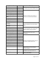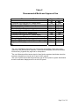
1
LABORATORY PROCEDURE
BD Phoenix™ PMIC/ID Panels
BD Phoenix™ PMIC Panels
BD Phoenix™ PID Panels
INTENDED USE
The BD Phoenix™ Automated Microbiology System is intended for the in vitro rapid
identification (ID) of Gram Positive bacteria from pure culture belonging to the genera
Staphylococcus, Enterococcus, and other Gram Positive cocci and Gram Positive bacilli. The
BD Phoenix Automated Microbiology System is also intended for the quantitative determination
of antimicrobial susceptibility by minimal inhibitory concentration (MIC) of most Gram Positive
bacteria from pure culture belonging to the genera Staphylococcus and Enterococcus.
SUMMARY AND EXPLANATION OF THE TEST
Micromethods for the biochemical identification of microorganisms were reported as early as
19181. Several publications reported on the use of the reagent-impregnated paper discs and
micro-tube methods for differentiating enteric bacteria1-9. The interest in miniaturized
identification systems led to the introduction of several commercial systems in the late 1960s,
and they provided advantages in requiring little storage space, extended shelf life,
standardized quality control, and ease of use.
Many of the tests used in the Phoenix ID panels are modifications of the classical methods.
These include tests for fermentation, oxidation, degradation and hydrolysis of various
substrates. In addition to these, the Phoenix system utilizes chromogenic and fluorogenic
substrates as well as single carbon source substrates in the identification of organisms10,11.
The modern broth microdilution test used today has origins in the tube dilution test used in 1942
by Rammelkamp and Maxon to determine in vitro antimicrobial susceptibility testing of bacterial
isolates from clinical specimens12. The broth dilution technique involves exposing bacteria to
decreasing concentrations of antimicrobial agents in liquid media by serial two-fold dilutions.
The lowest concentration of an antimicrobial agent in which no visible growth occurs is defined
as the minimal inhibitory concentration (MIC).
The introduction in 1956 of a microtitrator system, using calibrated precision spiral wire loops
and droppers for making accurate dilutions rapidly allowed Marymont and Wentz to develop a
serial dilution antimicrobial susceptibility test (AST)13. The microtitrator system was accurate
and allowed the reduction in volumes of antimicrobial agents. The term microdilution appeared
in 1970 to describe the MIC tests performed in volumes of 0.1 mL or less of antimicrobial
solution14.
The Phoenix AST test is a modified miniaturized version of the micro-broth doubling dilution
technique. Susceptibility testing in the Phoenix system is performed through determination of
bacterial growth in the presence of various concentrations of the antimicrobial agent tested.
PRINCIPLES OF THE PROCEDURE
A maximum of 100 identification and antimicrobial susceptibility tests can be performed in the
Phoenix instrument at a time using Phoenix ID/AST combination panels. A sealed and
self-inoculating molded polystyrene tray, with 136 micro-wells containing dried reagents,
serves as the Phoenix disposable. The combination panel includes an ID side with dried
substrates for bacterial identification, an AST side with varying concentrations of antimicrobial
agents, and growth and fluorescent controls at appropriate well locations. The Phoenix system
utilizes an optimized colorimetric redox indicator for AST, and a variety of colorimetric and
Page 1 of 23
fluorometric indicators for ID. The AST Broth is cation-adjusted (e.g., Ca++ and Mg++) to
optimize susceptibility testing performance.
The Phoenix panel is comprised of a 51 well ID side and an 85 well AST side. The ID side
contains 45 wells with dried biochemical substrates and 2 fluorescent control wells. The AST
side contains 84 wells with dried antimicrobial agents and 1 growth control well. Panels are
available as ID only (Phoenix™ NID Panels, Phoenix™ PID Panels), AST only (Phoenix™
NMIC Panels, Phoenix™ PMIC Panels), or ID/AST combination (Phoenix™ NMIC/ID Panels,
Phoenix™ PMIC/ID Panels). Unused wells are reserved for future use.
Phoenix panels are inoculated with a standardized inoculum. Organism suspensions must be
prepared only with the BBL™ CrystalSpec™ or BD PhoenixSpec™ Nephelometer. Once
inoculated, panels are placed into the instrument and continuously incubated at 35°C. The
instrument tests panels every 20 minutes: on the hour; at 20 minutes past the hour; and again
at 40 minutes past the hour up to 16 hours if necessary. Phoenix panels are read only by the
instrument. Phoenix panels cannot be read manually.
Bacterial Identification: The ID portion of the Phoenix panel utilizes a series of conventional,
chromogenic, and fluorogenic biochemical tests to determine the identification of the organism.
Both growth-based and enzymatic substrates are employed to cover the different types of
reactivity in the range of taxa. The tests are based on microbial utilization and degradation of
specific substrates detected by various indicator systems. Acid production is indicated by a
change in the phenol red indicator when an isolate is able to utilize a carbohydrate substrate.
Chromogenic substrates produce a yellow color upon enzymatic hydrolysis of either
p-nitrophenyl or p-nitroanilide compounds. Enzymatic hydrolysis of fluorogenic substrates
results in the release of a fluorescent coumarin derivative. Organisms that utilize a specific
carbon source reduce the resazurin-based indicator. In addition, there are other tests that
detect the ability of an organism to hydrolyze, degrade, reduce, or otherwise utilize a substrate.
A complete list of taxa that comprises the Phoenix ID Database is provided in Table A.
Reactions employed by various substrates and the principles employed in the Phoenix ID
reactions are described in Table B.
Antimicrobial Susceptibility Testing: The Phoenix AST method is a broth based
microdilution test. The Phoenix system utilizes a redox indicator for the detection of organism
growth in the presence of an antimicrobial agent15. Continuous measurements of changes to
the indicator as well as bacterial turbidity are used in the determination of bacterial growth.
Each AST panel configuration contains several antimicrobial agents with a range of two-fold
doubling dilution concentrations. Organism identification is used in the interpretation of the MIC
values of each antimicrobial agent producing Susceptible, Intermediate, or Resistant (SIR)
result classifications.
A complete list of taxa for which the Phoenix system can provide AST results is provided in
Table A. The list of antimicrobial agents and concentrations available for susceptibility testing in
the Phoenix system is provided under Performance Characteristics.
There are antimicrobial agents for use with the Phoenix System that are not proven to be
effective for treating infections for all organisms listed in the taxa. For interpreting and reporting
results of antimicrobial agents that have been shown to be active against organism groups both
in vitro and in clinical infections refer to the individual pharmaceutical antimicrobial agent
labeling. Alternatively, refer to the most recent CLSI M100 Performance Standard, Table 1
“Suggested Groupings of US FDA-Approved Antimicrobial Agents That Should Be Considered
for Routine Testing and Reporting on Organisms by Clinical Microbiological Laboratories”16.
The components required for testing using the Phoenix system include: 1) Phoenix panels with
panel closures, 2) Phoenix ID Broth, 3) Phoenix AST Broth, 4) Phoenix AST Indicator solution
5) Phoenix Inoculation Station, 6) Phoenix Transport Caddy, 7) BBL CrystalSpec or BD
PhoenixSpec Nephelometer, 8) 25 µL pipettor and sterile tips, and 9) Miscellaneous lab
supplies (listed under Materials Required But Not Provided).
Page 2 of 23
Prior to inoculation, the Phoenix panel is placed on the Inoculation Station with the inoculation
ports at the top for filling. Separate inocula are added manually to the ID and AST ports. The
inocula flow down the panel in serpentine fashion, filling the panel wells as the liquid front
progresses toward the pad. The pad absorbs excess inoculum. Closures are manually inserted
in the fill ports. An air admittance port is located in the divider area of the panel lid to ensure
adequate oxygen tension in the panel for the duration of the test.
INGREDIENTS:
For a listing of biochemical substrates used in the Phoenix panel refer to Table B. The package
insert enclosed in the panel box provides a listing of the specific antimicrobial agents and
concentrations found in the panel.
PRECAUTIONS
For in vitro Diagnostic Use
All patient specimens and microbial cultures are potentially infectious and should be treated
with universal precautions. Please refer to CDC manual Bio-safety in Microbiological and
Biomedical Laboratories, 4th Edition, 1999, as well as other recommended literature.
Prior to discarding, sterilize specimen containers and other contaminated materials by
autoclaving.
Panels, once inoculated, should be handled carefully until placed in the instrument.
STORAGE AND HANDLING
Phoenix Panels: Panels are individually packaged and must be stored unopened at room
temperature (15 - 25°C). Do not refrigerate or freeze. Visually inspect the package for holes or
cracks in the foil package. Do not use if the panel or packaging appears to be damaged. If
stored as recommended, the panels will retain expected reactivity until the date of expiration.
Phoenix ID Broth: Tubes are packaged as 100 tube packs. Visually inspect the tubes for
cracks, leaks, etc. Do not use if there appears to be a leak, tube or cap damage or visual
evidence of contamination (i.e., haziness, turbidity). Store Phoenix ID Broth tubes at 2-25°C.
Expiration dating is shown on the tube label.
Phoenix AST Broth: Tubes are packaged as 100 tube packs. Visually inspect the tubes for
cracks, leaks, etc. Do not use if there appears to be a leak, tube or cap damage or visual
evidence of contamination (i.e., haziness, turbidity). Store Phoenix AST Broth tubes at 2-25°C.
Expiration dating is shown on the tube label.
Phoenix AST Indicator Solution: The indicator solution is individually pouched and packaged
as a package of 10 dropper bottles. Visually inspect the bottle for cracks, leaks, etc. Do not use
if there appears to be a leak, bottle or cap damage or any change from a dark blue color. Store
Phoenix AST Indicator Solution at 2-8°C. Each bottle contains enough solution to test up to 100
panels. Expiration dating is shown on the box, pouch, and bottle label and is for unopened
bottles. An opened bottle is stable for up to 14 days if stored at 2-8°C. Be sure the bottle is
held vertically when dispensing the AST Indicator Solution.
SPECIMEN COLLECTION AND PROCESSING
The Phoenix system is not for use directly with clinical specimens. Only pure culture isolates of
Gram Positive organisms are acceptable for testing. The test isolate must be a pure culture. It
is recommended that cultures be no more than 24 hours old unless additional incubation is
required to achieve sufficient growth.
Page 3 of 23
Isolates must be tested with a Gram stain test to assure the appropriate selection of Phoenix
panel type. Once the Gram stain reaction is confirmed, select the appropriate Phoenix panel for
inoculation (e.g., PMIC/ID panel for use with Gram Positive organisms). Selection of the
incorrect panel type could lead to incorrect results.
For AST testing in the Phoenix system, isolates recovered from non-selective media are
recommended. It is recommended that media containing antibiotics not be used for organisms
to be tested in the Phoenix system. Selective media may inhibit some strains of bacteria;
therefore, caution must be used when selecting isolated colonies from these media.
For ID and AST testing, refer to Table C for recommended media.
For ID only testing of Gram Positive organisms, isolates from one of the following media may
be used: Trypticase™ Soy Agar without blood, Columbia Colistin Nalidixic Acid (CNA) Agar
with 5% sheep blood and Phenylethanol Agar (PEA).
When swabs are used, only cotton-tipped applicators should be used to prepare the inoculum
suspensions. Some polyester swabs may cause problems with inoculation of the panels.
The usefulness of the Phoenix system or any other diagnostic procedure performed on clinical
specimens is directly influenced by the quality of the specimens themselves. It is strongly
recommended that laboratories employ methods discussed in the Manual of Clinical
Microbiology17 for specimen collection, transport, and placement on primary isolation media.
Inoculum for use on the Phoenix system is prepared by the CLSI recommended direct colony
suspension method18. Due to variations in inoculum concentrations prepared with McFarland
standards, use of the BBL CrystalSpec or BD PhoenixSpec nephelometer is required for
adjusting the test inoculum prior to use in the Phoenix system.
It is highly recommended that the purity of the inoculum be checked by preparing a purity plate.
See “Purity Check” below.
MATERIALS REQUIRED
Materials Provided:
·
Phoenix Panels
·
Phoenix ID Broth
·
Phoenix AST Broth
·
Phoenix AST Indicator Solution
·
Phoenix Inoculation Station
·
Phoenix Transport Caddy
·
BBL™ CrystalSpec™ or BD PhoenixSpec™ Nephelometer and Standards
·
25 µL pipettor and sterile tips
·
50 µL pipettor and sterile tips
·
2 Pipette stands
Materials Required But Not Provided:
·
Gram stain reagents
·
Sterile cotton swabs
Page 4 of 23
·
Non-selective culture plated media (e.g., Trypticase™ Soy Agar with 5% Sheep Blood)
·
Incubators
·
Biohazard disposable container
·
Markers, etc
PHOENIX TEST PROCEDURE
Note: The Phoenix instrument should always be powered on. If it is not, power on the
instrument and allow 2 hours for the instrument to warm up before loading panels.
Prepare the Phoenix instrument to receive new panels as described in the BD Phoenix System
User’s Manual (“Operation, Daily System Maintenance”).
Care should be exercised when handling Phoenix panels. You should handle panels by the
sides only to avoid marking, smudging or obscuring the front or back of the panel in any way.
Accession barcode labels affixed to a Phoenix panel should:
·
Not be of fluorescent material
·
Not cover any Phoenix panel reaction wells
·
Not cover the Phoenix panel sequence number barcode
Broth and Panel Preparation:
1.
Confirm the Gram stain reaction of the isolate before proceeding with the inoculum
preparation for use in the Phoenix instrument. Once the Gram stain reaction is confirmed,
select the appropriate Phoenix panel for inoculation. Selection of the incorrect panel type
could lead to incorrect results.
2. Examine the pouch, and do not use the panel if the pouch is punctured or opened.
Remove the panel from the pouch. Discard the desiccant. Do not use the panel if there is
no desiccant or if the desiccant pouch is torn. Note: Panels must be used within 2 hours
of being removed from the pouch.
3. Place the panel on the Inoculation Station with ports at the top and pad on the bottom.
4. Label a Phoenix ID Broth tube with the patient’s specimen number. Using aseptic
technique, pick colonies of the same morphology with the tip of a sterile cotton swab (do
not use a polyester swab) or a wooden applicator stick from one of the recommended
media. See Table C.
5. Suspend the colonies in the Phoenix ID Broth (4.5 mL).
6. Cap the tube and vortex for 5 seconds.
7. Allow approximately ten seconds for air bubbles to surface. Tap the tube gently to aid in
eliminating bubbles.
8. Confirm default settings for inoculum density before inoculating panels. Insert the tube into
the BBL CrystalSpec or BD Phoenix Spec Nephelometer. Make sure the tube is inserted as
far as it will go. Note: Only the BD PhoenixSpec Nephelometer can be used to make
inoculum densities of 0.25 McFarland. (Refer to the BBL CrystalSpec Nephelometer or
BD PhoenixSpec product insert for correct usage instructions and calibration verification.)
9. If the inoculum density is set to 0.5 for the panel type being run, then a range of 0.50-0.60 is
acceptable. If the inoculum density is set to 0.25 for the panel type being run, then a range
Page 5 of 23
of 0.20-0.30 is acceptable. If the density of organisms is low, you can add colonies from the
isolate. Re-vortex the sample and reread to confirm that the correct density has been
achieved. If the density of organisms exceeds 0.6 McFarland, follow the steps below to
dilute the broth. It is very important to accurately indicate the level of the liquid in the tube
since this volume is needed to adequately fill the wells in the panel. Note: The
standardized bacterial suspension in ID broth must be used within 60 minutes of
preparation.
a Using a marker, mark the broth level in the over-inoculated Phoenix ID Broth tube.
b Using a sterile pipette, aseptically add fresh Phoenix ID Broth to the inoculum. Only
Phoenix ID Broth may be used to dilute the inoculum.
c Vortex the tube and allow to sit for 10 seconds.
d Place the tube in the nephelometer and remeasure the turbidity of the suspension.
• If the reading is greater than 0.6, repeat Steps b-d.
• If the reading is 0.5 – 0.6, go to Step e.
e Using a sterile pipette, aseptically remove excess broth to the original level indicated by
the mark on the tube created in Step a.
Remove excess broth to avoid overfilling the panel. Also, do not remove too much broth,
as there may be insufficient broth to adequately fill the panel.
f Broth may now be used to inoculate the Phoenix AST Broth and/or the Phoenix Panel.
10. If you are performing identification only, proceed to Step 15 and continue the procedure.
11. Label a Phoenix AST Broth tube (8.0 mL) with the patient’s specimen number. Holding the
AST Indicator Solution bottle vertically, add one free-falling drop of AST indicator solution
to the AST broth tube. Invert to mix. DO NOT VORTEX. Note: Allow AST Indicator
Solution to warm to room temperature before dispensing into AST broth. The
unused portion of the indicator should be returned to 2º- 8ºC as soon as possible.
Do not store at room temperature for more than 2 hours. Opened bottles should be
discarded after 14 days from initial opening. If volume other than one drop is added
inadvertently, discard the tube and use a fresh tube of AST broth. After the addition
of the Indicator to AST broth, the mixed solution can be stored in the dark, at room
temperature, for as long as 8 hours. Tubes must be used within 2 hours after the
addition of the indicator solution if exposed to light.
12. If an inoculum density of 0.50 – 0.60 was used, transfer 25 µL of the bacterial suspension
from the ID tube into the AST broth tube. If an inoculum density of 0.20 – 0.30 was used,
transfer 50 µL (use 2 shots if utilizing a 25 µL pipettor) of the bacterial suspension from the
ID tube into the AST broth tube. Note: Panels must be inoculated within 30 minutes of
the time that the AST inoculum is prepared.
13. Cap the AST tube and invert several times to mix. Do not vortex.
14. Wait a few seconds for air bubbles to surface. Tap the tube gently to aid in eliminating
bubbles.
15. Pour the ID tube inoculum into the fill port on the ID side of the panel (51-well side). Allow
the fluid to traverse down the tracks before moving the panel. If using an AST (only) panel,
DO NOT inoculate the ID side of the panel. Retain the ID tube for a purity check.
Page 6 of 23
16. Pour the AST tube inoculum into the fill port on the AST side of the panel (85-well side).
Allow the fluid to traverse down the tracks before moving the panel.
17. Before placing panel closure, check for residual droplets of inoculum on the edge of the fill
ports. If a droplet is present, remove the droplet with absorbent material. The used
absorbent material must be discarded along with your biohazard waste.
18. Snap on the panel closure. Make sure that the closure is fully seated.
Visually inspect panels to be sure each of the wells is full. Look at both sides of the panel.
Make certain that the wells are not overfilled. If any of the wells are unfilled or overfilled,
inoculate a new panel. Note: Panels must be loaded into the instrument within 30
minutes of inoculation. Panels must be kept in the inoculation station after
inoculation until the excess fluid has been completely absorbed by the pad. Panels
should stay vertical in the transport caddy until loaded into the instrument.
Inoculated panels should be handled with care. Avoid knocking or jarring the panel.
Purity Check
1. Using a sterile loop, recover a small drop from the inoculum fluid either before or after
inoculation of the panel.
2. Inoculate an agar plate (any appropriate medium) for a purity check.
3. Discard inoculum fluid tube and cap in a biohazard disposal container.
4. Incubate the plate for 24-48 hours at 35°C under appropriate conditions.
ID Inoculum Density Flexibility
You may run the ID portion of a panel in the opposite mode from what is configured by
darkening well A17 on the back of a panel before placing the panel in the instrument. This
allows you to run a panel at an inoculum density of 0.20 – 0.30 even if you are configured for a
density of 0.5 for that particular panel type. Likewise, you can run the panel at an inoculum
density of 0.50 – 0.60 if you are configured for a density of 0.25.
There is no way to alter the density setting during Panel Login. To use a panel in the opposite
density mode, using a black Sharpie™ (permanent marker), blacken the A17 well entirely. See
the BD Phoenix System User’s Manual (Operation, ID Inoculum Density Flexibility) for position
of well A17.
For instructions for panel login and loading, refer to the BD Phoenix System User’s Manual
(“Panel Login” and “Inserting Panels in the Instrument”).
Current Instrument
Inoculum Density
Configuration
Inoculum
Concentration
Desired for Test
Panel
Amount of ID Inoculum to
Add to AST Broth**
Well A-17
0.50
0.25
50 µL
Blackened
0.25
0.50
25 µL
Blackened
** If also running AST
Page 7 of 23
USER QUALITY CONTROL
In order to ensure appropriate set up procedure and acceptable performance of the system, the
following organisms are recommended to be tested. The user is advised to review the
individual AST panel formats to determine if all test strains need to be tested for routine
laboratory Quality Control. Refer to the Package Insert that accompanies the Phoenix panels
for expected ID results and AST results for QC organisms.
For instructions for QC panel login and loading, refer to the BD Phoenix System User’s Manual
(“Panel Login” and “Inserting Panels in the Instrument”).
ID (PMIC/ID and PID panels):
Staphylococcus aureus ATCC™ 29213
Enterococcus faecalis ATCC™ 29212
AST (PMIC/ID, PMIC panels):
Staphylococcus aureus ATCC™ 29213
Enterococcus faecalis ATCC™ 29212
Staphylococcus aureus ATCC™ 25923 (PMIC/ID panels only. QC for Nitrocefin.)
Enterococcus faecalis ATCC™ 51299
For the most reliable results, it is recommended that the QC organisms be subcultured at least
twice on two consecutive days onto TSA II with 5% Sheep Blood Agar before use in the
Phoenix system.
Compare recorded results to those listed in the Package Insert. If discrepant results are
obtained, review test procedures as well as confirm purity of the quality control strain used
before contacting BD Diagnostics Technical Services Department. Unacceptable QC results
are documented as “Fail” and acceptable QC results are documented as “Pass” on the QC
Report.
RESULTS
Organism identification will appear on the Phoenix Report Form with a probability percentage
from the Phoenix database based on the substrate reaction profile. Results from each
substrate will appear as +, -, V, or X for each reaction. The MIC results and Interpretive
Categorical Results (SIR) will be shown for the appropriate organism/antimicrobial agent
combinations.
Special messages will be shown when the BDXpert™ System detects results that are of
particular clinical interest.
Further information concerning results obtained from the Phoenix system can be found in the
BD Phoenix System User’s Manual (“Obtaining Results”).
Messages
Error messages may appear if the system detects unexpected reactivity due to inappropriate
procedure or instrument malfunction. For a complete listing of error codes and their meaning
refer to the BD Phoenix System User’s Manual (“System Alerts”, “Needs Attention” and
“Troubleshooting”).
Page 8 of 23
Special Notes
In general, the Phoenix System provides a MIC for all organisms at any of the concentrations
defined on a specific panel. For certain antimicrobic/organism combinations a specific
minimum or maximum MIC is reported even if there is a lower or higher concentration on the
panel. These MIC values are applied by the software and are reported out as less than or equal
to (</=) for the minimum MIC or greater than (>) for the maximum MIC. The table below
provides the range for these special antimicrobic/organism combinations.
Antimicrobial Agent
Oxacillin
Organism(s)
Coagulase negative
Applied Range (µg/mL)
0.0625-1.0
staphylococci
Penicillin
Staphylococcus spp.
0.0625-1.0
Enterococcus spp.
1.0-32
Gentamicin
Staphylococcus epidermidis
<4 and >16*
Moxifloxacin
Enterococcus spp. other
0.25-8
than E. faecium
* MICs of 4, 8, 16 not reported
LIMITATIONS OF THE PROCEDURE
See the package insert shipped with the panel for specific organism/antimicrobial limitations.
General
A Gram stain test is required for the selection of the appropriate Phoenix panel types. Accurate
identification and/or AST results may not be made without this test.
Use only well-isolated bacterial colonies from one of the recommended primary isolation
media. See Table C. Media containing esculin should not be used. Use of mixed colonies
could result in inaccurate identification and/or AST interpretations.
If the instrument inoculum density is configured to 0.5 (for the panel type being used), an
inoculum density of 0.50 – 0.60 must be met. Only the BBL CrystalSpec or BD Phoenix Spec
Nephelometer can be used to measure the inoculum density.
If the instrument inoculum density is configured to 0.25 (for the panel type being used), an
inoculum density of 0.20 – 0.30 McFarland must be met. Only the BD PhoenixSpec
Nephelometer can be used to measure inoculum density for this range.
Phoenix panels can be read only by the Phoenix instrument. Visual interpretation of the
Phoenix panels is not possible. Any attempt to manually interpret results from the panel may
lead to misidentification and/or inaccurate AST interpretations.
Identification
The unique panel environment combined with the shortened incubation time may result in
Phoenix panel reactions varying from those obtained using conventional biochemical media.
Page 9 of 23
Antimicrobial Susceptibility Testing
After the addition of the Phoenix AST Indicator Solution to the AST broth tubes, mix by
inversion. DO NOT VORTEX. Vortexing may cause air bubbles to form in the AST broth, which
can result in inappropriate filling of the Phoenix panel during inoculation.
Because of the low probability of occurrence or special growth requirements some organisms
included in the ID taxa are not included in the AST database. These organisms will display the
message “Organism not included in the AST database. Perform alternate method.”
For some organism/antimicrobial combinations, the absence of resistant strains precludes
defining any result categories other than “susceptible.” For strains yielding results suggestive of
a “nonsusceptible” category, organism identification and antimicrobial susceptibility test results
should be confirmed. Subsequently, the isolates should be saved and submitted to a reference
laboratory that will confirm the result using the CLSI reference dilution method.
PERFORMANCE CHARACTERISTICS
Gram Positive Identification
In two internal studies, the performance of the Phoenix Gram Positive identification was
evaluated. The 0.5 inoculum density configuration and the 0.25 inoculum density configuration
were tested with 696 strains (0.5) and 755 strains (0.25), respectively. Results were evaluated
against commercial and non-commercial methods.
The Phoenix Gram Positive identification performance is outlined below:
Species Level
McFarland
Agreement
No Agreement
No ID
0.5
95.4%
3.9%
0.7%
0.25
98.0%
1.6%
0.4%
An internal study was performed to simulate inter-site reproducibility. The identification results
obtained using the Phoenix system were compared with expected results. This performance
testing demonstrated intra-site and inter-site reproducibility of at least 95% or greater.
Gram Positive Susceptibility
Clinical, stock, and challenge isolates were tested across multiple clinical sites to determine
Essential Agreement (EA) and Category Agreement (CA) of the Phoenix system to the CLSI
broth microdilution reference method. Essential Agreement occurs when the MIC of the
Phoenix system and the reference method agree exactly or is within ± 1 dilution of each other.
Category Agreement occurs when the Phoenix system results agree with the reference method
with respect to the CLSI categorical interpretative criteria (susceptible, intermediate, resistant).
The table below summarizes the data from these studies.
Additionally, testing performed at multiple clinical sites demonstrated at least 95%
reproducibility or greater within ± 1 doubling dilution for all antimicrobial agents listed in the
table below.
DRUG CLASS
DRUG NAME
DRUG
CODE
DRUG
RANGE
EA
N
EA
%
CA
N
CA
%
(µg/mL)
5-Fluoroquinolone
Gatifloxacin
GAT
0.25-8
1180
98.6
1180
90.1
5-Fluoroquinolone
Levofloxacin
LVX
0.25-8
1878
96.8
1878
95.1
Page 10 of 23
5-Fluoroquinolone
Moxifloxacin
MXF
0.125-8
1777
96.0
1777
90.1
5-Fluoroquinolone
Norfloxacin
NOR
0.25-16
1252
96.9
1252
97.4
5-Fluoroquinolone
Ofloxacin
OFX
0.25-8
1184
98.7
1184
98.2
Aminoglycoside
Gentamicin
GM
0.25-16
1223
91.9
1223
95.2
Aminoglycoside
Gentamicin
Synergy
GMS
500
—
—
763
98.6
Streptomycin
Synergy
STS
1000
—
—
756
97.8
Aminoglycoside
Tobramycin
NN
0.5-16
953
93.5
797
98.5
Ansamycin
Rifampin
RA
0.25-32
1261
98.3
1261
98.2
B-Lac/B-Lac. Inh
Ampicillin/
Clavulanate
0.25/0.12-32/16
871
94.1
871
96.7
0.06-32
475
93.3
475
98.5
2/1-32/16
1240
97.2
1240
97.3
Aminoglycoside
AMC
B-Lactam Pen.
Ampicillin
AM
B-Lactam Pen Inh
Ampicillin/
sulbactam
SAM
B-Lactam Pen.
Oxacillin
OX
0.06-4
1231
95.4
1231
96.6
B-Lactam Pen.
Penicillin
P
0.06-32
1256
93.6
1256
97.5
Carbapenem
Meropenem
MEM
0.5-16
620
98.4
1198
96.6
Cephem
Cefazolin
CZ
2-32
597
99.5
597
99.7
Cephem
Cefoxitin
FOX
1-32
1164
96.3
1164
90.1
Cephem
Cephalothin
0.5-64
904
96.2
904
98.0
Folate Antagonist
TrimethoprimSulfamethoxazole
0.5/9.5- 16/304
634
96.4
634
97.9
CF
SXT
Glycopeptide
Vancomycin
VA
0.5-32
1981
97.7
1981
99.4
Macrolide
Erythromycin
E
0.06-8
1395
95.0
1395
94.6
Lincosamide
Clindamycin
CC
0.12-8
1242
98.2
1242
98.7
Lipopeptide
Daptomycin*
DAP
0.125-32
1568
97.4
1568
98.8
Nitrofurantoin
Nitrofurantoin*
FM
16-512
1757
99.8
1757
99.8
Oxazolidinone
Linezolid
LZD
0.25-32
1454
91.1
1454
95.3
Phenicol
Chloramphenicol
1-32
1447
93.4
1447
93.4
Streptogramin
Quinupristin/
Dalfopristin
SYN
0.25-4
2019
94.5
1500
95.5
Tetracycline
TE
0.5-16
2040
96.9
2040
96.5
Tetracycline
C
a
The ability of the system to detect resistance for this antimicrobic with Enterococcus species
and Staphylococcus species is unknown because resistant organisms were not available at the
time of comparative clinical testing.
Page 11 of 23
REFERENCES
1. Bronfenbrenner, J., and Schlesigner, M.J. 1918. “A Rapid Method for the Identification of
Bacteria Fermenting Carbohydrates,” Am. J. Public Health. 8:922-923.
2. Arnold, W.M., Jr., and Weaver, R.H. 1948. “Quick Microtechniques for Identification of
Cultures - I. Indole production,” J. Lab. Clin. Med. 33:1334-1337.
3. Bachmann, B., and Weaver, R.H. 1951. “Rapid Microtechnics for Identification of Cultures
- V. Reduction of Nitrates to Nitrites,” Am. J. Clin. Pathol. 21:195-196.
4. Hannan, J., and Weaver, R.H. 1948. “Quick Microtechniques for the Identification of
Cultures - II. Fermentations,” J. Lab. Clin. Med. 33:1338-1341.
5. Hartman, P.A. 1968. Paper strip and disc methods, p. 123-132. Miniaturized
microbiological methods. Academic Press, New York.
6. Sanders, A.C., Faber, J.E., and Cook, T.M. 1957. “A Rapid Method for the Characterization
of Enteric Pathogen Using Paper Discs,” Appl. Microbiol. 5:36-40.
7. Synder, M.L. 1954. “Paper Discs Containing Entire Culture Medium for the Differentiation
of Bacteria,” Pathol. Bacteriol. 67:217-226.
8. Soto, O.B. 1949. “Fermentation Reactions with Dried Paper Discs Containing
Carbohydrate and Indicator,” Puerto Rican J. Publ. Hlth. Trop. Med. 25:96-100.
9. Weaver, R.H. 1954. “Quicker Bacteriological Results,” Am. J. Med. Technol.
20:14-26.
10. Kämpfer, P., Rauhoff, O., and Dott, W. 1991. “Glycosidase Profiles of Members of the
Family Enterobacteriaceae,” J. Clin. Microbiol. 29:2877-2879.
11. Manafi, M., Kneifel, W., and Bascomb, S. 1991. “Fluorogenic and Chromogenic Substrates
Used in Bacterial Diagnostics,” Microbiol. Rev. 55:335-348.
12. Rammelkamp, C.H. and Maxon, T. 1942. “Resistance of Staphylococcus aureus to the
Action of Penicillin,” Proc. Soc. Biol. and Med. 51:386-389.
13. Marymont, J.H. and Wentz, R.M. 1966. “Serial Dilution Antibiotic Sensitivity Testing with
the Microtitrator System,” Am. J. Clin. Pathol. 45:548-551.
14. Gavan, T.L., and Town, M.A. 1970. “A Microdilution Method for Antibiotic Susceptibility
Testing: An Evaluation,” Am. J. Clin. Pathol. 53:880-885.
15. Lancaster, M.V. and Fields, R.D. 1996. Antibiotic and Cytotoxic Drug Susceptibility Assays
Using Resazurin and Poising Agents. U.S. Patent #5,501,959.
16. CLSI. M100-S15 Performance Standards for Antimicrobial Susceptibility Testing; Fifteenth
Informational Supplement. January, 2005.
17. Murray, Patrick R., et al. ed., Manual of Clinical Microbiology, 8th Edition, ASM Press,
Washington, D.C., 2003.
18. CLSI. M7-A6 Methods for Dilution Antimicrobial Susceptibility Tests for Bacteria That Grow
Aerobically; Approved Standard—Sixth Edition. January, 2003.
Manufactured by
Becton, Dickinson and Company
7 Loveton Circle
Sparks, MD 21152 USA (800) 638-8663
Made in USA
Page 12 of 23
TECHNICAL INFORMATION
Approved by:_________________________
Date Effective:________________________
Supervisor:___________________________Date:____________________
Director:_____________________________Date:____________________
Reviewed:____________________________
9/2006
Phoenix, BDXpert, BBL CrystalSpec, PhoenixSpec, Trypticase and BD are trademarks of
Becton, Dickinson and Company. ATCC is a trademark of American Type Culture Collection.
Sharpie is a trademark of Sanford. CHROMagar is a trademark of Dr. A. Rambach. ©2005 BD.
Table A
Taxa for ID/AST Determination
There are antimicrobial agents for use with the Phoenix system that are not proven to be
effective for treating infections for all organisms listed in this section. For interpreting and
reporting results of antimicrobial agents that have shown to be active against organism groups
both in vitro and in clinical infections refer to the individual pharmaceutical antimicrobial agent
labeling. Alternatively, refer to the most recent CLSI M100 Performance Standard, Table 1
“Suggested Groupings of US FDA-Approved Antimicrobial Agents That Should be Considered
for Routine Testing and Reporting on Organisms by Clinical Microbiological Laboratories.”
Gram Positive (0.5 McFarland)
Gram Positive Taxa1
ID, AST, ID/AST
Aerococcus urinae
ID
Aerococcus viridans
ID
Alloiococcus otitidis
ID
Arcanobacterium haemolyticum
ID
Arcanobacterium pyogenes
ID
Bacillus cereus
ID
Bacillus circulans
ID
Bacillus coagulans
ID
Bacillus licheniformis
ID
Bacillus megaterium
ID
Bacillus pumilus
ID
Bacillus sphaericus
ID
Bacillus subtilis
ID
Bacillus thuringiensis
ID
Page 13 of 23
Brevibacillus brevis
ID
Brevibacterium species
ID
Cellulomonas turbata
ID
Corynebacterium amycolatum
ID
Corynebacterium bovis
ID
Corynebacterium diphtheriae
ID
Corynebacterium jeikeium
ID
Corynebacterium kutscheri
ID
Corynebacterium matruchotii
ID
Corynebacterium minutissimum
ID
Corynebacterium propinquum
ID
Corynebacterium pseudodiphtheriticum
ID
Corynebacterium pseudotuberculosis
ID
Corynebacterium renale
ID
Corynebacterium striatum
ID
Corynebacterium ulcerans
ID
Corynebacterium urealyticum
ID
Corynebacterium xerosis
ID
Dermabacter hominis
ID
Dermacoccus nishinomiyaensis
ID
Enterococcus asini
AST
Enterococcus avium
ID/AST
Enterococcus casseliflavus
ID/AST
Enterocooccus cecorum
AST
Enterococcus columbae
AST
Enterococcus dispar
AST
Enterococcus durans
ID/AST
Enterococcus faecalis
ID/AST
Enterococcus faecium
ID/AST
Enterococcus flavescens
AST
Enterococcus gallinarum
ID/AST
Enterococcus gilvus
AST
Enterococcus haemoperoxidus
AST
Enterococcus hirae
ID/AST
Enterococcus malodoratus
AST
Page 14 of 23
Enterococcus moraviensis
AST
Enterococcus mundtii
AST
Enterococcus pallens
AST
Enterococcus pseudoavium
AST
Enterococcus raffinosus
ID/AST
Enterococcus ratti
AST
Enterococcus saccharolyticus
AST
Enterococcus solitarius
AST
Enterococcus sulfureus
AST
Erysipelothrix rhusiopathiae
ID
Gardnerella vaginalis
ID
Gemella haemolysans
ID
Gemella morbillorum
ID
Globicatella sanguinis
ID
Helcococcus kunzii
ID
Kocuria kristinae
ID
Kocuria rosea
ID
Kocuria varians
ID
Kytococcus sendentarius
ID
Lactococcus garvieae
ID
Lactococcus lactis ssp. cremoris
ID
Lactococcus lactis ssp. hordniae
ID
Lactococcus lactis ssp. lactis
ID
Lactococcus plantarum
ID
Lactococcus raffinolactis
ID
Leifsonia aquatica
ID
Leuconostoc citreum
ID
Leuconostoc lactis
ID
Leuconostoc mesenteroides ssp cremoris
ID
Leuconostoc mesenteroides ssp mesenteroides
ID
Leuconostoc pseudomesenteroides
ID
Listeria grayi
ID
Listeria innocua
ID
Listeria ivanovii
ID
Listeria monocytogenes
ID
Page 15 of 23
Listeria welshimeri
ID
Macrococcus caseolyticus
ID
Micrococcus luteus
ID
Micrococcus lylae
ID
Oerskovia xanthineolytica
ID
Paenibacillus alvei
ID
Paenibacillus macerans
ID
Pediococcus acidilactici
ID
Pedicococcus damnosus
ID
Pediococcus dextrinicus
ID
Pediococcus parvulus
ID
Pediococcus pentosaceus
ID
Rhodococcus equi
ID
Rothia dentocariosa
ID
Rothia mucilaginosa
ID
Staphylococcus arlettae
AST
Staphylococcus aureus
ID/AST
Staphylococcus auricularis
ID/AST
Staphylococcus capitis
ID/AST
Staphylococcus capitis ssp. capitis
ID/AST
Staphylococcus capitis ssp. ureolyticus
ID/AST
Staphylococcus caprae
ID/AST
Staphylococcus carnosus
ID/AST
Staphylococcus chromogenes
ID/AST
Staphylococcus cohnii
ID/AST
Staphylococcus cohnii ssp. cohnii
ID/AST
Staphylococcus cohnii ssp. urealyticum
ID/AST
Staphylococcus condimenti
AST
Staphylococcus delphini
AST
Staphylococcus epidermidis
ID/AST
Staphylococcus equorum
ID/AST
Staphylococcus felis
ID/AST
Staphyloccus fleurettii
AST
Staphylococcus gallinarum
ID/AST
Staphylococcus haemolyticus
ID/AST
Page 16 of 23
Staphylococcus hominis
ID/AST
Staphylococcus hyicus
ID/AST
Staphylococcus intermedius
ID/AST
Staphylococcus kloosii
ID/AST
Staphylococcus lentus
ID/AST
Staphylococcus lugdunensis
ID/AST
Staphylococcus lutrae
AST
Staphylococcus muscae
AST
Staphylococcus pasteuri
ID/AST
Staphylococcus piscifermentans
AST
Staphylococcus pulvereri
AST
Staphylococcus saccharolyticus
AST
Staphylococcus saprophyticus
ID/AST
Staphylococcus schleiferi
ID/AST
Staphylococcus schleiferi ssp. coagulans
ID/AST
Staphylococcus schleiferi ssp. schleiferi
ID/AST
Staphylococcus sciuri
ID/AST
Staphylococcus simulans
ID/AST
Staphylococcus succinus ssp. casei
AST
Staphylococcus succinus ssp succinus
AST
Staphylococcus vitulinus
ID/AST
Staphylococcus warneri
ID/AST
Staphylococcus xylosus
ID/AST
Streptococcus acidominimus
ID
Streptococcus agalactiae (Strep. group B)
ID
Streptococcus anginosus
ID
Streptococcus bovis I (Strep. group D)
ID
Streptococcus bovis II (Strep. group D)
ID
Streptococcus canis
ID
Streptococcus constellatus
ID
Streptococcus cristatus
ID
Streptococcus dysgalactiae ssp. dysgalactiae
ID
Streptococcus dysgalactiae ssp. equisimilis
ID
Streptococcus equi
ID
Streptococcus equi ssp. equi
ID
Page 17 of 23
Streptococcus equi ssp. zooepidemicus
ID
Streptococcus equines
ID
Streptococcus gordonii
ID
Streptococcus intermedius
ID
Streptococcus mitis
ID
Streptococcus mitis/pneumoniae
ID
Streptococcus mutans
ID
Streptococcus oralis
ID
Streptococcus parasanguinis
ID
Streptococcus pneumoniae
ID
Streptococcus porcinus
ID
Streptococcus pyogenes
1
(Strep. group A)
ID
Streptococcus salivarius
ID
Streptococcus sanguinis
ID
Streptococcus sobrinus
ID
Streptococcus uberis
ID
Streptococcus vestibularis
ID
Not all species encountered during clinical performance evaluations.
Gram Positive (0.25 McFarland)
Gram Positive Taxa1
ID, AST, ID/AST
Aerococcus urinae
ID
Aerococcus viridans
ID
Alloiococcus otitidis
ID
Dermacoccus nishinomiyaensis
ID
Enterococcus avium
ID/AST
Enterococcus casseliflavus
ID/AST
Enterococcus durans
ID/AST
Enterococcus faecalis
ID/AST
Enterococcus faecium
IID/AST
Enterococcus gallinarum
ID/AST
Enterococcus hirae
ID/AST
Enterococcus raffinosus
ID/AST
Gemella haemolysans
ID
Page 18 of 23
Gemella morbillorum
ID
Globicatella sanguinis
ID
Helcococcus kunzii
ID
Kocuria kristinae
ID
Kocuria rosea
ID
Kocuria varians
ID
Kytococcus sendentarius
ID
Lactococcus lactis ssp. cremoris
ID
Lactococcus lactis ssp. hordniae
ID
Lactococcus plantarum
ID
Leuconostoc citreum
ID
Leuconostoc lactis
ID
Leuconostoc mesenteroides ssp. mesenteroides
ID
Listeria innocua
ID
Listeria monocytogenes
ID
Macrococcus caseolyticus
ID
Micrococcus luteus
ID
Micrococcus lylae
ID
Pediococcus acidilactici
ID
Pedicococcus damnosus
ID
Pediococcus dextrinicus
ID
Pediococcus parvulus
ID
Pediococcus pentosaceus
ID
Rothia mucilaginosa
ID
Staphylococcus aureus
ID/AST
Staphylococcus auricularis
ID/AST
Staphylococcus capitis
ID/AST
Staphylococcus caprae
ID/AST
Staphylococcus carnosus
ID/AST
Staphylococcus chromogenes
ID/AST
Staphylococcus cohnii ssp. cohnii
ID/AST
Staphylococcus cohnii ssp. urealyticum
ID/AST
Staphylococcus epidermidis
ID/AST
Staphylococcus equorum
ID/AST
Staphylococcus felis
ID/AST
Page 19 of 23
Staphylococcus gallinarum
ID/AST
Staphylococcus haemolyticus
ID/AST
Staphylococcus hominis
ID/AST
Staphylococcus hyicus
ID/AST
Staphylococcus intermedius
ID/AST
Staphylococcus kloosii
ID/AST
Staphylococcus lentus
ID/AST
Staphylococcus lugdunensis
ID/AST
Staphylococcus pasteuri
ID/AST
Staphylococcus saprophyticus
ID/AST
Staphylococcus schleiferi ssp. coagulans
ID/AST
Staphylococcus schleiferi ssp. schleiferi
ID/AST
Staphylococcus sciuri
ID/AST
Staphylococcus simulans
ID/AST
Staphylococcus vitulinus
ID/AST
Staphylococcus warneri
ID/AST
Staphylococcus xylosus
ID/AST
Streptococcus agalactiae (Strep. group B)
ID
Streptococcus anginosus
ID
Streptococcus bovis (Strep. group D)
ID
Streptococcus bovis I (Strep. group D)
ID
Streptococcus bovis II (Strep. group D)
ID
Streptococcus constellatus
ID
Streptococcus cristatus
ID
Streptococcus equi
ID
Streptococcus gordonii
ID
Streptococcus group C/G (large colony)
ID
Streptococcus intermedius
ID
Streptococcus mitis
ID
Streptococcus mutans
ID
Streptococcus oralis
ID
Streptococcus parasanguinis
ID
Streptococcus pneumoniae
ID
Streptococcus porcinus
ID
Streptococcus pyogenes (Strep. group A)
ID
Page 20 of 23
1
Streptococcus salivarius
ID
Streptococcus sanguinis
ID
Streptococcus sobrinus
ID
Streptococcus uberis
ID
Streptococcus vestibularis
ID
Not all species encountered during clinical performance evaluations.
Table B
List of Reagents and Principles Employed
in the Phoenix System
Substrate Name
Code
4MU-BD-CELLOBIOSIDE
L-ALANINE-AMC
M_BDCEL
A_LALT
4MU-BD-GLUCOSIDE
M_BDGLU
L-PROLINE-AMC
A_LPROB
L-PYROGLUTAMIC
ACID-AMC
A_LPYR
L-PHENYLALANINE-AMC
A_LPHET
L-TRYPTOPHAN-AMC
A_LTRY
4MU-PHOSPHATE
M_PHOS
METHIONINE-AMC
A_META
4MU-AD-GLUCOSIDE
M_ADGLU
ARGININE-ARGININE-AMC
A_ARARR
GLYCINE-PROLINE-AMC
A_GLPRB
4MU-BD-GLUCURONIDE
M_BDGLC
L-LEUCINE-AMC
A_LLEUH
4MU-N-ACETYL-BD-GLUCOS
AMINIDE
M_NAG
L-ARGININE-AMC
A_LARGH
4MU-PHOSPHATE (with
Trehalose)
M_PHOT
L-HISTIDINE-AMC
A_LHIST
L-ISOLEUCINE-AMC
A_LISO
4MU-BD-GALACTOSIDE
M_BDGAL
Principle
Enzymatic hydrolysis of the amide or glycosidic
bond results in the release of a fluorescent
coumarin or 4-methylumbelliferone derivative.
Page 21 of 23
Substrate Name
Code
Principle
Resistance to the antimicrobial agents results
in a reduction of the resazurin based indicator.
COLISTIN
C_CLST
POLYMYXIN B
C_PXB
D-GLUCONIC ACID
C_DGUA
3-METHYL GLUTARIC ACID
C_3MGA
D-FRUCTOSE
C_DFRU
IMINODIACETIC ACID
C_IMN
ALPHA-KETOGLUTARIC
ACID
C_KGA
D-MANNITOL
C_DMNT
3-METHYLADIPIC ACID
C_MAA
THYMIDINE
C_THY
FLOURESCENT POSTIVE
CONTROL
FLR_CTL
FLUORESCENT POSITIVE
CONTROL
FLR_CTL
ALANINE-ALANINE-PNA
N_ALALH
L-PROLINE-PNA
N_LPROT
VALINE-ALANINE-PNA
N_VAALA
PNP-AD-GLUCOSIDE
P_PAGLU
PNP-PHOSPHATE
P_PHOL
BETA-GENTIOBIOSE
R_BGEN
D-SUCROSE
R_DSUC
MALTOTRIOSE
R_MTT
N-ACETYL-GLUCOSAMINE
R_NGU
D-TREHALOSE
R_DTRE
D-TAGATOSE
R_DTAG
MALTOSE
R_MAL
DEXTROSE
R_DEX
UREA
S_URE
Hydrolysis of urea and the resulting ammonia
change results in pH rise and change in
fluorescent indicator.
ESCULIN
T_ESC
Hydrolysis of esculin results in a black
precipitate in the presence of ferric ion.
NITROCEFIN
L_NCF
Enzymatic hydrolysis of the β-lactam ring
results in a color change.
Utilization of a carbon source results in a
reduction of the resazurin based indicator.
Control to standardize fluorescent substrate
results.
Enzymatic hydrolysis of the colorless amide
substrate releases yellow p-nitroaniline.
Enzymatic hydrolysis of the colorless aryl
substituted glycoside release yellow
p-nitrophenol.
Utilization of carbohydrate results in lower pH
and change in indicator (phenol red).
Page 22 of 23
Table C
Recommended Media and Approved Use
Recommended Media
Trypticase Soy Agar with 5% Sheep Blood
Bromthymol Blue (BTB) Lactose Agar
BBL™ CHROMagar™ Orientation
Chocolate Agar
Columbia Agar with 5% Horse Blood
Columbia Agar with 5% Sheep Blood
Columbia CNA Agar with 5% Sheep Blood
Cystine-Lactose-Electrolyte-Deficient (CLED) Agar
Phenylethyl Alcohol Agar
Trypticase™ Soy Agar without Blood
Trypticase™ Soy Agar with Lecithin and Tween™
80
Approved Use
ID
AST
Yes
Yes
2
Yes
Yes
Yes
Yes1
Yes
Yes
Yes
Yes
Yes
Yes
Yes
No
Yes3
Yes
Yes
No
Yes
No
Yes
No
1
The use of CHROMagar Orientation may produce false susceptibility results when testing
erythromycin with Gram positive organisms. Antimicrobial susceptibility test results should be
confirmed using Trypticase Soy Agar with 5% Sheep Blood.
2
The use of Bromthymol Blue lactose Agar for Gram Positive organism identification should be
restricted to Staphylococci for both the 0.5 and 0.25 GP systems.
3
The use of Cystine-Lactose-Electrolyte Deficient Agar for Gram positive organism identification
should be restricted to Staphylococci for the 0.25 GP system.
Page 23 of 23