1
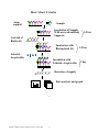
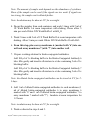
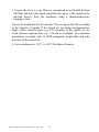
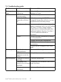
RayBio® Human Cytokine Antibody Array
C Series 1000
Patent Pending Technology
User Manual (Revised February 4, 2009)
RayBio® Human Cytokine Antibody Array C series 1000
(Combination of Array 6 & 7 Cat# AAH-CYT-1000)
Please read manual carefully before starting experiment
RayBiotech, Inc.
We Provide You with Excellent
Protein Array Systems and Service
Tel:(Toll Free) 1-888-494-8555 or 770-729-2992; Fax: 1-888-547-0580;
Website:www.raybiotech.com Email: [email protected]
RayBiotech, Inc.
RayBio® Human Cytokine Antibody Array C Series 1000 Protocol
TABLE OF CONTENTS
I.
Introduction……..……………………………...
2
How It Works………………………..…………
4
Materials Provided……………………………..
5
Additional Materials Required…………………
5
III. Overview and General Considerations…………
6
A. Preparation of Samples………………………
6
B. Handling Array Membrane………………….
6
C. Incubation……………………………………
6
IV. Protocol…………………………………………
7
A. Blocking and Incubation…………………….
7
B. Detection…………………………………….
9
Interpretation of Results………………………..
10
VI. Troubleshooting Guide…………………………
14
VII. Reference List………………………………….
15
II.
V.
Cytokine Antibody Arrays are RayBiotech patent-pending technology.
RayBio® is the trademark of RayBiotech, Inc.
RayBio® Human Cytokine Antibody Array C Series 1000
1
I. Introduction
All cell functions, including cell proliferation, cell death and differentiation,
as well as maintenance of health status and development of disease, are
controlled by a multitude of genes and signaling pathways. New techniques
such as cDNA microarrays have enabled us to analyze global gene
expression 1-3. However, almost all cell functions are executed by proteins,
which cannot be studied simply through DNA and RNA techniques.
Experimental analysis clearly shows a disparity between the relative
expression levels of mRNA and their corresponding proteins 4. Therefore,
analysis of the protein profile is critical. Currently, two-dimensional
polyacrylamide SDS page coupled with mass spectrometry is the mainstream
approach to analyzing multiple protein expression levels 5,6. However, the
requirement of sophisticated devices and the lack of quantitative
measurements greatly limit their broad application. Thus, effective study of
multiple protein expression levels has been complicated, costly are timeconsuming until now.
Our RayBio® Human Cytokine Antibody Array is the first commercially
available protein array system 7-11. By using the RayBiotech system,
scientists can rapidly and accurately identify the expression profiles of
multiple cytokines in several hours inexpensively.
The RayBiotech kit provides a simple format and highly sensitive approach
to simultaneously detect multiple cytokine expression levels from
conditioned media, patient’s sera, cell lysate, tissue lysates and other sources.
Traditionally, cytokines are detected by using ELISA; however,
RayBiotech’s approach has several advantages over ELISA. First and most
importantly is that our approach can detect many cytokines simultaneously.
Secondly, sensitivity is greatly increased. As little as 4 pg/ml of MCP-1 can
be detected using the protein array format. In contrast, at least 40 pg/ml of
MCP-1 is required to produce a clear signal in an ELISA assay. Furthermore,
the detection range is much greater than ELISA. For example, the detection
range of IL-2 varies from 25 to 250,000 pg/ml using RayBiotech technology,
whereas the detection range varies only within 100-1000 fold in a typical
RayBio® Human Cytokine Antibody Array C Series 1000
2
ELISA. Therefore, the detection range is greater with protein array compared
with ELISA. Additionally, the variation is lower than ELISA as well. As
determined by densitometry, the variation between two spots ranged from 0
to 10% in duplicated experiments. In contrast, variation (about 20%) in
ELISA is much higher. Finally, the system is much quicker and can be much
easier to adapt to high-throughput techniques.
Pathway-specific array systems allow investigators to focus on the specific
problem and are becoming an increasingly powerful tool in cDNA
microarray system. RayBiotech’s first protein array system, known as
RayBio® Human Cytokine Antibody Array, is particularly useful in
comparison with the human cytokine cDNA microarray system. Besides the
ability to detect protein expression, RayBiotech’s system is a more accurate
reflection of active cytokine levels because it only detects secreted cytokines,
and no amplification step is needed. Furthermore, it is much simpler, faster,
environmentally friendlier, and more sensitive.
Simultaneous detection of multiple cytokines undoubtedly provides a
powerful tool to study cytokines. Cytokines play an important role in innate
immunity, apoptosis, angiogenesis, cell growth and differentiation 12.
Cytokines are involved in most disease processes, including cancer and
cardiac diseases. The interaction between cytokines and the cellular immune
system is a dynamic process. The interactions of positive and negative
stimuli, and positive as well as negative regulatory loops are complex and
often involve multiple cytokines.
Without doubt, simultaneous detection of multiple cytokines provides a
powerful tool to study cytokines.
1. LPS induces the interaction of a transcription factor, LPS-induced TNF-a
factor, and STAT6(B) with effects on multiple cytokines. Tang X, Marciano
DL, Leeman SE, Amar S. PNAS. April 5, 2005 vol. 102 no. 14 5132-5137
2. HIV-1-mediated apoptosis of neuronal cells: Proximal molecular mechanisms
of HIV-1-induced encephalopathy. Xu Y, Kulkoshy J, Pomerantz RJ. PNAS.
2004 May 4, 2004 Vol. 101 No. 18.
RayBio® Human Cytokine Antibody Array C Series 1000
3
3. Synergistic increases in intracellular Ca(2+), and the release of MCP-1,
RANTES, and IL-6 by astrocytes treated with opiates and HIV-1 Tat. GLIA.
2005 Apr 15;50(2):91-106.
4. Bone Marrow Stroma Influences Transforming Growth Factor-β Production in
Breast Cancer Cells to Regulate c-myc Activation of the Preprotachykinin-I
Gene in Breast Cancer Cells. Oh HS, Moharita A, Rameshwar P. Cancer Res.
64, 6327-6336.
5. Recombinant Herpes Simplex Virus Type 1 (HSV-1) Codelivering
Interleukin-12p35 as a Molecular Adjuvant Enhances the Protective Immune
Response against Ocular HSV-1 Challenge J Virol. Mar. 2005 Vol. 79, No. 6.
6. Dysregulated Inflammatory Response to Candida albicans in a C5-Deficient
Mouse Strain. Alaka Mullick, Miria Elias, Serge Picard, Philippe Gros.
Infect Immunity, Oct. 2004, p. 5868-5876.
7. Leukotriene B4 Strongly Increases Monocyte Chemoattractant Protein-1 in
Human Monocytes Li Huang, Annie Zhao, Frederick Wong, Julia M. Ayala,
Jisong Cui Arterioscler Thromb Vascul Biol. 2004;24:1783-1788
8. Human CD1d-unrestricted NKT cells release chemokines upon Fas
engagement. Giroux M and François Denis. Yan Xu, Joseph Kulkoshy, Roger
j. Pomerantz. Blood. prepublished online September 2, 2004; DOI
10.1182/blood-2004-04-1537
9. Monitoring the response of orthotopic bladder tumors to granulocyte
macrophage colony-stimulating factor therapy using the prostate-specific
antigen gene as a reporter. Wu Q, Esuvaranathan K, Mahendran R. Clin
Cancer Res. 2004 Oct 15; 10(20):6977-84.
10. Neuroglial activation and neuroinflammation in the brain of patients with
autism (p NA). Vargas DL, Nascimbene C, Krishnan C, Zimmerman AW,
Pardo CA. Ann Neurology. 2005 Jan 1; DOI: 10.1002/ana.20315
11. Cytokine profiling of macrophages exposed to Porphyromonas gingivalis,
its LPS or its FimA. Zhou Q, Desta T, Graves DT, Amar S. Infect Immunity
(IAI). 2005 Feb;73(2):935-43.
RayBio® Human Cytokine Antibody Array C Series 1000
4
Here’s how it works
Array
support
Sample
Incubation of Sample
With arrayed antibody
Supports
Cocktail of
Biotin-Ab
Incubation with
Biotinylated Ab
1-2 hrs
1-2 hrs
Labeled –
streptavidin
Incubation with
Labeled- streptavidin
2 hrs
Detection of signals
Data analysis and graph
RayBio® Human Cytokine Antibody Array C Series 1000
5
II. Materials Provided
Upon receipt, all components of the RayBio® Human Cytokine Antibody
Array kit should be stored at -20°C to -80°C. At -20°C to -80°C the kit will
retain complete activity for up to 6 months. Once thawed, the array
membranes and 1X Blocking Buffer should be kept at –20°C and all other
component should be stored at 4°C. After thawing the reagents, the kit must
be used within three months, and please use the kit within six months of
purchase.
• RayBio® Human Cytokine Antibody Array membranes (2/4/8 array
membranes 6 and 2/4/8 array membranes 7).
• Biotin-Conjugated Anti-Cytokines (1/2/4 tubes, each tube for two
membranes)
• 1,000X HRP-Conjugated Streptavidin (50 μl)
• 1X Blocking Buffer (25/50 ml)
• 20X Wash Buffer I (10/20 ml)
• 20X Wash Buffer II (10/20 ml)
• 2X Cell Lysis Buffer (10/20 ml)
• Detection Buffer C (1.5/2.5 ml)
• Detection Buffer D (1.5/2.5 ml)
• Eight-Well Tray (1 each)
• Manual
Additional Materials Required
•
•
•
•
Small plastic boxes or containers
Orbital shaker
Plastic sheet protector or SaranWrap
Kodak X-Omat AR film (REF 165 1454) and film processor
or Chemiluminescence imaging system
RayBio® Human Cytokine Antibody Array C Series 1000
6
III. Overview and General Considerations
A. Preparation of Samples
• Use serum-free conditioned media if possible.
• If serum-containing media is required, use an uncultured media aliquot
as a negative control sample, since many types of sera contain
cytokines.
• For cell lysates and tissue lysates, we recommend using RayBio® Cell
Lysis Buffer to extract proteins from cell or tissue (e.g. using
homogenizer). Dilute 2X RayBio® Cell Lysis Buffer with H2O (we
recommend adding proteinase inhibitors to Cell Lysis Buffer before
use). After extraction, spin the sample down and save the supernatant
for your experiment. Determine protein concentration.
• We recommend using per membrane:
o 1 ml of Conditioned media (undiluted), or
o 1 ml of 2-fold to 5-fold diluted sera or plasma, or
o 50-500 μg of total protein for cell lysates and tissue lysates (use
~200-250 μg of total protein for first experiment) Dilute the
lysate at least 10 fold with 1 X blocking buffer.
Note: The amount of sample used depends on the abundance of cytokines.
More of the sample can be used if the signals are too weak. If the signals
are too strong, the sample can be diluted further.
If you experience high background, you may further dilute your sample.
B. Handling Array Membranes
• Always use forceps to handle membranes, and grip the membranes by
the edges only.
• Never allow the array membranes to dry during experiments.
RayBio® Human Cytokine Antibody Array C Series 1000
7
C. Incubation
• Completely cover the membranes with sample or buffer during
incubation, and cover the eight-well tray with a lid to avoid drying.
• Avoid foaming during incubation steps.
• Perform all incubation and wash steps under gentle rotation.
• Several incubation steps such as step 2 (blocking), step 3 (sample
incubation), step 8 (biotin-Ab incubation) or step 11 (HRP-streptavidin
incubation) may be done at 4°C for overnight. Please make sure to
cover the 8 well plate tightly to prevent evaporation.
IV. Protocol
A. Blocking and Incubation
1. Place one array membrane 6 (top left corner marked with “6”) and one
array membrane 7 (top left corner marked with “7”) into same well of
the provided eight-well tray (“6” or “7” marked side is the antibody
printed side).
2. Add 2 ml 1X Blocking Buffer and incubate at room temperature for 30
min to block membranes. Add some Blocking Buffer between the two
membranes. Make sure there are no bubbles between membranes.
3. Decant Blocking Buffer from each container, and incubate membranes
with sample at room temperature for 1 to 2 hours. Dilute sample using
1X Blocking Buffer if necessary.
Note: We recommend using 1.2 ml of undiluted conditioned media or 1.2
ml of 2-fold to 5-fold diluted sera or plasma or ~200-250 ug (range: 50500 ug) of total protein for cell lysates and tissue lysates. Dilute the lysate
at least 10 fold with 1X blocking buffer. Add some samples between
array membrane 6 and 7. Make sure there are no bubbles between
membranes.
RayBio® Human Cytokine Antibody Array C Series 1000
8
Note: The amount of sample used depends on the abundance of cytokines.
More of the sample can be used if the signals are too weak. If signals are
too strong, the sample can be diluted further.
Note: Incubation may be done at 4°C for overnight.
4. Decant the samples from each container, and wash 3 times with 2 ml of
1X Wash Buffer I at room temperature with shaking. Please allow 5
min per wash. Dilute 20X Wash Buffer I with H2O.
5. Wash 2 times with 2 ml of 1X Wash Buffer II at room temperature with
shaking. Allow 5 min per wash. Dilute 20X Wash Buffer II with H2O.
6. From this step, place array membrane 6 (marked with “6”) into one
well and array membrane 7 (with “7”) into another well.
7. Prepare working solution for biotin-conjugated antibodies.
Add 100 μl of 1x blocking buffer to the Biotin-Conjugated Antibody 6
tube. Mix gently and transfer all mixture to a tube containing 2 ml of 1x
blocking buffer.
Add 100 μl of 1x blocking buffer to the Biotin-Conjugated Antibody 7
tube. Mix gently and transfer all mixture to a tube containing 2 ml of 1x
blocking buffer.
Note: the diluted biotin-conjugated antibodies can be stored at 4°C for 23 days.
8. Add 1 ml of diluted biotin-conjugated antibodies to each membrane (1
ml of diluted biotin-conjugated antibodies 6 to array membrane 6
marked with “6” and 1 ml of diluted biotin-conjugated antibodies 7 to
array membrane 7 marked with “7”). Incubate at room temperature for
1-2 hours.
Note: incubation may be done at 4°C for overnight.
9. Wash as directed in steps 4 and 5.
RayBio® Human Cytokine Antibody Array C Series 1000
9
10. Add 2 ml of 1,000 fold diluted HRP-conjugated streptavidin (e.g. add
2 µl of HRP-conjugated streptavidin to 1998 µl 1X Blocking Buffer) to
each membrane.
Note: mix the tube containing 1,000X HRP-Conjugated Streptavidin
well before use since precipitation may form during storage.
11. Incubate at room temperature for 2 hours.
Note: incubation may be done at 4°C for overnight.
12. Wash as directed in steps 4 and 5.
B. Detection
* Do not let the membrane dry out during detection. The detection
process must be completed within 40 minutes without stopping.
1. Proceed with the detection reaction.
Add 250 μl of 1X Detection Buffer C and 250 μl of 1X Detection Buffer
D for one membrane; mix both solutions; Drain off excess wash buffer
by holding the membrane vertically with forceps. Place membrane
protein side up (“6” or “7” mark is on the protein side top left corner) on a
clean plastic sheet (provided in the kit). Transfer the mixed Detection
Buffer onto the membrane and incubate at room temperature for 2
minutes. Ensure that the detection mixture is completely and evenly
covers the membrane without any air bubbles.
2. Drain off any excess detection reagent by holding the membrane
vertically with forceps and touching the edge against a tissue. Gently
place the membrane, protein side up, on a piece of plastic sheet (“6” or
“7” mark is on the protein side top left corner). Cover with another piece
of plastic sheet on the array. Gently smooth out any air bubbles. Avoid
using pressure on the membrane.
RayBio® Human Cytokine Antibody Array C Series 1000
10
3. Expose the array to x-ray film (we recommend to use Kodak X-Omat
AR film) and detect the signal using film developer, or the signal can be
detected directly from the membrane using a chemiluminescence
imaging system.
Expose the membranes for 40 seconds. Then re-expose the film according
to the intensity of signals. If the signals are too strong (background too
high), reduce exposure time (e.g. 5-30 seconds). If the signals are too
weak, increase exposure time (e.g. 5-20 min or overnight). Or re-incubate
membranes overnight with 1x HRP-conjugated streptavidin, and redo
detection in the second day.
4. Save membranes in –20°C to –80°C for future references.
RayBio® Human Cytokine Antibody Array C Series 1000
11
V. Interpretation of Results:
The following figure shows a representative RayBio® Human Cytokine
Antibody Array membrane probed with patient’s plasma and. Membranes
were exposed to Kodak X-Omat film at room temperature for 1 minute.
The biotin-conjugated IgG produces positive signals, which can be used to
identify the orientation and to compare the relative expression levels
among the different membranes.
One important parameter is background. To obtain the best results, we
suggest that several exposures be attempted. We also strongly recommend
using a negative control in which the sample is replaced with an
appropriate mock buffer according to the array protocol, particularly
during your first experiment.
Typical results using RayBio® Cytokine Antibody arrays
By comparing the signal intensities, relative expression levels of
cytokines can be made. The intensities of signals can be quantified by
densitometry. The positive control can be used to normalize the results from
different membranes being compared. The signals also can be detected and
quantified by using a chemiluminescence-imaging device.
The RayBio® Analysis Tool is a program specifically designed for
analysis of RayBio® Cytokine Antibody Arrays. This tool will not only assist
in compiling and organizing your data, but also reduces your calculations to
a “copy and paste.” Call RayBiotech, Inc. at 770-729-2992 for ordering
information.
RayBio® Human Cytokine Antibody Array C Series 1000
12
RayBio® Human Cytokine Antibody Array 6 (60)
a
b
c
d
e
f
g
h
i
j
k
l
m
n
1
POS
POS
NEG
NEG
Blank
Angiogenin
BDNF
BLC
BMP-4
BMP-6
CK β 8-1
CNTF
EGF
Eotaxin
2
POS
POS
NEG
NEG
Blank
Angiogenin
BDNF
BLC
BMP-4
BMP-6
CK β 8-1
CNTF
EGF
Eotaxin
3
Eotaxin-2
Eotaxin-3
FGF-6
FGF-7
Fit-3 Ligand
Fractalkine
GCP-2
GDNF
GM-CSF
I-309
IFN-γ
IGFBP-1
IGFBP-2
IGFBP-4
4
Eotaxin-2
Eotaxin-3
FGF-6
FGF-7
Fit-3 Ligand
Fractalkine
GCP-2
GDNF
GM-CSF
I-309
IFN-γ
IGFBP-1
IGFBP-2
IGFBP-4
5
IGF-I
IL-10
IL-13
IL-15
IL-16
IL-1α
IL-1β
IL-1ra
IL-2
IL-3
IL-4
IL-5
IL-6
IL-7
6
IGF-I
IL-10
IL-13
IL-15
IL-16
IL-1α
IL-1β
IL-1ra
IL-2
IL-3
IL-4
IL-5
IL-6
IL-7
7
Leptin
LIGHT
MCP-1
MCP-2
MCP-3
MCP-4
M-CSF
MDC
MIG
MIP-1δ
MIP-3α
NAP-2
NT-3
PARC
8
Leptin
LIGHT
MCP-1
MCP-2
MCP-3
MCP-4
M-CSF
MDC
MIG
MIP-1δ
MIP-3α
NAP-2
NT-3
PARC
9
PDGF-BB
RANTES
SCF
SDF-1
TARC
TGF-β1
TGF-β 3
TNF-α
TNF-β
Blank
Blank
Blank
Blank
POS
10
PDGF-BB
RANTES
SCF
SDF-1
TARC
TGF-β1
TGF-β 3
TNF-α
TNF-β
Blank
Blank
Blank
Blank
POS
RayBio® Human Cytokine Antibody Array 7 (60)
a
b
c
d
e
f
g
h
i
j
k
l
m
n
1
POS
POS
NEG
NEG
Blank
Acrp30
AgRP
Angiopoietin-2
Amphiregulin
Axl
bFGF
b-NGF
BTC
CCL-28
2
POS
POS
NEG
NEG
Blank
Acrp30
AgRP
Angiopoietin-2
Amphiregulin
Axl
bFGF
b-NGF
BTC
CCL-28
3
CTACK
Dtk
EGF-R
ENA-78
Fas/TNFRSF6
FGF-4
FGF-9
GCSF
GITR-Ligand
GITR
GRO
GRO-α
HCC-4
HGF
4
CTACK
Dtk
EGF-R
ENA-78
Fas/TNFRSF6
FGF-4
FGF-9
GCSF
GITR-Ligand
GITR
GRO
GRO-α
HCC-4
HGF
5
ICAM-1
ICAM-3
IGFBP-3
IGFBP-6
IGF-I SR
IL-1 R4/ST2
IL-1 RI
IL-11
IL-12 p40
IL-12 p70
IL-17
IL-2 R alpha
IL-6 R
IL-8
6
ICAM-1
ICAM-3
IGFBP-3
IGFBP-6
IGF-I SR
IL-1 R4/ST2
IL-1 RI
IL-11
IL-12 p40
IL-12 p70
IL-17
IL-2 R alpha
IL-6 R
IL-8
7
I-TAC
Lymphotactin
MIF
MIP-1α
MIP-1β
MIP-3β
MSP-α
NT-4
Osteoprotegerin
Oncostatin M
PIGF
sgp130
sTNF RII
sTNF-RI
8
I-TAC
Lymphotactin
MIF
MIP-1α
MIP-1β
MIP-3β
MSP-α
NT-4
Osteoprotegerin
Oncostatin M
PIGF
sgp130
sTNF RII
sTNF-RI
9
TECK
TIMP-1
TIMP-2
Thrombopoietin
TRAIL R3
TRAIL R4
uPAR
VEGF
VEGF-D
Blank
Blank
Blank
Blank
POS
10
TECK
TIMP-1
TIMP-2
Thrombopoietin
TRAIL R3
TRAIL R4
uPAR
VEGF
VEGF-D
Blank
Blank
Blank
Blank
POS
Notes:
o
o
o
o
o
o
IL-12 reacts with both IL-12p40 and IL-12p70.
IL-12p70 only recognizes IL-12p70.
GRO reacts with CXCL1, CXCL2 and CXCL3 (GRO alpha, beta, and
gamma, respectively).
GRO-α reacts only with CXCL1.
VEGF reacts with VEGF-165 and VEGF-121
TGF-beta 1 reacts only with active form of TGF-beta 1
We also offer Custom Human Cytokine Antibody Arrays. You can select the cytokines of interest from
the following list and we will produce the customized array at an affordable price. For more
information, please visit our website, www.raybiotech.com.
RayBio® Human Cytokine Antibody Array C Series 1000
13
Human Custom Antibody Array List (285 proteins)
4-1BB/TNFRSF9
CNTF
GDNF
IL-18 R alpha
MIP-1 alpha
SCF
ACE-2
Cripto-1
GITR
IL-18 R beta
MIP-1 beta
SCF R
Activin A
CRP
GITR Ligand
IL-1ra
MIP-1 delta
SDF-1 alpha
Adiponectin/Acrp30
CTACK/CCL27
GM-CSF
IL-2
MIP-3 alpha
SDF-1 beta
Adipsin/Factor D
CTLA-4
GRO
IL-2 R alpha
MIP-3 beta
sgp130
AFP
CXCL16
GRO-a
IL-2 R beta
MMP-1
Shh N
AgRP(ART)
DAN
Growth Hormom
IL-2 R gamma
MMP-2
Siglec-5
ALCAM
Decorin
HB-EGF
IL-21 R
MMP-3
Siglec-9
Angiogenin
DKK-1
HCC-4/CCL16
IL-22
MMP-7
sTNF RII
Angiopoietin-1
DKK-3
hCGa, intact
IL-28A/IFN-lambda
MMP-8
sTNT RI
Angiopoietin-2
DKK-4
HGF
IL29/IFN-lambda 1
MMP-9
TACE
Angiostatin
DPPIV/CD26
HVEM
IL-3
MMP-10
TARC
ANGPTL4
DR6
I-309
IL-31
MMP-13
TECK/CCL25
AR (amphiregulin)
Dtk
ICAM-1
IL-4
MPIF-1
TGF-alpha
Axl
E-Cadherin
ICAM-2
IL-5
MSP a Chain
TGF-beta 1
B7-1(CD80)
EDA-A2
ICAM-3
IL-5 R alpha
NAP-2
TGF-beta 2
Bate2 M
EGF
IFN-gamma
IL-6
NCAM-1
TGF-beta 3
BCAM
EGF R
IGFBP-1
IL-6 sR
NGF R
Thyroglobulin
BCMA/TNFRSF17
EG-VEGF/PK1
IGFBP-2
IL-7
Nidogen-1/Entactin
Tie-1
Tie-2
BDNF
ENA-78
IGFBP-3
IL-8
NrCAM
beta IG-H3
Endoglin
IGFBP-4
IL-9
NRG1-beta 1/HRG1-beta 1
TIM-1
Betacellulin (BTC)
Endostatin
IGFBP-5
IL-9 R
NT-3
TIMP-1
bFGF
Eotaxin
IGFBP-6
Insulin
NT-4
TIMP-2
BLC
Eotaxin-2
IGF-I
IP-10
Oncostatin M
TIMP-4
BMP-4
Eotaxin-3
IGF-I sR
I-TAC/CXCL11
Osteopontin
TNF-alpha
BMP-5
EpCAM/TROP1
IGF-II
LAP(TGF-b1)
Osteoprotegerin
TNF-beta
BMP-6
ErbB2
IL-1 alpha
Leptin R
PAI-I
TPO
BMP-7
ErbB3
IL-1 beta
LEPTIN(OB)
PARC
TRAIL R1
b-NGF
Erythropoietin R (EPO R)
IL-1 R4/ST2
LH
P-Cadherin
TRAIL R2
BTC
E-Selectin
IL-1 sRI
LIF
PDGF R alpha
TRAIL R3
CA125
Fas Ligand
IL-1 sRII
LIGHT
PDGF R beta
TRAIL R4
CA15-3
Fas/TNFRSF6
IL-10
LIMPII/SR-B2
PDGF-AA
Trappin-2/Elafin
CA19-9
Fcr RIIB/C
IL-10 R alpha
Lipocalin-2/NGAL
PDGF-AB
TREM-1
Carbonic Anhydrase IX(CA9)
Ferritin
IL-10 R beta
L-Selectin
PDGF-BB
TROY
Cardiotrophin-1 (CT-1)
FGF-4
IL-11
Lymphotactin
PECAM-1
TSH
Cathepsin S
FGF-6
IL-12 p40
LYVE-1
Platelet Factor 4
TSLP
CCL14a/HCC-1
FGF-7
IL-12 p70
Marapsin/Pancreasin
PlGF
u PAR
CCL21/6ckine
FGF-9
IL-13
MCP-1
Procalcitonin/Calcitonin
Ubiquitin+1
CCL28/VIC
FLRG
IL-13 Ra1
MCP-2
Prolactin
VCAM-1
CD14
Flt-3 Ligand
IL-13 Ra2
MCP-3
PSA-free
VE-Cadherin
CD23/Fc epsilon RII
Follistatin
IL-15
MCP-4
PSA-total
VEGF
CD27
Fractalkine
IL-16
MCSF
P-selectin
VEGF R2
CD30
FSH
IL-17
M-CSF R
RAGE
VEGF R3
CD40
Furin
IL-17B
MDC
RANK
VEGF-C
CD40 Ligand
Galectin-7
IL-17C
MICA
RANTES
VEGF-D
CEA
GCP-2
IL-17F
MICB
Resistin
CEACAM-1
GCSF
IL-17R
MIF
S-100b
CK beta 8-1
GDF-15/MIC-1
IL-18 BPa
MIG
SAA
RayBio® Human Cytokine Antibody Array C Series 1000
14
RayBiotech, Inc., the protein array pioneer company, strives to research and develop
new products to meet demands of the biomedical community. RayBio’s patent-pending
technology allows detection of 274 cytokines, chemokines and other proteins in a single
experiment. Our format is simple, sensitive, reliable and cost effective. Products
include: Cytokine Arrays, Chemokine Arrays, ELISA kits, Phosphotyrosine kits, EIA
kits, Recombinant Proteins, Antibodies, and custom services.
1. Antibody arrays
2. Cytokine antibody array
Human cytokine antibody arrays
Mouse cytokine antibody arrays
Rat cytokine antibody arrays
Pathway- or disease-focused antibody arrays
Inflammation antibody array
Angiogensis antibody array
Chemokine antibody array
Growth factor antibody array
MMP antibody array
Atherosclerosis antibody array
Adipokine antibody arrays
Antibody analysis tool, software
3.
4.
5.
6.
7.
8.
9.
ELISA
Cell-based phosphorylation assay
Custom antibody arrays
Antibody
Recombinant protein
Cytokine protein arrays
Quantibody arrays for quantitative measurement of cytokine and other
protein concentration.
10. Phosphorylation antibody arrays
11. Biotin label-based antibody arrays for high density antibody arrays
12. EIA
13. Peptide
RayBio® Human Cytokine Antibody Array C Series 1000
15
RayBiotech also provides excellent custom service:
1. Antibody arrays
2. Protein arrays
3. Peptide synthesis
4. Production of recombinant protein and antibody
5. Peptide arrays
6. Phosphorylation arrays
7. ELISA
8. EIA
9. Assay development
Just simply send your samples and we will do the assay for you.
Technology transfer program
Have you developed technologies or reagents of interest to the scientific and research
community? RayBiotech can help you commercialize your technologies, reagents and
dream.
RayBio® Human Cytokine Antibody Array C Series 1000
16
VI. Troubleshooting guide
Problem
Cause
Weak signal or no 1. Taking too much time
signal
for Detection.
2. Film developer does
not work properly.
3. Did not mix HRPstreptavidin well before
use.
Recommendation
1. The whole Detection process must be
completed in 30 min.
2. Fix film developer.
3. Mix tube containing 1,000X HRPConjugated Streptavidin well before use since
precipitation may form during storage.
4. Sample is too dilute.
4. Increase sample volume, (e.g. using undilute
sample) or using more cells (e.g. seed 2 million
cells. After 1 or 2 days, change complete
medium with low serum medium and collect
conditioned medium 2 day later. Use about 1
to 2 ml of conditioned medium for
experiment).
5. Other.
1. Reduce blocking concentration by diluting
in 1X Wash Buffer II.
2. Slightly increase HRP concentrations.
3. Slightly increase biotin-antibody
concentrations.
4. Expose film for overnight to detect weak
signal.
Uneven signal
1. Bubbles formed
1. Remove bubble during incubation.
during incubation.
2. Membranes were not 2. Completely cover membranes with solution.
completely covered by
solution.
High background 1. Exposure to x-ray file 1. Decrease exposure time.
is too long.
2. Membranes were
2. Completely cover membranes with solution
allowed to dry out during during experiment.
experiment.
3. Sample is too
concentrated.
RayBio® Human Cytokine Antibody Array C Series 1000
3. Use more diluted sample.
17
Selected References Using RayBiotech Products
12. Classification and prediction of clinical Alzheimer's diagnosis based
on plasma signaling proteins. Ray S, Britschgi M, Herbert C,
Takeda-Uchimura Y, et al. Nature Med. 2007; 13(11):1359-1362.
13. Systemic Endocrine Instigation of Indolent Tumor Growth Requires
Osteopontin. McAllister SS, Gifford AM, Greiner AL, Kelleher SP,
Saelzer MP, et al. Cell. 2008;133:994-1005.
14. Chemokine Signaling via the CXCR2 Receptor Reinforces
Senescence. Acosta JC, O'Loghlen A, Banito A, Guijarro MV,
Augert A, et al. Cell. 2008;133: 1006-1018.
15. Apocrine Cysts of the Breast: Biomarkers, Origin, Enlargement, and
Relation with Cancer Phenotype. Celis JE, Gromov P, Moreira JMA,
Cabezon T, et al. Mol Cell Proteomics. 2006;5:432-483.
16. Towards discovery-driven translational research in breast cancer.
Celis JE, Moreira JMA, Gromova I, Cabezon T, et al. FEBS J.
2005;272:
17. Cytokine expression in pediatric subperiosteal orbital abscesses. Fu
SY, Su GY, McKinley SH, Yen MT. Can J Ophthalmol.
2007;42:865-869.
18. Production of Chemokines by Perivascular Adipose Tissue: A Role
in the Pathogenesis of Atherosclerosis? Henrichot E, Juge-Aubry
CE, Pernin A, Pache J-C, et al. Aterioscler Thromb Vasc Biol.
2005;25:2594-2599.
19. Viral Interactions in Human Lymphoid TissueL Human Herpesvirus
7 Suppresses the replication of CCF5-Tropic Human
Immunodeficiency Virus Type 1 via CD4 Modulation. Lisco A,
Grivel J-C, Biancotto A, Vanpouille C, et al. J Virol. 2007;
81(2):708-717.
RayBio® Human Cytokine Antibody Array C Series 1000
18
20. Adipokines oversecreted by omental adipose tissue in human
obesity. Maury E, Ehala-Aleksejev K, Guiot Y, Detry R,
Vandenhooft A, Brichard SM. Am J Physiol Endocrinol Metab.
2007; 293:E656-E665.
21. Atrial natriuretic peptide inhibits the production of adipokines and
cytokines linked to inflammation and insulin resistance in human
subcutaneous adipose tissue. Moro C, Klimeakova E, Lolmede K,
Berlan M, et al. Diabetologia. 2007; 50:1038-1047.
22. Role of Human Valve Interstitial Cells in Valve Calcification and
Their Response to Atorvastatin. Osman L, Yacoub MH, Latif N,
Amrani M, Chester AH. Circulation. 2006; 114(suppl. I):I547-I552.
23. Monocyte Chemotactic Protein-1 Mediates Prostate Cancer-Induced
Bone Resorption. Lu Y, Cai Z, Xiao G, Keller ET, et al. Cancer
Res. 2007; 67(8):3646-3653.
24. Innate Response by Ficolin Binding in Apoptotic Placenta is
Associated with the Clinical Syndrome of Preeclampsia. Wang CC,
Yim KW, Poon TCW, Choi WK, et al. Clin Chem. 2007;53:42-52.
25. Adaptive secretion of granulocyte-macrophage colony-stimulating
factor (GM-CSF) mediates imatinib and nilotinib resistance in
BCR/ABL+ progenitors via JAK-1/STAT-5 pathway activation.
Wang Y, Cai D, Brendel C, Barett C, Erben P, et al. Blood.
2007;109:2147-2155.
26. Cytokine Antibody Arrays: A Promising Tool to Identify Molecular
Targets for Drug Discovery. Huang, Combinatorial Chemistry &
High Throughput Screening. 2003, 6,79-99
RayBio® Human Cytokine Antibody Array C Series 1000
19
RayBio® Human Cytokine Antibody Array C Series 1000
20
Note:
RayBio® is the trademark of RayBiotech, Inc.
Cytokine protein arrays are RayBiotech patent-pending technology.
This product is intended for research only and is not to be used for clinical
diagnosis. Our produces may not be resold, modified for resale, or used to
manufacture commercial products without written approval by RayBiotech,
Inc.
Under no circumstances shall RayBiotech be liable for any damages arising
out of the use of the materials.
Products are guaranteed for three months from the date of purchase when
handled and stored properly. In the event of any defect in quality or
merchantability, RayBiotech’s liability to the buyer for any claim relating to
products shall be limited to replacement or refund of the purchase price.
Kodak X-OmatTM is the trademark of Eastman Kodak Company.
This product is for research use only.
©2009 RayBiotech, Inc.
RayBio® Human Cytokine Antibody Array C Series 1000
21