1
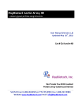
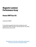
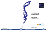
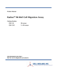
Instructions
For Research Use Only. Not For Use In Diagnostic Procedures
Directed In Vivo Angiogenesis Assay (DIVAA™)
Catalog #: 3450-048-K
Table of Contents
Directed In Vivo Angiogenesis
Section
I.
II.
III.
IV.
V.
VI.
Assay (DIVAA™)
Catalog #: 3450-048-K
48 Samples
VII.
VIII.
IX.
X.
Title
Background
Precautions and Limitations
Materials Supplied
Materials Required But Not Supplied
Reagent Preparation
Assay Protocols
A. Preparing for Implantation
B. Implanting Angioreactors
C. FITC-Lectin Detection
D. Calcein AM Detection
E. FITC-Dextran Detection
Data Interpretation
Troubleshooting
References
Appendices
A. Reagent Composition
B. Related Products from Trevigen
Page
1
1
1
2
2
3
4
7
9
9
10
11
12
13
14
©2011, Trevigen, Cultrex, PathClear and CultreCoat are registered trademarks, and DIVAA,
CellSperse, and AngioRack are trademarks, of Trevigen, Inc. Teflon is a registered trademark of Dupont Corporation.
i
E1/3/11v1
I. Background
IV. Materials/Equipment Required But Not Supplied
Please read the entire Instructions for Use prior to performing tests. Trevigen’s
TM
Directed In Vivo Angiogenesis Assay (DIVAA ), is the first in vivo system for
the study of angiogenesis that provides quantitative and reproducible results.1
The DIVAA system was developed for, and qualified using nude mice. Therefore,
optimization will be necessary for normal mouse strains.
During the course of the assay, implant grade silicone cylinders closed at one
end, called angioreactors, are filled with 20 µl of Trevigen's basement membrane
extract (BME) premixed with or without angiogenesis modulating factors. These
angioreactors are then implanted subcutaneously in the dorsal flanks of nude
mice. If filled with angiogenic factors, vascular endothelial cells migrate into, and
proliferate in the BME to form vessels in the angioreactor. As early as nine days
post-implantation, there are enough cells to determine an effective dose
response to angiogenic factors. The sleek design of the angioreactor provides a
standardized platform for reproducible and quantifiable in vivo angiogenesis
assays. Compared to the plug assay5, the angioreactor prevents assay errors
due to absorption of BME by the mouse. In addition, the angioreactor uses only a
fraction of the materials conserving both BME and test compounds used, and up
to four angioreactors may be implanted in each mouse, giving more data for
analysis. Trevigen’s DIVAATM has been used in evaluating the inhibition of
angiogenesis by TIMP-2,2 to study angiogenesis in matrix metalloprotease
(MMP)-2-deficient mice1 and enhancement of angiogenesis associated with
adrenomedullin3 and CD974. Trevigen’s DIVAATM was designed for assessing
angiogenesis activation by test compounds, and sufficient angiogenic factors are
provided for 8 FGF-2 controls and 8 positive controls.
II. Precautions and Limitations
1.
2.
For Research Use Only. Not for use in diagnostic procedures.
The physical, chemical, and toxicological properties of the products contained within
the Directed In Vivo Angiogenesis Assay may not yet have been fully investigated.
Therefore, Trevigen recommends the use of gloves, lab coats, and eye protection
while using any of these chemical reagents. Trevigen assumes no liability for damage
resulting from handling or contact with these products. MSDS sheets are available.
III. Materials Supplied
Catalog#
3450-048-01
Description
Angioreactors
3450-048-03
BME, Growth Factor Reduced
PathClear®
10X Wash Buffer
3450-048-04
FGF-2
3450-048-05
CellSperse™
3450-048-06
200X FITC-Lectin
3450-048-07
25X FITC-Lectin Diluent
3450-048-08
Heparin Solution
3450-048-B9
FGF-2(300 ng)/VEGF(100 ng)
3450-048-02
1
Quantity
48 units
Storage
4 oC
6 x 200 µl
-20 oC
25 ml
4 oC
100ng/10 µl
-20 oC
15 ml
-20 oC
250 µg/50 µl
4 oC
400 µI
4 oC
10 µl: 2 mg/ml
4 oC
10 µl
-20 oC
E1/3/11v1
Equipment
1. Mouse Cages/Facility
2. Laminar Flow Hood or Clean Room
3. Pipette helper
4. Micropipettor
5. CO2 incubator
6. Fluorescent plate reader or microscope equipped with fluorescein long pass filter
7. 500 ml graduated cylinder
8. Fine-point forceps
10. Fine-point cartilage forceps
11. Dissection scissors
12. Surgical scissors
13. Skin stapler
14. Scalpel
15. AngioRack™ (Catalog# 3450-048-09; sold separately)
Reagents
1. Nude Mice
2. Deionized water
3. DMEM, 10% FBS
4. 100 mg/ml Ketamine HCL (anesthesia)
5. 20 mg/ml Xylazine (analgesic)
6. Calcein AM
7. FITC-Dextran
8. Angiogenic-modulating factors (except FGF-2)
Disposables
1. Black 96 well fluorescence assay plate
2. Serological pipettes
3. Microscope slides and coverslips
4. Micropipettor tips
V. Reagent Preparation
1.
10X Wash Buffer
Dilute 25 ml of 10X Wash Buffer in 225 ml of sterile, deionized water.
2.
FGF-2 (100 ng)
Add 1 µI of Heparin Solution to 10 µI of FGF-2(100 ng), and gently pipette
up and down to mix immediately before addition to BME.
2
E1/3/11v1
3.
FGF-2(300 ng)/VEGF(100 ng)
Add 1 µI of Heparin Solution to 10 µI of FGF-2(300 ng)/VEGF(100 ng), and
gently pipette up and down to mix immediately before addition to BME.
4.
25X FITC-Lectin Diluent
Dilute 400 µI of 25X FITC-Lectin Diluent in 9.6 ml of sterile, deionized water.
5.
200X FITC-Lectin
Dilute 50 µI of 200X FITC-Lectin in 10 ml of 1X FITC-Lectin Diluent.
5.
6.
VI. Assay Protocol
eliminated by centrifuging 250 x g for 5 minutes at 4 oC.
Prepare to fill angioreactors. Angioreactors must be kept chilled on ice
prior to filling, whether inside microtubes or situated in an AngioRackTM.
Place angioreactors in the AngioRackTM. Add 20 µl of BME with or without
modulating factors to each angioreactor using a pre-chilled, sterile gelloading tip; see Figure 1. Be careful not to introduce bubbles into the
angioreactor. One tube will fill eight angioreactors; see Figure 2.
Once the eight angioreactors are filled, immediately invert angrioreactors and transfer to a sterile microtube, and place at 37 oC for 1 hour to
promote gelling (inverting angioreactors during gelling prevents the
formation of a meniscus at the open end of the angioreactor). Repeat
for the remainder of the angioreactors.
Note: The entire procedure must be conducted under sterile conditions
using aseptic technique to prevent contamination and subsequent infection in nude mice. The use of normal mice will require optimization.
A. Preparing Angioreactors for Implantation
1.
2.
3.
4.
Thaw Growth Factor Reduced BME at 4 oC, on ice, overnight prior to
assay. BME is to be kept on ice until gelling in step 6.
Pre-chill all pipette tips, angioreactors, AngioRackTM (Catalog# 3450048-09; sold separately), and angiogenesis modulating factors at 4 oC,
and keep BME on ice.
Working on ice, add angiogenic factors to one tube (200 µl) of Growth
Factor Reduced BME. Each tube of BME is sufficient for 8 angioreactors.
Add 10 µl of FGF-2 (100 ng) (Cat# 3450-048-04) or 10 µl of FGF-2(300
ng)/VEGF (100 ng) (Cat# 3450-048-B9), and 1 µl of Heparin Solution per
200 µl of BME to use for the positive control angioreactors. Add 11 µL of
sterile PBS, or test solvent per 200 µl BME to use for the negative control
angioreactors.
Still working on ice, add test angiogenesis modulating factors to the
remaining microtubes of Growth Factor Reduced BME; do not add more
than 10% total volume (over-diluting BME may compromise polymerization). Gently pipette up and down to mix test or control factors and BME;
be careful not to introduce bubbles into the BME. Bubbles may be
B. Implanting Angioreactors
7.
8.
9.
3
E1/3/11v1
Anesthetize each mouse immediately before implantation. Recommended: one part anesthesia, 100 mg/ml Ketamine HCL (not included), to
four parts analgesic, 20 mg/ml Xylazine (not included), injected
subcutaneously.
In a laminar flow hood using forceps, remove angioreactor from microtube; cap and save microtube for step 6. See Figure 3 for implant
preparation.
Incision should be made on the dorsal-lateral surface of a nude mouse,
approximately 1 cm above the hip-socket; see Figure 4. Start by
pinching back the skin and making a small cut using dissecting scissors.
Then extend cut to 1 cm in length, being careful not to puncture underlying tissues.
4
E1/3/11v1
10. Implant angioreactors into the dorsal flank of a mouse with the open end
opposite the incision; up to 2 angioreactors may be planted on each
side for a total of 4 angioreactors per mouse. See Figure 5 for implantation procedure and closure of the incision. Distribute angioreactors with like pairs in each mouse; see Figure 6 for recommended
distribution.
11. Maintain mice for 9 to 15 days; this step requires optimization. Longer
maintenance periods result in more vascularization.
5
E1/3/11v1
6
E1/3/11v1
C. FITC-Lectin Detection
12. After maintenance period, humanely euthanize mice. Exposure to CO2
levels greater than 70% for 5 minutes should be adequate.
13. Remove a 2 cm perimeter of skin surrounding angioreactors using
dissection scissors. Using a scalpel, cut along open end of angioreactor
to sever any vessels that may be growing into it. Recover angioreactor
using dissection forceps.
14. Carefully remove the bottom cap of the angioreactors with a sterile razor
blade, and using a sterile 200 µl pipette tip, push BME/vessel complex
out of angioreactor into the sterile microtube. See Figure 7 for vascularization in DIVAA™ Reduced Growth Factor BME plus FGF-2/VEGF.
7
E1/3/11v1
15. Rinse inside of each angioreactor with 300 µl of CellSperse™ and
transfer into a microtube. Dispose of empty angioreactors. Cap tube, and
incubate at 37 oC to digest BME and create a single cell suspension. This
may take 1 – 3 hours.
16. Dilute 25 mL DIVAA™ 10X Wash Buffer to 250 mL using deionized
water, and label “DIVAA™ Wash Buffer.”
17. Centrifuge digested BME at 250 x g for 5 minutes at room temperature
to collect cell pellets and insoluble fractions, and discard supernatant.
Resuspend pellet in 500 µl of DMEM, 10% FBS to allow for cell surface
receptor recovery, and incubate at 37 oC for one hour.
18. Centrifuge cells at 250 x g for 10 minutes at room temperature to collect
cell pellets. Resuspend pellet in 500 µl of DIVAA™ Wash Buffer to wash
cells, and centrifuge again. Discard supernatant and repeat wash two
more times.
19. Dilute 400 µl DIVAA™ 25X FITC-Lectin Dilution Buffer to 10 ml using
deionized water, and label “DIVAA™ FITC-Lectin Dilution Buffer.”
20. For each angioreactor, dilute 1 µl DIVAA™ 200X FITC-Lectin to 200 µl
using DIVAA™ FITC-Lectin Dilution Buffer, and label “DIVAA™ FITCLectin.”
21. Resuspend pellet in 200 µl of DIVAA™ FITC-Lectin, and incubate at 4 oC
overnight.
22. Centrifuge at 250 x g, and remove supernatant. Wash pellet three times
in DIVAA™ Wash Buffer as indicated in step 12.
8
E1/3/11v1
D. Optional Protocol for Calcein-AM Detection (not included in the
DIVAA kit).
1.
2.
3.
4.
5.
6.
7.
8.
After maintenance period, humanely euthanize mice. Exposure to CO2
levels greater than 70% for 5 minutes should be adequate.
Harvest angioreactors. Remove a 2 cm perimeter of skin surrounding
angioreactors using dissection scissors. Using a scalpel, cut along open
end of angioreactor to sever any vessels that may be growing into it.
Recover angioreactor using dissection forceps.
Carefully remove the bottom cap of the angioreactors with a razor
blade, and using a sterile 200 µl pipette tip, push BME/vessel complex
out of angioreactor into the sterile microtube. See Figure 6 for vascularization in DIVAA™ RGF BME plus angiogenic factors.
Rinse inside of angioreactors with 300 µl of CellSperse™ into microtube.
Dispose of empty angioreactors. Cap tube, and incubate at 37 oC to digest
BME and create a single cell suspension. This may take 1 – 3 hours.
Dilute 25 ml DIVAA™ 10X Wash Buffer to 250 ml using deionized
water, and label “DIVAA™ Wash Buffer.”
Centrifuge digested BME at 250 x g for 5 minutes at room temperature
to collect cell pellets and insoluble fractions, and discard supernatant.
Resuspend pellet in 500 µl of DIVAA™ Wash Buffer to wash cells, and
centrifuge again. Discard supernatant and repeat wash two more times.
Add 100 µl of 1 µM Calcein AM (in DIVAA™ Wash Buffer), and incubate
at 37 oC for 60 minutes.
Measure fluorescence in 96-well plates (excitation 485 nm, emission
510 nm); some fluorometers may require adjustment of Gain for an
optimal range of values (please consult your equipment user manual).
E. Optional Protocol for Dextran-FITC Detection (not included in
DIVAATM kit).
4.
5.
6.
Rinse inside of angioreactors with 300 µl of CellSperse™ into microtube.
Dispose of empty angioreactors. Cap tube, and incubate for 1 hour at 37 oC.
Clear incubation mix by centrifugation, 15,000 x g for 5 minutes at room
temperature.
Measure fluorescence of supernatant in 96-well plates (excitation 485
nm, emission 510 nm); some fluorometers may require adjustment of
Gain for an optimal range of values (please consult your equipment user
manual).
VII. Data Interpretation
Values for cell invasion will be expressed in Relative Fluorescent Units (RFUs).
Calculate the mean for each condition and its corresponding standard deviation.
Differences in conditions may be evaluated using a paired student’s t-test. For
inter-assay comparison, it may be more practical to compare relative invasion:
Relative invasion = Test sample (RFU) / Negative Control (RFU)
Data is usually plotted in a bar graph as such (amounts shown are per reactor):
Evaluation of Angiogeneis Activation Using DIVAA
8
7
Relative Invasion
23. Suspend pellet in 100 µl of DIVAA™ Wash Buffer for fluorometric determination.
24. Measure fluorescence in 96-well plates (excitation 485 nm, emission
510 nm); some fluorometers may require adjustment of Gain for an
optimal range of values (please consult your equipment user manual).
6
5
4
3
2
1
0
neg CTRL
1.
2.
3.
100 ng FGF
After maintenance period, inject 100 µl of 25 mg/ml Dextran-FITC in
DIVAA™ Wash Buffer via tail vein, and after 20 minutes, humanely
euthanize mice. Exposure to CO2 levels greater than 70% for 5 minutes
should be adequate.
Harvest angioreactors. Remove a 2 cm perimeter of skin surrounding
angioreactors using dissection scissors. Using a scalpel, cut along open
end of angioreactor to sever any vessels that may be growing into it.
Recover angioreactor using dissection forceps.
Carefully remove the bottom cap of the angioreactors with a razor
blade, and using a sterile 200 µL pipet tip, push BME/vessel complex
out of angioreactor into the sterile microtube. See Figure 7 for vascularization in DIVAA™ RGF BME with angiogenic factors.
9
E1/3/11v1
37.5 ng FGF/
12.5 ng VEGF
Data provided by John Basile
10
E1/3/11v1
VIII. Troubleshooting
Problem
Troubleshooting Guide
Problem
Cause
Use a more concentrated
compound formulation (do not
dilute BME more than 10%)
BME integrity has been
compromised by
inappropriate
shipping/storage or
contamination
Use new BME
Inadequate mixing of BME
and test compound
Mix BME and test com-pound
thoroughly by gently pipeting up
and down
Air pockets in angioreactor
Variability in
Assay
Inadequate mixing of BME
and test compound
Mix BME and test compound
thoroughly by gently pipeting
up and down
Air pockets in angioreactor
Do not use angioreactors
containing air pockets
Invert angioreactors when
gelling
Do not use angioreactors
containing air pockets
No or low
signal in
positive control
Invert angioreactors when
gelling
Implant up to 2 angio-reactors
in each preformed pocket in
dorsal flanks subcutaneously,
open end first inside pocket.
Improper implantation
Insufficient receptor
recovery after CellSperse™
treatment
High background in
negative
control
Solution
Solution
BME has been over diluted
BME does
not gel in
angioreactor
Cause
Use nude mice
Insufficient washing of cells
after FITC-Lectin Staining
Wash cells again in 1X Wash
Buffer
Implantation period is too
long
Reduce and optimize
implantation period
Gain is improperly set on
fluorometric plate reader
Adjust gain on fluoro-metric
plate reader within optimal
range
11
Allow cell surface receptors
to recover for 1 hour by
incubating cell in culture
media containing 10% FBS
Implantation period was not
sufficient to elicit angiogenic
response
Gain is improperly set on
fluorometric plate reader
Add Heparin to FGF-2 and mix
well before adding to BME
Extend and optimize
implantation period
Adjust gain on fluorometric
plate reader within optimal
range
IX. References
1.
E1/3/11v1
Insufficient receptor
recovery after CellSperse™
treatment
Omitting or inadequate mixing
of Heparin in FGF-2
Allow cell surface receptors to
recover for 1 hour by
incubating cell in culture media
containing 10% FBS
Use of C57Bl/6 mice
Improper implantation
Implant up to 2 angioreactors in each pre-formed
pocket in dorsal flanks
subcutaneously, open end
first inside pocket.
2.
3.
Guedez L, Rivera AM, Salloum R, Miller ML, Diegmueller JJ, Bungay PM,
Stetler-Stevenson WG. 2003. Quantitative assessment of angiogenic response
by the Directed In Vivo Angiogenesis Assay. American J Pathol 162:1431-1439.
Seo D, Li H, Guedez L, Wingfield PT, Diaz T, Salloum R, Wei B, StetlerStevenson WG. 2003. TIMP-2 Mediated inhibition of angiogenesis: An MMPindependent mechanism. Cell 114:171-180.
Martinez A, Vos M, Guedez L, Kaur G, Chen Z, Garayoa M, Pio R, Moody T,
Stetler-Stevenson WG, Kleinman HK, Cuttitta F. 2002. The effects of
adrenomedullin overexpression in breast tumor cells. J Natl Cancer Inst.
94:1226-37.
12
E1/3/11v1
4.
5.
6.
Wang T, Ward Y, Tian L, Lake R, Guedez L, Stetler-Stevenson WG, Kelly K.
2005. CD97, an adhesion receptor on inflammatory cells, stimulates
angiogenesis through binding integrin counterreceptors on endothelial cells.
Blood 105:2836-44.
Lee MS, Moon EJ, Lee SW, Kim MS, Kim KW, Kim YJ. 2001. Angiogenic activity
of pyruvic acid in in vivo and in vitro angiogenesis models. Cancer Res.
61:3290-3.
Basile JR, Holmbeck K, Bugge TH, Gutkind JS. 2007. MT1-MMP controls tumorinduced angiogenesis through the release of semaphorin 4D. J Biol Chem.
282:6899-905.
B. Related products available from Trevigen.
Catalog#
3450-048-SK
3450-048-IK
3470-096-K
A. Reagent Composition
2.
FGF-2(300 ng)/VEGF(100 ng) (Cat# 3450-048-B9)
300 ng FGF and 100 ng VEGF
3471-096-K
X. Appendices
1.
9.
3455-024-K
Angioreactor (Cat# 3450-048-01)
The angioreactor is a one centimeter long cylinder that is sealed on one
end and houses 20 µl total volume. It is made of implant-grade silicone
and provided sterile. Angiogenesis is directed into the cylinder at the
open end in response to angiogenesis modulating factors.
Growth Factor Reduced Basement Membrane Extract (BME) (Cat#
3450-048-02) BME is an extract from Engelbreth-Holm-Swarm (EHS)
tumor composed primarily of Laminin I, Collagen IV, and Entactin. BME
provides an angiogenesis permissive matrix for vessel formation in
response to angiogenic factors.
3.
10X Wash Buffer (Cat# 3450-048-03)
Proprietary buffer formulation.
5.
CellSperseTM (Cat# 3450-048-05)
A neutral metalloprotease from Bacillus polymyxa that provides for BME
digestion and gentle cell dissociation.
6.
200X FITC-Lectin (Cat# 3450-048-06)
Fluorescence labeled Griffonia Simplicifolia Lectin I binds to alpha-Dgalactosyl and N-acetyl galactosaminyl groups on the surface of
endothelial cells.
7.
25X FITC-Lectin Diluent (Cat# 3450-048-07)
Proprietary buffer formulation.
8.
Heparin Solution (Cat# 3450-048-08)
2 mg/mL Heparin.
13
3484-096-K
3455-096-K
3456-096-K
3457-096-K
3458-096-K
3465-096-K
3465-024-K
Size
48 samples
48 samples
96 tests
96 tests
24 inserts
96 samples
96 samples
96 samples
96 samples
96 samples
12 samples
Accessories:
Catalog#
3400-010-01
3446-005-01
3440-100-01
3442-050-01
3447-020-01
3410-010-01
3420-001-01
3416-001-01
3421-001-01
3417-001-01
3439-100-01
3438-100-01
3445-048-01
3430-005-02
3431-005-02
3432-005-02
3433-005-02
3437-100-K
3450-048-05
E1/3/11v1
Description
Cultrex® DIVAATM Starter
Cultrex® DIVAATM Inhibition Kit
Cultrex® In Vitro Angiogenesis Assay Endothelial
Cell Invasion Kit
Cultrex® In Vitro Angiogenesis Assay Tube
Formation Kit
24 Well BME Cell Invasion Assay
CultreCoat® 96 well BME-Coated Cell Invasion
Optimization Assay
Cultrex® 96 well BME Cell Invasion Assay
Cultrex® Laminin I Cell Invasion Assay
Cultrex® Collagen I Cell Invasion Assay
Cultrex® Collagen IV Cell Invasion Assay
Cultrex® 96 Well Cell Migration Assay
Cultrex® 24 Well Cell Migration Assay
Description
Mouse Laminin I
3-D Culture MatrixTM Laminin I
Rat Collagen I
Bovine Collagen I
3-D Culture MatrixTM Collagen I
Mouse Collagen IV
Human Fibronectin PathClear®
Bovine Fibronectin
Human Vitronectin PathClear®
Bovine Vitronectin
Poly-D-Lysine
Poly-L-Lysine
3-D Culture MatrixTM BME
BME with Phenol Red, PathClear®
BME with Phenol Red, Growth Factor
Reduced, PathClear®
Cultrex® BME, PathClear®
Cultrex® BME Growth Factor Reduced, PathClear®
Cultrex® Cell Staining Kit
CellSperseTM
Cultrex®
Cultrex®
Cultrex®
Cultrex®
Cultrex®
Cultrex®
Cultrex®
Cultrex®
Cultrex®
Cultrex®
Cultrex®
Cultrex®
Cultrex®
Cultrex®
Cultrex®
14
Size
1 mg
5 ml
100 mg
50 mg
100 mg
1 mg
1 mg
1 mg
50 μg
50 μg
100 ml
100 ml
15 ml
5 ml
5 ml
5 ml
5 ml
100 ml
15 ml
E1/3/11v1
The product accompanying this document is intended for research use only and
is not intended for diagnostic purposes or for use in humans.
Trevigen, Inc.
8405 Helgerman Ct.
Gaithersburg, MD 20877
Tel: 1-800-873-8443 • 301-216-2800
Fax: 301-560-4973
e-mail: [email protected]
www.trevigen.com
15
E1/3/11v1