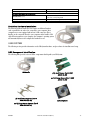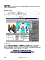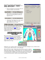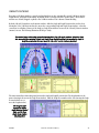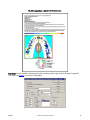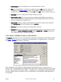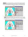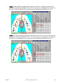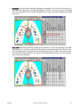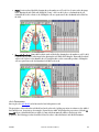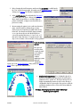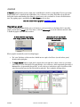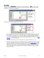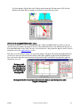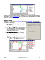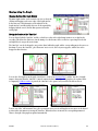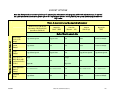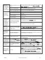
1
T-Scan III User Manual
®
computerized occlusal analysis system
12/14/05
T-Scan II User Manual (Rev I)
i
T-Scan III User Manual
®
v. 5.x
computerized occlusal analysis system
Tekscan, Inc . 307 West First St reet , South Boston, MA 02127
Te l: 61 7 .46 4 .45 00 /8 00 .248 .366 9 fa x : 61 7 .4 64 .4 266
Email: market ing@t ekscan.com web: www.tekscan.com
12/14/05
T-Scan II User Manual (Rev I)
i
Table Of Contents
WELCOME TO T-SCAN III....................................................................................................... 1
GETTING ASSISTANCE .....................................................................................................................................1
DECLARATION OF CONFORMITY ..........................................................................................................................2
WARRANTY INFORMATION ................................................................................................................................6
INTRODUCTION ...........................................................................................................................................7
CLINICAL APPLICATION ...................................................................................................................................7
QUICK START .............................................................................................................................................8
HARDWARE & INSTALLATION ............................................................................................... 11
COMPUTER REQUIREMENTS ............................................................................................................................11
EVOLUTION SYSTEM ....................................................................................................................................11
Evolution Component Identification ...................................................................................................................... 12
Evolution Handle ............................................................................................................................................... 12
Evolution Hardware Installation ........................................................................................................................... 15
USB SYSTEM............................................................................................................................................15
USB Component Identification.............................................................................................................................. 15
USB Handle....................................................................................................................................................... 16
USB Hardware Installation ................................................................................................................................... 19
SENSORS ................................................................................................................................................19
SOFTWARE ..................................................................................................................... 21
MAIN WINDOW .........................................................................................................................................21
TITLE BARS .............................................................................................................................................21
MENU ...................................................................................................................................................22
TOOLBAR................................................................................................................................................22
MAIN STATUS BAR ......................................................................................................................................25
REAL-TIME WINDOW ...................................................................................................................................25
2-D MOVIE WINDOW ..................................................................................................................................28
3-D MOVIE WINDOW ..................................................................................................................................32
GRAPH WINDOW ........................................................................................................................................33
GRAPH ZOOM WINDOW ................................................................................................................................36
LEGEND (COLOR SCALE) ...............................................................................................................................37
SMART DOCKING ........................................................................................................................................38
Local Smart Docking........................................................................................................................................... 39
Global Smart Docking ......................................................................................................................................... 39
Smart Docking: Graph Windows ............................................................................................................................ 39
AUTOMATIC WINDOW CONFIGURATION ................................................................................................................40
MULTIPLE MODE .......................................................................................................................................41
KEYBOARD SHORTCUTS .................................................................................................................................42
THE MAIN MENU .......................................................................................................................................43
File Menu ......................................................................................................................................................... 43
Edit Menu......................................................................................................................................................... 49
View Menu........................................................................................................................................................ 50
Options Menu.................................................................................................................................................... 61
Set User Preferences ........................................................................................................................................ 64
Settings ........................................................................................................................................................ 71
Movie Menu ...................................................................................................................................................... 72
Window Menu ................................................................................................................................................... 74
Help Menu ........................................................................................................................................................ 74
08/01/07
T-Scan III User Manual (Rev L)
i
RECORDING OCCLUSAL DATA ............................................................................................... 77
TAKING A RECORDING ..................................................................................................................................77
AUTONAMING ...........................................................................................................................................80
AUTOMATIC SENSITIVITY ADJUSTMENT .................................................................................................................81
REVIEWING A RECORDING ..............................................................................................................................82
INCLUDING COMMENTS WITH A RECORDING ...........................................................................................................83
LINKING A PHOTO TO A MOVIE FRAME .................................................................................................................86
ADVANCED RECORDING OPTIONS ......................................................................................................................91
Recording Settings.............................................................................................................................................. 91
CR (Centric Relation) Mode............................................................................................................................... 93
Turbo Mode .................................................................................................................................................. 94
Manual Sensitivity Adjustment.............................................................................................................................. 95
USING THE PATIENT FILE MANAGEMENT SYSTEM ....................................................................... 97
PATIENTS & THE PATIENT LIST .........................................................................................................................97
Accessing the Patient List ..................................................................................................................................... 97
Searching the System for a Patient ......................................................................................................................... 98
Patient Records ................................................................................................................................................. 98
Adding a New Patient to the System........................................................................................................................ 98
Opening an Existing Patient's Record ................................................................................................................... 100
Identifying a Patient by Diagnosis/Procedure ......................................................................................................... 101
Removing a Patient from the System .................................................................................................................... 102
PATIENT MOVIES .....................................................................................................................................103
Adding a New Movie to the System........................................................................................................................ 103
Adding an Existing Movie to the System................................................................................................................. 106
Exporting and Importing a Movie ........................................................................................................................ 107
Marking a Movie by Diagnosis or Procedure ........................................................................................................... 109
Opening an Existing System Movie ....................................................................................................................... 110
Removing a Movie from the System ...................................................................................................................... 112
AUTOMATIC PATIENT SYSTEM BACKUP FILES.........................................................................................................113
ANALYZING OCCLUSAL FORCE DATA...................................................................................... 115
DISPLAY OPTIONS ....................................................................................................................................115
2-D / 3-D Views ............................................................................................................................................... 115
COF & COF Trajectory........................................................................................................................................ 116
IP, Max and Delta............................................................................................................................................. 119
Arch Model, Table & Parameters........................................................................................................................... 120
Arch Model ................................................................................................................................................. 120
Arch Table .................................................................................................................................................. 123
Arch Parameters ........................................................................................................................................... 128
ADDING A LINE........................................................................................................................................131
Adding a Line to a Window................................................................................................................................. 131
GRAPHS ...............................................................................................................................................133
Displaying a graph ........................................................................................................................................... 133
Graph Data..................................................................................................................................................... 134
A-B / C-D Increment/Differential Lines ................................................................................................................. 135
Anterior and Posterior....................................................................................................................................... 136
Graph Preferences ............................................................................................................................................ 137
Manipulating the Graph..................................................................................................................................... 138
Graph Zoom.................................................................................................................................................... 142
Resizing the Graph Zoom window........................................................................................................................ 142
COPY & EXPORT OPTIONS ................................................................................................. 143
08/01/07
T-Scan III User Manual (Rev L)
ii
COPY OPTIONS........................................................................................................................................143
EXPORT OPTIONS .....................................................................................................................................145
PRINTING ..................................................................................................................... 147
PRINT SETUP ..........................................................................................................................................147
PRINTING GRAYSCALE .................................................................................................................................149
TROUBLESHOOTING ........................................................................................................ 151
TROUBLESHOOTING TABLE ...........................................................................................................................151
APPENDIX A: BACKGROUND FOR COF ANALYSIS........................................................................ 153
HOW THE CENTER OF FORCE IS CALCULATED ........................................................................................................153
RATIONALE FOR CENTER OF FORCE TARGET ..........................................................................................................153
APPENDIX B: USING THE EXTERNAL TRIGGER & TRIG-SYNCH BOX ................................................. 155
SETTING UP THE SOFTWARE FOR EXTERNAL TRIGGERING ............................................................................................155
INSTALLING THE TS-100 HARDWARE ON YOUR SYSTEM .............................................................................................156
APPENDIX C: VIDEO FEATURES ........................................................................................... 159
USING THE TRIG-SYNCH BOX TO TAKE A VIDEO......................................................................................................159
USING VIDEO OPTIONS TO ANALYZE PRESSURE DATA ................................................................................................159
Opening a Linked Video..................................................................................................................................... 159
SYNCHRONIZING AND EDITING .......................................................................................................................160
Synchronizing and Editing a Video ...................................................................................................................... 161
APPENDIX D: BIORESEARCH INTEGRATION MODULE ................................................................. 163
USING THE BIOEMG INTEGRATION MODULE ........................................................................................................163
APPENDIX E: BIBLIOGRAPHY.............................................................................................. 167
BIBLIOGRAPHIC DATA ................................................................................................................................167
APPENDIX F: THIRD-PARTY SOFTWARE SUPPORT ..................................................................... 169
THIRD-PARTY SOFTWARE SUPPORT DATA ...........................................................................................................169
BRIDGING THE TEKSCAN DATABASE WITH OTHER APPLICATIONS ....................................................................................170
08/01/07
T-Scan III User Manual (Rev L)
iii
WELCOME TO T-SCAN III
GETTING ASSISTANCE
Tekscan, Inc. will provide technical assistance for any difficulties you may experience using your T-Scan III system for
90 days from the system shipping date. After 90 days, Tekscan offers annual Technical Support and System Maintenance
Plans or customer support at our standard rates per incident. An incident is defined as one single issue or problem.
Contact Tekscan for additional T-Scan III sensors. Standard sensors are available in two sizes; small and large. Contact
us for current pricing and availability.
The flexible manufacturing process used to make sensors allows us to design custom sensors for applications in which
standard sensors are not suitable. Custom pressure -sensitive materials can be formulated to produce a sensor whose
sensitivity is well matched to a particular application. Contact your Tekscan representative to discuss custom sensors for
your special applications.
Write, call, or fax us with any concerns or questions. Our knowledgeable support staff will be happy to help
you. Comments and suggestions are always welcome.
Tekscan, Inc.
307 West First Street
South Boston, MA 02127-1309
Phone: (617) 464-4500
Or
(800) 248-3669 in U.S. and Canada
Fax: (617) 464-4266
E-mail: [email protected]
Or visit our website at: www.tekscan.com
Copyright © 2007 by Tekscan, Inc. All rights reserved. No part of this publication may be reproduced, transmitted,
transcribed, stored in a retrieval system, or translated into any language or computer language, in any form or by any
means without the prior written permission of Tekscan, Inc., 307 West First Street, South Boston, MA 02127-1309.
Tekscan, Inc. makes no representation or warranties with respect to this manual. Further, Tekscan, Inc. reserves the
right to make changes in the specifications of the product described within this manual at any time without notice and
without obligation to notify any person of such revision or changes.
T-Scan is a registered trademark of Tekscan, Inc.
Microsoft Windows and MS-DOS are registered trademarks of Microsoft Corporation.
08/01/07
T-Scan III User Manual (Rev L)
1
DECLARATION OF CONFORMITY
Tekscan is ISO 9001:2000 & 13485 registered.
The Tekscan T-Scan III System has been tested and conforms to the following standards for a "Component Medical
Device - Tactile Sensor System":
Europe
EN-60601-1, EN55011, IEC601-1-2, IEC801-2, IEC801-3, IEC801-4, IEC801-5
USB Handle Conformity
Safety
EMC
US
UL60601-1
IEC60601-1-2
Canada
CSA60601-1
IEC60601-1-2
Europe
EN60601-1
EN60601-1-2
World
CB
CB
Test Standards/Methods:
BS EN61326:1998 - EMC for Laboratory Test and Measurement Equipment
BS EN60601-1-2:2002 - EMC for Medical Electrical Equipment
CFR 47, Part 15 - Emissions for Unintentional Radiators (15.107, 15.109)
EN61000-4-2:1995 - Electrostatic Discharge Immunity
EN61000-4-3:1996 - Radiated Electromagnetic Field Immunity
EN61000-4-4:1995 - Electrical Fast Transient Immunity
EN61000-4-5:1995 - Surge Immunity
EN61000-4-6:1996 - Conducted Disturbances RF Immunity
EN61000-4-8:1993 - Power Frequency Magnetic Immunity
EN61000-4-11:1994 - Voltage Dips and Dropouts
EN61000-3-2:2000 - Harmonic Current Emissions
EN61000-3-3:1995 w/corr. July 1997 - Voltage Fluctuations and Flicker
Test Parameters:
BS EN 61326:1998 – Class B Emissions Limits (Default to Class A)
EN55011:1998 - Group 1, Class B (Default to Class A)
CFR 47, Part 15 - Class B (Default to Class A)
EN61000-4-2:1995 - ±6kV Contact, ±8kV Air
EN61000-4-3:1996 - 3V/m, 80MHz to 2.5GHz, w/ 80% 1kHz AM
EN61000-4-4:1995 - ±2kV Mains, ±1kV I/Os
EN61000-4-5:1995 - ±2kV Common Mode, ±1kV Differential Mode
08/01/07
T-Scan III User Manual (Rev L)
2
EN61000-4-6:1996 - 3Vrms, 150kHz to 80MHz, w/ 80% 1kHz AM
EN61000-4-8:1993 - 50Hz and 60Hz
EN61000-4-11:1994 – <5% 0.5 Cycles, 40% 5 Cycles, 70% 25 Cycles, <5% 5 Sec.
EN61000-3-2:2000 – A14 Class A Harmonic Emissions Limits
EN61000-3-3:1995 w/corr. July 1997 – Voltage Fluctuation and Flicker Limits
Warnings:
1. Medical electrical equipment needs special precautions regarding EMC and needs to be installed and put into
service according to the EMC information provided in the accompanying documents.
2. Portable and Mobile RF Communications Equipment can affect medical electrical equipment.
3. The use of accessories and cables other than those specified by the manufacturer as replacement parts may
result in increased emissions or decreased immunity of the equipment or system.
4. Only use Tekscan supplied battery packs and power sources to avoid damaging the system.
5. Do not use or attach any components that are not explicitly stated within this manual.
6. Do not connect any additional multiple portable socket outlet(s) or extension cord(s) to the system.
7. EMC (Electro-Magnetic Charge) can interfere with the system. If this occurs, or if there is a high level of noise
on your display screen, try moving to a location that is not in proximity to other electrical devices (such as
Televisions, radios, and cell phones).
8. ESD (Electro-Static Discharge) can halt the system. If the system stops functioning, shut down the system by
turning the power switches on all attached parts off. Also shut down the software. Then turn on the system and
restart the software. If problem persists, make sure the humidity in the room is >30% and refrain from
touching patient after equipment is installed and powered up. If you are still having difficulty in operating the
system, contact your local Tekscan representative.
9. The computer used with the applied part must be at a minimum
approved to 60950-1. If the computer is to be used within the
patient environment then it must also be approved to IEC606011 or have a medically approved isolation transformer between
the computer and the mains voltage. This setup must be tested
by a qualified technician to meet the requirements of IEC606011-1.
10. If a non-medically approved computer is being used, do not
touch both the computer and the patient at the same time.
11. If using parts other than those explicitly stated within this
manual, always follow the manufacturer’s cleaning instructions.
12. Do not disinfect the computer.
13. Dispose of applied parts in accordance with Federal and State
guidelines pertaining to computer equipment.
14. Sensor Replacement/Disposal: If using the MatScan, HR Mat,
Presto-Scan or Walkway systems, when ordering new sensors,
the sensor/sensor housing should be returned to Tekscan.
Tekscan will use the original sensor housing when replacing these sensors. Always dispose of sensors in
accordance with Federal and State guidelines pertaining to medical biohazardous waste.
15. Protection against electric shock: Internally powered equipment.
16. No user-serviceable parts. Do not try to service or take apart any Tekscan hardware. Consult with your Tekscan
representative if a component is not working correctly, or is not working as it should.
17. Tekscan systems (including sensors and scanning handles) are intended for use only in non-invasive medical
applications.
08/01/07
T-Scan III User Manual (Rev L)
3
Guidance Tables
Table 201 – Guidance and Manufacturer’s Declaration – Emissions
All Equipment and Systems
Guidance and Manufacturer’s Declaration - Emissions
The EH-1 is intended for use in the electromagnetic environment specified below. The customer or user of the EH-1
should ensure that it is used in such an environment.
Emissions Test
RF Emissions
CISPR 11
Compliance
Group 1
RF Emissions
CISPR 11
Harmonics
IEC 61000-3-2
Flicker
IEC 61000-3-3
Class B
Class A
Complies
Electromagnetic Environment – Guidance
The EH-1 uses RF energy only for its internal
function. Therefore, its RF emissions are very low
and are not likely to cause any interference in
nearby electronic equipment.
The EH-1 is suitable for use in all establishments,
including domestic, and those directly connected
to the public low-voltage power supply network
that supplies buildings used for domestic
purposes.
Table 203 – Guidance and Manufacturer’s Declaration – Emissions
Life-Supporting Equipment and Systems
Guidance and Manufacturer’s Declaration – Emissions
The EH-1 is intended for use in the electromagnetic environment specified below. The customer or user of the EH-1
should ensure that it is used in such an environment.
Immunity Test
Conducted RF
IEC 61000-4-6
Radiated RF
IEC 61000-4-3
IEC 60601 Test Level Compliance Level
3 Vrms (Outside ISM)
(V1)Vrms
10Vrms (In ISM Bands) (V2)Vrms
150 kHz to 80 MHz
(E1)V/m
10 V/m
80 MHz to 2.5 GHz
Electromagnetic Environment – Guidance
Portable and mobile communications equipment
should be separated from the EH-1 by no less
than the distances calculated/listed below:
D=(3.5/V1)(Sqrt P) D=(12/V2)(Sqrt P)
D=(3.5/E1)(Sqrt P) 80 to 800 MHz
D=(7/E1)(Sqrt P) 800 MHz to 2.5 GHz
where P is the max power in watts and D is the
recommended separation distance in meters.
Field strengths from fixed transmitters, as
determined by an electromagnetic site survey,
should be less than the compliance levels (V1 and
E1).
Interference may occur in the vicinity of
equipment containing a transmitter.
08/01/07
T-Scan III User Manual (Rev L)
4
Table 204 – Guidance and Manufacturer’s Declaration – Emissions
Equipment and Systems that are NOT Life-supporting
Guidance and Manufacturer’s Declaration – Emissions
The EH-1 is intended for use in the electromagnetic environment specified below. The customer or user of the EH-1
should ensure that it is used in such an environment.
Immunity Test
Conducted RF
IEC 61000-4-6
IEC 60601 Test
Level
3 Vrms
150 kHz to 80 MHz
Compliance
Level
(3)Vrms
(3)V/m
Radiated RF
IEC 61000-4-3
3 V/m
80 MHz to 2.5 GHz
Electromagnetic Environment – Guidance
Portable and mobile communications equipment should
be separated from the EH-1 by no less than the distances
calculated/listed below:
D=(1.16)(Sqrt P)
D=(1.16)(Sqrt P) 80 to 800 MHz
D=(2.33)(Sqrt P) 800 MHz to 2.5 GHz
where P is the max power in watts and D is the
recommended separation distance in meters.
Field strengths from fixed RF transmitters, as determined
by an electromagnetic site survey, should be less than the
compliance levels in each frequency range.
Interference may occur in the vicinity of equipment
containing a transmitter.
Table 206 – Recommended Separation Distances between portable and mobile RF Communications equipment
and the EH-1
Equipment and Systems that are NOT Life-supporting
Recommended Separations Distances for the EH-1
The EH-1 is intended for use in the electromagnetic environment in which radiated disturbances are controlled. The
customer or user of the EH-1 can help prevent electromagnetic interference by maintaining a minimum distance
between portable and mobile RF Communications Equipment and the EH-1 as recommended below, according to the
maximum output power of the communications equipment.
Max Output Power
(Watts)
0.01
0.1
1
10
100
08/01/07
Separation (m)
150kHz to 80MHz
D=(3.5/V1)(Sqrt P)
0.11
0.36
1.16
3.68
11.66
Separation (m)
80 to 800MHz
D=(3.5/E1)(Sqrt P)
0.11
0.36
1.16
3.68
11.66
T-Scan III User Manual (Rev L)
Separation (m)
800MHz to 2.5GHz
D=(7/E1)(Sqrt P)
0.27
0.86
2.72
8.60
27.22
5
WARRANTY INFORMATION
Tekscan, Inc.
Limited 1-Year Warranty
1. WARRANTY. Tekscan, Inc. warrants to the original purchaser of this product that should it prove defective by
reason of improper workmanship and/or materials:
A. Tekscan Systems and Components:
For one year from the date of original purchase at retail, Tekscan will repair or replace, at our option, any
defective part without charge for the part or labor if an inspection proves the claim. Parts used for
replacement may be used or rebuilt, and are warranted for the remainder of the original warranty period.
B. Tekscan Sensors:
Tekscan will replace any Tekscan Sensor which fails due to manufacturing defect if an inspection proves the
claim. Claims must be made within 30 days of purchase.
2. TO OBTAIN WARRANTY SERVICE, call Tekscan at 1-800-248-3669, (617) 464-4500 in MA, for further
instructions. Should you be asked to deliver your product to Tekscan, Inc. in Boston, MA, shipping expenses
are the purchaser’s responsibility. Proof of purchase is required when requesting warranty service.
3. THIS WARRANTY DOES NOT COVER defects caused by modification, alteration, repair or service of the
enclosed product by anyone other then Tekscan or an authorized Tekscan service center, physical abuse to,
misuse of, the product or operation thereof in a manner contrary to the accompanying instructions, or
shipment of the product to Tekscan or an authorized Tekscan service center for service. This warranty also
excludes all costs arising from installation, cleaning or adjustments of user controls. Consult the operating
manual for information regarding user controls.
1. ANY EXPRESS WARRANTY NOT PROVIDED HEREIN, AND ANY REMEDY FOR BREACH OF CONTRACT WHICH,
BUT FOR THIS PROVISION MIGHT ARISE BY IMPLICATION OR OPERATION OF LAW, IS HEREBY EXCLUDED
AND DISCLAIMED. THE IMPLIED WARRANTIES FOR THE MERCHANTABILITY AND OF FITNESS FOR ANY
PARTICULAR PURPOSE ARE EXPRESSLY LIMITED TO A TERM OF ONE YEAR. SOME STATES DO NOT ALLOW
LIMITATIONS ON HOW LONG AN IMPLIED WARRANTY LASTS, SO THAT THE ABOVE LIMITATION OR
EXCLUSION MAY NOT APPLY TO YOU. THE WARRANTIES SET FORTH HEREIN ARE IN LIEU OF ANY AND ALL
OTHER WARRANTIES EXPRESS OR IMPLIED INCLUDING THE WARRANTY OF MERCHANTABILITY AND FITNESS.
THE BUYER ACKNOWLEDGES THAT NO OTHER REPRESENTIONS WERE MADE TO HIM OR RELIED UPON BY
HIM WITH RESPECT TO THE QUALITY AND FUNCTION OF THE GOODS SOLD HEREIN. NO PERSON, FIRM OR
CORPORATION IS AUTHORIZED TO ASSUME FOR US ANY LIABILITY IN CONNECTION WITH THE SALE OF
THESE GOODS.
5. UNDER NO CIRCUMSTANCES shall Tekscan, Inc. be liable to purchaser or any other person for any special or
consequential damages, whether arising out of breach of warranty, breach of contract, or otherwise. Some
states do not allow the exclusion or limitation of incidental or consequential damages, so that the above
limitation or exclusion may not apply to you.
08/11/03 — FORM-200-057-B
08/01/07
T-Scan III User Manual (Rev L)
6
INTRODUCTION
This manual describes how to use Tekscan’s T-Scan III Computerized Occlusal Analysis. T-Scan III is a reliable and
easy-to-use clinical diagnostic device that senses and analyzes occlusal contact forces using paper-thin, disposable
sensors. The T-Scan III system comes with a full-featured Patient File Management system, which makes storing patient
records and tracking occlusal recordings simple, and makes the system an integral component of the clinical
workstation for occlusal diagnosis and treatment.
The T-Scan III software offers features that allow the user to:
•
•
•
Record the patient’s occlusal contact data
View the patient’s tooth contacts and associate them with specific teeth
Analyze the data, with the force and time relationships of the contacts displayed as color contour images
showing:
o
o
o
o
•
Instant IP
Center of Force
Center of Force Trajectory
More advanced features, such as: Max, Delta, Graph, Graph Zoom and 3-D Columns
Manage patient records and movie files through the use of an intuitive database
The T-Scan III system is comprised of the Microsoft (MS) Windows-based T-Scan III software, the associated
hardware, and patented Tekscan sensors. The system’s versatility allows you to copy occlusal contact data (as an image
or text file of actual force percentage values) and paste it into other Windows applications, or to print this information
to any of the hundreds of Windows-compatible color or grayscale printers.
This manual provides a thorough description of the system’s features and capabilities. Follow the Quick Start section as
a guideline, and refer to specific sections for more detailed instructions on how to use each feature.
CLINICAL APPLICATION
The T-Scan III technology has application in all phases of dentistry in which occlusal diagnosis and treatment are
involved, and remains the only practical quantitative method to analyze the occlusion.
The analytical software displays, such as "Center of Force (COF)" and "Center of Force Trajectory," provide in-depth
understanding of the overall balance of the occlusion, which cuts to the heart of occlusal analysis. Center of Force
analysis allows the dentist to examine the total effect of restorative dentistry on the patient’s maximum closure and
excursions, and is as easy as the click of a mouse.
Other features, such as instant "IP" and "Max," summarize critical occlusal data and give the dentist valuable assistance
in the diagnosis and treatment planning of occlusal disease, such as trauma caused by transient forces and interceptive
contacts. T-Scan III also uses subtractive technology, as shown in the "Delta" feature, to isolate slides and other
pathology from the occlusion, providing previously unavailable information.
Since the introduction of the original product in 1987, the use of computerized occlusal analysis has gained importance
in implant dentistry, patient education, and complex restorative and TMJ diagnosis and treatment.
It is clear that access to occlusal force and time data is critical to the discerning clinician, whether he or she is doing
something as complex as tracking the changing forces on implants in relation to natural teeth, or simply perfecting the
occlusion on a bite guard. T-Scan III has a wide range of clinical applications and, because of this, Tekscan provides
T-Scan application guides to assist the T-Scan operator. These application guides automatically install to the desktop
in a folder called “Manuals” when loading the software.
08/01/07
T-Scan III User Manual (Rev L)
7
For many practitioners, patient education is the most important application of the technology. The T-Scan III software
displays make occlusal problems easily understandable, increasing patient involvement and treatment acceptance. TScan III provides a valuable source of previously unavailable information, which can become a permanent part of the
patient’s record.
QUICK START
Welcome to Quick Start! This section is a quick look at how to use your T-Scan III system, and is intended to get you
started using your system in just a few easy steps. This Quick Start procedure should be followed as a general outline; it
will give you the basics on how to view tooth contact data in a Real-time window, record this data, play the recording
back, and analyze the occlusal information. However, it is highly recommended that you read the entire manual before
using the system in a patient test.
Note: You must read the entire manual before performing any tests, as this only provides general
guidelines.
Note: Familiarity with MS Windows is assumed.
Note: This procedure assumes that the proper hardware and software has been successfully
installed on your system (refer to the Installation section).
The following outlines the proper procedures to take a basic recording with your T-Scan III system:
1. Make sure that the T-Scan III software has been successfully installed on your system, and the sensor support
and sensor are correctly inserted into the Evolution or USB handle. The handle must be plugged into the
computer, and recognized.
2. Double click the shortcut icon that has been placed on your desktop, or click on the Start button at the bottom
left of the screen, select Programs, and then click the T-Scan III icon to run the program.
3. The Main window opens with the Patient List.
4. In the Patient List ("Patients" dialog), click New Patient. A new Patient Record is opened , along with the Arch
Table. Enter the new patient’s information (at a minimum, you must enter the patient’s first and last names).
Ensure the central incisor width is correctly entered, so that embrasure lines are approximately representative
of the patient’s bite. Refer to the Adding a New Patient to the System section for further information.
5. Click the New Movie button in the Patient Record. A blank Real-time window appears for the new patient.
The left side of the Real-time status bar should display "Sensor OK." If the sensor is not aligned correctly, the
message displays "MISALIGNED!" and the program prompts you to “Please insert a sensor into the handle”. If
you get this message, remove and reinsert the sensor into the handle, ensuring that the sensor is right side up,
and the handle latch is completely down. If the "Sensor OK" message still does not appear, consult the
Troubleshooting section.
6. Place the sensor in the patient’s mouth, with the sensor support pointer between the two central incisors. Keep
the handle as parallel to the occlusal plane as possible.
7. Press the Record Start & Stop button on the handle. Wait for your computer to “beep.” This means the system
is ready to record. If your computer does not have sound, or you do not hear the beep for some reason, look at
the bottom of the Real-time window; the system is ready to record if the status bar displays "Rec. Frame 0 of
300".
08/01/07
T-Scan III User Manual (Rev L)
8
8. Have the patient bite down normally on the sensor. The first tooth contact causes the system to begin recording
(refer to the Recording Occlusal Data section for details). While recording is in progress, the Real-time Status
Bar displays the frame count progress.
9. When the correct number of frames have been collected, recording automatically stops. If you want to manually
stop recording before this time, press the Record Start & Stop button on the handle once again. The recording
stops.
10. As soon as recording is completed, the Real-time window becomes a 2-D Movie window, which is divided into
two equal colored boxes (one green for the left side and one red for the right side) around the mid-sagittal
plane. A 3-D Movie window, Graph window and Graph Zoom window are also automatically opened for the
current movie. The Graph and Graph Zoom windows contain color-coded "traces" representing the forces
inside each of the colored boxes in the 2-D Movie window.
The recorded occlusal data can now be reviewed and analyzed. To review the recording, use the movie controls
on your toolbar. Refer to the Reviewing a Recording section for further information.
11. To save your movie as a file that can be opened later, select Save Movie from the File menu. The newly
recorded movie is automatically named using the patient’s first, last and middle names. Refer to the
Autonaming section for further information.
The movie is then saved into the patient’s folder in the "C:\TScan\Database\Movies" directory. The file is saved
as a T-Scan III "movie" file (extension *.fsx).
When you save a movie, you are asked whether or not you would like the movie to be part of the database.
Refer to the Using the Patient File Management System section for more information.
12. You may now analyze the recorded data. The following View menu options are essential for the analysis of
occlusal forces:
•
•
COF (Center of Force)
& COF Trajectory
Add Line
•
IP (Intercuspal Position)
•
Delta
•
MAX (Maximum Cumulative Force)
•
Arch Model
Note: Refer to the Analyzing Occlusal Force Data section for a detailed description of each option.
13. Copy the force data from the Movie or Graph window. To do this, select Copy from the Edit menu. Alternately,
you can hover your mouse over a window and right-click to select Copy. The active window is copied to the MS
Windows clipboard as both text and an image (bitmap) that can be pasted into other Windows programs. Refer
to the Copy & Export Options section for more information.
14. To print the active window, first select Print Setup from the "File" menu and choose your desired Print Setup
options. When your options have been selected, click OK. Next, Select Print Preview from the "File" menu to
review the printed output display. If you are happy with the results, click the Close button, and then select Print
from the "File" menu. Finally, click OK in the "Print" dialog. The active window is printed. Refer to the Printing
section for more details.
You have completed the Quick Start section. You should now be familiar with T-Scan III, and see how easy it is
to view, save, and analyze the occlusal forces on the sensor. The rest of the manual gives you detailed
instructions on how to use the system.
08/01/07
T-Scan III User Manual (Rev L)
9
HARDWARE & INSTALLATION
This section presents instructions for hardware installation, and also provides the system requirements necessary to
support the T-Scan III Occlusal Diagnostic system.
COMPUTER REQUIREMENTS
Suggested Minimum PC Requirements for all Tekscan systems (Desktop or Laptop)
•
Pentium 600 Mhz processor (Intel Dual Core 1.6 Ghz processor recommended)
•
128 MB RAM (512 MB RAM recommended)
•
1 GB hard drive
•
1 CD ROM drive
•
1 USB Port (not required for TireScan, F-Scan, HR Mat, Walkway, Grip, Hoof, or Saddle PCI-based
systems). USB 2.0 required for VersaTek systems.
•
1 PCI full length expansion slot (required only for TireScan, F-Scan, HR Mat, Walkway, Grip, Hoof, or
Saddle PCI-based systems)*
•
Windows 2000 (SP4) or XP (SP2), or Vista operating system**
* F-Scan, HR Mat, Walkway, Grip, Hoof, and Saddle USB-based systems are also available. TireScan is only available
as a PCI-based system.
* Using a PCI-based system with a laptop will require a PCMCIA Cardbus to PCI expansion system. One such system
is Magma model CB1F made by www.magma.com. Before purchase, ensure your laptop computer is also
compatible with the magma system.
** PCI-based systems can also be run on Windows 98SE or ME operating systems.
Requirements for Video Add-on (purchased separately and in addition to the above Suggested Minimum PC
Requirements)
•
20 GB Hard drive (ATA-33, 7200 RPM speed)
•
Firewire (iLink or IEEE1394) port
•
DV (digital video) format camcorder with Firewire (iLink or IEEE1394) port*
* Currently, DV format is the only video format supported for capturing video directly using Tekscan software.
Cameras that use other formats than the standard DV format are not compatible for video capture. With video
cameras using other non-DV formats, a third party software must be used to transfer video from the camera to the
computer.
EVOLUTION SYSTEM
The following section provides information on the Evolution System hardware, and procedures for installation and
setup.
08/01/07
T-Scan III User Manual (Rev L)
11
Evolution Component Identification
The following table provides a view of all the components that ship with your Evolution System:
Evolution Handle
(10) Small Sensors (Part # ZSEN
(2501) T1-HD)
and (10) Large Sensors (Part # ZSEN
(2001) T1-HD)
(2) Small Sensor Supports (Part #
ZSEN-SUPPORT-SM)
System Software
and (2) Large Sensor Supports (Part #
ZSEN-SUPPORT-LG)
User Manual
Evolution System Carrying Case
Evolution Handle
The Evolution Handle gathers the data from the sensor and processes it so that it can be sent easily to the computer. The
buttons on the sensor handle may also be used to start or stop a recording. This function is referred to as ‘Remote
Recording’.
The Evolution Handle has a latch on its topside. In the ‘Up’ position, the latch retracts the contact pins inside the handle
to allow insertion of the sensor tab. The sensor tab is placed into the sensor handle. The handle’s attached USB cable is
then connected directly to your computer via the USB port.
08/01/07
T-Scan III User Manual (Rev L)
12
The following displays the buttons and
their functionality for the Evolution
Handle:
The following shows an image of the
Evolution Handle with attached USB cable:
The following label is printed on all Evolution
Handles displaying the compliancy, model
number, warnings and parameters:
•
•
•
•
•
Sensor OK Green LED Indicator: A green light here indicates that the sensor is correctly inserted into the
handle and a Real-Time window can be opened.
Record Mode Green LED Indicator: A green light here indicates that the sensor is recording pressure data
and transferring that data to your computer.
New Real-Time Window Button: This will open a new Real-Time window in the software, so that you can
begin recording pressure data.
Record Start & Stop Button: Use this button to start a recording or stop a recording that is in progress.
Power Green & Yellow LED Indicator: When yellow, this light indicates that the handle is receiving power,
but is not yet initialized. When Green, this light indicates that the handle is receiving power and has been
initialized by the computer (i.e.: the device shows up under the Windows device manager).
To insert a sensor into the Evolution Handle:
1. Select a sensor support (either large or small to match the sensor to be used with the patient).
2. Ensure that the Evolution Handle latch, and the sensor support’s locating pointer, are both facing up.
3. Insert the sensor support into the Evolution Handle.
4. Lift the latch on the top of the Evolution Handle.
5. Slide the sensor tab (with ‘This Side UP’ facing upwards) under the pointer on the support, and into the
handle. Slide the sensor in until it reaches its mechanical stop. Do not force the sensor into the handle!
08/01/07
T-Scan III User Manual (Rev L)
13
6. Close the latch completely.
Once the sensor is inserted into the handle, connect the
attached USB cable to the computer’s USB port. Your
computer should automatically detect the new hardware and
configure it for your system. If the driver is not found, insert
the Tekscan software CD that came with your system and have
the computer locate the driver on your CD. Then follow the onscreen instructions.
After following this procedure and starting the software, the message below the real-time window for the sensor should
be “Sensor OK.” If the message is “MISALIGNED,” re-insert the sensor using the above procedure. If the “Sensor OK”
message still does not appear, consult the Troubleshooting section.
Caution! Do not allow the handle to hang from the sensor. The handle may become damaged,
resulting in a misaligned sensor.
Evolution Handle Maintenance and Care
•
The Evolution Handle cannot be autoclaved.
•
Do not let any liquid drip onto the electronics inside the handle. If this occurs, the handle will stop working
and must be allowed to dry for 24 hours. You can use your air syringe, however, to significantly reduce this
drying time. Do not attempt to dry out the handle using any other method, or you may destroy the delicate
electronics.
•
To properly clean the device, be sure to wipe down the handle and sensor supports with a 70% Isopropyl
Alcohol solution. To do this, slightly dampen a cloth with the alcohol solution, careful not to soak or saturate
the cloth. Then wipe the handle and sensor supports after each use.
Evolution Handle Specifications
COMMUNICATION/DATA ACQUISITION:
COMMUNICATION PROTOCOL TO HOST COMPUTER
SCAN SPEED
DIGITAL PRESSURE RESOLUTION
ELECTRICAL:
POWER SOURCE
POWER CONSUMPTION
MECHANICAL:
USB CABLE:
LENGTH in (mm)
WEIGHT lbs (kgs)
HANDLE ENCLOSURE:
SIZE LxWxH in (mm)
OPEN LEVER HEIGHT in (mm)
WEIGHT lbs (kgs)
AMBIENT OPERATING CONDITIONS:
TEMPERATURE: oF (oC)
08/01/07
USB 1.1 or 2.0 Compatible, 12 Mbps
Up to 100 Hz. (~500 Hz with Turbo Mode).
8 BIT
Host Computer’s USB BUS
200mA MAX at 5V
180 (4572)
0.40 (0.18)
5.42x2.25x1.88 (137.7x57.2x47.6)
4.30 (109.2)
0.40 (0.18)
14 to 131 (-10 to 55) Prolonged use at high
Temperatures should be avoided
T-Scan III User Manual (Rev L)
14
HUMIDITY: %
PRESSURE: psi (kPa)
STORAGE AND TRANSPORT CONDITIONS:
0 to 90 (non condensing)
1.7 to 14.7 (11.6 to 101.3) (sea level to 50,000 ft.)
TEMPERATURE: oF (oC)
-4 to 131 (-20 to 55) Short-Term
41 to 104 (5 to 40) Long-Term
0 to 90 (non condensing)
1.7 to 14.7 (11.6 to 101.3) (sea level to 50,000 ft.)
HUMIDITY: %
PRESSURE: psi (kPa)
Evolution Hardware Installation
The Tekscan Evolution handle does not require an additional interface
card or parallel box in order to be connected to your computer. Most
computers now come equipped with at least 2 USB connectors. These
handles can be connected directly to your computer via the handle’s USB
cable. When inserted into your computer, the computer’s operating system
will automatically detect and configure the hardware for use.
USB SYSTEM
The following section provides information on the USB System hardware, and procedures for installation and setup.
USB Component Identification
The following table provides a view of all the components that ship with your USB System:
USB Handle
(10) Small Sensors (Part # ZSEN
(2501) T1-HD)
and (10) Large Sensors (Part # ZSEN
(2001) T1-HD)
(2) Small Sensor Supports (Part #
ZSEN-SUPPORT-SM)
System Software
and (2) Large Sensor Supports (Part #
ZSEN-SUPPORT-LG)
08/01/07
T-Scan III User Manual (Rev L)
15
User Manual
USB System Carrying Case
Type CF equipment
F-type applied part which provides a higher degree of protection of electric shock than that provided
by type BF applied parts.
USB Handle
The USB Handle gathers the data from the sensor and processes it so that it can be sent easily to the computer. The
buttons on the sensor handle may also be used to start or stop a recording. This function is referred to as ‘Remote
Recording’.
The USB Handle has a latch on its topside. In the ‘Up’ position, the latch retracts the contact pins inside the handle to
allow insertion of the sensor tab. The sensor tab is placed into the sensor handle. The handle’s attached USB cable is
then connected directly to your computer via the USB port.
08/01/07
T-Scan III User Manual (Rev L)
16
The following displays the buttons and their
functionality for the USB Handle:
The following shows an image of the USB
Handle with attached USB cable:
The following label is printed on all USB
Handles displaying the compliancy, model
number, warnings and parameters:
•
•
•
•
08/01/07
Sensor OK Green LED Indicator: A green light here indicates that the sensor is correctly inserted into the
handle and a Real-Time window can be opened.
Record Mode Green LED Indicator: A green light here indicates that the sensor is recording pressure data
and transferring that data to your computer.
New Real-Time Window Button: This will open a new Real-Time window in the software, so that you can
begin recording pressure data.
Record Start & Stop Button: Use this button to start a recording or stop a recording that is in progress.
T-Scan III User Manual (Rev L)
17
•
Power Green & Yellow LED Indicator: When yellow, this light indicates that the handle is receiving power,
but is not yet initialized. When Green, this light indicates that the handle is receiving power and has been
initialized by the computer (i.e.: the device shows up under the Windows device manager).
To insert a sensor into the Evolution Handle:
1. Select a sensor support (either large or small to match the sensor to be used with the patient).
2. Ensure that the USB handle latch, and the sensor
support’s locating pointer, are both facing up.
3. Insert the sensor support into the USB Handle.
4. Lift the latch on the top of the USB Handle.
5. Slide the sensor tab (with ‘This Side UP’ facing
upwards) under the pointer on the support, and into
the handle. Slide the sensor in until it reaches its
mechanical stop. Do not force the sensor into the
handle!
6. Close the latch completely.
Once the sensor is inserted into the handle, connect the attached USB cable to the computer’s USB port. Your computer
should automatically detect the new hardware and configure it for your system. If the driver is not found, insert the
Tekscan software CD that came with your system and have the computer locate the driver on your CD. Then follow the
on-screen instructions.
After following this procedure and starting the software, the message below the real-time window for the sensor should
be “Sensor OK.” If the message is “MISALIGNED,” re-insert the sensor using the above procedure. If the “Sensor OK”
message still does not appear, consult the Troubleshooting section.
Caution! Do not allow the handle to hang from the sensor. The handle may become damaged,
resulting in a misaligned sensor.
USB Handle Maintenance and Care
•
•
•
The USB Handle cannot be autoclaved.
Do not let any liquid drip onto the electronics inside the handle. If this occurs, the handle will stop working
and must be allowed to dry for 24 hours. You can use your air syringe, however, to significantly reduce this
drying time. Do not attempt to dry out the handle using any other method, or you may destroy the delicate
electronics.
To properly clean the device, be sure to wipe down the handle and sensor supports with a 70% Isopropyl
Alcohol solution. To do this, slightly dampen a cloth with the alcohol solution, careful not to soak or saturate
the cloth. Then wipe the handle and sensor supports after each use.
USB Handle Specifications
COMMUNICATION/DATA ACQUISITION:
COMMUNICATION PROTOCOL TO HOST COMPUTER
SCAN SPEED
DIGITAL PRESSURE RESOLUTION
ELECTRICAL:
POWER SOURCE
POWER CONSUMPTION
08/01/07
USB 1.1 or 2.0 Compatible, 12 Mbps
Up to 9.84 Hz.
8 BIT
Host Computer’s USB BUS
200mA MAX at 5V
T-Scan III User Manual (Rev L)
18
MECHANICAL:
USB CABLE:
LENGTH in (mm)
WEIGHT lbs (kgs)
HANDLE ENCLOSURE:
SIZE LxWxH in (mm)
OPEN LEVER HEIGHT in (mm)
WEIGHT lbs (kgs)
AMBIENT OPERATING CONDITIONS:
180 (4572)
0.40 (0.18)
5.42x2.25x1.88 (137.7x57.2x47.6)
4.30 (109.2)
0.40 (0.18)
14 to 131 (-10 to 55) Prolonged use at high
Temperatures should be avoided
0 to 90 (non condensing)
1.7 to 14.7 (11.6 to 101.3) (sea level to 50,000 ft.)
TEMPERATURE: oF (oC)
HUMIDITY: %
PRESSURE: psi (kPa)
STORAGE AND TRANSPORT CONDITIONS:
-4 to 131 (-20 to 55) Short-Term
41 to 104 (5 to 40) Long-Term
0 to 90 (non condensing)
1.7 to 14.7 (11.6 to 101.3) (sea level to 50,000 ft.)
TEMPERATURE: oF (oC)
HUMIDITY: %
PRESSURE: psi (kPa)
USB Hardware Installation
The Tekscan USB handle does not require an additional interface card or
parallel box in order to be connected to your computer. Most computers now
come equipped with at least 2 USB connectors. These handles can be connected
directly to your computer via the handle’s USB cable. When inserted into your
computer, the computer’s operating system will automatically detect and
configure the hardware for use.
SENSORS
The T-Scan III sensor is an ultra-thin (.004", 0.1 mm), flexible printed circuit that detects your patient’s occlusal
forces. These sensors are made up of 1370 active pressure sensing locations for the large sensor (#2001), and 1122
pressure sensing locations for the small sensor (#2501). These sensing locations are referred to as ‘sensing elements’,
or ‘sensels’. The ‘sensels’ are arranged in rows and columns on the sensor. Each sensel can be seen as an individual
square on the computer screen by selecting the 2-D display mode. The output of each sensel is divided into 256
increments, and displayed as a value (raw sum) in the range of 0 to 255 by the software.
The following shows the small (left)
and large (right) T-Scan Sensor:
08/01/07
The following shows the small (left)
and large (right) T-Scan Sensor
Support:
T-Scan III User Manual (Rev L)
19
Note: Sensor supports are not interchangeable. The small support must be used with the small
sensor, and the large support with the large sensor.
Maintenance and Care
• The same sensor should not be used on different patients. You may use the same sensor on the same patient
across multiple sessions, however those sensors should also be disinfected with a 70% Isopropyl Alcohol
solution after each use. To do this, slightly dampen a cloth with the alcohol solution, careful not to soak or
saturate the cloth. Then wipe the sensor.
• Sensors should always be stored flat in either the envelope in which they were shipped, or some other
protective cover. Improper storage can adversely affect sensor life.
• Sensor Replacement/Disposal: Always dispose of sensors in accordance with Federal and State guidelines
pertaining to medical biohazardous waste.
08/01/07
T-Scan III User Manual (Rev L)
20
SOFTWARE
The T-Scan III software is compatible with Microsoft (MS) Windows 2000/XP/Vista. This section is an overview of the
components that make up the software.
MAIN WINDOW
When you start the T-Scan III program, the Main Window is displayed on your computer screen. The Main Window
consists of the Title Bar, Menu, Toolbar, and Main Status Bar. The Main Window may also contain one or more Realtime Window(s), Movie Window(s), Graph Window(s), and Legend. The window that is selected is considered the
active window. Only one window (Movie, Real-time, Graph, or Legend) can be active at one time, and the title bars of
the other windows will be grayed out. In the image below, the 2-D Movie Window is the active window because its title
bar is blue, while all other title bars are gray. The Main Window, and its components, are shown in the figure below.
TITLE BARS
Each window (Main, Real-time, Movie, Graph, and Graph Zoom) has a Title Bar along the top, which displays the
name (title) of that window (e.g. "Real-time 1" or "Movie1.fsx") and the patient’s name. If that window is paused,
"(Paused)" is added to the title. If a recording is in progress, "(Rec)" is added to the title. If the movie has been marked
by procedure, the Title Bar will show the procedure name (e.g. “protrusion”).
The following shows the Title Bar for a 2-D Movie window.
08/01/07
T-Scan III User Manual (Rev L)
21
Each Title Bar has the usual MS Windows control button at the left end, and Minimize, Maximize and Close buttons at
the right end (see figure above). Only one window may be active at any one time; the active window’s title bar is blue by
default, while any de-activated windows’ title bars are gray by default (though these colors may be different if you
changed your MS Windows desktop color schemes).
MENU
The Menu provides the pull-down menus used to control the T-Scan III program. Each Menu Bar option is described
in detail in the Main Menu section.
When no Movie or Real-time windows are open, the Menu Bar contains only the File, Options, and Help menus (see
figure below).
Some menu items contain shortcuts that can
be used on the keyboard. For example, in the
image at right, the Play Forward command
can be accessed by pressing Ctrl+F or F5 on
the keyboard. This will play the movie forward,
and may be easier than accessing the
command with the mouse. In addition, some
menu items may contain an arrow at the end of
the command. This indicates there is a submenu with additional commands available. In
the image below, hovering the cursor over the
Play Speed command opens a submenu with
additional Play Speed options.
Some menu items have an ellipsis (...) after
the command. This indicates additional
command parameters must be filled out in
order for the command to function. In this
case, clicking on the command will open a
dialog where these parameters can be input.
For example, in the image at right, the Arch
Parameters command requires additional
input from the user. Clicking on this
command opens the "Arch Parameters"
dialog where this information can be
entered.
TOOLBAR
Most T-Scan III functions can be performed using the Toolbar. The Toolbar provides most of the same options as the
Menu, but with icons replacing pull-down menus. Most icons on the Toolbar have a corresponding pull-down item in
the Menu; however, only the most frequently used items on the Menu Bar have an icon on the Toolbar.
08/01/07
T-Scan III User Manual (Rev L)
22
Note that not all Toolbar functions are available at all times. When a function is unavailable, it
will be "grayed out." The following displays the Toolbar and all associated icons.
There are optional commands that can be added and removed from this standard Toolbar (under Options -> Set User
Preferences -> "Toolbar" tab).
The following image displays these optional Toolbar items. Items in black are default icons and cannot be removed
from the Toolbar. Items in blue are default icons, but can be hidden by unchecking them in the "Toolbar" tab above.
Items in red are optional, and can be added to the Toolbar by checking them within the "Toolbar" tab above.
08/01/07
T-Scan III User Manual (Rev L)
23
The Toolbar can be relocated (separately) in the Main Window by clicking and dragging the edge of the Toolbar with
the mouse (anywhere outside an icon location). Release the mouse to place the Toolbar on another screen location
(shown below). To dock the Toolbar back in its original location, click and drag the Toolbar with the mouse and drop
it on top of the Menu along the left edge of the screen.
If you place the cursor on an edge of the Toolbar, a two-way
arrow will appear, and it’s shape and size can be changed (shown
at right).
The Toolbar can be hidden (disabled)
or displayed (enabled). To hide the
Main Toolbar, access the "Toolbar" command from the "View" menu (shown at left).
The Toolbar can be toggled on and off by clicking this menu item. When the Toolbar option is enabled (and the Main
Toolbar is visible), it will have a checkmark beside it in the pull-down menu.
Toolbar enabled (default - checked). The Toolbar is visible.
Toolbar disabled (unchecked). The Toolbar is hidden.
Note: The 2-D and 3-D Movie window Toolbars are unaffected by this command.
08/01/07
T-Scan III User Manual (Rev L)
24
MAIN STATUS BAR
The Main Status Bar provides information about what is happening on your screen at any given moment. When the
cursor is over a Toolbar icon, the left side of the status bar will give a description of that icon; otherwise, it indicates
"Ready".
The image at right displays the main Status Bar indicating the sensor is ready.
The image at left displays the main Status Bar when the cursor is
located over an icon on the Toolbar. A "Tool Tip" is displayed under
the cursor location, and the Status Bar explains the function of the
icon.
When the mouse cursor is over a Real-time or Movie window, the right side of the
main Status Bar will give the cursor position ("Row, Col:"), and the pressure
("Load") on the sensor at that point (see image below). This "Load" display is
helpful if you want to know the exact pressure at a point, instead of just the
pressure range shown by the Legend.
Note that the cursor position corresponds to the actual row and column on the sensor (the origin,
or zero point, is the top left corner of the movie or real-time window).
REAL-TIME WINDOW
The Real-time window displays the sensor’s color-coded tooth
contact data as it is happening, or in real time. If no pressure is
on the sensor, a blank Real-time window will appear (shown
below). The T-Scan III software provides numerous options for
displaying, recording and analyzing the real-time data.
A Real-time window can be opened by clicking the "New
Recording" icon on the Toolbar, or by selecting New Recording
(under the "File" menu). Alternately, clicking the New Movie
button in an open Patient Record also accesses a new Real-time
window.
08/01/07
T-Scan III User Manual (Rev L)
25
The Real-Time Movie can be opened by one of the following 3 methods:
Clicking the Toolbar
icon
Selecting the "New Recording" command
under the "File" menu
Pressing the "New Movie" button
in an open Patient Record
Real-time Movie Window Toolbar
The image at right displays the Toolbar options available from
the Real-time Movie window. A brief explanation of each option
found on this Toolbar is listed below.
•
2-D View: Displays a 2-D view of the Movie. Refer to
the View Menu for further information.
•
2-D Contours View: Displays a 2-D Contours view of
the Movie. Refer to the View menu for further
information.
•
3-D Columns: Displays a three-dimensional image, in which higher occlusal contact forces are shown as
relative peaks, or columns. Differences in occlusal force can be distinguished by the colors ranging from red
(greatest) to blue (lowest), as well as by the height of each column.
•
Comments: Opens the Comments dialog, where comments can be entered for the Movie. Refer to the Edit
Menu for further information.
•
Mirror: This is used to invert the view on-screen. Both the 2-D and 3-D Movie windows are affected, and their
views are mirrored to the opposite side of the screen. This option is a "toggle" which can flip the view
horizontally back and forth. Refer to the View Menu for further information.
•
Center of Force (COF) Trajectory: Illustrates the balance of the occlusion in the Movie window, using a
graphic COF target and COF marker. This button is a 2-way toggle which toggles between the following: 1. COF
marker displayed and 2. COF turned off. Refer to the View Menu for further information.
•
Force Distribution Percentages: Displays the percentage values of force for each tooth location, as it relates
to the overall force value of the Movie. Note: there is no equivalent Menu command for this Toolbar button.
In addition, sometimes the percentages can be cut off on-screen. To avoid this, change the Numbering
System to be displayed "outside" within the Numbering System tab in the Set User Preferences dialog.
•
Arch Table: Opens additional Tabular data for the teeth within the movie. This information can be changed or
set by the user. Refer to the View Menu for further information.
•
Record: Begins a recording. If the "Triggering" option is enabled (Options -> Recording Settings ->
Advanced dialog / button), the system waits until a first contact is sensed by the sensor. This means the system
is ready to record. The Real-time window Status Bar displays "Rec. Frame 0 of 800." If no "Triggering" is
enabled, the system will begin to record data as soon as the Record button is pressed.
08/01/07
T-Scan III User Manual (Rev L)
26
•
Turbo Mode (Evolution handle only): This option forces the software to function at the fastest possible speed
(highest hz. or maximum "Frames to record" within the Advanced Data Acquisition Parameters). The amount
of Frames that can be recorded per second will vary depending on the speed of the computer. Refer to the
Turbo Mode section for further information.
•
CR (Centric Relation) Recording Mode: When this mode is in effect, the Real-time window uses the CR Mode
recording settings ("CR recording time" and "CR Sensitivity") in the "Recording Settings dialog (Options ->
Recording Settings) to record occlusal data; otherwise, the Real-time window uses the standard IP mode
settings ("IP recording time" and "IP Sensitivity") to record occlusal data. Refer to the Recording Settings
section for information on how to change any of these settings. Refer to the CR (Centric Relation) Mode section
for more information about CR Mode.
Real-time Window Status Bar
The Real-time Window Status Bar is located at the bottom of the Real-time window, and displays the "sensor status" (or
the "frame count" of a recording in progress), and the "contact force." The sensor status is displayed (on the left side)
as either "Sensor OK" or "MISALIGNED!" If the message is "MISALIGNED!" this usually indicates the sensor is not
properly inserted into the sensor handle. Refer to the Troubleshooting section for advice on how to rectify this situation.
Status Bar indicating the sensor is
correctly aligned.
Misaligned sensor Status Bar message.
In the event that a sensor becomes misaligned (for example, if the
handle is suddenly lifted in the open position), the message shown at
right appears onscreen:
This message alerts you to possible causes of a misaligned sensor.
The total contact
force on the sensor
is reported, as
"Force," at the right
side of the Real-time Status Bar (shown at left).
The center of the status bar displays a flashing red and white icon to
warn you if the Legend’s upper or lower limit has been changed (refer
to Legend section). In addition to this visual indicator, the legend icon
on the Toolbar and the Legend's color indicator also flash simultaneously on-screen.
08/01/07
T-Scan III User Manual (Rev L)
27
The following image displays the areas of the screen that flash if the Legend's upper or lower limits
are changed.
Clicking the left-most section of the Status Bar (where the indicator displays "Sensor OK") can be used to "pause" the
Real-time Window (refer to the View menu for further information). When the indicator is popped out and the text turns
red, it means that the screen is "paused" (see figure below).
Status Bar indicating the Real-time
Window is not "paused."
Status Bar indicating the Real-time
Window is "paused."
2-D MOVIE WINDOW
The 2-D Movie Window displays a previously recorded movie. Only
T-Scan III recordings, with the extension *.fsx, can be opened. The
software provides many options for viewing and analyzing recorded
occlusal data, which are described in later sections.
Movie files can be opened by selecting Open Movie (in the "File"
menu), or by clicking the Open Movie button in an open Patient
Record.
08/01/07
T-Scan III User Manual (Rev L)
28
The Movie window can be opened by one of the following 2 methods:
Selecting the "Open Movie" command under the "File"
menu
Pressing the "Open Movie" button in an open
Patient Record
By default, when a Movie window is opened, the following related windows are opened automatically: 3-D Movie,
Graph, and Graph Zoom (shown below). Both the Graph and Graph Zoom windows are locked to the 2-D Movie
window. If the 2-D Movie window is closed, both the Graph and Graph Zoom windows are also closed. The same is true
in reverse. Opening a 2-D Movie window opens both the Graph and Graph Zoom windows automatically.
2-D Movie Window Toolbar
The image at right displays the Toolbar options
available from the 2-D Movie window. A brief
explanation of each option found on this Toolbar is
listed below.
•
08/01/07
2-D View: Displays a 2-D view of the Movie.
Refer to the View Menu for further
information.
T-Scan III User Manual (Rev L)
29
•
2-D Contours View: Displays a 2-D Contours view of the Movie. Refer to the View Menu for information.
•
Comments: Opens the Comments dialog, where comments can be entered for the Movie. Refer to the Edit
Menu for further information.
•
Mirror: This is used to invert the view on-screen. Both the 2-D and 3-D Movie windows are affected, and their
views are mirrored to the opposite side of the screen. This option is a "toggle" which can flip the view
horizontally back and forth. Refer to the View Menu for further information.
•
Intercuspal Position (IP): Advances the current movie to the frame in which maximum intercuspation, or the
largest area of tooth contact, occurs. Refer to the View Menu for further information.
•
Center of Force (COF) Trajectory: Illustrates the balance of the occlusion in the Movie window, using a
graphic COF target and COF marker. This button is a 3-way toggle which toggles between the following: 1. COF
turned off, 2. COF marker displayed, and 3. Both COF marker and COF Trajectory displayed. Refer to the View
Menu for further information.
•
Delta: Displays an image created by calculating the MAX frame from 1 to IP, then subtracting the IP frame.
Refer to the View Menu for further information.
•
Force Distribution Percentages: Displays the percentage values of force for each tooth location, as it relates
to the overall force value of the Movie. Note: there is no equivalent Menu command for this Toolbar button.
In addition, sometimes the percentages can be cut off on-screen. To avoid this, change the Numbering
System to be displayed "outside" within the Numbering System tab in the Set User Preferences dialog.
•
Anterior and Posterior: Breaks the movie into Anterior and Posterior sections, so that data can be obtained
for each area. Refer to the Anterior and Posterior section for further information. Note: there is no equivalent
Menu command for this Toolbar button.
•
Show Graph: Displays or hides the Graph and Graph Zoom windows automatically. This is a toggle button.
When the Graph is displayed, the 2-D window is divided into two boxes (left and right), and the Percentage of
Total Force is listed for each side at the bottom middle of the 2-D Window. Refer to the View Menu for further
information.
•
Arch Table: Opens additional Tabular data for the teeth within the movie. This information can be changed or
set by the user. Refer to the View Menu for further information.
•
Save Movie: Saves the current movie to your hard drive. Refer to the File Menu for further information.
•
Enable Smart Docking: With this option turned on, all windows related to the 2-D Movie window are docked
together. With it turned off, windows can be moved around independently of one another on-screen.
Movie Window Status Bar
The Movie window Status Bar is located at the bottom of the Movie window. The elapsed time, which is the current
elapsed time versus the total length of the movie, is displayed on the left side. The total contact force on the sensor (for
the current frame) is reported as "Force" on the right side. If Delta or MAX (in the "View" menu) is in effect, the left
side will read "Max IP minus IP" or "Max IP" respectively.
Movie window Status Bar with elapsed time displayed on the left and contact force on the right.
Movie window Status Bar with Delta
enabled under the "View" menu.
08/01/07
Movie window Status Bar with Max
enabled under the "View" menu.
T-Scan III User Manual (Rev L)
30
The Status Bar can be further customized by going into
Options -> Set User Preferences -> "Status Bar" tab. The
following outlines each option:
•
Frame Count: Displays the current frame of the movie
versus the total frame count for the Movie.
•
Relative Time (seconds): Displays the elapsed time,
which is the current elapsed time versus the total
length of the movie.
•
Absolute Date / Time: Displays the date (day / month /
year) and Time (hours / minutes / seconds /
milliseconds) the movie was created.
The center of the Status Bar displays a flashing red and
white icon to warn you if the Legend’s upper or lower limit
has been changed (refer to the Legend section). In
addition to this visual indicator, the legend icon on the
Toolbar and the Legend's color indicator also flash
simultaneously on-screen.
The following image displays the areas of the
screen that flash if the Legend's upper or lower
limits are changed.
When a movie window is opened, both 2-D and 3-D Views
of the movie are automatically opened, along with the
related Graph and Graph Zoom windows. To the left of the
"Force" display in the Status Bar is an icon called "Show
this view only," which allows the user to open or close
multiple windows in one step.
Clicking this icon in a 2-D Movie window closes the opposing 3-D window. Clicking this icon in a 3-D Movie window
closes the opposing 2-D Movie window, as well as both graphs.
This icon also acts as a toggle, so that if only one Movie window is open, the icon display is
different and clicking it switches to "Multiple Window View." If the 2-D Movie window is the
only open Movie window, clicking the icon from this window opens the 3-D Movie window.
If, on the other hand, the 3-D Movie window is the only open Movie window, clicking the
icon from this window opens the 2-D Movie window and both related graphs.
08/01/07
T-Scan III User Manual (Rev L)
31
Clicking the left-most section of the Movie window Status Bar (where the indicator displays the elapsed time) can be
used to "pause" the Real-time Window (refer to the View menu for further information). When the indicator is popped
out and the text turns red, it means that the screen is "paused."
Status Bar indicating the Real-time
Window is not "paused."
Status Bar indicating the Real-time
Window is "paused."
3-D MOVIE WINDOW
The 3-D Movie Window displays a previously recorded movie. Only TScan III recordings, with the extension *.fsx, can be opened. The
software provides many options for viewing and analyzing recorded
occlusal data, which are described in later sections. The 3-D Movie
window is similar to the 2-D Movie window, but has fewer options along
the Toolbar and data display is represented by a 3-D model of forces along
the arch, as opposed to a flat 2-D representation.
Movie files can be opened by selecting Open Movie (in the "File" menu),
or by clicking the Open Movie button in an open Patient Record.
The Movie window can be opened by one of the following 2 methods:
Selecting the "Open Movie" command under the "File"
menu
08/01/07
Pressing the "Open Movie" button in an open
Patient Record
T-Scan III User Manual (Rev L)
32
By default, when a Movie window is opened, the following related windows are opened automatically: 2-D Movie,
Graph, and Graph Zoom (shown below). The 3-D Movie window is independent of all these other windows. Closing the
3-D Movie window leaves the other 3 windows open on-screen. Closing any of the other windows leaves the 3-D Movie
window open.
3-D Movie Window Toolbar
The image at right displays the Toolbar options available from the 3-D Movie window. For
information about each of these options, refer to the View Menu.
Movie Window Status Bar
Refer to the 2-D Movie Window section for information related to the 3-D Movie window
status bar, as the status bar is the same for both the 2-D Movie window and 3-D Movie
window.
GRAPH WINDOW
The Graph Window displays a graph,
which compares left versus right side
occlusion, shown as a percentage of
the total force for an entire movie.
The graph includes color-coded
traces, which represent the
percentage of Maximum Force
(represented by the gray line), as
well as the forces inside the two equal
colored boxes in the active Movie
window (green for left side and red
for right side – see image at right).
08/01/07
T-Scan III User Manual (Rev L)
33
A vertical solid black time line marks the elapsed time, and current movie frame, at which the force percentages inside
the two colored boxes in the 2-D Movie window are being displayed. A graph window is automatically displayed when
you record a movie or open a previously recorded movie. In order to view the graph, the user must select Graph from
the "View" menu, or click the Show Graph icon on the 2-D Movie window Toolbar to display a graph for the active
Movie window.
To show or hide the Graph display,
click "Graph" from the "View" menu.
Alternately, the user can click the
"Show Graph" icon on the 2-D Movie
window Toolbar.
To the right of the Graph in the window, the name of the corresponding Movie window is displayed, followed by the
actual force percentage values. If the information on the right side of the Graph window is not fully visible, this area can
be expanded by dragging (with the cursor) the line that separates it from the actual graph. Refer to the Analyzing
Occlusal Force Data section for more information on the T-Scan III Graphing features.
Further Graph options can be set up under the "Set User Preferences"
dialog (under the "Options" menu). The following "Graph" tab
parameters are outlined below.
•
•
•
Show Total Force: When checked, the total force (gray
trace) is depicted in the graph, and the “% of Total Force”
value is displayed to the right of the graph.
Show A-B Increment: Shows two dotted lines (A and B) that
can be used to measure locations along the X-axis of the
graph. These lines are moveable by clicking and dragging
them to the desired location.
Show C-D Increment: Shows two additional dotted lines (C
and D) that can be used to measure locations along the Xaxis of the graph. These lines are moveable by clicking and
dragging them to the desired location.
The following shows all four lines within a Graph window.
Note that the exact location of the lines along the X-axis is
reported on the right side of the Graph window.
08/01/07
T-Scan III User Manual (Rev L)
34
Clicking on the Anterior & Posterior icon on the 2-D Movie window
Toolbar adds two additional Graph traces in the Graph window. These
are also color-coded and their output (Time) is displayed on the right
side of the Graph window (shown here).
Clicking the Anterior and Posterior icon separates the
Movie window into quadrants (shown right).
Once the Movie is subdivided into quadrants, they are
displayed within the Graph window as shown below. Each
trace is color-coded for the corresponding section of the
window, and the output is displayed on the right side of
the Graph.
Refer to the Graphs section for further information on using the Graph for data analysis.
08/01/07
T-Scan III User Manual (Rev L)
35
GRAPH ZOOM WINDOW
The Graph Zoom acts as a secondary graph which displays a
zoomed-in view of the Main Graph window. The Graph Zoom
cannot be enlarged or reduced in size and is permanently fixed to
display 1.5 seconds of the main Graph window. As most occlusal
force activity occurs and is captured in a very short duration of
time with a lot of activity, it is sometimes difficult to view the
subtle nuances that occur in the Graph window. This is where the
Graph Zoom can be useful to display activity down to the
millisecond. It provides a high resolution view of activity that is
occurring within the main Graph during this short duration.
Moving the black time line in either direction within the Graph window dynamically updates the time line in the Graph
Zoom window. This aids the user to target and hone in on a specific area of the Movie. Think of the Graph window as a
large scale map of the Movie, and the Graph Zoom is the smaller Inset map which displays a detailed view of a specific
area of the Movie.
Refer to the Graph Zoom section for further information about the Graph Zoom features.
08/01/07
T-Scan III User Manual (Rev L)
36
LEGEND (COLOR SCALE)
The Legend is a segmented force color scale, which shows the range of colors
displayed in any open Movie or Real-time windows, and their associated nominal
forces. Differences in occlusal force in these windows can be distinguished by the
colors ranging from red (highest forces) to blue (lowest forces). All windows that
are open simultaneously will be controlled by one legend, making it very easy to
make comparisons between windows. The Legend’s key does not have any units. A
legend can be placed in the Main Window by selecting Set Legend from the
"Options" menu, or by clicking the corresponding Toolbar icon.
You can adjust the legend settings by sliding either of the two pointers, which allow
you to alter the colors displayed in the view window. Dragging the small triangles to
the right of the color scale upward or downward makes adjustments to the Legend. If
the lower triangle is moved up, all sensed forces below the level of this triangle will not be displayed. This feature can be
used to suppress the display of low-level noise, which may be distracting. If the upper triangle is moved down, the full
range of colors will be displayed for the resulting compressed range. In cases where the sensed forces are at the lower
end of the legend scale, this has the effect of increasing the color range of the display. All view windows dynamically
update as you slide a pointer. The legend settings are saved
until you exit the T-Scan III program.
If the Legend's upper or lower limit is changed, the center of
the Movie window Status Bar displays a flashing red and white
icon that warns the user of changes to the Legend's
parameters. In addition to this visual indicator, the legend icon
on the Toolbar and the Legend's color indicator also flash
simultaneously on-screen.
The image at right displays the areas of the screen
that flash if the Legend's upper or lower limits are
changed.
Note: Changing the legend settings only affects the
view within the Movie windows; it does not affect
the data in any way.
When you click the right mouse button while the cursor is over a Legend, the following additional options become
available:
•
•
•
Move: If you select Move, a four-way arrow appears, and the arrow keys (on your
keyboard) can then be used to move the Legend in the window. Simply selecting the
Legend's Title Bar with the cursor and dragging it to another location in the window can
also move it.
Close: Select Close to remove the Legend from the window.
Copy: If you select Copy, the Legend will be copied to the MS Windows clipboard. By
selecting Edit -> Paste or Paste Special, the Legend can then be pasted into other
Windows applications.
Note: in order to access this context-sensitive menu, the cursor must be located in the
space outside the legend area, but inside the legend window (as shown in the image at
right).
08/01/07
T-Scan III User Manual (Rev L)
37
SMART DOCKING
Every time a 2-D Movie window is opened, its related windows are also automatically opened (3-D Movie window,
Graph window, and Graph Zoom window. By default, when a 2-D Movie window is opened, these 3 other related
windows are docked (snapped or glued) to the 2-D Movie window. This is known as Smart Docking.
Both the 2-D and 3-D windows are the master windows, while the Graph and Graph Zoom windows are the slaves.
Moving the 2-D or 3-D Movie windows also moves the corresponding Graph and Graph Zoom windows, so that the
connection is not broken. However, if the Graph or Graph Zoom windows are moved, the 2-D and 3-D movie windows
(masters) are not. The following illustration will help to clarify:
The master / slave relationship means that moving the 2-D or 3-D movie windows (shaded in blue)
also moves the Slave windows (Graph and Graph Zoom shaded in yellow). But moving the Graph or
Graph Zoom windows does not move the 2-D or 3-D movie windows (masters).
The same master/slave relationship exists between the Graph and Graph Zoom windows. The Graph window is the
master (moving it also moves the Graph Zoom window), while the Graph Zoom window is the slave (moving the Graph
Zoom window does not
move the Graph window).
The image at right
displays the Master
Graph window (shaded
blue) and the Slave
Graph Zoom window
(shaded yellow)
08/01/07
T-Scan III User Manual (Rev L)
38
Local Smart Docking
To disable Smart Docking, click the 2-D window's Smart Docking icon (shown at right). Once disabled, all windows
move independently from one another. Click the Smart Docking icon once again, and the windows are docked together
again.
Global Smart Docking
If multiple 2-D movies are open on-screen, you can disable
Smart Docking for all windows at once using the menu
command (Windows -> Disable Smart Docking). To enable
Smart Docking once again, go to Windows -> Enable Smart
Docking. All the 2-D movies are docked with their related
windows.
Smart Docking: Graph Windows
The Graph and Graph Zoom windows are "docked" to the 2-D Movie window by default. This means that when a 2-D
Movie window is moved from one location to another on-screen, the Graph or Graph Zoom window moves with it. To
disable this feature, right-click on an empty area of the Graph or Graph Zoom window. A context-sensitive menu opens.
Select Disable Smart Docking. When disabled, the Graph or Graph Zoom window moves independently of the other
windows on-screen.
Accessing the "Disable Smart Docking" command from a Graph window.
08/01/07
T-Scan III User Manual (Rev L)
39
Accessing the "Disable Smart Docking" command from a Graph Zoom window.
The Smart Docking command is a toggle. To enable Smart Docking once again, go back into the context-sensitive menu
and select Enable Smart Docking.
AUTOMATIC WINDOW CONFIGURATION
The 2-D movie window is the Master window for all others. This means that moving it also moves all other windows. It
also has a special status, in that if you move it too far to the edge, the program is smart enough to realize your other
windows will run out of room and automatically moves the two windows closest to the edge to the opposite side of the
2-D window. In this way, the windows cannot run out of room. Note that even though moving the 3-D Movie window
does move the 2-D Movie window (as well as the Graph and Graph Zoom windows), this will not cause any other
windows to move opposite the 3-D Movie window automatically. The following images will help to clarify:
In this image, the 2-D Movie window is moved (by clicking and dragging the Title bar) from the
right to left edge of the Main window. Notice how the 3-D Movie window and Graph Zoom window
are located on the left side of the 2-D window.
08/01/07
T-Scan III User Manual (Rev L)
40
As it approaches the edge and obscures the 3-D and Graph Zoom windows, they are automatically moved
to the right side of the 2-D Movie window, providing more space so they can clearly be viewed.
The same relationship occurs with the Graph window and Graph Zoom window. When moving the Graph window
towards an edge (and obscuring the Graph Zoom window), the Graph Zoom window is moved to the opposing side.
Note that moving the Graph Zoom window does not move the Graph window automatically, and therefore, the Graph
window is never automatically moved.
MULTIPLE MODE
Multiple mode can be accessed via the View menu or as a toggle button on the main toolbar. With this mode turned on,
two or more Movie or Real-time windows can be opened on-screen simultaneously. If this option is turned off, only one
Movie or Real-time window can be open at any one time (Single mode). Attempting to open a
second movie closes the first movie before opening the second. Multiple mode is useful and
should be turned on (checked) if the user wishes to review more than one movie at a time.
Turning this mode off (unchecked) is useful if the user does not wish to review multiple
movies at once, and reduces clutter on-screen. The default state for this toggle button is
disabled (Single mode).
Note: Even with Multiple mode disabled, you can still select multiple movies to open via the
Patients dialog. However, only the first selected movie opens. To open multiple movies, first enable
Multiple Mode, and then select multiple movies to open using the Patients dialog. Refer to Using
the Patient File Management System for more information.
When Multiple mode is in effect, the last save made to the movie windows (before exiting) saves the size of the windows.
When those saved movies are later re-opened in multiple mode, they will appear on-screen with the same size. This
feature helps when comparing multiple movies side by side by providing a remembered on-screen appearance.
08/01/07
T-Scan III User Manual (Rev L)
41
KEYBOARD SHORTCUTS
Keyboard shortcuts are available to activate the most often-used software commands with a single keystroke or
keystroke combination. They are extremely useful for Reviewing a Recording and are often faster than using their
Toolbar or Menu equivalents. The following table lists all available shortcut keys for the T-Scan III software.
Keyboard Combination
Ctrl + "B"
Ctrl + "C"
Ctrl + "F"
Ctrl + "M"
Ctrl + "N"
Ctrl + "O"
Ctrl + "P"
Ctrl + "R"
Ctrl + "S"
Ctrl + "T"
Ctrl + "V"
Ctrl + "X"
Ctrl + F9
Ctrl + F12
Shift + F1
Shift + Spacebar
------
08/01/07
Single Key
--F5
---F9
F2 or F3
-F4
<- (left arrow)
-> (right arrow)
----Spacebar
F1
F6
^ (up arrow)
v (down arrow)
T-Scan III User Manual (Rev L)
Shortcut Function
Play Reverse
Copy
Play Forward
Edit Comments
New Recording
Open
Print
Record
Save
Stop Playback
Play Previous Frame
Play Next Frame
Print Preview
Exit
Context Help
Play Continuously (loop)
Play / Stop (Toggle)
Access help
New Patient
Jump to Last Frame
Jump to First Frame
42
THE MAIN MENU
The Main Menu section is a comprehensive look at each option in
the Menu Bar. The most frequently used items in the Main Menu also
have an icon on the Toolbar.
Note: Familiarity with Microsoft Windows is assumed.
Note: Not all menu items are available all of the time. When an option is unavailable, it will
appear "grayed out."
File Menu
The "File" menu contains commands related to opening, saving, printing and closing any existing movies or new movies
within the software. Each command found under the "File" menu is outlined below.
Note: Many of the options found under the "File" menu have equivalent Keyboard Shortcuts that
can be used to review the movie. Often times, these keyboard shortcuts are faster than using their
Toolbar or Menu equivalents.
08/01/07
T-Scan III User Manual (Rev L)
43
Patients: Opens the "Patients" dialog, which allows you to access your patient database. From this dialog you can open
a patient’s record, add a new patient, remove a patient, and view and manipulate a patient’s information. Refer to the
Using the Patient File Management System section for more information.
New Patient: Allows you to create a
record for a new patient, without closing
and restarting the T-Scan III program.
This menu item closes all open windows
(asking the user whether to save any
unsaved movies or data) and displays a
blank patient record, as well as resetting
all display options, clearing all comment
field entries, and setting the sensitivity to
the default value. From the blank patient
record, you can enter a new patient’s
information and open a new Real-time window for that patient. Refer to the Using the Patient File Management System
section for more information.
Note: If you close the application, then restart the program and open a Real-time window, the
display options (e.g. 3-D, COF, Show Graph, etc.) and sensitivity will be recalled.
08/01/07
T-Scan III User Manual (Rev L)
44
Import Patient: Allows you to import a patient's entire
record, including all of their recorded movies, from another
program. When this option is selected, the user can select the
patient's "Patient File" (with the extension *.tpm), and begin
importing his or her data.
The user can also import a patient's information by clicking
the Import Patient & Movies button in the Patient List (see
image below). Refer to the Using the Patient File Management
System section for more information.
Open Patient: Opens the Patient Record for the last
patient that was selected (active patient). From this
patient record, you can change the patient’s information,
open the patient’s movies, record a new movie, or delete
movies. Refer to the Using the Patient File Management
System section for more information.
New Recording: Opens a new Real-time window for the
active patient. The new Real-time window becomes the
active window when it is opened, even if a Movie window
is currently open (and Multiple Mode is enabled).
08/01/07
T-Scan III User Manual (Rev L)
45
When New Recording is selected, the window size and position,
display options (e.g. 3-D, COF, etc.), comment field entries, and
sensitivity setting are all remembered from the previous Real-time
window.
Open Movie: Allows you to open a previously recorded T-Scan
III movie. When this menu item is selected, an "Open" dialog
appears (shown below), and the user must select the movie file
name and location. Only files with the *.fsx file extension may be
opened.
If
openin
ga
movie
that has
not been entered into the T-Scan III database, a message
appears, prompting you to add the movie to the database. If
you click "Yes," to add the movie to the patient database, and
the movie is not currently saved in the default directory
(TScan/Database/Movies), you will be asked to move the file
to that directory. Refer to the Using the Patient File
Management System section for more information.
Link Video (Optional): Opens a video and links the movie and video together. When Play is selected, both movie and
video play simultaneously. Refer to the Appendix C: Video Features section for further information.
Link Photo: The Link Photo feature allows you to open a
photo file and link it to any frame of a Tekscan (*.fsx) movie.
Since the photo is linked to a frame within the movie, it
becomes possible to associate one or several photos to a
single movie. The amount of photos is dependant only on the
number of frames available for the movie. Conversely, you
can only associate one photo per frame. You cannot have
multiple photos linked to a single frame. The following image
file formats are supported: *.bmp; *.jpg; *.gif; *.tif; *.pcx;
*.tga. Refer to the Linking a Photo to a Movie Frame section
for further information.
Close: Closes the currently active Movie or Real-time window.
Close All: Closes all windows (Movie, Real-time, Graph, and Graph Zoom) that are currently open. Note that you are
prompted to save any Movie or Real-time windows that have been changed. You can close individual windows using the
Close option.
Save Movie: Saves the newly recorded Real-time window as a file (*.fsx). The name of this file is automatically
generated, based on the patient’s name, and is saved in the “Database/Movies” directory. When Save Movie is
selected, the "Add Movie to Database" dialog is displayed, and the user is asked whether or not they want to save the
movie to the database.
08/01/07
T-Scan III User Manual (Rev L)
46
The Save Movie item is unavailable when a Movie window is active. The user must select the Save Movie As item if they
want to save the movie with a different name or in another location (directory).
Save Movie As: Saves the current movie as a file, with the specific name and location of there users choice. This menu
item is also available when a Movie window is active, and can be used to save a movie under more than one file name,
or to change the file name that was automatically assigned. When Save Movie As is selected, the "Add Movie to
Database" dialog is displayed, and the user is asked whether or not they want to save the movie to the database. T-Scan
III movie files must have the extension *.fsx.
Save All: Saves all open Movie windows that have been changed since they were last saved. The user can save individual
Movie windows using the Save Movie or Save Movie As options.
Print: Allows the user to print the currently active Real-time or Movie window to an MS Windows-compatible printer.
The window is printed exactly as it appears on-screen, and includes the options selected in Print Setup.
Print Preview: Shows exactly how the page will appear when printed. Print preview is recommended to ensure that the
setup is correct. Refer to the Printing section for more information.
08/01/07
T-Scan III User Manual (Rev L)
47
The following displays a typical Print Preview screen.
Print Setup: Provides a number of printing options that control the printed output, and how this printed output will
look. Refer to the Printing section for more information.
08/01/07
T-Scan III User Manual (Rev L)
48
(File List): The area between the "Print Setup" and "Exit"
commands is the File List, which displays the last four files that
were opened by the software. Clicking directly on one of these files
opens it within the software. This is handy if you need to quickly
open any one of the previous four movies.
Exit: Closes the T-Scan III software.
Edit Menu
The "Edit" menu contains options that relate to changes for the
current movie. Two main options are located on this Toolbar:
Copy and Comments. Each are outlined below.
Copy: Saves all, or a selected area of, the currently active Movie, Real-time, Graph, or Graph Zoom window to the MS
Windows clipboard as an image (bitmap). At the same time, text information relating to the window, such as the
window name, date and time, sensor information, time between frames, units and the contents of the comments fields is
also transferred to the clipboard. To paste this information into another Windows document, select Edit -> Paste
Special in that application, and then select either "bitmap" (graphic) or "text" to place it in the correct form.
The Copy option can also be found in the Context-Menu by clicking the right mouse button
while the cursor is over a Real-time, Movie, Graph, or Graph Zoom window. Refer to the
Analyzing Occlusal Force Data section for more information on the Copy menu item, as well
as other available Copy options.
The Copy command found within the context-sensitive menu when rightclicking over a Movie window (see image at right).
Note: For further information on how to export information from Tekscan
software into other software applications, please refer to the Copy & Export
Options section.
Comments: Opens a dialog box that allows you to view, add, and edit the comments
associated with the active Movie or Real-time window. The comment fields can be filled in
before a recording is taken (i.e. in a Real-time window), between recording and saving, or after saving. The
"Comments" dialog also displays the information from the Patient Record (i.e. Name, Patient ID, Gender, Date of Birth)
for easier movie identification and record keeping.
08/01/07
T-Scan III User Manual (Rev L)
49
If a Movie window is active, you may enter information in each field, but cannot change the field names. Once
comments have been added or altered in a Movie, the recording must be saved again in order to save these comment
changes.
If a Real-time window is active, the user can enter information in each field, and also add, delete, and change the field
names. Once comments are changed in a Real-time window, they are remembered until New Patient is selected from
the "File" menu, or the T-Scan III program is closed. If this Real-time window is recorded and saved as a movie, the
window’s comments are also saved. If the field names are changed and saved in a newly recorded movie, any later
recordings are automatically updated with these field names.
The Comments option can also be selected by clicking the right mouse button while the cursor is in a Real-time or
Movie window, and selecting Comments, or by accessing the "Comments" tab in the "Settings" dialog (under the
"Options" menu).
Right-clicking over a Movie window opens a
context-sensitive menu, where "Comments"
can be accessed.
Another way to view Comments for a Movie is going to
"Options -> Settings" and selecting the "Comments" tab.
Refer to the Including Comments with a Recording section for more information.
View Menu
The "View" menu items and corresponding Toolbar icons control the primary T-Scan III display and analysis
functions. When any of these View options are selected, it affects all open windows. Press the Ctrl key at the same time
to apply the selected option to only the "active" (current) window. Each command found under the "View" menu is
outlined below.
Note that the sensor force data does not change when changing modes, only the appearance of the
display.
08/01/07
T-Scan III User Manual (Rev L)
50
Note: Most options within the "View" menu are only accessible if a "Real-Time," "2-D" or "3-D"
Movie window is active. Most view options are grayed out if a "Graph" or "Graph Zoom" window is
active.
2-D displays the image in two-dimensional form, with differences in
occlusal force represented by colors ranging from red (greatest) to
blue (lowest). This display looks the closest to the actual raw output
of the sensor, and individual (square) sensels are seen. When 2-D
display is selected, a dot is visible beside it in the menu, and its
Toolbar icon on the 2-D Movie window is depressed (shown at right).
2-D Contour displays the pressures as a two-dimensional, contoured
image, with differences in occlusal force represented by colors
ranging from red (greatest) to blue (lowest). The sharp corners of
the sensor output are smoothed, making the pressure boundaries
easier to discern. 2-D Contours is the default view. This view most
closely resembles articulating paper marks. When 2-D Contours
display is selected, a dot is visible beside it in the menu, and its
Toolbar icon on the 2-D Movie window is depressed (shown at left).
08/01/07
T-Scan III User Manual (Rev L)
51
3-D Columns: Displays a three-dimensional image, in which higher
occlusal contact forces are shown as relative peaks, or columns.
Differences in occlusal force can be distinguished by the colors ranging
from red (greatest) to blue (lowest), as well as by the height of each
column. The relative peaks provide the clinician with visual insight into the
gradients within each color band. When 3-D Columns display is selected,
a dot is visible beside it in the menu.
When a movie is opened, the 3-D Movie window is automatically opened
by default, displaying this "3-D Columns" view. This window can be closed
independently of the other windows. When you are in 3-D Columns mode,
the "Rotate" option becomes available; you may click on the screen with
the mouse cursor to rotate the display 90 degrees clockwise.
Show COF (Trajectory): This menu item is enabled by default. When
enabled, the COF Trajectory icon is displayed in the 2-D Movie window
Toolbar. When disabled, the COF Trajectory icon is not displayed and is
therefore unavailable in the 2-D Movie window. See below for a complete description of the COF Trajectory.
Show COF (Trajectory) enabled in the "View"
menu. This is the default setting. The COF
Trajectory icon can be used to toggle the various
COF Trajectory states (discussed below).
Show COF (Trajectory) disabled in the "View"
menu. The COF Trajectory is "grayed out" and
unaccessible from the Toolbar.
COF Trajectory: This illustrates the "balance" of the occlusion in the active window using a graphic COF "marker" and
"trajectory." The COF marker pinpoints the location of the sum of the total force of the occlusal contacts. The COF
Trajectory displays the history of the path of the Center of Force from the beginning of the recording to the current
frame. The movement of the COF as the patient closes his/her teeth together can be tracked by playing a movie one
frame at a time with COF Trajectory toggled on. The COF Trajectory is represented on the screen by a red and white line
that "trails" the COF marker.
There is also a COF Target, which represents the ideal location of the center of force in a normal subject. This is
displayed on-screen by two "target" circles and a crosshair to denote the center "target."
The COF (Center of Force) and COF Trajectory have three states associated with the icon on the 2-D Movie window
Toolbar:
1. Off: The COF, COF Trajectory and COF Target are all turned off or hidden from view.
2. COF / COF Trajectory on: The COF, COF Trajectory and COF Target are all viewable on-screen.
3. COF on: The COF and COF Target are viewable on-screen.
Note: in a Real-time window, the COF icon only has two states: the first and third outlined above.
08/01/07
T-Scan III User Manual (Rev L)
52
Position number 1: Off.
Position #2: COF / COF Trajectory on.
Position #3: COF on.
The COF marker is the same red and white icon that represents COF on the 2-D Movie window Toolbar. When COF is
enabled, it has a checkmark beside it in the menu, and the 2-D Movie window Toolbar icon is depressed. This option is
not available in 3-D display mode.
In a Real-time window, the COF Trajectory is unavailable. Only the COF marker is displayed. Refer to the Analyzing
Occlusal Force Data section for more detailed information.
IP (Intercuspal Position): When a new movie is recorded, or an existing movie is opened, it is automatically advanced
to the frame in which maximum intercuspation occurs, or the largest area of tooth contact. The IP is a useful reference
for a number of common dental procedures. This option takes into account only those forces that are above the noise
threshold (in the Recording Settings dialog) and the legend’s lower limit. IP is only available when a Movie window is
active, and is unavailable when "MAX" or "Delta" are in effect.
08/01/07
T-Scan III User Manual (Rev L)
53
In the image below, the Movie is opened and automatically advances to the frame in which
maximum intercuspation occurs (IP). In this example, IP occurs at 3.477 seconds into the Movie,
which is displayed on the Status Bar.
To disable this feature, open the Set User Preferences dialog
(under the "Options" menu), and uncheck the "Auto
Intercuspal Position (IP)" checkbox. When unchecked, any
new movie that is recorded, or existing movie that is opened,
does not advance to the IP location. Instead, the window opens
to the first frame.
Max (Maximum Cumulative Force): This allows the user to
view the maximal force of closure for any bite collected over a
series of frames. It does this by displaying a composite movie
frame of the maximum force that each sensel reached over the
selected series of frames. Max allows the dentist to analyze
both static and dynamic data from a recording by providing a
history of the highest tooth contact forces on all teeth, and may
be different from IP, which represents only a single frame of a
recording. Tooth contacts that may have been transient
interceptive contacts show up in Max but may not be present in
IP.
Max can be viewed for the entire movie or from the beginning
of the movie to the IP frame. Max is most useful when viewed over the series of movie frames from the beginning of a
closure to IP, and this is the default ("From 1 -> IP"). To view the Max for the entire movie, open the Set User
Preferences dialog (under the "Options" menu) and under "IP and Max," select "For Whole Movie."
Max is only available when a Movie window is active. When it is enabled, the Movie window Status Bar displays "Max"
(for whole movie) or "Max IP" (for 1 -> IP), and the movie playback options are unavailable. If the COF Trajectory
display is enabled while Max is in effect, the trajectory for the entire range of frames is displayed. The Max option is
"toggled" on and off; when enabled, it has a checkmark beside it in the "View" menu.
08/01/07
T-Scan III User Manual (Rev L)
54
Delta: The Delta displays an image created by calculating the Max
frame from 1 to IP, then subtracting the IP frame. The Delta feature is
intended to identify the differences between the Max and IP movie
frames, and is used to demonstrate areas representing slides and
potential interceptive contacts otherwise difficult or impossible to
register. Perhaps the most clinically useful application of this option is
identifying differences in occlusal contacts during the guided closure
from centric relation to IP (CR-MIP slide). Many dentists believe this
analysis is key to understanding the health of the occlusion, and Delta
can clearly isolate these contacts for easy identification.
Delta takes into account only those forces that are above the noise
threshold (in the Recording Settings dialog under the Options menu)
and the legend’s lower limit.
Delta is only available when a Movie window is active, and is
unavailable when "Max" or "IP" are in effect. The Delta option is "toggled" on and off; when enabled, it has a
checkmark beside it in the menu, and the Movie Status Bar displays "Max IP minus IP." The movie playback options are
not available when Delta is selected.
The three figures below demonstrate the maximal force of a recorded closure from the beginning of the bite to IP (Max
- shown left), the frame showing IP and the Delta (shown center), or difference in tooth contacts between Max and IP
(shown right).
Arch Model: The Arch Model Superimposes a model of the maxillary arch, as well as an "arch" outline on the tooth
contact data in the active 2-D Movie window. The Arch Model uses a combination of the contact energy outline of the 2D Contour, which defines the arch, and the proportionality of human teeth, to establish the tooth interface
approximations across the arch (source, Ash/Wheeler).
The Arch Outline, which is drawn around the 2-D occlusal force image, provides some additional information for
analyzing your recordings. The Arch Model and Arch Outline are both saved with a movie file.
08/01/07
T-Scan III User Manual (Rev L)
55
Arch Model enabled.
Arch Model disabled.
When a new movie is recorded, or an existing movie is opened, the Arch
Model is automatically displayed. This is the default setting. To disable
this behavior, go to the "Arch Model" tab in the "Set User Preferences
dialog (under the "Options" menu). The following options can be set by
the user.
•
Show Arch Model Automatically (New Real-Time Views):
With this option checked, a "Teeth Outline" (yellow trace) is
drawn around the 2-D occlusal force image automatically each
time a new Real-Time window is opened. When this option is
unchecked, no Arch lines are displayed.
•
Show Outline with Arch Model (All Views): With this option
checked, an "Arch Model" is always displayed.
Arch Model is only
available when a
Movie window is active. This option is "toggled" on and off; when it
is enabled, it has a checkmark beside it in the menu. Refer to the
Arch Model section for further information.
Arch Table: Opens additional Tabular data for the teeth within the
movie. This information can be changed or set by the user either
prior to recording (from a Real-time window) or post recording
(within a Movie window). The parameters that can be changed are
blocked off in white cells. The data within gray cells cannot be
changed. Clicking in any of the white cell blocks places a green
checkmark to note an enabled parameter within the Movie.
08/01/07
T-Scan III User Manual (Rev L)
56
The following are the parameters outlined in the Arch Table:
Missing Closed, Missing Open, Implant, Bridge, Crown,
Deciduous, Extra Tooth, Pontic, and Removable Prosthetic.
The Numbering System and Tooth Width (mm) are
automatically set by the software, and cannot be changed or
altered by the user. Refer to the Arch Table section for further
information.
Arch Parameters: Opens a dialog for the active Movie window,
from which the user can either adjust the patient’s arch model,
or import an arch model from an earlier recording.
Before changing the Arch Parameters,
make sure that "Arch Model" is enabled under the "View" menu (shown at left).
You can fine tune the arch model to "best fit" the patient’s contacts by clicking on the up and
down arrows of the Central Incisor width option at the top of the dialog. You can import an arch model from another
recording by clicking "From External File" radio button, then clicking the Browse button. This launches the "Open"
dialog where the user can select the original file’s name and location.
Refer to the Arch Parameters section for more information.
Show Graph: Divides the active Movie window into two equal colored boxes (one red and one green) at the midsagittal plane, and creates a "Relative Force vs. Time" graph (as well as a related
"Graph Zoom"), with color-coded "traces" representing the forces inside each
colored box. When the Graph is displayed, the Percentage of Total Force is listed for
each side at the bottom middle of the 2-D Window (shown at right).
A graph is automatically created when you record a movie or open a previously recorded movie file. To close the Graph
and Graph Zoom, as well as remove the color-coded divisions within the 2-D Movie window, click the "Show Graph"
option again under the "View" menu or click the "Show Graph" icon on the 2-D Movie window Toolbar.
08/01/07
T-Scan III User Manual (Rev L)
57
Show Graph enabled.
Show Graph disabled.
A Graph is only available when a Movie window is active. It is not available with a Real-time window, as there is no data
to graph. This option is "toggled" on and off; when it is enabled, it has a checkmark beside it in the menu, and its 2-D
Movie window Toolbar icon is depressed (shown in the images above). Refer to the Graph Window and Graphs section
for more detailed information on graphing occlusal data.
Add Line: Allows you to place a line in the active Movie or Real-time window, which can be used to measure the actual
distance between two points on the sensor. When "Add Line" is selected, a line symbol is added to the cursor, and
clicking the cursor at the desired point in a Movie or Real-time window may then place a line. A Line can also be added
by pressing the Shift key and clicking the cursor in an open window, or by clicking the right mouse button with the
cursor over an active window, and selecting Add Line. Refer to the Adding a Line section for more information.
The following shows the Add Line option enabled (below left) and the Line placement after the
mouse is clicked (below right).
Pause: Causes the active Movie or Real-time window to "freeze," or to ignore all movie playback or recording
commands. When a window is paused, The left side of the Status Bar for the Movie or Real-time window is depressed
and the text turns red (shown below).
08/01/07
Pause disabled for a Real-time window
Pause disabled for a Movie window
Pause enabled for a Real-time window
Pause enabled for a Movie window
T-Scan III User Manual (Rev L)
58
Clicking on this area of the Status bar also toggles Pause on and off for
the window. When a Movie or Real-time window is paused, a
checkmark is placed beside it in the pull-down menu, and "(Paused)"
is added to the title bar at the top of the window (shown at right). If a
Movie is paused, its accompanying graph will also be paused.
Zoom To: Allows you to resize the display inside the active
Movie or Real-time window. Percentages refer to the actual
size of the sensor. Zoom percentages are available up to
400%, and any size from 0 to 400% can be entered in the
"Custom Fit" area. The default size is "Fit in Window."
When you have a 3-D Movie window active, accessing "Zoom
To" provides an additional zooming feature on the right side
of the dialog. The software allows the user to change the
vertical size of the 3-D view by adjusting the "Percent
Vertical" slider between 10 and 100%, or by entering a custom value.
Rotate: Turns the entire 3-D Movie window view clockwise in 90-degree increments from its current orientation. Press
the Ctrl key simultaneously to rotate the view in the counterclockwise direction. This option does not affect the 2-D
Movie window. As an alternative, clicking directly on the 3-D Movie window rotates the view clockwise, and holding the
Ctrl key down and clicking on the 3-D Movie window rotates the view counterclockwise.
The following shows the original view when the 3-D Movie window is opened (below left), and
after the "Rotate" button command is clicked (below right).
08/01/07
T-Scan III User Manual (Rev L)
59
Mirror: This is used to invert the view on-screen. Both the 2-D and 3-D Movie windows are affected, and their views are
mirrored to the opposite side of the screen. This option is a "toggle" which can flip the view horizontally back and forth.
Normal view without columns mirrored:
After columns are mirrored:
Multiple Mode: With this mode turned on, two or more Movie or Real-time windows can be opened on-screen
simultaneously. If this option is turned off, only one Movie or Real-time window can be open at any one time (Single
mode). Attempting to open a second movie closes the first movie before opening the second. Multiple mode is useful
and should be turned on (checked) if the user wishes to review more than one movie at a time. Turning this mode off
(unchecked) is useful if the user does not wish to review multiple movies at once, and reduces clutter on-screen. The
default state for this toggle button is disabled (Single mode). Refer to the Multiple Mode section for further information.
08/01/07
T-Scan III User Manual (Rev L)
60
Toolbar: This is used to display or hide the Main Toolbar. The Toolbar can be toggled on and off by clicking this menu
item. When the Toolbar option is enabled (and the Main Toolbar is visible), it will have a checkmark beside it in the
pull-down menu. Refer to the Toolbar section for a detailed description of all Toolbar features. Note that the 2-D and 3D Movie window Toolbars are unaffected by this command.
Toolbar enabled (default -- checked). The Toolbar is visible.
Toolbar disabled (unchecked). The Toolbar is hidden.
Status Bar: This is used to display or hide the Movie and Real-time Status Bars simultaneously. The Main Status Bar
remains unaffected. These status bars can be toggled on and off by clicking this menu item. When the Status Bar option
is in effect for the active window, it will have a checkmark beside it in the pull-down menu.
Status Bars enabled (default -- checked). The Status Bars are visible.
Status Bars disabled (unchecked). The Status Bars are hidden.
Options Menu
The "Options" menu provides commands that relate to setting
software parameters and special software functions. Each
command found under the "Options" menu is outlined below.
Set Legend: Opens the Legend in the Main Window. The Legend is a segmented force color scale that shows the range
of colors displayed in any open Movie or Real-time windows, and their associated nominal forces. Differences in
occlusal force in these windows can be distinguished by the colors ranging from red (greatest) to blue
(lowest).
The Legend settings can be adjusted by sliding either of two pointers to the right of the scale; this alters the
colors that are displayed in the open windows. If the lower pointer is moved up, all sensed forces below
the level of this pointer are not displayed. If the upper pointer is moved down, the full range of colors are
displayed for the resulting compressed range. The legend, legend marker in the Real-time or Movie
window, and Legend icon on the toolbar all flash to provide a visual indication that the Legend's settings
have been altered.
All open windows are controlled by one legend, and are automatically updated when the pointer is moved.
Refer to the Legend (Color Scale) section for further information.
08/01/07
T-Scan III User Manual (Rev L)
61
Hardware Information: Displays a dialog that shows which
Tekscan hardware is attached to the system and how it is set up.
This dialog box tells you the Receiver (Evolution or USB) that is
connected between the handle and the computer, and the number
of Handles that are connected. Your setup is correct if the
Evolution or USB is listed as the Receiver, and one (1) Handle is
listed. The other information (Address, IRQ Line, and DMA
Channel) describes the computer’s internal setup, and is intended
primarily for service personnel.
Recording Settings:
Displays the
Recording settings
dialog, which
enables you to
adjust the CR
(Centric Relation) and IP (Intercuspal Position) mode recording
settings. In this dialog, you can enter the length of time you want to
record occlusal force data ("recording time"), and manually adjust the
sensitivity for each of these modes. When CR Mode is in effect, the system
will use the CR Mode recording time and sensitivity settings; otherwise,
the system will use the IP mode settings. Recording Settings can only be
accessed when a Real-time window is open, and are unavailable from a
2-D or 3-D Movie window.
If you click on the Advanced button, the "Data Acquisition Parameters"
dialog opens, and the user is able to change some of the more advanced
movie recording parameters. From here, the number of Frames to record, and either the Period (elapsed time between
frames) or Frequency (frames per second) can be entered. At the bottom of the dialog box is the "Noise Threshold"
field, which is used to filter out unwanted low level force readings (noise), which is sometimes caused by bending and
crinkling of the sensor.
If you select the "Enable Triggering" option, recording will not start until the patient applies contact force to the sensor.
Once data collection begins, it continues until the specified number of frames are collected. By default, the "Enable
Triggering" option is checked, and triggering is in effect (shown above). Refer to Recording Settings for more
information.
08/01/07
T-Scan III User Manual (Rev L)
62
Set User Preferences: Allows you to customize certain operational features of the system according to your
preferences. Refer to Set User Preferences for information on each of the user preference tabs and each of their
options.
Settings: Displays the "Settings" dialog for the currently active Movie or Real-time window. This dialog allows the user
to view the window’s general information, comments, and sensitivity setting, all in one place.
Refer to the Settings section for further information about the Settings dialog.
08/01/07
T-Scan III User Manual (Rev L)
63
Set User Preferences
Set User Preferences Allows you to customize certain
operational features of the system according to your
preferences. You may enable/disable any of the options found
within this dialog. The following outlines each tab (and all
related options) found in the "Set User Preferences" dialog.
Note: Each "Set User Preferences" screen images
shown below displays the default options for the
software.
Numbering System
The "Numbering System" tab provides the option to display
numbers within the Movie window to indicate the location of
each tooth. The following outlines each option:
•
Display Numbering System: If no checkmark is
placed in this box, the numbering system is turned off
and all options within this tab are "grayed out." The Movie window does not display any numbering system for
tooth location. Placing a checkmark in this box opens up the other options on this tab and displays numbers
next to each tooth location within the Movie window.
The following displays the Movie window without any Numbering System applied:
08/01/07
T-Scan III User Manual (Rev L)
64
•
Universal: Displays the "Universal" numbering system (2-15
from left to right in relation to the patient – shown at right).
•
ISO: Displays the "ISO" numbering system (11-17 from the
center to left; 21-27 from the center to right, in relation to the
patient – shown below).
•
UK: Displays the "UK" numbering system (as shown at right).
•
Inside: The numbering system is displayed along the inside
of the 2-D occlusal force image, as shown in the previous
images.
•
Outside: The numbering system is displayed along the
outside of the 2-D occlusal force image (shown below).
08/01/07
T-Scan III User Manual (Rev L)
65
Arch Model
The "Arch Model" tab provides parameters that affect the surrounding
Arch lines in the Movie window.
•
Show Arch Model Automatically (New Real-Time Views):
With this option checked, a "Teeth Outline" (yellow trace) is
drawn around the 2-D occlusal force image automatically each
time a new Real-Time window is opened. When this option is
unchecked, no Arch lines are displayed.
•
Show Outline with Arch Model (All Views): With this option
checked, an "Arch Model" is always displayed.
•
Anterior/Posterior Default Position Multiplier: This factor is
used in the 2D Movie window when the "Anterior and
Posterior" icon is pressed, and the window is further
subdivided into four quadrants (left and right, anterior and
posterior). The Central Incisor mm is multiplied by this factor
to calculate the horizontal line default position. The default is 2.15. A higher value moves the division line
closer to the posterior, while a lower value moves the line closer to the anterior.
Note: in order to view the Arch Model Outline, it must also be enabled (checked) under the "View"
menu ("View -> Arch Model").
Sensitivity
The parameters on the "Sensitivity" tab provide automated Sensitivity
checks for each new patient or each new movie that is created.
•
Auto Check Sensitivity: When this option is selected
(checked), the system will check the sensitivity adjustment after
the initial recording in each new patient session. If it
determines that a beneficial adjustment can be made for that
patient’s bite, it will change the sensitivity setting, report how
many levels of adjustment were necessary, and ask whether or
not you would like to redo the movie.
When this option is selected, two other options become
available: "New patient only" and "Every new recording." One of
these options must be selected. These options are outlined
below.
•
08/01/07
New Patient Only: Selecting this option automatically checks the sensitivity each time a new patient is created.
The system will make an adjustment only once for each
new patient (after recording the first movie for each new
patient). Following this single adjustment, the clinician
should be aware of recording results and make manual
changes using the "Manual Sensitivity Adjustment"
feature, located in the middle of the Arch at the bottom
of the Movie window (shown at right). Manually setting
the Sensitivity can only be done prior to taking a new
recording.
T-Scan III User Manual (Rev L)
66
•
Every New Recording: Selecting this option
automatically checks the sensitivity each time a new
recording is created.
Toolbar
The Toolbar tab provides optional toolbar icons that can be
displayed on the main software Toolbar.
The following image displays the optional Toolbar items. Items
in black are default icons and cannot be removed from the
Toolbar. Items in blue are default icons, but can be hidden by
unchecking them in the "Toolbar" tab above. Items in red are
optional, and can be added to the Toolbar by checking them
within the "Toolbar" tab above.
IP and MAX
These options set parameters for opening any new movie
automatically to the IP (Intercuspal Position). The following
outlines each option:
•
Auto Intercuspal Position (IP): When this is in
effect, any new movie that is recorded, or existing
movie that is opened, is automatically forwarded to
the IP frame.
•
Max: You can elect to have the software perform the
"Max" calculation from the beginning of the movie up
to and including the IP frame ("For 1 -> Intercuspal
Position (IP)"), or for the entire movie ("For Whole
Movie").
08/01/07
T-Scan III User Manual (Rev L)
67
Diagnosis/Procedure
The "Diagnosis/Procedure" tab provides options for where the
Diagnosis/Procedure is displayed within the software.
Show Diagnosis/Procedure: If a procedure (e.g. "left lateral" or "ipco") is selected when saving a recording to the patient database, this
procedure name can be displayed in one of two places:
In Title: Displays the Diagnosis/Procedure in the Title Bar of the
Movie Window
In View: Displays the Diagnosis/Procedure in the top right corner of
the Movie Window (within the window).
The following displays the diagnosis/procedure in both the
"Title" and "View".
New Patient View
The New Patient View provides options for how a new Real-time window is opened.
•
Default: 2-D: Opens each new Real-time window in "2-D" view mode.
•
Default: 2-D Contours: Opens each new Real-time window in "2-D Contours" view mode.
08/01/07
T-Scan III User Manual (Rev L)
68
The following displays the Default: 2-D
option:
The following displays the Default: 2-D
Contours option:
Graph
The "Graph" tab relates to parameters that you set for any graph
that opens along with the Movie window.
•
Show Total Force: When checked, the total force (gray
trace) is depicted in the graph, and the “% of Total
Force” value is displayed to the right of the graph.
•
Show A-B Increment/Differential: Shows two dotted
lines (A and B) which can be used to measure
locations along the X-axis of the graph.
•
Show C-D Increment/Differential: Shows two
additional dotted lines (C and D) which can be used to
measure locations along the X-axis of the graph.
08/01/07
T-Scan III User Manual (Rev L)
69
Blank Frames
The "Blank Frames" tab relates to how blank frames are treated
at the beginning and end of the movie. This is useful if there are
several blank frames from the time at which the recording is
triggered to the time data is actually recorded, as well as the
time at which data stops being recorded to the time at which the
recording ends. This helps to trim these frames from the
beginning and end of the Movie.
Auto Remove Blank Frames: When one or both of the items in
"Remove Blank Frames" is selected, the system will eliminate
blank frames automatically. When using the BioEMG Integration
Module, this feature is disabled.
From Beginning of Movie: Removes any blank frames from the
beginning of the movie. The frames between the time the
recording is initiated to the time at which data begins to occur
are removed.
From End of Movie: Removes any blank frames from the end of the movie. The frames between the time the data stops
to the time at which the recording ends are removed.
Center of Force (COF)
The "Center of Force (COF)" tab is used to set parameters for
the COF marker within the Movie window. Note that the Center
of Force (COF) must be enabled using the COF icon on the
Movie window Toolbar for any of these parameters to be
relevant.
•
Threshold: (O-100): Only those force values that are
above the COF Trajectory "Threshold" value (relative to
the maximum force within the movie) are displayed
when COF Trajectory is selected (under the "View"
menu).
•
From Sensor Top: The COF "Target Position" enables
you to adjust the location of the automatically
generated COF Target. You can type in a value for the
distance "From Sensor Top," in millimeters, to position
the COF Target.
•
From Sensor Left: The COF "Target Position" enables you to adjust the location of the automatically generated
COF Target. You can type in a value for the distance "From Sensor Left," in millimeters, to position the COF
Target.
08/01/07
T-Scan III User Manual (Rev L)
70
Status Bar
The "Status Bar" tab provides options which display on the left
side of the Movie Window Status Bar. The following outlines
each option:
•
Frame Count: Displays the current frame of the movie
versus the total frame count for the Movie.
•
Relative Time (seconds): Displays the elapsed time,
which is the current elapsed time versus the total length
of the movie.
•
Absolute Date / Time: Displays the date (day / month /
year) and Time (hours / minutes / seconds /
milliseconds) the movie was created.
Settings
The Settings dialog (located under the "Options" menu) displays information about the
currently active Movie or Real-time window. This dialog allows the user to view the
window’s general information, comments, and sensitivity setting, all in one place.
"Settings" can also be selected by clicking the right mouse button with the cursor over a
window, and selecting "Settings" (shown at right).
To access the desired information for the window, click on
one of the following tabs in the dialog box:
•
08/01/07
General: The general information includes the
sensor data, such as the sensor type and number of
rows and columns, and the movie data, such as the
noise threshold and the selected units (shown at
right).
T-Scan III User Manual (Rev L)
71
•
Comments: This tab (shown at right)
displays the comments associated with the
current window. If you want to make any
changes to these comments, click the
Change Comments button. The "Comments"
dialog is displayed. Any changes to the
Comments are displayed immediately. Refer
to the Including Comments with a Recording
section for more information.
•
Sensitivity: This tab (shown below) displays
the sensitivity setting of the current Movie or
Real-time window. Refer to the Recording
Occlusal Data section for details on manually
adjusting the sensitivity.
Movie Menu
The "Movie" menu commands and corresponding
Toolbar icons control the movie playback and
recording functions. The playback menu items and
icons control all open movies in unison, regardless of
which movie is active. Press the Ctrl key
simultaneously if you want the selected playback
option to only affect the active window. Each
command found under the "Movie" menu is outlined
below.
•
Play Forward: Plays the recording forward, starting at the current frame. To play forward continuously, press
Shift+Spacebar. To Pause the playback, press the Spacebar.
•
Play Backward: Plays the recording in reverse, starting at the current frame.
08/01/07
T-Scan III User Manual (Rev L)
72
•
Stop: Stops either playback or recording. If a recording is in progress, Stop causes the software to stop
recording the active Real-time window, even if the selected number of movie frames has not yet been recorded.
You can also stop recording by pressing the F4 key. The Ctrl key does not affect the Stop option.
•
Play Speed: Adjusts the speed at which the recording is played back. The user can choose from one of the
following speeds: Slowest, Medium Slow, Normal (default), Medium Fast, and Fastest. The play speed can be
adjusted either before or during movie playback.
•
Next Frame: Plays the recording forward one frame each time it is clicked.
•
Step Forward 10 msec: This option progresses the recording forward 10 milliseconds each time the option is
selected from the menu or Main Toolbar. This is useful for high resolution movies, where more precision is
required when reviewing the movie.
•
Previous Frame: Plays the recording in reverse one frame each time it is clicked.
•
Step Backward 10 msec: This option rewinds the recording 10 milliseconds each time the option is selected
from the menu or Main Toolbar. This is useful for high resolution movies, where more precision is required
when reviewing the movie.
1. Last Frame: Positions the recording at the end (final frame) of the movie.
•
First Frame: Positions the recording at the beginning
(frame) of the movie.
•
Record: This is used to initiate a recording (movie).
Once Record is selected, the software waits until the
first sensor contact, and then begins recording frames
of occlusal force data as it appears in the current
Real-time window. If "Triggering" is disabled (Under
Options -> Recording Settings -> Advanced
button/dialog), recording begins as soon as Record
is selected. You can also begin recording by pressing
the Ctrl+R keyboard combination or F3 key.
When data is being recorded, "(Rec)" is added to the
Real-time window title bar, and the window’s status
bar displays each frame number as it is recorded.
Movies created by the software are stored as *.fsx
files.
08/01/07
T-Scan III User Manual (Rev L)
73
Window Menu
The "Window" menu provides functions that relate to the windows that are active on-screen. Each command found
under the "Window" menu is outlined below.
Enable Smart Docking: Turns on docking for all windows that are open on-screen. Clicking this menu option groups
each 2-D Movie window and their related windows together. Not to be confused with the Smart docking icon on the 2-D
Movie window, which only turns on/off smart docking for that specific movie, this option enables smart docking for all
open movie windows on-screen.
Disable Smart Docking: Turns off docking for all windows that are open on-screen. Clicking this menu option makes
every open window on-screen independent of each other. Not to be confused with the Smart docking icon on the 2-D
Movie window, which only turns on/off smart docking for that specific movie, this option disables smart docking for all
open movie windows on-screen.
Tile (Vertically or Horizontally): Use this command to arrange multiple opened windows in a non-overlapped
fashion, either vertically (side by side) or horizontally.
Arrange Icons: Use this command to arrange the icons for minimized windows at the bottom of the Main Window.
(Window List): Displays the names of the currently open Real-time, Movie, Graph, and Graph Zoom windows. A
checkmark is displayed next to the name of the "active" window.
Help Menu
The "Help" menu provides commands to access the Help File
and general information about the software. Each command
found under the "Help" menu is outlined below.
Contents: Displays the "Table of Contents" for the Help information.
Search Displays an index of the available Help topics.
08/01/07
T-Scan III User Manual (Rev L)
74
About T-Scan III: Displays the "About T-Scan III" dialog box, which gives some basic information about the Tekscan
software.
Technical Support: Provides information on how to contact Tekscan for technical assistance.
08/01/07
T-Scan III User Manual (Rev L)
75
RECORDING OCCLUSAL DATA
T-Scan III allows you to capture your patient’s occlusal force data over a period of time through a process called
recording. A T-Scan III recording is similar to a video recording, since you can rewind your recording, play it and
replay it as many times as you wish. These force data recordings are also referred to as movies.
Note: Unlike video recordings, T-Scan III movies must be explicitly saved in order to be available
for future review.
TAKING A RECORDING
The T-Scan III system is designed to be easy to operate
and ready to record occlusal data from start up. You may
take a recording using the buttons on the Handle, or
using the buttons in the software Menu and
Toolbar. Both of these methods are described in this
procedure. Follow the steps below to begin recording:
1. Place the sensor support into the Evolution, or
USB Handle; depending on your hardware
configuration.
2. Open the handle latch. Insert the sensor into the
handle, ensuring that the display “This Side UP”
on the sensor is correctly oriented.
3. When you start up the T-Scan III program, the
"Patients" dialog is displayed. Select (highlight)
a patient in the list, then click the Open Patient button, or begin a new patient record by clicking the New
Patient button (shown above right).
4. When the new or existing "Patient Record" is
opened, click the New Movie button to open
a new Real-time window for the patient
(shown at right). Make sure that the Realtime window status bar displays “Sensor
OK.” If it displays "Misaligned!" then reinsert
the sensor into the sensor handle. If it still
displays "Misaligned!" then refer to the
Troubleshooting section.
To open a new Real-time window using the
sensor handle, first make sure that the
correct patient record is active. Press the
New Real-Time Window button on the
scanning handle to activate a new window
(note: to close the real-time window, press
this button once again). To open a new
Real-time window using the menu, select
New Recording from the File menu. You
can alternately select the New Recording
icon on the Toolbar.
08/01/07
T-Scan III User Manual (Rev L)
77
Selecting a new recording from the "File" menu.
Selecting a new recording from the Toolbar.
5. Place the sensor in the patient’s mouth, with the sensor support pointer
between the two central incisors. Keep the scanning handle as parallel to
the occlusal plane as possible.
6. Press the Record Start / Stop button on the handle to begin recording, or
alternately, click the Record icon on the Toolbar in the software. To select
this option using the menu, select Record from the Movie menu. You can
also select this option by pressing the Ctrl+R keyboard combination or F2
key.
Selecting "Record" from the "Movie" menu.
Selecting "Record" from the Real-time window
Toolbar.
Note: The "Record" button is located on the Real-time window, not the Main Toolbar.
7. Wait for your computer to “beep.” This means the system
is ready to record. If your computer does not have
sound, or you do not hear the beep for some reason,
look at the bottom of the Real-time window. The system
is ready to record if the Status Bar displays "Rec. Frame
0 of 800."
8. Have the patient bite down normally on the sensor. The
first tooth contact will cause the system to begin
recording (this is called "Triggering" the recording).
While recording is in progress, the Real-time Status Bar
shows the frame count progress (shown at right).
By default, the first contact on the sensor will trigger the
recording, and 20 frames that lead up to that trigger
event are automatically included at the beginning of the
movie. This assures that if there is any light contact
leading up to the contact that triggers the movie, it will be included in the recording. By using the Triggering
option, the operator needs to be careful not to allow anything to press on the sensor before the intended event
occurs, as this may cause premature recording. The operator should also coach the patient not to bite the
sensor too soon to avoid premature recording. Refer to the Recording Settings section for further information.
08/01/07
T-Scan III User Manual (Rev L)
78
9. When the correct number of frames has been collected, recording automatically stops. If you want to manually
stop recording before this time, press the Record Start / Stop button on the scanning handle. You may also
manually stop recording by selecting Stop from the Movie menu, clicking the Stop icon on the Toolbar, or
pressing the F4 key on the keyboard.
Selecting "Stop" from the "Movie" menu.
Selecting "Stop" from the Main Toolbar.
10. As soon as a recording is complete, the Real-time window becomes a Movie window. You may now review and
analyze the recorded occlusal force data (refer to the Reviewing a Movie and Analyzing Occlusal Force Data
sections). If the first recording is not successful for any reason, you can easily create another recording
following the above steps.
Once recording is complete,
a number of functions are
automatically performed. The
system sensitivity is checked
(and adjusted if necessary),
any blank frames are
removed from the movie,
Graph and Graph Zoom
windows are displayed, the
Movie and Graph windows
are tiled (vertically), an
"Arch Model" is displayed,
the "COF Trajectory" is
displayed, and the new movie
is automatically forwarded to
the maximum Intercuspal
Position (IP) frame. These
functions (except for the
"COF Trajectory") can be
adjusted or disabled in the
Set User Preferences dialog.
12. If the system finds the default sensitivity setting does not
match your recording, it will adjust the sensitivity setting,
and provide a prompt on-screen asking whether or not you
would like to re-record the movie with this updated setting.
Click the Keep the recording and review the data button if
you would like to override this setting and keep the
recorded movie "as is."
Note that clicking the "Keep the recording and review the
data" button may provide you with inaccurate readings.
Most of the time you will want to redo the movie with the sensitivity changed to get better results.
Refer to the Automatic Sensitivity Adjustment section for further information.
08/01/07
T-Scan III User Manual (Rev L)
79
13. The newly recorded movie is automatically named, using the active patient’s first, last and middle names. Refer
to the Autonaming section for further details. To save this movie as a file, using this "automatic" name, select
Save Movie from the File menu. The "Add Movie to Database" dialog is displayed, and you may choose whether
or not you would like the movie to be part of the database, change any associated "Comments" (Change
Comments button) and associate a "Diagnosis / Procedure" to the movie. Refer to the Using the Patient File
Management System section for more information.
If this is the first saved recording for this patient, a folder is created inside the "Database/Movies" folder on
your hard drive using the patient’s last, first, and middle names. For example, a patient named John R. Smith
would have the following folder titled "Smithjohnr" created on the hard drive. By default, this patient’s saved
movies are placed in this folder.
If you wish to save the movie as a different file name or in
a different location / path on the hard drvie, select Save
Movie As from the File menu. T-Scan III movie files are
saved with the extension *.fsx.
AUTONAMING
T-Scan III makes it easy to keep track of a patient’s occlusal recordings by automatically naming saved movies. Once
they are recorded, patient movie files are named using the first four letters of the patient’s last name, the first letter of
the patient’s first name, and a two-digit number. For example, assume the patient’s last name is Smith and first name is
John, and this is the first movie you are recording for this patient. The movie will be named smitj01, where:
smit
j
01
08/01/07
are the first four characters of the patient’s last name
is the first character of the patient’s first name
is the number assigned sequentially by the system to each movie (i.e., the second movie for John Smith
will be number 02)
T-Scan III User Manual (Rev L)
80
When you record your first patient movie, a folder named "Database" is created in the same directory as the T-Scan III
program, and a folder named "Movies" is placed inside the "Database" folder. A folder is then created for the patient
inside the "Movies" folder, using the patient’s last, first, and middle names. For example, for a patient named John R.
Smith, the folder would be titled "Smithjohnr." By default, this patient’s saved movies are placed in this folder.
AUTOMATIC SENSITIVITY ADJUSTMENT
The T-Scan III software has an automatic sensitivity adjustment, which aligns the sensor’s output with the biting force
of the patient. The system does this by measuring the contact force for the bite and distributing this force over a
predetermined range, represented by the color range of red (highest force) to blue (lowest force). This adjustment
ensures a reliable range of bite force, which highlights the relative differences of the significant contacts without
overloading the sensor.
The system does not check the sensitivity setting by default.
However, you can specify whether or not to enable Automatic
Sensitivity Adjustment after the initial recording in each new
patient session or after each new recording. To do this, go to the
Set User Preferences dialog (under the "Options" menu).
The following outlines the options available within the
"Sensitivity" tab on the "Set User Preferences" dialog.
•
Auto Check Sensitivity: When this option is selected
(checked), the system will check the sensitivity
adjustment after the initial recording in each new
patient session.
When this option is selected, two other options become
available: "New patient only" and "Every new
recording." One of these options must be selected.
These options are outlined below.
•
New Patient Only: Selecting this option automatically checks the sensitivity each time a new patient is created.
The system will make an adjustment only once for each new patient (after recording the first movie for each
new patient). Following this single adjustment, the
clinician should be aware of recording results and
make manual changes using the "Manual Sensitivity
Adjustment" feature, located in the middle of the
Arch at the bottom of the Movie window (shown at
right). Manually setting the Sensitivity can only be
done prior to taking a new recording.
•
Every New Recording: Selecting this option
automatically checks the sensitivity each time a new recording is created.
08/01/07
T-Scan III User Manual (Rev L)
81
If the Automatic Sensitivity Adjustment determines that a beneficial
adjustment can be made for that patient’s bite, it will change the
sensitivity setting, report how many levels of adjustment were
necessary, and ask whether or not you would like to redo the movie
(shown at right).
Note that clicking the "Keep the recording and review the
data" button may provide you with inaccurate readings.
Most of the time you will want to redo the movie with the
sensitivity changed to get better results. Refer to the
Automatic Sensitivity Adjustment section for further
information.
The system will make a single adjustment; after this, the sensitivity is no longer checked by the system and any
adjustments are left to the clinician. The clinician should be aware of subsequent movie results and make manual
changes, if necessary. Refer to the Manual Sensitivity Adjustment section for instructions.
Keeping this feature enabled is recommended, because without Automatic Sensitivity, the data may not be a true
assessment of the relative force of the occlusal contact data if you make an incorrect manual adjustment.
REVIEWING A RECORDING
Once you have made a recording (movie), it is available for your review. Your movie consists of a certain number of
frames of occlusal force data that is "captured" during the recording period. This section reviews the basics of movie
playback.
The playback options control all open movie windows (and corresponding graphs) in unison, regardless of which
movie is active. Press Ctrl on your keyboard if you have more than one movie open, and you want the selected playback
option to affect only the active window. If a movie window is "Paused," it will not respond to the playback commands.
Playing back the recording
1. Either record a current Real-time window, or open a T-Scan
III movie file (with the extension *.fsx).
2. Use the Movie menu commands and corresponding Toolbar
icons to review the movie, as follows:
Note: Many of the options found under the "Movie" menu
or on the "Toolbar" have equivalent Keyboard Shortcuts
that can be used to review the movie. Often times, these
keyboard shortcuts are faster than using their Toolbar or
Menu equivalents.
08/01/07
•
First Frame: Positions the recording at the beginning (frame) of the movie.
•
Play Backward: Plays the recording in reverse, starting at the current frame.
•
Step Back 10 msec: This option rewinds the recording 10 milliseconds each time the option is selected
from the menu or Main Toolbar. This is useful for high resolution movies, where more precision is
required when reviewing the movie.
T-Scan III User Manual (Rev L)
82
•
Previous Frame: Plays the recording in reverse one frame each time it is clicked.
•
Stop: Stops either playback or recording. If a recording is in progress, Stop causes the software to stop
recording the active Real-time window, even if the selected number of movie frames has not yet been
recorded. You can also stop recording by pressing the F4 key. The Ctrl key does not affect the Stop option.
•
Next Frame: Plays the recording forward one frame each time it is clicked.
•
Step Forward 10 msec: This option progresses the recording forward 10 milliseconds each time the
option is selected from the menu or Main Toolbar. This is useful for high resolution movies, where more
precision is required when reviewing the movie.
•
Play Forward: Plays the recording forward, starting at the current frame. To play forward continuously,
press Shift+Spacebar. To Pause the playback, press the Spacebar.
•
Last Frame: Positions the recording at the end (final frame) of the movie.
•
Play Speed: Adjusts the speed at which the recording is played back. The user can choose from one of the
following speeds: Slowest, Medium Slow, Normal (default), Medium Fast, and Fastest. The play speed
can be adjusted either before or during movie playback.
INCLUDING COMMENTS WITH A RECORDING
The Comments option allows you to view, add, and edit the contents of the comment fields associated with the currently
active Movie or Real-time window. The comment fields can be filled in before a recording is taken (i.e. in a Real-time
window), between recording and saving, or after saving.
These comment fields can be useful for recording information about a particular session, such as the doctor or
clinician’s name, or treatment method. The "Comments" dialog (shown above) displays the information from the Patient
Record (i.e. Name, Patient ID, Gender, Date of Birth, etc.), and allows you to create additional comment fields and add
free-form comments. In this way, Comments can serve as a Medical History, and an Evaluation and Treatment Record
for each patient.
08/01/07
T-Scan III User Manual (Rev L)
83
The "Comments" dialog is accessed by selecting Comments from the Edit menu, clicking the Comments icon on the 2D Movie window or Real-time window Toolbar, or clicking the right mouse button while the cursor is in a Movie or
Real-time window, and selecting Comments.
Accessing the "Comments"
dialog via the "Edit" menu.
Accessing the "Comments"
dialog via the 2-D Movie
window Toolbar.
Accessing the "Comments"
dialog by right-clicking over
the Movie window and
accessing the Contextsensitive menu.
You can view the comments for an "active" window by
selecting Settings from the Options menu, and clicking on
the "Comments" tab. Click on the "Change Comments"
button on this page to display the "Comments" dialog box.
The title of each of the white spaces in the "Comments"
dialog is referred to as a "field name", and the entered
information is referred to as a "field entry". The gray areas
(patient information) are uneditable fields, because text
cannot be entered directly into them. These gray areas
contain information pulled from the "Patient Record," and
can only be
changed directly
in the Patient
Record.
If a Movie window is active, you may enter information in each field, but
cannot change the field names. Once comments have been added or altered
in a Movie, the recording must be saved again in order to save these
comment changes. When saving a movie to the database, you can click on
the "Change Comments" button in the "Add Movie to Database" dialog to
make any final changes before saving the movie.
If a Real-time window is active, you may enter information in each field, and
also add, delete and change the field names. Once Comments are changed
in a Real-time window, they are remembered until New Patient is selected
from the File menu, or the software is closed. If this Real-time window is
recorded and saved as a movie, the window’s comments are also saved.
08/01/07
T-Scan III User Manual (Rev L)
84
Editing a field name
1. With a Real-time window as the
active view, select Comments from
the "Edit" menu, or from the Main
Toolbar. The "Comments" dialog
opens.
2. Click the Change Fields button to
access the "Change Fields" dialog
(see figure below), which allows
you to add, delete and change field
names.
3. In the "Field Name List," select
(highlight) the field name you want to
edit (only one may be selected at a
time). The currently highlighted field is
displayed in the edit field (under "Field
Name") at the bottom of the dialog
(shown at right).
4. Use the edit buttons to edit the
currently highlighted field. The buttons’
names and functions are as follows:
•
Add: This adds a new field
name. You can type in the new
field name under "Field
Name."
•
Delete: Removes the
highlighted field name.
•
Delete All: Removes all
field names.
•
Move Up: Moves the
currently highlighted field
up one row.
•
Move Dn: Moves the
currently highlighted field
down one row.
5. When you are satisfied with the
changes, click OK. The changes are remembered across sessions.
08/01/07
T-Scan III User Manual (Rev L)
85
LINKING A PHOTO TO A MOVIE FRAME
The Link Photo feature allows you to open a photo file and link it to any frame of a Tekscan (*.fsx) movie. Since the
photo is linked to a frame of the movie, it becomes possible to associate one or several photos to a single movie. The
amount of photos is dependant only on the length of the movie. Conversely, you can only associate one photo per frame.
You cannot have multiple photos linked to a single frame. The following image file formats are supported:
*.bmp;*.jpg;*.gif;*.tif;*.pcx;*.tga.
Note: The “Photo Link” option is only available if a movie is open within the Tekscan software. If
there is no open movie, the “Link Photo” option is grayed out and unavailable.
The following highlights some benefits from using this feature:
•
Procedure documentation is enhanced for all or any part of a movie. You can link several photos to a single
movie, thereby documenting each step of a given procedure.
•
Create a visual catalog for your movies. Once a photo is associated to a frame of the movie, you can instantly
recognize when the movie was recorded by viewing the linked photo. In addition, you can include comments to
the movie to explain why the photo is linked to the movie.
Associating a Photo to a Movie Frame
Follow the steps below to associate a photo to a frame of the movie:
1. Open the movie for which you would like the photo
associated (shown at right).
2. Advance the movie to the time index for which you would like
the photo associated (shown below left). You must select a
time index for your movie. If you do not, the photo is
automatically associated with the beginning of the movie at
time index "0."
3. Go to File -> Link Photo (shown below right).
08/01/07
T-Scan III User Manual (Rev L)
86
4. The “Open” dialog displays, where you can
select a photo file from your hard drive. Locate
the appropriate file, highlight it, and click
Open.
5. The photo file opens and snaps to the right
edge of the movie window.
Only one photo can be linked to each frame of the movie. If you attempt to link a photo to a frame which already has a
linked photo, the following message opens on-screen asking if you would like to overwrite the existing photo with the
new photo:
When there is only one photo linked to a particular frame of the movie, removing it causes the following warning
message to appear on-screen:
08/01/07
T-Scan III User Manual (Rev L)
87
Toolbar and Status Bar Options
When a photo is linked to the frame of a movie,
the following toolbar and status bar options are
available from the top and bottom of the photo
window, respectively:
•
Select Photo: This option lists all
photos that are linked to the current
movie. Clicking on the drop-down
arrow allows you to select from any of
these linked photos. When a photo is
selected, the movie automatically jumps
to the corresponding linked frame.
•
Add Photo: Allows you to add a photo
and link it to a movie frame.
•
Remove Current Photo: Allows you to
remove the currently displayed photo,
thereby removing its link to the movie’s
frame.
•
Copy Photo: This command copies the photo to the windows clipboard. You can then paste the photo to any
other application that supports photo files, such as Microsoft Word or Excel.
•
Select Size (Zoom): Allows you to quickly select from a number of preset zoom factors. Options are 25%,
33%, 50%, 75%, 100% and Fit to Window. These scale factors are relative to the original photo size. You can
also drag from the bottom-right edge of the window to expand or contract the photo and photo window, while
maintaining the photo’s aspect ratio. Note: the photo’s current scale is also displayed as a percentage within the
middle of the status bar.
Movie Playback
During movie playback, when one photo is linked to a movie, the photo is continually displayed for the entire duration
of the movie.
If more than one photo is linked to the movie, when the movie is played, the photo switches to the next photo at the
frame for which the photo is linked. For example, if you associate Image1 to frame 1 and Image2 to frame 193, when
the movie plays from the beginning, Image1 is displayed until the movie reaches frame 193. When the movie reaches
frame 193, Image2 is displayed until it reaches another frame for which a photo is associated, or until the end of the
movie if no other photos are linked to the movie.
Smart Docking
By default, the photo is docked to the right side of the movie window (called “Smart Docking”). When you drag the
movie around the screen (by clicking on the Title Bar of the movie and dragging with your mouse), the photo window
moves with the movie. This helps you identify photo windows in situations where you have several movie and photo
windows open on-screen at once.
08/01/07
T-Scan III User Manual (Rev L)
88
Conversely, you can move the photo window independently of
the movie window by clicking and dragging on the photo
window’s Title Bar. The reason for this is so that you can dock
the photo window to a different side of the movie window. If,
for example, you want the photo to dock to the left side of the
movie window instead of the right, you can click and drag the
photo window’s title bar and move the photo window to the
right side of the movie window. When you then move the movie
window around the screen, the photo window automatically
docks to the left side of the movie window.
If you prefer to unhinge the photo window entirely so that the
movie window moves independently from the photo window,
right click and select Disable smart docking from the context
menu (see image at right). You can now move both windows
independently of one another.
Copying Linked Photos
Using the Copy Photo command located as an icon on the
toolbar, you can copy any photo to the windows clipboard. The
copy can then be pasted to any other application that supports
photo files, such as Microsoft Word, PowerPoint or Excel.
When you copy and paste the photo in this manner, the photo
is always displayed at its original photo size. If you want to
keep the photo’s size the same as what you see on-screen,
right-click over the photo window. From the context menu,
select Copy Photo (Current View Size). When the photo is
pasted into other programs, the size will remain consistent
with the photo’s current view displayed within the Tekscan
software.
Missing Photo Links
When a photo is linked to a frame of the movie, there is a connection that is
made between the photo file location and the movie. If the photo is moved, this
connection is broken. When this happens, your photo is no longer displayed
when the movie is opened (see image at right).
To correct this situation, first remove the file using the Remove Current Photo
icon on the toolbar, and then link the photo once again by clicking the Add
Photo icon on the toolbar. When the “Open” dialog is displayed, select the
new location for the photo; where the photo was moved. The photo is visible
once again.
08/01/07
T-Scan III User Manual (Rev L)
89
Printing the Movie Frame and Photo
To print the photo without the movie, first select the photo window by clicking the photo window’s Title Bar. It will turn
blue. Then go to File -> Print. The Photo prints out along with the Tekscan software version, Frame number to which
the photo is associated, and full path to the file’s location on your hard drive (see image below).
If you would like to print both the movie and photo window at the same time, first select the movie window by clicking
the movie window’s Title Bar. It will turn blue. Then go to File -> Print Setup. Ensure the “Photo” checkbox has a
checkmark located in it (see image below). This is checked by default.
Click OK to close this dialog box. Then go to File -> Print. Both the movie frame and photo are printed at the same
time.
08/01/07
T-Scan III User Manual (Rev L)
90
ADVANCED RECORDING OPTIONS
This section describes some of the more advanced options when taking a recording with the T-Scan III system, such as
Recording Settings and Manual Sensitivity Adjustment.
Recording Settings
The T-Scan III system is factory set with default settings that will provide
an excellent recording of your patient’s occlusion; however, you have the
ability to adjust these parameter values. Follow the instructions below to
change these default parameters.
3. Select Recording Settings from the Options menu. The
"Recording Settings" dialog is displayed. Here you can adjust the
IP (Intercuspal Position) mode and CR (Centric Relation) mode
recording settings, and access the more advanced recording
options.
When the system is in CR Mode, it uses the "CR recording time"
and "CR Sensitivity" settings; otherwise, it will use the "IP
recording time" and "IP Sensitivity" settings.
Note: The "BioPAK recording time" option is only available
with the BioResearch Integration Module.
2. To change the total length of time that the system records occlusal data, enter a value (in seconds) in either the
"IP recording time" or "CR recording time" field. You may enter any value into these fields as long as it falls
into the allowable range displayed above the field (in brackets). The default recording time for IP mode is 10
seconds, and for CR Mode is 12 seconds.
3. To adjust the system’s sensitivity setting, click on either the "IP Sensitivity" or "CR Sensitivity" button. An "Adjust
Sensitivity" dialog opens, and you can manually adjust the sensitivity for that mode. The default sensitivity for
"IP mode" is "Default" and "CR mode" is "Mid-2." Refer to the Manual Sensitivity Adjustment section for more
information.
Default "IP mode" setting: Default.
08/01/07
Default "CR mode" setting: Mid-2.
T-Scan III User Manual (Rev L)
91
4. Click the Advanced button for additional options. The "Data Acquisition Parameters" dialog opens, and you are
able to change the following recording parameters:
Note:Allowable ranges for recording parameters are shown in parentheses.
•
Frames to Record: The total number of frames to be recorded. The default number of "Frames to
record" is 1000.
•
Period: The amount of time between frames. The default is 0.01 seconds
•
Frequency: The number of frames that are recorded each second. The default is 100 frames per
second.
•
Duration: The Duration, which is the same as the "Recording time," is calculated based on the other
parameters and displayed in the dialog box. The default is a recording time of 10 seconds.
5. Wrinkling of the sensor may sometimes produce force readings ("noise") to appear in your Real-time window
when no force is applied to the sensor. You can eliminate such noise from your data by entering a higher value
in the "Noise Threshold" field. The software sets forces equal to or less than this "Noise Threshold" to a value of
zero. The default "Noise Threshold" value is "18."
Note: An alternate way to mask noise is to raise the minimum
value (lower arrow) in the Legend. This does not eliminate the
noise from the recording however, it only keeps it from being
displayed.
6. With the "Triggering" feature enabled, once the dentist or clinician initiates a recording, the T-Scan III system
waits to begin recording occlusal force data. When it senses the first contact on the sensor, the recording
begins. Once recording begins, it continues until the specified number of frames ("Frames to record") are
collected.
By default, the "Enable Triggering" option is checked, and triggering is in effect. If you wish to disable this
feature, click (to de-select) the "Enable Triggering" checkbox.
08/01/07
T-Scan III User Manual (Rev L)
92
It should be noted that 20 frames are added to the beginning of the recording if the recording is triggered and
there is a wait time between pressing the “Record” button and the first contact. This is termed "pre-triggering,"
and is visible within the graph once the recording has been taken. If there is contact on the sensor while the
“Record” button is pressed, these 20 frames are not added to the movie and the recording starts on the first
frame.
Pre-triggering assures that if there is any light contact leading up to the contact that triggers the movie, it will be
included in the recording. By using the Triggering option, the operator needs to be careful not to allow
anything to press on the sensor before the intended event occurs, as this may cause premature recording. Also
coach the patient not to bite the sensor too soon to avoid premature recording.
The majority of the time you can visually see whether or not a
movie has been triggered by looking at the graph after the
recording has been taken. If the 20 frames are there, you know
that the recording was triggered.
The following displays the 20 frames of blank data that are
added to the beginning of the Movie. The red trace denotes the
start of forces acting on the sensor.
7. When you are satisfied with the values in the "Data Acquisition Parameters" dialog, click OK. These settings
remain in effect until you change them (even if you close the T-Scan III program), or until the "Default"
button is clicked. Click the Default button to set all the parameters to their original values.
CR (Centric Relation) Mode
When CR Recording Mode is selected from the 2-D Movie window Toolbar, the current
Real-time window switches to CR (Centric Relation) Mode.
When this mode is in effect, the Real-time window uses the CR Mode recording settings ("CR recording time" and "CR
Sensitivity") to record occlusal data; otherwise, it uses the standard IP mode settings ("IP recording time" and "IP
Sensitivity"). Refer to the Recording Settings section for information on how to change any of these settings.
The importance of isolating the Centric Relation Prematurity when performing an Occlusal Equilibration has been
advocated by numerous authors . It is believed that when the mandible is properly positioned in Centric Relation, both
articular discs are properly positioned between the head of the mandibular condyle and the superior aspect of the
eminentia, such that there is no stimulus for antagonistic muscle hyperactivity .
1-4
5
Generally, the Centric Relation Prematurity can be located by various methods involving operator-guided mandibular
positioning. The method advocated by Dawson, known as Bimanual Manipulation , has been widely recognized and
accepted as a predictable way to guide the mandible into the Centric Relation position. The first occlusal contact that
results from the Bimanual Manipulation procedure is known as the “Centric Relation Prematurity”.
6-8
8
Historically, to locate the Centric Relation Prematurity (first tooth contact), the operator relied on the patient to
describe the general location of the first tooth-to-tooth contact that they “felt” as the guided jaw manipulation brought
the first few teeth into contact. With the T-Scan III system, you can locate the Centric Relation Prematurity simply by
viewing the first contact that increases in force, as displayed on your computer monitor.
The CR Mode settings were designed to be more appropriate for procedures involving the Centric Relation Prematurity
and guided closure analysis. The default “recording time” and “frames to record” for CR Mode is 12 seconds and 1200
frames, respectively. The default ‘sensitivity’ setting is higher for CR Mode than for IP mode ("Mid-2" versus "Default"),
because the dentist is creating the force of closure by pressing on the mandible and the muscles of mastication are
disengaged, so this higher sensitivity setting may be necessary to identify the early contacts in guided closure.
08/01/07
T-Scan III User Manual (Rev L)
93
1.
2.
3.
4.
5.
6.
7.
8.
Dawson, PE. Diagnosis and Treatment of Occlusal Problems, ed. 2. St Louis, CV Mosby Co. 1989.
Schuyler, CH. Fundamental principles in the correction of occlusal disharmony, natural and artificial. JADA, Pp. 1193-1202, July, 1935
Glickman, l. Clinical Periodontology, ed. 4, Philadelphia, 1972, W.B. Saunders Co.
Long, JH. Occlusal adjustment. JPD 30:706-714, 1973.
Dawson, PE. Diagnosis and Treatment of Occlusal Problems. Ed. 2. St Louis, CV Mosby Co. 1989. Pp. 31.
Long, JH. Location of the terminal hinge axis by intraoral means. JPD 23:11, 1970.
Lucia, VO. A technique for recording centric relation. JPD 14:492, 1964.
Dawson, PE. Diagnosis and Treatment of Occlusal Problems, ed. 2. St Louis, CV Mosby Co. 1989. Pp. 41-47.
Turbo Mode
Turbo Mode allows you to maximize the number of frames that can be captured per second. This can be beneficial for
high-speed applications, when trying to achieve the maximum frame rate. The frame rate depends on the amount of
load on the sensor. Typical occlusion will be about 300 Hz (300 frames per second). Turbo mode captures data at
approximately 500 Hz (500 frames per second). With turbo mode enabled, rapid, transitory events can be captured at
the sacrifice of consistent frame rate. Recordings captured using turbo mode will have many more frames than movies
made at slower speeds.
Note: The Turbo Mode option is only available with the Evolution System. It is not available with
the USB system.
To enable "Turbo Mode", do the following:
1. Go into Options -> Recording Settings (shown at right).
2. When the "Recording settings" dialog opens, click the Advanced button
(shown at right).
3. When the "Data Acquisition Parameters" dialog opens, place a checkmark in the Turbo Mode checkbox
(shown below).
When a checkmark is placed in the "Turbo Mode" checkbox, the
"Period" and "Frequency" parameters are unavailable. This means
that the hardware is operating at its maximum capacity; capturing as
many frames as possible. The maximum frequency is approximately
500 frames/second.
08/01/07
T-Scan III User Manual (Rev L)
94
Manual Sensitivity Adjustment
Although the T-Scan III system provides an automatic sensitivity adjustment, certain clinical procedures may require
manual adjustment of this setting. For example, guided closure analysis is aided by selecting a higher than normal
sensitivity setting, because the dentist is creating the force of closure by pressing on the mandible and the muscles of
mastication are disengaged. In this example, the highest sensitivity setting may be necessary to identify the early contacts
in guided closure.
The sensitivity adjustment feature allows you to manually adjust the sensitivity settings for your system, when necessary.
The manual adjustment is not recommended, however; and the Auto Check Sensitivity (Set User Preferences) feature
should be enabled whenever possible.
The T-Scan III system offers two recording modes with different sensitivities.
•
IP (Intercuspal Position): This is the default recording mode. The IP sensitivity level is set to the same level
that is used in the current patient’s last treatment session. For new patients, it is set to the "Default" level. After
the first recording for each new session, the software performs the automatic sensitivity adjustment (if
enabled). IP Sensitivity is designed to remember different sensitivity levels for each patient to match the varying
levels of bite force at full closure.
•
CR (Centric Relation): CR sensitivity is used when the CR button is pushed. By default, it is set to Mid-2. The
CR sensitivity level is the same with all patients and does not change with the automatic sensitivity adjustment.
For more information, refer to CR (Centric Relation) Mode.
08/01/07
T-Scan III User Manual (Rev L)
95
Advanced users may also find that it is more clinically efficient to select a mid-range setting for routine use, but care
must be taken not to overly distort the optimal range of the data distribution. Collecting data with the auto-adjust
sensitivity feature disabled results in the higher range forces being masked because the sensor is prevented from
differentiating them due to lack of range.
Manually adjusting the sensitivity
There are two ways to manually adjust the sensitivity, as follows:
1. Sensitivity adjustment buttons are located at the bottom of the Real-time
window. Clicking the blue downward arrow on the left decreases sensitivity.
Clicking the red upward arrow on the right increases sensitivity. The current
level of sensitivity is displayed in the center.
Note: Manual sensitivity adjustment is only available from a Real-time
window.
2. With a Real-time window open, select Recording Settings
from the Options menu. The "Recording Settings" dialog
opens. Here you can adjust the sensitivity settings for both
the "IP mode" and "CR mode."
There are 8 possible sensitivity settings, ranging from "Low-1
(Min)" to "High-2 (Max)." Click on the desired sensitivity
setting radio button to select it, or click the T-Scan III
Default (Default) button to bring the sensitivity back to the
default level.
Adjust the sensitivity while your patient bites on the sensor,
so you can see the results immediately, and can ensure that a
good color range is achieved for the applied forces. When
you are satisfied with your choice, click the OK button to exit
the dialog and have your changes take effect.
Note: You can view the sensitivity setting at any time by selecting "Settings" from the "Options"
menu, and clicking the "Sensitivity" tab.
08/01/07
T-Scan III User Manual (Rev L)
96
USING THE PATIENT FILE MANAGEMENT SYSTEM
The T-Scan III software comes with a full-featured patient file management system. This system can be used to store
each patient’s information (first and last name, patient ID, etc.), as well as store and track each movie associated with
that patient. This system is a powerful tool for managing client records. It will help to cut down on paperwork and make
keeping track of your patient’s movie files simple.
PATIENTS & THE PATIENT LIST
The "Patients List" feature enables you to manage your
patient files. The "Patients" dialog is opened
automatically when you start the T-Scan III software.
From here, you can view a list of the patients in the
management system, enter a new patient or remove an
existing patient from the system, or open an existing
patient record. It also provides a patient search option
and the ability to identify patients that have had certain
procedures performed.
Accessing the Patient List
Select Patients from the File menu, or click the
Patients icon in the Main Toolbar. The "Patients"
dialog is opened.
Note that this dialog box is opened automatically when you start the software.
08/01/07
T-Scan III User Manual (Rev L)
97
Patients are listed on the left side of the "Patients" dialog. When you select (highlight) a patient in this list, the patient’s
name and ID number are displayed in the field above the list, and the patient’s pertinent information is displayed to the
right. This information consists of the patient’s first, last and middle names, patient ID, date of birth, gender, date and
time the patient was entered into the system, and a list of all of the patient’s movies (T-Scan III recordings).
Searching the System for a Patient
The following procedure should be used to search the
Patient File Management System for a patient.
1. Open the "Patient List," as described in the
Accessing the Patient List section.
2. In the top right corner of the "Patients" dialog,
type the patient’s last name into the "Search by
last name" field. The first patient with the
entered last name is highlighted in the list.
Patient Records
A Patient Record can contain the following
information: patient’s first, last, and middle names,
patient ID, gender, date of birth, a list of available
movies (T-Scan III recordings), and each of these
movies’ information. The patient record also includes an option, called "Identify Movies by Procedure", which allows
you to distinguish movies based on the procedure
used in that patient session. In addition, the
Movie Diagnosis/Procedure is displayed for each
movie, and can also be changed by clicking the
Change button on the right.
The movies are listed (on the left side of the
patient record) by the date and time they were
recorded. When a movie in this list is selected
(highlighted), all of the information pertaining to
that movie is displayed to the right. The movie
information consists of the date and time that the
movie was created, any comments that were
saved with the movie, the movie file name, and
the path (location) at which the movie files is
stored.
Adding a New Patient to the
System
The following procedure should be used to add a
new patient to the Patient File Management
System:
08/01/07
T-Scan III User Manual (Rev L)
98
1. Select New Patient from the File menu, or click on the New Patient icon in the Main Toolbar, to create a new
patient record. Alternately, you can also create a new patient record by opening the "Patients" dialog box, and
clicking the New Patient button.
Creating a new patient from the "File"
menu.
Creating a new patient
from the Toolbar.
Creating a new patient
from the "Patients"
dialog.
2. A blank Patient Record is opened along with the Arch Table, where you can enter the patient’s information. The
only information that is required to create a new patient record is the patient’s first and last name. In this
patient record, however, you may also enter the patient’s middle name, gender, date of birth, and patient ID.
Note: If the Arch Table is closed, you cannot reopen it from the New Patient dialog. You can,
however, open it from the Real-time or 2-D Movie windows at any point in time after the patient is
created.
Note: If the Central Incisor Width is adjusted from the "New Patient" dialog, the Arch Table is not
automatically updated. However, once the patient is created and the Arch Table is opened from a
new Real-time window, the adjusted incisor width is displayed, and can be adjusted directly
within the Arch Table.
08/01/07
T-Scan III User Manual (Rev L)
99
3. The patient’s information is saved as a "Patient Record" when you click the OK button. You can click Cancel if
you do not wish to save the patient’s information.
Opening an Existing Patient's Record
When you have a movie open on-screen which is associated to a Patient, the "Open Patient" icon and menu item become
available. This option opens the currently selected patient's record. If you have more than one movie open on-screen
and these multiple movies are associated with different patients, first select the movie whose patient file you wish to
open so that it is the active window (the patient's name is in the title bar of the movie window for quick visual
reference), and then click the Open Patient icon on the toolbar, or menu. The following outlines this procedure.
1. Select Open Patient from the File menu, or click the Open Patient icon on the Main Toolbar to open the last
patient record that was accessed. You can also access this client record by opening the "Patients" dialog, and
clicking on the Open Patient button.
Opening a patient from
the Toolbar.
Opening a patient from the "File" menu.
Opening a patient from
the "Patients" dialog.
As long as there are patients in the file
management system, one patient’s
record is always active. The last patient
record that was accessed is active until
you add a new patient or open another
patient record. When you select Open
Patient (shown above), the active patient
record is opened (shown at right).
2. To open a different patient record, open
the "Patients" dialog, select (highlight)
the client whose record you want to
open, and either double-click on the
patient’s name or click the Open Patient
button (shown below).
08/01/07
T-Scan III User Manual (Rev L)
100
Identifying a Patient by
Diagnosis/Procedure
The Patient List includes an option that
allows you to view which patients have had a
certain procedure performed, for easier
identification. Patients cannot be "marked;"
individual client movies must be "marked"
when they are saved to the system. This is
described in the Marking a Movie by
Diagnosis or Procedure section. The
following instructions describe how to
identify a patient by a specific procedure.
1. Open the "Patients" dialog. This can
be done by selecting Patients from
the File menu, or clicking on the
Patients icon on the Main Toolbar.
2. In the lower right corner of this dialog is the "Identify
by Procedure" field. If you have entered any
diagnosis/procedures, they will be identified in this
drop-down field.
Note: It is possible to add procedures to this list. This
is described in the Patient Movies section.
3. Click on the desired procedure. An asterisk (*) will be
placed next to the name of any patients that have had the procedure performed, and also next to the specific
movie file to the right.
08/01/07
T-Scan III User Manual (Rev L)
101
4. If you would like to change the
diagnosis/procedure at a later date, click the
Change button shown below.
5. The "Change Diagnosis/Procedure" dialog opens, where you can overwrite the description associated to the
movie file. When you are finished, click OK.
6. The new diagnosis/procedure is now associated to the movie file. This feature makes it easy for you to organize
and sort your movie files using a short and meaningful description.
Warning! Changing the diagnosis/procedure is a global change. This means all movie files in the
database that were associated to the previous diagnosis/procedure are now re-associated to the
new diagnosis/procedure automatically. Use caution when applying this change feature.
Removing a Patient from the System
The following procedure can be used to remove a patient from the Patient File Management System:
1. Open the "Patients" dialog. This can be done by selecting Patients from the File menu, or clicking on the
Patients icon in the Main Toolbar.
Opening a patient from the "File" menu.
Opening a patient
from the Toolbar.
2. Select (highlight) the patient you wish to remove
from the system, and click on the Delete Patient
button.
3. If there are no movies associated with the Patient,
you will see the following message:
08/01/07
T-Scan III User Manual (Rev L)
102
Click the OK button to delete the patient from your database
system. If you have movies associated with the patient, you will see
the following message:
•
•
Keep all files on hard disk: This retains all the movie
files associated with the patient, but deletes the patient
from the database. The movies are still available on your
hard drive and can be associated to other patients, if desired.
•
Confirm file deletion one by one: This option will go through each file one by one and ask you if you
want to delete each file. For each file that is associated with the patient, you will see the following
message:
Delete all files from hard disk: This option will delete both the patient and all movie files associated with
the patient. It is the all-inclusive way to delete all traces of the patient, and therefore should be used with
caution. Once this option is used, you cannot retrieve the patient, or their associated movie files. They are
permanently deleted.
PATIENT MOVIES
This section contains information on how to work with your patients' movies within your T-Scan III Patient File
Management System.
Adding a New Movie to the System
The following procedure outlines how to add a new movie to the Patient File Management System.
1. When you initially open the software, a Real-time window opens (assuming the Handle is properly connected).
Record a movie. Refer to the Recording Occlusal Data section for details and instructions.
2. Once the movie is recorded, it is not yet associated with
any patient. You must decide whether or not you want the
movie associated with a patient. To associate a movie to a
patient, and save the movie under that patient's movie list,
go to File -> Save Movie.
08/01/07
T-Scan III User Manual (Rev L)
103
The following dialog opens (shown at right).
Click on the patient's name. This is the patient you
want associated to the movie. The patient's name is
displayed under the "Selected Patient" field as
shown below.
Click the OK button. The movie is now saved and associated to your patient. Once you associate a patient to a
movie, any subsequent Real-time windows you open or movies you take are also associated with this same
patient.
3. Patient movie files are automatically named, using the first four letters of the patient’s last name, the first letter
of the patient’s first name and a two-digit number. For example, with a recording of a patient named John B.
Smith, the first recording would be titled ‘SmitJ01’. Refer to the Autonaming section for more detailed
information.
4. If you simply close the recording, before saving it as a file, you will be
asked whether or not you want to save changes to the movie. Click Yes
to display the "Add Movie to Database" dialog box.
5. When you record your first patient movie, a folder named "Movies" is created in the same directory as the TScan III program. A folder is then created for the client inside "Movies", using the patient’s last, first, and
middle names. For example, with a patient named John B. Smith, the folder would be titled ‘Smithjohnb’. By
default, all of this patient’s saved movies will be placed in this
folder.
6. If you want to change the comments associated with the movie,
before it is saved, click the Change Comments button in the
"Add Movie to Database" dialog box. The Comments dialog is
displayed, and you can make changes or additions to the
comment fields.
Creating a New Movie associated to a Different Patient
The following procedure should be used to switch to a different patient within the Patient File Management System and
associate new movies to that patient.
08/01/07
T-Scan III User Manual (Rev L)
104
1. Go to the Patients command under the File menu, or click the Patients button on the Toolbar.
Opening a patient from the "File" menu.
Opening a patient from the Toolbar.
2. This opens the Patients dialog, where you can
open a new patient (shown at right).
3. Once this new patient is open, any subsequent
Real-time windows you open or movies you take
are also associated with this same patient.
Recording a series of movies
If you decide you would like to record a series of movies,
you can continuously open new Real-time windows and
create several recordings in a row without saving.
Provided the movies are all going to be associated with the
same patient, you can select one of the unsaved movies,
and go to File -> Save Movie. Once within the "Save
Movie" dialog, click the Save All button and all the
unsaved open movies are saved and associated to the
current patient at once. This saves you time, as you do not have to individually save each movie one after another.
To save a movie without associating it to a patient within your database, instead go to File -> Save Movie As. The
following prompt opens.
08/01/07
T-Scan III User Manual (Rev L)
105
If you select Yes, the Patient List opens, where you can select a Patient with which to associate the movie. However, if
you select No, the following "Save" dialog opens, where you can save the movie anywhere on your computer.
Adding an Existing Movie to the System
If you chose not to add the movie to the system when saving, or if the movie was recorded with a previous version of
software, you may want to add this movie to the system at a later time. Follow the instructions below to add an existing
movie to the Patient File Management System.
1. Select Open Movie from the File menu. When the "Open" dialog is displayed, highlight the desired movie and
click the Open button.
2. You will receive a message stating that the movie is not currently found in the system, and asking you whether
or not you would like to add it to the system. Click Yes to open the movie and save it as part of the Patient File
Management System. Click No to simply open the movie, without adding it to the system.
08/01/07
T-Scan III User Manual (Rev L)
106
3. The "Add Movie to Patient Record" dialog,
which is almost identical to the Patients dialog,
is displayed. Select (highlight) the desired
patient, and either double-click on the
patient’s name or click the OK button to add
the movie to that patient’s record.
4. If you add a movie to the system that is not
found in the "Movies" directory (folder), you
will receive a message that recommends that
you move the movie to this directory. You may
either click OK to add the movie to the system
anyway; or click Cancel, move the movie file to
the appropriate directory, and then add it to
the system. If you add the movie to the system,
without placing it in the "Movies" directory,
you may have trouble locating this file at a later
time.
Exporting and Importing a Movie
When a movie has been recorded on another computer, you may wish to import it into the Patient File Management
system.
1. Record a movie, then select Save Movie from the File
menu. The movie is saved in the Patient File Management
system.
2. Open the "Patient Record" dialog by selecting Patients from the File menu or clicking on the Patients button
on the Toolbar.
Opening the "Patients" dialog from the
"File" menu.
08/01/07
Opening the "Patients"
dialog from the Toolbar.
T-Scan III User Manual (Rev L)
107
Click the Export Patient & Movies button. A dialog box appears, prompting you to select Yes to export all
movies for the client, or No to export only the selected movie(s) for the patient. The movie is placed in the
export folder.
3. When you are ready to
import the patient,
select Import patient
from the File menu,
or open the patients
dialog and click the
Import patient &
Movies button
(shown below). A
dialog opens, in
which the contents of
the Export folder is
displayed. Select the
patient movie files you
wish to import to the
system, and then click
the Open button. All
selected patient movie
files are exported.
08/01/07
T-Scan III User Manual (Rev L)
108
Marking a Movie by Diagnosis or Procedure
When saving a movie to the Patient File Management System, you have the option of "marking" it by the diagnosis or
procedure that was performed. This information is saved with the recording, and can be used to "sort" movies later.
1. Record a movie, then select Save Movie from the "File" pull-down menu. The "Add Movie to Database" dialog
is displayed. This dialog shows the movie’s information, such as the date and time it was created, the file name
(automatically generated) and path, as well as the patient’s information.
2. At the bottom of the "Add Movie to Database" dialog is the Diagnosis/Procedure field. By default, this field is
set to "None," and you can enter any Diagnosis/Procedure
that applies to your application. Keep in mind when
entering information into this field that it remains within
your database as a future option from which you can
select; available for selection on all future patient movies.
For this reason, you may want to draft a short list of
diagnosis/procedures from which to select, and enter
them into this field until you have a standard list. Otherwise, you can end up with a very lengthy list of options.
3. Select the desired diagnosis/procedure, or type in the name of a diagnosis/procedure you wish to add to this
list, to "mark" the movie. Later, in the Patient List or a Patient Record, an asterisk (*) is placed next to this
movie’s title and/or the patient’s name when you select this procedure in the "Identify Movies by
Diagnosis/Procedure" field. Refer to the Identifying a Patient by Diagnosis/Procedure section for further
information.
08/01/07
T-Scan III User Manual (Rev L)
109
4. Once a movie is "marked", the diagnosis/procedure name is
displayed in the Movie Window and Title bar as well as the
header information of any printouts. You can disable these two
features in "Diagnosis/Procedure" tab in the Set User
Preferences ("Options" menu) dialog. The following outlines
the parameters that can be changed.
•
Show Diagnosis/Procedure: If a procedure (e.g. "left
lateral" or "ip-co") is selected when saving a
recording to the patient database, this procedure
name can be displayed in one of two places:
•
In Title: Displays the Diagnosis/Procedure in the Title
Bar of the Movie Window
•
In View: Displays the Diagnosis/Procedure in the top
right corner of the Movie Window (within the
window).
The following displays the diagnosis/procedure in
both the "Title" and "View".
To change the display of this Diagnosis/Procedure in
any printouts, go to Print Setup in the File menu and
uncheck "Comments" in this dialog. Refer to the
Printing section for further information.
Opening an Existing System Movie
The following procedure should be performed in order to open an existing system movie:
1. Select Open Movie from the File menu. Select (highlight) the movie you want to open, and click on the Open
button.
08/01/07
T-Scan III User Manual (Rev L)
110
There are two options as follows:
•
If you open a movie that is not currently
included in the system, a dialog appears,
asking whether or not you would like to
add the movie to the Patient File
Management System. Click Yes to add the
movie to the system (shown at right).
The "Add Movie to Patient Record"
window opens. Select the patient for
which the movie is to be associated,
or click the New Patient button to
create a new patient, and associate the
movie to the new patient. When you
are finished, click OK. The movie
automatically opens.
•
If you want to choose a movie from a
list of a patient’s system movies, open
that patient’s record. From the list,
select (highlight) the movie you
would like to open, and either
double-click on the movie, or click on
the "Open Movie" button.
You also have the ability to select (and open) multiple movies in a patient record. To select more
than one movie, hold down the Ctrl key and click on each of the movies. To select a group of
movies, hold down the Shift key, then click on the first and last movies in the group. All movies
between these two are selected.
08/01/07
T-Scan III User Manual (Rev L)
111
2. The movie opens on-screen in a movie window.
Removing a Movie from the System
The following procedure outlines how to remove a movie from the Patient File Management System:
1. Open an existing client record, as described in the Opening an Existing Patient's Record section.
2. On the left side of the dialog, select
(highlight) the movie you wish to
remove from the system, and then click
on the Delete Movie button.
A warning message opens on-screen
asking if you are sure you want to
delete the selected movie. If you are
sure this is what you want to do, click
the Delete File button. If you change
your mind, click the Keep File button.
If you want to cancel out of this dialog
altogether, click the Cancel Deletion
button.
08/01/07
T-Scan III User Manual (Rev L)
112
AUTOMATIC PATIENT SYSTEM BACKUP FILES
Each time you close the program, a new backup file is created automatically. If you have made any changes to your
system, these changes are saved. This helps to protect the data stored within your system.
The first time the backup is created, the file -- "tekscan1.mdb" -- is saved in your C:/windows/system directory. If you
backup the system a second time, an additional file -- "tekscan2.mdb" -- is saved. Any further backups will cause the
earliest backup file to be replaced (overwritten).
Note: The standard system backup directory is "C:\windows\system." The name of this directory
may vary slightly, depending on your installation of MS Windows.
08/01/07
T-Scan III User Manual (Rev L)
113
ANALYZING OCCLUSAL FORCE DATA
As you review Real-time or recorded occlusal force data, you will want to make use of the many diagnostic analysis tools
that T-Scan III provides. The main analysis options are: Display, Lines, and Graphs. Each of these features provides you
with a number of ways to present the data, which enables you to focus on specific aspects of the dynamic force
relationships of the contacts. The following sections describe how to best utilize these analysis options.
DISPLAY OPTIONS
The following T-Scan III display options are essential in the analysis of occlusal data. 2-D and 3-D Views, COF / COF
Trajectory, IP (Intercuspal Position), Max (Maximum Cumulative Force), Delta, and Arch Model. These analyses
are designed to enable the clinician to summarize the occlusal contacts in useful formats, with the force and time
relationships of the contacts displayed as color images.
Note that "IP," "Max," "Delta" and "Arch Model" are not available in Real-time mode .
These features, such as IP and Max, assist the dentist in the diagnosis and treatment planning of occlusal disease, such
as trauma caused by transient forces and interceptive contacts. T-Scan III also provides subtractive technology (Delta)
to isolate slides and other pathology from the occlusion, providing previously unavailable information. Center of Force
analysis allows the dentist to examine the total effect of restorative dentistry on the patient’s maximum closure and
excursions, and is as easy as the click of a mouse.
Note: When any of these View options is selected, it affects all open windows. Press the Ctrl key at
the same time to apply the selected option to only the ‘active’ window.
2-D / 3-D Views
2-D displays the image in two-dimensional form, with differences in
occlusal force represented by colors ranging from red (greatest) to
blue (lowest). This display looks the closest to the actual raw output
of the sensor, and individual (square) sensels are seen. When 2-D
display is selected, a dot is visible beside it in the menu, and its
Toolbar icon on the 2-D Movie window is depressed (shown at
right).
2-D Contour
displays the
pressures as a
two-dimensional,
contoured image,
with differences in
occlusal force
represented by
colors ranging from red (greatest) to blue (lowest). The sharp
corners of the sensor output are smoothed, making the pressure
boundaries easier to discern. 2-D Contours is the default view. This
view most closely resembles articulating paper marks. When 2-D
Contours display is selected, a dot is visible beside it in the menu,
and its Toolbar icon on the 2-D Movie window is depressed (shown
at left).
08/01/07
T-Scan III User Manual (Rev L)
115
3-D Columns: Displays a three-dimensional image, in which higher
occlusal contact forces are shown as relative peaks, or columns.
Differences in occlusal force can be distinguished by the colors ranging
from red (greatest) to blue (lowest), as well as by the height of each
column. The relative peaks provide the clinician with visual insight into the
gradients within each color band. When 3-D Columns display is selected,
a dot is visible beside it in the menu.
When a movie is opened, the 3-D Movie window is automatically opened
by default, displaying this "3-D Columns" view. This window can be closed
independently of the other windows. When you are in 3-D Columns
mode, the "Rotate" option becomes available; you may click on the screen
with the mouse cursor to rotate the display 90 degrees clockwise.
COF & COF Trajectory
The COF and COF Trajectory illustrates the "balance" of the occlusion in
the active window using a graphic COF "marker" and "trajectory." The COF marker pinpoints the location of the sum of
the total force of the occlusal contacts. This is done by calculating the sum of the medio-lateral and antero-posterior
force moments of the recorded contacts, and presenting this data by superimposing the COF marker on the tooth
contact data.
The COF Trajectory displays the history of the path of the Center of Force from the beginning of the recording to the
current frame, in relation to the COF target. The movement of the COF as the patient closes his/her teeth together can be
tracked by playing a movie one frame at a time with "COF Trajectory" toggled on. The trajectory is represented onscreen by a red a white line that "trails" the COF marker.
This powerful analysis tool can be used to determine the influence of interceptive contacts, estimate the stability of a
closure, and track the trajectory of an excursive movement.
Viewing COF and COF Trajectory on-screen
The COF (Center of Force) and COF Trajectory have three states associated with the icon on the 2-D Movie window
Toolbar:
1. Off: The COF, COF Trajectory and COF Target are all turned off or hidden from view.
2. COF / COF Trajectory on: The COF, COF Trajectory and COF Target are all viewable on-screen.
3. COF on: The COF and COF Target are viewable on-screen.
Note: in a Real-time window, the COF icon only has two states: the first and third outlined above.
08/01/07
T-Scan III User Manual (Rev L)
116
Position number 1: Off.
Position #2: COF / COF Trajectory on.
Position #3: COF on.
The COF marker is the same red and white icon that represents COF on the 2-D Movie window Toolbar. When COF is
enabled, it has a checkmark beside it in the menu, and the 2-D Movie window Toolbar icon is depressed. This option is
not available in 3-D display mode.
In a Real-time window, the COF Trajectory is unavailable. Only the COF marker is displayed. Refer to the Analyzing
Occlusal Force Data section for more detailed information.
COF Target
The COF marker is shown in relation to a dual elliptical target, which represents the ideal location of the center of force
for any maximum intercuspation closure, and acts as a guide with respect to a normal occlusion. The inner ellipse
shows the area within which the COF of 68% of a normal population is found, and the outer ellipse shows the area
within which the COF of 95% of a normal population is found.
08/01/07
T-Scan III User Manual (Rev L)
117
The COF target is displayed on-screen by two "target" circles and a crosshair to denote the center "target."
The relationship between the Center of Force marker and target is not intended to be an absolute measure of normal
occlusion, but to serve as a guide for the dentist to compare the patient’s occlusion to that of a group of normal
subjects.
The rationale for the center of force target came from the work of Maness et al, who described the mean location of the
distribution of occlusal contacts in maximum intercuspation; and from Mizui, et al, who quantified occlusal force in the
intercuspal position (see Appendix A. Background for COF Analysis).
Positioning the COF Target
The user has the ability to adjust the position of the COF target in Set
User Preferences (under the Options menu) if it is necessary. These
options are outlined below.
Note that the Center of Force (COF) must be enabled
using the COF icon on the Movie window Toolbar for any
of these parameters to be relevant.
•
Threshold: (O-100): Only those force values that are
above the COF Trajectory "Threshold" value (relative to the
maximum force within the movie) are displayed when COF
Trajectory is selected (under the "View" menu). In this
way, the user has some control over which frames of the
movie are used to calculate the COF Trajectory.
•
From Sensor Top: The COF "Target Position" enables you
to adjust the location of the automatically generated COF
Target. You can type in a value for the distance "From Sensor Top," in millimeters, to position the COF Target.
•
From Sensor Left: The COF "Target Position" enables you to adjust the location of the automatically generated
COF Target. You can type in a value for the distance "From Sensor Left," in millimeters, to position the COF
Target.
Showing/Hiding the COF (Trajectory)
The Show COF (Trajectory) "View" menu item is enabled by default. When enabled, the COF Trajectory icon is
displayed in the 2-D Movie window Toolbar. When disabled, the COF Trajectory icon is not displayed and is therefore
unavailable in the 2-D Movie window.
Show COF (Trajectory) enabled in the "View"
menu. This is the default setting. The COF
Trajectory icon can be used to toggle the various
COF Trajectory states (discussed below).
08/01/07
Show COF (Trajectory) disabled in the "View"
menu. The COF Trajectory is "grayed out" and
unaccessible from the Toolbar.
T-Scan III User Manual (Rev L)
118
IP, Max and Delta
IP (Intercuspal Position)
When a new movie is recorded, or an existing movie is opened, it is
automatically advanced to the frame in which maximum
intercuspation occurs, or the largest area of tooth contact. The IP is a
useful reference for a number of common dental procedures. This
option takes into account only those forces that are above the noise
threshold (in the Recording Settings dialog) and the legend’s lower
limit. IP is only available when a Movie window is active, and is
unavailable when
"MAX" or "Delta" are
in effect.
In the image below,
the Movie is opened
and automatically
advances to the
frame in which maximum intercuspation occurs (IP). In
this example, IP occurs at 3.477 seconds into the Movie,
which is displayed on the Status Bar.
To disable this feature, open the Set User Preferences dialog (under the
"Options" menu), and uncheck the "Auto Intercuspal Position (IP)"
checkbox. When unchecked, any new movie that is recorded, or
existing movie that is opened, does not advance to the IP location.
Instead, the window opens to the first frame.
Max (Maximum Cumulative Force)
This allows the user to view the maximal force of closure for any bite
collected over a series of frames. It does this by displaying a composite
movie frame of the maximum force that each sensel reached over the
selected series of frames. Max allows the dentist to analyze both static
and dynamic data from a recording by providing a history of the highest
tooth contact forces on all teeth, and may be different from IP, which
represents only a single frame of a recording. Tooth contacts that may
have been transient interceptive contacts show up in Max but may not
be present in IP.
Max can be viewed for the entire movie or from the beginning of the
movie to the IP frame. Max is most useful when viewed over the series
of movie frames from the beginning of a closure to IP, and this is the
default ("From 1 -> IP"). To view the Max for the entire movie, open
the Set User Preferences dialog (under the "Options" menu) and under
"IP and Max," select "For Whole Movie."
08/01/07
T-Scan III User Manual (Rev L)
119
Max is only available when a Movie window is active. When it is enabled, the Movie window Status Bar displays "Max"
(for whole movie) or "Max IP" (for 1 -> IP), and the movie playback options are unavailable. If the COF Trajectory
display is enabled while Max is in effect, the trajectory for the entire range of frames is displayed. The Max option is
"toggled" on and off; when enabled, it has a checkmark beside it in the "View" menu.
Delta:
The Delta displays an image created by calculating the Max frame
from 1 to IP, then subtracting the IP frame. The Delta feature is
intended to identify the differences between the Max and IP movie
frames, and is used to demonstrate areas representing slides and
potential interceptive contacts otherwise difficult or impossible to
register. Perhaps the most clinically useful application of this option
is identifying differences in occlusal contacts during the guided
closure from centric relation to IP (CR-MIP slide). Many dentists
believe this analysis is key to understanding the health of the
occlusion, and Delta can clearly isolate these contacts for easy
identification.
Delta takes into account only those forces that are above the noise
threshold (in the Recording Settings dialog under the Options
menu) and the legend’s lower limit.
Delta is only available when a Movie window is active, and is unavailable when "Max" or "IP" are in effect. The Delta
option is "toggled" on and off; when enabled, it has a checkmark beside it in the menu, and the Movie Status Bar
displays "Max IP minus IP." The movie playback options are not available when Delta is selected.
The three figures below demonstrate the maximal force of a recorded closure from the beginning of the bite to IP (Max
- shown left), the frame showing IP and the Delta (shown center), or difference in tooth contacts between Max and IP
(shown right).
Arch Model, Table & Parameters
This section explains the concepts of the Arch Model, Arch Table and Arch Parameters.
Arch Model
The Arch Model Superimposes a model of the maxillary arch, as well as an "arch" outline on the tooth contact data in
the active 2-D Movie window. The Arch Model uses a combination of the contact energy outline of the 2-D Contour,
which defines the arch, and the proportionality of human teeth, to establish the tooth interface approximations across
the arch (source, Ash/Wheeler).
08/01/07
T-Scan III User Manual (Rev L)
120
The Arch Outline, which is drawn around the 2-D occlusal force image, provides some additional information for
analyzing your recordings. The Arch Model and Arch Outline are both saved with a movie file.
Arch Model enabled.
Arch Model disabled.
When a new movie is recorded, or an existing movie is opened, the
Arch Model is automatically displayed. This is the default setting. To
disable this behavior, go to the "Arch Model" tab in the "Set User
Preferences dialog (under the "Options" menu). The user can set the
following options.
•
Show Arch Model Automatically (New Real-Time Views):
With this option checked, a "Teeth Outline" (yellow trace) is
drawn around the 2-D occlusal force image automatically each
time a new Real-Time window is opened. When this option is
unchecked, no Arch lines are displayed.
•
Show Outline with Arch Model (All Views): With this option
checked, an "Arch Model" is always displayed.
•
Anterior/Posterior Default Position Multiplier: This factor is
used in the 2D Movie window when the "Anterior and
Posterior" icon is pressed, and the window is further subdivided into four quadrants (left and right, anterior
and posterior). The Central Incisor mm is multiplied by this factor to calculate the horizontal line default
position. The default is 2.15. A higher value moves the division line closer to the posterior, while a lower value
moves the line closer to the anterior.
Note: in order to view the Arch Model Outline, it must also be enabled (checked) under the "View"
menu ("View -> Arch Model").
Arch Model is only available when a Movie window is active. This option is "toggled" on and off; when it is enabled, it
has a checkmark beside it in the menu.
08/01/07
T-Scan III User Manual (Rev L)
121
When a recording (movie) is saved for the first time, the Arch Model is stored in the Patient Database. This Arch Model
is used for each new recording with this patient. Changing the Arch and saving this file as a new movie can save
revisions to this Arch Model. This brings up a prompt asking whether the new arch model should replace the existing
one in the database. Whenever a movie is opened that has a different Arch Model, there is a message asking whether the
new database arch should be used for this movie. To permanently save the new database arch to any existing movies,
you must re-save the movie with the new Arch Model using the Save As command (in the File menu) and replace the
original movie. Refer to Arch Parameters for more information on changing the Arch Model.
Embrasure Lines
The embrasure lines are the black lines that define tooth locations and follow the Arch Model. It is important to note
that the active region for each tooth is defined by a four-sided polygon that can be drawn from the end of each
embrasure line to the corresponding end of the embrasure lines on either side. In the image below left, the black lines
are the embrasure lines, while the red lines in the image below right denote a theoretical drawing of the active area for
the tooth. Note that the Red lines are purely theoretical, and will never be seen within the software. Any pressure sensed
outside this "Active" area is not included within any analysis.
Black embrasure lines as they are seen
within the software:
08/01/07
A theoretical diagram of the "Active"
region of the sensor. Each tooth is
enclosed within a four-sided polygon.
T-Scan III User Manual (Rev L)
122
Arch Table
The Arch Table opens additional Tabular data for the teeth within the
movie. This information can be changed or set by the user either prior to
recording (from a Real-time window) or post recording (within a Movie
window). The parameters that can be changed are displayed in white cells.
The data within gray cells cannot be changed. Clicking in any of the white
cell blocks places a green checkmark to note an enabled tooth parameter
within the Movie.
The following are the parameters outlined in the Arch Table: Missing
Closed, Missing Open, Implant, Bridge, Crown, Deciduous, Extra
Tooth, Pontic, and Removable Prosthetic. The Numbering System and
Tooth Width (mm) are automatically set by the software, and cannot be
changed or altered by the user.
Accessing the Arch Table
The Arch Table is automatically opened when a New Patient is created (shown below).
Note: If the Arch Table is closed, you cannot reopen it from the New Patient dialog. You can,
however, open it from the Real-time or 2-D Movie windows at any point in time after the patient is
created.
Note: If the Central Incisor Width is adjusted from the "New Patient" dialog, the Arch Table is not
automatically updated. However, once the patient is created and the Arch Table is opened from a
new Real-time window, the adjusted incisor width is displayed, and can be adjusted directly
within the Arch Table.
Note: Changing central incisor width will re-distribute tooth sizes.
08/01/07
T-Scan III User Manual (Rev L)
123
You can also access the Arch Table directly from the "View" menu or from any Real-time or 2-D Movie window.
Accessing the Arch Table from the
"View" menu.
Clicking the Arch Table icon from the
2-D Movie window Toolbar
Adjusting Tooth Positions and Properties
The positions and properties of individual teeth can be modified independently. The position of each tooth can be
adjusted by dragging the hash mark that separates each tooth. In addition, individual teeth can be labeled to mark
distinguishing features. Using the Arch Table, you can elect to mark specific teeth with these individual features by
clicking in the appropriate cells next to the tooth numbers.
Note: Right clicking on a tooth number also brings up a context-sensitive menu with a list of
features that can be applied to the tooth.
The following outlines the available features for marking each tooth:
•
08/01/07
Missing Closed: Teeth that are labeled as "Missing Closed," are designated by the red hash mark separating the
two teeth on either side of the missing tooth and the missing tooth number is removed. Tooth 12 is missing in
the image below. Opening the Arch Table and clicking the "Missing Closed" cell for tooth 12 places a
checkmark in the cell, noting that this tooth is missing. The rest of the table parameters for this tooth are
grayed out and unavailable. Clicking this cell once again removes the checkmark and places the tooth back into
the Arch Model.
T-Scan III User Manual (Rev L)
124
Missing Open: When a tooth is labeled "Missing Open," the tooth in the arch model is replaced with a gray shaded
area. This means the tooth is missing and there is a gap in the Arch Model. Tooth 12 is missing in the image below.
Opening the Arch Table and clicking the "Missing Open" cell for tooth 12 places a checkmark in the cell, noting that
this tooth is missing. Clicking this cell once again removes the checkmark and places the tooth back into the Arch
Model.
•
08/01/07
Implant: Implant teeth are labeled in red with an “I” next to the tooth number (I-12). Tooth 12 is an Implant
in the image below. Opening the Arch Table and clicking the "Implant" cell for tooth 12 places a checkmark in
the cell, noting that this tooth is an implant. Clicking this cell once again removes the checkmark and red label
for the tooth.
T-Scan III User Manual (Rev L)
125
•
Bridge: A bridge is labeled by changing the tooth number to red. Tooth 12 is a bridge in the image below.
Opening the Arch Table and clicking the "Bridge" cell for tooth 12 places a checkmark in the cell, noting that
this tooth is a bridge. Clicking this cell once again removes the checkmark and red label for the tooth.
•
Crown: Crown teeth are labeled by changing the tooth number to red. Tooth 12 is a crown in the image below.
Opening the Arch Table and clicking the "Crown" cell for tooth 12 places a checkmark in the cell, noting that
this tooth is a crown. Clicking this cell once again removes the checkmark and red label for the tooth.
08/01/07
T-Scan III User Manual (Rev L)
126
•
Deciduous: Deciduous teeth are labeled by changing the tooth number to red. Tooth 12 is a deciduous tooth
in the image below. Opening the Arch Table and clicking the "Deciduous" cell for tooth 12 places a checkmark
in the cell, noting that this tooth is a bridge. Clicking this cell once again removes the checkmark and red label
for the tooth.
•
Extra Tooth: Extra teeth are labeled by changing the tooth number to a red plus sign. Opening the Arch Table
and clicking on tooth 12 adds an additional tooth directly above (shown in the image below). This affects the
teeth surrounding tooth 12, and extra teeth cannot be added between teeth 9-15. These teeth cells are grayed
out in the table. Clicking the "Extra Tooth" cell removes the checkmark from the table as well as removing the
extra tooth from the Arch Model.
08/01/07
T-Scan III User Manual (Rev L)
127
•
Pontic: Pontic teeth are labeled by changing the tooth number to red. Tooth 12 is a Pontic tooth in the image
below. Opening the Arch Table and clicking the "Pontic" cell for tooth 12 places a checkmark in the cell,
noting that this tooth is a Pontic tooth. Clicking this cell once again removes the checkmark and red label for
the tooth.
•
Removable Prosthetic: Removable Prosthetic teeth are labeled by changing the tooth number to red. Tooth 12
is a Removable Prosthetic in the image below. Opening the Arch Table and clicking the "Removable Prosthetic"
cell for tooth 12 places a checkmark in the cell, noting that this tooth is a removable prosthetic. Clicking this
cell once again removes the checkmark and red label for the tooth.
Arch Parameters
Once in place, the Arch Model can be fine-tuned to best fit the patient’s teeth.
The proportions of the teeth in the Arch Model are based on the rule of golden proportions in reference to the width of
the Central Incisors. Increasing or decreasing the "Central Incisor width" field changes the proportions of all teeth. TScan III selects a default Central Incisor width of 8.5 mm, which automatically addresses the majority of the
population. The following procedure should be followed in order to adjust the Patient's Arch Model Parameters.
08/01/07
T-Scan III User Manual (Rev L)
128
1. Before changing the Arch Parameters, make sure that Arch Model is enabled under
the "View" menu (shown at right). Also make sure the 2-D Movie window is active by
clicking on its Title Bar. The Title Bar is highlighted in blue.
2. Select Arch Parameters (located under the View menu - shown at right). This opens a dialog for the active
Movie window, from which you can either adjust the
patient’s Arch Model, or import an arch model from a
previous recording.
3. After measuring the patient’s incisor with your Iawanson
gauge, enter this value by clicking the up or down
arrows next to the "Central Incisor width" field (shown
below left). Note that the Arch Model changes instantly
so you can observe the new Arch Model directly onscreen. The standard deviations of +1 and -1 are +0.5
mm and -0.5 mm; this range of 11 positions in 0.1 mm
steps (8.0 to 9.0 mm) covers nearly 90% of the population.
Tooth Numbering
Systems
Once the arch
model has been
created, the teeth
can be labeled
using three
common
numbering systems
within the
"Numbering
System" tab on the Set User Preferences dialog (under the Options
menu). These options are outlined below.
•
Display Numbering System: If no checkmark is placed in
this box, the numbering system is turned off and all options
within this tab are "grayed out." The Movie window does not
display any numbering system for tooth location. Placing a
checkmark in this box opens up the other options on this tab
and displays numbers next to each tooth location within the
Movie window.
The image at left displays the Movie window without any
Numbering System applied.
08/01/07
T-Scan III User Manual (Rev L)
129
•
Universal: Displays the "Universal" numbering system (2-15
from left to right in relation to the patient – shown at right).
•
ISO: Displays the "ISO" numbering system (11-17 from the
center to left; 21-27 from the center to right, in relation to the
patient – shown below).
•
UK: Displays the "UK" numbering system (as shown
below)
•
Inside: The numbering system is displayed along the inside
of the 2-D occlusal force image, as shown in the previous
images.
•
Outside: The numbering system is displayed along the
outside of the 2-D occlusal force image (shown below).
Importing Arch Model Parameters
You can also elect to import an Arch Model from another recording. This may be useful merely for observation or for
incorporation as the model in your currently active movie.
08/01/07
T-Scan III User Manual (Rev L)
130
To Import Arch Model Parameters from a previously recorded movie, click the "From External File" radio button. You
can then either type the stored movie’s file name and entire path in the "File Name" field, or click the Browse button.
The Browse button launches the "Open" dialog where you can select the original file’s name and location (shown
below).
ADDING A LINE
A Line can be placed into an open Movie or Real-time window, and used to measure the distance between two specific
points on the sensor. This option is a very useful tool that allows you to quickly determine dimensions inside the mouth.
Adding a Line to a Window
The following procedure can be used to add a Line to the Movie or Real-time window.
Note: up to 4 Lines can be added to any Real-time or Movie window.
1. Select Add Line from the View menu. You may also click the right mouse button while the cursor is over a
window, and select "Add Line."
Selecting "Add Line" from the "View" menu.
Selecting "Add Line" from the right-click contextsensitive menu.
2. When Add Line is selected, the "Line" symbol is added to the cursor over a Real-time or Movie window. Click
the mouse in the desired window, and a gray Line is placed at that point. The actual distance (on the sensor)
between the Line’s endpoints is displayed at the midpoint of the line.
08/01/07
T-Scan III User Manual (Rev L)
131
Note: You can also add a line by pressing the "Shift" key and clicking the cursor in an open
Movie or Real-time window.
The following shows the Add Line option enabled (below left) and the Line placement after the
mouse is clicked (below right).
3. Once placed into a window, the Line can be repositioned so that it connects the desired areas. Move or resize
the Line using one of the following two methods:
•
Place the cursor anywhere on the line; a four-way arrow appears, and the entire Line can be moved in
any direction. Hold down the mouse and drag the line to the desired position. Then let go of the
mouse.
•
Place the cursor on one of the endpoints on the Line. A four-way arrow appears. The endpoint can
now be moved in any direction. Hold down the mouse and drag the endpoint to the desired position.
Then let go of the mouse.
4. If you want to remove a Line, either click the right mouse button on the line, and
select "Delete," or select (click on) the line and press the "Delete" key on your
keyboard (shown at right).
08/01/07
T-Scan III User Manual (Rev L)
132
GRAPHS
T-Scan III graphing features provide a simple way to study bilateral occlusal force relationships. The force percentage
display enables you to compare the left / right balance of an occlusion in a recording. This feature also provides the
dentist with a better understanding of the timing of occlusal events, such as total time for closure in IP and disclusion
time. The graphing option is unavailable when 3-D Columns mode is in effect.
Note: You cannot create a graph for a Real-time window .
Displaying a graph
A graph is automatically displayed when you record a movie or open a previously recorded movie file. To turn off the
graph, you must uncheck Graph in the "View" menu, or click the Show Graph icon on the 2-D Movie window Toolbar
to disable the graph for the active Movie window.
To show or hide the Graph display,
click "Graph" from the "View" menu.
Alternately, the user can click the
"Show Graph" icon on the 2-D Movie
window Toolbar.
When a graph is automatically created, two things happen:
•
The "active" Real-time or Movie window is divided into two equal-colored boxes (one red and one green)
about the mid-sagittal plane
•
The Graph Window displays a graph, which compares left versus right side occlusion, shown as a percentage
of the total force for an entire movie. The graph includes color-coded traces, which represent the percentage of
Maximum Force (represented by the gray line), as well as the forces inside the two equal colored boxes in the
active Movie window (green for left side and red for right side). This is outlined in the figure below.
08/01/07
T-Scan III User Manual (Rev L)
133
Graph Data
The graph displays the Relative Force (% of Total Force) for the left and right sides vs. Time (in seconds), for the
entire movie. A vertical "time line" marks the elapsed time, and current movie frame, at which the force percentages
inside the two colored boxes are being displayed.
The information to the right of the graph includes the following:
•
The "% of Maximum Force." This is noted on the graph by a gray trace that shows the force at that "time line"
position (frame) as a percentage of the total force. The "total force" is calculated by adding the left and right
force values for a frame. This gray "trace" is on by default, but may be disabled by deselecting "Show Total
Force" under the "Graph" tab in the Set User Preferences dialog (under the Options menu). Refer to the Graph
Preferences section for further information.
•
The amount of time from the start of the "A" Line (OT-A) to the ending "B" Line (OT-B), in seconds.
•
The location of the graph, denoted by a vertical solid black time line. This marks the elapsed time, and current
movie frame, at which the force percentages inside the two colored boxes in the 2-D Movie window are being
displayed. In other words, this is the location of the graph, which is in synch with the 2-D Movie window. Just
below the elapsed time are the force percentages for the left (green) and right (red) sides for any specific
movie frame. An ideal occlusion would have 50% of the occlusal force on the left side and 50% on the right
during a normal closure.
08/01/07
T-Scan III User Manual (Rev L)
134
To lock the timeline, click the Time in the 2-D Movie window Status Bar. The time turns red. This locks the
Timeline in the Graph. When you attempt to move the line, it snaps back into position.
A-B / C-D Increment/Differential Lines
The dashed lines marked "A" and "B" in the graph defines the "A-B Increment/Differential" lines. There are also two
other lines that can be added to the graph ("C" and "D"). These additional two lines can be added by selecting "Show CD Increment/Differential" in the "Graph" tab of the "Set User Preferences" dialog (under the "Options" menu). For more
information, refer to the Graph Preferences section.
These lines are used to mark two separate sets of positions (frames) of the movie. The A-B Increment/Differential lines
can be used to denote the start and end of the Occlusion Time (OT-A and OT-B) and the C-D Increment/Differential
lines can be used to denote the start and end of the Disclusion Time (DT-C and DT-D). To the right of the graph, the
elapsed times are noted.
The image at right
shows the Differential
lines (note: these lines
are called Differential
lines when using IP
recording mode).
The image below shows the Increment lines (note: these lines are called Increment lines when
using CR recording mode).
08/01/07
T-Scan III User Manual (Rev L)
135
"Disclusion Time" (a term coined by Dr. Robert B. Kerstein, DMD) is the length of time that the posterior teeth, the
molars and premolars, are engaged and meshed together during the chewing function. The longer this time of meshing,
the longer the posterior teeth push against each other and compress each other into their tooth sockets, causing a
number of TMJ disorders.
Moving the Incremental/Differential lines
To move one of the Increment/Differential lines, simply place your mouse cursor over it. The mouse cursor changes
to a double-sided arrow. Click and drag the two-way arrow to the desired position. When these lines are moved, the
corresponding numerical values to the right of the graph are updated automatically.
Note: Be careful not to confuse the Timeline with the Increment/Differential lines. The Timeline is
solid, while the Increment/DIfferential lines are dashed.
Anterior and Posterior
Clicking on the Anterior & Posterior icon on the 2-D Movie window
Toolbar adds two additional Graph traces in the Graph window. These
are also color-coded and their output (Time) is displayed on the right
side of the Graph window.
Clicking the
Anterior and
Posterior icon
separates the
Movie window into
quadrants (shown
at right).
If you want to
change the size of
these four
quadrants, you can place the cursor at the intersection (center
point). A four-way arrow appears. Click and drag the center point in
any direction, and let go of the mouse when the quadrants are in the
desired position (shown at left).
Once the Movie is subdivided into quadrants, they are displayed
within the Graph window as shown below. Each trace is color-coded for the corresponding section of the window, and
the output is displayed on the right side of the Graph.
08/01/07
T-Scan III User Manual (Rev L)
136
The Anterior/Posterior line is positioned automatically. The default value is 2.15. This value can be changed within the
"Arch Parameters" tab of the Set User Preferences dialog. Refer to this section for further information on how to adjust
settings for the Anterior/Posterior functionality.
Graph Preferences
Graph options can be set up under the Set User Preferences dialog (under the Options menu). The following "Graph"
tab parameters are outlined below.
•
Show Total Force: When checked, the total force (gray trace)
is depicted in the graph, and the “% of Total Force” value is
displayed to the right of the graph.
•
Show A-B Increment/Differential: Shows two dotted lines (A
and B) which can be used to measure locations along the Xaxis of the graph. These lines are moveable by clicking and
dragging them to the desired location.
•
Show C-D Increment/Differential: Shows two additional
dotted lines (C and D) which can be used to measure locations
along the X-axis of the graph. These lines are moveable by
clicking and dragging them to the desired location.
The following shows all four lines within a Graph window.
Note that the exact location of the lines along the X-axis is
reported on the right side of the Graph window.
08/01/07
T-Scan III User Manual (Rev L)
137
Manipulating the Graph
Changing the Size of the Graph Window
The main Graph window can be resized to any size you desire by
clicking and dragging on the outer edges of the Graph window.
In the same way, if the information on the right side of the
Graph window is not fully visible, this area can be expanded by
dragging (with the cursor) the line that separates it from the
actual graph.
Moving the Location of the "Time Line"
You can observe both the "time line" and the occlusal force values to the right change in unison as you play back a
recording. This allows the clinician to view the timing of occlusal events, and to see the force percentages for the left
and right sides for any specific movie frame.
The "time line" can also be dragged to any position (time) within the graph, and the corresponding movie also moves to
that frame. To move the "time line," place the mouse cursor over it. A two-way arrow appears, and the line can be
dragged in either direction.
You can also click anywhere in the graph (careful not to click on any increment lines), and the "time line" instantly
jumps to that point. When the "time line" is moved, numerical values to the right of the graph are also updated. If the
accompanying 2-D Movie window is paused, the "time line" is also paused, and you will not be able to move it.
To move one of the "A-B Increment" lines, place your mouse cursor over it, and drag the two-way arrow to the desired
position in the same manner as moving the "time line." When these lines are moved, the corresponding numerical
values to the right of the graph are updated automatically.
08/01/07
T-Scan III User Manual (Rev L)
138
Moving the Location of the Left and Right Boxes
You may also want to change the size of the left and right boxes in the Movie window. To do this, place the cursor on the
line that divides the boxes; a two-way arrow appears, and that line can be dragged in either direction. To remove one of
the boxes completely, drag the line all the way to the edge of the window and let go of the mouse. The graph is
automatically updated.
To move the box, click and drag from one location to the other. Notice the graph in the initial
position (below right)
Once the box is moved, the graph updates with new information for the left and right positions.
You can also divide the Movie window into four boxes. Refer to the Anterior and Posterior section for further
information on how to do this.
You can also dock and undock graph windows. Refer to the Smart Docking section for further information.
08/01/07
T-Scan III User Manual (Rev L)
139
Closing/Opening a Graph from the Context Menu
You can utilize the right-click context menu to open and close a graph as well as to delete it. Hover your mouse over the
2-D Movie window's Left/Right division line. The cursor becomes a two-way arrow. Right click and select Graphs to
uncheck it (by default this is checked, meaning the Graph and Graph Zoom windows are displayed). When unchecked,
the 2-D window division line switches to gray and the Graph and Graph Zoom windows are closed. Right click over the
gray division line and click Graphs again to bring back both Graph and Zoom Graph windows.
Uncheck Graphs in the 2-D Movie window's division line context menu.
This results in removal of the Graph and Graph Zoom windows, but maintains the division line
and left/right boxes.
08/01/07
T-Scan III User Manual (Rev L)
140
To delete the Graph from the right-click context menu select Delete. A warning message asks if you are sure you want to
remove the selected object. Click OK. This removes the Graph and Graph Zoom windows, and also removes the division
line (and left/right boxes) from the 2-D Movie Window. To access the Graph and Graph Zoom windows at this point,
click the Show Graph icon on the 2-D Movie window toolbar.
Click Delete from the 2-D Movie window's division line context menu.
A message asks if you are certain you want to delete the selected object. Click "OK."
This results in the Graph and Graph Zoom window being removed, as well as the division line and
left/right boxes in the 2-D Movie window.
08/01/07
T-Scan III User Manual (Rev L)
141
Closing a graph
To close a graph window, select Graph from the View menu, or click the Show Graph icon in the 2-D Movie window.
You can also close a graph by clicking the ‘X’ in the top right corner of the Graph window’s Title Bar.
Note: A graph will close automatically if the corresponding 2-D Movie window is closed.
Note: Closing the "Graph" window automatically closes the "Graph Zoom" window as well. Closing
the "Graph Zoom" window does not close any other open windows. To make the "Graph Zoom"
window visible again, toggle "Graph" from the "View" menu twice, or click the "Show Graph" icon
in the 2-D Movie window twice (first to close the graph and then again to open it once more).
Graph Zoom
The Graph Zoom acts as a secondary graph that displays a zoomedin view of the Main Graph window. The Graph Zoom cannot be
enlarged or reduced in size and is permanently fixed to display 1.5
seconds of the main Graph window. As most occlusal force activity
occurs and is captured in a very short duration of time with a lot of
activity, it is sometimes difficult to view the subtle nuances that
occur in the Graph window. This is where the Graph Zoom can be
useful to display activity down to the millisecond. It provides a
high-resolution view of activity that is occurring within the main
Graph during this short duration.
Moving the black time line in either direction within the Graph window dynamically updates the time line in the Graph
Zoom window. This aids the user to target and hone in on a specific area of the Movie. Think of the Graph window as a
large-scale map of the Movie, and the Graph Zoom is the smaller Inset map which displays a detailed view of a specific
area of the Movie.
Smart Docking
You can also dock and undock the Graph Zoom window. Refer to the Smart Docking section for further information.
Resizing the Graph Zoom window
To resize the window, click and drag from any side or corner of the window. The aspect ratio of the Graph Zoom
window is fixed and always maintained, no matter what size it becomes. This means both the height and width of the
window is changed at once. You cannot change one without changing the other.
08/01/07
T-Scan III User Manual (Rev L)
142
COPY & EXPORT OPTIONS
COPY OPTIONS
Note: For further information on how to export information from Tekscan software into other
software applications, please refer to the “Copy & Export Options” section found in your system
software Help File under the “Help” menu.
T-Scan III provides a number of copy options that allow you to compare movie frames, create reports or documents
using actual color “movie” frames, and view the window information in a spreadsheet or word processing program.
The Copy command saves the currently “active” Real-time window, 2-D or 3-D Movie Frame, Graph, or Graph Zoom to
the MS Windows clipboard as an image (bitmap). At the same time, some general text information relating to the
window, including the window name, date and time, sensor information, time between frames, header information, and
comments, is also transferred to the clipboard. When you want to paste this information from the clipboard into another
document, select Edit>>Paste Special in that program, and then select either “bitmap” or “text” to get the data in the
desired form.
You may copy T-Scan III window data using one of the following methods:
1. To copy an entire 2-D or 3-D Movie, Real-time, Graph or Graph Zoom window: Click on the desired
window to make it “active.” Select Copy from the Edit menu (or click the corresponding Toolbar icon), or
click the right mouse button while the cursor is above the window and select Copy. The “active” window is
copied as an image (bitmap), and its header information will be copied as a text file, to the MS Windows
clipboard.
2. To copy only a specific area of a Movie or Real-time window: Position the mouse pointer at one corner of
the region to be copied. Click and hold down the left mouse button, and drag the cursor to the other edge of
the region to copy; a selection box will be drawn around the area, and a prompt opens asking if you would like
to keep the Copy Box. Click Yes. After a selection box has been created, it may be moved or resized by clicking
on its border and dragging to the new location, or clicking on a resize handle and dragging to the desired size.
Note that the mouse cursor changes shape to a double-sided arrow or four way arrow to indicate the allowable
actions.
Once the selection box is in place, select Copy from the “Edit” menu (or click the corresponding Toolbar
icon), or click the right mouse button while the cursor is over the window and select Copy. The area inside the
selection box is copied as an image (bitmap), and the window’s header information is copied as a text file, to
the MS Windows clipboard. Press the “Delete” key on your keyboard to remove the selection box.
3. To copy the Legend: Right mouse click anywhere on the Legend (color scale), and select Copy. The legend is
copied to the clipboard as an image (bitmap) only. The Copy command in the “Edit” menu does not affect the
Legend.
4. To copy the entire screen: Press the “Print Screen” key on your computer’s keyboard. A picture of the entire
screen, including any open 2-D or 3-D Movie windows, Real-time window, or Graph and Graph Zoom
windows, as well as the Toolbar and Menu, are placed into the Windows clipboard as an image (bitmap) only.
From there, you can paste this image into any other compatible program.
08/01/07
T-Scan III User Manual (Rev L)
143
EXPORT OPTIONS
Note: This chart provides an overview of what can be exported from Tekscan into other programs and how this information can be exported.
For a full explanation of each option, please refer to the “Copy & Export Options” section found in your system software Help File under the
“Help” menu.
Where do you want to use the exported information?
Word Processor (MS Word)
Presentation (MS PowerPoint)
Spreadsheet (MS Excel)
Image File
(Static - .BMP)
Video File
(Dynamic - .AVI)
Printout
(Hard Copy)
Report File
(PDF)
What do you want to export from Tekscan?
Method Used to export data
Movie Frame
(2-D or 3-D
Image)
Copy & Paste Special
Copy & Paste
N/A
Print
Acrobat or PDF995
Legend
(Image)
Copy & Paste
N/A
N/A
Print
Acrobat or PDF995
Graph
(Image)
Copy & Paste Special
Copy & Paste
N/A
Copy & Paste Special
Copy & Paste Special
Whole Movie or
Whole Movie with
synchronized video
(Video)
SnagIt
N/A
N/A
or
SnagIt
N/A
or
Hypercam
Hypercam
Entire Application
Window
(Image)
Alt + Print Screen
Alt + Print Screen
N/A
Alt + Print Screen
Alt + Print Screen
Comments
(Text)
Copy & Paste
N/A
N/A
Print
Acrobat or PDF995
08/01/07
T-Scan III User Manual (Rev L)
145
PRINTING
The Print function allows you to print the contents of the currently active Movie window, including comments and
graphs, to any of the hundreds of printers which MS Windows supports. The window is printed exactly as it appears onscreen, and includes the options selected in Print Setup.
PRINT SETUP
Print Setup allows you to select parameters to generate a final output of your T-Scan III movie along with Graphs and
Comments to a printer. Follow the instructions below to select your print options.
1. Select Print Setup from the File menu.
Print Setup gives you a number of options on how your printed page will
look. Most of the options are standard MS Windows printing options.
2. In the "Content" section, you can specify
exactly what will appear on the printout.
You make selections by clicking on an
option. A checkmark is placed to the left
of your choice. Unavailable options are
"grayed out." Each of these options is
described below:
Note: By
default, all
available
"Content"
options are
enabled
(checked), and
will print along with the "active" movie.
•
Fields: Prints any fields that were entered under Comments in the File menu, as well as the standard
patient fields (patient’s first, last, and middle name, patient ID, gender, and date of birth). Refer to the
Including Comments with a Recording section for more information on entering and changing fields.
•
Comments: Prints the comments that were entered under Comments in the File menu. In addition, if the
movie is "marked by procedure," the procedure name is listed in the header information. Refer to the
"Including Comments with a Recording" section for more information on comments, and the Marking a
Movie by Diagnosis or Procedure section for information on how to "mark" movies by procedure.
•
View: Prints the contents of the selected 2-D Movie window. If this option is not selected, no occlusal
force (sensor) data is printed.
•
Graph: Prints any graphs that correspond to an "active" 2-D Movie window (it is unavailable when a Realtime window is "active"). When this option is selected, the Graph is listed to the left. You can click on the
name of the graph to select (highlight) or deselect it for printing.
Note: If a graph does not print, or is not visible in the Print Preview, check the "Graph" Print Setup option.
•
08/01/07
Photo: Prints the associated Photo that is linked to the Movie. Refer to Linking a Photo to a Movie Frame
for further information.
T-Scan III User Manual (Rev L)
147
There are also view options located at the bottom right of the dialog called "Scaling the
View." Select your choice based on your preference. These two options are outlined
below.
•
Fit on Page or Scale: These selections are mutually exclusive for a window. If
Fit on Page is selected (default), the size of the image will be set to the largest
printable area. If Scale is selected, you may enter the percentage of the actual
sensor that is displayed.
3. When you are satisfied with the Print Setup, click OK. Then select Print
Preview from the "File" menu, to see how your printout will look.
If some information is missing or you wish to make any other Print
Setup changes, close out of the Print Preview, and make any necessary adjustments in the Print Setup dialog (as
outlined in step 2 above).
The following displays a typical Print Preview screen.
08/01/07
T-Scan III User Manual (Rev L)
148
4. Select Print from the "File" menu (shown at right).
Enter any Printer-specific options. As printers vary, your make and
model will most likely be different than the one shown here. Consult your
printer's documentation for an explanation of your printer's options.
When you are ready to Print, click the OK button in your "Print" dialog.
If you receive an error message while printing, or printing does not begin in a timely manner, close any Realtime windows and/or stop recording data. If printing does not resume immediately, re-send the print job to
your printer. If you are still having problems, consult your Printer's documentation.
PRINTING GRAYSCALE
The T-Scan III software also allows you to print the color screen display as a grayscale image. When a grayscale (noncolor) printer is installed, and is selected as the default printer (in the Print Setup or "Print" options in the File menu),
the image is printed in grayscale.
In color printouts, the color scale (legend) goes from blue (lowest forces) to red (highest forces). The T-Scan III
software does not simply print these colors in black and white, because red and blue will both print dark and appear to
be at the same force.
When printing grayscale, the software automatically adjusts the Legend to divide the 13 possible force ranges into 13
shades of gray, going from light (low forces) to dark (high forces). The corresponding areas on the printout of the
display window is adjusted to agree with the Legend. In this way, the differences in occlusal force are much more easily
discerned.
Note: This option is intended for grayscale printers, and may not work correctly if printing to a
color printer, with the "print grayscale" option selected in your printer driver dialog. If you are in
doubt whether or not you have a grayscale printer, consult your printer's documentation.
08/01/07
T-Scan III User Manual (Rev L)
149
TROUBLESHOOTING
If your T-Scan III system is not performing as it should, first check the status
of the hardware. To do this, select Hardware Information from the Options
menu. The "Hardware Information" dialog (shown at right) is displayed.
In addition, the Troubleshooting Table within this section gives some
guidelines for troubleshooting problems with your T-Scan III system.
Tekscan, Inc. will provide technical assistance for any difficulties you may
experience using your T-Scan III system. Write, call or fax us with any
concerns or questions. Our knowledgeable support staff will be happy to help
you.
Tekscan Technical Support can be reached between 8:30 AM and 7:00 PM Eastern Standard Time by calling:
(617) 464-4500 or (800) 248-3669 in U.S. and Canada
Or at any time via fax or e-mail:
Fax: (617) 464-4266 E-mail: [email protected]
TROUBLESHOOTING TABLE
Symptoms
Probable Cause
Action
Handle is not connected to computer.
System starts up with Software is unable to identify or locate
no Real-time display. hardware.
Electronics are plugged into a laptop
computer with a docking station.
Sensor is misaligned or was p bulled out of
the handle.
Sensor is inserted upside down in the
handle.
Exit software, connect handle to computer and re-start software.
Broken or damaged sensor.
In the Real-time
display, or during
recording, colors
appear on the screen Electrical "noise" sources are nearby.
where there is no
force on the sensor.
Replace the sensor.
Contact Tekscan for technical assistance.
Disconnect the docking station from the laptop.
Remove and re-insert sensor.
Remove the sensor. Make certain that the This Side UP label is facing
the handle latch.
Clean the terminal sections of the sensor carefully with a cloth
The terminals of the sensor are dirty.
moistened with alcohol.
The Real-time Status
Cut a clean piece of drawing Mylar® the same width as the handle
Bar displays
contact area. Insert this strip in the handle and latch. Pull the paper
"MISALIGNED!"
The contact pins in the handle are dirty.
out of the handle. Turn the Mylar® over and repeat to clean the other
instead of "Sensor
set of contacts.
OK."
Two sensors are stuck together.
Remove sensors and peel apart. Re-insert one sensor.
Replace sensor with a new one. A defective sensor will be replaced by
Sensor is defective.
Tekscan (if it is unused).
Turn power off and remove handle cable from the computer. Check
Cable is unplugged or not properly plugged
to see if any of the cable connector pins are bent or broken. Carefully
in.
reconnect the handle cable. Turn the power back on.
08/01/07
Remove the electronics or relocate the T-Scan III equipment. Check
to see that power is coming into the computer.
Raise the "Noise Threshold" setting under the "Acquisition
Parameters" dialog (under Options -> Recording Settings ->
Advanced button).
T-Scan III User Manual (Rev L)
151
Decrease the Legend's lower limit, or the "Noise Threshold" setting under
No forces are
Insufficient contact pressure to activate
the "Acquisition Parameters" dialog (under Options -> Recording
indicated in the Real- the sensor.
Settings -> Advanced button).
time window when
force is applied to the
Two sensors are stuck together.
Remove sensors and peel apart. Re-insert one sensor.
sensor, and the
"Sensor OK" indicator Alignment may not be correct, even
Remove the sensor from the handle and reinsert.
is on.
though the "Sensor OK" indicator is lit.
Sensor is misaligned.
Remove the sensor from the handle and reinsert.
The terminal section of the sensor is Clean the terminal sections of the sensor carefully with a cloth moistened
An entire row or
dirty.
with alcohol.
column will not
respond to applied
force.
The sensor is damaged.
Replace the sensor.
The sensor is defective.
Replace sensor with a new one. A defective sensor is replaced by Tekscan
(if it is unused).
Computer runs slow,
or the hard drive
appears to be
The system does not have adequate
especially active (as resources (memory) for all of the
seen by the hard
currently open applications.
drive's LED), even
when you are not
doing anything.
Software gives
message, "Cannot
start recording."
Close all applications other than T-Scan III, if possible.
Increase the amount of memory in your computer.
Reduce the number of "Frames to Record" in the "Acquisition Parameters"
The software does not have enough
available memory to record a movie of (under Options -> Recording Settings -> Advanced button).
the length selected.
Increase the amount of memory in your computer.
An error is received
while printing, or
printing does not
Printer will not support simultaneous Close any Real-time windows and/or stop recording data. If printing doesn't
begin in a timely
data acquisition and printing.
resume immediately, re-send the print job.
manner.
An error occurs when
opening a movie from The database path is incorrect.
the database.
The patient database
All files (probably *.dll) were not
that was installed on
installed correctly by the T-Scan III
the network can no
installation program.
longer be located.
Open the "Patient Record" and select the movie you wish to open. The path
is displayed in the lower section. Make sure that the path is correct, and the
movie is still in that folder.
Un-install the T-Scan III software, then reboot the computer. After the
computer is running again, press the Ctrl + Alt + Del keys
simultaneously. When the "Close Program" dialog is displayed, close all
programs except "Explorer" and "Systray." Reinstall T-Scan III.
Click Start at the bottom left of the screen, and then select Settings ->
Patients that you've You are running your system over a
Control Panel. Double-click the ODBC icon. Double-click "Patient
entered are not listed network, and the network database is
Registration;" the database file's path is displayed. Click the Select button,
in the database.
not selected.
locate the patient database file, and then click OK.
Close the Tekscan Application. Right-click on the Tekscan Shortcut icon
(application icon in the start menu or on the desktop), and select
Properties. Navigate to the “Shortcut” tab, and click on the Find Target
Contents or Search
button. The Tekscan executable is found and highlighted. Double click this
button under Help
Known Issue.
executable file to open the application. You should now be able to access
menu does not open
the Help File via Help -> Contents or Help -> Search.
the Help File.
Note: This should re-establish the link between the Help File and the
Application, and you should only have to perform this procedure once.
08/01/07
T-Scan III User Manual (Rev L)
152
APPENDIX A: BACKGROUND FOR COF ANALYSIS
This section provides further background for analyzing the Center of Force.
HOW THE CENTER OF FORCE IS CALCULATED
The location of the Center of Force marker is determined by calculating the sum of the medio-lateral force moment of
the tooth contacts about the mid-sagittal axis, and the mean of the left and right leverage arms in relation to the incisive
plane, according to the following formulas:
Left-Right Moment = Sum (contact force x contact distance right) - (contact force x contact distance left) and is
displayed in Newton-millimeters (N-mm).
The anterior-posterior location of the COF marker is found by calculating the left lever and right lever statistics from the
incisal plane as follows:
Left Lever = Sum (force at contact x distance from the incisal plane) / Sum (left contacts), displayed in millimeters.
Similarly the right lever is calculated, and the mean of the left and right leverage arms gives the average anteriorposterior distance of the marker from the incisive plane, which is described by the following formula:
Mean L-R lever = Right lever + Left lever / 2
These statistics allow the practitioner to identify the location of the Center of Force for any bite automatically by selecting
the COF option from the View menu.
RATIONALE FOR CENTER OF FORCE TARGET
The rationale for the COF target came from the work of Maness, et al, who described the mean location of the
distribution of occlusal contacts in maximum intercuspation; and from Mizui, et al, who quantified occlusal force in the
intercuspal position. Both investigators described a left-right balance of occlusal contacts about the mid-sagittal plane
with the center of the COF target located approximately in the region representing the mesial aspect of the first molar.
Maness, et al, identified the anterior-posterior center of the distribution of contacts in maximum intercuspation as 28
mm. distal to the incisal plane and Mizui, et al found the center at approximately 34 mm. The mean of these measures,
31 mm., represents the center of the target on the mid-sagittal plane and is used as an approximation of the ideal
location of the Center of Force in a normal subject.
The size of the target is taken from the standard deviation of the anterior-posterior and medio-lateral measure of the
distribution of occlusal contacts described by Maness, and is 7.65 mm. and 6.48 mm., respectively. These dimensions
describe an ellipse with a radius represented by two standard deviations, which describes 68% and 95% of normal
subjects. The center of the target lies approximately 31 millimeters distal from the incisal plane. This is the location of
the Center of Force target and is used as an approximation of the ideal location of the center of force in a normal
subject.
It must be emphasized that this analysis is provided to the dentist as an estimate of normal and the dentist must
determine the significance of the data in relation to other pertinent facts.
08/01/07
T-Scan III User Manual (Rev L)
153
APPENDIX B: USING THE EXTERNAL TRIGGER & TRIG-SYNCH BOX
As an option, you may purchase additional software that will enable your Tekscan system to receive an external signal
recording. The trigger box was developed to provide this trigger signal so that the user can synchronize more than one
file. These two can also be used in combination with the optional software feature video playback to synchronize the
digital video (*.avi, *.mpg, *.mpeg, *.mp2, *.mps, *.mpe) with the pressure data from a Tekscan system.
Note: The external trigger cannot be used concurrently with the Synchronizing Pulse because they
use the same serial (COM) port.
SETTING UP THE SOFTWARE FOR EXTERNAL TRIGGERING
1. Select Acquisition Parameters from the
Options pull-down menu. This will bring up
the Data Acquisition Parameters dialog box
(shown at right).
2. Click in the check box to Enable Triggering.
The Triggering button, on the right side of the
dialog box, will become available.
3. Click the 'Triggering' button on the right side
of the dialog box. This brings up the
'Triggering' dialog box (below).
4. In the 'Start Triggering’ dialog box, select ‘External’ as the Start
Method. When the software receives input from the COM
(serial) port, recording begins.
5. In the 'Stop Triggering' area, select 'Frame count,' 'Last
contact,' 'External,' or 'None' as the Stop Method.
Select ‘Frame Count’ if you wish the software to stop when it
has recorded the set number of frames. If you select this
option, you must enter the frame count in the Triggering dialog
box under ‘Stop Frame Count.’ If you enter a different number
of frames in this field than what was entered in the Data
Acquisition Parameters dialog box, the software will stop recording at the Frame Count you entered under
Triggering.
Select ‘External’ if the software will receive an external signal to stop recording from a device attached to your
computer.
Select ‘None’ if you wish to stop recording manually, or if you want the entire movie as specified in the Data
Acquisition Parameters dialog box to be recorded. When None is selected, recording will stop only when you
select Stop from the Movie pull-down menu, click on the Stop icon in the Toolbar, press <F4> on your
keyboard, or the correct number of frames (as entered in the 'Frames to Record' field) have been recorded.
6. When you are satisfied with your choices, click 'OK' to return to the Data Acquisition Parameters dialog box.
7. In the Data Acquisition Parameters dialog box, click 'OK' to accept and save your settings.
08/01/07
T-Scan III User Manual (Rev L)
155
8. Once a recording is initiated by the user (by selecting Record or Snapshot from the Main Menu or Toolbar),
pressure data collection will not start until the start trigger event occurs. If no frames of data have been
collected (i.e.: no trigger event was received), and recording is stopped, the system will revert to Real-time
mode.
9. The recording will stop when specified by the ‘Stop Triggering’ event selections. If you wish to stop recording
manually, select Stop from the Movie menu, click on the Stop icon in the Toolbar, or press the <F4> key on
your keyboard.
The TS-100 box is used to generate an external trigger to be transmitted to the Tekscan software. The TS-100 can be
used in one of three ways:
1. The Trigger switch can be pushed by hand to trigger a recording.
2. External devices (such as an electric eye or light beam) can be connected to generate input that will trigger
recording in the software.
3. External devices can be connected to receive output from the trigger box for other data gathering purposes.
Each time the trigger is pushed, the TS-100 generates a single pulse to connected devices. Pushing the button
more than once, or having the inputs generate multiple square waves, will generate multiple pulses to the
outputs on the trigger box.
INSTALLING THE TS-100 HARDWARE ON YOUR SYSTEM
1. Enable External Triggering in the Tekscan software (as described above).
2. Exit the Tekscan software.
3. Connect the serial cable to the serial port you selected when enabling external triggering. When the TS-100 is
connected to the computer via the serial port, the batteries are being charged.
4. Connect the other end of the cable to the 'To Serial Port' terminal on the Trig-Synch box.
5. Plug in the External LED cable.
6.
(Optional) Plug in the External Power cord. The included external power supply can be used to power the box
when the batteries are low. In addition, the external power supply will recharge the batteries.
7.
(Optional) Plug in any other positive or negative Inputs to the channels labeled either 'Negative Input' or
'Positive Input.' Connecting other inputs will activate the trigger instead of using the trigger switch.
8.
(Optional) Plug in any other positive or negative Outputs to the channels labeled either 'Negative Output' or
'Positive Output.' The TS-100 will generate one pulse to these outputs each time the button is depressed or
each time an input device generates a square wave.
9. Toggle the power switch to ‘On’. The power indicator should light.
08/01/07
T-Scan III User Manual (Rev L)
156
10. Restart the Tekscan software.
Negative Input: Connecting this channel to
ground will activate the trigger instead of using
the Trigger Switch.
Positive Input: Supplying +4V to this channel
will activate the trigger instead of using the
Trigger Switch.
Positive Output: This channel initially produces
a 0V output and then rises to a +4 output when
the trigger is activated.
Negative Output: This channel initially
produces a +4V output and falls to a 0V output
when the trigger is activated.
08/01/07
T-Scan III User Manual (Rev L)
157
APPENDIX C: VIDEO FEATURES
The video software will enable you to record a video of your test at the same time you record pressure data using the
Tekscan sensor. The Tekscan software enables you to import video information for review and analysis side by side with
recorded pressure data. Synchronization of a physical image with the pressure data can aid in analyzing pressure data
created by ordinary movements during the test. Before a video can be recorded, the software must be set up for External
Triggering. However, External Triggering and Pre-triggering (if available for your software) cannot be used
concurrently. If you have purchased the video option, your Toolbar will have additional buttons.
Important: To use these features with the Tekscan software, you must have an open COM (serial)
port and Windows Media version 6.4 or higher. Tekscan recommends the use of the Tekscan TrigSync (TS-100) box for this feature.
USING THE TRIG-SYNCH BOX TO TAKE A VIDEO
1. To operate the TS-100 by hand as an external trigger, set up your camera so that both the test area and the LED
are within the camera's field of view.
2. Start the camera recording video information before triggering movie recording with the software.
3. When you are ready to start recording seating pressure data, push the trigger Switch. The LED will come on and
the software will begin recording. The LED signal visible in the video can later be used to synchronize the video
with the movie data recorded by the software.
4. Once the video has been recorded, it can be edited using the Video Functions.
Note: Be sure to save the movie data that was recorded at the same time as the video. Videos
recorded by the external video camera must be saved separately.
USING VIDEO OPTIONS TO ANALYZE PRESSURE DATA
Before the Tekscan software can use a video, the video must be converted to a digital movie and stored on your
computer. The software can use any of the following file extensions: *.avi, *.mpg, *.mpeg, *.mp2, *.mps, *.mpe.
Videos are linked together by time, not by the content of the video and movies. When saving your videos, be sure to
name them descriptively so that the videos can later be linked to the correct seating pressure movie. If a pressure data
movie is opened first, any video opened in the software will be linked to the movie, regardless of whether or not the
video is related to the movie of pressure data.
Note that the buttons in the Toolbar and the VCR buttons in the video window affect both the movie and video if they are
linked. If the movie and video are unlinked, the Toolbar buttons operate only the movie of pressure data, while the VCR
buttons in the video window affect only the video. Additionally, the Toolbar buttons
play frames at the rate specified in the Acquisition Parameters dialog box. The VCR
buttons in the video window play the frames at the acquisition rate of the video
camera used to record the video.
Opening a Linked Video
1. Open the .fsx movie file of seating pressure data that you wish to
synchronize with the video.
2. Using one of the three buttons (right) on the Toolbar, open the video.
08/01/07
T-Scan III User Manual (Rev L)
159
•
Open Linked Video: Opens a video and links the movie and video together. When Play is selected, movie
and video will play simultaneously.
•
Standalone: Opens a video that is not linked to a movie. When Play is selected, movie and video will play
separately.
•
Separate: Allows you to unlink a movie and video and play them independently of one another. If you wish
to re-link the movie and video, simply click the button again.
SYNCHRONIZING AND EDITING
To synchronize a linked movie and video, it is necessary to manipulate them independently of one another. Once they
have been synchronized, they can be linked together to play simultaneously for analysis. Use the buttons on the VCR bar
(below) in the video window to manipulate the video:
•
Save Video: Saves the newly edited video as a file. This will overwrite the original video converted to digital
format. Video files must have the extension *.avi, *.mpg, *.mpeg, *.mp2, *.mps, or *.mpe.
•
Save As: Saves the current video with the specific file name and location of your choice. Video files must have
the extension *.avi, *.mpg, *.mpeg, *.mp2, *.mps, or *.mpe.
•
Copy Current Frame: Places a copy of the current frame of the video on the Windows clipboard. This frame
can then be pasted into other Windows applications as a graphic (bitmap).
•
Back One Frame: Moves the video back one frame from the current view.
•
Forward One Frame: Moves the video forward one frame from the current view
•
Mark Start Point: Allows you to mark the precise frame in the video when seating pressure data recording was
begun. This will allow you to then edit the video so it can be synchronized exactly with the movie.
•
Mark End Point: Allows you to mark the precise frame in the video when the seating pressure data recording
was stopped. This will allow you to edit the video so it can be synchronized exactly with the movie.
•
Cut Beginning: Cuts all frames prior to the current frame being displayed in the video window.
•
Cut the Rest: Cuts all frames after the current frame being displayed in the video window.
•
Reload Original Video: Discards any changes you have made to the video and reverts to the last saved version
of the video.
•
Reload Edited Video: Allows you to reload any changes you have made to the original video.
08/01/07
T-Scan III User Manual (Rev L)
160
Synchronizing and Editing a Video
1. Click the 'Unlink' button on the Toolbar. It will appear as depressed.
2. Use the scroll bar (at right) in the video window
to scroll the movie until you see the LED light.
3. Click the “Start Point" button on the Video VCR
button bar.
4. Click the 'Unlink' button to re-link the video. The button should appear to pop out.
5. Click the Play button on the Toolbar to play the movie and video simultaneously.
6. If the movie and video are not precisely synchronized, repeat steps 1-5
until you are satisfied.
7. Scroll the movie until you have reached the start point.
8. Click "Cut Beginning” button. This brings up the Video dialog (at right).
Select one of the Compressors from the pull-down menu. The quality of
your video will depend both on the original compression used when your video was converted to digital format
and saved on your computer, as well as on the decompression you select when editing the video. In general,
the higher the compression, the smaller the file but the lower the quality of the video playback. The lower the
compression, the higher the quality of the video playback, but the larger the file. If your system has the
capacity, Tekscan recommends selecting Full Frames (Uncompressed). If you must select compression,
Tekscan recommends “DV Video Encoder,” “Microsoft Video 1,” or “Indeo.” Microsoft Video 1 comes with
Windows; Indeo is a video compression and decompression program available free of charge from Ligos.
Indeo's video compression and decompression software maintains high quality video playback while still
providing high compression of the file. It is downloadable at:
http://www.ligos.com/index.phtml?n1=products&n2=indeo&n3=video
If you select compression, you will be able to enter more settings. Compression Quality also affects quality of
the video. The higher the number selected, the better the quality and the larger the size of the file. If the
number selected is smaller, quality of the video will be degraded, but the file will be smaller. If your computer
is having difficulty playing the video, select a smaller compression number. When you select compression, the
Configure button will become available. Tekscan recommends that you leave the settings at the defaults. If you
make a mistake, click the 'Reload Original Video' button on the VCR bar in the video window.
9. Click the 'Save Video' or 'Save Video As' button to save your edited video.
10. If you have reached the end of the recorded seating pressure data, but the video is still playing, the excess video
can also be removed. Play the linked movie and video until the end of the movie. Use the scroll bar at the
bottom of the video window to scroll the video if necessary.
11. Click the 'Mark End Point' button on the VCR bar in the video window.
12. Play the movie and video to check that you have marked the end point correctly.
When you are satisfied, click the 'Cut the Rest' button on the VCR bar in the video window. This will again bring
up the Video Compression dialog box. Select one of the Compressors from the pull-down menu. Select the
same Compressor that you chose when you cut the beginning frames. If you make a mistake, click the 'Reload
Original Video' button on the VCR bar in the video window.
13. Click the 'Save Video' or 'Save Video As' button to save your edited video.
08/01/07
T-Scan III User Manual (Rev L)
161
APPENDIX D: BIORESEARCH INTEGRATION MODULE
The BioEMG Integration Software synchronizes and integrates the data from the T-Scan III with electromyography data
of the BioEMG module. The following instructions are on how to operate the additional synchronization and integration
features included in the Integration Software. Otherwise, both systems function the same as in the non-integrated
version. If you need information on how to operate either system, please refer to their respective manuals.
USING THE BIOEMG INTEGRATION MODULE
Activating the Integration mode
To be in the integration mode, you must start the corresponding program by clicking the BioEMG icon in the T-Scan
III program’s main menu bar or by clicking the T-Scan III icon in the BioEMG program.
To acquire integrated data, the operator can do any of the following:
1. Activate BioEMG from the T-Scan III program by clicking the BioEMG icon on the main menu bar.
2. Activate the T-Scan III program from the BioEMG program by clicking the T-Scan III icon after you have
selected an existing patient or entered new patient data.
3. Once both programs are active by clicking on the integration icon, the recording procedures are the same as if
you were using the programs independently.
Note: When using the Integration module, the “trigger sensor” feature is not available. The systems
will start recording when “Recording” is activated.
Note: The default recording time is 30 seconds. This period can be changed within the "Data
Acquisition Parameters" dialog under the "Options" menu in the T-Scan III program.
Saving integrated data
When you save integrated data from either program, the data automatically saves in the other program if both files are
open at the initial time of saving the data.
Note: If you include comments and procedures in an integrated file, they are saved in both programs.
Comments feature
When you initially save a file, the comment text
strings are shared between the programs. This
ensures the same message in the corresponding
file of both BioEMG and T-Scan III. Comments,
changed after initially saving a file in one
program, will not automatically update in the
other program. In this case, if you need the same
text in the other program, you will need to make
the change manually in the other program.
The T-Scan III procedure line is automatically
passed into the BioEMG comment line. Editing
the "quick entry" comment list in either program will not automatically update the "quick entry" list in the other
program. If you need the same "quick entry" list in the other program, the change needs to be made manually.
08/01/07
T-Scan III User Manual (Rev L)
163
Closing integrated files
If you are closing an un-saved recording of simultaneously acquired data, it closes both the T-Scan III and BioEMG
recordings simultaneously. If you are reviewing saved integrated data in both programs, the files will need to be closed
individually.
Identifying corresponding integrated data files in either program
T-Scan III files that have corresponding BioEMG files are automatically labeled with EMG in the procedure line.
Screen layout in integration mode
By default in the integration mode, both programs are tiled vertically. You can change this setting to horizontal by going
into Options -> Settings and clicking on the "View Settings" tab.
The following shows an image of the integration between the T-Scan III program on the right and the BioEMG module
on the left.
08/01/07
T-Scan III User Manual (Rev L)
164
Changing recording time
1. The integration module has a 10 second default recording time.
2. This setting can be changed in both programs. If you change the setting in one program, you should change it
in the other.
3. To change the recording time for the:
•
T-Scan II: go to Options -> Acquisition Parameters. Change the setting within the "BioPAK recording
time" field.
•
BioEMG: see the preset section of the BioEMG operator manual.
Time synchronization
The cursor of the T-Scan III graph and the BioEMG raw data window are synchronized. Changing the cursor position
in either program will synchronize the cursor position in the corresponding program.
Reviewing files
When the integration mode is active (see “Activating the Integration Mode” above), opening an integrated file in one
program will open the corresponding file in the other program.
Ordering the T-Scan III - BioEMG Integration Software
If you do not have the T-Scan III - BioEMG Integration Software and are considering a purchase, please note that the
Integration Software consists of two pieces:
1. The first piece is an additional optional feature that needs to be ordered and added to the T-Scan III software.
2. The second piece is the additional BioEMG optional feature that needs to be ordered from BioResearch.
In addition, you will need to upgrade your software(s) to be compatible. The integration mode requires T-Scan III
version 4.0 or later and BioEMG version 2.1 or later.
08/01/07
T-Scan III User Manual (Rev L)
165
APPENDIX E: BIBLIOGRAPHY
In this section you will find cited works used within the Manual documentation.
BIBLIOGRAPHIC DATA
Ash, Major M.; Wheeleris Dental Anatomy, Physiology and Occlusion, .B. Saunders Company, Seventh Edition, 1993.
Maness, W.L., Benjamin M., Podoloff, R., Bobbick, A., Golder, R.: Computerized Occlusal Analysis: A New Technology,
Quintessence International, Vol. 18, No. 18, 1987, pp 287-292.
Christensen, G.: Clinical Research Associates Newsletter, Vol. 13, Issue 5, May 1989.
Maness, W.L., Podoloff, R. Distribution of Occlusal Contacts in Maximum Intercuspation, J Prosthet Dentistry, Vol. 62,
No. 2, August 1989, pp238-242.
Mizui, M., Nabeshima, F., Tosa, J., Tanaka, M., Kawazoe, T.: Quantitative Analysis of Occlusal Balance in Intercuspal
Position using the T-Scan II System, Internat J Prosthodontics, Vol. 7, No. 1, 1994, pp62-71.
Kerstein, R.B., Wright, N.: A Electromyographic and Computer Analysis of Patients Suffering from Chronic Myofascial
Pain Dysfunction Syndrome; Pre and Post Treatment With Immediate Complete Anterior Guidance Development. J of
Prosthet Dentistry, Vol. 66, No. 5, 1991, pp 677-686.
Dario, L.D., How Occlusal Forces Change in Implant Patients: A Clinical Research Report, JADA, Vol. 126, August 1995,
pp 1130-1132.
Waltz, M., The T-Scan II System For Occlusal Registration. General Dentistry, Vol. 39, No. 6, 1991, pp 451-454.
Kerstein, R.B.: A Comparison of Traditional Occlusal Equilibration and Immediate Complete Anterior Guidance
Development. Cranio, Vo. 11, No. 2, 1993, pp126-140.
Maness, W.L.: The Future of Diagnostic Workstations, Computers in Clinical Dentistry, Proceedings of the First
International Conference, Quintessence Publishing, 1993, pp 204-215.
08/01/07
T-Scan III User Manual (Rev L)
167
APPENDIX F: THIRD-PARTY SOFTWARE SUPPORT
In this section you will find information on Tekscan Inc.'s Third-Party Software Support; a list of companies (and their
customer technical support information) whose software has the capability to link to T-Scan III. Please note that this
list may change frequently.
THIRD-PARTY SOFTWARE SUPPORT DATA
Maxident
Phone number
E-Mail Address
Website
1-800-663-7199
[email protected]
http://www.maximsoftware.com/
BioResearch Associates Inc.
Phone number
E-Mail Address
Website
(800) 251-2315
[email protected]
http://www.biojva.com/
Dentrix Dental Systems
Phone number
E-Mail Address
Website
(800) 735-5518 (choose option 2 for the Digital Technology support department)
[email protected]
http://www.dentrix.com/
Pre-version 11 link item# 140-0272; Version 11 link item# 140-0269
EagleSoft
Phone number
Fax number
E-Mail Address
Website
(800) 475-5036
(217) 342-4571
[email protected]
http://www.eaglesoft.net/
Orthotrac
Phone number
Fax number
E-Mail Address
Website
(800) 766-7846
(770) 447-5920
Classic: [email protected] / Office: [email protected]
http://www.orthotrac.com/
Practice Outlook
Phone number
E-Mail Address
Website
(800) 247-6173
[email protected]
http://www.practiceoutlook.com/
Practice Works
Phone number
E-Mail Address
Website
(800) 603-4438
[email protected]
http://www.practiceworks.com/
SoftDent
Phone number
E-Mail Address
Website
(800) 262-8593
(410) 785-3677
http://www.softdent.com/
08/01/07
T-Scan III User Manual (Rev L)
169
BRIDGING THE TEKSCAN DATABASE WITH OTHER APPLICATIONS
The following guidelines are for users interested in having an alternate application open an existing patient or create a
new patient within the Tekscan patient database.
Introduction
Tekscan has a simple command line interface so that other programs can start the Tekscan software and send across a
limited amount of patient information necessary to create the patient in our database or open an existing patient in our
database. Thereafter, whenever the link button is executed in the user's application, it will start the Tekscan software
and open the active patient’s record in Tekscan. The user can then continue with a recording or analysis in Tekscan.
This ability is included in the Tekscan software, and does not require any additional module or add-on. It would,
however, require a button in the corresponding software to activate and pass the information to Tekscan.
Example
The following example illustrates opening John Smith's patient record using the T-Scan III Tekscan software:
1. Go to Start -> Run. The "Run" dialog opens.
2. Type C:\tekscan\tscan2\tscan.exe -lSmith -fJohn -i123, for example. Alternately you can right-click on the
Tscan icon on your desktop and select Properties. Navigate to the shortcut tab and type
C:\tekscan\tscan2\tscan.exe -lSmith -fJohn -i123 in the "Target" field.
3. Execute the command, and the Tekscan software searches through the database. If John Smith is found in the
database, then John Smith's patient record opens on-screen. If John Smith is not found in the database, then a
new patient record is created for John Smith, and this new patient record opens on-screen.
Command Line Parameters
You can pass the following parameters in the command line when you start the Tekscan software executable:
tscan.exe [-fFirstname [-mMiddlename] -lLastname -iPatientid [-dBirthday] [-jBirthmonth] [-yBirthyear] [gGender]]
•
•
No spaces are allowed in the individual parameters.
The parameter sequence is irrelevant.
Example: “tscan.exe -fJohn -lSmith -iBT1000 -d07 -j02 -y1962 -g2”
•
•
•
•
-f (lowercase F): Firstname can be any alphanumerical string, Tekscan truncates it at 15 characters.
-m (lowercase M): Middlename can be any alphanumerical string, Tekscan truncates it at 15 chars (optional).
-l (lowercase L): Lastname can be any alphanumerical string, Tekscan truncates it at 20 chars.
-i (lowercase I): Patientid can be any alphanumerical string, Tekscan truncates it at 20 chars.
"Patientid" is the “key” link between the starter program and Tekscan software. This Patientid can be any sequence of
characters or letters the user desires. The semantics of the Patientid is irrelevant. Tekscan only makes a case-insensitive
alphanumeric search in the software database. If the same string is found, the patient is treated as a current patient in
the software, and his/her record is opened. If the patient is not found, the patient is treated as a new patient. If the same
Patientid is found, but there is a discrepancy between first, last or middle names, then a warning message is displayed.
If you already have several patients within your Tekscan Patient Database, and wish to create a link from an external
application, the recommended procedure is to change the Patient information (Patientid, First Name, Last Name, etc.)
within the Tekscan Patient Database to match that of the external application. This ensures duplicate records are not
created if there is a mismatch between the two sets of information.
08/01/07
T-Scan III User Manual (Rev L)
170
•
•
•
•
-d (lowercase D): Birth day can be 1-31 (optional).
-j (lowercase J): Birth month can be 1-12 (optional).
-y (lowercase Y): Birth year can be 1900-2100 (optional).
-g (lowercase G): Gender can be 1 for females and 2 for males (optional).
Note: The patient’s birth date is optional. However, if using the BioResearch Integration Module ,
both the Patientid and Birth day, Birth month and Birth year fields are mandatory.
Current Limitations
•
•
•
08/01/07
Tekscan does not provide the ability to modify current patients or patient parameters within the Tekscan patient
database.
Tekscan does not currently support the DDE interface or HL7 protocol.
Information is not passed back from the Tekscan software to other applications.
T-Scan III User Manual (Rev L)
171