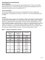
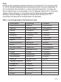
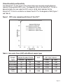
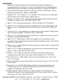
1
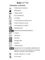
REF
TM
60501
Catalog number
TnI
Troponin l
For the Rapid, Quantitative Determination of
Troponin I in human Whole Blood, Plasma or Serum
MF
Manufacturer:
Princeton BioMeditech Corporation
4242 US Route 1
Monmouth Junction, NJ 08852
www.pbmc.com
Manufactured for:
LifeSign LLC
71 Veronica Avenue
Somerset, NJ 08873
Tel: 1 800. 526.2125
www.lifesignmed.com
Authorized Representative:
MT Promedt Consulting GmbH
Altenhofstrasse 80
66386 St. Ingbert, Germany
+49-68 94-58 10 20
P-56611
Version # 226-10/24/06
Status First™ TnI
Explanation of Symbols
CE Marking of Conformity
Manufacturer
Catalog number
Expiry date/Use by
Lot number
In Vitro Diagnostic medical device
Consult instructions for use
Store between 2°C and 8°C
Manufactured for
European Authorized Representative
Contents
Test Device
Data Chip
Package Insert
PIP
PIP
PIP
PIP
PIP
PIP
PIP
PIP
PIP
PIP
PIP
PIP
PIP
PIP
PIP
PIP
PIP
PIP
PIP
Desiccant
Transfer Pipette
Intended Use: For the quantitative determination of
Troponin I as an aid in the diagnosis of myocardial
infarction and acute coronary syndrome
Contains sufficient for <n> tests
Do not reuse
Status First™ TnI
For in vitro diagnostic use
Intended Use
StatusFirst™ TnI (Troponin I) is a rapid test for in vitro quantitative determination of Troponin I
in human whole blood, plasma or serum. The device is intended for use with the DXpress™
Reader to provide quantitative results as an aid in the diagnosis of myocardial infarction and
acute coronary syndrome (ACS).
Summary and Explanation
Acute coronary syndrome (ACS) is the leading cause of morbidity and mortality among both
men and women, affecting more than 13.9 million people in the US. The presentation of ACS
is varied and symptoms may develop suddenly, with acute myocardial infarction (AMI) being
the most dramatic of presentations. Annually, AMI affects approximately 1.1 million people in
the US, with a 30% mortality rate.
The World Health Organization recommends diagnosis of AMI with a positive indication on at
least two of these three criteria: (1) patient history and physical examination; (2)
electrocardiogram; (3) changes in blood levels of cardiac protein markers.1 Patient history and
physical examination are critical and must be considered, but often provide insufficient
information to differentiate AMI from other cardiac abnormalities. Electrocardiogram, together
with the patient history and physical examination, is useful but diagnostic in only about 50% of
AMI patients. ST segment elevation or depression and Q wave formation are typical indicators
of AMI. Patients hospitalized with ACS have been found to be high risk ST-segment elevation
myocardial infarction (STEMI) or moderate-high risk non-ST-segment elevation myocardial
infarction (NSTEMI) patients.2 However, it may be difficult to diagnose AMI in NSTEMI
patients.3 Therefore, measurement of sensitive and specific cardiac protein markers in the
blood provides an extremely useful differential diagnostic tool enabling prompt management of
patients presenting with symptoms of AMI.
Troponin I and troponin T are two specific contractile proteins of the cardiac muscle regulatory
complex that have gained popularity as cardiac specific markers for AMI. Three subunits of the
troponin complex (I, T, C) are bound with tropomyosin to actin in the thin filament of the
myofibril. Cardiac troponin I (cTnI) prevents muscular contraction and is exclusively present in
the cardiac muscle.4 The Troponin complex, together with tropomyosin, forms the main
component that regulates the Ca2+ sensitive ATPase activity of actomyosin in striated muscle
(skeletal and cardiac).5 Different isoforms of TnI exist in the skeletal and cardiac muscles (sTnI
and cTnI, respectively) with distinct structural heterogeneity between these isoforms that allow
Page 1
the production of isoform-specific antibodies.6 Specifically, the amino terminal amino acid
sequence of cTnI has 31 amino acid residues that are not present in sTnI. The utility of
determining the serum levels of the different isoforms of TnI has been studied.7
Detection of cTnI in the serum has been investigated as an aid in the determination of
myocardial damage in patients with AMI.8 Determining the serum level of cTnI provides
diagnostic value in identifying patients with AMI. The temporal relation of release of cTnI into
the serum has been investigated and compared to the other established cardiac markers, such
as creatine kinase MB (CK-MB), myoglobin and Troponin T.9,10 Following AMI, cTnI is released
into the circulation with levels exceeding the upper reference limit of normal within 4–6 hours,
and peak levels are reached after 12-24 hours.11 This early release profile is similar to CK-MB.
However, CK-MB levels return to normal values in about 72 hours, while levels of cTnI remain
elevated for 5–7 days.
The release of cTnI into the blood involves myocardial damage, primarily as a result of AMI;
however, such damage may occur from causes other than AMI, such as unstable angina,
congestive heart failure, and ischaemic damage due to coronary artery by-pass surgery.13
Further, the distinct structure of cTnI and the availability of highly specific detection methods
for cTnI have increased the utility of this marker for the diagnosis of AMI in complex clinical
conditions that involve skeletal muscle damage. The high specificity of cTnI measurements for
the identification of myocardial damage has been demonstrated in conditions such as the
perioperative period, after marathon runs, and following blunt chest trauma.11,12 Levels of other,
non-cardiac specific protein markers such as CK-MB and myoglobin are typically elevated in
the blood following such skeletal muscle damage.14,15
Principle
The StatusFirst™ TnI test device utilizes biotin coupled anti-TnI antibody/streptavidin solidphase chromatographic immunoassay technology to quantitatively determine the concentration
of TnI in human whole blood, plasma and serum specimens. After a sample has been
dispensed into the sample well, the StatusFirst™ TnI test device is placed in the DXpress™
Reader. The DXpress™ Reader displays the TnI concentration 15 minutes after sample
addition. The DXpress™ Reader is programmed to convert the intensity of the test band (as
indicated by the “TnI” line on the test device) into a concentration of TnI automatically by using
lot specific calibration factors supplied with each box of test devices. The TnI concentration in
the sample correlates with the intensity of the test band.
Reagents
The StatusFirst™ TnI test device contains all required reagents including dye conjugated
monoclonal anti-TnI antibody, biotin conjugated polyclonal anti-TnI antibody and streptavidin
immobilized at the test band. No other reagents are required.
Page 2
Materials Provided
Each box contains the following:
• 20 StatusFirst™ TnI test devices, each individually sealed in a foil pouch with a
desiccant and a single use transfer pipette.
• 1 lot specific Data Chip with calibration information
• 1 package insert
Materials/Equipment Required But Not Provided
1. DXpress™ Reader, part no. LSR-2000
2. Commercially available TnI Controls for external Quality Control (QC)
Precautions and Warnings
• For in vitro diagnostic use only.
• Carefully follow the instructions for use.
• Wear disposable gloves while handling patient samples.
• Patient samples, used test devices and transfer pipettes should be treated as if
potentially infectious and should be discarded as biohazard materials according to local
regulations.
• Thoroughly wash hands afterwards and observe the appropriate regulations/procedures
for disposal of all used materials (samples, test devices, and transfer pipettes).
• The result obtained from the StatusFirst™ TnI test device does not provide a definitive
diagnosis and should be interpreted by the physician in conjunction with other
laboratory test results and patient clinical findings.
• Avoid cross contamination of samples by using a new transfer pipette for each sample.
• Keep the test device in the sealed pouch until ready for use.
• Do not use the test device if the pouch is damaged or the seal is broken.
• Do not use the test device after the expiration date printed on the pouch.
• This is a quantitative test and therefore no visual interpretation of the result should be
made.
• The test must be read at 15 minutes after sample addition to ensure an accurate result.
Storage and Stability
• Store the StatusFirst™ TnI test device between 2° and 8°C (35° to 46°F) until the
expiration date printed on the pouch is reached.
• The StatusFirst™ TnI test device in its sealed pouch is stable at room temperature
(18° to 30°C/ 64° to 86°F) for 15 days, provided the expiration date printed on the pouch
is not exceeded.
Page 3
Sample Collection and Preparation
• The StatusFirst™ TnI test device is to be run using whole blood, serum or plasma
samples collected using EDTA or lithium heparin as the anticoagulant.
• Whole blood samples must be tested within 4 hours and plasma or serum samples
must be tested within 8 hours of collection.
• Whole blood, serum or plasma samples should be stored and/or transported in
refrigerated conditions.
• If longer storage is required, plasma or serum samples should be kept frozen at -20°C
(-4°F) or lower.
• Allow patient samples to equilibrate to room temperature prior to testing.
• Grossly hemolyzed samples should not be used.
• Mix the blood specimen gently inverting the tube several times before using.
• For frozen samples, thaw completely and mix thoroughly before use.
PROCEDURE
DXpress™ Reader
Consult the DXpress™ Reader User Manual.
For DXpress™ Reader installation, startup and complete instructions refer to the DXpress™
Reader User Manual. The operator must consult the DXpress™ Reader User Manual prior to
use and become familiar with the processes and quality control procedures.
QC Procedure
Self Check
Each time the DXpress™ Reader is turned on, Self Check is automatically performed. Self
Check takes about 15 seconds. PASS or FAIL results will be displayed/printed when testing is
completed. All Self Check items should pass before testing patient samples. The operator may
then proceed to “Calibration QC.”
If the DXpress™ Reader is left on or in power save mode, the operator should perform Self
Check daily as follows:
1. From the Main Menu, select [2] RUN QC.
2. Select [1] SELF CHECK.
3. Self Check takes about 15 seconds. PASS or FAIL results will be displayed/printed when
testing is completed.
PIP
Page 4
Calibration QC
Each day of patient testing, use the Calibration Set (see DXpress™ Reader manual) to ensure
the DXpress™ Reader functions properly:
1. From the Main Menu, select [2] RUN QC.
2. Select [2] CALIBRATION QC.
3. Scan (or manually enter) the Operator ID barcode.
4. Scan the Calibrator ID barcode, found on the back of the Calibrator.
5. Insert the Calibrator into the reader – be sure to close the Tray – and press ENTER.
Follow prompts displayed on the screen.
6. Calibration QC result will be displayed/printed when the calibration is completed.
Calibration QC should pass before running daily patient testing.
7. Blank Calibration is not necessary for TnI product. Select [CANCEL] and press ENTER.
External QC
Perform External QC once for each new lot of StatusFirst™ TnI test devices.
1. From Main Menu, select [2] RUN QC.
2. Select [3] EXTERNAL QC.
3. Using external controls, follow the same procedure as if running a patient sample; see
section “Testing Patient Sample Procedure” below. The only difference is that RUN
PATIENT requires a Patient ID, whereas EXTERNAL QC requires a Sample ID.
Uploading Lot-Specific Calibration Information using the Data Chip
When you start to use a new lot of test devices, the Reader will prompt the operator to insert
the Data Chip that is supplied in the test kit. Insert the Data Chip with the corresponding test
device lot number, select OK to continue and follow the prompts.
Page 5
Testing Patient Sample Procedure
There are three modes of testing patient samples (Read-Now, Batch, Scheduler). The operator
is free to choose the mode most suitable for the particular task at hand. The Scheduler mode
is described below as an example:
1. Open the pouch and remove the test device.
2. Label the test device with the patient ID.
3. Place the test device on a level surface.
4. Testing the Patient Sample on the DXpress™ Reader:
• From the Main Menu, select [1] RUN PATIENT.
• Scan (or manually enter) lot number barcode from the pouch.
• Confirm test device information (type of test device and lot number) as displayed on
the screen and press ENTER.
• Scan (or manually enter) the Operator ID barcode.
• Scan (or manually enter) the Patient ID barcode.
• Select either Blood or Plasma/Serum, as appropriate.
• From the Incubation Time window, select SCHEDULER.
• Within 30 seconds do the following:
Draw patient sample solution up to the line on the transfer pipette while holding
the transfer pipette vertically, and dispense into the sample well. When drawing
sample into the transfer pipette, avoid introducing air bubbles. Do not touch the
sample well or test device with the tip of the transfer pipette.
Fill sample up to line
• Press ENTER.
• Insert the test device in the Reader tray, close the Reader tray.
• After 15 minutes of incubation the DXpress™ Reader will automatically display the
results on the screen.
• Results may be printed by pressing the PRINT button.
• At this point the test device may be removed and appropriately discarded.
Report and Interpretation of Results
• The range of TnI concentrations reported by the test device system is 0.05 ng/mL to 30 ng/mL.
Results below or above this range will be shown as < 0.05 ng/mL or > 30 ng/mL, respectively.
• Valid results will be displayed on the Reader as: Control: Valid
TnI: xx.xx ng/mL
• Invalid results: If the sample fails to migrate properly or the reagents fail, the Reader
will display: Control: Invalid***
TnI: Invalid***
In this case, repeat the test with a new test device.
Page 6
Recommended Decision Threshold Values
The clinical cutoff of StatusFirst™ TnI was determined as 0.2 ng/mL TnI when compared to
Access® AccuTnI™† and Triage® Cardiac Panel††. However, laboratories should establish their
own diagnostic cutoff concentration based on the clinical practice at their respective
institutions.
Registered trademark of Beckman Coulter
Registered trademark of Biosite
†
††
QUALITY CONTROL
External Controls
Good laboratory practice includes the use of external controls to ensure proper test device
performance. It is recommended that prior to using a new lot or shipment of StatusFirst™ TnI
test devices, the performance of the lot be confirmed by testing with external controls (see
section “Materials/Equipment Required But Not Provided”) to ensure the test devices will
deliver the correct test result. The frequency of QC testing should be determined according to
individual laboratory standard QC procedures. Upon confirmation of the expected results, the
test devices are ready for use with patient samples. If external controls do not perform as
expected, do not use the test devices and contact LifeSign Technical Services at 1-800-526-2125.
Internal Controls
StatusFirst™ TnI test device has a built-in control that satisfies the requirements of testing a
control on a daily basis. The control line is an internal positive procedural control. A distinct
reddish-purple control line should appear at the control position if the test is performed
properly, an adequate sample volume is used, the sample and reagent are wicking on the
membrane, and the reagents at the control line are reacting with the conjugate-color indicator.
In addition, a clear background may be considered a negative procedural control. If the test is
performed correctly and the device is working properly, the background in the result window
will become clear and provide a distinct result. The DXpress™ Reader will report “Control:
Valid” and the test result for TnI concentration in ng/mL unit when Internal Control QC is
satisfied.
LIMITATIONS
• The results of the StatusFirst™ TnI test should be used in conjunction with the total
clinical presentation of the patient and other laboratory information available. Medical
decisions should not be based on a single point in time test result of the StatusFirst™
TnI test. The published guideline recommends collecting at least three blood samples
during the early triage period.16
• Other substances and/or factors not listed, e.g., technical or procedural errors, may
interfere with the test and cause false results.
Page 7
PERFORMANCE CHARACTERISTICS
Limit of Detection
The limit of detection (LoD) represents the lowest concentration of TnI that can be reliably
differentiated from zero. The LoD of 0.05 ng/mL was determined according to Clinical and
Laboratory Standards Institute (CLSI) guideline EP17-A.
Limit of Quantitation
The limit of quantitation (LoQ) is the lowest TnI concentration that can be reproducibly
measured with a total coefficient of variation of at most 20%. The LoQ was determined to be
0.1 ng/mL.
Linearity
Human cardiac troponin complex from the National Institute of Standards & Technology (NIST)
was spiked into human blood to a final concentration of 32 ng/mL cardiac troponin I, and then
plasma was separated from the whole blood. The plasma from the spiked blood was 2-fold
serially diluted into troponin I negative plasma for a total of ten values spanning the measuring
range of the StatusFirst™ TnI test. Each level was tested in ten replicates. The data were
analyzed in accordance with CLSI EP6-A and demonstrated linearity of the test device across
the measuring range from 0.063 to 32 ng/mL. The data are shown below in Table 1.
Table 1. Linearity of StatusFirst™ TnI test
Expected conc.
(ng/mL)
Measured conc.
(ng/mL)
0.063
0.070
0.25
0.261
1
1.021
4
4.078
0.125
0.5
2
8
16
32
0.146
% Recovery
113.8%
116.8%
104.4%
0.546
109.2%
2.151
107.6%
8.393
17.439
32.569
102.1%
102.0%
104.9%
109.0%
101.8%
Page 8
Precision
The precision of the StatusFirst™ TnI test device was determined using samples where TnI
from NIST was spiked at four concentrations (Table 2). The within-day and total precision
studies were performed in two runs per day, in six replicates per run at each concentration
level, for 15 days with three DXpress™ Readers. The within-run and total variances and
coefficients of variation (CVs) were computed according to CLSI guideline EP5-A.
Table 2. Precision Data
Mean level
Average Within-run
Average Total
(ng/mL)
Std. dev. (ng/mL)
CV (%)
Std. dev. (ng/mL)
CV (%)
0.25
0.04
14.4
0.04
14.8
1.73
17.2
1.78
17.6
0.29
2.0
10.0
14.4
3.70
20.0
0.31
18.0
3.70
15.1
18.4
CROSS-REACTIVITY AND INTERFERING SUBSTANCES
Potentially Cross-Reactive Substances
The following cardiac proteins and peptides were tested for potential cross-reactivity in the
StatusFirst™ TnI test device at the maximum concentration of substance indicated (Table 3).
No substance demonstrated significant cross-reactivity (i.e., all cross-reactivities < 0.05%)
when added to sample containing a TnI concentration of approximately 0.5–0.7 ng/mL.
Table 3. Cross-reactivity study
Protein/Peptides
Tropomyosin
Actual Conc. (µg/mL) Cross-reactivity (%)
1
0.002
1
0.016
1
0.003
1
0.000
cTnT
0.5
cMyosin light chain
0.2
CK-MB
1
cTnC
Myoglobin
BNP
NT-proBNP
1
0.005
0.048
0.005
0.000
Page 9
Drugs
Sixty drugs were assessed for potential interference in the StatusFirst™ TnI test device (Table
4). The list of drugs encompassed common prescription and over-the-counter compounds, as
well as medications often prescribed in a cardiac related patient population. The drugs were
tested at concentrations as recommended in the CLSI Approved Guideline EP7-A ‘Interference
Testing in Clinical Chemistry,’ or at least three times the highest concentration reported
following a therapeutic dosage. No significant interference with the StatusFirst™ TnI
measurement was observed for the drugs listed in the table below.
Table 4. List of drugs tested for the interference study
Acetaminophen
Dipyridamole
Oxazepam
Actetylsalicylic acid (Aspirin)
Dopamine
Oxytetracycline
Alteplase
Erythromycin
Phenytoin
Amiodarone
Furosemide
Probenecid
Ampicillin
Heparin
Propranolol
Allopurinol
Ambroxol
Amlodipine Besylate
Enalapril maleate
Fluvastatin (Lescol)
Glyburide
Phentobarbital
Pravastatin
Procainamide
Ascorbic acid (vitamin C)
Hydralazine
Quinidine
Atorvastatin calcium
Indomethacin
Sulfamethoxazole
Atenolol
Hydrochlorothiazide
Caffeine
Isosorbide dinitrate
Chloramphenicol
Methaqualone
Captopril
Spironocactone
Theophylline
Lisinopril
L-thyroxine
Chlordiazepoxide
Methyl-DOPA
Verapamil
Clopidogrel bisulphate
Nicotine
Cinnarizine
Cyclosporine A
Diclofenac
Milrinone lactate
Warfarin
Nifedipine
Nitrofurantoin
Digitoxin
Nitroglycerin
Diltiazem
Nystatine
Digoxin
Trimethoprim
Noraminopyren (Dipyrone)
Page 10
Other Potentially Interfering Substances
When added to a sample containing TnI, hemoglobin (up to 0.5 g/dL), bilirubin (up to 20
mg/dL), triglycerides (up to 1 g/dL) and human albumin (up to 16 g/dL) did not interfere with
the recovery of TnI.
Hook Effect
No high dose hook effect was observed for TnI concentrations up to 500 ng/mL.
Blood, Plasma, and Serum Correlation
Blood, plasma and serum comparison study was performed with clinical samples.
A comparison analysis between blood, plasma and serum showed the following correlations.
StatusFirst™ TnI EDTA Plasma
= 0.958 x StatusFirst™ TnI EDTA Whole blood (EDTA) – 0.002 ng/mL
r2 = 0.95
StatusFirst™ TnI Lithium Heparin Plasma
= 0.946 x StatusFirst™ TnI Whole blood (Lithium Heparin) – 0.010 ng/mL
r2 = 0.96
StatusFirst™ TnI serum = 1.197 x StatusFirst™ TnI Plasma (EDTA)
r2 = 0.98
CLINICAL PERFORMANCE
A total of 228 patients’ whole blood and plasma specimens collected in EDTA and/or lithium
heparin tubes were tested. TnI concentrations in plasma samples from lithium heparin tubes
were determined by both Access® AccuTnI™ and the StatusFirst™ TnI test. TnI concentrations
in whole blood samples from EDTA tubes were determined by both Triage® Cardiac Panel and
StatusFirst™ TnI test. TnI levels in plasma from EDTA tubes and in whole blood from lithium
heparin tubes were tested on the StatusFirst™ TnI test only and used to determine the
correlation of different matrices and anticoagulants.
The sensitivity and specificity of the StatusFirst™ TnI test was evaluated against Access®
AccuTnI™ and the StatusFirst™ TnI performance was correlated to Access® AccuTnI™ or
Triage® Cardiac Panel.
Page 11
Clinical Sensitivity and Specificity
The StatusFirst™ TnI test results in this clinical study were analyzed using the Receiver
Operator Characteristics (ROC) curve. The diagnostic utility of the StatusFirst™ TnI test is
demonstrated by the area under the ROC curve of >0.99, which indicates that the
StatusFirst™ TnI test is as effective as Access® AccuTnI™ in the diagnosis of AMI (Figure 1
and Table 5).
Figure 1. ROC curve comparing with Access® AccuTnI™
Table 5. Area under Curve (AUC) with different sample types
Sample
Types
EDTA WB
EDTA Plasma
Sample Numbers Tested
Positive
Negative
92
90
94
Li Heparin WB
89
Li Heparin
Plasma
93
AUC
90
0.998
94
0.998
93
0.995
0.998
95% Conf. Interval
Lower
Upper
0.988
1.003
0.996
0.996
0.996
1.001
1.001
1.001
Page 12
To determine the best cutoff, a series of presumed cutoff values were set up and the
corresponding sensitivities and specificities were compared (Table 6).
Table 6. Sensitivity and Specificity Compared to Access® AccuTnI™
StatusFirst™
EDTA WB
EDTA Plasma
Li Heparin WB
Li Heparin Plasma
TnI Cutoff
(ng/mL) Sensitivity (%) Specificity (%) Sensitivity (%) Specificity (%) Sensitivity (%) Specificity (%) Sensitivity (%) Specificity (%)
0.1
100
99
100
93
100
0.3
94
99
95
98
96
0.2
0.4
0.5
99
86
81
99
100
100
99
88
85
98
99
99
82
100
98
96
99
96
92
100
85
100
95
99
98
93
100
86
98
100
A cutoff of 0.2 ng/mL TnI is recommended for diagnosis of AMI, as this yields optimal
performance of 99% sensitivity and >96% specificity relative to Access® AccuTnI™. However,
laboratories should establish their own diagnostic cutoff concentration based on the clinical
practice at their respective institutions.
Correlation of StatusFirst™ TnI vs Access® AccuTnI™ and Triage® Cardiac Panel
The following correlation equations were obtained by comparing StatusFirst™ TnI to Access®
AccuTnI™ and Triage® Cardiac Panel. Since each company uses different TnI standard
material for the test calibration, the correlation factor would differ. StatusFirst™ TnI uses NIST
standard for the calibration, as designated by the American Association for Clinical Chemistry.
StatusFirst™ TnI Lithium Heparin Plasma
= 0.323 x AccuTnI™ Plasma (Lithium Heparin) + 0.007 ng/mL
r2 = 0.93 with n = 182
StatusFirst™ TnI Whole blood (EDTA)
= 0.623 x Triage® Whole blood (EDTA) + 0.065 ng/mL
r2 = 0.92 with n = 95
Page 13
REFERENCES
1. World Health Organization. Report of the Joint Internaltional Society and Federation of
Cardiology/World Health Organization Task Force on Standardization of Clinical Nomenclature.
Nomenclature and criteria for diagnosis of ischemic heart disease. Circulation 1979; 59:607-9.
2. Veterans Health Administration, Ischemic heart disease: [inclusive of JCAHO AMI]). Veterans
Health Administration Report. 2005 Mar. NQMC:000931
3. Heidenreich, P.A., Go, A., Melsop, K.A., et al. Prediction of risk for patients with unstable angina.
AHRQ Publication No. 01-E001 December, 2000; Number 31.
4. Mehegan, J.P. & Tobacman, L.S., Cooperative interaction between troponin molecules bound to
the cardiac thin filament. J. Biol. Chem. 266(2): 966-972 (1991).
5. Ebashi, S., Ca2+ and the contractile proteins. J. Mol. Cell Cardiol. 16(2): 129-136(1984).
6. Bodor, GS. et al., Development of monoclonal antibodies for an assay of cardiac troponin-I and
preliminary results in suspected cases of myocardial infarction. Clin. Chem. 38(11):2203-2214
(1992).
7. Takahashi, M. et al., Use of immunoassay for measurement of skeletal troponin-I utilizing isoformspecific monoclonal antibodies. Clin Biochem 29(4): 301-308 (1996).
8. Adams, J.E., et al., Biochemical markers of myocardial injury. Is MB creatin kinase the choice for
the 1990s? Circulation 88:750-763 (1993).
9. Apple, F.S. et al., Cardiac troponin, CK-MB, and myoglobin for the early detection of acute
myocardial infarction and monitoring of reperfusion following thrombolytic therapy. Clinica Chimica
Acta 237(1-2): 59-66 (1995).
10. Adams, J.E. et al., Improved detection of cardiac contusion with cardiac troponin I. American Heart
J, 131(2): 308-312 (1996)
11. Adams, J.E. et al., Diagnosis of perioperative myocardial infarction with measurement of cardiac
troponin I. N. Eng. J. Med. 330(10): 670-674 (1994).
12. Machler, H. et al., Preoperative myocardial cell damage in patients with unstable angina
undergoing coronary artery bypass graft surgery. Anesthesiology 81(6): 1317-1320 (1994).
13. Hossein-Nia, M. et al., Cardiac troponin I release in heart transplantation. Ann. Thorac. Surg.
61(1): 277-278 (1996).
14. Wu, A. et al., Prognostic value of cardiac troponin I in patients with chest pain. Clin. Chem. 42(4):
651-652 (1996).
15. Braunwald, E. et al., ACC/AHA 2002 guideline update for the management of patients with
unstable angina and non-ST-segment elevation myocardial infarction: ACC/AHA Practice
Guidelines, ACC (Bethesda, MD) & AHA , 2002
16. Wu HBA, Apple PS, Gibler B, Jesse RL et al. National Academy of Clinical Biochemistry
Standards of Laboratory Practice: Recommendations for the use of cardiac markers in coronary
artery disease. Clin. Chem. 1999, 45(7):1104-1121
Manufactured by PBM Princeton BioMeditech Corporation 4242 US Route 1 Monmouth Junction NJ 08852
U.S.A. Tel 1-732-274-1000 Fax 1-732-274-1010 www.pbmc.com