1
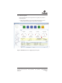
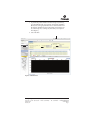
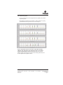
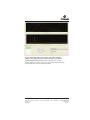
Technical Bulletin
PowerPlex® 5-Dye Matrix
Standards, 3100/3130
INSTRUCTIONS FOR USE OF PRODUCTS DG4700.
Note: These matrix fragments are compatible with the
ABI PRISM® 3100 and 3100-Avant and Applied Biosystems 3130,
3130xl, 3500 and 3500xL Genetic Analyzers.
PRINTED IN USA.
Revised 7/12
Part# TBD024
PowerPlex® 5-Dye Matrix
Standards, 3100/3130
All technical literature is available on the Internet at: www.promega.com/protocols/
Please visit the web site to verify that you are using the most current version of this
Technical Bulletin. Please contact Promega Technical Services if you have questions on use
of this system. E-mail: [email protected].
1. Description..........................................................................................................2
2. Product Components and Storage Conditions ............................................2
3. Instrument Preparation and Spectral Calibration Using
the Applied Biosystems 3500 and 3500xL Genetic Analyzers ..................3
A. Matrix Sample Preparation.................................................................................3
B. Instrument Preparation .......................................................................................5
4. Instrument Preparation and Spectral Calibration Using
POP-4™ Polymer and Data Collection Software, Version 2.0
or Version 3.0 (ABI PRISM® 3100 and 3100-Avant and Applied
Biosystems 3130 and 3130xl Genetic Analyzers) .........................................9
A. Matrix Sample Preparation.................................................................................9
B. Instrument Preparation .....................................................................................11
5. Instrument Preparation and Spectral Calibration Using
POP-7™ Polymer and Data Collection Software, Version 3.0
(Applied Biosystems 3130 and 3130xl Genetic Analyzers)......................14
A. Matrix Sample Preparation...............................................................................14
B. Instrument Preparation .....................................................................................16
6. Troubleshooting...............................................................................................19
A. Applied Biosystems 3500 and 3500xL Genetic Analyzers ...........................19
B. ABI PRISM® 3100 and 3100-Avant and Applied Biosystems
3130 and 3130xl Genetic Analyzers .................................................................20
7. Related Products ..............................................................................................21
Promega Corporation · 2800 Woods Hollow Road · Madison, WI 53711-5399 USA
Toll Free in USA 800-356-9526 · Phone 608-274-4330 · Fax 608-277-2516 · www.promega.com
Printed in USA.
Revised 7/12
Part# TBD024
Page 1
1.
Description
Proper generation of a spectral calibration file is critical to evaluate multicolor
systems with the ABI PRISM® 3100 and 3100-Avant and Applied Biosystems
3130, 3130xl, 3500 and 3500xL Genetic Analyzers. The PowerPlex® 5-Dye Matrix
Standards, 3100/3130(a,b), consists of DNA fragments labeled with five different
fluorescent dyes: one tube contains a DNA fragment labeled with fluorescein,
one tube contains a DNA fragment labeled with JOE, one tube contains a DNA
fragment labeled with TMR-ET, one tube contains a DNA fragment labeled
with CXR-ET, and one tube contains a DNA fragment labeled with CC5.
These matrix fragments are mixed and used on the ABI PRISM® 3100 or 3100Avant or Applied Biosystems 3130, 3130xl, 3500 or 3500xL Genetic Analyzer to
perform a spectral calibration on a specified dye set. The spectral calibration
should be performed on dye set G5. Once generated, this file is applied during
sample detection to calculate the spectral overlap between the five different
dyes and separate the raw fluorescent signals into individual dye signals. The
PowerPlex® 5-Dye Matrix Standards, 3100/3130, was developed for use with
the 5-dye PowerPlex® Systems. A matrix should be generated for each
individual instrument. Protocols to operate the ABI PRISM® 3100 or 3100-Avant
or Applied Biosystems 3130, 3130xl, 3500 or 3500xL Genetic Analyzer should be
obtained from the manufacturer.
2.
Product Components and Storage Conditions
Product
PowerPlex® 5-Dye Matrix Standards, 3100/3130
Not for Medical Diagnostic Use. Includes:
•
•
•
•
•
•
25µl
25µl
25µl
25µl
25µl
1.25ml
Size
25µl (each dye)
Cat.#
DG4700
Fluorescein Matrix, 5-Dye (3100/3130)
JOE Matrix, 5-Dye (3100/3130)
TMR-ET Matrix, 5-Dye (3100/3130)
CXR-ET Matrix, 5-Dye (3100/3130)
CC5 Matrix, 5-Dye (3100/3130)
Nuclease-Free Water
Storage Conditions: Store all components at –30°C to –10°C in a nonfrost-free
freezer. The matrix standards fragments are light-sensitive and must be stored in
the dark. We strongly recommend that the matrix standards be stored with postamplification reagents and used separately with different pipettes, tube racks, etc.
Do not store the diluted matrix standard fragments.
Promega Corporation · 2800 Woods Hollow Road · Madison, WI 53711-5399 USA
Toll Free in USA 800-356-9526 · Phone 608-274-4330 · Fax 608-277-2516 · www.promega.com
Part# TBD024
Page 2
Printed in USA.
Revised 7/12
3.
Instrument Preparation and Spectral Calibration Using the Applied
Biosystems 3500 and 3500xL Genetic Analyzers
Materials to Be Supplied by the User
• 95°C dry heating block, water bath or thermal cycler
• crushed ice or ice-water bath
• centrifuge compatible with 96-well plates
• aerosol-resistant pipette tips
• 3500/3500xL capillary array, 36cm
• performance optimized polymer 4 (POP-4™) in a pouch for the 3500 or
3500xL
• anode buffer container with 1X buffer
• cathode buffer container with 1X buffer
• conditioning reagent pouch for the 3500 or 3500xL
• MicroAmp® optical 96-well plate and septa
• Hi-Di™ formamide (Applied Biosystems Cat.# 4311320)
!
The quality of formamide is critical. Use Hi-Di™ formamide. Freeze formamide in
aliquots at –20°C. Multiple freeze-thaw cycles or long-term storage at 4°C may cause
breakdown of formamide. Poor-quality formamide may contain ions that compete
with DNA during injection, which results in lower peak heights and reduced
sensitivity. A longer injection time may not increase the signal.
!
Formamide is an irritant and a teratogen; avoid inhalation and contact with skin.
Read the warning label, and take the necessary precautions when handling this
substance. Always wear gloves and safety glasses when working with formamide.
For additional information on performing spectral calibration, refer to the Applied
Biosystems 3500/3500xL Genetic Analyzer User Guide.
3.A. Matrix Sample Preparation
!
There may be instrument-to-instrument variation in the sensitivity of
detection. The dilutions described here may need to be optimized in
individual laboratories, depending on the sensitivity of each Applied
Biosystems 3500 or 3500xL Genetic Analyzer.
1. Thaw the matrix standards on ice. Mix each matrix standard by vortexing
each tube for 5–10 seconds prior to use. Do not centrifuge the matrix
standards as this may cause the DNA to be concentrated at the bottom of
the tube. Place on ice.
2. Initial dilution of concentrated fragments: Before combining the matrix
standards, dilute the individual matrix standards 1:10 in Nuclease-Free
Water, as described below. Vortex for 5–10 seconds to mix. Place on ice.
Component
Fluorescein
Concentrated Dye
5µl
Nuclease-Free Water
45µl
JOE
5µl
45µl
TMR-ET CXR-ET
5µl
5µl
45µl
45µl
CC5
5µl
45µl
Promega Corporation · 2800 Woods Hollow Road · Madison, WI 53711-5399 USA
Toll Free in USA 800-356-9526 · Phone 608-274-4330 · Fax 608-277-2516 · www.promega.com
Printed in USA.
Revised 7/12
Part# TBD024
Page 3
3.A. Matrix Sample Preparation (continued)
3. Fragment mix (using 1:10 dilutions of matrix standards): After the initial
dilution in Step 2, combine the 1:10 dilution of each matrix standard as
directed below. Vortex for 5–10 seconds to mix.
Component
Hi-Di™ formamide
Fluorescein from initial dilution
JOE from initial dilution
TMR-ET from initial dilution
CXR-ET from initial dilution
CC5 from initial dilution
Volume
665µl
7.0µl
7.0µl
7.0µl
7.0µl
7.0µl
4. On the Applied Biosystems 3500xL Genetic Analyzer, only wells A1 to H3
of the 96-well plate are used for spectral calibration. Load 25μl of fragment
mix prepared in Step 3 into each of the 24 wells. After placing the septa on
the plate, briefly centrifuge the plate to remove bubbles.
On the Applied Biosystems 3500 Genetic Analyzer, only wells A1 to H1 of
the 96-well plate are used for spectral calibration. Load 25μl of fragment
mix prepared in Step 3 into each of the 8 wells. After placing the septa on
the plate, briefly centrifuge the plate to remove bubbles.
5. Denature samples at 95°C for 3 minutes, then immediately chill on crushed
ice or in an ice-water bath for 3 minutes. Denature samples just prior to
loading the instrument.
6. Place the plate in the 3500 series 96-well standard plate base, and cover
with the plate retainer. Place the plate assembly in Position A on the
autosampler with the labels facing you.
Promega Corporation · 2800 Woods Hollow Road · Madison, WI 53711-5399 USA
Toll Free in USA 800-356-9526 · Phone 608-274-4330 · Fax 608-277-2516 · www.promega.com
Part# TBD024
Page 4
Printed in USA.
Revised 7/12
3.B. Instrument Preparation
We have found that the use of fresh polymer and new capillary array results in
an optimal spectral.
9247TA
1. Set the oven temperature to 60°C, then select the Start Pre-Heat icon at least
30 minutes prior to the first injection to preheat the oven (Figure 1).
Figure 1. The Dashboard. The arrow highlights the preheat options.
Promega Corporation · 2800 Woods Hollow Road · Madison, WI 53711-5399 USA
Toll Free in USA 800-356-9526 · Phone 608-274-4330 · Fax 608-277-2516 · www.promega.com
Printed in USA.
Revised 7/12
Part# TBD024
Page 5
3.B. Instrument Preparation (continued)
2. To perform a spectral calibration with the Promega 5-dye chemistry, a new
dye set should be created. If a new dye set was created previously, proceed
to Section 3.B, Step 2.c.
a. To create this new dye set, navigate to the Library, highlight “Dye Sets”
and select “Create”.
9325TA
b. The Create a New Dye Set tab will appear (Figure 2). Name the Dye Set,
select “Matrix Standard” for the Chemistry and select “G5 Template”
for the Dye Set Template. Select “Save”.
Figure 2. Create New Dye Set.
Promega Corporation · 2800 Woods Hollow Road · Madison, WI 53711-5399 USA
Toll Free in USA 800-356-9526 · Phone 608-274-4330 · Fax 608-277-2516 · www.promega.com
Part# TBD024
Page 6
Printed in USA.
Revised 7/12
c. To perform the spectral calibration with the Promega 5-dye chemistry,
go to the Maintenance tab, select “Spectral”, and under the Calibration
Run tab, choose the appropriate fields: Choose “Matrix Standard” from
the Chemistry Standard drop-down menu and the new Promega 5-dye
set created in Step 2b (i.e., Promega G5) from the Dye Set drop-down
menu (Figure 3).
d. Select “Start Run”.
Figure 3. Calibration Run.
Promega Corporation · 2800 Woods Hollow Road · Madison, WI 53711-5399 USA
Toll Free in USA 800-356-9526 · Phone 608-274-4330 · Fax 608-277-2516 · www.promega.com
Printed in USA.
Revised 7/12
Part# TBD024
Page 7
3.B. Instrument Preparation (continued)
3. If fewer than the recommended number of capillaries pass, the spectral
calibration run will be repeated automatically up to three times. Upon
completion of the spectral calibration, check the quality of the spectral in
the Capillary Run Data display (Figure 4), and choose either “Accept” or
“Reject”.
9327TA
Note: Refer to the 3500 Series Data Collection Software Version 1.0 HID User
Manual for the criteria recommended by Applied Biosystems when
accepting or rejecting a spectral calibration.
Figure 4. The Capillary Run Data display.
Promega Corporation · 2800 Woods Hollow Road · Madison, WI 53711-5399 USA
Toll Free in USA 800-356-9526 · Phone 608-274-4330 · Fax 608-277-2516 · www.promega.com
Part# TBD024
Page 8
Printed in USA.
Revised 7/12
4.
Instrument Preparation and Spectral Calibration Using POP-4™ Polymer
and Data Collection Software, Version 2.0 or Version 3.0 (ABI PRISM®
3100 and 3100-Avant and Applied Biosystems 3130 and 3130xl Genetic
Analyzers)
Materials to Be Supplied by the User
• 95°C dry heating block, water bath or thermal cycler
• crushed ice or an ice-water bath
•
centrifuge compatible with 96-well plates
•
•
•
•
•
•
3100 or 3130 capillary array, 36cm
performance optimized polymer 4 (POP-4™) for the 3100 or 3130
10X genetic analyzer buffer with EDTA
MicroAmp® optical 96-well plate
aerosol-resistant pipette tips
Hi-Di™ formamide (Applied Biosystems Cat.# 4311320)
!
The quality of formamide is critical. Use Hi-Di™ formamide. Freeze formamide in
aliquots at –20°C. Multiple freeze-thaw cycles or long-term storage at 4°C may cause
breakdown of formamide. Poor-quality formamide may contain ions that compete
with DNA during injection, which results in lower peak heights and reduced
sensitivity. A longer injection time may not increase the signal.
!
Formamide is an irritant and a teratogen; avoid inhalation and contact with skin.
Read the warning label, and take appropriate precautions when handling this
substance. Always wear gloves and safety glasses when working with formamide.
4.A. Matrix Sample Preparation
There may be instrument-to-instrument variation in the sensitivity of
detection. The dilutions described here may need to be optimized in
individual laboratories, depending on the sensitivity of each ABI PRISM® 3100
or 3100-Avant or Applied Biosystems 3130 or 3130xl Genetic Analyzer. The
optimal dilution may differ for each dye fragment. You also may need to
adjust injection time or voltage in Section 4.B to achieve a passing spectral
calibration. Peak heights in the range of 1,000–4,000RFU are ideal. Peak heights
above 750RFU are required.
The same plate of matrix standards can be re-injected up to four times. To
re-inject the same matrix standards plate, add an injection by selecting “Plate
Manager”, then “Edit”. Select “Edit” again in the top left corner of the window,
then select “Add sample run”. Injection time and voltage may require
optimization to obtain peak heights above 750RFU and below the saturation
point.
1. Thaw the matrix standards on ice. Mix each matrix standard by vortexing
for 5–10 seconds prior to use. Do not centrifuge the matrix standards as this
may cause the DNA to be concentrated at the bottom of the tube. Place on
ice.
Promega Corporation · 2800 Woods Hollow Road · Madison, WI 53711-5399 USA
Toll Free in USA 800-356-9526 · Phone 608-274-4330 · Fax 608-277-2516 · www.promega.com
Printed in USA.
Revised 7/12
Part# TBD024
Page 9
4.A. Matrix Sample Preparation (continued)
2. Initial dilution of concentrated fragments: Before combining the matrix
standards, dilute the individual matrix standards 1:10 in Nuclease-Free
Water, as described below. Vortex for 5–10 seconds to mix. Place on ice.
Component
Fluorescein
Concentrated Dye
5µl
Nuclease-Free Water
45µl
JOE
5µl
45µl
TMR-ET CXR-ET
5µl
5µl
45µl
45µl
CC5
5µl
45µl
3. Fragment mix (using 1:10 dilutions of matrix standards): After the initial
dilution in Step 2, combine the 1:10 dilutions as directed below. Vortex for
5–10 seconds to mix.
Component
Hi-Di™ formamide
Fluorescein from initial dilution
JOE from initial dilution
TMR-ET from initial dilution
CXR-ET from initial dilution
CC5 from initial dilution
Volume
475µl
5.0µl
5.0µl
5.0µl
5.0µl
5.0µl
4. On the ABI PRISM® 3100 and Applied Biosystems 3130xl Genetic Analyzers,
16 wells are used for spectral calibration on 16 capillaries (wells A1 through
H2 of a 96-well plate). Load 25µl of fragment mix prepared in Step 3 into
each of the 16 wells. Briefly centrifuge the plate to remove bubbles.
On the ABI PRISM® 3100-Avant and Applied Biosystems 3130 Genetic
Analyzers, four wells are used for spectral calibration on four capillaries
(wells A1 through D1 in a 96-well plate). Load 25µl of fragment mix into
each of the four wells. Briefly centrifuge the plate to remove any bubbles.
5. Denature samples at 95°C for 3 minutes, then immediately chill on crushed
ice or in an ice-water bath for 3 minutes. Denature samples just prior to
loading the instrument.
Promega Corporation · 2800 Woods Hollow Road · Madison, WI 53711-5399 USA
Toll Free in USA 800-356-9526 · Phone 608-274-4330 · Fax 608-277-2516 · www.promega.com
Part# TBD024
Page 10
Printed in USA.
Revised 7/12
4.B. Instrument Preparation
We have found that the use of fresh polymer and new capillary array results in
an optimal spectral.
8283TA
Representative raw data are shown in Figure 5. Another example of a passing
spectral calibration in the Spectral Viewer is shown in Figure 6.
Figure 5. Representative data for the PowerPlex® 5-Dye Matrix Standards,
3100/3130, on the Applied Biosystems 3130 Genetic Analyzer using POP-4™
polymer and Data Collection Software, Version 3.0. Figure shows the CC5,
CXR-ET, TMR-ET, JOE and fluorescein peaks in the raw data profile from each of
the four capillaries in the Capillaries Viewer.
Promega Corporation · 2800 Woods Hollow Road · Madison, WI 53711-5399 USA
Toll Free in USA 800-356-9526 · Phone 608-274-4330 · Fax 608-277-2516 · www.promega.com
Printed in USA.
Revised 7/12
Part# TBD024
Page 11
8198TA
4.B. Instrument Preparation (continued)
Figure 6. Representative data for the PowerPlex® 5-Dye Matrix Standards,
3100/3130, on the Applied Biosystems 3130 Genetic Analyzer using POP-4™
polymer and Data Collection Software, Version 3.0. Figure shows the CC5
(orange), CXR-ET (red), TMR-ET (yellow), JOE (green) and fluorescein (blue) peaks
in the Spectral Viewer from one of the four capillaries.
1. Prepare matrix samples as previously described in Section 4.A.
Note: Differences in instrument sensitivity may result in peak imbalance or
reduced peak height of the matrix standards. You may need to adjust
injection time or voltage to achieve a passing spectral calibration. You also
may need to prepare a new plate and adjust the dilution of individual
matrix standards. Peak heights in the range of 1,000–4,000RFU are ideal.
Peak heights above 750RFU and below the saturation point of the
instrument are required.
2. Perform the spectral calibration as described in the ABI PRISM® 3100 or
3100-Avant or Applied Biosystems 3130 or 3130xl Genetic Analyzer User’s
Manual with the following modifications:
a. In the Module Manager, select “New.” Select “Spectral” in the Type
drop-down list, and select “Spect36_POP4” in the Template drop-down
list. Confirm or change the following settings:
Inj. kV:
Inj. Secs:
Data Delay Time:
Run time:
1.2
12
400
700 seconds
Promega Corporation · 2800 Woods Hollow Road · Madison, WI 53711-5399 USA
Toll Free in USA 800-356-9526 · Phone 608-274-4330 · Fax 608-277-2516 · www.promega.com
Part# TBD024
Page 12
Printed in USA.
Revised 7/12
b. Create a new name for the run module, then select “OK”.
c. In the Protocol Manager, under Instrument Protocols select “New”.
Type a name for your protocol.
Make the following selections in the Protocol Editor:
•
•
•
•
•
•
“Spectral” in the Type drop-down list
“G5” in the DyeSet drop-down list
“POP4” for the polymer
“36” in the Array Length drop-down list
“Matrix Standard” for the chemistry
Select the spectral module you created in the previous step in the
Run Module drop-down list.
Finally, select “Edit Parameters”, and make the following modifications:
• Change the lower condition bound to 4.0, and change the upper
condition bound to 12.0.
• Confirm that the Minimum Quality Score (“Q value”) is 0.95.
Select “OK” in the Edit Parameters window, and select “OK” in the
Protocol Editor.
Note: The condition number (“C value”) obtained when generating a
spectral calibration will vary with the instrument. After obtaining a
spectral calibration that performs acceptably, the condition bounds
range in the previous step may be narrowed to more critically evaluate
C values for subsequent spectral calibrations.
d. In the Plate Manager, create a new plate record as described in the
instrument user’s manual. In the dialog box that appears, select
“Spectral Calibration” in the Application drop-down list, and select
“96-well” as the plate type. Add entries in the owner and operator
windows, name the plate and select “OK.”
e. In the spectral calibration plate editor dialog box, place sample names in
the appropriate cells. In the Instrument Protocol column, select the
protocol you created in Step 2.c. Ensure that this information is present
for each row that contains a sample name. Select “OK.”
f. Run your plate as described in the instrument user’s manual.
g. Upon completion of the run, check the status of the spectral calibration
in the Event Log window. For the ABI PRISM® 3100 and Applied
Biosystems 3130xl Genetic Analyzers, we recommend that a minimum of
12 of the 16 capillaries pass calibration. For the ABI PRISM® 3100-Avant
and Applied Biosystems 3130 Genetic Analyzers, we recommend that a
minimum of three of four capillaries pass calibration. If fewer than the
recommended numbers of capillaries pass, repeat the spectral
calibration.
Promega Corporation · 2800 Woods Hollow Road · Madison, WI 53711-5399 USA
Toll Free in USA 800-356-9526 · Phone 608-274-4330 · Fax 608-277-2516 · www.promega.com
Printed in USA.
Revised 7/12
Part# TBD024
Page 13
5.
Instrument Preparation and Spectral Calibration Using POP-7™ Polymer
and Data Collection Software, Version 3.0 (Applied Biosystems 3130
and 3130xl Genetic Analyzers)
Materials to Be Supplied by the User
• 95°C dry heating block, water bath or thermal cycler
• crushed ice or an ice-water bath
•
centrifuge compatible with 96-well plates
•
•
•
•
•
•
3130 or 3130xl capillary array, 36cm
performance optimized polymer 7 (POP-7™) for the 3130 or 3130xl
10X genetic analyzer buffer with EDTA
MicroAmp® optical 96-well plate
aerosol-resistant pipette tips
Hi-Di™ formamide (Applied Biosystems Cat.# 4311320)
!
The quality of formamide is critical. Use Hi-Di™ formamide. Freeze formamide in
aliquots at –20°C. Multiple freeze-thaw cycles or long-term storage at 4°C may cause
breakdown of formamide. Poor-quality formamide may contain ions that compete
with DNA during injection, which results in lower peak heights and reduced
sensitivity. A longer injection time may not increase the signal.
!
Formamide is an irritant and a teratogen; avoid inhalation and contact with skin.
Read the warning label, and take appropriate precautions when handling this
substance. Always wear gloves and safety glasses when working with formamide.
5.A. Matrix Sample Preparation
There may be instrument-to-instrument variation in the sensitivity of
detection. The dilutions described here may need to be optimized in
individual laboratories, depending on the sensitivity of each Applied
Biosystems 3130 or 3130xl Genetic Analyzer. The optimal dilution may differ
for each dye fragment. You also may need to adjust injection time or voltage in
Section 5.B to achieve a passing spectral calibration. Peak heights in the range
of 1,000–4,000RFU are ideal. Peak heights above 750RFU are required.
The same plate of matrix standards can be re-injected up to four times. To
re-inject the same matrix standards plate, add an injection by selecting “Plate
Manager”, then “Edit”. Select “Edit” again in the top left corner of the window,
then select “Add sample run”. Injection time and voltage may require
optimization to obtain peak heights above 750RFU and below the saturation
point.
1. Thaw the matrix standards on ice. Mix each matrix standard by vortexing
for 5–10 seconds prior to use. Do not centrifuge the matrix standards as this
may cause the DNA to be concentrated at the bottom of the tube. Place on
ice.
Promega Corporation · 2800 Woods Hollow Road · Madison, WI 53711-5399 USA
Toll Free in USA 800-356-9526 · Phone 608-274-4330 · Fax 608-277-2516 · www.promega.com
Part# TBD024
Page 14
Printed in USA.
Revised 7/12
2. Initial dilution of concentrated fragments: Before combining the matrix
standards, dilute the individual matrix standards 1:10 in Nuclease-Free
Water, as described below. Vortex for 5–10 seconds to mix. Place on ice.
Component
Fluorescein
Concentrated Dye
5µl
Nuclease-Free Water
45µl
JOE
5µl
45µl
TMR-ET CXR-ET
5µl
5µl
45µl
45µl
CC5
5µl
45µl
3. Fragment mix (using 1:10 dilutions of matrix standards): After the initial
dilution in Step 2, combine the 1:10 dilutions as directed below. Vortex for
5–10 seconds to mix.
Component
Hi-Di™ formamide
Fluorescein from initial dilution
JOE from initial dilution
TMR-ET from initial dilution
CXR-ET from initial dilution
CC5 from initial dilution
Volume
475µl
5.0µl
5.0µl
5.0µl
5.0µl
5.0µl
4. On the Applied Biosystems 3130xl Genetic Analyzer, 16 wells are used for
spectral calibration on 16 capillaries (wells A1 through H2 of a 96-well
plate). Load 25µl of fragment mix prepared in Step 3 into each of the
16 wells. Briefly centrifuge the plate to remove bubbles.
On the Applied Biosystems 3130 Genetic Analyzers, four wells are used for
spectral calibration on four capillaries (wells A1 through D1 in a 96-well
plate). Load 25µl of fragment mix into each of the four wells. Briefly
centrifuge the plate to remove any bubbles.
5. Denature samples at 95°C for 3 minutes, then immediately chill on crushed
ice or in an ice-water bath for 3 minutes. Denature samples just prior to
loading the instrument.
Promega Corporation · 2800 Woods Hollow Road · Madison, WI 53711-5399 USA
Toll Free in USA 800-356-9526 · Phone 608-274-4330 · Fax 608-277-2516 · www.promega.com
Printed in USA.
Revised 7/12
Part# TBD024
Page 15
5.B. Instrument Preparation
We have found that the use of fresh polymer and new capillary array results in
an optimal spectral.
10608TA
Representative raw data are shown in Figure 7. Another example of a passing
spectral calibration in the Spectral Viewer is shown in Figure 8.
Figure 7. Representative data for the PowerPlex® 5-Dye Matrix Standards,
3100/3130, on the Applied Biosystems 3130 Genetic Analyzer using POP-7™
polymer and Data Collection Software, Version 3.0. Figure shows the CC5, CXR-ET,
TMR-ET, JOE and fluorescein peaks in the raw data profile from each of the four
capillaries in the Capillaries Viewer.
Promega Corporation · 2800 Woods Hollow Road · Madison, WI 53711-5399 USA
Toll Free in USA 800-356-9526 · Phone 608-274-4330 · Fax 608-277-2516 · www.promega.com
Part# TBD024
Page 16
Printed in USA.
Revised 7/12
10609TA
Figure 8. Representative data for the PowerPlex® 5-Dye Matrix Standards,
3100/3130, on the Applied Biosystems 3130 Genetic Analyzer using POP-7™
polymer and Data Collection Software, Version 3.0. Figure shows the CC5
(orange), CXR-ET (red), TMR-ET (yellow), JOE (green) and fluorescein (blue) peaks
in the Spectral Viewer from one of the four capillaries.
Promega Corporation · 2800 Woods Hollow Road · Madison, WI 53711-5399 USA
Toll Free in USA 800-356-9526 · Phone 608-274-4330 · Fax 608-277-2516 · www.promega.com
Printed in USA.
Revised 7/12
Part# TBD024
Page 17
5.B. Instrument Preparation (continued)
1. Prepare matrix samples as previously described in Section 5.A.
Note: Differences in instrument sensitivity may result in peak imbalance or
reduced peak height of the matrix standards. You may need to adjust
injection time or voltage to achieve a passing spectral calibration. You also
may need to prepare a new plate and adjust the dilution of individual
matrix standards. Peak heights in the range of 1,000–4,000RFU are ideal.
Peak heights above 750RFU and below the saturation point of the
instrument are required.
2. Perform the spectral calibration as described in the Applied Biosystems
3130 or 3130xl Genetic Analyzer User’s Manual with the following
modifications:
a. In the Module Manager, select “New.” Select “Spectral” in the Type
drop-down list, and select “Spect36_POP7” in the Template drop-down
list. Confirm or change the following settings:
Inj. kV:
Inj. Secs:
Data Delay Time:
Run time:
1.2
18
275
700 seconds
b. Create a new name for the run module, then select “OK”.
c. In the Protocol Manager, under Instrument Protocols select “New”.
Type a name for your protocol.
Make the following selections in the Protocol Editor:
•
•
•
•
•
•
“Spectral” in the Type drop-down list
“G5” in the DyeSet drop-down list
“POP7” for the polymer
“36” in the Array Length drop-down list
“Matrix Standard” for the chemistry
Select the spectral module you created in the previous step in the
Run Module drop-down list.
Finally, select “Edit Parameters”, and make the following modifications:
• Change the lower condition bound to 4.0, and change the upper
condition bound to 12.0.
• Confirm that the Minimum Quality Score is 0.95.
Select “OK” in the Edit Parameters window, and select “OK” in the
Protocol Editor.
Note: The condition number (“C value”) obtained when generating a
spectral calibration will vary with the instrument. After obtaining a
spectral calibration that performs acceptably, the condition bounds
range in the previous step may be narrowed to more critically evaluate
C values for subsequent spectral calibrations.
Promega Corporation · 2800 Woods Hollow Road · Madison, WI 53711-5399 USA
Toll Free in USA 800-356-9526 · Phone 608-274-4330 · Fax 608-277-2516 · www.promega.com
Part# TBD024
Page 18
Printed in USA.
Revised 7/12
d. In the Plate Manager, create a new plate record as described in the
instrument user’s manual. In the dialog box that appears, select
“Spectral Calibration” in the Application drop-down list, and select
“96-well” as the plate type. Add entries in the owner and operator
windows, name the plate and select “OK.”
e. In the spectral calibration plate editor dialog box, place sample names in
the appropriate cells. In the Instrument Protocol column, select the
protocol you created in Step 2.c. Ensure that this information is present
for each row that contains a sample name. Select “OK.”
f. Run your plate as described in the instrument user’s manual.
g. Upon completion of the run, check the status of the spectral calibration
in the Event Log window. For the Applied Biosystems 3130xl Genetic
Analyzers, we recommend that a minimum of 12 of the 16 capillaries
pass calibration. For the Applied Biosystems 3130 Genetic Analyzers, we
recommend that a minimum of three of four capillaries pass calibration.
If fewer than the recommended numbers of capillaries pass, repeat the
spectral calibration.
6.
Troubleshooting
For questions not addressed here, please contact your local Promega Branch Office or Distributor.
Contact information available at: www.promega.com. E-mail: [email protected]
6.A. Applied Biosystems 3500 and 3500xL Genetic Analyzers
Symptoms
Causes and Comments
Fewer than the recommended
number of capillaries passed the
spectral calibration
Matrix standards were too dilute. Matrix
samples that are too dilute will result in low
peak heights, which may result in spectral
calibration failure or bleedthrough or
oversubtraction in other dye colors. Decrease
the dilution of each fragment in Step 2 of
Section 3.A.
Matrix standards were too concentrated. Matrix
samples that are too concentrated may result in
spectral calibration failure or bleedthrough or
oversubtraction in other dye colors. Increase the
dilution of each fragment in Step 2 of Section 3.A.
Samples were not denatured completely.
Incomplete denaturation can cause extra peaks.
Heat-denature samples for the recommended
time, and cool on crushed ice or in an ice-water
bath immediately prior to loading the capillary.
Do not cool samples in a thermal cycler or
–20°C freezer.
Promega Corporation · 2800 Woods Hollow Road · Madison, WI 53711-5399 USA
Toll Free in USA 800-356-9526 · Phone 608-274-4330 · Fax 608-277-2516 · www.promega.com
Printed in USA.
Revised 7/12
Part# TBD024
Page 19
6.B. ABI PRISM® 3100 and 3100-Avant and Applied Biosystems 3130 and
3130xl Genetic Analyzers
Symptoms
Causes and Comments
Fewer than the recommended
number of capillaries passed the
spectral calibration
Peak heights for the matrix standards were too
low. Increase the injection voltage or time. If
the matrix peak heights are below 1,000RFU,
decrease the dilution of each fragment in Step 2
of Section 4.A or 5.A, and repeat the spectral
calibration.
Peak heights for the matrix standards were too
high. Decrease the injection voltage or time.
Matrix sample peak heights that are too high
may result in spectral calibration failure. If
matrix sample peak heights are too high
(>5,000RFU), increase the dilution of each
fragment in Step 2 of Section 4.A or 5.A, and
repeat the spectral calibration.
For best spectral calibration results, use a fresh
bottle of polymer, fresh buffer and water, and a
capillary array with fewer than 100 injections.
All capillaries failed spectral
calibration
Monitoring fragment migration in the
Capillaries Viewer during the spectral
calibration run can provide information that
will be useful for troubleshooting purposes.
Re-inject the spectral calibration plate, and
monitor the Capillaries Viewer during the run.
Note any unusual peak formations or extremely
high or low peak heights. Based on information
obtained while watching the Capillary Viewer,
it may be necessary to adjust the run conditions.
Samples were degraded due to improper
storage. Store matrix standards at –30°C to
–10°C and protected from light. Do not store in
the freezer door, and do not store in a frost-free
freezer.
The CXR-ET-labeled matrix fragment came off
too early. Decrease the data delay time, and
re-inject the matrix standards.
Multicolor peaks were detected prior to the
CXR-ET peak. Increase the data delay time, and
re-inject the matrix standards.
Promega Corporation · 2800 Woods Hollow Road · Madison, WI 53711-5399 USA
Toll Free in USA 800-356-9526 · Phone 608-274-4330 · Fax 608-277-2516 · www.promega.com
Part# TBD024
Page 20
Printed in USA.
Revised 7/12
Symptoms
Causes and Comments
Spectral failure due to improper
C value
Confirm that the parameters in the run module
are set correctly between 4.0 and 12.0 (see
Sections 4.B and 5.B).
Clean the instrument, and install a new array
and fresh polymer.
A dramatic increase or change in the C value
between spectral runs on a single instrument
that has not been serviced between the runs
may indicate an instrument problem that
requires service.
Spectral failure due to improper
Q value
7.
Clean the instrument, and install a new array
and fresh polymer.
Related Products
Product
PowerPlex® Y23 System
PowerPlex® 21 System
PowerPlex® 18D System
PowerPlex® ESX 16 System
PowerPlex® ESX 17 System
PowerPlex® ESI 16 System
PowerPlex® ESI 17 Pro System
Size
50 reactions
200 reactions
200 reactions
200 reactions
800 reactions
100 reactions
400 reactions
100 reactions
400 reactions
100 reactions
400 reactions
100 reactions
400 reactions
Cat.#
DC2305
DC2320
DC8902
DC1802
DC1808
DC6711
DC6710
DC6721
DC6720
DC6771
DC6770
DC7781
DC7780
Size
50ml
6,250µl (5 × 1,250µl)
50µl (each dye)
Cat.#
P1193
DW0991
DG4600
Not for Medical Diagnostic Use.
Accessory Components
Product
Nuclease-Free Water
Water, Amplification Grade
PowerPlex® 5-Dye Matrix Standards, 310*
*Not for Medical Diagnostic Use.
Promega Corporation · 2800 Woods Hollow Road · Madison, WI 53711-5399 USA
Toll Free in USA 800-356-9526 · Phone 608-274-4330 · Fax 608-277-2516 · www.promega.com
Printed in USA.
Revised 7/12
Part# TBD024
Page 21
(a)This product or portions thereof is manufactured and sold under license from GE Healthcare under patents Austria Pat. No.
E236994, Australia Pat. No. 692230, Belgium Pat. No. 0743987, Switzerland Pat. Nos. 0743987 and 0851867, Germany Pat. Nos.
19581489, 69530286.8 and 0851867, EP Pat. Nos. 0743987 and 0851867, Spain Pat. Nos. 2197193 and 2173310, France Pat. Nos.
0743987 and 0851867, United Kingdom Pat. Nos. 0743987 and 0851867, Italy Pat. Nos. 0743987 and 0851867, Liechtenstein Pat.
Nos. 0743987 and 0851867, Netherlands Pat. Nos. 0743987 and 0851867, Sweden Pat. Nos. 0743987 and 0851867, U.S. Pat. Nos.
5,654,419, 5,688,648, 5,869,255, 6,177,247, 5,707,804, 6,028,190, 6,544,744, 7,015,000 and 5,728,528, Canada Pat. No. 2231475,
Japan Pat. No. 3066984 and other pending and foreign patent applications.
End User Terms and Conditions
Acceptance. These terms and conditions shall govern the purchase, use, transfer and acceptance of the products described in
the purchase order, quotation or invoice, which products are sold and distributed by Promega to the buyer/transferee of such
products (the "End User"). The transfer/sale of products to the End User is expressly conditional upon End User's acceptance
of these terms and conditions.
Restrictions on Use. End Users are specifically not authorized to and are forbidden from reselling, transferring or distributing
any products either as a stand alone product or as a component of another product. The right to use the products does not, in
and of itself, include or carry any right of the End User to any GE Healthcare Bio-Sciences Corp.'s technology or intellectual
property other than expressly provided herein. End Users may not use sequence(s) in an attempt to reverse engineer
parameters of any of GE Healthcare Bio-Sciences Corp. proprietary products or services.
Disclaimer of Warranties. GE Healthcare Bio-sciences Corp. provides no warranties to end user (statutory or implied),
including without limitation, as to product quality, condition, description, merchantability or fitness for a particular purpose,
and all such warranties are hereby expressly disclaimed. GE Healthcare Bio-Sciences Corp. hereby expressly disclaims any
warranty regarding results obtained through the use of the products, including without limitation any claim of inaccurate,
invalid or incomplete results.
Exclusion of Liability. GE Healthcare Bio-Sciences Corp. and its affiliates shall have no liability to an End User, including,
without limitation, for any loss of use or profits, business interruption or any consequential, incidental, special or other
indirect damages of any kind, regardless of how caused and regardless of whether an action in contract, tort, strict product
liability or otherwise.
(b)TMR-ET, CXR-ET and CC5 dyes are proprietary.
© 2009, 2011, 2012 Promega Corporation. All Rights Reserved.
PowerPlex is a registered trademark of Promega Corporation.
ABI PRISM and MicroAmp are registered trademarks ofApplera Corporation. Hi-Di, POP-4 and POP-7 are trademarks of
Applera Corporation.
Products may be covered by pending or issued patents or may have certain limitations. Please visit our Web site for more
information.
All prices and specifications are subject to change without prior notice.
Product claims are subject to change. Please contact Promega Technical Services or access the Promega online catalog for the
most up-to-date information on Promega products.
Promega Corporation · 2800 Woods Hollow Road · Madison, WI 53711-5399 USA
Toll Free in USA 800-356-9526 · Phone 608-274-4330 · Fax 608-277-2516 · www.promega.com
Part# TBD024
Page 22
Printed in USA.
Revised 7/12