1
IGF-1 (Mouse/Rat) ELISA
For the quantitative determination of IGF-1 in serum or plasma of mice and rats.
Please read carefully due to Critical Changes, e.g., SODWHZDVKLQJLQVWUXFWLRQV
For Research Use Only. Not For Use In Diagnostic Procedures.
Catalog Number:
Size:
Version:
22-IG1MS-E01
96 wells
240112 Version 5 - ALPCO January 30, 2012
US Customers - For Research Use Only. Not for Use in Diagnostic Procedures
1
Mouse/Rat IGF-I ELISA
- is suited for IGF-I determination in serum and plasma of mice and rats
- high sensitive: 0.029 ng/ml analytical sensitivity; required sample volume is very small
- is fast: incubation time a total of 2 hours
- Single Standards with 0.5, 2.5, 6, 12, 18 ng/ml recombinant IGF-I are provided in the Kit
- 2 Control Sera are provided for quality control
- uses high affinity antibodies against m/r IGF-I
- Microtiter plates are separately breakapart
Intended Use
Measurement of IGF-I in mouse / rat serum and plasma.
INTRODUCTION
Beside different cell culture models and studies with human patients, mice and rats are suitable model
organisms for basic research and pre-clinical studies.Thus, this test system was developed as a tool
for IGF-I measurements in mice and rat for usage in research and pre-clinical studies. Even if the
comparability of mice and humans is limited some background information on the human IGF-I
system follows:
Insulin-like growth factors (IGF) I and II play a pivotal role in regulating the proliferation and
differentiation of many cell types (1-3). IGF-I is identical with Somatomedin C (Sm-C) (4) and has a
molecular weight of 7649 daltons (5). Its major regulators are growth hormone (GH) and nutrition (6).
In contrast to many other peptide hormones, IGFs are avidly bound to specific binding proteins
(IGFBP). The seven IGFBPs which are known at present (7,8,22) either bind IGF-I and IGF-II with
similar affinities or show a preference for IGF-II (9,10).
A major problem of IGF-I measurement results from the interference of IGFBPs in the assay. Direct
determinations in untreated serum samples (11) give false values because of the extremely slow
dissociation of the IGF-I/IGFBP-3 complexes during the assay incubation. Depending on the ratio
IGF-I to IGFBP in the sample interference comes up (see example Figure 1):
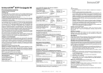
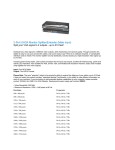
Figure 1.
Interference of IGFBP in IGF-I measurements.
Known concentrations of IGF-I were assayed in the presence of
0.5 ng (left) or 5 ng (right) hIGFBP-3 by a conventional ()and by
the IGFBP-blocked assay (*).
US Customers - For Research Use Only. Not for Use in Diagnostic Procedures
2
Therefore, various techniques were applied to physically separate IGF-I from its binding proteins
before measurement, including (a) size exclusion chromatography under acidic conditions, (b) solidphase extraction and (c) acid-ethanol extraction (2,12,13). These techniques, however, are either
inconvenient or time-consuming or give incomplete and not-reproducible recoveries. The most widely
used method is the acid-ethanol extraction (13,14) with a recovery of only 70-80 % of IGFBP-bound
IGF-I as a result of co-precipitation. The absolute results of such an extraction are therefore false low
(15). The extraction removes the IGFBPs only insufficiently and leads to reduction in sensitivity of the
assay due to pre-dilution of the samples by the extraction procedure. Furthermore, the remaining
IGFBP may still interfere in the assay. In addition, the acid-ethanol extraction is ineffective in
specimens other than serum or plasma (e.g. cell culture media), in which determination of IGF-I is
already difficult enough due to the fact that IGFBPs are frequently present at large excess.
To avoid these difficulties, an uncomplicated assay was developed, in which special sample preparation is
not required before measurement.
Reagents Provided
Microtiter plate, ready for use: Microtiter plate with 96 wells, dived up in 12
strips of 8 wells (separately breakapart) coated with anti-mouse/rat IGF-I
antibody.
Standards A-E, lyophilised, contain recombinant IGF-I. Standard values are
between 0.5 - 18 ng/ml (0.5, 2.5, 6, 12 und 18 ng/ml) IGF-I and have to be
reconstituted in 1 ml (each) Sample Buffer PP. 50 µl per well are used in the
assay. If the standards are required for more than one assay process we
recommend to store the reconstituted Standards frozen at -20°C. Attention:
Standards should be thawed only once – where required please store aliquoted
in adequate volumes.
Sample buffer PP, 125 ml, ready for use, please use for the reconstitution of
Standards A-E, Control Sera KS1 & KS2 and for the serum dilution.
Control Sera KS1 & KS2, each 500 µl lyophilised: Sera have to be
reconstituted in 500 µl Sample Buffer PP. The IGF-I target values and the
respective ranges are given on the vial labels. The dilution of the Control Sera
KS 1&2 should be according to the dilution of the respective samples, the target
values should be obtained by multiplication with the respective dilution factor.
Antibody Conjugate AK, 7 ml, ready for use, contains the biotinylated antiIGF-I antibody. Use 50 µl for each well in the assay.
Enzyme Conjugate EK, 12 ml, ready for use, contains horseradisch-peroxidase
conjugate to streptavidin, use 100 µl for each well in the assay.
Washing Buffer (WP), 50 ml, 20 X concentrated solution. Washing Buffer
(WP) has to be diluted 1:20 with distilled or demineralised water before use (e.g.
add the complete contents of the flask (50 ml) into a graduated flask and fill up
with A.dest. to 1000 ml). Attention: After dilution the Washing Buffer is only 4
weeks stable, dilute only according to requirements.
1)
MTP
2)
CAL
3)
DILU
4)
Control
5)
Ab
6)
CONJ
7)
WASHBUF
20x
8)
SUBST
Substrate (S), 12 ml, ready for use, horseradish-peroxidase-(HRP)-substrate,
stabilised H2O2 Tetramethylbencidine.
9)
H2SO4
Stopping Solution (SL), 12 ml, ready for use, 0.2 M sulphuric acid, Caution
acid!
10)
Sealing tape for covering of the microtiter plate, 2 x, adhesive.
US Customers - For Research Use Only. Not for Use in Diagnostic Procedures
3
Materials Required but not provided
Precision pipettes and multichannel pipettes with disposable plastic tips
Vortex-mixer
Microtiter plate shaker (350 rpm)
Microtiter plate washer (recommended)
Microplate reader ("ELISA-Reader") with filter for 450 and 620nm (or ≥590 nm)
Polyethylene PE/Polypropylene PP tubes for dilution of samples
WARNINGS AND PRECAUTIONS
For research use only. For professional use only.
Before starting the assay, read the instructions completely and carefully. Use the valid version of the
package insert provided with the kit. Be sure that everything is understood.
Before use, all kit components should be brought to room temperature at 20 - 25°C. Precipitates in
buffers should be dissolved before use by thorough mixing and warming.
Do not mix reagents of different lots. Do not use expired reagents.
The microplate contains snap-off strips. Unused wells must be stored at 2 - 8°C in the sealed foil
pouch and used in the frame provided.
Caution: This kit contains material of human and/or animal origin.
No known test methods can offer total assurance of the absence of infectious agents; therefore all
components and specimens should be treated as potentially infectious.
Following components contain < 0.01% 2-Methyl-4-isothiazolin-3-one solution as preservative:A-E,
AK, EK, PP
R34
R43
S26
S36/37
S45
Irritating to eyes and skin
Sensibilisation through skin contact possible
In case of contact with eyes, rinse immediately with plenty of water and seek medical advice
Wear suitable protective clothing and gloves
In case of accident or if you feel unwell seek medical advice
Following components contain < 0.01% (w/w) 5-chloro-2-methyl 2H isothiazol-3-one and 2-methyl-2Hisothiazol-3-one as preservative: A-E, AK, EK, PP, WP
R36/38
R43
S26
advice S28.1
Irritating to eyes and skin
Sensibilisation through skin contact possible
In case of contact with eyes, rinse immediately with plenty of water and seek medical
After contact with skin, wash immediately with plenty of water
Stop solution contains 0.2 M Sulfuric Acid (H 2 SO 4 )
R36/38
S26
S28.1
S36/37
Irritating to eyes and skin
In case of contact with eyes, rinse immediately with plenty of water and seek medical
advice
After contact with skin, wash immediately with plenty of water
Wear suitable protective clothing and gloves.
Pipetting of samples and reagents must be done as quickly as possible and in the same sequence for
each step. Use separate pipette tips for each sample, control and reagent to avoid cross
contamination. Use reservoirs only for single reagents. This especially applies to the substrate
reservoirs. Using a reservoir for dispensing a substrate solution that had previously been used for the
conjugate solution may turn solution colored. Do not pour reagents back into vials as reagent
contamination may occur. Mix the contents of the microplate wells thoroughly to ensure good test
results. Do not reuse microwells. Do not let wells dry during assay; add reagents immediately after
completing the rinsing steps.
US Customers - For Research Use Only. Not for Use in Diagnostic Procedures
4
TMB-Substrate (S) contains 3,3´,5,5´ Tetramethylbenzidine. Store and Incubate in the dark.
R20/21/R22
R36/37/38
S26
S28.1
S36/37
Harmful by inhalation, in contact with skin and if swallowed
Irritating to eyes, respiratory system and skin
In case of contact with eyes, rinse immediately with plenty of water and seek medical
advice
After contact with skin, wash immediately with plenty of water
Wear suitable protective clothing and gloves
General first aid procedures:
Skin contact: Wash affected area thoroughly with water. Discard contaminated cloths and shoes.
Eye contact: In case of contact with eyes, rinse immediately with plenty of water at least 15 minutes.
In order to assure an effectual rinsing spread the eyelids.
Ingestion: If swallowed, wash out mouth thoroughly with water. Immediately see a physician.
Do not eat, drink or smoke in these areas.
Never pipette the materials with the mouth.
Spilled material must be wiped off immediately and should become disinfected. Clean contaminated
areas and equipment with a suitable detergent.
PRINCIPLE
This ELISA for mouse/rat IGF-I is a so-called Sandwich-Assay. It utilizes two specific and high affinity
antibodies for this protein. The IGF-I in the sample
binds to the immobilized first antibody on the
microtiter plate, the biotinylated and StreptavidinPeroxidase conjugated second specific anti-IGF-IAntibody binds in turn to the immobilized IGF-I. In
the closing substrate reaction the turn of the colour
will be high specific catalysed, quantitatively
depending on the IGF-I-level of the samples.
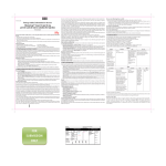
In order to dissociate IGF-I from the IGFBPs, the
samples must be diluted in an acidic buffer
(Figure 2). The diluted samples are then pipetted
into the wells, by this the pH-value will be
neutralized. After neutralization of the samples,
the excess IGF-II occupies the IGF-binding sites
of the binding proteins, thus allowing the
measurement of resulting free IGF-I. With this
method, the IGFBPs are not removed, but their
function and therefore their interference in the
assay is neutralized. Due to the extremely low
cross-reactivity of the IGF-I antibody with IGF-II,
the excess of IGF-II does not disturb the
interaction with IGF-I.
The test runs like a conventional ELISA using a
Streptavidin-Peroxidase-Enzyme Conjugate.
Figure 2: Principle of the IGFBP blocked IGF-I ELISA
US Customers - For Research Use Only. Not for Use in Diagnostic Procedures
5
Specimen
Serum samples as well as Heparin-, EDTA- and Citrat-Plasma samples are suited. Possible dilution of the
sample by the anticoagulant must be considered.
Influence of Heparin (30IE/mL), EDTA (6,8mM) and NaCitrat (0,015M) on the measurement of IGF-I
has been investigated in recovery experiments. Buffer solution was enriched with recombinant IGF-I
and the above mentioned substances. No significant influence on the recovery of IGF-I was detected,
on average the recovery of recombinant material in comparison to enriched PBS was 108%.
Cell culture medium is suitable as sample matrix after predilution of 1:2 with Sample Buffer PP.
Haemolytic reactions have to be avoided. The blood has to be allowed to clot and after complete
clotting, serum is separated by centrifugation.
Storage of the samples
Storage at RT max. 2 days
Storage at –20°C
max. 2 years
More than 2 freeze/thaw cycles are not recommended. IGF-I in samples was found to be unstable
under repeated freeze/thaw cycles, measured values declined respectively.
Sample Preparation
Samples have to be diluted at least 1:10 in Sample Buffer (PP).
A serum dilution of 1:100 is in general suitable. However, the IGF-I levels can vary individually
significant, we would therefore recommend to check this and adjust the dilution respectively.
After reconsititution, mix gently and incubate at room temperature for at least 15 minutes, but no
longer than 2 hours.
Technical Notes
The assay has to be conducted strictly according the test protocol herein.
Reagents with different lot numbers cannot be mixed. The microtiterplate and reagents are stable until
the indicated expiry if stored unopened and protected from sunlight at 2 – 8°C.
Bring all reagents to room temperature (20 - 25°C) before use. Possible precipitations in the buffers
have to be resolved before usage by mixing and / or warming.
Incubation at room temperature means: 20-25°C
The incubation steps should be performed at mean rotation frequency of a particularly suitable
microtitre plate shaker. We are recommending 350 rpm. Due to certain technical differences
deviations may occur, in case the rotation frequency must become adjusted. Insufficient shaking may
lead to inadequate mixing of the solutions and thereby to low optical densities, high variations and/or
false values, excessive shaking may result in high optical densities and/or false values.
Proper washing is of basic importance for a secure, reliable and precise performance of the test.
Incomplete washing is common and will adversely affect the test outcome. Possible consequences
may be uncontrolled unspecific variations of measured optical densities, potentially leading to false
results calculations of the examined samples. Effects like high background values or high variations
may indicate washing problems.
All washing must be performed with the provided washing buffer diluted to usage concentration.
Washing volume per washing cycle and well must be 300 µl at least.
US Customers - For Research Use Only. Not for Use in Diagnostic Procedures
6
The danger of handling with potentially infectious material must be taken into account.
When using an automatic microtitre plate washer, the respective instructions fur use must be carefully
followed. Device adjustments, e.g. for plate geometry and the provided washing parameters, must be
performed. Dispensing and aspirating manifold must not scratch the inside well surface. Provisions
must be made that the remaining fluid volume of every aspiration step is minimized. Following the last
aspiration step of each washing cycle, this could be controlled, and possible remaining fluid could
then be removed, by inverting the plate and repeatedly tapping it dry on non fuzzy absorbent tissue.
Manual washing is an adequate alternative option. Washing Buffer may be dispensed via a
multistepper device, a multichannel pipette, or a squirt bottle. The fluid may be removed by
dynamically swinging out the microtitre plate over a basin. If aspirating devices are used, care has to
be taken that the inside well surface is not scratched. Subsequent to every single washing step, the
remaining fluid should be removed by inverting the plate and repeatedly tapping it dry on non fuzzy
absorbent tissue.
Standards and Controls
For the reconstitution of the lyophilised components (Standards A - E and Control Sera KS1 &KS2)
the kit Sample Buffer PP has to be used. It is recommended to keep reconstituted reagents at room
temperature for 15 minutes and then to mix them thoroughly but gently (no foam!) with a Vortex mixer.
The reconstituted standard and controls can be stored for 2 months at –20°C. Repeated freeze/thaw cycles
have to be avoided.
Washing Buffer
The required volume of washing buffer is prepared by 1:20 dilution of the provided 20-fold concentrate
with deionised water. The diluted Washing Buffer is stable for 4 weeks at 2-8°C. It has to be at room
temperature for usage!
Microtiter plate
Unused microtiter plate stripes have to be stored airtight together with the desiccant bag at 2-8°C.
The labelled expiry is not influenced in case of proper storage.
US Customers - For Research Use Only. Not for Use in Diagnostic Procedures
7
Assay Procedure
All determinations (Standards, Control Sera KS1 & KS2 and samples) should be assayed in duplicate.
For optimal results, accurate pipetting and adherence to the protocol are recommended.
When performing the assay, the Standards, Control Sera and the samples should be pipette as fast
as possible (e.g., <15 minutes).
All incubations have to conducted at room temperature (20-25°C)
To avoid distortions due to differences in incubation times, Antibody (AK) and Enzyme Conjugate (EK) as
well as the following Substrate Solution S should be added to the plate in the same order and in the same
time interval as the samples. Stop Solution SL should be added to the plate in the same order as the
Substrate Solution.
1) Add 50 µl Antibody Conjugate AK in all wells used
2) Add 50 µl Sample Buffer PP into the first two wells (These wells serve as blanks). Subsequently
add 50 µl Standard or 50 µl of diluted Control Sera or diluted samples into wells designated for
standards, controls or samples.
3) Cover the wells with sealing tape and incubate the plate for 1 hour at room temperature (shake
at 350 rpm )
4) After incubation, aspirate the contents of the wells and wash the wells 5 times 300 µl Washing
Buffer WP / well. The washing buffer should incubate for at least for 15 seconds/cycle. Tap the
plate firmly against several layers of folded paper towels to remove excess washing buffer.
5)
Following the last washing step pipette 100 µl of the Enzyme Conjugate EK in each well.
6) Cover the wells with sealing tape and incubate the plate for 0.5 hour at room temperature
(shake 350 rpm).
7)
After incubation wash the wells 5 times with Washing Buffer as described in step 4
8)
Pipette 100 µl of the TMB Substrate Solution in each well.
9)
Incubate the plate for 30 minutes in the dark at room temperature (20 - 25°C).
10) Stop the reaction by adding 100 µl of Stopping Solution.
11) Measure the colour reaction within 30 minutes at 450nm (reference filter ≥590 nm).
US Customers - For Research Use Only. Not for Use in Diagnostic Procedures
8
Calculation of Results
Establishing the Standard Curve
For the evaluation of the assay it is preconditioned that the absorbance values of the blank should be
below 0.25, these of standard E should be above 1.0.
Samples, which yield higher absorbance values than Standard E are beyond the standard curve, for
reliable determinations these samples should be tested anew with a higher dilution.
Standards are provided in the following concentrations (use the concentration unit as preferred):
Standard
ng/ml
A
0.5
B
2.5
C
6
D
12
E
18
1)
Calculate the mean absorbance value for the blank from the duplicated determination (well
A1/A2).
2)
Subtract the mean absorbance of the blank from the mean absorbances of all other values.
3)
Plot the standard concentrations on the x-axis versus the mean value of the absorbance of the
standards on the y-axis.
4)
Recommendation: Calculation of the standard curve should be done by using a computer
program. A higher-grade polynomial, or four parametric logistic (4-PL) curve fit or nonlinear regression are usually suitable for the evaluation (as might be spline or point-to-point
alignment in individual cases).
5)
The IGF-I concentration of the diluted sample or the diluted control sera KS1&2 in ng/ml (or
µg/ml according the chosen unit for the standards) is calculated in this way, the IGF-I
concentration of the undiluted sample and of KS1 & KS2 is calculated by multiplication with
the respective dilution factor.
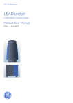
The exemplary shown standard curve in Fig.3 cannot be used for calculation of your test results. You
have to establish a standard curve for each test you conduct!
Exemplary calculation of the IGF-I concentration of a diluted sample:
Measured extinction of your sample
Measured extinction of the blank
0.70
0.02
Your measurement programm will calculate the IGF-I concentration of the diluted sample
automatically by using the difference of sample and blank for the calculation. You only have to
determine the most suitable curve fit (here: polynomial 3rd degree).
US Customers - For Research Use Only. Not for Use in Diagnostic Procedures
9
In this exemplary case the following equation is solved by the programm to calculate the IGF-I
concentration in the sample:
y
= -0.031924+0.2606x-0.010994x2 +0.0019791x3
3.0921= x
If the dilution factor (1:100) is taken into account the IGF-I concentration of the undiluted sample is
309.21 ng/mL
Fig. 3: Exemplary Standard Curve with a polynomial 3rd degree as curve fit.
Performance Characteristics
Standards
The Standards of this ELISA are prepared from recombinant IGF-I in concentrations of 0.5, 2.5, 6,
12, 18 ng/mL.
Sensitivity
The analytical sensitivity of this ELISA yields < 0.029 ng/mL
Interassay Variability
Mean
SD
VC%
n
Sample 1
471
24.81
5.3
10
Sample 2
744
41.8
5.6
10
Sample 3
347
23.3
6.72
16
Sample 2
Sample 3
Intraassay Variability
Mean
[µg/mL]
SD
VC%
n
Sample 1
165
8,0
4,9
10
794
47
5,9
10
514
22
4,2
10
US Customers - For Research Use Only. Not for Use in Diagnostic Procedures
10
Linearity
ng/mL
Probe 1
1:50
261
1:75
332
1:100
315
1:200
281
1:400
290
1:600
298
1:800
325
Probe 2
888
909
980
960
994
997
983
Method Comparison
Figure 4: Method comparison of this IGF-I ELISA and another commercially available IGF-I ELISA.
US Customers - For Research Use Only. Not for Use in Diagnostic Procedures
11
LITERATURE
1) Baxter RC. 1986 The somatomedins: insulin-like growth factors. Adv Clin Chem.25:49-115
2) Daughaday WH, Rotwein P. 1989 Insulin-like growth factors I and II. Peptide, messenger ribonucleic acid and
gene structures, serum, and tissue concentrations. Endocr Rev. 10:68-91
3) Spencer EM (Ed.) 1991 Modern Concepts of Insulin-Like Growth Factors. New York: Elsevier.
4) Klapper DG, Svoboda ME, Van Wyk JJ. 1983 Sequence analysis of somatomedin-C: confirmation of identity
with insulin-like growth factor-I. Endocrinology. 112:2215-2217.
5) Rinderknecht E, Humbel RE. 1978 The amino acid sequence of human insulin-like growth factor I and its
structural homology with proinsulin. J Biol Chem. 253:2769-2276.
6) Clemmons DR, Van Wyk JJ. 1984 Factors controlling blood concentration of somatomedin C. Clin Endocrinol
Metab. 13:113-143.
7) Ballard J, Baxter R, Binoux M, et al. 1989 On the nomenclature of the IGF binding proteins. Acta Endocrinol
(Copenh). 121:751-752.
8) Drop SLS. 1992 Report on the nomenclature of the IGF binding pro-teins. J. Clin Endocrinol Metab. 74:12151216.
9) Martin JL, Baxter RC. 1986 Insulin-like growth factor binding protein from human plasma. Purification and
characterization. J Biol Chem. 261:8754-8760.
10) Binkert C, Landwehr J, Mary JL, Schwander J, Heinrich G. 1989 Cloning, sequence analysis and expression
of a cDNA encoding a novel insulin-like growth factor binding protein (IGFBP-2). EMBO J. 8:2497-2502.
11) Furlanetto RW, Underwood LE, Van Wyk JJ, D'Ercole AJ. 1977 Estimation of somatomedin-C levels in
normals and patients with pituitary disease by radioimmunoassay.J. Clin Invest. 60:648-657.
12)
Daughaday WH, Kapadia M, Mariz I. 1987 Serum somatomedin binding proteins: physiologic
significance and interference in radioligand assay. J Lab Clin Med. 109:355-363.
13)
Breier BH, Gallaher BW, Gluckman PD. 1991 Radioimmunoassay for insulin-like growth factor-I:
solutions to some potential problems and pitfalls. J Endocrinol. 128:347-357.
14) Daughaday WH, Mariz IK, Blethen SL. 1980 Inhibition of access of bound somatomedin to membrane
receptor and immunobinding sites: a comparison of radioreceptor and radioimmunoassay of somatomedin in
native and acid-ethanol-extracted serum. J Clin Endocrinol Metab. 51:781-788.
15) Blum WF, Gallaher B, Ranke MB. 1992 An IGFBP-blocked IGF-I RIA that measures what it pretends to
measure: IGF-I. 74th Annual Meeting of the American Endocrine Society. 293.
16) Rosenfeld RG, Wilson DM, Lee PDK, Hintz RL. 1986 Insulin-like growth factors I and II in evaluation of
growth retardation. J Pediatr. 109:428-433.
17) Clemmons DR, Van-Wyk JJ, Ridgway EC, Kliman B, Kjellberg RN, Underwood LE. 1979 Evaluation of
acromegaly by radioimmunoassay of somatomedin-C.N.Engl J Med. 301:1138-1142
18) Zapf J, Walter H, Froesch ER. 1981 Radioimmunological determination of insulin-like growth factors I and II
in normal subjects and in patients with growth disorders and extrapancreatic tumor hypoglycemia. J Clin
Invest. 68:1321-1330.
19) Blum WF. 1992 Insulin-like growth factors and their binding proteins. In: Ranke MB, ed. Functional
Endocrinologic Diagnostics in Children and Adolescence. Mannheim: J + J Verlag; 102-117.
20) Rieu M, Girard F, Bricaire H, Binoux M. 1982 The importance of insulin-like growth factor (somatomedin)
measurements in the diagnosis and surveillance of acromegaly. J Clin Endocrinol Metab. 55:147-153.
21) Blum WF, Ranke MB, Bierich JR. 1988 A specific radioimmunoassay for insulin-like growth factor II: the
interference of IGF binding proteins can be blocked by excess IGF-I. Acta Endocrinol (Copenh).118:374-380.
22) Wilson EM, Oh Y, Rosenfeld RG (1997) Generation and characterization of an IGFBP-7 antibody:
Identification of 31 kD IGFBP-7 in human biological fluids and Hs578T human breast cancer conditioned
media. J Clin Endocrinol Metab Vol 82, 4:1301-1303
23)
Ranke MB, Schweizer R, Elmlinger MW, Weber K, Binder G, Schwarze CP, Wollmann HA (2000)
Significance of basal IGF-I, IGFBP-3 and IGFBP-2 measurements in the diagnostics of short stature in
children. Horm Res 54:60-68
24)
Ranke MB, Schweizer R, Elmlinger MW, Weber K, Binder G, Schwarze CP, Wollmann HA (2001)
Relevance of IGF-I, IGFBP-3 and IGFBP-2 measurements during GH treatment of GH-deficient and non-GHdeficient children and adolescents. Horm Res 55:155-124
US Customers - For Research Use Only. Not for Use in Diagnostic Procedures
12
SUMMARY – IGF-I (MOUSE/RAT) ELISA 22-IG1MS-E01
Reconstitution / Dilution of Reagents
Standards A-E
Reconstitution in Sample Buffer PP
1 ml
Control Serum KS1
Reconstitution in Sample Buffer PP
500 µl
Control Serum KS2
Reconstitution in Sample Buffer PP
500 µl
Wash Buffer WP
dilute in A. dest. (eg. total volume of 50 ml in a
1:20
graduated flask and fill up to 1000 ml)
Sample + Control Sera KS1 and KS2: dilute 1:100 in Sample Buffer PP, mix
immediately, incubate at least for 15 min, max. 2h. Use 50 µl for each well in the assay.
Before conducting the assay equilibrate all reagents to room temperature.
Assay Procedure for Double Determinations:
Pipette
Reagent
50 µl
Antibody Conjugate AK
50 µl
50 µl
50 µl
50 µl
50 µl
50 µl
50 µl
50 µl
50 µl
Sample Buffer PP (blank)
Standard A (0.5 ng/ml)
Standard B (2.5 ng/ml)
Standard C (6 ng/ml)
Standard D (12 ng/ml)
Standard E (18 ng/ml)
Control Serum KS1
Control Serum KS2
Samples
Position
in all wells used
A1 and A2
B1 and B2
C1 and C2
D1 and D2
E1 and E2
F1 and F2
G1 and G2
H1 and H2
following wells
Cover the wells with the sealing tape.
Incubation: 1 h at RT, 350 rpm
5x 300 µl
100 µl
Aspirate the contents of the wells and wash 5x with 300
µl Wash Buffer WP
Enzyme Conjugate EK
each well
each well
Incubation: 30 min at RT, 350 rpm
5x 300 µl
100 µl
Aspirate the contents of the wells and wash 5x with 300
µl Wash Buffer WP
Substrate S
each well
each well
Incubation: 30min in the dark RT
Stop Solution SL
100 µl
each well
Measure the absorbance within 30 min at 450 nm with ≥ 590 nm as reference wavelength.
US Customers - For Research Use Only. Not for Use in Diagnostic Procedures
13