1
Product overview
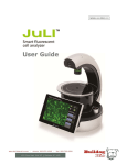
A fully-automated, fluorescence live cell imaging system that acquires cell images directly from various cell culture
plates (6 to 384 well) and dishes in an incubator. JuLI™ Stage supports multi-channel fluorescent colors, multiple objective lenses, and sensitive filter-based optics to optimize for various live cell assays. In addition, it enables users to support quantified cellconfluence results with low variation and the growth curve using image-based analysis with bright
field.
Compact, compatible and automated fluorescence cell imaging system of live cell imaging from
inside your cell culture incubator!
Suitable for the following applications:
• Live cell imaging (time lapse)
• Cell culture quality control
• HCS cell imaging analysis (6 to 384 well)
• Cell-based assay optimization
• Image stitching
• Angiogenesis
• Scratch-wound assay
• Neurite growth
• Angiogenesis
• Protein expression
• Kinetic imaging and cell differentiation
• Cell growth monitoring
• Cell culture optimization
• Stem cell development
• Cell proliferation
• Cell cytotoxicity
• Cell migration study
• Cell differentiation
JuLI™Stage, New paradigm for cell based assay
5