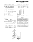
1
US-7a
Fetus Ultrasound Examination Phantom
SPACE FAN-ST
Instruction
Manual
Product Supervision :
Kiyoko Kabeyama RN, RM, PhD
Professor Midwifery & Women's Health, School of Human Health
Science, Graduate School of Medicine, Kyoto University
Haruto Egawa, PhD
National Hospital Organization Kyoto Medical Center
Department of Obstetrics, Head Physician or Medical Director
Contents
●
Please read before training
General information
●
Training
Before training
Training session
After training
●
Training Items
back cover
The SPACE FAN-ST provides high quality training for routine second trimester screening. This phantom contains
a 23 week fetus with full anatomy placed in the uterus that can be scanned with 2D and 3D transducers.
Included life-size fetus model facilitates demonstration and three dimensional understanding.
* Ensure that the ultrasonic diagnostic equipment which you use is properly quality controlled.
Training Items:
Anatomy
Fetal size assessment:
23 week fetus (26cm).
Fetal anatomy assessment:
head and brain, heart and lung, abdominal organs,
spine and bones.
Placental localization
Fetus: skeletal structure, brain with septum lucidum,
heart with four chambers, lungs, spleen, kidneys, aorta,
UV, UA, and the external genital.
Don’t mark on the phantom with pen or leave printed materials contacted on its surface.
Ink marks on the phantom will be irremovable.
Caution
●
Training Items
Assessment of development and condition of fetus,
Whole fetus assessment - head, breast, abdomen,
spine, limbs, and genitalia
■ Fetus measurement - BPD, AC, and FL
■
presentation, position, fetal habitus, sexuality and so on.
■
* compatible with 2D and 3D probes.
amount, placenta, and umbilical cord
① Measurement of fetus (measuring at 3 points)
BPD: biparietal diameter - measures BPD using the transparent
septum as landmarks .
AC: Abdominal circumference - measures AC using the stomach,
abdominal aorta and umbilical vein as landmarks.
FL: Femur length - measures the total length of the bone.
Estimates the weight by measuring at 3 points to evaluate the
development condition of the fetus.
BPD
AC
FL
estimate the fetal weight
②
Measures the maximum
vertical depth of the
(the maximum amniotic
③ Assessment of head, breast, abdomen, spine and so on
●
of the brain
Assessment of the spine and limbs
● Assessment of the cardiac chambers and blood vessels,
inclination, and the lung
● Assessment of the viscera such as the stomach, kidney,
and bladder and the vasculature
④ Assessment of umbilical cord and placenta
Scan the umbilical cord
and blood vessels,
placenta attachment
condition, and placenta
position.
●
blood vessels
umbilical cord
brain (transparent septum)
cardiac chambers
stomach
kidney
abdominal aorta
bladder
⑤ Determination of fetus presentation
(cephalic or breech)
Rotate the phantom
unit and determine the
fetus presentation
(cephalic or breech).
breech
⑥ Determination of sex
(This product represents
a male fetus.)
male fetus
* This product does not reproduce the heart beat and the bloodstream.
Please contact manufacturer with any discrepancies in this manual or product feedback. Your cooperation is reatly appreciated.
Please read
General information
Set includes
Before your first use, please ensure that you have all components listed below.
a
b
c
d
mother body torso ・・・・・・・・・・・・・・・・・・・・
ultrasound pregnant uterus phantom ・・・・
fetus demonstration model ・・・・・・・・・・・・・
DVD Tutorial Manual ・・・・・・・・・・・・・・・・・・
Carrying case ・・・・・・・・・・・・・・・・・・・・・・・・・
Instruction manual (this leaflet)
1
1
1
1
1
d
DOs and DON’T s
DO s
DON’Ts
Always attach the protection bowl in the cavity of the
mother body torso.
Do not remove the hard plastic protection bowl. Ensure to use
it on the torso when setting the phantom unit on. Putting the
phantom on the torso directly without the bowl may lead to
discoloring or deformation of the torso.
Handle with care
The materials for phantom and models are special composition
of soft resin. Please handle with care at all times.
Never wipe the phantom or models with
thinner or organic solvent.
Don’t mark on the phantom with pen or
leaveprinted materials contacted on their
surface.
Ink marks on the models will be
irremovable.
Cleaning and care
Please clean the phantom completely every time after you
finish the training.
The remaining lubricating gel may deteriorate the phantom.
Keep the training set at room temperature, away from heat,
moisture and direct sunlight.
Please note:
The color of the phantom may change over time, though, please be assured that this is not deterioration
of the material and the ultrasonic features of the phantom stay unaffected.
This phantom has a thin layer of coating for protective purposes which may cause minor wrinkling.
However, it has no effect on the ultrasound image.
1
Training
Before training
Training session
After training
1 Before training
① Set the phantom unit on the mother body torso in the
desired direction. You can rotate the phantom to change
the presentation and position of the fetus.
② Apply ultrasound gel on the phantom unit
directly. Use plenty of gel.
2 Training session
Fetus assessment and measurement
BPD
AC
FL
estimate the fetal weight
① Place the probe on the phantom unit . The
keys for ultrasound training are listed on the
back cover of this manual.
3 After training
① Wipe off the gel completely with wet wipes
not to leave the gel on the surface of the
phantom and the torso. You can wash the
phantom unit and the protection bowl with
water.
Do not wash the body torso with water.
2