1
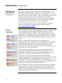
Mouse Cytokine 20-Plex
Panel
For simultaneous quantitative determination
of FGF-basic, GM-CSF, IFN-γ, IL-1α, IL-1β,
IL-2, IL-4, IL-5, IL-6, IL-10, IL-12 (p40/p70),
IL-13, IL-17, IP-10, KC, MCP-1, MIG, MIP-1α,
TNF-α and VEGF in mouse serum and tissue
culture supernatant
Catalog no. LMC0006
Rev. 1.1
16 June 2010
PRLMC0006
Corporate Headquarters
Invitrogen Corporation
1600 Faraday Avenue
Carlsbad, CA 92008
T: 1 760 603 7200
F: 1 760 602 6500
E: [email protected]
For country-specific contact information visit our web site at www.invitrogen.com
User Manual
Table of Contents
Table of Contents......................................................................................................iii
Kit Contents and Storage .......................................................................................... iv
Introduction.................................................................................. 1
Overview ................................................................................................................... 1
Methods ........................................................................................ 4
Before Starting........................................................................................................... 4
Preparing Reagents .................................................................................................... 7
Assay Procedure ...................................................................................................... 10
Instrument Setup...................................................................................................... 17
Performance Characteristics and Limitations of the Procedure ............................... 18
Troubleshooting....................................................................................................... 19
Appendix .................................................................................... 22
Technical Support.................................................................................................... 22
Purchaser Notification ............................................................................................. 23
References ............................................................................................................... 24
Higher Multiplexing ................................................................................................ 25
Protocol Summary ................................................................................................... 27
Plate Plan Template ................................................................................................. 28
Explanation of Symbols........................................................................................... 29
iii
Kit Contents and Storage
Storage
All components of the Mouse Cytokine 20-Plex Panel are shipped
at 2 to 8°C. Upon receipt, store all kit components at 2 to 8°C. Do
not freeze.
Contents
The components and amounts included in the Mouse Cytokine
20-Plex Panel are listed below.
Reagents Provided
Mouse 20-Plex Antibody Bead Solution (1X) (contains 7.5 mM
sodium azide)
100 Test Kit
2.5 mL × 1 vial
Mouse 20-Plex Biotinylated Antibody Concentrate (10X) (contains 1 mL × 1 vial
0.1% sodium azide)
Mouse 20-Plex Lyophilized Standard (contains 0.1% sodium azide) 2 vials
Wash Solution Concentrate (20X) (contains 0.1% sodium azide)
15 mL × 1 bottle
Assay Diluent (contains 0.1% sodium azide)
15 mL × 1 bottle
Incubation Buffer (contains 0.05% sodium azide)
12 mL × 1 bottle
Biotin Diluent (contains 3.3 mM thymol)
12 mL × 1 bottle
Streptavidin-RPE Concentrate (10X) (contains 0.1% sodium azide) 1 mL × 1 vial
Streptavidin-RPE Diluent (contains 3.3 mM thymol)
12 mL × 1 bottle
96-well Filter Plate
1 × 96-well plate
iv
Introduction
Overview
Purpose
Invitrogen’s Multiplex Bead Immunoassay Kits are developed to
maximize flexibility in experimental design, permitting the
measurement of one or multiple proteins in panels designed by the
researcher. The Mouse Cytokine 20-Plex Panel contains all the
reagents that are intended for use with the 100™ or 200™ dual laser
detection system manufactured by Luminex Corporation and sold by
Invitrogen and other vendors.
For Research Use Only. CAUTION: Not for human or animal
therapeutic or diagnostic use.
Background
Information
Advances in the field of cell biology have defined a complex and
interdependent set of extracellular and intracellular signaling
molecules that control normal cell function. Therefore, there is
growing interest among researchers as well as drug discovery
groups in simultaneously monitoring multiple components of
signaling pathways. Solid phase multiplex protein assays are the
tools of choice in these studies as they maximize efficiency by
simultaneously profiling several proteins within individual samples.
Invitrogen’s Multiplex Bead Immunoassays are solid phase protein
immunoassays that use spectrally encoded antibody-conjugated
beads as the solid support. The spectral beads are suitable for use in
singleplex assays or may be mixed for multiplex assays according
to the researcher’s requirements. Each assay is carefully designed
and tested to assure that sensitivity, range, and correlation are
maximized. The assay is performed in a 96-well plate format and
analyzed with a Luminex® 100™ or 200™ instrument which
monitors the spectral properties of the capture beads while
simultaneously measuring the quantity of associated fluorophore.
Standard curves generated with this assay system extend over
several orders of magnitude of concentrations, while the sensitivity
and quantitation of the assays are comparable to ELISAs (Enzyme
Linked-Immuno-Sorbent Assays). Assay standards are calibrated to
NIBSC (National Institute for Biological Standards and Controls)
reference preparations, when available, to assure accurate and
reliable results.
Continued on next page
1
Overview, Continued
Background
Information,
Continued
Invitrogen’s Mouse Cytokine 20-Plex Panel is designed for the
quantitative determination of FGF-basic, GM-CSF, IFN-γ, IL-1α,
IL-1β, IL-2, IL-4, IL-5, IL-6, IL-10, IL-12 (p40/p70), IL-13, IL-17,
IP-10, KC, MCP-1, MIG, MIP-1α, TNF-α and VEGF in serum and
tissue culture supernatant. This kit has not been tested for
multiplexing with other markers. Should user elect to multiplex
this kit with other Luminex kits, the assay conditions should be
determined empirically for each specific application.
Visit the Invitrogen web site for a current listing of available
Invitrogen multiplex bead immunoassays and reagents, at
www.invitrogen.com/luminex.
The xMAP® technology combines the efficiencies of multiplexing
up to 100 different proteins for simultaneous analysis, with
reproducibility similar to ELISA. The technology uses 5.6 μm
polystyrene beads which are internally dyed with red and infrared
fluorophores of differing intensities. Each bead is given a unique
number, or bead region, allowing differentiation of one bead from
another.
Assay
Overview
Antibody Conjugated Beads
IL-5
IFN-γ
IL-2
IL-10
Analyte Capture
IL-5
IFN-γ
IL-2
IL-10
Detection Antibody
IL-5
IFN-γ
IL-2
IL-10
Analyte Detection
2
Beads of defined spectral properties are conjugated to
protein-specific capture antibodies and added along with samples
(including standards of known protein concentration, control
samples, and test samples), into the wells of a filter-bottom
microplate and where proteins bind to the capture antibodies over
the course of a 2 hour incubation.
After washing the beads, protein-specific biotinylated detector
antibodies are added and incubated with the beads for 1 hour.
During this incubation, the protein-specific biotinylated detector
antibodies bind to the appropriate immobilized proteins.
After removal of excess biotinylated detector antibodies,
streptavidin conjugated to the fluorescent protein, R-Phycoerythrin
(Streptavidin-RPE), is added and allowed to incubate for
30 minutes. The Streptavidin-RPE binds to the biotinylated detector
antibodies associated with the immune complexes on the beads,
forming a four-member solid phase sandwich.
After washing to remove unbound Streptavidin-RPE, the beads are
analyzed with the Luminex detection system. By monitoring the
spectral properties of the beads and the amount of associated
R-Phycoerythrin (RPE) fluorescence, the concentration of one or
more proteins can be determined.
Experimental Overview
Experimental
Outline
The experimental outline for using the Mouse Cytokine 20-Plex
Panel is shown below.
Prewet wells
Add beads
Wash
Add incubation buffer, standard,
and samples then incubate for 2
hours
Wash
Add detection antibody
then incubate for 1 hour
Wash
Add streptavidin-RPE
then incubate for 30 minutes
Wash
Resuspend and acquire data
using Luminex Detection system
3
Methods
Before Starting
Materials
Required but
Not Provided
•
Luminex® xMAP® system with data acquisition and analysis
software (Invitrogen, Cat. no. MAP0200), contact Invitrogen
for instrument and software placement services, see page 22
•
Filtration vacuum manifold for bead washing (Pall, Cat. no.
5017 is recommended)
•
Sonicating water bath
•
Vortex mixer
•
Orbital shaker (small diameter rotation recommended)
•
Calibrated, adjustable, precision pipettes, preferably with
disposable plastic tips (A manifold multi-channel pipette is
desirable)
•
Distilled or deionized water
•
Glass or polypropylene tubes
•
Aluminum foil or opaque 96-well plate cover (Invitrogen, Cat.
no. PC10)
Continued on next page
4
Before Starting, Continued
Procedural
Notes
Review the procedural notes below before starting the protocol.
•
Before mixing plexes, check that each protein is represented by
a unique bead region. Up to 10 bead concentrates (singleplexes
or pre-mixed multiplexes) can be combined to increase the number
of proteins measured.
•
Do not invert the filter plates during the assay. The filter plates
are designed to be used in conjunction with a vacuum manifold
(do not exceed 5 mm Hg) and emptied from the bottom.
•
Do not freeze any component of this kit. Store kit components at 2
to 8°C when not in use. Allow all reagents to warm to room
temperature before use (air-warm all reagents at room temperature
for at least 30 minutes, or alternatively, in a room-temperature
water bath for 20 minutes (except plate and standard vials).
•
The fluorescent beads are light-sensitive. Protect the beads from
light to avoid photobleaching of the embedded dye. Use aluminum
foil to cover test tubes used in the assay. Cover filter plates
containing beads with an opaque or aluminum foil-wrapped plate
cover. Since the amber vial does not provide full protection, keep
the vial covered in the box or drawer when not in use.
•
Do not expose beads to organic solvents.
•
Do not place filter plates on absorbent paper towels during loading
or incubations, as liquid may be lost due to contact wicking. An
extra plate cover is a recommended surface to rest the filter plate.
Following plate washing, remove excess liquid and blot from the
bottom of the plate by pressing the plate on clean paper towels.
•
When pipetting reagents, maintain a consistent order of addition
from well-to-well to ensure equal incubation times for all wells.
•
To prevent filter tearing, avoid touching the filter plate membrane
with pipette tips.
•
Do not use reagents after kit expiration date.
•
It is recommended that in-house controls be included with every
assay. If control values fall outside pre-established ranges, the
assay may be suspect. Contact Invitrogen Technical Support for
product and technical assistance.
•
Do not mix or substitute reagents with those from other lots or
sources.
Continued on next page
5
Before Starting, Continued
Recommended
Plate Plan
•
Handle all blood components and biological materials as
potentially hazardous. Follow standard precautions as
established by the Centers for Disease Control and Prevention
and by the local Occupational Safety and Health
Administration when handling and disposing of infectious
agents.
•
This kit contains materials with small quantities of sodium
azide. Sodium azide reacts with lead and copper plumbing to
form explosive metal azides. Upon disposal, flush drains with
a large volume of water to prevent azide accumulation. Avoid
ingestion and contact with eyes, skin and mucous membranes.
In case of contact, rinse affected area with plenty of water.
Observe all federal, state and local regulations for disposal.
It is recommended that a plate plan be designed before starting the
assay. A plate plan template is provided on page 28. The following
is a suggested plate plan:
A
1
2
B
B
B
Std 7 Std 7
C
Std 6 Std 6
D
Std 5 Std 5
E
Std 4 Std 4
F
Std 3 Std 3
G
Std 2 Std 2
H
Std 1 Std 1
3
4
5
6
7
8
9
10 11 12
B= blank (Assay Diluent), Standards 7 through 1, lowest
concentration to highest.
The remainder of the plate is available for controls and samples
which may be run as a singlet or in duplicate, as desired.
Note
6
Running all standards, samples and controls in duplicate is
recommended.
Preparing Reagents
Introduction
Review the information in this section before starting. The Mouse
Cytokine 20-Plex Panel includes both antibody bead reagents and
buffer reagents. Prepare components of the Mouse Cytokine
20-Plex Panel according to instructions below.
Note: Bring all reagents and samples to room temperature before
use.
Preparing
Wash Solution
Upon storage at 2 to 8°C, a precipitate may form in the 20X Wash
Solution Concentrate. If this occurs, warm the 20X Wash Solution
Concentrate to 37°C and mix until the precipitate is dissolved.
1. Prepare a 1X Working Wash Solution for use with a 96-well
plate by transferring the entire contents of the Wash Solution
Concentrate bottle to a 500 mL container (or equivalent) and
then add 285 mL of deionized water. Mix well.
2. The 1X Working Wash Solution is stable for up to 2 weeks
when stored at 2 to 8°C.
Note: To prepare smaller volumes of 1X Working Wash Solution,
mix 1 part of 20X concentrate with 19 parts of deionized water.
Mix well.
Sample
Preparation
Guidelines
•
Serum and tissue culture supernatants are suitable for use
with Invitrogen’s Multiplex Bead Immunoassays. Additional
sample types may be suitable but have not been thoroughly
validated. If possible, avoid the use of hemolyzed or lipemic
sera. The appropriate sample types are defined on the
Technical Data Sheet included with this multiplex panel.
•
Collect samples in pyrogen/endotoxin-free tubes. Centrifuge,
separate, and transfer samples to polypropylene tubes for
storage.
•
Analyze samples shortly after collection or thawing. Freeze
samples after collection if samples will not be tested
immediately. Avoid multiple freeze-thaw cycles of frozen
samples. Thaw completely and mix well (do not vortex)
prior to analysis.
•
Clarify all samples by centrifugation (1,000 × g for
10 minutes) and/or filter prior to analysis to prevent clogging
of the filter plates.
•
In the event that the sample concentrations exceed the
standard curve, dilute samples and reanalyze. Dilute the
serum samples in Assay Diluent and dilute tissue culture
supernatants in the corresponding tissue culture medium.
Continued on next page
7
Preparing Reagents, Continued
Guidelines for
Standard
Curve
Preparation
•
Each kit comes with 2 complete sets of standard vials, so that
2 runs on the plate can be made with freshly prepared
standards.
•
Reconstitute the protein standard within 1 hour of performing
the assay. All standards are calibrated to NIBSC preparations,
when available. Additional standards are available from
Invitrogen custom services
•
Before performing standard mixing and serial dilutions
confirm reconstitution volumes on the Technical Data Sheet,
included in the Singleplex Bead Kit(s).
•
The concentrations of the protein components of the standard
are indicated on the Technical Data Sheet.
•
Perform standard dilutions in glass or polypropylene tubes.
•
When using serum samples, reconstitute the standard with
Assay Diluent provided.
•
If using other sample types (e.g., tissue culture supernatant),
reconstitute the standard with a mixture, composed of 50%
Assay Diluent and 50% of the matrix which closely
resembles the sample type (50%/50% mixture). For example:
When the sample type is RPMI medium containing 5% FBS,
the standards should be reconstituted in a mixture composed
of 50% Assay Diluent and 50% RPMI containing 5% FBS.
Important
Protein standards may be analyzed alone, or combined with other
protein standards for higher levels of multiplexing. Do not
combine more than 4 vials.
Reconstituting
Lyophilized
Standards
1.
2.
3.
To the standard vial(s), add the suggested reconstitution
volume of the appropriate diluent (see table on next page). Do
not vortex. When mixing or reconstituting protein
solutions, always avoid foaming.
Replace the vial stopper and allow the vial to stand
undisturbed for 10 minutes.
Gently swirl and invert the vial 2 to 3 times to ensure
complete reconstitution and allow the vial to sit at room
temperature for an additional 5 minutes.
Continued on next page
8
Preparing Reagents, Continued
One vial of standard
Reconstitute the standard vial in the suggested
reconstitution volume, usually 1 mL, of appropriate diluent.
Two vials of standards
Reconstitute each vial with 0.5 mL of appropriate diluent.
Combine 300 μL from each vial and mix by gently
pipetting up and down 5 to 10 times.
Three vials of standards Reconstitute each vial with 0.333 mL of appropriate
diluent. Combine 200 μL from each vial and mix by gently
pipetting up and down 5 to 10 times.
Four vials of standards
Preparing
Standard
Curve
Reconstitute each vial with 0.250 mL of appropriate
diluent. Combine 150 μL from each vial and mix by gently
pipetting up and down 5 to 10 times.
The standard curve is made by serially diluting the reconstituted
standard in Assay Diluent for serum samples or a mixture of 50%
Assay Diluent and 50% tissue culture medium for tissue culture
supernatant samples. See below. Do not vortex. Mix by gently
pipetting up and down 5 to 10 times.
Serum: Assay Diluent
Tissue culture: 50% Assay Diluent/ 50% Tissue Culture Medium
Discard all remaining reconstituted and diluted standards after
completing assay. Return the Assay Diluent to the kit.
9
Assay Procedure
Method of
Washing
Incomplete washing adversely affects assay results. Perform all wash
steps with the Wash Solution supplied with the kit. All phases of the
assay, including incubations, washing steps, and loading beads, are
performed in the filter bottom plate supplied with the kit.
1.
2.
3.
4.
5.
6.
To wash beads, place the filter plate on the vacuum manifold
and aspirate the liquid with gentle vacuum (do not exceed
5 mm Hg). Excessive vacuum can cause the membrane to tear,
resulting in antibody bead loss. Prevent any vacuum surge by
opening and adjusting the vacuum on the manifold before
placing the plate on the manifold surface.
Stop the vacuum pressure as soon as the wells are empty. Do
not attempt to pull the plate off the vacuum manifold while the
vacuum is still on or filter plate damage may occur. Release the
vacuum prior to removing the plate.
If solution remains in the wells during vacuum aspiration, do
not detach the bottom of the 96 well filter plate. In some
cases, minor clogs in the filter plate may be dislodged by
carefully pressing the bottom of the plate under the clogged
well with the pointed end of a 15 mL plastic conical tube. Place
the filter plate on a clean paper towel and use a gloved thumb or
a 1 mL Pasteur pipette bulb to plunge the top of the clogged
well. Empty all clogged wells entirely before continuing the
washes. Note: Do not attempt to repetitively pull vacuum on
plates with clogged wells. This can compromise the unclogged
wells and bead loss may occur.
After all wells are empty, lightly tap or press the filter plate onto
clean paper towels (hold the plate in the center for tapping) to
remove excess fluid from the bottom of the filter plate. Do not
invert plate.
Following the last aspiration and plate taps, use a clean
absorbent towel to blot the bottom of the filter plate before
addition of next liquid phase or data acquisition step.
Do not leave plate on absorbent surface when adding
reagents.
Continued on next page
10
Assay Procedure, Continued
Reverse
Pipetting
Recommendation
Note
Online Tool
To reduce bubbles and loss of reagents due to residual fluid left in
pipette tips, use the recommended reverse pipetting technique.
1. To reverse pipette, set the pipette to the appropriate volume
needed. Note: Do not reverse pipette volumes <20 µL.
2. Press the push-button slowly to the first stop and then press
on past it. Note: the amount past the first stop will depend on
the volume of liquid available to aspirate from.
3. Immerse the tip into the liquid, just below the meniscus.
4. Release the push-button slowly and smoothly to the top
resting position to aspirate the set volume of liquid.
5. Place the end of the tip against the inside wall of the recipient
vessel at an angle.
6. Press the push button slowly and smoothly to the first stop.
Some liquid will remain in the tip, this should not be
dispensed.
7. Remove the tip, keeping the pipette pressed to the first stop.
Bring all reagents and samples to room temperature before use.
Go to http://www.invitrogen.com/luminex under Multiplex Solution
Tools, click Luminex® Calculation Worksheet for auto calculation
of all assay dilutions.
Continued on next page
11
Assay Procedure, Continued
Analyte
Capture
1.
2.
3.
4.
5.
6.
Note
Analyte
Capture
Determine the number of wells required for the assay.
Use an adhesive plate cover to seal any unused wells. This will
keep the wells dry for future use.
Pre-wet the designated assay wells by adding 200 μL of
Working Wash Solution into designated wells. Incubate plate
15 to 30 seconds at room temperature.
Aspirate the Working Wash Solution from the wells using the
vacuum manifold.
Vortex the 1X Antibody Beads for 30 seconds, then sonicate
for at least 30 seconds immediately prior to use in the assay.
Pipette 25 μL of the bead solution into each well. Once the
beads are added to the plate, keep the plate protected from
light.
When multiplexing other proteins with this 20-Plex Panel
Aspirate the liquid by gentle vacuum using the vacuum manifold.
And prepare a 1X Antibody Bead solution refer to Higher
Multiplexing instructions on page 25.
7.
Add 200 μL Working Wash Solution to the wells. Allow the
beads to soak for 15 to 30 seconds.
8. Aspirate the Working Wash Solution from the wells with the
vacuum manifold. Repeat this washing step.
9. Blot the bottom of the filter plate on clean paper towels to
remove any residual liquid.
Note: Place the filter plate on a plate cover or non-absorbent
surface before all incubations.
Continued on next page
12
Assay Procedure, Continued
Analyte
Capture,
Continued
10. Pipette 50 μL Incubation Buffer into each well.
11. To wells designated for the standard curve, pipette 100 μL of
appropriate standard dilution.
12. To the wells designated for the sample, pipette 50 μL Assay
Diluent followed by 50 μL sample to each well or 50 μL
in-house controls, if used.
13. Cover filter plate containing beads with an aluminum
foil-wrapped plate cover. Incubate the plate for 2 hours at room
temperature on an orbital shaker. Shaking should be sufficient to
keep beads suspended during the incubation (500-600 rpm).
Larger radius shakers will need a lower speed and smaller radius
shakers will typically handle higher speeds without splashing.
14. Ten to fifteen minutes prior to the end of this incubation,
prepare the biotinylated detector antibody, and then proceed to
Analyte Detection, Step 1.
Preparing 1X
Biotinylated
Antibody
The Biotinylated Antibody is supplied as a 10X concentrate and
must be diluted prior to use.
To prepare a 1X Biotinylated Antibody stock, dilute 10 μL of 10X
Biotinylated Antibody in 100 μL of Biotin Diluent per assay well.
Each well requires 100 μL of the diluted Biotinylated Antibody.
See table below for examples of volumes to combine.
Note: Dilution factor is 1:11 for extra pipetting volume.
Number of
Wells
Vol. 10X Biotinylated Antibody
Concentrate
Vol. Biotin Diluent
24
0.24 mL
2.4 mL
32
0.32 mL
3.2 mL
40
0.40 mL
4.0 mL
48
0.48 mL
4.8 mL
56
0.56 mL
5.6 mL
64
0.64 mL
6.4 mL
72
0.72 mL
7.2 mL
80
0.80 mL
8.0 mL
88
0.88 mL
8.8 mL
96
0.96 mL
9.6 mL
Continued on next page
13
Assay Procedure, Continued
Analyte
Detection
1.
2.
3.
4.
5.
6.
After the 2 hour capture bead incubation, remove the liquid
from wells by aspiration with the vacuum manifold.
Add 200 μL of Working Wash Solution to the wells. Allow the
beads to soak for 15 to 30 seconds, then aspirate with the
vacuum manifold. Repeat this washing step.
Blot the bottom of the filter plate on clean paper towels to
remove residual liquid.
Add 100 μL of prepared 1X Biotinylated Detector Antibody
(page 13) to each well and incubate the plate for 1 hour at
room temperature on an orbital shaker. Shaking should be
sufficient to keep the beads suspended during incubation
(500-600 rpm).
Prepare the Luminex® 100™ or 200™ instrument during this
incubation step. Refer to the Luminex Instrument Quick
Reference card provided in kit. Refer to the Technical Data
Sheet for all bead regions and standard concentration values.
Ten to fifteen minutes prior to the end of the detector
incubation step, prepare the Streptavidin-RPE, and then
proceed with Assay Reading, Step 1.
Continued on next page
14
Assay Procedure, Continued
The Streptavidin-RPE is supplied as a 10X concentrate and must
Preparing
Streptavidin-RPE be diluted prior to use. Protect Streptavidin-RPE from light
during handling.
To prepare a 1X Streptavidin-RPE stock, dilute 10 μL of 10X
Streptavidin-RPE in 100 of μL Streptavidin-RPE Diluent per assay
well. Each well requires 100 μL of the diluted Streptavidin-RPE.
See table below for examples of volumes to combine.
Note: Dilution factor is 1:11 for extra pipetting volume.
Number of
Wells
Vol. 10X
Streptavidin-RPE Concentrate
Vol. Streptavidin-RPE
Diluent
24
0.24 mL
2.4 mL
32
0.32 mL
3.2 mL
40
0.40 mL
4.0 mL
48
0.48 mL
4.8 mL
56
0.56 mL
5.6 mL
64
0.64 mL
6.4 mL
72
0.72 mL
7.2 mL
80
0.80 mL
8.0 mL
88
0.88 mL
8.8 mL
96
0.96 mL
9.6 mL
Continued on next page
15
Assay Procedure, Continued
Assay
Reading
16
1.
Remove the liquid from wells by aspiration with the vacuum
manifold.
2. Add 200 μL Working Wash Solution to the wells. Allow the
beads to soak for 15 to 30 seconds, then aspirate with the
vacuum manifold. Repeat this washing step.
3. Blot the bottom of the filter plate on clean paper towels to
remove residual liquid.
4. Add 100 μL of prepared 1X Streptavidin-RPE to each well and
incubate the plate for 30 minutes at room temperature on an
orbital shaker. Shaking should be sufficient to keep the beads
suspended during incubation (500-600 rpm).
5. Remove the liquid from wells by aspiration with the vacuum
manifold.
6. Wash beads by adding 200 μL Working Wash Solution to the
wells; allow the beads to soak for 10 seconds, then aspirate
with the vacuum manifold. Repeat this washing step 2
additional times for a total of 3 washes.
7. Blot the bottom of the filter plate on clean paper towels to
remove residual liquid.
8. Add 100 μL of Working Wash Solution to each well. Shake
the plate on an orbital shaker (500-600 rpm) for 2 to 3 minutes
to resuspend the beads.
Note: If the plate cannot be read on the day of the assay, cover
and store the plate in the dark overnight at 2 to 8°C for reading
the following day without significant loss of fluorescent
intensity. Aspirate Working Wash Solution from stored plates
and add 100 μL fresh Working Wash Solution. Place the plate
on an orbital shaker for 2 to 3 minutes at 500-600 rpm prior to
analysis.
9. Uncover the plate and insert the plate into the XY platform of
the Luminex® 100™ or 200™ instrument, and analyze the
samples.
10. Determine the concentration of samples from the standard
curve using curve fitting software. It is recommended to use
the five parameter algorithm with a weighted function (1/y2),
depending on the software package used.
Instrument Setup
Helpful guides for Luminex 100™and 200™ users.
1.
2.
3.
4.
5.
6.
7.
Assign the appropriate Bead Region (refer to the kit-specific
technical data sheet) to each analyte.
We recommend that the user count 100 events/bead region.
Set Minimum Events to 0.
Set Sample Size to 50 μl.
Set Flow Rate to 60 μl/minute.
For Invitrogen kits we recommend an initial Double
Discriminator (DD) gate setting of 7,800-15,200. (This
setting may vary among instruments and must be determined
by the user.)
Collect Median RFU.
Note: All Invitrogen Multiplex Luminex Kits are qualified at low
PMT setting.
17
Performance Characteristics and
Limitations of the Procedure
Refer to analyte specific Technical Data Sheet for performance
Performance
Characteristics claims.
Procedure
Limitations
18
•
Do not extrapolate the standard curve beyond the highest or
lowest standard point; the dose-response and data collected in
these regions may be non-linear and should be considered
inaccurate. Note: In some cases, further dilution of the
standard beyond 7 points may be possible to extend the low
end of the standard curve.
•
Dilute samples that are greater than the highest standard with
Assay Diluent or appropriate matrix diluent; reanalyze these
samples and multiply results by the appropriate dilution
factor.
•
Samples are diluted in the assay 1:2 (50 μL of sample and
50 μL of Assay Diluent) relative to the standards. Be sure
to account for this dilution factor during sample
calculations.
•
The influence of various drugs, aberrant sera (hemolyzed,
hyperlipidemic, jaundiced, etc.) and the use of biological
fluids in place of serum, plasma and tissue culture
supernatant samples have not been thoroughly investigated.
The rate of degradation of analytes in various matrices may
not have been investigated. The immunoassay literature
contains frequent references to aberrant signals seen with
some sera, attributed to heterophilic antibodies. Though such
samples have not been seen to date, the possibility of this
occurrence cannot be excluded.
Troubleshooting
Introduction
Refer to the table below to troubleshoot problems encountered
with the use of Invitrogen’s Multiplex Bead Kits on the Luminex®
platform.
To troubleshoot problems with the Luminex® instrument, refer to
the manual supplied with the instrument.
For more troubleshooting solutions, visit
www.invitrogen.com/luminex.
Problem
Cause
Solution
During data
analysis,
insufficient and/or
erratic bead count
is observed
Bead aggregation
Make sure to vortex the beads for
30 seconds and then sonicate the
beads for at least 30 seconds prior to
beginning the assay, to break up any
bead aggregates.
Empty wells and add fresh wash
buffer. Shake for 2 to 3 minutes to
resuspend the beads.
Loss of beads due to the
filter plate membrane
tearing
To prevent membrane tearing, place
pipette tips on the side of the well,
rather than straight down onto the
membrane when dispensing liquid
into the wells.
Turn the vacuum manifold on before
placing the filter plate on the top to
prevent vacuum surge. When
evaluating a new vacuum manifold,
adjust the vacuum force so that 3
seconds are required to empty
0.2 mL from the wells of a plate.
Clog in instrument or
probe
Remove probe, sonicate for
5 minutes, rinse the probe and
reinstall. Run an unclog protocol.
See instrument manual.
Probe height set
incorrectly
Readjust the instrument probe
height. If it is too low, it could
puncture the well membrane. If it is
too high, air could be pulled up with
the liquid which may appear as bead
fragments to the instrument.
Continued on next page
19
Troubleshooting, Continued
Problem
Cause
Solution
During washing
steps, the vacuum
manifold does not
aspirate the liquid
from wells of the
filter plate
The filter plate is clogged
Dislodge the clog by gently pushing
the pointed end of a 15 mL plastic
conical tube into the bottom of the
plate under the clogged well. This
procedure clears the small opening
in the plastic casing.
Dislodge by placing the filter plate
on a clean paper towel and use a
gloved thumb or a 1 mL Pasteur
pipette bulb to plunge the top of the
clogged well.
To prevent filter plate clogging,
clarify samples by centrifugation at
1,000 × g for 10 minutes prior to
analysis. Some samples may also
require filtration prior to analysis.
In-house controls
perform differently
in subsequent
assays
Lack of a tight seal
Hold the plate firmly against the
vacuum manifold to form a tight
seal. If only a partial plate is being
analyzed, cover the empty wells with
a self-adhesive plate seal.
Incorrect concentration
entered in data analysis
software
The standard proteins included in
Invitrogen’s Singleplex Bead Kits
are calibrated to NIBSC
preparations, whenever possible.
This calibration assures lot-to-lot
consistency in performance.
However, the concentration of the
reconstituted standards may vary
with each new lot of standard.
Therefore, it is important to check
the concentration of the standard
listed on the Technical Data Sheet,
and to verify all concentration values
entered into the data analysis
software.
Check standard reconstitution and
dilution as described on page 9.
Improper reconstitution or
dilution of the standard
Continued on next page
20
Troubleshooting, Continued
Problem
Cause
Solution
Leaky plate
Solution remains on the
bottom of the wells after
vacuum aspiration,
causing wicking and
leakage of well contents
during next incubation
After final wash step and plate taps,
use a clean absorbent towel to blot
the bottom of the filter plate before
addition of next liquid phase or data
acquisition step.
Filter plate membrane
tearing
Excessive vacuum can cause the
membrane to tear, resulting in
antibody bead loss. Prevent any
vacuum surge by opening and
adjusting the vacuum on the
manifold before placing the plate on
the manifold surface.
21
Appendix
Technical Support
World Wide
Web
Contact Us
Visit the Invitrogen website at www.invitrogen.com for:
•
Technical resources, including manuals, Technical Data
Sheet, quick calculation worksheet, application notes,
MSDSs, FAQs, formulations, citations, handbooks, and more
•
Complete Technical Support contact information
•
Access to the Invitrogen Online Catalog
•
Additional product information and special offers
For more information or technical assistance, call, write, fax, or
email. Additional international offices are listed on our website
(www.invitrogen.com).
Corporate Headquarters:
Invitrogen Corporation
5791 Van Allen Way
Carlsbad, CA 92008 USA
Tel: 1 760 603 7200
Tel (Toll Free): 1 800 955 6288
Fax: 1 760 602 6500
E-mail:
[email protected]
Japanese Headquarters:
Invitrogen Japan
LOOP-X Bldg. 6F
3-9-15, Kaigan
Minato-ku, Tokyo 108-0022
Tel: 81 3 5730 6509
Fax: 81 3 5730 6519
E-mail:
[email protected]
European Headquarters:
Invitrogen Ltd
Inchinnan Business Park
3 Fountain Drive
Paisley PA4 9RF, UK
Tel: +44 (0) 141 814 6100
Tech Fax: +44 (0) 141 814 6117
E-mail:
[email protected]
Invitrogen Corporation
542 Flynn Road
Camarillo, CA 93012 USA
Tel (Toll Free): 1 800 955 6288
E-mail: [email protected]
MSDS
Requests
22
Material Safety Data Sheets (MSDSs) are available at
www.invitrogen.com/msds.
Purchaser Notification
Limited Use
Label License
No. 330:
Luminex®
Assay Product
By opening the packaging containing this Assay Product (which
contains fluorescently labeled microsphere beads authorized by
Luminex Corporation) or using this Assay Product in any manner, you
are consenting and agreeing to be bound by the following terms and
conditions. You are also agreeing that the following terms and
conditions constitute a legally valid and binding contract that is
enforceable against you. If you do not agree to all of the terms and
conditions set forth below, you must promptly return this Assay
Product for a full refund prior to using it in any manner.
You, the customer, acquire the right under Luminex Corporation's
patent rights, if any, to use this Assay Product or any portion of this
Assay Product, including without limitation the microsphere beads
contained herein, only with Luminex Corporation’s laser based
fluorescent analytical test instrumentation marketed under the name
Luminex Instrument.
Limited
Warranty
Invitrogen is committed to providing our customers with high-quality
goods and services. Our goal is to ensure that every customer is 100%
satisfied with our products and our service. If you should have any
questions or concerns about an Invitrogen product or service, contact
our Technical Support. Invitrogen warrants that all of its products will
perform according to specifications stated on the certificate of analysis.
The company will replace, free of charge, any product that does not
meet those specifications. This warranty limits Invitrogen
Corporation’s liability only to the cost of the product. No warranty is
granted for products beyond their listed expiration date. No warranty is
applicable unless all product components are stored in accordance with
instructions. Invitrogen reserves the right to select the method(s) used
to analyze a product unless Invitrogen agrees to a specified method in
writing prior to acceptance of the order. Invitrogen makes every effort
to ensure the accuracy of its publications, but realizes that the
occasional typographical or other error is inevitable. Therefore
Invitrogen makes no warranty of any kind regarding the contents of
any publications or documentation. If you discover an error in any of
our publications, please report it to our Technical Support. Invitrogen
assumes no responsibility or liability for any special, incidental,
indirect or consequential loss or damage whatsoever. The above
limited warranty is sole and exclusive. No other warranty is made,
whether expressed or implied, including any warranty of
merchantability or fitness for a particular purpose.
23
References
The references below demonstrate the success customers achieve
when using Invitrogen Multiplex Assays. For a complete list, visit
www.invitrogen.com/luminex.
1.
Chang, D. H., et al. (2005) Sustained expansion of NKT cells and antigenspecific T cells after injection of α-galactosyl-ceramide loaded mature dendritic
cells in cancer patients. J. Exp. Med. 201:1503–1517.
2.
Kinter, A., et al. (2004) CD25+CD4+ regulatory T cells from the peripheral
blood of asymptomatic HIV-infected individuals regulate CD4+ and CD8+ HIVspecific T cell immune responses in vitro and are associated with favorable
clinical markers of disease status. J. Exp. Med. 200:331–343.
3.
Pickering, A., et al. (2004) Cytokine response to infection with bacillus anthracis
Spores. Infect. Immun. 72:6382–6389.
4.
Piqueras, B., et al. (2006) Upon viral exposure, myeloid and plasmacytoid
dendritic cells produce 3 waves of distinct chemokines to recruit immune
effectors. Blood 107(6):2613–2618.
5.
Raza, K., et al. (2005) Early rheumatoid arthritis is characterized by a distinct
and transient synovial fluid cytokine profile of T cell and stromal cell origin.
Arthritis Res. & Ther. 7(4):R784–R795.
6.
Rice, P., et al. (2005) Oral delivery and gastrointestinal absorption of soluble
glucans stimulate increased resistance to infectious challenge. J. Pharmacol.
Exp. Ther. 314(3):1079–1086.
7.
Szodoray, P., et al. (2004) Circulating cytokines in primary Sjorens Syndrome
determined by a multiplex cytokine system. Scand. J. Immunol. 59:592–599.
8.
Talwar, S., et al. (2006) Gene expression profiles of peripheral blood leukocytes
after endotoxin challenge in humans. Physiol. Genomics 25:203–215.
9.
Wille-Reece, U., et al. (2004) Immunization with HIV-1 Gag protein conjugated
to a TLR7/8 agonist results in the generation of HIV-1 Gag-specific Th1 and
CD8+ T cell responses. J. Immunol. 172:449–456.
10. Williams, D.L., et al. (2005) Modulation of the phosphoinositide 3–kinase
pathway alters innate resistance to polymicrobial sepsis. J. Immunol. 174:7676–
7683.
11. Zacharowski, K., et al. (2006) Toll-like receptor 4 plays a crucial role in the
immune-adrenal response to systemic inflammatory response syndrome. Proc.
Natl. Acad. Sci. USA 103(16):6392-6397.
For Research Use Only. CAUTION: Not for human or animal therapeutic or diagnostic
use.
24
Higher Multiplexing
Introduction
Invitrogen’s Multiplex Bead Kits are designed to permit maximal
flexibility in experimental design. These kits can be used alone, or
combined with other Singleplex Bead Kits to enable the
development of higher level multiplexed assays designed by the
researcher. Instructions for combining bead mixtures and
Biotinylated Antibody mixtures in the development of multiplexed
assays are presented below.
Important
Before preparing multiplexed assays, it is important to verify that
each analyte is represented by a unique bead region. This assures
the compatibility of each bead in the development of multiplexed
assays. Up to 10 bead concentrates (pre-mixed panel multiplexes
and/or singleplexes) can be combined to increase the number of
proteins being monitored.
Multiplexing
Antibody
Beads
After loading the 1X 20-Plex bead solution prepare the following:
The volume required for a multiplexed assay can be calculated by
using the formulas presented below.
Total Volume of 1X Antibody Bead Mixture Required
0.0275 mL x (# of wells) = ________________
Volume of each 10X Antibody Bead Concentrate Required
Total Volume of 1X Antibody Bead Mixture Required = ______
11
Working Wash Solution Required
[Total Volume of 1X Bead Mixture] - [(Volume 10X Beads) x
(# of plexes)] =________
To prepare a 1X Antibody Bead mixture for a multiplexed assay,
pipette the beads and Working Wash Solution (using the volumes
calculated with the formulas presented above) into a foil wrapped
tube. Vortex the tube for 30 seconds, then sonicate for 30 seconds.
The mixture is ready to be used in a multiplexed assay.
If desired, premixed beads can be stored overnight at 2 to 8°C
Continued on next page
25
Higher Multiplexing, Continued
Multiplexing
Biotinylated
Antibodies
The volume required for a multiplexed assay can be calculated by
using the formulas presented below.
Total Volume of 1X Biotinylated Antibody Mixture Required
0.110 mL x (# of wells) = ________________
Volume of each 10X Biotinylated Antibody Concentrate
Required
Total Volume of 1X Biotinylated Antibody Mixture Required = ___
11
Biotin Diluent Solution Required
[Total Volume of 1X Biotinylated Antibody Mixture] [(Volume 10X Biotinylated Antibody) x (# of plexes)]
=________
To prepare a Biotinylated Antibody mixture for a multiplexed
assay, pipette the Biotinylated Antibody Concentrates and Biotin
Diluent (using the volumes calculated with the formulas presented
above) into a tube. Mix gently. The mixture is ready to be used in a
multiplexed assay.
If desired, premixed 1X Biotinylated Antibody can be stored
overnight at 2 to 8°C.
Multiplexing
Standards
26
Protein standards may be analyzed alone, or combined with other
protein standards for higher levels of multiplexing. Do not
combine more than 4 vials. See page 9.
Protocol Summary
Pre-wet plate
Add 25 μL 1X antibody-coated beads
and 200 μL Wash Solution
Wash 1 × 200 µL
Sample type:
Standard
Serum/plasma/tissue culture supernatant
Add 50 μL
incubation buffer
Add 50 μL
incubation buffer
Add 50 μL
assay diluent
Add 100 μL
standard
Add 50 μL
sample
Shake for 2 hr at RT in the dark
Wash 2 × 200 µL
Add 100 μL 1X detector antibody
Shake for 1 hour at RT in the dark
Wash 2 × 200 µL
Add 100 μL 1X SAV-RPE
Shake for 30 min at RT in the dark
Wash 3x 200 µL
Add 100 μL wash buffer
Shake for 2-3 min
Read in Luminex® Detection System
Total time: 3.5 hr
Protein
Bead
Detector
antibody
R-phycoerythrin
(RPE) 27
28
A
B
C
D
E
F
G
H
1
2
3
4
Kit Catalog number: ____________________
Plate ID: ______________________________
5
6
7
8
9 10 11 12
Lot Number: __________________________
Date: ________________________________
Plate Plan Template
Explanation of Symbols
Symbol Description
[-]
Symbol
Description
Catalogue Number
Batch code
Research Use Only
In vitro diagnostic medical device
Use by
Temperature limitation
Manufacturer
European Community authorised representative
Without, does not contain
Protect from light
[+]
With, contains
Consult accompanying documents
Directs the user to consult instructions for use (IFU), accompanying the product.
Copyright © Invitrogen Corporation. 16 June 2010
29
Notes
Corporate Headquarters
Invitrogen Corporation
5791 Van Allen Way
Carlsbad, CA 92008
T: 1 760 603 7200
F: 1 760 602 6500
E: [email protected]
For country-specific contact information visit our web site at www.invitrogen.com
User Manual