1
© 2012 Nature America, Inc. All rights reserved.
protocol
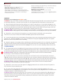
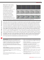
Figure 6 | Example application: selected
a
area illumination of C. elegans. (a) Frames
from supplementary video 2 of reference 12
demonstrating direct muscle control
of a paralyzed animal using patterned
light. (b) Sequential frames from
supplementary video 3 of reference 12 showing
b
a bar of light passing over the worm
from the posterior to the anterior region
as the animal is freely crawling. Initially,
the worm is traveling forward; however,
when the light reaches the anterior
mechanosensory neurons expressing
c
ChR2 (middle frame), the worm quickly
reverses direction. (c) Sequential frames
from supplementary video 8 of reference 12
showing the multispectral dynamic
capacity of the illumination system. The worm
is illuminated with blue light in the region of
the anterior mechanosensory neurons, which express ChR2, thus eliciting a reversal. The worm is subsequently illuminated with green light in the
region of the command interneurons, which express the hyperpolarizing MAC, thus halting the reversal. Scale bars, 250 µm.
used for direct control of muscle contractions in a paralyzed worm. The worm strain (ZX299: lin-15(n765ts); zxEx22[pmyo-3::
ChR2(H134R)::YFP; lin-15 + ]) expresses ChR2 in the muscle cells; therefore, when illuminated with blue light the muscles
will contract2. Animals are immobilized with ivermectin (0.01 mg ml−1 solution), which hyperpolarizes motor neurons but
leaves muscles fully functional43. By dynamically altering the illumination pattern on the basis of the shape of the animal
and optically activating muscle contraction, the animal can be optogenetically controlled (Fig. 6a and ref. 12, http://www.
nature.com/nmeth/journal/v8/n2/extref/nmeth.1555-S3.mov). A second experiment demonstrates the ability to dynamically
alter projection patterns at high resolution in a moving animal. Using animals expressing ChR2 in the gentle touch sensory
neurons (AQ2334: lite-1(ce314); ljIs123[pmec-4::ChR2; punc-122::RFP]), a bar of light was scanned along the body of the
animal while it crawls freely. When the illumination reaches either the anterior or posterior sensory neurons, the animal will
reverse or accelerate, respectively (Fig. 6b and ref. 12, http://www.nature.com/nmeth/journal/v8/n2/extref/nmeth.1555S4.mov). A final demonstration uses the full capacity of the system for dynamic, multicolor illumination. A strain expressing
excitatory ChR2 in the gentle touch sensory neurons and inhibitory MAC in the command interneurons (ZX899: lite-1(ce314);
ljIs123[pmec-4::ChR2; punc-122::RFP]; zxEx621[pglr-1::MAC::mCherry; pelt-2::GFP]) was subjected to blue illumination (thus
exciting ChR2) in the region containing the anterior sensory neurons, thus initiating a reversal, and it was subsequently
illuminated with green light (exciting MAC), thus inhibiting signal transmission and halting the reversal (Fig. 6c and
ref. 12, http://www.nature.com/nmeth/journal/v8/n2/extref/nmeth.1555-S9.mov).
Acknowledgments We thank members of the Caenorhabditis Genetic Center,
W. Schafer, Y. Tanizawa (Medical Research Council-Laboratory of Molecular
Biology, Cambridge, UK) and E. Boyden (Massachusetts Institute of Technology)
for reagents; the US National Institutes of Health (H.L.), the Alfred P. Sloan
Foundation (H.L.), the Human Frontier Science Program Organization (S.J.H.),
the Deutsche Forschungsgemeinschaft (grants GO1011/2-1, SFB807-P11,
FOR1279-P1, EXC115/1) and the Schram Foundation (A.G.) for funding.
We also thank J. Andrews and B. Parker in the Georgia Institute of Technology
School of Chemical and Biomolecular Engineering machine shop and D. Woodyard
in the glass shop.
AUTHOR CONTRIBUTIONS J.N.S. designed and constructed the illumination
system. J.N.S. and M.M.C. characterized the system. S.J.H. and A.G. contributed
to reagents and provided valuable discussions. J.N.S., M.M.C., S.J.H., A.G. and
H.L. designed the experiments. J.N.S. and H.L. prepared the manuscript.
COMPETING FINANCIAL INTERESTS The authors declare no competing financial
interests.
Published online at http://www.natureprotocols.com/.
Reprints and permissions information is available online at http://www.nature.
com/reprints/index.html.
1. Nagel, G. et al. Channelrhodopsin-2, a directly light-gated cation-selective
membrane channel. Proc. Natl. Acad. Sci. USA 100, 13940–13945 (2003).
2. Zhang, F. et al. Multimodal fast optical interrogation of neural circuitry.
Nature 446, 633–639 (2007).
3. Chow, B.Y. et al. High-performance genetically targetable optical neural
silencing by light-driven proton pumps. Nature 463, 98–102 (2010).
4. Papagiakoumou, E. et al. Scanless two-photon excitation of
channelrhodopsin-2. Nat. Methods 7, 848–854 (2010).
5. Andrasfalvy, B.K., Zemelman, B.V., Tang, J.Y. & Vaziri, A. Two-photon
single-cell optogenetic control of neuronal activity by sculpted light.
Proc. Natl. Acad. Sci. USA 107, 11981–11986 (2010).
6. Nagel, G. et al. Light activation of channelrhodopsin-2 in excitable cells
of Caenorhabditis elegans triggers rapid behavioral responses. Curr. Biol.
15, 2279–2284 (2005).
7. Liewald, J.F. et al. Optogenetic analysis of synaptic function. Nat. Methods
5, 895–902 (2008).
8. Mahoney, T. et al. Intestinal signaling to GABAergic neurons regulates a
rhythmic behavior in Caenorhabditis elegans. Proc. Natl. Acad. Sci. USA
105, 16350–16355 (2008).
9. Liu, Q., Hollopeter, G. & Jorgensen, E. Graded synaptic transmission at
the Caenorhabditis elegans neuromuscular junction. Proc. Natl. Acad. Sci.
USA 106, 10823–10828 (2009).
nature protocols | VOL.7 NO.2 | 2012 | 219