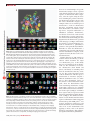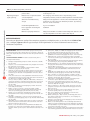
1
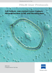
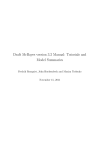
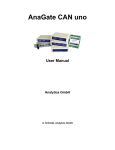
PROTOCOL
Spectral karyotyping analysis of human and mouse
chromosomes
Hesed M Padilla-Nash, Linda Barenboim-Stapleton, Michael J Difilippantonio & Thomas Ried
Genetics Branch, Center for Cancer Research, National Cancer Institute, US National Institutes of Health, 50 South Drive-MSC 8010, Bethesda, Maryland 20892, USA.
Correspondence should be addressed to H.M.P.-N. ([email protected]).
Published online 31 January 2007; doi:10.1038/nprot.2006.358
Classical banding methods provide basic information about the identities and structures of chromosomes on the basis of their
unique banding patterns. Spectral karyotyping (SKY), and the related multiplex fluorescence in situ hybridization (M-FISH),
are chromosome-specific multicolor FISH techniques that augment cytogenetic evaluations of malignant disease by providing
additional information and improved characterization of aberrant chromosomes that contain DNA sequences not identifiable
using conventional banding methods. SKY is based on cohybridization of combinatorially labeled chromosome-painting probes
with unique fluorochrome signatures onto human or mouse metaphase chromosome preparations. Image acquisition and
analysis use a specialized imaging system, combining Sagnac interferometer and CCD camera images to reconstruct spectral
information at each pixel. Here we present a protocol for SKY analysis using commercially available SkyPaint probes, including
procedures for metaphase chromosome preparation, slide pretreatment and probe hybridization and detection. SKY analysis
requires approximately 6 d.
INTRODUCTION
Cytogenetic studies in cancer are complicated by the fact that
many chromosomal aberrations are often difficult to characterize. Initially, researchers relied on classical banding (G-, Q- and
C-banding) methods1–3. These methods helped distinguish individual chromosomes by visualizing physical landmarks (called
bands) and provided chromosome-specific banding patterns for
each species examined. Cytogenetic techniques became much
more versatile through the introduction of hybridization of fluorescently labeled region-specific DNA probes. This molecular
cytogenetic technique is now widely known as FISH4–6.
SKY is also based on the hybridization of fluorescently labeled
DNA probes; however, the probe used is complex, generally consisting of up to 55 individually generated chromosome-specific
probes. SKY has proven exceedingly valuable for the comprehensive
analysis of cytogenetic abnormalities associated with malignant
disease and has been applied to a large series of samples derived
from hematological malignancies and solid tumors7–11. In fact,
since its invention in 1996, more than 500 papers have been published that applied SKY for the analysis of various chromosomal
preparations. SKY results can now be submitted to an interactive database12, which facilitates data analyses (SKY, M-FISH and
CGH), storage and retrieval of recurrent chromosome breakpoints
found in many cancers and constitutional disorders (http://www.
ncbi.nlm.nih.gov/sky/skyweb.cgi). Alternatively, M-FISH13 can be
used for characterizing karyotypes14. Although the hybridization
principle is identical to SKY, M-FISH uses a set of fluorochromespecific optic filters (five or seven different filters), rather than the
single custom-designed filter used in SKY. Both SKY and M-FISH
achieve the same goal—that is, identification of individual chromosomes in different colors. Both methods are more costly than
conventional banding methods, as they require expensive materials
(probes), equipment and software.
The systematic analysis of chromosomal abnormalities in cancer cells using SKY allows for the characterization of novel and
hidden chromosomal translocations, identification of complex
rearrangements15 and reconstruction of clonal evolution events
during cancer progression, and it has revealed the role of unstable
chromosome rearrangements, such as jumping translocations,
occurring as tissue-specific genomic imbalances11. Although SKY
allows for the identification of which particular piece of DNA may
be contained within a chromosome aberration, the spectral image
alone does not provide information as to the specific region of
the chromosome localized within a rearrangement. For this, one
must rely on the banding pattern of 4,6-diamidino-2-phenylindole (DAPI). Good-quality chromosome preparations are also
key to the success of SKY analysis, if the chromosome preparations are of inferior quality—for example, if the slides are old or
have too much cytoplasm surrounding the chromosomes—SKY
analysis can be less than optimal. Thus, the strengths and limitations of SKY analysis are somewhat dependent on the skills of the
user. Maximizing the amount of information gained through the
use of this technique is greatly facilitated by a general knowledge
and expertise in cytogenetics—that is, familiarity with the chromosomal banding patterns of the species being analyzed, as well
as an understanding of the principles of how the software interprets the acquired information. For assistance in both identification of the banding patterns and determination of breakpoints of
aberrant human chromosomes, researchers use the nomenclature
rules defined in ref. 16. For mouse chromosomes, the nomenclature rules used can be found at http://www.informatics.jax.org/
mgihome/nomen/gene.shtml.
SKY greatly assists in identifying chromosomal regions involved
in homogenous staining regions and double minute chromosomes, regardless of size and numbers. However, with respect to
homogenous staining regions and double minute chromosomes,
again, SKY has its limitations, in that these aberrations often contain multiple genes and/or DNA regions that are tightly linked;
resolving these details therefore often requires additional FISH
NATURE PROTOCOLS | VOL.1 NO.6 | 2006 | 3129
PROTOCOL
hybridizations with either gene-loci probes or specific chromosome or chromosome-arm paints17. SKY has also proven to be
very useful for the study of constitutional chromosome abnormalities arising from de novo balanced translocations (in which
chromosomal regions are relocated in a reciprocal manner and
no genetic material is lost) and unbalanced translocations (which
are, by definition, associated with genomic imbalances) that occasionally involve chromosome exchanges between very small DNA
regions, especially at the telomeres18–20. In prenatal diagnosis, the
difference between the outcome of a balanced translocation and
an unbalanced one may represent the difference between a normal
child and one with mild-to-severe birth defects. SKY is useful
in determining the origin of supernumerary marker chromosomes that are occasionally seen in prenatal specimens (appearing as unbanded small chromosome fragments using conventional banding methods). It is imperative that the chromosomal
origin of these markers be determined, as genomic imbalances
may result in birth defects and or mental retardation21, and some
markers may have considerable prognostic significance.
SKY has also been applied for the reconstruction of chromosomal rearrangements that have occurred during the course
of chromosome evolution, a field referred to as comparative
BOX 1 SLIDE PRETREATMENT WITH PEPSIN TO REMOVE CYTOPLASM
1. Equilibrate the slide in a coplin jar containing 2× SSC for 5 min, without shaking, at room temperature. The solution should cover all
the slides.
▲ CRITICAL STEP Size of coplin jar used depends on number of slides being pretreated.
2. Dilute the 20 mg ml–1 RNase A stock solution (slide pretreatment solution 1) 1:200 in 2× SSC, made and kept at room temperature
until use.
3. Apply 120 µl to a 24 mm × 60 mm coverglass, invert slide metaphase-spread-side down onto the liquid bearing coverglass, and
reinvert slide, taking care not to scratch the slide.
▲ CRITICAL STEP The purpose of this step is to digest DNA molecules surrounding the chromosomes that would interfere with the
hybridization and result in high fluorescent background. Ensure that sample is in full contact with the liquid and that air bubbles have
been eliminated.
4. Incubate slides in a moist, lightproof, waterproof hybridization box (as described in Step 29) at 37 °C for 45 min.
5. Carefully remove coverglass by gently tapping the sides of slide onto a solid surface; the coverglass should slide off easily.
6. Rinse the slide by placing in a coplin jar containing 2× SSC for 5 min at room temperature, with shaking.
7. Repeat Box Step 6 two more times, with shaking (at room temperature), using fresh 2× SSC each time.
8. Add 2–30 µl pepsin stock solution (slide pretreatment solution 2) into an empty, clean 100-ml glass beaker, then add 100 ml 0.01 M
HCl that has been prewarmed to 37 °C in a water bath, and adjust the pH to 2.0 using 1.0 N HCl. Mix well and pour appropriate volume
into a coplin jar sufficient to cover slide.
▲ CRITICAL STEP It is very important that the pepsin be added to the clean beaker first and not directly into the HCL. If the pepsin is
added to the HCL, it does not dissolve into solution.
9. Incubate slide in coplin jar for 30 s–5 min at 37 °C.
▲ CRITICAL STEP The time of pepsin treatment and amount of pepsin stock solution to be used is dependent on (i) the amount of
cytoplasm surrounding the metaphase spreads, as observed with a light microscope using phase objectives before slide pretreatment,
and (ii) the age of the slide—slides with excess cytoplasm, or older than 6 months, may require longer treatment with pepsin (3–5
min) and higher concentrations of pepsin (10–30 µl). After exposure to pepsin, the slide can be placed into a Petri dish containing 1×
PBS and examined using an inverted microscope, to see whether longer pepsin treatment is required. If so, place the slide back into
the coplin jar containing the pepsin-acid mixture.
10. Wash the slide in 1× PBS for 5 min at room temperature, with shaking.
11. Repeat Box Step 10, using fresh 1× PBS.
12. Wash the slide once for 5 min at room temperature in 1× PBS/MgCl2 (slide pretreatment solution 3), with shaking.
13. Place the slide in a coplin jar containing 1% (vol/vol) formaldehyde/1× PBS/MgCl2 (slide pretreatment solution 4) and incubate
slide (not shaking) for 10 min at room temperature (when finished with procedure decant solution into an appropriate waste
container).
14. Wash slide for 5 min in 1× PBS at room temperature, shaking.
15. Dehydrate slide in an alcohol series (70%, 90% and 100% ethanol), using different coplin jars, at room temperature, without
shaking, for 3 min each.
16. Air-dry the slide until the ethanol has evaporated (1–2 min), on the workbench.
17.Check chromosomes to see whether the morphology has been preserved, and select an area for hybridization.
▲ CRITICAL STEP After slide pretreatment, the chromosomes should retain their original morphology and should not have the
appearance of being overdigested by the enzyme. If they look hollow, they are not generally useful for SKY hybridization, and there
is no reversal of this step. If there are enough slides available, using additional slides with different pepsin concentrations and times
is highly recommended to achieve the best results. Redo the entire procedure if the pretreatment does not sufficiently remove the
cytoplasm surrounding the chromosome.
? TROUBLESHOOTING
3130 | VOL.1 NO.6 | 2006 | NATURE PROTOCOLS
PROTOCOL
cytogenetics22. By using SKY, one can determine and compare
homologous regions of genomes between closely related species—for example, between human and a gibbon (Hylobates
concolor)23. These analyses can be complemented by performing the reverse experiment—that is, hybridizing differentially
labeled flow-sorted gibbon chromosomes onto human metaphase spreads. This is referred to as cross-species hybridization24. Recently, SKY analysis has also been used in other species, such as rat. Newly developed rat probes have been used to
determine the homologous regions of rat, mouse and human
chromosomes, as well as to characterize the karyotypes of rat
tumors25.
The study of chromosomal aberrations in mice is extremely
demanding, because the chromosomes of this species are all acrocentric (that is, the primary constriction, or centromere, is located
at one end of the chromosome) and of similar size and morphology. Therefore, subtle aberrations such as insertions or translocations involving small regions are often unrecognizable. Murine
models of human carcinogenesis are widely used to delineate
genetic mechanisms that determine tumor initiation and progression26. SKY as a genome-scanning method for detection of chromosomal aberrations has now been applied to a wide variety of
murine models of human cancer, including numerous models for
hematological malignancies and solid tumors27–30. Although SKY
has been applied to vertebrate organisms closely related to human
or mouse, thus far commercially made SKY probes and analysis
software are only available for human, mouse and rat.
Experimental design
The protocol we present uses SkyPaint probes (Applied Spectral
Imaging). The SKY analysis protocol described herein consists
of six parts: (i) preparation of metaphase chromosome spreads
(Steps 1–20); (ii) slide pretreatment and slide and probe denaturation (Box 1 and Steps 21–26); (iii) hybridization (Steps 27–29)
and (iv) detection (Steps 30–49) of the SKY probes; and (v) tips
for image acquisition and (vi) image analysis (Step 50) (see Fig. 1).
Preparation of metaphase chromosome spreads for SKY analysis
requires 1 d. Slide pretreatment and denaturation of both the slide
and the probe takes 2 h, at which point one can pause before continuing with the hybridization steps. The hybridization protocol,
which includes denaturation of the SKY probe as well as that of the
target metaphase chromosomes on the slide, is performed on day
1. For the actual hybridization of human or mouse SKY probes, the
procedure continues for 48–72 h at 37 °C. The subsequent detection steps take approximately 5 h (see Fig. 1). Image acquisition
and analysis can take place immediately after the detection steps,
but can also be delayed and the slides can be stored in the dark at
4 °C. We highly recommend analyzing the slides within 1 week of
completion of the SKY detection procedures.
Preparation of metaphase spreads for SKY. This requires
(i) knowledge of the ideal culture conditions of the specimen
studied to obtain actively dividing cells, and (ii) recognizing the
conditions necessary to yield spreads free of cytoplasm, which
include (iii) the optimum humidity required for the individual chromosome preparations to ensure proper spreading of
chromosomes onto glass slides and eliminate the possibility of
chromosomes overlapping31. If none of these conditions are
met, hybridization with SKY and or FISH probes will be suboptimal, thereby impeding an accurate analysis of the results.
Figure 1 | Protocol timeline flowchart. Shown are the steps in a SKY
hybridization and analysis.
Excess cytoplasm surrounding the chromosomes can be reduced
by pretreating the slides containing the metaphase preparations
with enzymes such as trypsin or pepsin that digest the surrounding cellular proteins. This facilitates access of the DNA probe to
its target sequence on the chromosome as well as reducing the
amount of background signal that arises when the DNA probe
nonspecifically binds the proteins (see Fig. 2). However, overtreatment of the slides can also reduce hybridization efficiency
through degradation of the target DNA or excessive removal of
chromosome-associated histone proteins, leading to a distortion
of the chromosome morphology. Should this occur, we recommend repeating this phase of the experiment with new slides.
SKY probe generation and labeling. SKY probes consist of a
cocktail of chromosome-specific painting probes of the target species. The chromosome-painting probes used for SKY analysis of
human or mouse genomes are prepared by amplification of flowsorted chromosomes using degenerate oligonucleotide–primed
(DOP)-PCR32. Chromosome sorts that contain all 22 human
homologs and the X and Y human chromosomes, or 19 mouse
homologs and the X and Y mouse chromosomes, can be requested
for a fee from the Cambridge Resource Centre for Comparative
NATURE PROTOCOLS | VOL.1 NO.6 | 2006 | 3131
PROTOCOL
a
b
10×
c
Genomics (http://www.vet.cam.ac.uk/genomics). SKY probes can
either be in-house–generated probes33–35 or be purchased from
Applied Spectral Imaging (ASI, http://www.spectral-imaging.
com/). ASI-produced SkyPaint probes undergo stringent quality
controls and their chromosome specificity is tested by hybridization of all probes individually using FISH. Probes are labeled with
nucleotide analogs, either conjugated directly to fluorochromes,
such as Spectrum orange, Rhodamine green or Texas Red, or
indirectly via small nonfluorescent molecules—for example, biotin or digoxygenin, which are visualized using avidin- and antibody-conjugated fluorochromes. As five distinct fluorochromes
are distinguishable from one another using the SKY system, each
Figure 3 | SKY imaging system. (a) Diagrammatic
representation of the SKY imaging system.
(1) Xenon lamp source. (2) Phase filter. (3) SKY
filter cube, which reflects specific wavelengths of
light (blue ‘waves’). (4) ×63 objective. (5) Slide
containing sample, which will fluoresce when
excited (red ‘waves’). (6) BG38 filter used to
reduce far-red wavelengths, which may contribute
to excessive background. (7) SKY SpectraCube,
which contains the Sagnac interferometer used
to split the emitted light. (8) CCD camera, which
captures the DAPI counter-stain as a grayscale
image. (b) Photo of the SD300 SpectraCube
mounted on a Leica DMXRA epifluorescence
microscope. Red arrows indicate features
depicted in a. The BG38 filter (6) is controlled by
a lever on the right-hand side of the microscope
(not visible in this image). This figure has been
reproduced in part and reprinted with permission
from Current Protocols in Human Genetics, Chapter
4.4 (John Wiley & Sons)36.
3132 | VOL.1 NO.6 | 2006 | NATURE PROTOCOLS
Figure 2 | Effects of humidity and removal of excess cytoplasm by slide
pretreatment on hybridization efficiency. (a) When cells are not kept in
the hypotonic solution for enough time, the chromosomes become trapped
inside the cytoplasm (white arrows). Altering the humidity at which
the metaphase preparation is dropped onto the microscope slide may
sometimes alleviate this problem; otherwise, hybridization efficiency will
be compromised. (b) When normal human metaphase spreads are pretreated
with pepsin and hybridized with a whole chromosome paint probe labeled
with tetramethyl rhodamine isothyocyanate (TRITC), the hybridization will
be successful. (c) Here, a whole chromosome paint-labeled with TRITC was
hybridized to a different normal human metaphase spread and the slide was
not pretreated with pepsin. Note the weaker intensity of the hybridization
signal (red color).
chromosome is labeled with a unique combination of these
tagged nucleotides. To further enhance the chromosome specificity of the probe cocktail, the labeled chromosome-painting probes
are denatured and combined with an excess of CotI DNA (which
represents a fraction of the genome enriched for highly repetitive elements, such as long and short interspersed elements, Alu
repeats and so forth, that are distributed throughout all the chromosomes). This step is known as preannealing and ensures that
any labeled non–chromosome-specific repetitive elements in the
chromosome-painting probe form double-stranded DNA with
their complementary sequences found in the CotI DNA. These
repetetive elements are thereby prevented from hybridizing to the
metaphase chromosome spreads.
Probe hybridization and detection for SKY analysis. The
principles of SKY probe hybridization and detection are very
similar to those of FISH. The probe is placed in direct contact
with denatured metaphase chromosomes (which are attached
to glass slides), allowing for Watson-Crick base-pairing to occur
between complementary sequences in the labeled probe and the
metaphase chromosomes. This hybridization step is allowed
to proceed for 24 h to 3 d, depending on the age and conditions
of the starting material on the slide (older slides tend to require
longer hybridization times). The slides are then washed to remove
PROTOCOL
with short chromosomes, with some overlaps, with debris and with large numbers of
chromosomes are also informative, especially in determining whether the cells manifest chromosome instability (random gains
and losses) or consist of several subclones
with different karyotypes. In contrast, if the
initial metaphase-spread preparations are
made properly, it will not be necessary to
acquire overcondensed chromosomes (too
a
b
c
much colcemid) or incomplete metaphase
d
spreads (too strong a hypotonic shock).
Avoid imaging areas containing bright
spots and nuclei, as that can also complicate the analysis. If the metaphase spread
is too large to fit in the region of interest,
acquire two separate images and analyze
each as an individual, combining the data
after analysis, but make sure to have overlapping regions so that the two images can
Figure 4 | Normal human male (46 chromosomes, XY) metaphase spread analyzed by SKY. (a) InvertedDAPI image of a chromosome metaphase spread. The banding pattern is similar to G-banding but
be fit together.
accentuates the heterochromatic region of some chromosomes (for example, 1, 9, 16 and Y). (b) Same
Spectrum-based classification of the
metaphase with RGB display colors. (c) Same metaphase spread with classification pseudo-colors.
specific wavelengths of all chromosomes,
(d) Karyotype of the same metaphase spread. Arrows in panels a–c identify the X and Y chromosomes.
in combination with karyotype analysis
of the images, is achieved using SkyView
software (ASI), following the User Manual
unhybridized probe and nonspecifically hybridized probe (that supplied with the software. Additional information regarding the
is, probe that has formed double bonds with sequences in the technical aspects of the ASI system can be retrieved from the ASI
chromosomes that are not fully complementary). Those chromo- webpage (http://www.spectral-imaging.com/), which includes
somes labeled directly with fluorescently conjugated nucleotides online support and tutorials. After image analysis, the chromodo not require anything further for their visualization under the somes can be viewed with their specific fluorescent signature
microscope. Nonfluorescensly-labeled probes, in contrast, require (display colors) and classification colors (pseudo-color), and
further detection with a molecule such as Avidin (for the detec- their specific banding pattern (inverted DAPI) (see Figs. 4–7).
tion of biotin) or an antibody, either of which has a fluorochrome For optimal classification, make sure there is at least one normal
attached to it. In addition to the chromosome-specific fluorescent copy of each chromosome in the metaphase being analyzed. If
label, we use DAPI to introduce a grayscale chromosome banding
pattern that is similar to Giemsa-trypsin (G)-banding.
Image acquisition and analysis. This is accomplished by viewing
the slides with an epifluorescence microscope equipped with hardware and software designed for visualizing SKY images (see Fig. 3
and ref. 36). The epifluorescence microscope should be equipped
with a ×63 oil-immersion objective, a single custom-designed triple band-pass filter (SKY filter cube) that allows for the simultaneous excitation of all fluorochromes, and a DAPI filter cube. Image
acquisition requires illumination from a 150-W xenon lamp. The
emitted light is passed through the collection optics and then the
Sagnac interferometer, where an optical path difference is created
(see Fig. 3). The interferogram measures every pixel detected by
the CCD camera. Using Fourier transformation, it mathematically
retrieves the wavelengths of the emitted light from the combinatorially labeled chromosomes and thereby identifies a spectral signature for each pixel of the CCD camera image.
When beginning image acquisition, first find the area of hybridization using the SKY filter cube; next, systematically scan the
slide up and down using the DAPI filter. If the fluorescence colors are bright, the acquisition time can be reduced. Conversely, if
Figure 5 | Metaphase spread from a human bladder cancer cell line
the hybridization signals are weak, the time of acquisition can be hybridized with a SKY probe. This aneuploid cell line, J82, contains multiple
increased. When selecting metaphase spreads, do not acquire only chromosomal aberrations. White arrows identify some of the chromosome
the best-looking spreads, or the results may be biased. Metaphases translocations.
NATURE PROTOCOLS | VOL.1 NO.6 | 2006 | 3133
PROTOCOL
Figure 6 | Mouse spectral karyotyping. (a) SKY image of a metaphase spread from a mouse deficient
for the gene mutated in the human disease ataxia telangectasia was overlayed onto the corresponding
DAPI-banded image. White arrows indicate two chromosome translocations frequently observed in this
mouse model system, T(12;14) and Dic(14;15). (Using mouse cytogenetic nomenclature rules, T denotes
a translocation where the chromosome listed first contains the centromere; Del denotes a deletion; Dic
denotes a chromosome containing two centromeres (dicentric), one from each one of the contributing
chromosomes.) The arrowhead indicates the presence of a centromere at the fusion of chromosomes 14
(yellow) and 15 (blue) in the dicentric chromosome, Dic(14;15). (b) Karyotyping corresponding to the
metaphase spread shown in a. Each chromosome is represented from left to right by the display color, the
inverted DAPI counter-stain and the classification pseudo-color. Numbers to the right of each translocation
chromosome indicate the chromosomal origin of each fragment. T(13;14) is an example of an unbalanced
translocation, resulting from loss of some genomic material from chromosome 13 and the gain of a portion
of chromosome 14.
Figure 7 | SKY karyotype of a metaphase spread from a pancreatoblastoma cell line. Left, inverted DAPI
chromosomes; right, classification pseudo-colors. Some of the aberrations consist of multiple chromosomal
segments, as indicated by numbers to right of chromosomes. Assignments of these chromosome segments
were subsequently confirmed with whole chromosome–painting probes to rule out the sandwich effect.
This effect is visible on the chromosome 11 aberration (asterisk). Only the chromosome 15 at the
boundary of chromosome segments 11 and 4 was verified; hence, the segment identified as coming
from chromosome 12 is an artifact resulting from the sandwich effect, or merging of two overlapping
fluorochromes. There is no normal chromosome 7 in this karyotype. To classify any segments that involve
chromosome 7, a red lasso was drawn around a chromosome 7 segment (identified by its banding pattern
and spectral color) contained in the derivative chromosome 4, the chromosome 7 segment was manually
placed in the chromosome 7 compartment, and finally the karyotype was reclassified.
3134 | VOL.1 NO.6 | 2006 | NATURE PROTOCOLS
there are no normal examples of a specific
chromosome, attempt to locate a portion
of the missing chromosome (this requires
expertise in chromosome-band recognition), select the region (using the ‘lasso’
tool) containing the portion of the missing chromosome and place the piece into
the correct chromosome category17. For
example, if there are no normal copies of
chromosome 7, look for chromosomes
that have recognizable regions derived
from chromosome 7 (for instance, resulting from a reciprocal translocation or
unbalanced (derivative) chromosome),
use the lasso tool to draw a circle around
the chromosome 7 portion of the derivative chromosome and manually place that
fragment into the chromosome 7 position
of the karyotype (see Fig. 7). This enables
the software to correctly identify the spectrum of chromosome 7, particularly as
pertains to that particular hybridization.
Remember to delete the chromosome 7
segment that was generated with the lasso
tool after classification is complete, as the
segment selected by the lasso tool is a ‘virtual’ chromosome.
Further intricacies of SKY analysis to
consider include the following. (i) If the
software cannot determine the origin
of a chromosome piece, it will classify
this piece as belonging to the Y chromosome. (ii) The centromeres of acrocentric chromosomes (both human and
mouse) will classify as something other
than the chromosomes that they reside
on. (iii) The pseudo-autosomal regions
of the X chromosome will show up as a
translocations or insertions (assigned a
different classification color). (iv) There
may be other classification color(s) at
the junction of the translocation breakpoint, where the fluorochromes from
each contributing chromosome overlap,
giving what is referred to as a ‘sandwich’
effect, which results in incorrect classification of the region (see Fig. 7). Sometimes
these regions really do represent a small
amount of material from a third chromosome (see Fig. 7). The definitive determination of such chromosomal aberrations
requires confirmation of SKY results with
chromosome-specific FISH probes17. For
accurate descriptions of karyotypes and
human aberrations, use the nomenclature
rules of ref. 16. For nomenclature rules
applying to mouse, refer to the Jackson
laboratory nomenclature (http://jaxmice.
jax.org/info/nomenclature_hints.html).
PROTOCOL
MATERIALS
REAGENTS
Cell-culture media
AlexaFluor 680 F (ab′) 2 fragment of goat anti-mouse IgG (H + L) (cat. no.
A21059, Invitrogen)
▲ CRITICAL Do not repeatedly freeze and thaw antibodies used in detection
protocols. Store aliquots according to manufacturer’s directions and record
expiration dates. Keep antibodies out of the light when using.
Colcemid (cat. no. 295-892, Roche)
Cy5-conjugated StreptAvidin (cat. no. 016-170-084, Jackson ImmunoResearch
Laboratories) ▲ CRITICAL Do not repeatedly freeze and thaw antibodies used
in detection protocols Store aliquots according to manufacturers directions and
record expiration dates. Keep antibodies out of the light when using.
Ethyl alcohol (ethanol), anhydrous 200 proof (cat. no. AC615090020, Fisher
Scientific)
Hydrochloric acid (HCl), 1 N (cat. no. AC12421-0010, Fisher Scientific)
! CAUTION HCl is a corrosive acid and is irritating to respiratory system. Make all
solutions in a chemical fume hood. Contact your waste management department
for proper disposal protocol.
Hypotonic solution, 0.075 M KCl in H2O (cat. no. 10575-082, Invitrogen)
Monoclonal anti-digoxygenin clone DI-22 mouse ascites fluid (cat. no. D8156,
Sigma-Aldrich) ▲ CRITICAL Do not repeatedly freeze and thaw antibodies used
in detection protocols. Store aliquots according to manufacturer’s directions and
record expiration dates.
PBS, 1×, pH 7.4, without calcium and without MgCl2 (cat. no. 10010, GIBCO,
Invitrogen)
SkyPaint probe (Applied Spectral Imaging)
Trypsin (cat. no. 25200-056, Invitrogen)
EQUIPMENT
Thermotron humidity chamber (model CDS-5, Thermotron)
Clinical centrifuge (for example, Allegra 6R (equipped with the GH3.8 rotor),
Beckman Coulter)
Biological safety cabinet (for example, Forma 1280 Class II, Thermo Scientific)
Reciprocal shaking water baths (2) (for example, model 15-455, Fisher Scientific)
Test tube rocker (for example, model 12-815-6, Fisher Scientific)
Slide warmer (for example, model 77, Fisher Scientific)
Thermomixers (2) (Eppendorf, model 5436, Brinkmann Instruments)
Vortex mixer (for example, Daigger vortex Genie 2, A. Daigger & Co.)
Microcentrifuge (for example, model V, VWR Scientific)
Microscope slides, glass (for example, cat no. 12-544-7, Fisher Scientific)
Microscope coverglass, 18 mm × 18 mm (for example, cat. no. 48368040, VWR
Scientific)
Microscope coverglasss, 24 mm × 60 mm (for example, cat. no. 48393106, VWR
Scientific)
Rubber cement (local craft or drug store)
Hybridization chamber (box), 6 in × 8 in (for example, Tupperware container,
local retail)
DAPI filter cube (Chroma Technology)
SKY filter cube (Chroma Technology)
Inverted tissue-culture microscope (for example, Zeiss)
Light microscope equipped with a ×16 and a ×40 dry objective (for example,
Model Axiovert, Zeiss), for observing slides during preparation of spreads.
Epifluorescence microscope equipped with high-end objectives (for example,
Leica)
▲ CRITICAL Epifluorescence microscopes from Zeiss, Nikon or Olympus
would alternatively be suitable. We use a ×63 oil-immersion objective for image
acquisitions.
SpectraCube (Applied Spectral Imaging)
150-W xenon lamp (for example, Opti Quip)
REAGENT SETUP
Fixative for metaphase slide preparation Combine methanol (cat. no. 301602, Mallinckrodt Chemicals) and acetic acid (cat. no. V193-14, Mallinckrodt
Chemicals) at a 3:1 ratio (vol/vol).
! CAUTION Methanol is a poison, and highly flammable. Use only with adequate
ventilation (chemical fume hood). Wear gloves when handling this agent.
Contact your waste management department for proper disposal protocol.
▲ CRITICAL Freshly prepare before use. Can be stored before procedure (1–2
h) at 4 °C.
Slide pretreatment solution 1 (RNase A stock solution, 20 mg ml–1 % wt/vol).
Dissolve RNase A (cat. no. 109169, Roche Molecular Biochemicals) to a
final concentration of 20 mg ml–1 in sterile water, boil 15 min, cool to room
temperature (22 °C). Store aliquots, 100 µl each, at –20 °C.
Slide pretreatment solution 2 (pepsin stock solution, 100 mg ml–1)
Dissolve pepsin (cat. no. P-6887, Sigma-Aldrich) into sterile water, to a final
concentration of 100 mg ml–1; keep on ice until aliquoted. Store aliquots, 20 µl
each, at –20 °C.
Slide pretreatment solution 3 (1× PBS/MgCl2) Add 50 ml of 1 M MgCl2 (cat.
no. 351-033-060, Quality Biological) to 950 ml 1× PBS. Final volume is 1 l. Store
at room temperature indefinitely.
Slide pretreatment solution 4 (1% vol/vol formaldehyde/1× PBS/MgCl2)
Add 2 ml of 37% formaldehyde (cat. no. 5016, Mallinckrodt Chemicals) to 73
ml 1× PBS/MgCl2. Make fresh for each experiment, mix well and store at room
temperature until use. ! CAUTION Formaldehyde is flammable, carcinogenic
and a poison. Formaldehyde should be dispensed in a chemical fume hood
and only handled when wearing gloves. Contact your waste management
department for proper disposal protocol.
Slide denaturation solution (70% vol/vol deionized formamide/2× SSC,
or FA/SSC) Combine 10 ml of 20× SSC (cat. no. 1666681, Roche Molecular
Biochemicals), 20 ml dH2O and 70 ml deionized formamide (cat. no. 9342,
Ambion). Adjust to pH 7.25 with 1.0 N HCl. Mix well; store as 1-ml aliquots at
–20 °C. ! CAUTION Formamide causes eye and skin irritation and is a known
mutagen. Wear gloves when handling this reagent and dispense under a
chemical fume hood. Contact your waste management department for proper
disposal protocol. ▲ CRITICAL Denature slides using deionized formamide, as
it contains fewer impurities than conventional formamide.
Wash solution 1 (50% vol/vol formamide/2× SSC) Combine 100 ml
formamide (cat. no. 47670, Sigma-Aldrich) with 20 ml 20× SSC and 80 ml
ddH2O. Adjust pH to 7.0 using 1.0 N HCl. Mix well and use fresh; prewarm and
maintain temperature in a water bath set at 45 °C until use.
Wash solution 2 (1× SSC) Add 25 ml of 20× SSC to 475 ml dH2O. Mix well and
use fresh; prewarm and maintain temperature in a water bath set at 45 °C until
use.
Wash solution 3 (4× SSC/Tween 20) Add 200 ml of 20× SSC to 799 ml of
dH2O, and add 1 ml of polyoxyethylene-sorbitan monolaurate (Tween-20; cat.
no. P-1379, Sigma-Aldrich). Mix well and use fresh; prewarm in a water bath at
45 °C until use.
Blocking solution (BSA) Add 0.30 g of bovine serum albumin (BSA) Fraction
V, protease-free, lyophilizate (cat. no. 03-117-332-001, Roche Molecular
Biochemicals) to 10 ml of 4× SSC/Tween 20. Mix well and use fresh; prewarm
and maintain temperature in a water bath set at 37 °C until use.
2× SSC. Add 10 ml of 20× SSC to 90 ml of dH2O.
Carbonate-bicarbonate buffer (pH 9.0) Add 4 ml sodium bicarbonate, 0.5 M
(pH 8.13; cat. no. S-6297, Sigma-Aldrich) to 1 ml sodium carbonate, 0.5 M (pH
11.32; cat. no. S-6139, Sigma-Aldrich), and filter-sterilize. Store, sealed at room
temperature, up to 1 year.
Antifade solution Dissolve 50 mg 1,4-phenylenediamine dihydrochloride
(antifade; cat. no. P1519, Sigma-Aldrich) in 2 ml 1× PBS and adjust pH with
carbonate-bicarbonate buffer to achieve pH 8.0. Add 1× PBS to 5 ml. Mix
with 45 ml 86% (vol/vol) glycerol (cat. no. 11514011, Invitrogen). Leave on
test tube rocker for at least 1 h. Aliquots can be stored at –20 °C. ▲ CRITICAL
Add 2 ml buffer and check pH with pH paper; add dropwise 0.1 M HCl until
pH approaches 8.4. As the pH approaches 8.0, check the pH with a pH meter,
making sure to wash the pH meter’s probe thoroughly. If pH exceeds 8.0, the
procedure must be repeated.
DAPI solution Add 2 mg of DAPI (cat. no. 236276, Roche Molecular
Biochemicals) to 10 ml dH2O for stock solution at a concentration of 0.2 mg
ml–1. Store in aliquots at –20 °C. For working DAPI solution, add 40 µl of stock
solution to 100 ml of 2× SSC.
! CAUTION DAPI is a known mutagen; avoid contact with eyes and skin. Protect
DAPI solution from light; it can be stored at 4 °C for as long as 2 months.
EQUIPMENT SETUP
Fluorescence microscope and SKY imaging system See Figure 3. If interested
in setting up a microscope to perform SKY analysis, please directly contact
Applied Spectral Imaging (http://www.spectral-imaging.com/), as they are the
only suppliers of this system.
NATURE PROTOCOLS | VOL.1 NO.6 | 2006 | 3135
PROTOCOL
PROCEDURE
Metaphase preparation
1| Grow adherent and or suspension cell cultures in their preferred media under their preferred growth conditions. In the case
of adherent cell cultures, grow until they reach 60%–70% confluence (% confluence means the degree to which cells are making
contact with each other). This determination is made using an inverted tissue-culture microscope.
2| Add colcemid (0.1 g ml–1, 10 µl for every 1 ml medium) to cell cultures using sterile micropipettes.
3| Incubate cells for 30 min–1 h.
▲ CRITICAL STEP For fast-growing cultures, reduce time of exposure to colcemid, and for slower-growing cultures, incubate for
several hours with colcemid. Monitor cells throughout incubation period under the inverted tissue-culture microscope. Ideally,
cultures with approximately 10–15 dividing cells per field of view (cells appear round and shiny) are ready for harvesting of
metaphase chromosomes. For suspension and adherent cultures, one 75-cm2 flask should provide sufficient numbers of cells for
preparation of metaphase spreads.
4| Harvest either adherent cells (A) or suspension (B) cell cultures.
(A) Adherent cells.
(i) Remove medium from each flask with a sterile 10-ml pipette; save entire contents in a 50-ml conical centrifuge tube.
(ii) Add 5 ml sterile 1× PBS (pH 7.4) to each flask, rinse, and then transfer contents to 50-ml tube (the same tube as
described above).
(iii) Add 0.5 ml sterile trypsin to each flask and incubate until cells detach. Time will vary with each cell culture, as some cells
adhere to the bottom of the flask more than others, but is approximately 1–2 min. Hitting the flask may be required for
firmly attached cells.
▲ CRITICAL STEP Monitor cells under inverted microscope continuously, as some cultures are sensitive to trypsin.
(iv) Add 5 ml of growth medium; squirt the medium with a pipette onto the surface of the flask to remove the cells from the
sides of the flask or by hitting the flat part of your hand onto the bottom of the flask, and add the contents (the fresh
medium, which now contains the detached cells) to the same 50-ml conical centrifuge tube used in Steps 4A(i) and
4A(ii).
▲ CRITICAL STEP The fetal bovine serum (FBS) present in the medium used to grow the cells inactivates the trypsin.
(v) Spin cells in a clinical centrifuge at 175g (1,000 r.p.m.), for 5 min at room temperature.
(vi) Remove the supernatant carefully by vacuum aspiration, leaving 0.5 ml in the tube, and flick the tube with finger to
loosen the pellet.
(B) Suspension cultures.
(i) Transfer cultures to centrifuge tubes and spin at in a clinical centrifuge at 175g (1,000 r.p.m.) for 5 min at room
temperature.
(ii) Remove the supernatant carefully by vacuum aspiration, leaving 0.5 ml in the tube, and flick the tube with a finger to
loosen the pellet.
5| Add 2–50 ml hypotonic solution to the conical tube containing the cells in suspension and incubate at room temperature
for 15–25 min.
▲ CRITICAL STEP The volume of hypotonic solution added to each tube is variable and determined specifically by the final
amount of the cells in pellet. One does not need to measure the number of cells, but generally speaking, if the cell pellet is
just coating the bottom of the tube, add no more than 2 ml hypotonic solution; if there appears to be 0.5–1.0 ml of cells
at the bottom of the tube, add approximately 50 ml hypotonic solution. The cell-hypotonic mixture should appear slightly
cloudy and not clear. Some cultures (such as lymphocytes and plasma cytoma) are more sensitive, and the time of exposure
to the hypotonic solution should be approximately 15 min. Adherent cells typically require 20–25 min exposure. Chromosome
metaphase preparations on the slides will have excess cytoplasm if exposure to the hypotonic time is too short (see Fig. 2).
? TROUBLESHOOTING
6| Using a 1-ml pipette, add one drop of fresh methanol/acetic acid fixative (3:1 vol/vol) per ml of cell/hypotonic mixture
dropwise to the tube and invert to mix (this step will stop the hypotonic process and prefix the cells).
7| Spin cell suspension using a clinical centrifuge at 250g (1,200 r.p.m.) for 5 min at room temperature.
8| Remove the supernatant carefully, leaving 0.5 ml liquid, and flick the tube to loosen the pellet and fully resuspend all cells.
3136 | VOL.1 NO.6 | 2006 | NATURE PROTOCOLS
PROTOCOL
9| Add 5 ml fresh fixative down the side of the tube, very slowly, gently flicking pellet all the time.
▲ CRITICAL STEP To ensure that the final metaphase cell pellet is fully resuspended, it is crucial to add fixative very slowly
down the side of the centrifuge tube; otherwise, the cell pellet will remain full of clumps, metaphase spreads will become
trapped in the clumps and spreading will be compromised.
10| Pellet the cells at 250g (1,200 r.p.m.) for 5 min at room temperature.
11| Remove the supernatant and flick the tube to loosen the pellet and fully resuspend the cells.
12| Add 5 ml fresh fixative to wash the cell preparations.
13| Pellet the cells at 250g (1,200 r.p.m.) for 5 min at room temperature.
14| Remove the supernatant and flick the tube to loosen the pellet and fully resuspend the cells.
15| Add 1–5 ml fresh ice-cold fixative along the wall of the tube.
▲ CRITICAL STEP The final volume of fixative is determined by the desired concentration of cells on the slide. This requires
a visual examination of the final solution; it should appear slightly cloudy, yet translucent, when held up to the light. If too
much fixative is added, there may not be enough metaphases in the hybridization area of the slide for a thorough analysis. If
the fixative volume is too small, the cell density on the slide will be high, preventing the chromosomes from spreading well
and also resulting in too much cellular debris in close proximity to the metaphase spreads. This debris will nonspecifically
bind the fluorescent probe, resulting in brightly fluorescent ‘spots’ that will interfere with the image acquisition (owing to
pixel intensity saturation in the camera).
■ PAUSE POINT To store, tighten cap and wrap cap with laboratory film (Parafilm); store at –20 °C for short-term storage and
at –80 °C for several years.
16| In a humidity chamber or Thermotron set at 45% or 55% humidity at room temperature (for mouse or human cells,
respectively), drop ~15 µl of the fixed cell suspension onto a clean glass microscope slide and allow the slide to fully dry by
evaporation (approximately 1–2 min).
17| View the dried slide with a ×16 high-dry phase objective on a light microscope to determine final cell density, extent of
chromosome spreading and the presence or absence of cytoplasm.
▲ CRITICAL STEP The spreading of chromosomes and the integrity of chromosome structure are dependent on the evaporation
rate of the fixative, as determined by the percent humidity, temperature and success of the hypotonic procedure. If the
chromosomes are light gray in color, increase the humidity; if the chromosomes have excess cytoplasm (see Fig. 2a), appear
shiny or have a bright halo around them, decrease the humidity in the chamber.
? TROUBLESHOOTING
18| Dry the slides at 37 °C, or at room temperature if the humidity is low in the storage space.
▲ CRITICAL STEP Metaphase spreads on slides can be heated for 1 h at 80 °C or stored in a drying oven overnight (40 °C)
before hybridization; however, slides stored for one week at 37 °C yield optimum results.
19| Check the slide under a light microscope using a ×40 high-dry phase objective. If cytoplasm is present (gray particulate
matter surrounding the chromosomes; see Fig. 2a), pretreat the slides with pepsin as described in Box 1. If cytoplasm is not
visible (a clear light margin surrounds the metaphase chromosomes) and the chromosomes have good morphology (black in
color, not phase-light or shiny), there is no need for slide pretreatment with pepsin.
? TROUBLESHOOTING
20| Using a light microscope equipped with phase optics and ×10 and ×40 dry lenses, select an area for hybridization that will
fit under an 18 mm × 18 mm coverglass.
▲ CRITICAL STEP Selection of the region of the slide to be hybridized requires that there be sufficient numbers of
metaphase spreads to view and that the chromosome morphology has been preserved.
■ PAUSE POINT The slide(s) can be stored in a dry location until hybridization (1–3 d). Alternatively, they can be frozen at
–80 °C, sealed in bags containing desiccant to eliminate moisture.
NATURE PROTOCOLS | VOL.1 NO.6 | 2006 | 3137
PROTOCOL
Slide and probe denaturation
21| Warm SkyPaint probe in a Thermomixer set to 37 °C with shaking (1,100 r.p.m.) for 20 min, vortex and centrifuge briefly
in a microcentrifuge at 80g (1,000 r.p.m.) for 1–3 s at room temperature.
22| Denature the SKY probe in a Thermomixer set to 85 °C for 5 min and immediately place probe back into a different
Thermomixer or water bath set to 37 °C, to allow the labeled probe DNA to preanneal with the highly repetitive genomic
elements enriched in the CotI DNA fraction, which is also part of the SkyPaint probe, for approximately 1 h.
? TROUBLESHOOTING
23| During the time the SKY probe is incubating, begin the slide-denaturation steps by adding 120 µl of slide-denaturation
solution (formamide/2× SSC) to a 24 mm × 60 mm coverglass.
24| Invert the slide onto the coverglass; reinvert the slide, metaphase-spread-side up; next, make sure it is free of air
bubbles; and then place the slide onto a slide warmer set at 70 °C for mouse cells and 80 °C for human cells; incubate the
slides for 30 s–1.5 min.
▲ CRITICAL STEP Denaturation of the slide is a major determinant of the success of the hybridization of SKY probes. It is
important that the slide warmer is set to the right temperature: for mouse chromosomes, the slide warmer should be set
to 70 °C, and for human chromosomes to 80 °C. The age of slides, amount of cytoplasm and humidity at which cells were
dropped onto the slides determines the time required for denaturation. The correct timing is often determined empirically.
25| Immerse the slide into ice-cold 70% (vol/vol) ethanol for 3 min (this immediately arrests the heat denaturation
process, preventing the chromosomes from reannealing). Further dehydrate the slide by transferring it into 90% (vol/vol)
ethanol for 3 min, and then into 100% ethanol for 3 min, and air-dry (90% and 100% ethanol are kept in separate coplin
jars and held at room temperature).
▲ CRITICAL STEP Cool 70% ethanol to –20 °C by storing a tightly sealed 100-ml coplin jar inside a –20 °C freezer before
use; when ready to use, keep the coplin jar inside an ice bucket.
26| Examine the slide when dry under a microscope that has phase optics and determine whether the chromosome
morphology is still intact. If the morphology of the chromosomes remains intact, the chromosomes will hybridize. If the
chromosomes look bloated or ‘hollow’, do not hybridize this slide, as the hybridization procedure is unlikely to succeed.
Probe hybridization
27| Carefully apply the entire contents of the SkyPaint probe, using a 20-µl micropipette, onto the area selected for
hybridization, and gently place an 18 mm × 18 mm coverglass onto the region containing the probe.
▲ CRITICAL STEP Before adding the probe to the slide, determine the hybridization area as mentioned above, and mark it.
This can be done by gently etching the underside of the glass slide with a diamond pen, marking the sides of the slide with
a pencil or recording the Vernier (x and y) coordinates of the microscope stage and synchronizing these coordinates with
the microscope being used for image acquisition. Keep slide away from bright lights while adding the fluorescently labeled
probe.
28| Remove air bubbles by gently pushing on the coverglass with a forceps, being careful not to expel large amounts of the
hybridization solution. Seal the edges of the coverglass with rubber cement.
29| Place the slide into a hybridization chamber that is protected from ambient light, with sufficient moisture (~1–2 ml)
to prevent the rubber cement from cracking and allowing the hybridization solution to evaporate. Keep the hybridization
chamber in a 37 °C incubator for 48–72 h.
▲ CRITICAL STEP The hybridization chamber must be a lightproof and a waterproof box. Keep the box moist by adding
1–2 ml water inside the box or by placing a damp paper towel inside, taking care, of course, that the slide does not
come into contact with the water. Perform the hybridization procedure in an incubator. It is not necessary to have this
incubator contain CO2, as is found in tissue-culture incubators. Some older samples (specimens over 1 year) may require
longer incubation (4 d rather than 2 d) to achieve optimal hybridization.
Fluorescence detection
30| Remove the hybridization chamber from the incubator after 48–72 h of incubation.
3138 | VOL.1 NO.6 | 2006 | NATURE PROTOCOLS
PROTOCOL
31| Carefully remove the rubber cement from the slide, taking care not to drag the coverglass across the slide, thereby
scratching the metaphase preparations.
32| Place the slide into a coplin jar containing wash solution 1 (the size of the coplin jar used depends on the numbers
of slide in experiment: typically, a 50-ml coplin jar is ideal for 6, and a 100-ml coplin jar can accommodate 10–12 slides).
Wash slides with shaking at 45 °C for 5 min.
▲ CRITICAL STEP For all steps that indicate shaking of the slide, shake by placing the coplin jar inside a reciprocal
shaking water bath set at 45 °C and 45 r.p.m. Prewarm wash solutions 1, 2 and 3 in a water bath at 45 °C.
33| Repeat Step 32 two more times, with shaking, using fresh wash solution 1 each time (using the same coplin jar as in
Step 32).
34| After discarding wash solution 1 (using the same warm coplin jar), wash the slides with wash solution 2 a total of
three times, 5 min each, with shaking.
35| Discard wash solution 2 and wash once for 5 min at 45 °C with wash solution 3, with shaking.
36| Dispense 120 µl of blocking solution (BSA) onto a 24 mm × 60 mm coverglass, invert the slide metaphase-spread-side
down onto the liquid bearing coverglass, and immediately reinvert the slide, taking care not to scratch the slide during
this process and taking care to remove air bubbles.
▲ CRITICAL STEP Prewarm the blocking solution in a water bath at 37 °C.
37| Incubate slide at 37 °C for 30 min in the moist hybridization chamber (as described in Step 29).
38| After incubation, remove coverglass, taking care not to scratch the slide, and place the slide in a new coplin jar with
prewarmed wash solution 3. Wash by shaking for 5 min.
? TROUBLESHOOTING
39| At this point, prepare the first layer of detection reagents. Use mouse anti-digoxygenin at a 1:150 dilution and
StreptAvidin-Cy5 at a 1:200 dilution. Combine both reagents so that they are diluted into an appropriate volume of wash
solution 3, taking into account the total number of slides that will require the detection solution.
40| Place 120 µl of detection solution on a 24 mm × 60 mm coverglass for each slide. Shake off excess liquid from each
slide, invert the slide metaphase-spread-side down onto the liquid bearing coverglass, and immediately reinvert the slide,
taking care not to scratch the slide during this process and to gently remove air bubbles.
41| Place the slide (metaphase-spread-side up) inside the moist hybridization chamber (as described in Step 29) and
incubate for 1 h at 37 °C.
42| Wash the slide in wash solution 3, three times, using a coplin jar, for 5 min each, with shaking.
43| Prepare second layer of detection solution, which contains AlexaFluor 680 F (ab′) 2 fragment of goat anti-mouse IgG
(H + L) at a 1:200 dilution in wash solution 3. The final volume (using 120 µl per slide) will be determined by the number
of slides you are detecting.
44| Following similar procedures as in Steps 40–42, using the second layer of detection solution.
? TROUBLESHOOTING
45| Shake off excess wash solution 3 from slide. Place the slide in the DAPI solution for 5 min, with shaking at room temperature.
▲ CRITICAL STEP We do not recommend the use of other antifade products that already contain DAPI (for example,
VectaShield). These products prevent photobleaching of the far-red dyes less efficiently. Keep slides out of direct light
throughout DAPI staining procedure.
46| Wash the slide for 5 min in a coplin jar containing 2× SSC, with shaking, at room temperature.
NATURE PROTOCOLS | VOL.1 NO.6 | 2006 | 3139
PROTOCOL
47| Dehydrate the slide sequentially in three different coplin jars containing 70% (room temperature), 90% and 100% ethanol,
respectively, without shaking, keeping the slides out of ambient light, for 3 min in each solution.
48| Air-dry the slide in the dark (in a drawer, or on the workbench under a foil tent.
49| Once the slides is dry, add 35 µl antifade solution to a 24 mm × 60 mm coverglass, invert the slide metaphase-spread-side
down onto the liquid bearing coverglass and immediately reinvert the slide. Carefully remove any air bubbles.
■ PAUSE POINT Slides can be imaged immediately or, alternatively, can be stored at 4 °C protected from light, ideally no longer
than 1 week.
Image acquisition and analysis
50| The exact procedure for image capture will vary with the microscope and lamp system used. Please see the ‘Experimental
design’ section of the INTRODUCTION for further information.
● TIMING
Steps 1–20 (metaphase preparation and aging of slides): 1 week
Box 1, Steps 1–17 (slide pretreatment): day 1, ~2 h
Steps 21–26 (probe and slide denaturation): day 1, ~1.5 h;
Steps 27–29 (hybridization) day 1, ~15 min;
Steps 30–49 (detection): day 4, ~4.5 h;
Step 50: image acquisition, day 5, ~10–15 min per metaphase spread (this is dictated by the acquisition time, as assesed by the
ASI software); image analysis, day 6, refer to Figure 1.
? TROUBLESHOOTING
See Table 1.
Table 1 | Troubleshooting table.
Problem
Possible reason
Solution
No signal
Quality of metaphases
Slides are over 1 year old
If there is a cell pellet stored, try dropping new slides with better
humidity conditions and/or longer hypotonic conditions
Excess cytoplasm surrounding the
chromosomes
Try a stronger pepsin treatment; generally, the pepsin stock solution
(slide pretreatment solution 2) can be increased in 5-µl increments
Probe denaturation temperature
Check the actual temperature of the equipment used to denature the
probe
Slide denaturation temperature
Check the actual temperature of the instrument used to heat the slides
(water bath or hot plate). Always check the structural integrity of
the slide after pretreatment and denaturation (before hybridization);
chromosomes should not appear hollow or too shiny
Detection reagents are approaching
expiration
Always check the date on the detection reagents, as some expire within
3 months of purchase
Detection reagents were improperly
diluted
It is important to not overdilute the detection reagents; check the
concentrations
Antibodies were added in the wrong
order
It is important to first add the mouse anti-digoxygenin and then detect
that with the anti-mouse fluor
The wrong filter was used during
imaging
Always make sure to use the SKY filter cube for taking SKY images.
The chromosomes were too
phase-light from the start
When checking the slide, make sure to choose an area of hybridization
that contains darkly stained chromosomes, not light gray ones. If no
dark areas exist, try to drop new metaphases at a higher humidity
Forgot to use RNase and/or pepsin
These steps remove excess cytoplasm and RNA, allowing the probe to
make full contact with the DNA on the slide (see Box 1)
Preannealing time was too extensive
Longer preannealing times allow for unique single-copy genomic
sequences to form double-stranded DNA with their complementary
strands in the tube, rather than on the slide.
Weak signal
3140 | VOL.1 NO.6 | 2006 | NATURE PROTOCOLS
PROTOCOL
Table 1 | Troubleshooting table (continued).
High background/low
signal specificity
Wash solutions were stored a long
period of time
Always make up fresh solutions, especially wash 1. Make sure the pH is
in the range of 7–7.4
Detection was not performed at the
correct temperature
The stringency of the washes can be compromised by a lower
temperature; therefore, check the actual temperatures of the water baths
Slides were not denatured long
enough
Always check the appearance of chromosomes after denaturation; they
should look as good as when you started the procedure. Try increasing
time in 5- to 10-s increments
Insufficient hybridization time
Try a 72-h incubation
Excess cytoplasm surrounding the
chromosomes
See Box 1
BSA was not properly washed off
Always check the BSA concentration when making wash solution 4. Also
note the temperature of the wash solutions after Step 42
ANTICIPATED RESULTS
The results are dependent on quality of the metaphase preparations and SkyPaint probes, as described in the INTRODUCTION
(also, see Fig. 2). Figures 4–6 show typical examples of SKY hybridizations of normal and abnormal human and mouse
chromosome complements.
ACKNOWLEDGMENTS We thank E. Schröck and M. Liyange for their input into
the initial development of SKY for human and mouse, respectively, T. Knutsen for
editing of the original SKY protocols and B. Chen and J. Cheng for their technical
assistance.
COMPETING INTERESTS STATEMENT The authors declare that they have no
competing financial interests.
1.
Seabright, M. Rapid banding technique for human chromosomes. Lancet II,
971–972 (1971).
2. Caspersson, T. et al. Chemical differentiation along metaphase chromosomes.
Exp. Cell Res. 49, 219–222 (1968).
3. Arrigghi, F.F. & Hsu, T.C. Localization of heterochromatin in human
chromosomes. Cytogenetics 10, 81–86 (1971).
4. Landegent, J.E. Jansen in de Wal, N., Dirks, R. W., Baas, F. & van der Ploeg,
M. Use of the whole cosmid cloned genomic sequences for chromosomal
localization by non-radioactive in situ hybridization. Hum. Genet. 77, 366–
370 (1987).
5. Pinkel, D. et al. Fluorescence in situ hybridization with human chromosome
specific libraries: detection of trisomy 21 and translocation of chromosome 4.
Proc. Natl. Acad. Sci. USA 85, 9138–9142 (1988).
6. Cremer, T., Lichter, P., Borden, J., Ward, D.C. & Mannuelidis, L. Detection of
chromosome aberrations in metaphase and interphase tumor cells by in situ
hybridization using chromosome-specific library probes. Hum. Genet. 80,
235–246 (1988).
7. Schröck, E. et al. Multicolor spectral karyotyping of human chromosomes.
Science 273, 494–497 (1996).
8. Veldman, T., Vignon, C., Schrock, E., Rowley, J.D. & Ried, T. Hidden
chromosome abnormalities in haematological malignancies detected by
multicolour spectral karyotyping. Nat. Genet. 15, 406–410 (1997).
9. Macville, M. et al. Comprehensive and definitive molecular cytogenetic
characterization of HeLa cells by spectral karyotyping. Cancer Res. 59, 141–
150 (1999).
10. Hilgenfeld, E., Padilla-Nash, H., Schrock, E. & Ried, T. Analysis of B-cell
neoplasias by spectral karyotyping (SKY). Curr. Top. Microbiol. Immunol. 246,
169–174 (1999).
11. Padilla-Nash, H.M. et al. Jumping translocations are common in solid tumor
cell lines and result in recurrent fusions of whole chromosome arms. Genes
Chromosom. Cancer 30, 349–363 (2001).
12. Knutsen, T. et al. The interactive online SKY/M-FISH & CGH database and
the Entrez cancer chromosomes search database: linkage of chromosomal
aberrations with the genome sequence. Genes Chromosom. Cancer 44, 52–64
(2005).
13. Speicher, M.R., Ballard, S.G. & Ward, D.C. Karyotyping human chromosomes
by combinatorial multi-fluor FISH. Nat. Genet. 12, 368–375 (1996).
14. Geigl, J.B., Uhrig, S. & Speicher, M.R. Multiplex-fluorescence in situ
hybridization for chromosome karyotyping. Nat. Prot. 1, 1172–1184 (2006).
15. Padilla-Nash, H.M. et al. Molecular cytogenetic analysis of the bladder
carcinoma cell line BK-10 by spectral karyotyping. Genes Chromosom. Cancer
25, 53–59 (1999).
16. ISCN: An International System for Human Cytogenetic Nomenclature (Shaffer,
L.G. & Tommerup, N. (eds); S Karger, Basel, Switzerland, 2005).
17. Barenboim-Stapleton, L. et al. Pediatric pancreatoblastoma: histopathologic
and cytogenetic characterization of tumor and derived cell line. Cancer Genet.
Cytogenet. 157, 109–117 (2005).
18. Schröck, E. et al. Spectral karyotyping refines cytogenetic diagnostics of
constitutional chromosomal abnormalities. Hum. Genet. 101, 255–262
(1997).
19. Haddad, B.R. et al. Identification of de novo chromosomal markers and
derivatives by spectral karyotyping. Hum. Genet. 103, 619–625 (1998).
20. Ning, Y., Laundon, C.H., Schrock, E., Buchanan, P. & Ried, T. Prenatal
diagnosis of a mosaic extra structurally abnormal chromosome by spectral
karyotyping. Prenat. Diagn. 19, 480–482 (1999).
21. Cotter, P.D. et al. Prenatal diagnosis of minute supernumerary marker
chromosomes. Gynecol. Obstet. Invest. 60, 27–38 (2005).
22. Schrock, E. et al. Spectral karyotyping of human, mouse, rat and ape
chromosomes–applications for genetic diagnostics and research. Cytogenet.
Genome Res. 114, 199–221 (2006).
23. Muller, S. & Wienberg, J. “Bar-coding” primate chromosomes: molecular
cytogenetic screening for the ancestral hominoid karyotype. Hum. Genet.
109, 85–94 (2001).
24. Rens, W., Fu, B., O’Brien, P.C.M. & Ferguson-Smith, M. Cross-species
chromosome painting. Nat. Prot. 1, 783–790 (2006).
25. Buwe, A. et al. Multicolor spectral karyotyping of rat chromosomes.
Cytogenet. Genome Res. 103, 163–168 (2003).
26. Barkan, D., Montagna, C., Ried, T. & Green, J.E. Mammary gland cancer. in
Mouse Models of Human Cancer (ed. Holland, E.C.) 103–131 (Wiley-Liss,
Hoboken, New Jersey, 2004).
27. Liyanage, M. et al. Multicolour spectral karyotyping of mouse chromosomes.
Nat. Genet. 14, 312–315 (1996).
28. Weaver, Z. et al. Mammary tumors in mice conditionally mutant for Brca1
exhibit gross genomic instability and centrosome amplification yet display a
recurring distribution of genomic imbalances that is similar to human breast
cancer. Oncogene 21, 5097–5107 (2002).
29. Montagna, C. et al. The Septin 9 (MSF) gene is amplified and overexpressed in
mouse mammary gland adenocarcinomas and human breast cancer cell lines.
NATURE PROTOCOLS | VOL.1 NO.6 | 2006 | 3141
PROTOCOL
Cancer Res. 63, 2179–2187 (2003).
30. Coleman, A.E. et al. Previously hidden chromosome aberrations in
T(12;15)-positive BALB/c plasmacytomas uncovered by multicolor spectral
karyotyping. Cancer Res. 57, 4585–4592 (1997).
31. Barch, M., Knutsen, T. & Spurbeck, J. (eds.). The AGT Cytogenetics Laboratory
Manual (Raven Press, New York, 1997).
32. Telenius, H. et al. Cytogenetic analysis by chromosome painting using
DOP-PCR amplified flow sorted chromosomes. Genes Chromosom. Cancer 4,
257–263 (1992).
3142 | VOL.1 NO.6 | 2006 | NATURE PROTOCOLS
33. Amplification of flow sort DNA using DOP-PRC (SKY). Laboratory of Thomas
Ried Protocols http://www.riedlab.nci.nih.gov/protocols.asp (2005).
34. DOP-PCR secondary (SKY). Laboratory of Thomas Ried Protocols http://www.
riedlab.nci.nih.gov/protocols.asp (2005).
35. DOP-PCR labeling (SKY). Laboratory of Thomas Ried Protocols http://www.
riedlab.nci.nih.gov/protocols.asp (2005).
36. McNamara, G., Difilippantonio, M.J. & Ried, T. Microscopy and image analysis.
in Current Protocols in Human Genetics (ed. Miranker, L.) 4.4.1–4.4.34 (John
Wiley & Sons, New York, 2005).